Date post: | 19-Dec-2015 |
Category: |
Documents |
Upload: | gilbert-nash |
View: | 215 times |
Download: | 1 times |
© 2015 Pearson Education, Inc.
Figure 15.1a Organs of the urinary system.
Hepatic veins (cut)
Inferior vena cava
Adrenal gland
Aorta
Iliac crest
Rectum (cut)
Uterus (partof femalereproductivesystem)
(a)
Renal arteryRenal hilumRenal vein
Kidney
Ureter
Urinarybladder
Urethra
© 2015 Pearson Education, Inc.© 2014 Pearson Education, Inc.
Kidney
Renalpelvis
Ureter
Urinarybladder
Figure 24.18 Pyelogram.
© 2015 Pearson Education, Inc.
Figure 15.7 Position and shape of a distended and an empty urinary bladder in an adult man.
Umbilicus
Superior wallof distended bladder
Superior wallof empty bladder
Pubicsymphysis
© 2015 Pearson Education, Inc.© 2014 Pearson Education, Inc.
Peritoneum
Ureter
Rugae
Detrusor
Adventitia
Ureteric orifices
Trigone of bladder
Bladder neck
Internal urethralsphincter
Prostate
Prostatic urethra
Intermediate partof the urethra
External urethralsphincter
Urogenital diaphragm
Trigone
Urethra
External urethralorifice
Female.
Spongyurethra
Erectile tissueof penis
External urethralorifice
Male. The long male urethra has three regions: prostatic, intermediate, and spongy.
Figure 24.20 Structure of the urinary bladder and urethra.
© 2015 Pearson Education, Inc.
Figure 15.2a Internal anatomy of the kidney.
Renal column
Renal cortex
Major calyx
(a)
Minor calyx
Renalpyramid
© 2015 Pearson Education, Inc.
Figure 15.2b Internal anatomy of the kidney.
Renal column
Renal cortex
(b)
Minor calyx
Fibrous capsule
Renalpyramid
Cortical radiate vein
Cortical radiate artery
Arcuate vein
Arcuate artery
Interlobar vein
Interlobar artery
Segmental arteries
Renal vein
Renal artery
Renal pelvis
Major calyxUreter
© 2015 Pearson Education, Inc.
Homeostatic Imbalance 15.3 A urogram of a 35-year-old man after an injection of 80 ml of iodized contrast medium.
© 2015 Pearson Education, Inc.
Figure 15.3c Structure of the nephron.
PCTGlomerularcapsular space
Glomerular capillarycovered by podocytes
Efferent arteriole
Afferentarteriole
(c)
© 2015 Pearson Education, Inc.© 2014 Pearson Education, Inc.
Figure 24.7b Blood vessels of cortical and juxtamedullary nephrons.
Peritubularcapillary bed
Glomerulus
Efferentarteriole
Afferentarteriole
© 2015 Pearson Education, Inc.
Figure 15.3d Structure of the nephron.
Filtration slits
Podocytecell body
Footprocesses
(d)
© 2015 Pearson Education, Inc.
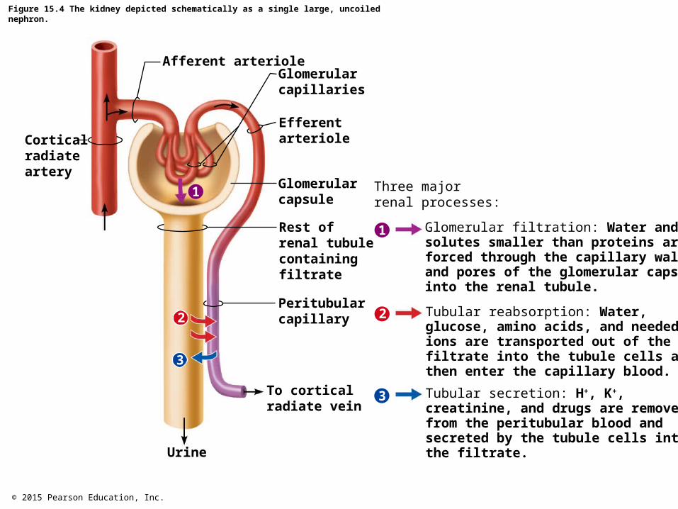
Figure 15.4 The kidney depicted schematically as a single large, uncoiled nephron.
3
1
2
1
3
2
Afferent arterioleGlomerularcapillaries
Efferentarteriole
Glomerularcapsule
Rest ofrenal tubulecontainingfiltrate
Peritubularcapillary
To corticalradiate vein
Urine
Corticalradiateartery
Three majorrenal processes:
Glomerular filtration: Water andsolutes smaller than proteins areforced through the capillary wallsand pores of the glomerular capsuleinto the renal tubule.
Tubular reabsorption: Water, glucose, amino acids, and neededions are transported out of thefiltrate into the tubule cells andthen enter the capillary blood.
Tubular secretion: H+, K+,creatinine, and drugs are removedfrom the peritubular blood andsecreted by the tubule cells intothe filtrate.
© 2015 Pearson Education, Inc.
Figure 15.4 The kidney depicted schematically as a single large, uncoiled nephron (1 of 2).
3
2
1
Afferent arterioleGlomerularcapillaries
Efferentarteriole
Glomerularcapsule
Rest ofrenal tubulecontainingfiltrate
Peritubularcapillary
To corticalradiate vein
Urine
Corticalradiateartery
© 2015 Pearson Education, Inc.
Figure 15.4 The kidney depicted schematically as a single large, uncoiled nephron (2 of 2).
1
Three majorrenal processes:
Glomerular filtration: Water andsolutes smaller than proteins areforced through the capillary wallsand pores of the glomerular capsuleinto the renal tubule.
Tubular reabsorption: Water, glucose, amino acids, and neededions are transported out of thefiltrate into the tubule cells andthen enter the capillary blood.
Tubular secretion: H+, K+,creatinine, and drugs are removedfrom the peritubular blood andsecreted by the tubule cells intothe filtrate.
2
3
© 2015 Pearson Education, Inc.
Figure 15.5 Sites of filtration, reabsorption, and secretion in a nephron.
Filtrate
Blood
Cortex
Medulla
H2OSalts (NaCl, etc.)HCO3− (bicarbonate)H+
UreaGlucose; amino acidsSome drugs
Reabsorption
Active transport
Passive transportSecretion
(active transport)
Nephronloop
H2O
NaCl
NaCl
H2O
NaCl
K+
H2O
Urea
Urine(to renal pelvis)
K+ andsomedrugs
Collectingduct
H+Some drugsand poisons
Glomerularcapsule
Proximal tubule Distal tubule
NaClNaClGlucose andamino acids
HCO3−
H2O
© 2015 Pearson Education, Inc.
Figure 15.8 The major fluid compartments of the body.
Volume = 40 LTotal body water
Volume = 12 L Volume = 25 L
60% body weight
40% body weight 80% of ECF
20% body weightVolume = 15 L
Intracellular fluid (ICF)
Extracellular fluid (ECF)
Interstitialfluid (IF)
Vo
lum
e = 3 L
, 20% o
f EC
F
Pla
sma
© 2015 Pearson Education, Inc.
Figure 15.9 The continuous mixing of body fluids.
Lungs Gastrointestinaltract
Kidneys
Bloodplasma
Interstitialfluid
Intracellularfluid in tissue cells
O2 CO2 Nutrients
O2 CO2 Nutrients
H2O,Ions
H2O
H2O,Ions
Ions
Nitrogenouswastes
Nitrogenouswastes
© 2015 Pearson Education, Inc.
Figure 15.10 Water intake and output.
Metabolism10%
Foods30%
Beverages60%
Feces 4%
Sweat 8%Insensiblelosses viaskin andlungs 28%
Urine 60%
Average intakeper day
Average outputper day
1500 ml1500 ml
750 ml 700 ml
200 ml
100 ml250 ml
250
0 m
l
© 2015 Pearson Education, Inc.
Figure 15.11 The thirst mechanism for regulating water intake.
Plasma solutes
Saliva
Dry mouth
Osmoreceptorsin hypothalamus
Hypothalamicthirst center
Sensation ofthirst; persontakes a drink
Water moistensmouth, throat;stretches stomach,intestine
Water absorbedfrom GI tract
Plasmasolutes
Initial stimulus
KEY:
Physiological response
Result
Increases, stimulatesReduces, inhibits
© 2015 Pearson Education, Inc.
Figure 15.12 Flowchart of mechanisms regulating sodium and water balance to help maintain blood pressure homeostasis.
Falling systemic blood pressure/volume
Reduced filtrate volumeor solute content inrenal tubules
JG cells of kidneys
Release
(+)(+)
Renin
Leads to
(+)
(+)
Inhibits baroreceptorsin blood vessels
Sympathetic nervoussystem
Systemic arterioles
Causes
Vasoconstriction
Results in
Peripheral resistanceAngiotensin II
formed in blood(+)
(+)(+)
Causes
Results in
Systemic arterioles Adrenal cortex
Secretes
Targets
Aldosterone
Kidney tubules
Vasoconstriction
Peripheral resistance
Na+ reabsorption (andH2O absorption)
Causes
Results in
Blood volume
Rising blood pressure
(+)
(+)
(+)
Causes
Release
(+)
Hypothalamicosmoreceptors
Posterior pituitary
ADH (antidiuretichormone)
Collecting ductsof kidneys
H2O reabsorption
= stimulates
Renin-angiotensin system
Neural regulation (sympatheticnervous system effects)
Effects of ADH release
KEY:
© 2015 Pearson Education, Inc.
Figure 15.13 Dissociation of strong and weak acids in water.
HCl H2CO3
H2CO3
H2CO3
H2CO3
H2CO3 HCO3−
HCO3−H+
H+
H+
H+
H+H+
H+
H+H+Cl−
Cl− Cl−
Cl−
Cl− Cl−
Cl−
(a) A strong acidsuch as HCldissociatescompletelyinto its ions.
(b) A weak acid suchas H2CO3 doesnot dissociatecompletely.
© 2015 Pearson Education, Inc.
Figure 15.13a Dissociation of strong and weak acids in water.
HCl
H+
H+
H+H+
H+
H+H+Cl−
Cl− Cl−
Cl−
Cl− Cl−
Cl−
(a) A strong acidsuch as HCldissociatescompletelyinto its ions.
© 2015 Pearson Education, Inc.
Figure 15.13b Dissociation of strong and weak acids in water.
H2CO3
H2CO3
H2CO3
H2CO3
H2CO3 HCO3−
HCO3−
H+
H+
(b) A weak acid suchas H2CO3 doesnot dissociatecompletely.
© 2015 Pearson Education, Inc.
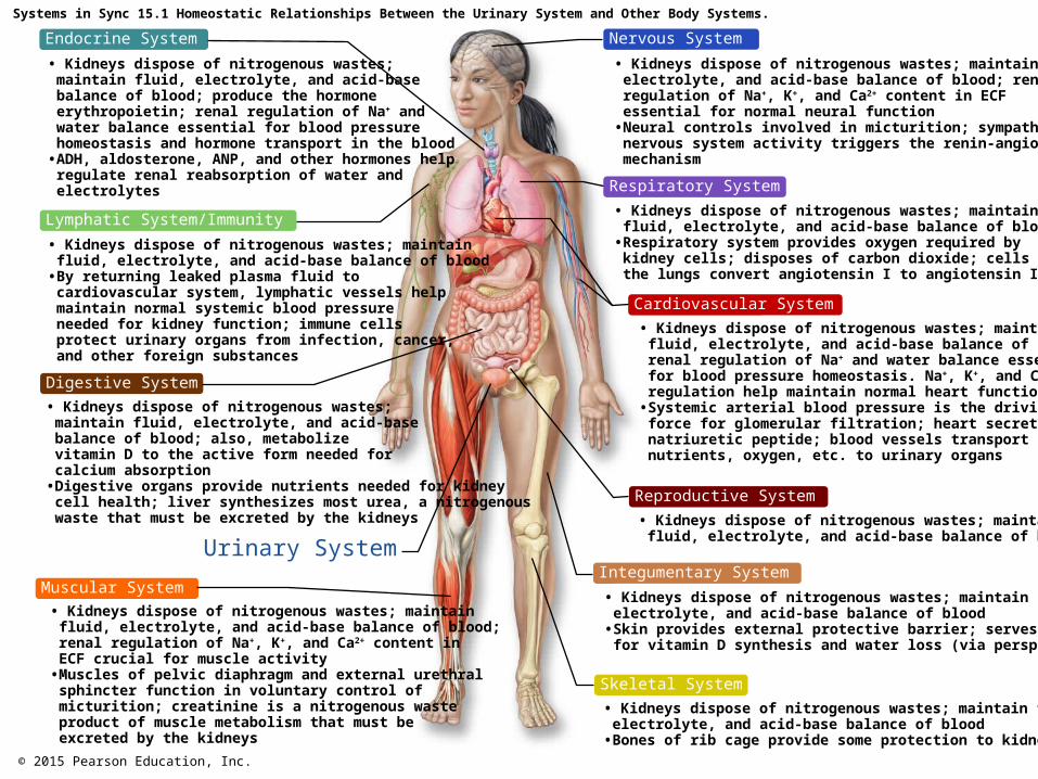
Systems in Sync 15.1 Homeostatic Relationships Between the Urinary System and Other Body Systems.
Endocrine System
Lymphatic System/Immunity
Digestive System
Urinary System
Muscular System
Nervous System
Respiratory System
Cardiovascular System
Reproductive System
Skeletal System
Integumentary System
• Kidneys dispose of nitrogenous wastes;maintain fluid, electrolyte, and acid-basebalance of blood; produce the hormoneerythropoietin; renal regulation of Na+ andwater balance essential for blood pressurehomeostasis and hormone transport in the blood
• ADH, aldosterone, ANP, and other hormones helpregulate renal reabsorption of water andelectrolytes
• Kidneys dispose of nitrogenous wastes; maintain fluid, electrolyte, and acid-base balance of blood
• By returning leaked plasma fluid tocardiovascular system, lymphatic vessels helpmaintain normal systemic blood pressureneeded for kidney function; immune cellsprotect urinary organs from infection, cancer,and other foreign substances
• Kidneys dispose of nitrogenous wastes; maintain fluid, electrolyte, and acid-base balance of blood; also, metabolizevitamin D to the active form needed forcalcium absorption
• Digestive organs provide nutrients needed for kidneycell health; liver synthesizes most urea, a nitrogenouswaste that must be excreted by the kidneys
• Kidneys dispose of nitrogenous wastes; maintain fluid, electrolyte, and acid-base balance of blood; renal regulation of Na+, K+, and Ca2+ content inECF crucial for muscle activity
• Muscles of pelvic diaphragm and external urethralsphincter function in voluntary control ofmicturition; creatinine is a nitrogenous wasteproduct of muscle metabolism that must beexcreted by the kidneys
• Kidneys dispose of nitrogenous wastes; maintain fluid, electrolyte, and acid-base balance of blood; renalregulation of Na+, K+, and Ca2+ content in ECFessential for normal neural function
• Neural controls involved in micturition; sympatheticnervous system activity triggers the renin-angiotensinmechanism
• Kidneys dispose of nitrogenous wastes; maintain fluid, electrolyte, and acid-base balance of blood
• Respiratory system provides oxygen required bykidney cells; disposes of carbon dioxide; cells inthe lungs convert angiotensin I to angiotensin II
• Kidneys dispose of nitrogenous wastes; maintain fluid, electrolyte, and acid-base balance of blood;renal regulation of Na+ and water balance essentialfor blood pressure homeostasis. Na+, K+, and Ca2+
regulation help maintain normal heart function• Systemic arterial blood pressure is the driving
force for glomerular filtration; heart secretes atrialnatriuretic peptide; blood vessels transportnutrients, oxygen, etc. to urinary organs
• Kidneys dispose of nitrogenous wastes; maintain fluid, electrolyte, and acid-base balance of blood
• Skin provides external protective barrier; serves as sitefor vitamin D synthesis and water loss (via perspiration)
• Kidneys dispose of nitrogenous wastes; maintain fluid, electrolyte, and acid-base balance of blood
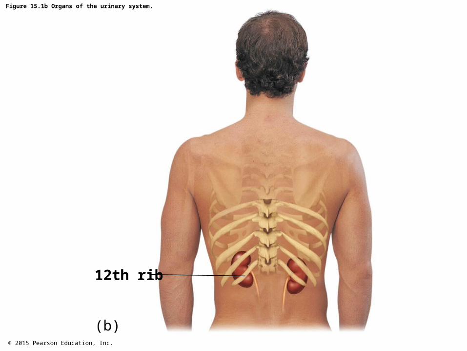
• Bones of rib cage provide some protection to kidneys