71
Stroke

Stroke

Global mortality

Cause of death—Dept. of Neurology?
1938-1950 1990-2002
Stroke
Infect.
Tumor
Tumor
Stroke

After MI 70-80%
Normal lifestyle
After stroke
10%

Differential diagnosis---CT/MRI!!!
5
Agyi infarktus 80% Vérzés 20%ischemia 80% sp. Hemorrhage 10-15%
Subarachnoidal bleeding

62 yrs stroke at admission
One day later
2 days later
CT

Large vessel occluded? Less chance for successful
intravenous lysis..
Alexandrov , J Int. Medicine 267; 209–219 2010.

Ischemic Stroke
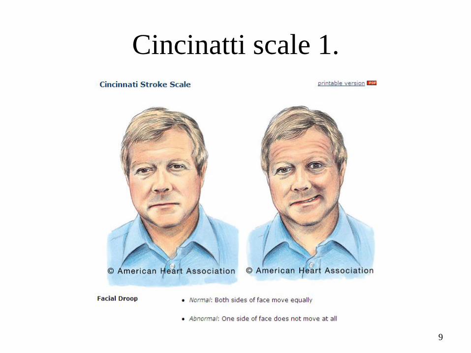
Cincinatti scale 1.
9

Cincinatti scale 2.
10

Cincinatti scale 3.
• Slurred speech, aphasia
11

TIA
(TRANSIENT ISCHEMIC
ATT.)• Transient symptoms
• Minutes
• No residual tissue deficit (MRI)
TIA is emergency!!! High risk
of devastating stroke

Ischemic Stroke
• 700 km axon/hour↓
• 2 millió neuron/min ↓
Time is brain!

Open the artery as soon as
possible
1 1,5 3 4,5 6 hours after stroke
45 patients
21 patients
9 patients
4 patients
2 patients

Lysis…….
Within 3 hours between 3-4,5 hours
worseimproved
better
worse
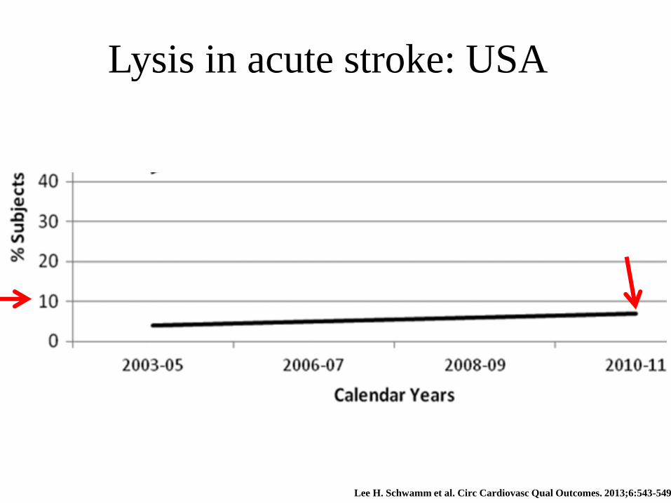
Lee H. Schwamm et al. Circ Cardiovasc Qual Outcomes. 2013;6:543-549
Lysis in acute stroke: USA

iv. Thrombolysis in Debrecen
2011 2012 2013 2014 2015 2016.12.12.
133 142 113 131 147 216
(%) 20 20 17 18 19% 23%
More than 1300 iv. lysis!

stroke
CT-!
CT+CT angio!
Stroke unit
Ambulance!
Debrecen stroke care
Personalised:
Iv?
intraarterial?
iv+ia?
mechanical thrombectomy

In basilar artery the time window could be 12 h (iv or ia. lysis)
6-8 hours IF ICA or MCA occlusion:intraarterial or
mechanical thrombectomy (MET) BUT start with iv.
In case of specific constellation of MRI(!) and age and infarct
volume → the time window could be 24 h!!!
Within 4,5 hours (some subgroups 3 hours) Iv. lysis if
small vessel occlusion
Time window?
depends on the vessel and time
elapsed after stroke?
time

If out of time window?
• 100-300 mg aspirin
• Monitoring of BP and ECG
• Do NOT decrease BP till 220/110 Hgmm!
• Pulsoximetry, 2-4 lit oxigen, if less 94%
• Normoglycemia
• LMWH or heparin to prevent DVT deep venous thromb.
• Nasogastric tube if dysphagia
• Antipyretic ther.
• If seizure antiepilept.
• antibiotics

After desoblieration therapy, the next
question:
cause of stroke?

Stroke: diagnosis
Blood
•Blood counts
•Glucose, ions
•Coagulation?
•lipids
•Immunol. tests
Heart
Function•BP monitoring
•ECG
•Holter ECG
morphology•TTE
•TEE
ultrasound
Carotid, vertebr-•ultrasound
•CT AG
•MRA
•DSA
Imaging
•CT•MRI
•Diff. WI•TCD
•DSA angiogr.
22

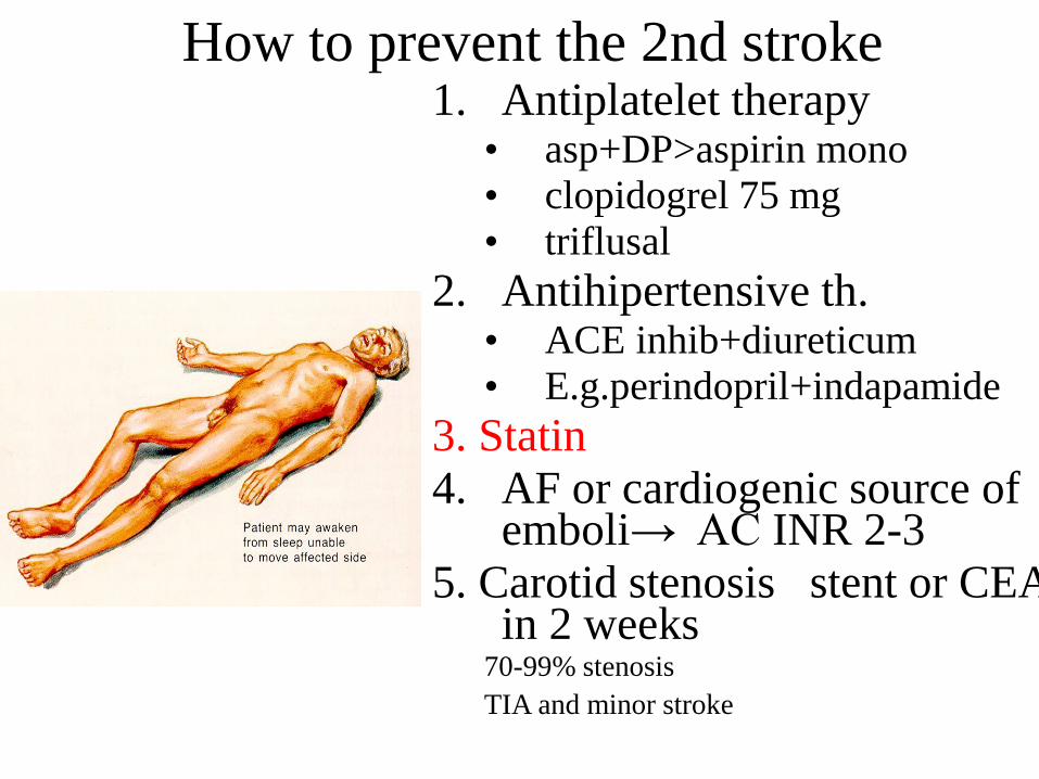
How to prevent the 2nd stroke?

Recurrent stroke
• 1 mo 30%
• 1 year 10%
• then 6-8%

How to prevent the 2nd stroke1. Antiplatelet therapy
• asp+DP>aspirin mono
• clopidogrel 75 mg
• triflusal
2. Antihipertensive th. • ACE inhib+diureticum
• E.g.perindopril+indapamide
3. Statin
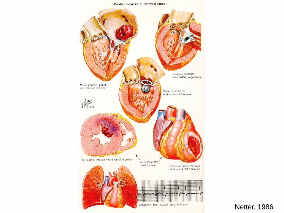
4. AF or cardiogenic source of emboli→ AC INR 2-3
5. Carotid stenosis stent or CEA in 2 weeks
70-99% stenosis
Only if TIA and minor stroke
Occlusion NO!! Plegic?NO!!!!!
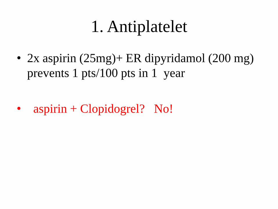
1. Antiplatelet
• 2x aspirin (25mg)+ ER dipyridamol (200 mg)
prevents 1 pts/100 pts in 1 year
• aspirin + Clopidogrel? No!

How to prevent the 2nd stroke1. Antiplatelet therapy
• asp+DP>aspirin mono
• clopidogrel 75 mg
• triflusal
2. Antihipertensive th. • ACE inhib+diureticum
• E.g.perindopril+indapamide
3. Statin
4. AF or cardiogenic source of emboli→ AC INR 2-3
5. Carotid stenosis stent or CEA in 2 weeks
70-99% stenosis
TIA and minor stroke
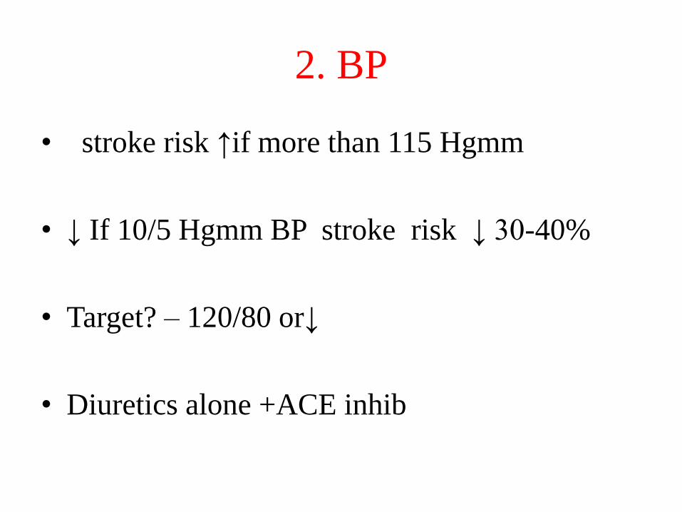
2. BP
• stroke risk ↑if more than 115 Hgmm
• ↓ If 10/5 Hgmm BP stroke risk ↓ 30-40%
• Target? – 120/80 or↓
• Diuretics alone +ACE inhib
How to prevent the 2nd stroke1. Antiplatelet therapy
• asp+DP>aspirin mono
• clopidogrel 75 mg
• triflusal
2. Antihipertensive th. • ACE inhib+diureticum
• E.g.perindopril+indapamide
3. Statin
4. AF or cardiogenic source of emboli→ AC INR 2-3
5. Carotid stenosis stent or CEA in 2 weeks
70-99% stenosis
TIA and minor stroke

3.Statin
• Statin if ≥LDL 2.6 mmol/l or signs of atherosclerosis
• target: LDL < 1.8 mmol/l
• Niacin or gemfibrosil if HDL-C low

How to prevent the 2nd stroke1. Antiplatelet therapy
• asp+DP>aspirin mono
• clopidogrel 75 mg
• triflusal
2. Antihipertensive th.• ACE inhib+diureticum
• E.g.perindopril+indapamide
3. Statin
4. AF or cardiogenic source of emboli→ AC INR 2-3
5. Carotid stenosis stent or CEA in 2 weeks
70-99% stenosis
TIA and minor stroke
Netter, 1986

4. If AF post-stroke
• Aspirin is NOT enough---if possible -----AC
with
– warfarin
– or NOAC (dabigatran, rivaroxaban, apixaban,
edoxaban----all as good as warfarin but less
bleeding and no need for INR control)

How to prevent the 2nd stroke1. Antiplatelet therapy
• asp+DP>aspirin mono
• clopidogrel 75 mg
• triflusal
2. Antihipertensive th.• ACE inhib+diureticum
• E.g.perindopril+indapamide
3. Statin
4. AF or cardiogenic source of emboli→ AC INR 2-3
5. Carotid stenosis stent or CEA in 2 weeks
70-99% stenosis
TIA and minor stroke
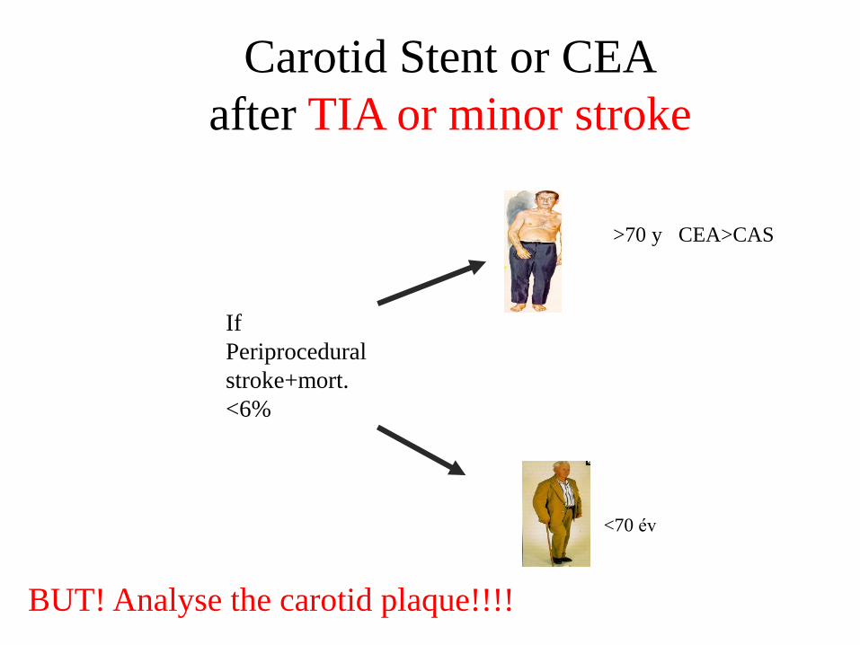
Carotid Stent or CEA
after TIA or minor stroke
If
Periprocedural
stroke+mort.
<6%
>70 y CEA>CAS
<70 év
BUT! Analyse the carotid plaque!!!!

Differential diagnosis---CT/MRI!!!
36
Agyi infarktus 80% Vérzés 20%ischemia 80% sp. Hemorrhage 10-15%
Subarachnoidal bleeding

Hemorrhage

38
Agyi infarktus 80% Vérzés 20%sp. Hemorrhage 10-15%
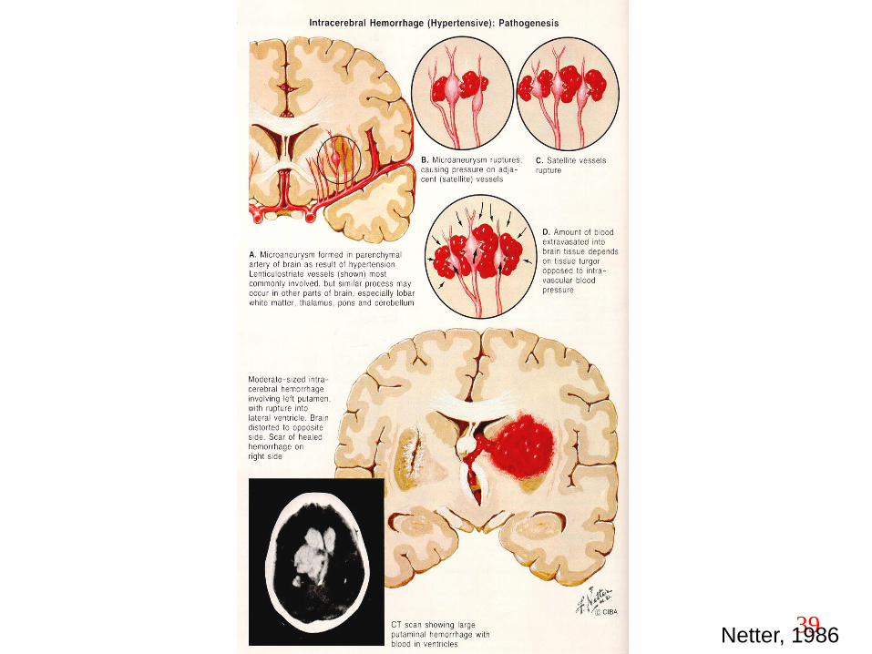
39Netter, 1986

40Netter, 1986

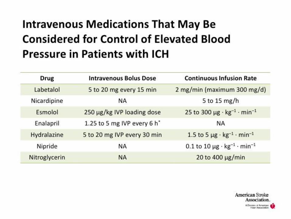
ICH
• neuroimaging with CT or MRI
• CT angiography and contrast CT may be
considered to identify patients at risk for
hematoma expansion
– structural lesions?
– vascular malformations?
– tumors?

VII
• FVIIa can limit the extent of hematoma expansion
• increase in thromboembolic risk with rFVIIa
• rFVIIa is not recommended
• intermittent pneumatic compression +elasticstockings
• After cessation of bleeding, low-dose sc. LMWheparin or unfractionated heparin may beconsidered for prevention of DVT with lack of mobility after 1 to 4 days from onset!!!!

Guidelines for the Management of Spontaneous
Intracerebral Hemorrhage ASA/AHA 2015
0

Prevention of Secondary Brain Injury
• Glucose should be monitored and
normoglycemia (range 4.4 to 6,1 mmol/L)
• seizures should be treated with antiepileptic
drugs
• Prophylactic anticonvulsant medication should
not be used

Surgery
• If Glasgow Coma Scale of 8 +clinical evidence
of transtentorial herniation,
• or significant intraventric. Hemorrhage (IVH)
• or hydrocephalus
• ventricular drainage in patients with
decreased level of consciousness

Surgery 2
• uncertain
• exceptions deteriorating cerebellar
hemorrhage
– with brainstem compression
– and/or hydrocephalus ASAP!
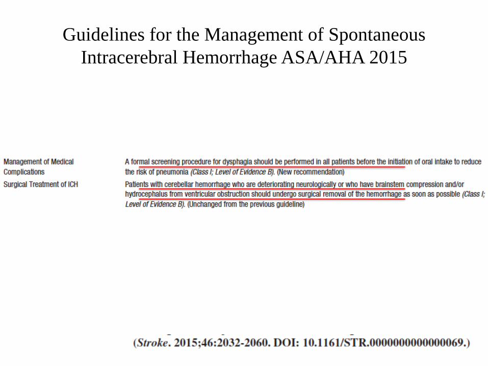
Guidelines for the Management of Spontaneous
Intracerebral Hemorrhage ASA/AHA 2015

Clot removal
• lobar clots >30 ml and within1 cm of the
surface, by standard craniotomy might be
considered
• The effectiveness
– of minimally invasive clot evacuation
• stereotactic
• or endoscopic aspiration with or without thrombolytic
usage is uncertain
– no clear evidence

Prevention of Recurrent ICH
• risk factors for recurrence:
– Lobar location
– older age,
– ongoing anticoagulation,
– presence of the apolipoprotein E ε2 or ε4 alleles,
– and greater number of microbleeds on MRI!!
• After the acute ICH period BP <140/90<130/80 if diabetes or chronic kidney disease, reasonable

What to do if concomittant risk for
cardiogenic stroke exists after ICH
• Anticoagulation after lobar ICH NO (but
antiplatelet) due to high risk of recurrence
• Anticoagulation YES after non lobar ICH (e.g.
basal ganglia)
• No heavy alcohol use
• insufficient data
– use of statin?
– physical or sexual activity

Agyi infarktus 80% Vérzés 20%
Subarachnoidal bleeding

53
SAH
10-20/100 000
•Thunderclap headache
•Vomitus, photophobia
•During physical exercise
But not always!
•Neck rigidity
• sometimes paresis
ACT sometimes negative!! Lp!
aneurysma multiplex?
Vasospasm 4-11 day
Netter 1986
The classic presentation of SAH can
include the following:
• Sudden onset of severe headache (the classic feature)
• Accompanying nausea or vomiting
• Symptoms of meningeal irritation
• Photophobia and visual changes
• Focal neurologic deficits
• Sudden loss of consciousness at the ictus
• Seizures during the acute phase

SAH
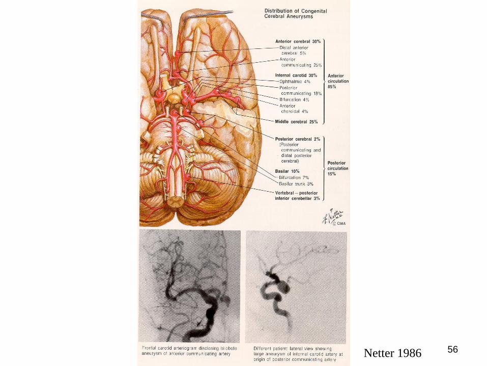
56Netter 1986

57Netter 1986
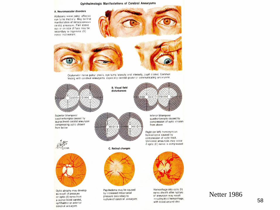
58Netter 1986

Signs present SAH include the
following:
• Sensory or motor disturbance (6%)
• Seizures (4%)
• Ptosis (3%)
• Bruits (3%)
• Dysphasia (2%)

Complications of SAH include the
following:
• Hydrocephalus
• Rebleeding
• Vasospasm
• Seizures
• Cardiac dysfunction

Diagnosis
• clinical suspicion combined with
• noncontrast CT,
• followed by lumbar puncture
• or CT angiography of the brain.
• further imaging to characterize the source of
the hemorrhage.

Laboratory studies
• Complete blood count
• Prothrombin time (PT)/activated partial
thromboplastin time (aPTT)
• Blood typing/screening
• Cardiac enzymes
• Arterial blood gas (ABG) determination

Imaging studies
• CT (noncontrast, contrast, or infusion)
• Digital subtraction cerebral angiography
• Multidetector CT angiography
• MRI (if no lesion is found on angiography)
• Magnetic resonance angiography (MRA; investigational for SAH)
Other diagnostic studies that may be warranted are as follows:
• Baseline chest radiograph
• ECG on admission
• Lumbar puncture and CSF analysis


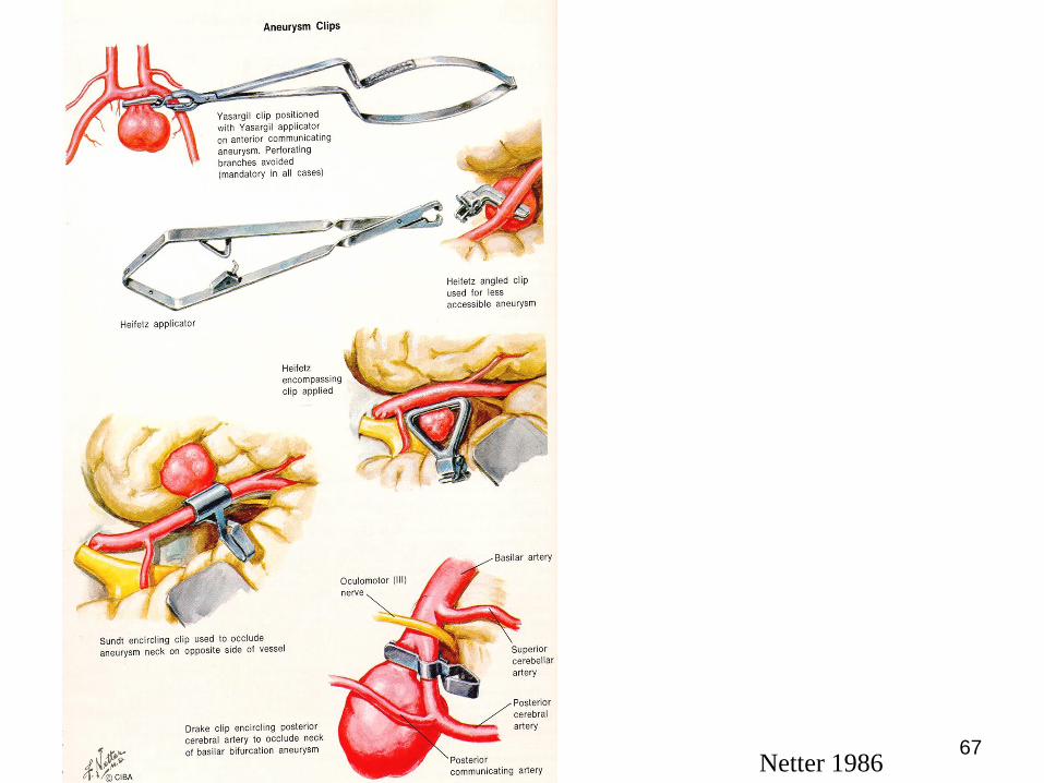
67Netter 1986

Aneurysma sac is filled
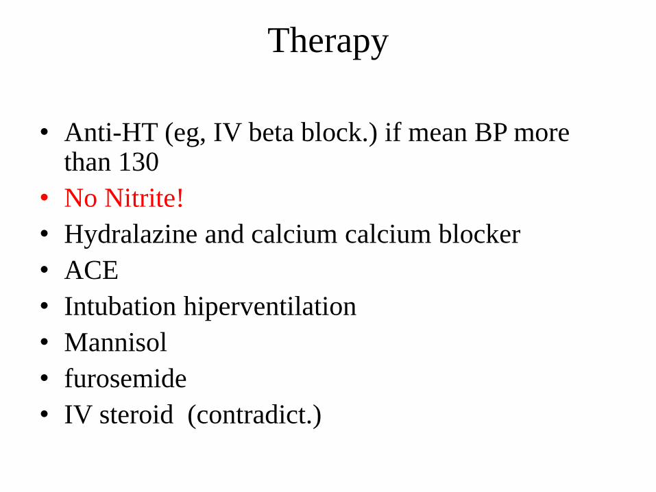
Therapy
• Anti-HT (eg, IV beta block.) if mean BP more than 130
• No Nitrite!
• Hydralazine and calcium calcium blocker
• ACE
• Intubation hiperventilation
• Mannisol
• furosemide
• IV steroid (contradict.)

Complications
• Re-bleeding
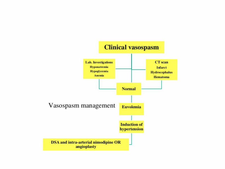
• Vasospasm (4-11. nap, Transcran. Doppler)
nimodipine
• Hydrocephalus
• Hyponatremia
• Seizures
• Lung edema, MI

Others
• Clipping the ruptured aneurysm
• Endovascular treatment
• The choice between coiling and clipping usually depends on the location of the lesion, the neck of the aneurysm, and the availability and experience of hospital staff.
• Screening is not recommended in the general population.
• However, it can lower cost and improve quality of life in patients at relatively high risk for aneurysm formation and rupture.
• Vasospasm between 4-11th after rupture! Transcranial Doppler


