Note: Within nine months from the publication of the mention of the grant of the European patent, any person may give notice to the European Patent Office of opposition to the European patent granted. Notice of opposition shall be filed in a written reasoned statement. It shall not be deemed to have been filed until the opposition fee has been paid. (Art. 99(1) European Patent Convention). Printed by Jouve, 75001 PARIS (FR) Europäisches Patentamt European Patent Office Office européen des brevets (19) EP 1 343 013 B1 (Cont. next page) & (11) EP 1 343 013 B1 (12) EUROPEAN PATENT SPECIFICATION (45) Date of publication and mention of the grant of the patent: 26.04.2006 Bulletin 2006/17 (21) Application number: 02290578.0 (22) Date of filing: 07.03.2002 (51) Int Cl.:G01N 33/68 (2006.01) G01N 33/50 (2006.01) A61K 31/715 (2006.01) A61K 31/66 (2006.01) (54) Methods of screening apoptosis modulating compounds, compounds identified by said methods and use of said compounds as therapeutic agents. Verfahren zum Screening von Verbindungen, die die Apoptose modulieren, derart identifizierte Verbindungen, sowie Verwendung der Verbindungen als pharmazeutische Agentien Procédés de criblage de substances modulatrices de l’apoptose, substances identifiées par ces procédés, et l’utilisation desdites substances comme agents thérapeutiques (84) Designated Contracting States: AT BE CH CY DE DK ES FI FR GB GR IE IT LI LU MC NL PT SE TR (43) Date of publication of application: 10.09.2003 Bulletin 2003/37 (73) Proprietors: • INSTITUT PASTEUR 75724 Paris Cédex 15 (FR)• CONSEJO SUPERIOR DE INVESTIGACIONES CIENTIFICAS E-28006 Madrid (ES)• CENTRE NATIONAL DE LA RECHERCHE SCIENTIFIQUE (CNRS)75794 Paris Cedex 16 (FR)(72) Inventors: • Garcia, Alphonse 75015 Paris (FR)• Cayla, Xavier 91220 Bretigny Sur Orge (FR)• Rebollo, Angelita 28049 Madrid (ES)• Ayllon, Véronica 28012 Madrid (ES)• Fleischer, Aarne 28005 Madrid (ES)(74) Representative: Desaix, Anne et al Ernest Gutmann - Yves Plasseraud S.A.S. 3, rue Auber 75009 Paris (FR)(56) References cited: WO-A-00/37088 WO-A-01/10888 WO-A-01/48236 US-A- 3 855 409 • GAJATE CONSUELO ET AL: "The antitumor ether lipid ET-18-OCH3 induces apoptosis through translocation and capping of Fas/CD95 into membrane rafts in human leukemic cells." BLOOD, vol. 98, no. 13, 15 December 2001 (2001-12-15), pages 3860-3863, XP002233039 December 15, 2001 ISSN: 0006-4971 • KABOURIDIS PANAGIOTIS S ET AL: "Cholesterol depletion disrupts lipid rafts and modulates the activity of multiple signaling pathways in T lymphocytes." EUROPEAN JOURNAL OF IMMUNOLOGY., vol. 30, no. 3, March 2000 (2000-03), pages 954-963, XP002233040 ISSN: 0014-2980 • AYLLON VERONICA ET AL: "Protein phosphatase 1alpha is a Ras-activated Bad phosphatase that regulates interleukin-2 deprivation-induced apoptosis." EMBO (EUROPEAN MOLECULAR BIOLOGY ORGANIZATION) JOURNAL, vol. 19, no. 10, 15 May 2000 (2000-05-15), pages 2237-2246, XP002233041 ISSN: 0261-4189 • REBOLLO A ET AL: "The association of Aiolos transcription factor and Bcl-xL is involved in the control of apoptosis." JOURNAL OF IMMUNOLOGY (BALTIMORE, MD.: 1950) UNITED STATES 1 DEC 2001, vol. 167, no. 11, 1 December 2001 (2001-12-01), pages 6366-6373, XP002233042 ISSN: 0022-1767
Transcript
Note: Within nine months from the publication of the mention of the grant of the European patent, any person may givenotice to the European Patent Office of opposition to the European patent granted. Notice of opposition shall be filed ina written reasoned statement. It shall not be deemed to have been filed until the opposition fee has been paid. (Art.99(1) European Patent Convention).
Printed by Jouve, 75001 PARIS (FR)
Europäisches Patentamt
European Patent Office
Office européen des brevets
(19)
EP
1 34
3 01
3B
1
(Cont. next page)
��&������������(11) EP 1 343 013 B1
(12) EUROPEAN PATENT SPECIFICATION
(45) Date of publication and mention of the grant of the patent: 26.04.2006 Bulletin 2006/17
(21) Application number: 02290578.0
(22) Date of filing: 07.03.2002
(51) Int Cl.: �G01N 33/68 (2006.01) G01N 33/50 (2006.01)
A61K 31/715 (2006.01) A61K 31/66 (2006.01)
(54) Methods of screening apoptosis modulating compounds, compounds identified by said methods and use of said compounds as therapeutic agents. �Verfahren zum Screening von Verbindungen, die die Apoptose modulieren, derart identifizierte Verbindungen, sowie Verwendung der Verbindungen als pharmazeutische Agentien
Procédés de criblage de substances modulatrices de l’apoptose, substances identifiées par ces procédés, et l’utilisation desdites substances comme agents thérapeutiques
(84) Designated Contracting States: AT BE CH CY DE DK ES FI FR GB GR IE IT LI LU MC NL PT SE TR
(43) Date of publication of application: 10.09.2003 Bulletin 2003/37
(73) Proprietors: • INSTITUT PASTEUR
75724 Paris Cédex 15 (FR) �• CONSEJO SUPERIOR DE INVESTIGACIONES
CIENTIFICASE-�28006 Madrid (ES) �
• CENTRE NATIONAL DE LA RECHERCHE SCIENTIFIQUE (CNRS) �75794 Paris Cedex 16 (FR) �
(72) Inventors: • Garcia, Alphonse
75015 Paris (FR) �• Cayla, Xavier
91220 Bretigny Sur Orge (FR) �• Rebollo, Angelita
28049 Madrid (ES) �• Ayllon, Véronica
28012 Madrid (ES) �• Fleischer, Aarne
28005 Madrid (ES) �
(74) Representative: Desaix, Anne et alErnest Gutmann - Yves Plasseraud S.A.S. 3, rue Auber75009 Paris (FR) �
• GAJATE CONSUELO ET AL: "The antitumor ether lipid ET- �18-�OCH3 induces apoptosis through translocation and capping of Fas/CD95 into membrane rafts in human leukemic cells." BLOOD, vol. 98, no. 13, 15 December 2001 (2001-12-15), pages 3860-3863, XP002233039 December 15, 2001 ISSN: 0006-4971
• KABOURIDIS PANAGIOTIS S ET AL: "Cholesterol depletion disrupts lipid rafts and modulates the activity of multiple signaling pathways in T lymphocytes." EUROPEAN JOURNAL OF IMMUNOLOGY., vol. 30, no. 3, March 2000 (2000-03), pages 954-963, XP002233040 ISSN: 0014-2980
• AYLLON VERONICA ET AL: "Protein phosphatase 1alpha is a Ras- �activated Bad phosphatase that regulates interleukin- �2 deprivation- �induced apoptosis." EMBO (EUROPEAN MOLECULAR BIOLOGY ORGANIZATION) JOURNAL, vol. 19, no. 10, 15 May 2000 (2000-05-15), pages 2237-2246, XP002233041 ISSN: 0261-4189
• REBOLLO A ET AL: "The association of Aiolos transcription factor and Bcl- �xL is involved in the control of apoptosis." JOURNAL OF IMMUNOLOGY (BALTIMORE, MD.: 1950) UNITED STATES 1 DEC 2001, vol. 167, no. 11, 1 December 2001 (2001-12-01), pages 6366-6373, XP002233042 ISSN: 0022-1767
2
EP 1 343 013 B1
• SIMONS K ET AL: "LIPID RAFTS AND SIGNAL TRANSDUCTION" NATURE REVIEWS MOLECULAR CELL BIOLOGY, MACMILLAN MAGAZINES, LONDON, GB, vol. 1, no. 1, October 2000 (2000-10), pages 31-39, XP001006200
EP 1 343 013 B1
3
5
10
15
20
25
30
35
40
45
50
55
Description
�[0001] The invention relates to the modulation of ap-optosis in mammalian cells. More particularly, the inven-tion provides methods for identifying novel pro-�apoptoticor anti-�apoptotic cellular polypeptides, methods ofscreening compounds which modulate apoptosis, andmethod of detecting early events of the apoptotic proc-ess.�[0002] Apoptosis or programmed cell death is an activeprocess in which cells induce their self- �destruction in re-sponse to specific cell death signals or in the absence ofcell survival signals. This active process is actually es-sential in the normal development and homeostasis ofmulticellular organisms. It is opposed to necrosis whichis cell death occurring as a result of severe injuriouschanges in the environment.�[0003] Apoptosis of a cell can be characterized at leastby
- the rapid condensation of the cell with collapse ofthe nucleus but preservation of membranes; or,
- cleavage of nuclear DNA at the linker regions be-tween nucleosomes to produce fragments which canbe easily visualized by agarose gel electrophoresisas a characteristic ladder pattern.
�[0004] Various pathologies occur due to a defective oraberrant regulation of apoptosis in the affected cells ofan organism. For example, defects that result in a de-creased level of apoptosis in a tissue as compared to thenormal level required to maintain the steady-�state of thetissue can promote an abnormal increase of the amountof cells in a tissue. This has been observed in variouscancers, where the formation of tumors occurs becausethe cells are not dying at their normal rate. Some DNAviruses such Epstein- �Barr virus, African swine fever virusand adenovirus, also inhibit or modulate apoptosis, there-by repressing cell death and allowing the host cell to con-tinue reproducing the virus.�[0005] On the contrary, a defect resulting in an in-crease of cell death in a tissue may be associated withdegenerative disorders wherein cells are dying at a high-er rate than they regenerate. This is observed in variousdisorders, such as AIDS, senescence, and neurodegen-erative diseases.�[0006] Compounds that modulate positively or nega-tively apoptosis can provide means for the treatment orthe prevention of these disorders. As a consequence,the delineation of apoptotic pathways provides targetsfor the development of therapeutic agents that can beused to modulate the response of a cell to apoptotic orcell survival signals.�[0007] Progresses have been made in identifying ex-tracellular, intracellular and cell surface molecules thatregulate apoptosis. Previous studies have focused onthe identification of specific cell death signals, (such asthe deprivation of growth factors, the FAS/TNF system,
genotoxic agents, glucocorticoïds...), members of theBcl-�2 family and ICE-�type proteases. But critical steps inapoptotic pathways remain to be identified.�[0008] Accordingly, there is still a need in identifyingthe cellular mechanisms involved in apoptotic pathways,and target for the development of therapeutic agents thatcan be used to modulate cell apoptosis.�[0009] Among the different transducing agents, theBcl-�2 family proteins act as an intracellular checkpoint inthe apoptotic pathway. The family is divided into two func-tional groups (médecine / sciences 97 ; 13: 384-6): theproteins that suppress cell-�death (anti-�apoptotic mem-bers such as Bcl- �2, Bcl- �xL, Bcl- �w, Bag-�1, Mcl-�1, A1) andthe proteins that promote cell death (pro- �apoptotic mem-bers such as Bim, Nix, Hzk, Bax, Bak, Bcl- �xs, Bad, Bik).The Bcl-�2 family has been defined by sequence homol-ogy based upon specific conserved motifs termed BCL-homology regions (BH1, BH2, BH3 and BH4 domains).BH1, BH2 and BH3 domains have been shown to beimportant in homodimerization or heterodimerization andin modulating apoptosis. Anti-�apoptotic molecules havea specific BH4 domain.�[0010] It has been proposed that the ratio of pro- �ap-optotic members to anti-�apoptotic members expressedin a cell determines whether this cell will respond to anapoptotic signal. Indeed, pro- �apoptotic and anti- �apoptot-ic members antagonized each other by forming inactiveheterodimers (Oltvai et al., 1993, Cell 74: 609-619), asa consequence only the balance may promote or preventa cell to undergo apoptosis.�[0011] More recently, it has been shown that phospho-rylation of Bcl- �2 proteins can also modulate their activity.Indeed, different anti-�apoptotic pathways are likely to beactivated by growth factors, involving phosphatidylinosi-tol 3 kinase (Pl3K), Akt kinase and Ras activated kinases.In particular, upon stimulation of cells with IL-�3 and NGF,the pro-�apoptotic Bad protein (Bcl-�XL/Bcl-�2 Associatedcell Death regulator, Downward, 1999, Nature Cell Biol1: 33-35) becomes serine phosphorylated, resulting inassociation to 14-3-3 protein (Hsu et al., 1997, Mol En-docrinol 11: 1858-1867). It was proposed that such in-teraction facilitates the translocation of phosphorylatedBad from the mitochondrial membrane to cytosolic com-partments, sequestering it therein and thus, preventingfurther interaction with other anti-�apoptotic Bcl-�2 mem-bers (US Pat No. 5,856,445). It was further shown thatassociation of 14-3-3 protein to Bad is dependent uponserine 155 phosphorylation of Bad (WO 0110888, Apop-tosis Technology Inc., 2001).�[0012] The results disclosed in the present inventionindicate that some pro- �or anti- �apoptotic proteins espe-cially of the Bcl-�2 family, are regulated through a newlyidentified subcellular localization that is in lipid raftsformed in the plasma membrane. This observation offersa way to a novel general mechanism of regulation of cellapoptosis that may play a role in the regulation of pro- oranti- �apoptotic molecules in response to cell death or cellsurvival signals.
1 2
EP 1 343 013 B1
4
5
10
15
20
25
30
35
40
45
50
55
�[0013] Localization of proteins to distinct subcellularcompartments, including membranes, is a critical eventin multiple cellular pathways. Plasma membranes ofmany cell types contain microdomains commonly re-ferred to as lipid rafts, which are biochemically distinctfrom bulk plasma membranes (Brown and London, 1998,Annu Rev Cell Dev Biol 14: 111-136). These domainsconsist of dynamic assemblies of sphingolipids and cho-lesterol. More specifically, the presence of saturated hy-drocarbon chains in sphingolipids allows for cholesterolto be tightly intercalated, leading to the presence of dis-tinct liquid-�ordered phases, and thereby more fluid, lipidbilayer. Lipid rafts can be isolated by subcellular fraction-ation and density gradient ultracentrifugation accordingto methods described in Hacki et al. (Oncogene 200019: 2286-2295) and Millan et al. (Eur J Immunol 199828: 2675-3684). They can also be visualized in intactcells by confocal microscopy using, for example, fluores-cently labelled cholera toxin subunit B (CTx) which bindsto the ganglioside GM1 (Harder et al., 1998, J Cell Biol141: 929-942). One key element of lipid rafts is that theycan include or exclude proteins to WO 01/48236 disclos-es methods for identifying agents which modulate theactivity of protein kinase C theta and the pharmaceuticaluse of such agents; said agents were found to inhibit theor binding of PKC-�θ with lipid rafts varying degrees.�[0014] In T cells, a number of proteins involved in signaltransduction such as IcK, Lat, copurify with lipid rafts iso-lated on sucrose gradient. Disruption of rafts integrity bya variety of methods inhibits early activation events, sup-porting a critical role for these domains in the recruitmentfor signalling and thus, in signal transduction from cellsurface receptors. For example, the antitumor ether lipid1-O-octadecyl- � 2-O-methyl- � rac-glycero-� 3- � phospho-choline (ET-�18- �OCH3; edelfosine) was shown to triggerapoptosis via translocation of Fas to lipid rafts and sub-sequent Fas recruitment by lipid rafts (Gajate and Mol-linedo, Blood, 2001, 98: 3860-3863).�[0015] The present invention results from the discov-ery of a novel mechanism of cellular regulation of theactivity of pro- or anti- apoptotic molecules in a cell bytranslocation of these molecules into lipid rafts under nonapoptotic conditions such as proliferative conditions orconditions where cells do not divide.�[0016] Indeed, the inventors have surprisingly foundthat interaction of a pro- �apoptotic protein, such as theBad protein, with rafts is an active process regulated bycytokines or growth factors. They have also shown thatsegregation of this molecule from rafts in cytokine orgrowth factor deprived-�cells is involved in the inductionof apoptosis and associated with raft disorganization.�[0017] The invention thus provides methods for iden-tifying cellular polypeptides which have pro-�apoptotic oranti- �apoptotic activity in a particular cell type.�[0018] The invention also provides means for screen-ing apoptosis modulating compounds which interferewith the newly identified mechanism of apoptosis regu-lation. Candidate compounds in this respect may either
interfere by blocking, preventing or stimulating translo-cation of one or several pro- or anti- apoptotic polypep-tides in lipid rafts under non apoptotic conditions. Can-didate compounds may in addition or alternatively inter-fere by disrupting or reconstituting lipid rafts in a cellwhich normally produce pro- or anti- apoptotic polypep-tides located in lipid rafts under non apoptotic conditions.According to another embodiment, candidate com-pounds may in addition or alternatively interfere by seg-regation of pro- or anti- apoptotic polypeptides from lipidrafts.�[0019] The invention also provides a compound capa-ble of modulating association of a pro- or anti-�apoptoticpolypeptide with lipid rafts.�[0020] The invention also provides a compound capa-ble of modulating transfer of a pro- or anti-�apoptoticpolypeptide between a lipid raft and another cellular lo-calization.�[0021] The invention also provides for the use of com-pounds capable of modulating lipid rafts formation or ofmodulating translocation of pro- or anti-�apoptotic pro-teins in rafts in the preparation of a medicine for the pre-vention and/or treatment of disorders induced by or as-sociated with a defective regulation of cell death as wellas of specific pathologies in which the death of infectedor deregulated cells may be at least art of a therapy.�[0022] Among the several advantages of the presentmethods, it should be noted that the apoptotic or nonapoptotic state of a cell can be determined according tothe present methods in a relatively short period of timeby analysing lipid raft organization. In particular, there isno need to quantify specific gene expression. The meth-ods of the invention are thus particularly appropriate forroutine high throughput screening of apoptosis modulat-ing compounds.�[0023] Furthermore, the invention provides methodsfor detecting early events of the apoptotic process in acell.�[0024] A first object of the invention is a method ofscreening cellular polypeptides for pro-�apoptotic or anti-apoptotic activity in a cell of a particular cell-�type, saidmethod comprising: �
a. culturing cells of said particular cell-�type undernon apoptotic conditions and culturing cells of saidparticular cell-�type under apoptotic conditions; and,b. determining subcellular localisation of said cellularpolypeptides in the cultured mammalian cells;
wherein a localization of a cellular polypeptide in lipidrafts in cells cultured under non apoptotic conditions anda segregation of said cellular polypeptide from lipid raftsin cells cultured under apoptotic conditions is indicativeof the pro-�apoptotic or an anti-�apoptotic activity of saidcellular polypeptide in said particular cell- �type.�[0025] In a particular embodiment, said cultured cellsare mammalian cells.�[0026] In a preferred embodiment, the cells cultured
3 4
EP 1 343 013 B1
5
5
10
15
20
25
30
35
40
45
50
55
under non apoptotic conditions are cultured under prolif-erative conditions. According to the methods of the in-vention, cells are considered to be cultured "under nonapoptotic conditions" when the proportion of cells under-going apoptotic process in the cell culture is relativelystable in time and does not represent more than 10%,preferably, more than 1% of the whole cell population(depending upon the cell-�type).�[0027] In a preferred embodiment, non apoptotic con-ditions are proliferative conditions.�[0028] On the contrary, cells are considered to be cul-tured "under apoptotic conditions" when the proportionof the cells undergoing apoptotic process increases dra-matically in time to reach, after a certain period, especiallyfor around 24 hours, from deprivation of growth or prolif-eration factor or by use of an apoptotic factor, more than50% of the whole cell population.�[0029] Cells which have undergone apoptotic processcan be characterized, for example, by specific cleavageof nuclear DNA which can be visualized on agarose gelelectrophoresis.�[0030] As used herein, the term "cellular polypeptide"refers to any polypeptide which is produced in a cell bygene expression. It can be a polypeptide naturally en-coded in said cell especially by a native gene, or apolypeptide not naturally encoded in said cell, meaningthat the gene encoding said polypeptide or a coding se-quence derived from said identified gene has been re-combined in the genome of the cell to obtain expression.It can be a mutated form of a naturally occurring polypep-tide and more specifically, a mutated form wherein themutation is involved in abnormal subcellular localisationof said polypeptide under proliferative growth conditions.�[0031] As used herein, the term "lipid rafts" refers todynamic assemblies of sphingolipids and cholesterol inplasma membranes forming microdomains with distinctliquid- �ordered phases, said microdomains stably retain-ing specific structures, such as gangliosides or polypep-tides such as Lck. Lipid rafts can be biochemically iso-lated by subcellular fractionation and density gradient ul-tracentrifugation according to the methods described inHacki et al. (Oncogene 2000 19: 2286-2295) and Millanet al. (Eur J Immunol 1998 28: 2675-3684) and in theexamples below. They can also be visualized in intactcells by confocal microscopy using, for example, fluores-cently labelled cholera toxin subunit B (CTx) which bindsto the ganglioside GM1 which accumulate in lipid rafts(Harder et al., 1998, J Cell Biol 141: 929-942). More spe-cifically, subcellular localization of a particular polypep-tide in lipid rafts is determined by double immunofluores-cence using a labelled marker detecting lipid rafts andanother labelled marker detecting the polypeptide to lo-calize. It can also be determined by analysing the pres-ence of said polypeptide in subcellular fractions contain-ing lipid rafts.�[0032] In a particular embodiment, the method of theinvention is appropriate to screen a polypeptide, whosestructure is known but whose function is unknown, for a
pro- or anti- apoptotic activity in a particular cell type. Inparticular, the one skilled in the Art can use the methodof the invention to screen polypeptides which are sus-pected to be involved in apoptosis regulation accordingto specific features such as specific structural domains.The screening of cellular polypeptides is however notnecessarily limited to cellular polypeptides of knownstructure.�[0033] Several pro-�apoptotic proteins have been iden-tified so far, however, expression pattern of these pro-teins may vary depending upon the cell type. The methodof screening is thus also useful in determining whethera putative cellular polypeptide, known to be pro- or anti-apoptotic in a certain cell-�type is involved in apoptosismodulation in another cell-�type.�[0034] In a particular embodiment, the screenedpolypeptides belong to the Bcl-�2 family. As mentionedhereabove, the Bcl-�2 family members are characterizedby sequence homology based upon specific conservedmotifs termed BCL- �homology regions (BH1, BH2, BH3and BH4 domains). Accordingly, their subcellular locali-sation can be determined by the use of a molecule whichspecifically recognizes a BH domain. Such moleculesencompass for example monoclonal antibodies or poly-clonal antibodies specifically recognizing a BH domain.One example of a molecule that interacts with BH3 motifin PP1a (Ayllon, et al. 2000. EMBO J.; 19: 2237-2246).Examples of molecules that interact with BH4 motif arereview in Admas, J.M. and Cory, S. (1998) Science, 281,1322). In a particular embodiment, a molecule which spe-cifically recognizes a BH4 domain is used to screen pref-erably for anti-�apoptotic molecules. In another particularembodiment, a molecule which specifically recognizes aBH3 domain is used to screen for pro- or anti- apoptoticmolecules. A combination of molecules recognizing thedifferent BH domains can also be used, for example toscreen for pro- �apoptotic molecules which have only theBH3 domain.�[0035] The method of the invention is also appropriateto screen novel polypeptides of the Bcl-�2 family whosestructure and function are unknown at least in part, butwhich can be easily isolated using molecular recognitionof their BH domain�(s). As a result, in a preferred embod-iment, said screened cellular polypeptides are first iso-lated from biochemically isolated lipid rafts of said mam-malian cells cultured in proliferative conditions by the useof a molecule which specifically recognizes a BH domain.More specifically, the polypeptides present in the isolatedlipid rafts can be separated on a gel and analysed byWestern Blot analysis using an antibody which recogniz-es a BH domain or by similar methods of protein analysis.The polypeptides recognized by a BH domain can beisolated and antibodies which recognize each isolatedpolypeptides can be produced according to usual meth-ods well known in the art. The subcellular localization ofone or more of the isolated polypeptides is then deter-mined according to the method of the invention usingsuch specific antibodies to screen for apoptotic activity.
5 6
EP 1 343 013 B1
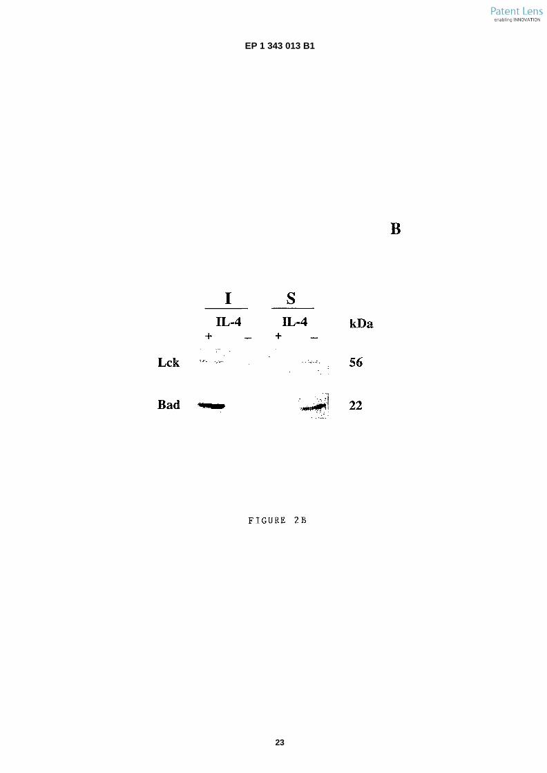
6
5
10
15
20
25
30
35
40
45
50
55
�[0036] The proteins which are associated with lipidrafts may have a transmembrane domains or have un-dergone post- �translational modifications such as myris-toylation. In another specific embodiment, said screenedcellular polypeptides are further isolated by the use of amolecule which specifically recognizes a mirystoylatedpolypeptide.�[0037] In a preferred embodiment, the method is car-ried out for screening cellular polypeptides of a cell typecharacterized by the production of Bad protein, i.e. Bad+
cell type. According to another preferred embodiment,the cells are characteristic of the immune system, andmost preferably are T cell lines.�[0038] Indeed, it is shown, in the examples below, thatthe pro-�apoptotic Bad protein is sequestered in lipid raftsof IL-�4 stimulated T-�cells and segregates from rafts in IL-4 deprived T-�cells.�[0039] More specifically, it is shown in the Examplethat Bad can be co- �purified with lipid-�rafts by subcellularfractionation and density gradient ultracentrifugationfrom cells under non-�apoptotic conditions, especially un-der proliferative conditions. These results indicate thatBad is strongly associated with lipid rafts in cells culturedunder non apoptotic conditions such as proliferative con-ditions.�[0040] Cellular polypeptides which physically interactswith Bad protein in isolated lipid rafts of Bad+ cells thusconstitute preferred putative polypeptides to screen forpro- or anti- apoptotic activity. Accordingly, in a preferredembodiment, the screened cellular polypeptides are iso-lated from isolated lipid rafts of Bad+ cells cultured underproliferative conditions and are selected among thepolypeptides which interact physically with the Bad pro-tein.�[0041] Naturally, the invention also pertains to the new-ly identified cellular polypeptides having pro- or anti-�ap-optotic activity and their use in providing means for mod-ulating apoptosis in cells, such as mammalian cells, ex-pressing these polypeptides.�[0042] It is another object of the invention to provide amethod of screening compounds for their capacity tomodulate apoptosis in cells, which produce pro- or anti-apoptotic polypeptides which are located in lipid raftswhen said cells are cultured under non apoptotic condi-tions, said method comprising: �
a) culturing said cells in a growth medium maintain-ing non apoptotic conditions;b) contacting said cultured cells with a candidatecompound;c) determining the level of one or several pro- or anti-apoptotic polypeptides associated to lipid rafts;d) selecting the compound which interferes with theassociation of one or several pro- or anti- apoptoticpolypeptides with lipid rafts, said compound havingthe capacity to modulate apoptosis.
�[0043] A compound interferes with the association of
a pro- or anti- �apoptotic polypeptide when it modifies saidassociation, including when it alters the chemical and/orthe physical nature of said association or when it providesor influences segregation of pro- or anti-�apoptoticpolypeptides from lipid rafts, or when it prevents said as-sociation, or also when it acts on and especially promotesdisruption of lipid rafts or more generally alter constitutionof lipid rafts.�[0044] The invention further relates to a method ofscreening compounds for their capacity to promote ap-optosis in cells, said method comprising
a) culturing cells in a growth medium maintainingnon apoptotic conditions; wherein said cells producea pro-�apoptotic protein which is located in lipid raftsunder non apoptotic conditions of said cells;b) contacting said cultured cells with a candidatecompound; and,c) determining the absence or the presence of lipidrafts in said cultured cells;d) in case of presence of lipid rafts, optionally deter-mining the level of pro-�apoptotic protein located inthe lipid rafts,
wherein the absence of lipid rafts in the plasma mem-brane of cells incubated with said candidate compoundor if determined, the reduced level of pro-�apoptotic pro-tein in the rafts is indicative that said compound promotesapoptosis.�[0045] The invention also relates to a method ofscreening compounds for their capacity to inhibit or pre-vent apoptosis of cells, said method comprising:�
a) culturing cells in a growth medium for maintainingnon-�apoptotic conditions; wherein said cells producea pro-�apoptotic protein which is located in lipid raftsunder non apoptotic conditions;b) contacting said cells with a candidate compound;c) culturing cells under apoptotic conditions; and,d) determining the absence or the presence of lipidrafts;e) in the case of presence of lipid rafts, optionallydetermining the level of pro-�apoptotic protein locatedin the lipid rafts,
wherein the presence of lipid rafts in the plasma mem-branes of cells incubated with said candidate compoundand optionally the maintained level of proapoptotic pro-tein in the rafts is indicative that said candidate compoundinhibits or prevents apoptosis.�[0046] In a preferred embodiment, the cells are cul-tured in a growth medium comprising at least a cytokineor a growth factor necessary for maintaining proliferativegrowth conditions and step c) of the method comprisesdepriving the cells of said cytokine or growth factor nec-essary for maintaining proliferative growth conditions.�[0047] As used herein, the term "compound" refers toinorganic or organic chemical or biological compounds
7 8
EP 1 343 013 B1
7
5
10
15
20
25
30
35
40
45
50
55
either natural (isolated) or synthetic, and especially en-compass nucleic acids, proteins, polypeptides, peptides,glycopeptides, lipids, lipoproteins and carbohydrates.�[0048] Any cells in which pro- or anti-�apoptotic proteinsmay be translocated in lipid rafts can be used in the meth-ods of the invention. In a preferred embodiment of themethods of the invention, cells are mammalian cells.�[0049] Mammalian cells which are used in the methodsof screening compounds can be any mammalian cellswhose cell survival can be controlled by a specific cy-tokine or growth factor. In preferred embodiments of theabove methods, the cultured mammalian cells are se-lected among those which produce the Bad protein as apro-�apoptotic protein. More specifically, preferred mam-malian cells which produce a Bad protein are selectedamong cells characteristic of the immune system, andmore preferably among T cells.�[0050] As used herein, the term "cytokine or growthfactor" refers to any molecule which is necessary to bepresent in a growth medium to prevent apoptotic processof a cultured cell and/or to promote cell proliferation.Known cytokines include any interleukin. Known growthfactors include the fibroblast growth factors, bFGF, aF-GF, FGF6, the hepatocyte growth factors HGF/SF, theepidermis growth factor, EGF and other characterizedgrowth factors such as IGF- �1, PDGF, LIF, VEGF, SCF,TGFb, TNFa, NGF, BMP, neuregulin, thrombopoïetinand growth hormone. Growth factors according to theinvention can include also, progestagenes and deriva-tives thereof (progesterone), oestrogens and derivativethereof (oestradiol), androgenes (testosterone), miner-alocorticoïds and derivatives thereof (aldosterone), LH,LH-�RH, FSH et TSH hormones, T3, T4, and retinoidicacid, calcitonine E2 and F2/alpha prostaglandins. Glu-cocorticoïds (natural or hemisynthetic, i.e. hydrocorti-sone, dexamethasone, prednisolone or triamcinolone),can also be used.�[0051] Cells characteristic of the immune system canbe advantageously cultured under stimulation with an in-terleukin for maintaining proliferative growth conditions.In particular, IL-�4, IL-�2 or IL- �9 interleukin can be used inthis context, or a mixture thereof.�[0052] However, any available apoptosis model canbe used to select the cell type and the factors for nonapoptotic conditions such as the growth factor or cy-tokine, used in the methods. As used herein, the term"apoptosis model" comprises any teaching providing away to control cell apoptosis in a cell culture of a specificcell type by the use of specific factors for non apoptoticconditions such as a specific cytokine or growth factor ora mixture thereof. Such apoptosis models are for exam-ple the control of IL- �4 stimulated T- �cell lines, IL3 and he-matopoietic progenitor, PC12 and CRH (corticotropin-�re-leasing hormone), HN9.10.�[0053] A candidate compound may modulate apopto-sis by blocking or preventing the association of said pro-or anti- apoptotic polypeptide with lipid rafts. In this con-text, the subcellular localisation of said pro- or anti- �ap-
optotic polypeptide in lipid rafts is no more observed incells incubated with the compound when compared tocells not incubated with the compound, or, at least, lipidrafts subcellular localisation of said pro- or anti-�apoptoticpolypeptide is significantly reduced when compared tocells not incubated with the compound.�[0054] It is another object of the invention to providecompounds capable of modulating association of a pro-or anti- �apoptotic polypeptide with lipid rafts.�[0055] Some of these compounds may modulate ap-optosis by preventing the association of a pro- or anti-apoptotic polypeptide with lipid rafts, by promoting seg-regation of a pro- or anti-�apoptotic polypeptide from lipidrafts or by promoting disruption of lipid rafts.�[0056] Some of these compounds may modulate ap-optosis by preventing the segregation of a pro- or anti-apoptotic polypeptide from lipid rafts, by promoting theassociation of a pro- or anti-�apoptotic polypeptide withlipid rafts or by promoting constitution of lipid rafts.�[0057] It is another object of the invention to providecompounds capable of modulating transfer of a pro- oranti- �apoptotic polypeptide between a lipid raft and anoth-er cellular localization.�[0058] Some of these compounds may modulate ap-optosis by preventing transfer of a pro- or anti-�apoptoticpolypeptide from a cellular localization, other than a lipidraft, to a lipid raft or from a lipid raft to another cellularlocalization.�[0059] Some of these compounds may modulate ap-optosis by promoting transfer of a pro- or anti- �apoptoticpolypeptide from a cellular localization, other that a lipidraft, to a lipid raft or from a lipid raft to another cellularlocalization.�[0060] In a preferred embodiment, the compounds ofthe invention modulating apoptosis are capable of mod-ulating the association of a pro-�apoptotic protein of Bcl-2 family, especially Bad, with lipid rafts or transfer of saidprotein between lipid rafts and another cellular localiza-tion.�[0061] Some of said compounds may promote apop-tosis in a Bad-�producing cell by preventing associationof Bad with lipid rafts, by promoting segregation of Badfrom lipid rafts or by promoting disruption of lipid rafts.�[0062] Some of said compounds may inhibit apoptosisin a Bad-�producing cell by promoting association of Badwith lipid rafts, by preventing segregation of Bad fromlipid rafts or by promoting constitution of lipid rafts.�[0063] In a particular embodiment, a compound of theinvention may inhibit apoptosis of a Bad-�producing cellby preventing transfer of Bad to mitochondria after Badsegregation from lipid rafts. Such a compound may in-teract with Bad by means of a motif similar to the lipidraft motifs which allow association of Bad to lipid rafts.�[0064] Lipid rafts subcellular localisation of said pro-or anti-�apoptotic polypeptide can be quantified by anyquantitative analysis methods available in the art. A sig-nificant reduction of lipid rafts subcellular localisation isobserved when the level of pro- or anti- �apoptotic polypep-
9 10
EP 1 343 013 B1
8
5
10
15
20
25
30
35
40
45
50
55
tide is reduced to at least 50%, preferably 90% in cellsincubated with the candidate compound or compared tocells not incubated with the candidate compound.�[0065] A candidate compound may modulate apopto-sis by blocking or preventing the segregation of said pro-or anti- apoptotic polypeptide from lipid rafts. In this con-text, the subcellular localisation of said pro- or anti- �ap-optotic polypeptide in lipid rafts of cells cultured underconditions promoting pro- or anti-�apoptotic protein seg-regation (e.g., apoptotic conditions for pro-�apoptotic pro-teins) remains observed in cells incubated with the com-pound when compared to cells not incubated with thecompound and lipid rafts subcellular localisation of saidpro- or anti-�apoptotic polypeptide is significantly main-tained when compared to cells not incubated with thecompound.�[0066] Lipid rafts subcellular localisation of said pro-or anti-�apoptotic polypeptide can be quantified by anyquantitative analysis methods available in the art. A sig-nificant maintenance is observed when the level of pro-or anti-�apoptotic polypeptide is maintained to at least50%, preferably 90% in cells incubated with the candi-date compound or compared to cells not incubated withthe candidate compound or compared to cells not incu-bated with the candidate compound.�[0067] By "determining the absence of lipid rafts", it isunderstood that lipid rafts in cells incubated with the can-didate compound are not detected, or at least, are de-tected as traces, or are detected in a significantly reducedlevel (i.e., minimum 20% less) with the methods dis-closed in the present invention as compared with a cul-ture of cells not incubated with the candidate compoundas a control. The proportion of lipid rafts in a cell can becompared between the cell cultures using any quantita-tive analysis methods available in the art. A significantreduction of lipid rafts is observed when their proportionis reduced from 20%, preferably is reduced to at least50%, preferably 90% in cells incubated with the candi-date compound to the control. For example, by confocalmicroscopy analysis, the profile of fluorescence can bequantified by image analysis of cells incubated with thecandidate compound and cells not incubated with thecandidate compound.�[0068] Similarly, by "determining the presence of lipidrafts", it is understood that lipid rafts in cells incubatedwith the candidate compound are detected in substan-tially the same amount as compared with a culture ofcells not incubated with the candidate compound as acontrol.�[0069] Methods to isolate lipid rafts have been de-scribed below. More specifically, it is possible to isolatelipid rafts from mammalian cells, by cell fractionating oversucrose gradient and immunoblotting subcellular frac-tions with markers specific for rafts in order to identifyrafts containing subcellular fractions. The presence orthe absence of lipid rafts can be thus determined in aspecific embodiment by the following steps
i) recovering the cultured cells incubated with saidcompound and resuspending said cells in a bufferappropriate for subcellular fractionation, such as gra-dient sucrose buffer;ii) ultracentrifugating the fractionated cells;iii) recovering the subcellular fraction which shouldcontain lipid rafts; and,iv) determining whether the recovered subcellularfraction contains ganglioside and/or lipid raft asso-ciated molecule�(s).
�[0070] As used herein, "the subcellular fraction whichshould contain lipid rafts" is the subcellular fraction cor-responding to the banded organelles of the gradientwhich contains lipid rafts in a gradient obtained with cellscultured under non apoptotic conditions. Naturally, in ap-optotic conditions, the corresponding cell fraction willcontain much less lipid rafts.�[0071] Lipid rafts and lipid rafts subcellular localisationof pro- or anti- �apoptotic polypeptides can also be directlyvisualized in intact cells by confocal microscopy using amolecular marker which specifically binds to a raft-�asso-ciated molecule or a ganglioside.�[0072] Such preferred molecular markers are, for ex-ample, the cholera toxin subunit B (CTx) which specifi-cally recognizes ganglioside GM1, anti- �Bad antibody oranti- �Lck antibody. More generally, any antibody directedto any cellular polypeptide newly identified according tothe method of the invention as described above can beused as a molecular marker specific for raft.�[0073] The invention also concerns the compoundsidentified by the methods of screening.�[0074] Such compounds identified by the above meth-ods of the invention are useful for the prevention and/ortreatment of disorders induced by or associated with adefective regulation of cell death or of specific patholo-gies where death of infected or deregulated cells may beat least part of a therapy.�[0075] The invention further provides a use of a com-pound capable of modulating lipid rafts formation, in thepreparation of a medicine for the treatment of disordersinduced by or associated with a defective regulation ofcell death.�[0076] In a preferred embodiment, said defective reg-ulation affects cells which produce Bad protein, morepreferably, cells of the immune system and most prefer-ably T- �cells.�[0077] When said defective regulation of cell death re-sults in an abnormal decrease of cell death, the usedcompound is preferably a compound which is capable ofdisrupting lipid rafts, thereby promoting apoptosis. Suchcompounds are for example, methyl-�β-�cyclodextrin or fil-ipin. Examples of disorders resulting in an abnormal de-crease of cell death are cancer diseases and especiallylymphoproliferative cancers, infectious diseases and es-pecially viral diseases, inflammatory diseases or auto-immune diseases.�[0078] Conversely, when defective regulation of apop-
11 12
EP 1 343 013 B1
9
5
10
15
20
25
30
35
40
45
50
55
tosis results in an abnormal increase of cell death, theused compound is preferably a compound capable ofreconstituting lipid rafts in the plasma membrane of cells,such as edelfosine thereby preventing apoptosis. Exam-ples of disorders resulting in an abnormal increase of celldeath is diseases associated to senescence, neuro-�de-generative diseases, including Alzheimer disease,ischemic cell death, wound-�healing or AIDS.�[0079] The invention provides new means to detectearly events of the apoptotic process. In particular, theinvention enables to identify the apoptotic state of a cellby determining the presence or the absence of lipid rafts.Accordingly, another object of the invention is an in vitromethod for the detection of a defective regulation of ap-optosis, in a sample of cells of an individual, said methodcomprising determining the presence or the absence oflipid rafts in said cells, wherein the absence of said lipidrafts is indicative of a defective regulation of apoptosis.�[0080] Examples of methods for determining the pres-ence or absence of lipid rafts in cells have already beendescribed above. In a preferred embodiment, the pres-ence or the absence of lipid rafts is determined by de-tecting the presence or absence of a pro-�apoptotic or ananti- �apoptotic protein which is known to be located in lipidrafts under proliferative growth conditions, such as theBad protein, or any other protein, and especially a cellularpolypeptide identified according to the method of the in-vention exposed above.�[0081] In a specific embodiment, said isolated cells arecells characteristic of the immune system of an individualaffected by a lymphoproliferative disease.�[0082] Naturally, the invention also concerns a use ofa compound appropriate for detecting the presence oflipid rafts, in the in vitro detection method describedabove.�[0083] Examples of a compound appropriate for de-tecting the presence of lipid rafts is a compound whichspecifically recognizes Bad protein, Lck protein or gan-glioside GM1. In a specific embodiment, said compoundused in the in vitro detection method is selected amongcholera toxin subunit B (CTx), anti- �Bad antibody and anti-Lck antibody.
LEGENDS TO THE FIGURES
Figure 1. Effect of IL-�4 on association of Bad to 14-3-3 protein
�[0084] Cytoplasmic extracts from 10 � 106 IL-�4-�stim-ulated or -deprived cells were immunoprecipitated withanti- �Bad or anti- �Raf antibodies and blotted with anti-14-3-3, anti-�Raf and anti- �Bad. Total extracts (lane T)were used as a positive control of 14-3-3 and Raft ex-pression. Similar results were obtained in three inde-pendent experiments.
Figure 2. Subcellular localization of Bad in IL- �4-stimu-lated or -deprived cells.
�[0085] A) Anti-�Bad, anti-�Lck (rafts), CTx- �Biotin (GM1ganglioside, rafts), anti- �caspase 3 (cytosol), anti-�calnex-in (endoplasmic reticulum, ER) and anti-�cytochrome C(mitochondria) immunoblot analysis of subcellular frac-tions from IL- �4-�stimulated or -deprived cells. The frac-tions (1 to 4) were prepared by sucrose gradient ultra-centrifugation and tested for their purity using antibodiesagainst mitochondria, rafts, ER and cytosol. Nuclear frac-tion is not shown in the blot (fraction 5). Protein loadedper well in each gradient fraction corresponds to that of5 � 106 cells. Total extracts, 30 Pg of protein. Similarresults were obtained in three independent experiments.B) IL- �4-�stimulated or -deprived cells were Triton X-�100extracted and fractionated in Optiprep flotation gradient.Fractions were collected from the top to the bottom ofgradient and analyzed by western blot. Only the first, in-soluble proteins (I) and the last fraction, soluble proteins(S) are shown. Similar results were obtained in two in-dependent experiments.
Figure 3. Rafts localization of Bad in IL-�4-stimulated cells
�[0086] A) IL- �4- �stimulated or -deprived cells werestained with CTx-�FITC and either anti- �Lck or anti- �Badantibodies as indicated, followed by Cy3-�labeled second-ary antibody and analyzed by confocal microscopy.�Similar results were obtained in three independent ex-periments. Single confocal sections show fluorescencein green (FITC) and red (Cy3). B) IL-�4-�stimulated or -de-prived cells were stained with anti-�Bad and anti- �mito-chondria antibodies, followed by FITC- and Cy3-�labeledsecondary antibodies and analyzed as above. Similarresults were obtained in three independent experiments.
Figure 4. Methyl-�β-�cyclodextrin (M- �β-�CD) treatment abolishes association of Bad to rafts and induces apop-tosis.
�[0087] A) IL-�4-�stimulated cells were serum-�starved for30 min and then treated with or without 10 mM M-�β-�CDfor 30 min at 37°C before incubation with CTx-�FITC andanti- �Bad or anti-�Lck antibodies, followed by Cy3- �labeledsecondary antibody. Then, cells were analyzed by con-focal microscopy. Similar results were obtained in twoindependent experiments. Single confocal sections showgreen (FITC) and red (Cy3) fluorescence. B) IL-�4-�stimu-lated cells were serum-�starved for 30 min and then treat-ed with or without 10 mM M-�β-�CD for 30 min at 37°C,then washed and transferred to complete medium sup-plemented with IL-�4. At different times, apoptosis wasmeasured. Sub G1 region of the fluorescence scale wasused to determine the percentage of cells present in theinitial step of apoptosis. Similar results were obtained intwo independent experiments. White bars, control cells;grey bars, M-�β-�CD-�treated cells.
13 14
EP 1 343 013 B1
10
5
10
15
20
25
30
35
40
45
50
55
Figure 5. Effect of IL- �4 on serine phosphorylation of Bad.
�[0088] A) Cytoplasmic extracts from IL- �4- �stimulated or-deprived cells were immunoprecipitated with anti- �Badantibody and blotted with anti-�Bad serine 136, 112 and155. As internal control, the blot was developed with anti-Bad. Positive control for serine 112 and 136 phosphor-ylation, IL-�2- �stimulated cells; positive control for serine155 phosphorylation of Bad, Bad-�transfected COS cells(C). B) Western blot from figure 2A was proved with anti-Bad serine 136 antibody. Molecular weight of the corre-sponding proteins is shown.
EXAMPLES
1. MATERIALS AND METHODS
1.1 Cells, lymphokines and reagents
�[0089] TS1αβ is a murine T cell line that can be prop-agated independently in IL-�2, IL-�4 or IL- �9 Cells were cul-tured in RPMI-�1640 as previously described (Pitton etal., 1993, Cytokine 5, 362-371). Murine rIL-�4 or super-natant of a HeLa subline transfected with PKCRIL-�4.neowas used as a source of murine IL-�4. Fluorescein isothi-ocyanate (FITC-)-labeled cholera toxin (CTx) B subunit,CTx-�Biotin and methyl-�β-�cyclodextrin (M-�β-�CD) were ob-tained from Sigma-�Aldrich (St. Louis, MO). Cy3- and Cy2-conjugated secondary antibodies were purchased fromMolecular Probes (Eugene, OR). Anti-�mitochondria se-rum (mito 2813; pyruvate dehydrogenase) was a gift fromDr A. Serrano (CNB, Madrid, Spain).
1.2 Immunoprecipitation and Western blot
�[0090] Cells (1 � 107) were IL- �4- �stimulated or -de-prived and lysed for 20 min at 4°C in lysis buffer (50 mMTris-�HCl pH 8, 1% Nonidet P-�40, 137 mM NaCl, 1 mMMgCl2, 1 mM CaCl2, 10% glycerol and protease inhibitormixture). Lysates were immunoprecipitated with the cor-responding antibody (Calbiochem Transduction Labora-tory). Protein A-�Sepharose was added for 1 h at 4°C and,after washing, immunoprecipitates were separated bySDS- �PAGE. Alternatively, cells were lysed in Laemmlisample buffer and protein extracts separated by SDS-PAGE, transferred to nitrocellulose, blocked with 5% nonfat dry milk in Tris-�buffered saline (TBS, 20 mM Tris HClpH 7.5, 150 mM NaCl) and incubated with primary anti-body in TBS/�0.5% non fat dry milk. Membranes werewashed with 0.05% Tween 20 in TBS and incubated withPO-�conjugated secondary antibody. After washing, pro-teins were developed using the ECL system.
1.3 Cell cycle analysis
�[0091] A total of 2 � 105 IL- �4-�stimulated cells treatedwith or without M-�β-�CD were washed, resuspended inPBS, permeabilized with 0.1 % Nonidet P-�40 and stained
with 50 Pg/ml propidium iodide (PI). At different times,samples were analyzed using a EpicsXL flow cytometer(Coulter, Hialeah, FL). Apoptosis was measured as thepercentage of cells in the sub-�G1 region of the fluores-cence scale having an hypodiploid DNA content. �Cell cycle was also analyzed by annexin staining. A totalof 2 � 103 cells were washed with ice-�cold PBS dilutedin ice-�cold binding buffer and stained with annexin andpropidium iodide. Samples were maintained on ice for10 min in the dark and then analyzed by flow cytometry.
1.4 Subcellular fractionation
�[0092] Subcellular fractionation was performed as pre-viously described (Hacki et al., 2000, Oncogene 19,2286-2295; Millan and Alonso, 1998, Eur. J. Immunol.28, 3675-3684). Briefly, IL-�4-�stimulated or -deprived cellswere washed in PBS and then resuspended for 2 min inextraction buffer STE (10 mM Hepes pH 7.4, 1 mM EDTA,0.25 mM sucrose, 2 Pg/ml aprotinin, 10 Pg/ml leupeptin,1 mM PMSF, 1 Pg/ml pepstatin). The extract was inspect-ed under the microscope and more than 95% of the cellswere lysed. The homogenates were applied to a lineargradient sucrose (0.73 to 1.9M) and ultracentrifuged at20,000 g overnight. The banded organelles were recov-ered by syringe, diluted with an equal volume of 10 mMHepes buffer and sedimented at the speed appropriatedfor the respective organelles. The purity of the organelleswas determined by Western blot using antibodies againstspecific markers: anti-�cytochrome C for mitochondria,anti- �Lck and CTx- �Biotin for rafts, anti-�calnexin for endo-plasmic reticulum (ER) and anti-�caspase 3 for cytosol.For preparation of cytosol, the homogenate was precen-trifuged at 750 g for 10 min to remove nuclei and unbrokencells, followed by a centrifugation a 100,000 g for 1 h toclear off the membranes.
1.5 CTx- �FITC labeling
�[0093] IL- �4- �stimulated or -deprived cells were fixedwith 1% paraformaldehyde for 5 min on ice, permeabi-lized and then incubated with CTx-�FITC (20 min, 6 Pg/ml)and anti-�Bad antibody for 1 h in PBS- �BSA. Cy3-�labeledsecondary antibody was added and incubated for 1 h.Finally, and after several washing steps, cells were incu-bated with methanol at -20°C for 10 min, mounted wihtVectashield medium, and analyzed by confocal micros-copy. The program used for quantification of sampleswas Leica TSC NT version 1.5.451 (Leica, Lasertechnik,Heidelberg, Germany).
1.6 Cholesterol depletion
�[0094] IL- �4- �stimulated serum-�deprived cells weretreated for 30 min at 37°C with 10 mM M-�β-�CD, washedand then incubated with CTx-�FITC and anti- �Bad or anti-Lck antibodies as above. Secondary antibody was addedand incubated for 1 h. Finally, cells were incubated with
15 16
EP 1 343 013 B1
11
5
10
15
20
25
30
35
40
45
50
55
methanol at -20°C for 10 min and mounted as describedabove.
1.7 Triton X- �100 flotation
�[0095] IL-�4-�stimulated or -deprived cells were lysed inTXNE buffer (50 mM Tris HCl pH 7.4, 150 mM NaCl, 5mM EDTA, 0.2% Triton X-�100) containing protease in-hibitor mixture. Detergent insoluble membranes wereisolated by ultracentrifugation (17,000 g, 4h, 4°C) in30-35% gradient of Optiprep as previously described(Mañes et al., 1999, EMBO J. 18, 6211-6220).
1.8 Isolation of mitochondria and S-�100 fraction
�[0096] Mitochondria were isolated using a modificationof the method described by Yang et al., 1997, Science275: 1129. Briefly, 20 � 106 cells were IL- �4 stimulatedor deprived, harvested, and washed with ice-�cold PBS.Cell pellet was suspended in 5 vol. ice-�cold buffer A (20Mm HEPES-�KOH (pH 7.5), 10mM KCl, 1,5 mM MgCl2,1mM EDTA, 1 mM EGTA, 1 mM DTT, 0.1 mM PMSF,and 250 mM sucrose) supplemented with protease in-hibitors. Cells were disrupted in a Dounce homogenizer(Kontes, Vineland, NJ), the nucleic were centrifuged(1.000 � g, 10 min. 4°C), and the supernatant was furthercentrifuged (1.000 � g. 15 min., 4°C). The resulting mi-tochondrial pellet was resuspended in buffer A and storedat -80°C. The supernatant was centrifuged (100,000 �g, 1 h, 4°C), and the resulting S- �100 fraction was storedat -80°C.
2. RESULTS
2.1 Bad associates with lipid rafts in IL-�4-stimulated cells
�[0097] It has been shown that after IL-�3-�stimulation,Bad becomes phosphorylated, resulting in associationto 14-3-3 protein. More recently, it has been shown thatIL- �2 induces Bad phosphorylation, but not associationwith 14-3-3 protein (Ayllón et al., 2001, J. Immunol. 166,7345-7352). Figure 1 shows that neither IL- �4-�stimulationnor IL- �4-�deprivation results in association of Bad to14-3-3 protein. As internal control, the interaction of Rafand the 14-3-3 protein is shown (Fig 1).�[0098] The subcellular distribution of Bad in IL-�4-�stim-ulated or -deprived cells has been analyzed. IL-�4-�stimu-lated or -deprived cells were lysed and fractionated oversucrose gradient. To validate the gradient protocol, frac-tions (1 to 4) were immunoblotted with markers for rafts(Lck and GM1 ganglioside), mitochondria (cytochromeC), endoplasmic reticulum (calnexin) and cytosol (cas-pase 3). Nuclear fraction (fraction 5) is not shown in theblot because there is not Bad localization in the nucleus.Rafts were detected by western blot in fraction 1 usinganti- �Lck antibody and CTx- �Biotin, which recognizes GM1ganglioside (Fig 2A). Most of Bad was found in rafts (frac-
tion 1), although a very small fraction was also presentin mitochondria (fraction 4), cytosol and endoplasmic re-ticulum (fraction 2). As internal control, Bad was ob-served in total extracts of IL-�4-�stimulated cells. Finally, ithas been observed that the fraction of Bad that is se-questered in lipid rafts is dephosphorylated.�[0099] It has been previously reported that Bcl- �2 is ex-pressed in IL- �2- �stimulated cells and Bcl- �XL in IL-�4 cul-tured cells (Gomez, J. et al, 1998, Oncogene 17: 1235).When IL-�4- �maintained cells are deprived of lymphokine,they undergo apoptosis. As early as 4 h after IL-�4 depri-vation, ≈ 9% of the cells were apoptotic, reaching 40%at 24h, whereas control IL-�4-�stimulated cells showed nosignificant level of apoptosis.�[0100] IL- �4-�deprivation induces disorganization ofrafts (fraction 1), which are not detected using either anti-Lck antibody or CTx-�Biotin. The mitochondria marker,which also contains other cellular structures with similardensity, is observed in fraction 4 and most of the caspase3 is cleaved, given a new protein of lower molecularweight. More interesting, Bad is almost undetectable incytosol and rafts are only observed in fraction 4, whichcorresponds to mitochondria and cellular structures withsimilar density (Fig 2A). This result strongly suggests anIL- �4-�dependent association of Bad with rafts and trans-location to mitochondria upon IL-�4-�deprivation. Raftswere also isolated by Triton X-�100 flotation gradient. Asshown in Fig 2B, Bad and Lck are detected in the deter-gent insoluble fraction (I) of IL- �4-�stimulated cells, whichcorresponds to lipid rafts. In IL-�4-�deprived cells, Bad andLck are detected in the fraction corresponding to solubleproteins (S). It has been observed that post-�translationalmyristoylation targets Bad to rafts (data not shown).�[0101] The subcellular localization of Bad was also an-alyzed in mitochondrial and cytosolic fractions of IL-�4-stimulated or -deprived cells. Bad was detected in themitochondrial fraction of IL-�4-�stimulated cells. Theamount of Bad associated with mitochondria increasedupon IL- �4 deprivation. Traces of Bad were detected inthe cytosolic fraction of IL-�4 stimulated or -deprived cells.The antiapoptotic molecule Bcl-�xL was weakly detectedin the mitochondrial fraction of IL-�4-�stimulated cells, in-creasing after IL-�4 deprivation. As an internal control ofprotein fractionation, the blot was probed with anti-�cas-pase 3 (cytosolic marker), anti- �mitochondria Mito 2813(pyruvate dehydrogenase, mitochondrial marker), andanti- �calnexin to show the lack of endoplasmic reticulumcontamination in mitochondrial preparation. Total ex-tracts (late T) were used as a positive control of calnexinexpression. Finally, the association of Bad with some Bcl-2 family members was explored. Coimmunoprecipitationexperiments of cytoplasmic proteins under IL-�4 stimula-tion or deprivation conditions using specific antibodieswere performed. Bad was detected by Western blot inanti- �Bcl-�XL immunoprecipitates of IL-�4 stimulated cells,decreasing throughout the starvation period analyzed.Probing the membrane with anti-�Bcl-�XL antibodiesshowed similar levels in all analyzed conditions.
17 18
EP 1 343 013 B1
12
5
10
15
20
25
30
35
40
45
50
55
�[0102] Bad association to rafts in IL-�4-�stimulated cellswas also analyzed in intact cells by confocal microscopy(Fig 3A). IL- �4-�stimulated or -deprived cells were incubat-ed with the raft marker cholera toxin B subunit (CTx-�FITC)before secondary labelling with anti-�Bad or anti-�Lck an-tibody. Double immunofluorescence analysis with anti-Bad and CTx-�FITC showed raft localization of Bad in thesurface of IL-�4- �stimulated cells. In marked contrast, adisorganization of rafts in IL-�4- �deprived cells was ob-served and consequently, no rafts localization of Bad inIL-�4- �deprived cells (Fig 3A). Double immunofluores-cence analysis with anti-�Lck and CTx-�FITC was used asa positive control of localization of Lck in membrane raftsof IL-�4-�stimulated cells. Lck associated with rafts was notdetected in IL- �4-�deprived cells (Fig 3A).�[0103] The profile of green and red fluorescence colo-calization was analyzed using the quantification softwareof Leica (TCS NT; Leica, Rockleigh, NJ). A high numberof green and red colocalization peaks was observed inthe membrane of IL-�4 stimulated cells stained with CTx-Lck or CTx-�Bad. On the contrary, the level of colocaliza-tion of green and red fluorescence was strongly reducedin IL-�4-�deprived cells.�[0104] This result suggests that Bad is preferentiallylocalized in lipid rafts in IL- �4- �stimulated cells and segre-gates from plasma membrane in IL-�4-�deprived cells.�[0105] Similar results of colocalization of Bad with lipidrafts were observed using freshly isolated thymocytesfrom mice.�[0106] Bad association with mitochondria in IL-�4-�de-prived cells was also analyzed in intact cells by confocalmicroscopy (Fig 3B). Double immunofluorescence anal-ysis with anti- �Bad and anti-�mitochondria antibodiesshows weak association of Bad to mitochondria in IL-�4-stimulated cells while there is a high fraction of Bad as-sociated to mitochondria in IL-�4-�deprived cells (Fig 3B).This separation of Bad from rafts correlates with its trans-location to mitochondria in IL-�4- �deprived cells, as shownby cellular fractionation and confocal microscopy (Fig 2Aand 2B).�[0107] The profile of green and red fluorescence colo-calization was also analyzed using quantification soft-ware (Leica) and showed moderate green and red colo-calization peaks in IL-�4-�stimulated cells stained with anti-Bad and anti-�mitochondria Abs. The level of colocaliza-tion of both fluorescences strongly increased in IL-�4 de-prived cells.
2.2 Association of Bad to lipid rafts is required for prevention of apoptosis
�[0108] Depletion of cellular cholesterol impairs theability of glycosyl phosphatidylinositol (GPI)-anchoredproteins to associate with lipid rafts. To examine whetherthere is a similar requirement of cholesterol for the asso-ciation of Bad with rafts, IL-�4-�stimulated cells were treat-ed for 30 min with or without 10 mM methyl-�β-�cyclodextrin(M-�β-�CD) in serum- �free medium to deplete cellular cho-
lesterol. Cells were then incubated with CTx-�FITC andlabeled with anti-�Bad or anti-�Lck antibodies. Serum de-pletion alone weakly disrupt the association of Lck or Badto lipid rafts (Fig 4A). However, M-�β-�CD treatment causesa severe disruption of raft formation and association ofLck and Bad with rafts in IL-�4-�stimulated cells (Fig 4A).This result indicates that disruption of raft formation bycholesterol depletion induces segregation of Bad and Lckfrom rafts in IL- �4-�stimulated cells.�[0109] Given that exclusion of Bad from rafts was alsoobserved in apoptotic IL-�4-�deprived cells (Fig 3A), it wasanalyzed whether Bad association to rafts and its integritywas necessary for prevention of apoptosis. For this pur-pose, IL-�4-�stimulated cells were treated for 30 min withor without M-�β-�CD in serum- �free medium, then washed,resuspended in IL-�4-�supplemented complete mediumand analyzed for induction of apoptosis at different times(Fig 4B). M-�β-�CD treated cells showed stronger level ofapoptosis compared with control non treated cells, reach-ing the highest level 5 hours after M- �β-�CD treatment.Eight hours upon treatment, the amount of apoptotic cellsdetected in treated and non treated cells were similarbecause addition of serum restores the lipid compositionof the membrane.�[0110] This result suggests that segregation of Badfrom rafts is involved in the induction of apoptosis. Post-translational modifications of Bad such as phosphoryla-tion, and its role in Bad localization in rafts or mitochon-dria was further analyzed. Figure 5A shows that IL-�4 in-duces serine 136 phosphorylation of Bad, but not serine112 and 155. Moreover, IL- �4-�deprivation induces serine136 dephosphorylation of Bad. Given that IL-�4 inducesserine 136 phosphorylation of Bad, western blot was rep-robed from Figure 2A with anti-�Bad serine 136 antibody.Figure 5B shows that while most of Bad is localized inrafts in IL-�4-�stimulated cells, only the weak cytosolic frac-tion of Bad is serine 136 phosphorylated. In IL-�4-�deprivedcells, traces of serine 136 phosphorylation are observedin cytosol and mitochondria. This result suggests thatdephosphorylated Bad is sequestered in rafts and IL- �4-deprivation induces segregation and translocation to mi-tochondria.�[0111] Subcellular localization of Bad enables to dis-cover how Bad function may be regulated by dynamicinteraction with lipid rafts or mitochondria. The distinctBad distribution and function is directly related to IL-�4-stimulation or -deprivation of the cells.�[0112] These data show that 14-3-3 protein does notcontrol the proapoptotic role of Bad, contrary to previousreports. On the basis of this result, the subcellular distri-bution of Bad in IL-�4-�stimulated or -deprived cells wasanalyzed. These results show that different plasma mem-brane fractions can be separated using subcellular frac-tionation sucrose ultracentrifugation gradient becauseraft markers were successfully resolved from non- �raftsmarkers. Rafts and mitochondria were also isolated byTriton X-�100 flotation gradient and differential centrifu-gations, respectively. There are precedents for reversible
19 20
EP 1 343 013 B1
13
5
10
15
20
25
30
35
40
45
50
55
raft association as has been shown following the move-ment of single fluorescence lipid molecules (Schutz etal., 2000, EMBO J. 19, 892-901). In addition, after acti-vation by ligand binding , the epidermal growth factor mi-grates out of rafts into bulk plasma membrane (Mineo etal., 1999, J. Biol. Chem. 274, 30636-30643). The asso-ciation of proteins with lipid rafts can be modulated be-cause some proteins may be excluded from rafts by as-sociation to other proteins (Field et al., 1995, Proc. Natl.Acad. Sci. USA. 92, 9201-9205). Association of Bad withrafts may be involved in steps leading to Bad inactivation,because rafts do not constitute the final site of activation.IL-�4-�deprivation induces segregation of Bad from rafts.This results suggests a two steps apoptotic process: first,segregation of Bad from rafts, that triggers apoptosis andsecond, disorganization of lipid rafts during apoptoticprocess. This is strongly suggested by results showingthat disruption of cholesterol rich rafts prevents Bad as-sociation and induces apoptosis in IL- �4- �stimulated M- �β-CD-�treated cells. Addition of fetal calf serum to IL-�4- �sup-plemented medium restores the lipid components of theplasma membrane, preventing progression of apoptosis.�[0113] Localization of proteins to distinct subcellularfractions is an essential step in multiple signaling path-ways, including apoptosis. According to this, it has beenshown that some signaling molecules are sequesteredin rafts. Cholesterol depletion disrupts lipid rafts and mod-ulates the activity of multiple signaling pathways in T lym-phocytes (Kabouridis et al., 2000, Eur. J. Immunol. 30,954-963). These results strongly suggest that in the ab-sence of association of Bad to 14-3-3 protein, Bad is se-questered in rafts, avoiding a proapoptotic role and as-sociation with partners. IL-�4 deprivation-�induced segre-gation of Bad from rafts correlates with translocation tomitochondria and induction of apoptosis. Restriction ofintermolecular interactions by sequestration in lipid raftshas been also described fro the α-�chain of the IL-�2R,avoiding its association with the β-�and γ-�chains of the IL-2R (Marmor, M; and M. Julius, 2001, Blood 98:�1489). Itis interesting to notice that in IL-�4-�stimulated cells, mostof cellular Bad localizes in rafts in a dephosphorylatedcondition while the weak cytosolic fraction is serine 136phosphorylated. These results show for the first time thesequestration of a pro-�apoptotic protein into lipid rafts asa mechanism that controls the availability of said proap-optotic protein.
Claims
1. A method of screening cellular polypeptides for pro-apoptotic or anti-�apoptotic activity in a cell of a par-ticular cell- �type, said method comprising
a. culturing cells of said particular cell-�type undernon apoptotic conditions and culturing cells ofsaid particular cell- �type under apoptotic condi-tions; and,
b. determining subcellular localisation of saidcellular polypeptides in the cultured cells;
wherein a localization of a cellular polypeptide in lipidrafts in cultured cells under non apoptotic conditionsand a segregation of said cellular polypeptide fromlipid rafts in cultured cells under apoptotic conditionsis indicative that said cellular polypeptide has a pro-apoptotic or an anti-�apoptotic activity in said partic-ular cell-�type.
2. The method of Claim 1, wherein said screened cel-lular polypeptides belong to the Bcl-�2 family and theirsubcellular localisation is determined by the use ofa molecule which specifically recognizes a BH do-main.
3. The method of Claim 2, wherein said screened cel-lular polypeptides are isolated from biochemicallyisolated lipid rafts of said cells cultured in non apop-totic conditions by the use of a molecule which spe-cifically recognizes a BH domain.
4. The method of any of Claims 2 to 3, wherein saidscreened cellular polypeptides are further isolatedby the use of a molecule which specifically recogniz-es a mirystoylated polypeptide.
5. The method according to any of Claims 1 to 4, where-in said cell- �type is characterized by the productionof Bad protein (Bad+ cell type), preferably is com-posed of cells of the immune system, and most pref-erably of T cell lines.
6. The method according to Claim 5, wherein saidscreened cellular polypeptides are isolated from bi-ochemically isolated lipid rafts of said cells culturedunder non apoptotic conditions and selected amongthe cellular polypeptides which physically interactwith the Bad protein.
7. The method according to any of Claims 2 to 6, where-in said BH domain is the BH4 domain.
8. The method according to any of Claims 2 to 6, where-in said BH domain is the BH3 domain.
9. A method of screening compounds for their capacityto modulate apoptosis in cells which produce pro- oranti- apoptotic polypeptides which are located in lipidrafts when said cells are cultured under non apop-totic conditions, said method comprising: �
a. culturing said cells in a growth medium formaintaining non apoptotic conditions;b. contacting said cultured cells with a candidatecompound;c. determining the level of one or several pro- or
21 22
EP 1 343 013 B1
14
5
10
15
20
25
30
35
40
45
50
55
anti- apoptotic polypeptides associated to lipidrafts ; and,d. selecting the compound which interferes withthe association of one or several pro- or anti-apoptotic polypeptides with lipid rafts, said com-pound having the capacity to modulate apopto-sis.
10. A method of screening compounds for their capacityto promote apoptosis in cells, said method compris-ing
a. culturing mammalian cells in a growth mediumfor maintaining non apoptotic conditions; where-in said cells produce a pro- �apoptotic proteinwhich is located in lipid rafts under non apoptoticconditions of said cells;b. contacting said cultured cells with a candidatecompound; and,c. determining the absence or the presence oflipid rafts in said cultured cells;d. in case of presence of lipid rafts, optionallydetermining the level of pro-�apoptotic protein lo-cated in the lipid rafts,
wherein the absence of lipid rafts in the plasma mem-brane of cells incubated with said candidate com-pound or if determined, the reduced level of pro-�ap-optotic protein in the rafts is indicative that said com-pound promotes apoptosis.
11. A method of screening compounds for their capacityto inhibit or prevent apoptosis of cells, said methodcomprising
a. culturing cells in a growth medium for main-taining non apoptotic conditions; wherein saidcells produce a pro-�apoptotic protein which islocated in lipid rafts under non apoptotic condi-tions;b. contacting said cells with a candidate com-pound;c. culturing cells under apoptotic conditions;d. determining the absence or the presence oflipid rafts;e. in the case of presence of lipid rafts, optionallydetermining the level of pro-�apoptotic protein lo-cated in the lipid rafts;
wherein the presence of lipid rafts in the plasmamembranes of cells incubated with said candidatecompound and optionally the maintained level of pro-apoptotic protein in the rafts is indicative that saidcandidate compound inhibits or prevents apoptosis.
12. The method according to Claim 9, 10 or 11, whereina pro-�apoptotic protein located in lipid rafts underproliferative conditions is the Bad protein.
13. The method of Claim 12, wherein said cells whichproduce a Bad protein are cells characteristic of theimmune system, preferably T cells.
14. The method according to any of Claims 9 to 13,wherein the presence or the absence of lipid rafts isvisualized by confocal microscopy.
15. The method according to any of Claims 9 to 13,wherein the presence or the absence of lipid rafts isdetermined by the following steps
i) recovering the cultured cells incubated withsaid compound candidate and resuspendingsaid cells in a buffer appropriate for subcellularfractionation, such as gradient sucrose buffer;ii) ultracentrifugating the fractionated cells and;iii) recovering the subcellular fraction whichshould contain lipid rafts;iv) determining whether the recovered subcel-lular fraction contains ganglioside and/or lipidraft associated molecule�(s).
16. The method according to Claim 14 or 15, whereinthe presence or the absence of lipid rafts is deter-mined by the use of a marker which specifically rec-ognizes ganglioside or a raft- �associated molecule.
17. The method according to Claim 16, wherein saidmarker is selected among cholera toxin subunit B(CTx), anti-�Bad antibody or anti- �Lck antibody.
18. The method of anyone of claims 1 to 18, whereinsaid cells are mammalian cells.
19. The method of anyone of claims 1 to 19, whereinsaid non apoptotic conditions are proliferative con-ditions.
20. The method of anyone of claims 9 to 13 or 15 to 18and 19, wherein said growth medium comprises atleast a cytokine or a growth factor necessary formaintaining proliferative growth conditions.
21. The method of anyone of claims 11, 17 or 20, whereinthe apoptotic conditions of step c) are obtained bydepriving the cells with said cytokine or growth factor.
22. The method of claim 20, wherein a cytokine or growthfactor necessary for maintaining proliferative growthconditions is an interleukin, preferably selectedamong IL-�4, IL- �2 or IL-�9, or a mixture thereof.
23. A use of a compound capable of reconstituting lipidrafts in the preparation of a medicine for the treat-ment of disorders induced by or associated with adefective regulation of cell death resulting in an ab-normal increase of cell death or of any specific pa-
23 24
EP 1 343 013 B1
15
5
10
15
20
25
30
35
40
45
50
55
thology in which cell death may be at least a part ofthe therapy.
24. The use of Claim 23, wherein said defective regula-tion affects cells which produce Bad protein.
25. The use of Claim 24, wherein said defective regula-tion affects cells of the immune system.
26. The use of Claim 23. wherein a pathology resultingin an abnormal increase of cell death is a diseaseassociated to senescence, neuro- �degenerative dis-ease Alzheimer, AIDS, ischemic cell death or wound-healing.
27. The use of any of Claims 23 and 26, wherein a com-pound capable of reconstituting lipid rafts is edelfo-sine.
28. An in vitro method for the detection of a defectiveregulation of apoptosis, in a sample of cells of anindividual, said method comprising determining thepresence or the absence of lipid rafts in said cells,wherein the absence of said lipid rafts is indicativeof a defective regulation of apoptosis.
29. The method according to Claim 28, wherein saidcells are cells of the immune system of an individualaffected by a lymphoproliferative disease.
30. The method of any of Claims 28 to 29, wherein thepresence or the absence of lipid rafts is determinedby detecting the presence or absence of a pro-�ap-optotic or an anti- �apoptotic protein which is knownto be located in lipid rafts under non apoptotic con-ditions of cells.
31. The method of Claim 30, wherein a pro-�apoptoticprotein known to be located in lipid rafts under nonapoptotic conditions is a Bad protein.
32. A use of a compound appropriate for detecting thepresence of lipid rafts, in the in vitro detection methodaccording to any of Claims 28 to 31.
33. The use of Claim 32, wherein a compound appropri-ate for detecting the presence of lipid rafts is a com-pound which specifically recognizes Bad protein, Lckprotein or ganglioside GM1.
34. The use of Claim 33, wherein said compound is se-lected among cholera toxin subunit B (CTx), anti-Bad antibody and anti-�Lck antibody.
35. A use of a compound capable of modulating a proteinof Bcl- �2 family rafts localization for the preparationof a medicine for the treatment of disorders inducedby or associated with a defective regulation of cell
death or of any specific pathology in which cell deathmay be at least a part of the therapy.
36. The use of claim 35, wherein said compound pro-mote a protein of Bcl- �2 family segregation from lipidrafts.
37. The use of claim 35, wherein said compound pro-motes a protein of Bcl-�2 family localization in lipidrafts.
38. An in vitro method for the detection of a defectiveregulation of apoptosis, in a sample of cells of anindividual, said method comprising determining thepresence or the absence of lipid rafts in said cells,wherein the presence of a great number of lipid raftscompared to normal cells is indicative of a defectregulation of apoptosis.
Patentansprüche
1. Verfahren, zum Screenen zellulärer Polypeptide hin-sichtlich pro-�apoptotischer oder antiapoptotischerAktivität in einer Zelle von einem bestimmten Zelltyp,wobei das Verfahren folgendes umfasst
a. Kultivieren von Zellen des bestimmten Zell-typs unter nicht-�apoptotischen Bedingungen,und Kultivieren von Zellen des bestimmten Zell-typs unter apoptotischen Bedingungen; undb. Bestimmen der subzellulären Lokalisation derzellulären Polypeptide in den kultivierten Zellen;
wobei eine Lokalisierung eines zellulären Polypep-tids in Lipidflößen in kultivierten Zellen unter nicht-apoptotischen Bedingungen und eine Segregationdes zellulären Polypeptids aus Lipidflößen in kulti-vierten Zellen unter apoptotischen Bedingungendarauf hinweist, dass das zelluläre Polypeptid einepro-�apoptotische oder eine anti- �apoptotische Aktivi-tät in dem bestimmten Zelltyp aufweist.
2. Verfahren von Anspruch 1, wobei die gescreentenzellulären Polypeptide der Bcl-�2-�Familie angehörenund ihre subzelluläre Lokalisierung durch die Ver-wendung eines Moleküls bestimmt wird, das spezi-fisch eine BH-�Domäne erkennt.
3. Verfahren von Anspruch 2, wobei die gescreentenzellulären Polypeptide aus biochemisch isolierten Li-pidflößen der bei nicht-�apoptotischen Bedingungenkultivierten Zellen durch Verwenden eines Molekülsisoliert werden, das spezifisch eine BH-�Domäne er-kennt.
4. Verfahren von mindestens einem der Ansprüche 2bis 3, wobei die gescreenten zellulären Polypeptide
25 26
EP 1 343 013 B1
16
5
10
15
20
25
30
35
40
45
50
55
ferner durch die Verwendung eines Moleküls isoliertwerden, das spezifisch ein myristoyliertes Polypep-tid erkennt.
5. Verfahren nach mindestens einem der Ansprüche 1bis 4, wobei der Zelltyp durch die Herstellung vonBad-�Protein gekennzeichnet ist (Bad+-Zelltyp),vorzugsweise aus Zellen des Immunsystems undam stärksten bevorzugt aus T-�Zelllinien besteht.
6. Verfahren nach Anspruch 5, wobei die gescreentenzellulären Polypeptide aus biochemisch isolierten Li-pidflößen der unter nicht-�apoptotischen Bedingun-gen kultivierten Zellen isoliert und aus den zellulärenPolypeptiden selektiert werden, welche mit demBad-�Protein physikalisch wechselwirken.
7. Verfahren nach mindestens einem der Ansprüche 2bis 6, wobei die BH-�Domäne die BH4-�Domäne ist.
8. Verfahren nach mindestens einem der Ansprüche 2bis 6, wobei die BH-�Domäne die BH3-�Domäne ist.
9. Verfahren zum Screenen von Verbindungen hin-sichtlich ihres Vermögens, Apoptose in Zellen zumodulieren, welche pro- oder anti- �apoptotische Po-lypeptide herstellen, welche in Lipidtlößen lokalisiertsind, wenn die Zellen unter nicht-�apoptotischen Be-dingungen kultiviert werden, wobei das Verfahrenfolgendes umfasst: �
a. Kultivieren der Zellen in einem Wachstums-medium zum Aufrechterhalten von nicht- �apop-totischen Bedingungen;b. Kontaktieren der kultivierten Zellen mit einerKandidatenverbindung;c. Bestimmen des Spiegels an einem oder meh-reren pro- oder anti- �apoptotischen Polypepti-den, welche mit Lipidflößen assoziiert sind; undd. Auswählen der Verbindung, welche in die As-soziation von einem oder mehreren pro-�oder an-ti-�apoptotischen Polypeptiden mit Lipidflößeneingreift, wobei die Verbindung die Fähigkeitaufweist, Apoptose zu modulieren.
10. Verfahren zum Screenen von Verbindungen hin-sichtlich ihres Vermögens, Apoptose in Zellen zu for-dern, wobei das Verfahren folgendes umfasst: �
a. Kultivieren von Säugerzellen in einemWachstumsmedium zum Aufrechterhalten vonnicht-�apoptotischen Bedingungen, wobei dieZellen ein pro-�apoptotisches Protein herstellen,welches unter nicht-�apoptotischen Bedingun-gen der Zellen in Lipidflößen lokalisiert ist;b. Kontaktieren der kultivierten Zellen mit einerKandidatenverbindung; undc. Bestimmen der Abwesenheit oder Gegenwart
von Lipidflößen in den kultivierten Zellen;d. im Falle der Gegenwart von Lipidflößen ge-gebenenfalls Bestimmen des Spiegels an pro-apoptotischem Protein, das in den Lipidflößenlokalisiert ist,
wobei die Abwesenheit von Lipidflößen in der Plas-mamembran von Zellen, welche mit der Kandidaten-verbindung inkubiert wurden, oder - sofern bestimmt- der verringerte Spiegel an pro-�apoptotischem Pro-tein in den Flößen darauf hinweist, dass die Verbin-dung Apoptose fördert.
11. Verfahren zum Screenen von Verbindungen hin-sichtlich ihres Vermögens, Apoptose von Zellen zuinhibieren oder zu verhindern, wobei das Verfahrenfolgendes umfasst: �
a. Kultivieren von Zellen in einem Wachstums-medium zum Aufrechterhalten von nicht- �apop-totischen Bedingungen, wobei die Zellen einpro-�apoptotisches Protein, welches unter nicht-apoptotischen Bedingungen in Lipidflößen loka-lisiert ist, herstellen;b. Kontaktieren der Zellen mit einer Kandidaten-verbindung;c. Kultivieren von Zellen unter apoptotischenBedingungen;d. Bestimmen der Abwesenheit oder der Gegen-wart von Lipidflößen;e. im Falle der Gegenwart von Lipidflößen ge-gebenenfalls Bestimmen, des Spiegels an pro-apoptotischem Protein, das in den Lipidflößenlokalisiert ist;
wobei die Gegenwart von Lipidflößen in den Plas-mamembranen von Zellen, welche mit der Kandida-tenverbindung inkubiert wurden, und gegebenen-falls der aufrechterhaltene Spiegel an pro-�apoptoti-schem Protein in den Flößen darauf hinweist, dassdie Kanndidatcnverbindung Apoptose inhibiert oderverhindert.
12. Verfahren gemäß Anspruch 9, 10 oder 11, wobei einpro-�apoptotisches Protein, das unter proliferativenBedingungen in Lipidflößen lokalisiert ist, das Bad-Protein ist.
13. Verfahren von Anspruch 12, wobei die Zellen, dieein Bad-�Protein herstellen, für das Immunsystemcharakteristische Zellen, vorzugsweise T-�Zellen,sind.
14. Verfahren nach mindestens einem der Ansprüche 9bis 13, wobei die Gegenwart oder Abwesenheit vonLipidflößen durch konfokale Mikroskopie sichtbargemacht wird.
27 28
EP 1 343 013 B1
17
5
10
15
20
25
30
35
40
45
50
55
15. Verfahren nach mindestens einem der Ansprüche 9bis 13, wobei die Gegenwart oder Abwesenheit vonLipidflößen durch die folgenden Schritte bestimmtwird: �
i) Gewinnen der kultivierten, mit dem Verbin-dungs-�Kandidaten inkubierten Zellen und Re-suspendieren der Zellen in einem Puffer, der fürsubzelluläre Fraktionierung geeignet ist, wie ei-nem Gradienten-�Saccharose-�Puffer;ii) Ultrazenlrifugieren der fraktionierten Zellen,und;iü) Rückgewinnen der subzellulären Fraktion,welche Lipidflöße enthalten sollte;iv) Bestimmen, ob die rückgewonnene subzel-luläre Fraktion Gangliosid und/�oder Lipidfloß-assoziierte Molekül�(e) enthält.
16. Verfahren gemäß Anspruch 14 oder 15, wobei dieGegenwart oder Abwesenheit von Lipidflößen durchdie Verwendung eines Markers bestimmt wird, derspezifisch Gangliosid oder ein Floß-�assoziiertes Mo-lekül erkennt.
17. Verfahren gemäß Anspruch 16, wobei der Markerunter Choleratoxin-�Untereinheit B (CTx), Anti-�Bad-Antikörper oder Anti-�Lck-�Antikörper gewählt wird.
18. Verfahren von mindestens einem der Ansprüche 1bis 18, wobei die Zellen Säugerzellen sind.
19. Verfahren von mindestens einem der Ansprüche 1bis 19, wobei die nicht-�apoptotischen Bedingungenproliferative Bedingungen sind.
20. Verfahren von mindestens einem der Ansprüche 9bis 13 oder 15 bis 18 und 19, wobei das Wachstums-medium mindestens ein Cytokin oder einen Wachs-tumsfaktor umfasst, welches�(r) notwendig zum Auf-rechterhalten von proliferativen Wachstumsbedin-gungen ist.
21. Verfahren von mindestens einem der Ansprüche 11,17 oder 20, wobei die apoptotischcn Bedingungenvon Schritt c) erhalten werden, indem man den Zel-len das Cytokin oder den Wachstumsfaktor entzieht.
22. Verfahren von Anspruch 20, wobei ein Cytokin oderWachstumsfaktor, welches �(r) notwendig zum Auf-rechterhalten von proliferativen Wachstumsbedin-gungen ist, ein Interleukin, vorzugsweise gewähltaus IL- �4, IL-�2 oder IL-�9, oder eine Mischung von die-sen ist.
23. Verwendung einer Verbindung, die zum Rekonstitu-ieren von Lipidflößen in der Lage ist, in der Herstel-lung eines Medikamentes für die Behandlung vonStörungen, welche induziert werden durch oder as-
soziiert sind mit einer fehlerhaften Regulierung desZelltodes, welche zu einer abnormen Erhöhung desZelltodes führt, oder jeglicher spezifischen Patholo-gie, in welcher Zelltod wenigstens ein Teil der The-rapie sein kann.
24. Verwendung von Anspruch 23, wobei die fehlerhafteRegulierung Zellen betrifft, welche Bad-�Protein pro-duzieren.
25. Verwendung von Anspruch 24, wobei die fehlerhafteRegulierung Zellen des Immunsystems betrifft.
26. Verwendung von Anspruch 23, wobei eine Patholo-gie, welche zu einer abnormen Erhöhung des Zell-todes führt, eine mit Senszenz assoziierte Krankheit,die neurodegenerative Alzheimer-�Krankheit, AIDS,ischämischer Zelltod oder Wundheilung ist.
27. Verwendung von mindestens einem der Ansprüche23 und 26, wobei eine Verbindung, die zum Rekon-stituieren von Lipidflößen in der Lage ist, Edelfosinist.
28. In- �vitro- �Verfahren zum Detektieren einer fehlerhaf-ten Regulierung von Apoptose in einer Probe vonZellen eines Individuums, wobei das Verfahren dasBestimmen der Gegenwart oder Abwesenheit vonLipidflößen in den Zellen umfasst, wobei die Abwe-senheit der Lipidflöße kennzeichnend für eine feh-lerhafte Regulierung von Apoptose ist.
29. Verfahren gemäß Anspruch 28, wobei es sich beiden Zellen um Zellen des Immunsystems eines In-dividuums handelt, das von einer lymphoproliferati-ven Krankheit betroffen ist.
30. Verfahren von mindestens einem der Ansprüche 28bis 29, wobei die Gegenwart oder Abwesenheit vonLipidflößen durch Detektieren der Gegenwart oderAbwesenheit eines pro-�apoptotischen oder einesanti-�apoptotischen Proteins bestimmt wird, von dembekannt ist, dass es unter nicht-�apoptotischen Be-dingungen von Zellen in Lipidflößen lokalisiert ist.
31. Verfahren von Anspruch 30, wobei ein pro-�apopto-tisches Protein, von dem bekannt ist, dass es unternicht- �apoptotischen Bedingungen in Lipidflößen lo-kalisiert ist, ein Bad-�Protein ist.
32. Verwendung einer Verbindung, die zum Detektierender Gegenwart von Lipidflößen geeignet ist, in demin vitro-Detektionsverfahren gemäß mindestens ei-nem der Ansprüche 28 bis 31.
33. Verwendung von Anspruch 32, wobei eine Verbin-dung, die zum Detektieren der Gegenwart von Lipid-flößen geeignet ist, eine Verbindung ist, welche spe-
29 30
EP 1 343 013 B1
18
5
10
15
20
25
30
35
40
45
50
55
zifisch Bad-�Protein, Lck- �Protein oder GangliosidGM1 erkennt.
34. Verwendung von Anspruch 33, wobei die Verbin-dung unter Choleratoxin-�Untereinheit B (CTx), Anti-Bad-�Antikörper und Anti-�Lck- �Antikörper gewähltwird.
35. Verwendung einer Verbindung, die zum Modulierender Lokalisierung eines Proteins der Bcl- �2-�Familiein Flößen in der Lage ist, zur Herstellung eines Me-dikamentes für die Behandlung von Störungen, dieinduziert werden durch oder assoziiert sind mit einerfehlerhaften Regulierung des Zelltodes, oder jegli-cher spezifischen Pathologie, in welcher Zelltod we-nigstens ein Teil der Therapie sein kann.
36. Verwendung von Anspruch 35, wobei die Verbin-dung die Segregation eines Proteins der Bcl- �2-�Fa-milie aus Lipidflößen fördert.
37. Verwendung von Anspruch 35, wobei die Verbin-dung die Lokalisierung eines Proteins der Bcl-�2-�Fa-milie in Lipidflößen fördert.
38. In- �vitro-Verfahren zum Detektieren einer fehlerhaf-ten Regulierung von Apoptose in einer Probe vonZellen eines Individuums, wobei das Verfahren dasBestimmen der Gegenwart oder Abwesenheit vonLipidflößen in den Zellen umfasst, wobei die Gegen-wart einer großen Anzahl an Lipidflößen im Vergleichzu normalen Zellen kennzeichnend für eine fehler-hafte Regulierung von Apoptose ist.
Revendications
1. Procédé de criblage de polypeptides cellulaires pourune activité pro-�apoptotique ou anti-�apoptotiquedans une cellule d’un type cellulaire particulier, leditprocédé comprenant :�
a. la culture de cellules dudit type cellulaire par-ticulier dans des conditions non apoptotiques etla culture de cellules dudit type cellulaire parti-culier dans des conditions apoptotiques ; etb. la détermination de la localisation sous-�cellu-laire desdits polypeptides cellulaires dans lescellules cultivées ;
dans lequel une localisation d’un polypeptide cellu-laire dans des radeaux lipidiques dans les cellulescultivées dans des conditions non apoptotiques etune ségrégation dudit polypeptide cellulaire depuisdes radeaux lipidiques dans les cellules cultivéesdans des conditions apoptotiques indiquent que leditpolypeptide cellulaire possède une activité pro-apoptotique ou anti-�apoptotique dans ledit type cel-
lulaire particulier.
2. Procédé selon la revendication 1, dans lequel lesditspolypeptides cellulaires criblés appartiennent à la fa-mille Bcl-�2 et leur localisation sous-�cellulaire est dé-terminée par l’utilisation d’une molécule qui recon-naît spécifiquement un domaine BH.
3. Procédé selon la revendication 2, dans lequel lesditspolypeptides cellulaires criblés sont isolés à partirde radeaux lipidiques biochimiquement isolés des-dites cellules cultivées dans des conditions nonapoptotiques par l’utilisation d’une molécule qui re-connaît spécifiquement un domaine BH.
4. Procédé selon l’une quelconque des revendications2 à 3, dans lequel lesdits polypeptides cellulairescriblés sont en outre isolés par l’utilisation d’une mo-lécule qui reconnaît spécifiquement un polypeptidemyristoylé.
5. Procédé selon l’une quelconque des revendications1 à 4, dans lequel ledit type cellulaire est caractérisépar la production de la protéine Bad (type cellulaireBad*), de préférence est composé de cellules dusystème immunitaire, et le plus préférentiellementde lignées de cellules T.
6. Procédé selon la revendication 5, dans lequel lesditspolypeptides cellulaires criblés sont isolés à partirde radeaux lipidiques biochimiquement isolés des-dites cellules cultivées dans des conditions nonapoptotiques et choisis parmi les polypeptides cel-lulaires qui, interagissent physiquement avec la pro-téine Bad.
7. Procédé selon l’une quelconque des revendications2 à 6, dans lequel ledit domaine BH est le domaineBH4.
8. Procédé selon l’une quelconque des revendications2 à 6, dans lequel ledit domaine BH est le domaineBH3.
9. Procédé de criblage de composés pour leur capacitéà moduler l’apoptose dans des cellules qui produi-sent des polypeptides pro- ou anti-�apoptotiques quisont situés dans des radeaux lipidiques quand les-dites cellules sont cultivées dans des conditions nonapoptotiques, ledit procédé comprenant :�
a. la culture desdites cellules dans un milieu decroissance pour maintenir des conditions nonapoptotiques ;b. la mise en contact desdites cellules cultivéesavec un composé candidat ;c. la détermination du niveau d’un ou plusieurspolypeptides pro- ou anti-�apoptotiques associés
31 32
EP 1 343 013 B1
19
5
10
15
20
25
30
35
40
45
50
55
à des radeaux lipidiques ; etd. la sélection du composé qui interfère avecl’association d’un ou plusieurs polypeptides pro-ou anti-�apoptotiques avec des radeaux lipidi-ques, ledit composé possédant la capacité demoduler l’apoptose.
10. Procédé de criblage de composés pour leur capacitéà favoriser l’apoptose dans des cellules, ledit procé-dé comprenant : �
a. la culture de cellules de mammifère dans unmilieu de croissance pour maintenir des condi-tions non apoptotiques ; dans lequel lesditescellules produisent une protéine pro-�apoptoti-que qui est située dans des radeaux lipidiquesdans des conditions non apoptotiques de culturedesdites cellules ;b. la mise en contact desdites cellules cultivéesavec un composé candidat ; etc. la détermination de l’absence ou de la pré-sence de radeaux lipidiques dans lesdites cel-lules cultivées;d. en cas de présence de radeaux lipidiques, ladétermination éventuelle du niveau de protéinepro-�apoptotique située dans les radeauxlipidiques ;
dans lequel l’absence de radeaux lipidiques dans lamembrane plasmatique de cellules incubées avecledit composé candidat ou, s’il est déterminé, le ni-veau réduit de protéine pro-�apoptotique dans les ra-deaux, indique que ledit composé favorise l’apopto-se.
11. Procédé de criblage de composés pour leur capacitéà inhiber ou prévenir l’apoptose de cellules, ledit pro-cédé comprenant :�
a. la culture de cellules dans un milieu de crois-sance pour maintenir des conditions nonapoptotiques ; dans lequel lesdites cellules pro-duisent une protéine pro-�apoptotique qui est si-tuée dans des radeaux lipidiques dans des con-ditions non apoptotiques ;b. la mise en contact desdites cellules avec uncomposé candidat ;c. la culture de cellules dans des conditionsapoptotiques ;d. la détermination de l’absence ou de la pré-sence de radeaux lipidiques ;e. en cas de présence de radeaux lipidiques, ladétermination éventuelle du niveau de protéinepro-�apoptotique située dans les radeauxlipidiques ;
dans lequel la présence de radeaux lipidiques dansles membranes plasmatiques de cellules incubées
avec ledit composé candidat et éventuellement leniveau maintenu de protéine pro-�apoptotique dansles radeaux indiquent que ledit composé candidatinhibe ou prévient l’apoptose.
12. Procédé selon la revendication 9, 10 ou 11, danslequel une protéine pro-�apoptotique située dans desradeaux lipidiques dans des conditions proliférativesest la protéine Bad.
13. Procédé selon la revendication 12, dans lequel les-dites cellules qui produisent une protéine Bad sontdes cellules caractéristiques du système immunitai-re, de’ préférence des cellules T.
14. Procédé selon l’une quelconque des revendications9 à 13, dans lequel la présence ou l’absence de ra-deaux lipidiques est visualisée par microscopie con-focale.
15. Procédé selon l’une quelconque des revendications9 à 13, dans lequel la présence ou l’absence de ra-deaux lipidiques est déterminée par les étapessuivantes : �
i) récupérer les cellules cultivées incubées avecledit composé candidat et remettre en suspen-sion lesdites cellules dans un tampon adapté àun fractionnement sous-�cellulaire, tel qu’un tam-pon de gradient de saccharose ;ii) ultracentrifuger les cellules fractionnées ; etiii) récupérer la fraction sous-�cellulaire qui de-vrait contenir des radeaux lipidiques ;iv) déterminer si la fraction sous-�cellulaire récu-pérée contient un ganglioside et/ou une�(des)molécule�(s) associée�(s) à des radeaux lipidi-ques.
16. Procédé selon la revendication 14 ou 15, dans lequella présence ou l’absence de radeaux lipidiques estdéterminée par l’utilisation d’un marqueur qui recon-naît spécifiquement un ganglioside ou une moléculeassociée à des radeaux.
17. Procédé selon la revendication 16, dans lequel leditmarqueur est choisi parmi la sous-�unité B de la toxinecholérique (CTx), un anticorps anti- �Bad et un anti-corps anti-�Lck.
18. Procédé selon l’une quelconque des revendications1 à 17, dans lequel lesdites cellules sont des cellulesde mammifère.
19. Procédé selon l’une quelconque des revendications1 à 18, dans lequel lesdites conditions non apopto-tiques sont des conditions prolifératives.
20. Procédé selon l’une quelconque des revendications
33 34
EP 1 343 013 B1
20
5
10
15
20
25
30
35
40
45
50
55
9 à 13 ou 15 à 18 et 19, dans lequel ledit milieu decroissance comprend au moins une cytokine ou unfacteur de croissance nécessaire pour maintenir desconditions de croissance prolifératives.
21. Procédé selon l’une quelconque des revendications11, 17 et 20, dans lequel les conditions apoptotiquesde l’étape c) sont obtenues en privant les cellules deladite cytokine ou dudit facteur de croissance.
22. Procédé selon la revendication 20, dans lequel lacytokine ou le facteur de croissance nécessaire pourmaintenir des conditions de croissance proliférativesest une interleukine, de préférence choisie parmi l’IL-4, l’IL-�2 et l’IL-�9, ou un mélange de celles-�ci.
23. Utilisation d’un composé susceptible de reconstituerdes radeaux lipidiques pour la préparation d’un mé-dicament destiné au traitement de troubles induitspar ou associés à une régulation défectueuse de lamort cellulaire résultant en une augmentation anor-male de la mort cellulaire ou de toute pathologie spé-cifique dans laquelle la mort cellulaire peut être aumoins une partie de la thérapie.
24. Utilisation selon la revendication 23, dans laquelleladite régulation défectueuse affecte des cellules quiproduisent la protéine Bad.
25. Utilisation selon la revendication 24, dans laquelleladite régulation défectueuse affecte des cellules dusystème immunitaire.
26. Utilisation selon la revendication 23, dans laquelleune pathologie résultant en une augmentation anor-male de la mort cellulaire est une maladie associéeà la sénescence, la maladie neurodégénératived’Alzheimer, le SIDA, la mort cellulaire ischémiqueou la cicatrisation.
27. Utilisation selon l’une quelconque des revendica-tions 23 et 26, dans laquelle un composé susceptiblede reconstituer des radeaux lipidiques est l’édelfo-sine.
28. Procédé in vitro pour la détection d’une régulationdéfectueuse de l’apoptose dans un échantillon decellules d’un individu, ledit procédé comprenant ladétermination de la présence ou de l’absence de ra-deaux lipidiques dans lesdites cellules, dans lequell’absence desdits radeaux lipidiques est indicatriced’une régulation défectueuse de l’apoptose.
29. Procédé selon la revendication 28, dans lequel les-dites cellules sont des cellules du système immuni-taire d’un individu affecté par une maladie lympho-proliférative.
30. Procédé selon l’une quelconque des revendications28 à 29, dans lequel la présence ou l’absence deradeaux lipidiques est déterminée en détectant laprésence ou l’absence d’une protéine pro-�apoptoti-que ou anti-�apoptotique qui est connue pour être si-tuée dans des radeaux lipidiques dans des condi-tions non apoptotiques de cellules.
31. Procédé selon la revendication 30, dans lequel uneprotéine pro-�apoptotique connue pour être situéedans des radeaux lipidiques dans des conditions nonapoptotiques est la protéine Bad.
32. Utilisation d’un composé approprié pour détecter laprésence de radeaux lipidiques, dans le procédé dedétection in vitro selon l’une quelconque des reven-dications 28 à 31.
33. Utilisation selon la revendication 32, dans laquelleun composé approprié pour détecter la présence deradeaux lipidiques est un composé qui reconnaîtspécifiquement la protéine Bad, la protéine Lck oule ganglioside GM1.
34. Utilisation selon la revendication 33, dans laquelleledit composé est choisi parmi la sous- �unité B de latoxine cholérique (CTx), un anticorps anti-�Bad et unanticorps anti- �Lck.
35. Utilisation d’un composé susceptible de moduler lalocalisation dans des radeaux d’une protéine de lafamille Bcl-�2 pour la préparation d’un médicamentdestiné au traitement de troubles induits par ou as-sociés à une régulation défectueuse de la mort cel-lulaire ou de toute pathologie spécifique dans laquel-le la mort cellulaire peut être au moins une partie dela thérapie.
36. Utilisation selon la revendication 35, dans laquelleledit composé favorise la ségrégation d’une protéinede la famille Bcl-�2 depuis des radeaux lipidiques.
37. Utilisation selon la revendication 35, dans laquelleledit composé favorise la localisation d’une protéinede la famille Bcl-�2 dans des radeaux lipidiques.
38. Procédé in vitro pour la détection d’une régulationdéfectueuse de l’apoptose dans un échantillon decellules d’un individu, ledit procédé comprenant ladétermination de la présence ou de l’absence de ra-deaux lipidiques dans lesdites cellules, dans lequella présence d’un grand nombre de radeaux lipidi-ques en comparaison de cellules normales est indi-catrice d’une régulation défectueuse de l’apoptose.