Chromosome dynamics in Bacillus subtilis – Characterization of the Structural Maintenance of Chromosomes (SMC) complex Dissertation for the doctor’s degree of natural sciences (Dr. rer. nat. corresponding to Ph.D.) submitted to the Fachbereich Biologie Philipps Universität Marburg by Judita Mascarenhas from Bhadravathi, India Marburg an der Lahn 2004
Transcript
Chromosome dynamics in Bacillus subtilis –
Characterization of the Structural Maintenance of Chromosomes
(SMC) complex
Dissertation
for the doctor’s degree of natural sciences
(Dr. rer. nat. corresponding to Ph.D.)
submitted to the Fachbereich Biologie
Philipps Universität Marburg
by
Judita Mascarenhas
from Bhadravathi, India
Marburg an der Lahn
2004
Von Fachbereich Bioloogie der Philipps-Universität Marburg als Dissertation am 27April 2004 angenommen.
1. Introduction 81.1 Basic mechanisms of bacterial replication and cell division 91.2 Organization of bacterial chromosome 111.2.1 Membrane attachments of nucleoids 121.2.2 The nucleoid structure is dynamic 131.3 Nucleoid-associated proteins 14 Hbsu 14 SASPs 151.3.1 Partition proteins 15 Plasmid segregation system in E. coli 15 Spo0J/Soj 161.3.2 Proteins involved in chromosome dynamics 17 Topoisomerases 17 SpoIIIE 18 PrfA 181.4 SMC - Structural/stable maintenance of chromosomes protein 191.4.1 Structure of SMC 191.4.2 SMC in eukaryotes 211.4.3 SMC in prokaryotes 221.5 Basis and aim of this work 26
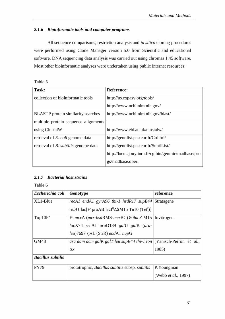
2. Materials and methods 272.1 Materials 272.1.1 Equipment used in this study 272.1.2 Materials and reagents 272.1.3 Kits 292.1.4 Antibodies 302.1.5 Oligonucleotides 302.1.6 Bioinformatic tools and computer programs 312.1.7 Bacterial host strains 312.1.8 Plasmids used in this study 312.2 Molecular biology methods 342.2.1 Growth Medium 342.2.2 Antibiotic Solutions 352.2.3 Techniques related to DNA 352.2.4 Agarose gel electrophoresis of DNA 352.2.5 Digestion of DNA by restriction enzymes 362.2.6 Ligation of vector and insert DNA 362.2.7 E. coli transformation 372.2.8 Preparation of plasmid DNA 372.2.9 Polymerase chain reaction - PCR 382.2.10 DNA sequencing 392.2.11 Primer annealing cloning 40
2.2.12 Site-directed Mutagenesis 402.3 Techniques related to RNA 412.3.1 RNA extraction 412.3.2 Primer extension 422.4 Techniques related to protein 432.4.1 Preparation of protein extracts 432.4.1 Separation of proteins by SDS-polyacrylamide gel electrophoresis 442.4.2 Protein staining with Coomassie blue 452.4.3 Western blotting 462.4.3.1 Immunodetection 462.4.3.2 Chemiluminescence-detection of proteins on nitrocellulose membrane 472.4.4 Purification by strep-tactin column 482.5 Bacillus genetics 492.5.1 Preparation of chromosomal DNA from Bacillus subtilis cells 492.5.2 Preparation of competent Bacillus subtilis cells 492.5.3 Transformation of Bacillus subtilis 502.5.4 Screening for gene integration at the amyE (amylase) locus 512.5.5 Promoter induction in Bacillus subtilis 512.5.6 PCR knockout technique for Bacillus subtilis 512.6 Microscopic techniques 532.6.1 Fluorescence microscopy - Principle 532.6.2 Vital stains used in fluorescence microscopy 542.6.3 Media used for microscopy 552.6.4 Preparation of slides for microscopy 56
3. Results 573.1 Identification of SMC interacting proteins - Historical observations 573.2 Phenotypic analysis of ypuG and ypuH 583.3 YpuG and YpuH - A new family of conserved proteins 613.4 Subcellular localization pattern of ScpA, ScpB, and SMC 643.5 Dynamic localization of SMC, ScpA, and ScpB 663.6 SMC, ScpA, ScpB are associated with DNA 693.7 Colocalization of ScpA, ScpB, and SMC 703.8 Interaction of ScpA, ScpB, and SMC in vivo 713.9 Specific localization depends on all three proteins of the complex 743.10 SMC complex requires active replication for its bipolar foci
segregation 763.11 SMC localization depends on DNA topology 773.12 SMC - A bacterial condensin protein 793.12.1 Effects of overproduction of SMC 793.12.2 SMC condenses from a single position on the nucleoid 803.13 Regulation of SMC 813.13.1 Growth phase dependent expression of SMC and ScpB 833.13.2 Stability of SMC 843.14 Involvement of SMC complex in repair 853.15 Identification and examination of SMC-like proteins in Bacillus subtilis 873.15.1 Analysis of YirY/SbcC function 883.15.2 Localization of AddAB 893.16 Topoisomerase IV - A chromosome segregator 90
4. Discussion 934.1 Fluorescence microscopy - changing the view of prokaryotes 100
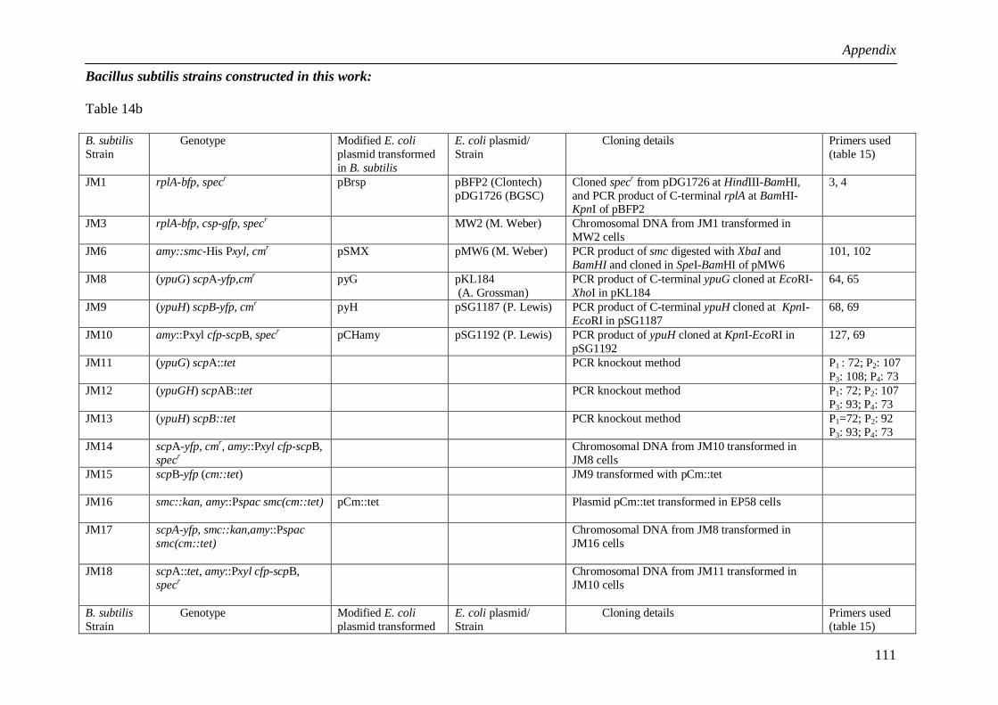
5. Appendix5.1 Specific polar localization of ribosomes in Bacillus subtilis depends on
active transcription. 1035.2.1 Strains used in this work 1095.2.2 List of plasmids and strains constructed in this work 1105.2.3 Primers used 1145.2.4 Primer annealing temperatures 116
6. References 117
1
ZusammenfassungAlle Zellen müssen ihr Erbmaterial verdoppeln und dafür Sorge tragen, daß
jede Tochterzelle einen kompletten Satz des Erbguts vor der Zellteilung erhält. In
Bakterien müssen die Chromosomen organisiert und kompaktiert werden, während sie
gleichzeitig dynamisch sein müssen, um laufende zelluläre Prozesse wie DNA
Reparatur, Rekombination, Transktiption und Replikation zu ermöglichen. SMC
(Structural Maintenance of Chromosome) Proteine bilden eine ubiquitäre
Proteinfamilie, die eine zentrale Rolle in verschiedenen Chromosomendynamiken
spielt. Das Hauptaugenmerk in dieser Arbeit ruht auf der Charakterisierung der SMC
Proteine und ihrer Partner aus Bacillus subtilis.
Genbanksuchen haben zu der Identifizierung zweier Interaktionspartner des
SMC Proteins geführt. Diese Proteine, ScpA und ScpB, sind in Bakterien und
Archaen konserviert. Die Deletion des scpA oder des scpB Gens führte zu einem der
Wachstum (unterhalb 23°C), dekondensierten Nukleoiden (zelluläre Struktur der
Chromosomen) und einem ausgeprägten Segregationsdefekt. Die gleichzeitige
Deletion der Gene erzeugte keinen veränderten Phänotyp, was zeigt, dass alle drei
Proteine im gleichen Aspekt der Chromosomen-Kondensation und Segregation
fungieren. Um ihre Funktion in vivo zu untersuchen, wurden die Proteine in Zellen
mit Hilfe von voll funktionellen GFP Fusionen lokalisiert. Alle drei Proteine bildeten
diskrete Foci in den Zellen, einem bis dato unbekannten Lokalisationsmuster, das sich
dynamisch während des Zellzyklus veränderte: zu Beginn des Zellzyklus befanden
sich die Foci in der Zellmitte, und nach der Verdopplung des Focus wanderten die
beiden Foci rasch entgegengesetzt in Richtung der Zellpole. In diesen bipolaren Foci
verblieben die drei Proteine für den Rest des Zellzyklus. Die Bildung des
Proteinkomplexes konnte durch Fluoreszenz Resonanz Energie Transfer (FRET) und
durch Depletionsstudien belegt werden. So konnte die Bildung der Foci nur in
Anwesenheit aller Proteine beobachtet werden, nicht jedoch in Abwesenheit eines der
drei Proteine. Die spezifische Lokalisierung des SMC Komplex hing auch von
fortlaufender DNA Replikation ab, von zellulärer Gyrase Aktivität (d.h. von der
Struktur der DNA), sowie von der ATPase-Aktivität von SMC. Die Überproduktion
von SMC führte zu einer Über-Kondensation der Nukleoide, wobei die Lokalisation
2
des SMC Komplexes erhalten blieb, was darauf hin deutet, daß die beobachteten Foci
aktive Kondensationszentren darstellen.
Weiterhin zeigten die Proteine des SMC Komplex wachstumsabhängige
Expression. SMC und ScpB waren nur in wachsenden Zellen vorhanden, und wurden
rasch beim Übergang in die Statonärphase abgebaut. Die Analyse der RNA Mengen
in verschiedenen Wachstumsphasen durch Primer Extensionsanalyse zeigte, daß das
smc Transkript im Übergang zur Stationärphase nicht abnimmt. Diese Experimente
zeigten einen bisher nicht identifizierten smc Promotor auf, und erbrachten den
Nachweis, daß SMC posttranskriptionell reguliert wird. Die smc, scpA, und scpB
Deletionsmutanten wiesen ebenfalls eine ausgeprägte Sensitivität gegenüber
Mitomycin C (MMC) auf, welches Doppelstrangbrüche (DSBs) in die DNA einführt.
Demnach wird der SMC Komplex ebenfalls für die Reparatur von DSBs benötigt.
Weiterhin wurde die Funktion des SMC Proteins YirY untersucht, welches
homolog zum DNA Reparatur Protein SbcC aus Escherichia coli ist. Die yirY
Deletion führte ebenfalls zu einer deutlichen Sensitivität zu MMC, was eine Rolle in
der DSB Reparatur belegt. In MMC behandelten Zellen bildete YirY Foci auf der
DNA, welche aktive DSB Reparaturzentren darstellen könnten. In Gegensatz dazu
waren die anderen Proteine aus dem gleichen Operon, AddA, AddB, and SbcD
überall in den Zellen vorhanden und bildeten keine speziellen Strukturen, was darauf
hindeutet, daß SbcC und AddAB in verschiedenen Reparaturwegen fungieren.
Die subzelluläre Lokalisation der Topoisomerase IV Untereinheiten ParC und
ParE wurde ebenfalls in dieser Arbeit beleuchtet. ParC lokalisierte auf dem gesamten
Nukleoid, ganz im Gegenteil zu einer früheren Studie, in der ParC ausschließlich in
der Nähe der Zellpole vorhanden war, wonach ParC eine spezialisierte Rolle bei der
Dekatenierung von Chromosomen zugesprochen wurde. Durch Überproduktion von
ParC und ParE wurden die Nukeloide noch stärker kompaktiert, was zusammen mit
der Lokalisierung eine generelle Rolle in der Chromosomenkompaktierung belegt.
Ein weiterer Aspekt in dieser Arbeit war die Lokalisierung von Ribosomen.
Das L1 Protein aus der großen Untereinheit lokalisierte in wachsenden Zellen in den
zytoplasmatischen Stellen, die das Nukeloid umgeben, wohingegen es in stationären
Zellen und nach Inhibition der Transkription überall in der Zelle vorlag. Demnach
hängt die spezifische Lokalisierung von Ribosomen von aktiver RNA Synthese in den
Zellen ab.
3
Insgesamt läßt sich schlußfolgern, daß die Lokalisation von Proteinen, die an
der Chromosomensegregation, DNA Reparatur und Translation beteiligt sind, ein
wesendlich definierteres Bild der räumlichen Funktion der Proteine in lebenden
Bakterien erbrachte.
4
Summary
All cells need to duplicate and separate their genetic material faithfully into the
future daughter cells before cell division takes place. In bacteria, the chromosome has
to be organized and compacted whilst, at the same time, it needs to be dynamic to
allow other ongoing cellular processes like repair, recombination, transcription,
replication and segregation to take place. SMC (Structural Maintenance of
Chromosome) protein belongs to a ubiquitous protein family that play crucial roles in
chromosome dynamics. The main interest of this work is to characterize the function
of the SMC protein in Bacillus subtilis.
Data base searches have led to the identification of two interaction partners of
SMC. These proteins, ScpA and ScpB are conserved among bacterial and archaeal
species possessing SMC. The scpA or scpB deletions showed a similar phenotype to
that of a smc disruption, namely temperature sensitive slow growth (below 23°C),
decondensed nucleoids and a strong segregation defect. Their simultaneous deletion
did not exacerbate the phenotype, suggesting that all the three proteins function in the
same pathway in chromosome condensation. To investigate their in vivo function, the
proteins where localized in the cells using functional GFP fusions. The subcellular
localization showed bipolar foci, a unique pattern of localization that was dynamic
and cell cycle dependent. The foci were present at mid-cell position in smaller cells
and separated towards opposite cell poles within a few minutes. The formation of a
complex between SMC, ScpA, and ScpB in vivo was confirmed using fluorescence
resonance energy transfer (FRET) and depletion studies. Formation of foci was only
seen in the presence of all three proteins, but not in the absence of any one of them.
The specific localization pattern of these proteins also depended on ongoing DNA
replication, on active gyrase and thus on DNA topology, as well as on SMC’s ATPase
activity. Overproduction of SMC led to increased compaction of nucleoids but the
localization was retained in the form of foci suggesting that the foci represent active
chromosome condensation centers.
The proteins of the SMC complex showed growth dependent protein
expression. SMC and ScpB proteins were present in actively replicating exponential
phase cells, but were rapidly depleted as the cells entered stationary phase. Analysis
with total RNA extracts from various growth phases by primer extension studies
5
showed a strong transcript for SMC that was present even in stationary phase. This
experiment led to the identification of a new promoter for smc, and suggests that SMC
is regulated at the protein level by a protease that is induced at the onset of stationary
phase. Smc, scpA, and scpB deletion mutant cells were also sensitive to Mitomycin C
(MMC) treatment, which induces double strand breaks (DSB) into DNA. This finding
revealed a role of the SMC complex in DSB repair.
I also investigated the role of YirY, a homolog of the DSB repair protein SbcC
which is a proposed member of SMC family. Upon disruption of yirY/sbcC, the cells
did not show any visible phenotype but the cells were sensitive to MMC, suggesting
its role in repair. SbcC formed foci only in MMC treated cells, so the foci in the cell
might represent a DNA repair centers. Other proteins located in the same operon as
SbcC, AddA, AddB, and SbcD, did not show any specific pattern of localization, but
were present throughout the cell and showed slight increase in their fluorescence
intensity after MMC treatment, suggesting that SbcC and AddAB function in different
in repair pathways.
The localization of topoisomerase IV subunits ParC and ParE has also been
investigated in this work. The fluorescent protein fusion of ParC localized throughout
the nucleoid, contrarily to the previously published bipolar localization as foci, which
had suggested a specialized function of topoisomerase IV in chromosome
decatenation. Upon over expression of ParC and ParE, the cells contained more
condensed nucleoids, revealing a general role of topoisomerase IV in global
chromosome compaction.
A further aspect of this work was the study of dynamic localization of
ribosomes. The large subunit ribosome protein L1 showed specific localization in the
cytoplasmic space surrounding the nucleoid in growing cells, and was seen diffused
throughout the cell in the stationary phase. The same effect was observed upon
inhibition of transcription, suggesting the dependence of specific ribosome
localization on active transcription.
In toto, localization of DNA segregation, DNA repair and the ribosomal
proteins has provided a more defined view of the spatial organization of these cellular
processes in live bacterial cells.
6
Abbreviations
ATP adenisine-5’- triphosphate
amyE gene coding for α-amylase
bp base pair
cDNA complementary DNA
Cmr chloramphenicol resistant
DAPI 4',6-diamidino-2-phenylindole
DSBR double strand break repair
dsDNA double stranded DNA
EDTA ethylene diamine tetra acetic acid
EM electron microscopy
EtBr ethidium bromide
Fig figure
FP fluorescent protein
FRET fluorescent resonance energy transfer
GFP/YFP/CFP green/ yellow/cyan fluorescent protein
h hour
IPTG isopropanol-b-D-thiogalactopyranoside
kb kilo base(s)
LB Luria-Bertani medium
MCS multiple cloning sites
min minute(s)
mls macrolide lincosamine streptogramidine B
MMC mitomycin C
nm nanometer
O.Dxxx optical density at xxx nm
Ori origin of replication
PCR polymerase chain reaction
RNase ribonuclease
RT room temperature
rpm revolutions per minute
SDS-PAGE sodium dodecylsulfate polyacrylamide gel electrophoresis
SMC structural maintenance of chromosome protein
Tm melting temperature of dsDNA
7
TB tris boric acid buffer
TE tris EDTA buffer
tetr tetracyclin resistance
Tris tris-(hydroxymethyl) aminomethane
U unit of enzyme activity
UV ultraviolet light
wt wild type strain
sum of
deletion
:: gene replacement at chromosome by double crossover
Introduction
8
1 Introduction
Life on earth persists because of its propagation through cell division - a
central cellular process that is shared by all living organisms. Before a cell divides it
has to duplicate a number of sub cellular components, most importantly the DNA
molecule(s) carrying the genetic information and depending on the organism,
organelles and then segregate them into the appropriate daughter cell compartments.
This process is maintained by well-coordinated action of many dedicated proteins that
make up a functional network whose complexity depends on the nature of the
respective organism. Although compared to eukaryotes, prokaryotic cell division
seems much simpler with most of the time only one major DNA molecule and lack of
membrane-dependent organelles, many basic principles are functionally conserved.
The DNA of bacterial chromosome is several thousand micrometers long and
therefore are condensed into a compact structure called ‘nucleoids’ that has the
diameter of only 0.5 µm (Rouviere-Yaniv et al., 1979). A typical bacterial cell
contains >250 different species of DNA binding proteins (Robinson and Kadonaga,
1998), which include DNA polymerases, topoisomerases, helicases, histone-like
proteins, etc. These proteins are associated with the nucleoid and take part in
chromosome organization during various cellular processes like replication,
recombination, repair, modification and transcription of DNA. One among these
players is the SMC protein, which belongs to a ubiquitous protein family and plays a
key role in maintaining chromosome organization. This work is focused on the in vivo
characterization of SMC and proteins interacting with SMC by making use of genetic
and microscopic approaches.
From the important model organisms Escherichia coli a Gram-negative
enterobacterium, Bacillus subtilis a Gram-positive soil bacterium, and Caulobacter
cresentus a dimorphic Gram-negative aquatic bacterium all of which have been under
thorough investigation for several years now, our laboratory decided to focus on
Bacillus subtilis which is a broadly distributed, rod-shaped micro-organism that
resides in the upper layers of soil. B. subtilis is a facultative aerobe and capable of
converting to anaerobic nitrate respiration under oxygen limiting conditions
(Hoffmann et al., 1995). It has the ability to form extremely resistant endospores in
response to nutrient deprivation or slow dehydration (Stragier and Losick, 1996) and
Introduction
9
because it is genetically easily accessible, it has been accepted as one of the best-
studied bacteria even before its genome was entirely sequenced a few years ago
(Kunst et al., 1997; Weber and Marahiel, 2003).
1.1 Basic mechanisms of bacterial replication and cell division
A cell divides only after molecular sensors have detected that its genetic
material, DNA, providing the molecular blueprint for daughter cells to survive, has
been faithfully duplicated in a damage-free manner. In eukaryotes, this sensor is a cell
division cycle molecule Cdc25, that turns on the proteins required for the actual cell
division event (Jinno et al., 1994). Precise DNA replication has to be followed by the
segregation process which involves a complex sequence of structural events termed
mitosis in eukaryotes. This is a marked difference compared to prokaryotes where
replication is not followed by but coupled to the segregation process and is therefore
coordinated with the cell growth and division (Helmstetter, C, 1996).
In bacteria, chromosome replication is initiated when a critical size of a
growing cell is reached (Messer, W., and Weigel, C, 1996). This parameter is called
the initiation mass. In Vibrio harveyi the protein CgtA was shown to be involved in
coupling of chromosome replication to cell growth and division. In B. subtilis, its
homologue, Obg, has been proposed to control DNA replication and regulate
initiation of sporulation by sensing the intracellular GTP level and stimulating the
activity of a phosphorelay system which in turn activates several proteins involved in
replication processes (Sikora-Borgula et al., 2002).
Initiation of replication commences when an ATP-bound replication initiation
protein DnaA binds to the AT rich DnaA boxes in the replication origin, OriC,
regions and causes local strand melting (Moriya et al., 1988). The DnaB helicase is
then recruited to the unwound region. Together with other proteins of the primosome
complex the strands are loaded on the DNA polymerase replication machinery, which
is located at the mid cell. In B. subtilis, the DNA now moves through a stationary
replisome complex (Lemon and Grossman, 1998) whereas in case of C. cresentus the
replisome is mobile (Jensen et al., 2002). During the replication process, replicated
chromosomes move outward towards each cell halves. The termination of replication
takes place through arresting of the replication forks by complex formation of the
Introduction
10
replication termination protein (RTP) with the ter sites located at approximately 172°
on the chromosome (Bussiere and Bastia, 1999). The replicated chromosomes are
thereafter separated by decatenation process involving topoisomerase IV and the site-
specific recombinases, CodV and RipX (Sciochetti and Piggot, 2000). Once the mid
cell region is cleared from the replicated chromosomes by FtsK/SpoIIIE, bacterial
cells assemble a ring like cytoskeletal structure at the division site, which is composed
of tubulin-like FtsZ protein that constricts the cellular membrane and forms the
septum. This FtsZ ring structure or the divisome is localized to the division site by the
Min proteins (Raskin and De Boer, 1997). The Min system plays an important role in
division site placement by inhibiting FtsZ ring formation at polar regions. It
comprises the MinC and MinD complex and the inhibitor protein which is called
DivIVA in B. subtilis and MinE in E. coli, that ensures the inhibition only at the polar
regions (Cha and Stewart, 1997; Edwards and Errington, 1997; Marston et al., 1998).
Under normal conditions, bacterial cell division is symmetric. However, B. subtilis
undergoes asymmetric division when conditions of nutrient limitation and high
population density result in the initiation of a sporulation pathway that culminates in
the formation of a heat- and desiccation-resistant spore. During sporulation, FtsZ
forms a septum close to one of the cell poles - a process regulated by the master
sporulation regulator, Spo0A (Levin and Losick, 1996; Stragier and Losick, 1996). In
case of Caulobacter, the cell cycle is inherently asymmetric, a sessile-stalked cell
undergoes asymmetric cytokinesis releasing a flagellated motile swarmer cell. This
motile cell has to re-differentiate into a sessile-stalker cell before becoming able to
undergo a further round of cell division (Wheeler et al., 1998). This is achieved by
repression of the replication process by a response regulator (CtrA) that is later
proteolyzed when the swarmer cell differentiates into a sessile cell. (Shapiro and
Losick, 2000).
The mechanism of bacterial chromosome partitioning was explained by an
‘extrusion-capture’ model proposed by (Lemon and Grossman, 2001) (fig. 1). This
model assumes that the energy from the replication factory is used to power
partitioning of the replicated chromosomes. The replicated chromosomes are captured
at the cell quarter position and are organized through compaction and supercoiling
which further assists the segregation process. This model was then further refined by a
‘push, direct, condense, hold and clear’ model by (Sawitzke and Austin, 2001) and
Introduction
11
assumes that daughter DNA strands are actively transported to the cell halves,
possibly by the Par proteins.
Fig. 1: Simplified model of bacterial cell cycle. DNA (grey lines), oriC (grey circles),terminus, terC (dark grey square), DNA polymerase (triangles), and cytokinetic ringFtsZ (dashed line). DNA replication initiates at the mid cell. The sister originsseparate out bidirectionally. The replication continues followed by compaction of anewly replicated DNA until there are two complete and separate chromosomes.Finally the cell divides medially by the FtsZ ring formation. Figure adapted from(Lemon and Grossman, 2001).
1.2 Organization of bacterial chromosome
With some exceptions such as Streptomycetes coelicolor that possesses linear
DNA and the Borrelia genus whose genome is made from linear DNA with hairpin
ends, most bacterial cells possess a closed circular genomic DNA molecule. As stated
earlier, the bacterial chromosome is about 1000-fold longer than the cell size (Drlica,
K., 1986) and is condensed into a compact structure called ‘nucleoid’. The bacterial
nucleoid is functionally analogous to the eukaryotic nucleus, e.g. the packing density
of the DNA in the nucleoid is like that of eukaryotic interphase nuclei and would thus
allow diffusion in and out of even large macromolecules (Kellenberger, 1991). Early
attempts to elucidate the nucleoid structure using techniques of fixation led to a
number of artefacts caused by the fixation technique itself. Nucleoids prepared from
the cryo-freeze substituted cells showed a central dense regions and long, thinner
Introduction
12
cytoplasmic protrusion-like clefts, see fig. 2 (Bohrmann et al., 1991; Hobot et al.,
1985):
Fig. 2: Schematic model of bacterial nucleoid from the sections of cryofixed, freezesubstituted E. coli cells. Figure adapted from Bohrmann et al., 1991.
The nascent RNA was shown to localize at the nucleoid periphery (Ryter and
Chang, 1975), hence these edges of chromosomal protrusions from the nucleoids were
interpreted as areas of a metabolically active nucleoid undergoing active transcription.
Furthermore, DNA from isolated nucleoids was shown to possess a negatively
supercoiled topology and these supercoils could not be relaxed by a single nick. This
observation was interpreted in terms of topologically independent chromosomal
domains that were calculated as 50 per genome for E. coli (Sinden and Pettijohn,
1981); (Drlica, 1986).
1.2.1 Membrane attachment of nucleoids
The compact nucleoid structure is maintained by membrane-DNA, protein-
DNA and RNA-DNA interactions (Guillen and Bohin, 1986). In B. subtilis or E. coli,
it was not possible to obtain membrane-free nucleoids (Harmon and Taber, 1977) (fig.
3), which led to the hypothesis that nucleoids are anchored to the membrane.
Specifically, in B. subtilis, the chromosome origin region isolates were enriched in
membrane fractions and these attachments are thought to facilitate chromosome
replication and segregation processes. DnaB was one among the proteins involved in
DNA attachment process playing an essential role in DNA replication and membrane
attachment of the Ori of replication of chromosomes (Laurent and Vannier, 1973). It
was shown that DnaB forms specific foci and localizes at the OriC region (Imai et al.,
2000). Recent investigations identified a novel protein RacA that localized near the
poles and on the nucleoid and acts as an adhesion component bridging the origin
region to the cell poles (Ben-Yehuda et al., 2003).
Introduction
13
Fig. 3: Membrane attachment of nucleoid- EM of isolated E. coli nucleoid spread inthe presence of spermidine. (Scale bar 1µm). Fig. adapted from ‘Escherichia coli andSalmonella typhimurium’, Cellular and Molecular Biology, Vol 1, 1987 (ASM press).
1.2.2 The nucleoid structure is dynamic
Nucleoids in rapidly growing cells appear in complex shapes (Zimmerman,
2003). This is due to the occurrence of cellular processes like transcription and
replication that require a very dynamic state of chromosomes. Moreover,
chromosomes appear to have a defined orientation within the cell which has been
determined using various origin region markers (Levin and Grossman, 1998; Losick
and Shapiro, 1999; Webb et al., 1997). Soon after replication, origin regions separate
from each other and move to opposite sides of the cell, while the terminus regions is
found in the mid cell (Teleman et al., 1998; Webb et al., 1997). The newly replicated
origin regions then position near the cell quarter regions for the next round of
replication. In vegetative cells of B. subtilis, the nucleoid appears as a discrete mass
Introduction
14
centered close to mid cell, with prominent gaps at each cell pole. Soon after the onset
of sporulation, the nucleoid undergoes a conformational change, in which it
approximately doubles in length so that it reaches from pole to pole in this state, it is
termed as axial filament (Errington, 2001). In spores, the nucleoid is packed into
donut-like ring of approximately 1 micrometer in diameter (Pogliano et al., 1995).
During germination, the ring-shaped nucleoid disappears and the nucleoid becomes
more dense while later in spore outgrowth the shape of the nucleoid is reverted to the
diffuse lobular shape seen in growing cells (Ragkousi et al., 2000).
Changes in the nucleoid structures have been observed during cell growth
phase and in various environmental stress conditions, depicting the altered
transcription. Upon cold shock, the nucleoid appears more condensed (Weber et al.,
2001). Elevated hydrostatic pressure perturbs cell division and nucleoid structure
(Welch et al., 1993) and the addition of transcription inhibitor rifamycin leads to
decondensed nucleoid in B. subtilis (Guillen and Bohin, 1986). Addition of
chloramphenicol in exponentially growing cells showed changes in appearance from
irregular spheres and dumbbells to large, brightly stained spheres and ovals, while the
late exponential phase cells showed elongated axial filament structures (Bylund et al.,
1993).
1.3 Nucleoid-associated proteins
DNA topology plays a critical role during dynamic chromosomal processes
and is affected by changes in growth phase, environmental stress situations, and by
several DNA-interacting proteins. Proteins that bind and organize DNA structure are
vital components of the cell. By interacting with their DNA substrate, they affect gene
expression, growth efficiency, and cell viability through change in the state of
chromosome condensation and relaxation. Some of the relevent nucleoid-associated
proteins are briefly discussed below.
HBsu - The histone-like protein in B. subtilis, belongs to a highly conserved
HU protein family. HBsu is coded by hbs gene, which is regulated by two promoters.
Throughout the cell cycle, HBsu is the most abundant protein associated with
Introduction
15
nucleoid, is essential for growth and differentiation and has been shown to modulate
DNA topology (Klein and Marahiel, 2002; Micka and Marahiel, 1992). HBsu was
extracted from isolated nucleoids and characterized by its ability to introduce negative
supercoils into DNA in the presence of topoisomerase I (Le Hegarat et al., 1993).
HBsu binds to DNA as homodimer in a sequence-independent manner with a
preference for curved DNA (Kohler and Marahiel, 1997). HBsu localizes to the
nucleoids in growing cell and colocalizes with SASPs (see below) on the ring-shaped
nucleoid of the germinating spores (Ross and Setlow, 2000). HBsu has also been
demonstrated to play a role as part of the bacterial signal recognition particle involved
in presecretory protein translocation (Nakamura et al., 1999) and in DNA repair and
recombination (Alonso et al., 1995).
SASPs - Small acid-soluble spore proteins are found in two forms, the alpha-
and beta-type, encoded by six sspA-F genes in B. subtilis that are expressed during
sporulation and are implicated in packaging of DNA in spores. The SASPs bind with
greater affinity to GC rich DNA regions and increase DNA persistence length
tremendously by changing the DNA conformation B to A (Mohr et al., 1991). SASPs
protect the spore chromosome against damages induced by heat, oxidizing agents,
desiccation, and UV irradiation (Mason and Setlow, 1987; Setlow and Setlow, 1995).
SASPs also act as an amino acid reservoir for protein synthesis during spore
germination (Setlow, 1988) and colocalize with the nucleoid until early germination
(Ross and Setlow, 2000). During the germination process, the donut shaped nucleoid
transforms into a more compact mass due to the degradation of most of the spore’s
pool of major alpha/beta-type SASPs (Ragkousi et al., 2000).
1.3.1 Partition proteins
Plasmid segregation system in E. coli. Plasmids are autonomously replicating
genetic entities that are ubiquitous in bacteria. Plasmids control their own replication
(Hiraga, 1992), utilizing the standard cellular replication machinery and are actively
segregated between daughter cells (Gordon and Wright, 2000; Hiraga, 2000). The
presence of a partition cassette (par) allows for the inheritance of the plasmid copy in
each daughter cell. This partition cassette encodes two structural genes and a cis-
acting parS site (a centromere analog). Par+ plasmids form clusters and localize to the
Introduction
16
cell poles, while Par- plasmids localize randomly in a cell (Weitao et al., 2000).
Plasmid partitioning either uses ParA, an ATPase with a Walker-type ATP-binding
motif or ParM, an actin-type-ATPase (Bignell and Thomas, 2001; van den Ent et al.,
2002). In both cases, ParB, a second partition protein, binds to the cis-acting DNA
partitioning parS site and recruits the ATPase into the nucleoprotein partition
complex (Bouet and Funnell, 1999). In the ParA system, plasmid pairs translocate to
the mid-cell position shortly before septation and are then propelled bidirectionally by
the partition apparatus into the daughter cell halves (Li and Austin, 2002). For
plasmid partition involving an actin-type ParM protein, extensive polymerization of
this protein is likely to direct plasmid movement during segregation (van den Ent et
al., 2002).
Spo0J and Soj constitute the Par protein system in B. subtilis and are
homologs of ParB and ParA proteins involved in plasmid and chromosome
segregation in E. coli. Spo0J and Soj were originally identified as proteins required
for an early stage of the sporulation pathway. The ParB homolog Spo0J controls the
expression of early acting sporulation genes which require expression of the Spo0A
transcription factor. Soj is related to the ParA ATPase family and is a transcriptional
regulator that functions antagonistically to Spo0J. The Spo0J and Soj have been
demonstrated to function as partition proteins in E. coli and were required for the
specific localization of plasmids at cell quarters when heterologously expressed in E.
coli (Yamaichi and Niki, 2000). The Soj-Spo0J system operates a checkpoint that
couples chromosome partitioning to developmental gene expression. When
chromosome partitioning is incomplete, Soj represses the activity of Spo0A. The
completion of partitioning results in Spo0J inactivating the Soj repression. Spo0J
binds to specifically conserved 16-bp parS sequences clustered around the soj-spo0J
operon. These sequences occur approximately ten times within the Ori region of B.
subtilis genome (Lin and Grossman, 1998). Immunofluorescence and GFP tagging of
Spo0J show that they localize as foci near the origin region (Lewis and Errington,
1997). Deletion of Spo0J affects nucleoid organization and segregation and leads to a
100-fold increase in anucleate cells suggesting its active role in chromosome
partitioning (Ireton et al., 1994); (Draper and Gober, 2002). Spo0J was also thought to
be involved in the spatial organization of Ori regions of the chromosome, since its
mutations led to defects in the orientation of the prespore chromosome. Soj oscillates
Introduction
17
from one pole to the other within the period of 20 seconds in a Spo0J-dependent
manner and in its absence, Soj localizes to the nucleoid (Marston and Errington,
1999a). Deletion of soj (parA homolog) does not result in a DNA segregation defect,
but it is required for the stability of parS-containing plasmids. In contrast to B.
subtilis, inactivation of either parA or parB in C. crescentus is lethal to the cell
(Marczynski and Shapiro, 2002).
1.3.2 Proteins involved in chromosome dynamics
Topoisomerases participate in maintaining chromosome function by adjusting
DNA topology appropriately to meet the requirements of changing conditions such as
temperature, growth phase, nutrient availability, etc. and facilitate fundamental
cellular processes such as chromosome segregation, transcription, and DNA
replication (Brill et al., 1987). Topoisomerases possess the unique ability to create a
transient break in a DNA molecule that allows the passage of one strand through
another and then religate the cut molecule (Hsieh and Brutlag, 1980). Type I
topoisomerases cleave one strand of the DNA duplex in an ATP-independent manner,
while type II topoisomerases cleave both strands and utilize ATP. Topoisomerase II
activity facilitates DNA replication and transcription by removing superhelical twists
that result from the progression of the DNA and RNA polymerases along the
chromosome (Koshland and Strunnikov, 1996). While the DNA molecules of
mesophilic bacteria are negatively supercoiled, which facilitates the DNA processes
of replication, transcription and recombination (Declais et al., 2001), those of
hyperthermophilic archaea possess positively supercoiled DNA that are maintained by
the activity of a unique enzyme termed reverse gyrase that protects their
chromosomes from denaturation (Lopez-Garcia and Forterre, 1999).
Bacillus subtilis harbours four topoisomerases: topA, coding for topoisomerase
I, unwinds DNA by removing negative supercoils and has been shown to play a role
in illegitimate plasmid recombination that allows recombination between non-
homologous sequences and recognizes a consensus sequence 5'-
A/(T)CAT(A)/(T)TA(A)/(A)(T)/(T)A-3' (Meima et al., 1998). TopB codes for
topoisomerase III, which has been characterized in E. coli, where it acts as a cellular
decatenase during the process of chromosome segregation. topB is a multicopy
suppressor of a topA null mutation (Broccoli et al., 2000). Both topA and topB are
Introduction
18
type I topoisomerases and required for proper chromosomal segregation in E. coli
(Zhu et al., 2001). Gyrase and topoisomerase IV finally constitute the type II
topoisomerases. Gyrase is formed by two subunits coded by gyrA and the ATP-
binding subunit gyrB. Similarly, parE and parC code for topoisomerase IV. Gyrases
are involved in the initial stages of replication easing the positive supercoils and the
topoisomerase IV acts at the final stage of replication as a decatenase (Huang et al.,
1998) and are essential for cell cycle progression and developmental regulation in
Caulobacter crescentus (Ward and Newton, 1997).
SpoIIIE is a DNA tracking protein with ATPase activity and a member of a
large family of bacterial proteins involved in DNA translocation (Dworkin, 2003;
Errington et al., 2001). It is required for complete segregation of chromosomal DNA
into the pre-spore during asymmetric division in sporulating B. subtilis. Chromosome
partitioning during sporulation differs from vegetative chromosome partitioning in
that it occurs after formation of the septum. SpoIIIE localizes to the prespore septum
where it is proposed to pump the remaining chromosome from the mother cell
compartment into the prespore (Wu and Errington, 1997). Mutations in the spoIIIE
gene prevent proper partitioning of one chromosome into the developing prespore
during sporulation but has no effect on partitioning in vegetatively dividing cells
(Pedersen and Setlow, 2000). The gene encoding SpoIIIE is expressed constitutively
and plays a role in chromosome segregation during vegetative growth by translocating
trapped DNA from enclosing septum during cell division (Pedersen and Setlow,
2000).
PrfA, the penicillin-binding protein-related factor A, also designated as RecU,
is located downstream in an operon with ponA, a penicillin-binding protein (PBP1)
involved in peptidoglycan crosslinking. PrfA/RecU is implicated in several cellular
processes such as cell wall synthesis, chromosome segregation, and DNA
recombination and repair (Pedersen and Setlow, 2000). A prfA deletion rendered cells
more sensitive to DNA-damaging agents, decreased the transformation efficiency
(Fernandez et al., 1998), and led to 0.9-3% anucleate cells and cells with abnormal
nucleoid staining patterns. Inactivation of prfA also exacerbated smc and spo0J
chromosome segregation phenotype and its overexpression in E. coli caused nucleoid
condensation (Pedersen and Setlow, 2000). PrfA has been shown to possess
Introduction
19
endonuclease activity and is structurally related to the restriction enzyme PvuII
(Rigden et al., 2002).
1.4 SMC - Structural/stable maintenance of chromosomes protein
SMC proteins are ubiquitous and are an essential part of a high order complex
which is involved in chromosome dynamics. In 1985, as pioneers of SMC research
Larinov and Strinnikov, observed an increase in the copy number of artificial mini
chromosomes in a Saccharomyces cerevisiae mutant with an impaired segregation
process. These mutants were mapped in four genes, AMC1, AMC2, AMC3, and
AMC4 (AMC = Arificial Mini Chromosome) which control the segregation of natural
chromosomes in yeast. AMCs were later rediscovered as SMC for stability of mini
chromosomes (Larionov and Strunnikov, 1987; Strunnikov et al., 1993). While
eukaryotes code for 6 different types of SMC protein represented by SMC1-6, to date,
prokaryotes contain only a single allele for an SMC homolog (Hirano, 2002).
1.4.1 Structure of SMC
The proteins belonging to the SMC family are large proteins in the range
between 110 and 170 kDa (Harvey et al., 2002) and share common principles in
domain organization: A globular N-terminus contains a conserved sequence
resembling a Walker A ATP-binding motif (G-NGSGKSN) and a C-terminal domain
harbors both the highly conserved LSGG motif signature, called C motif, and P-P-
DE-DAALD which corresponds to a Walker B motif (Walker et al., 1982). The N-
and C-terminal domains are connected via two long coiled coil domains (of variable
length) separated by a globular hinge domain of approx. 150 amino acids in length:
Fig. 4: Schematic diagram of domain organization in a typical SMC protein.
Introduction
20
Based on the primary sequence it was predicted that SMC proteins might form
antiparallel dimers, and that dimerization is probably mediated by inter- or
intramolecular interactions (Saitoh et al., 1994). Electron microscopic analysis of B.
subtilis SMC by (Melby et al., 1998), showed various conformations of SMC and the
most prominent ‘V’ shaped conformation, see fig. 5. In their model, they suggested
that SMC proteins form antiparallel dimers connected through the coiled coil
segments with the N- and C-termini of each monomer forming a head domain located
at the ends of a ‘V’-like structure (Melby et al., 1998):
Fig. 5: Electron micrographs from B. subtilis SMC representing the most commonconformations. Images were adapted from (Melby et al., 1998).
So far, no crystallization of the whole SMC molecule has been reported, which
might be due to its large and flexible nature, but several workgroups have come up
with the crystal structures of different domains. Analysis of the crystal structure of N-
terminal domain of MukB which is a member of SMC family and a functional analog
of SMC protein in E. coli, showed that the N- and C-terminal domains of SMC
molecules have to come together to create an ATPase activity pocket (van den Ent et
al., 1999). Rad50 is a member of eukaryotic SMC-like proteins and differs from other
true SMC in having shorter coiled coil arms and a conserved CxxC motif within the
hinge domain. The crystal structure of the Rad50 catalytic domain showed two ATP
molecules being sandwiched between the P loop of Walker A and the signature C
motif (Hopfner et al., 2000). Furthermore, the crystal structure of the head domain
Introduction
21
comprising the N- and the C-terminal domain of SMC from Thermotoga maritima,
showed close similarity to the ABC ATPases (Lowe et al., 2001).
Based on electron microscopic analysis of SMC and MukB, it was earlier
proposed that SMC forms antiparallel dimers mediated by the coiled coil interaction
between two different subunits, i.e. the catalytic ATP cassette or the head domain is
formed by the intermolecular interaction between the N- and the C-terminal domains
of the dimer (Melby et al., 1998). But the recent evidence from the crystal structure of
the SMC hinge domain and the hinge domain with a part of the coiled coils from
Thermotoga maritima showed that the hinge forms a donut-like dimer (Haering et al.,
2002), proving that the dimer formation mediated by the hinge and the head domain is
formed by the intramolecular interaction of N- and C-terminal domains of the same
SMC molecule. This view was supported by biochemical studies with various point
mutations at the hinge region and site-directed protein-protein cross linking
experiments (Hirano et al., 2001; Hirano and Hirano, 2002).
1.4.2 SMC in Eukaryotes
Eukaryotic SMC proteins have been well investigated in the model organisms
Saccharomyces cerevisiae, Caenorhabditis elegans, Drosophila, and Xenopus. So far,
six different SMC family members have been identified which take part in various
chromosomal events (Hagstrom and Meyer, 2003). SMC1 and SMC3 form a part of
cohesin, that acts like glue between sister chromatids, which is laid down during DNA
replication at S phase until the sister chromatids are subsequently segregated away
from each other in metaphase stage. The cohesin complex comprises the SMC1 and
SMC3 heterodimer and two non-SMC proteins, termed as Scc1 and Scc3, in S.
cerevisiae that helps the SMC’s fix to the sister chromatids (Koshland and Guacci,
2000). Once chromosomes are properly aligned in the mitotic spindle, cohesin is
cleaved by proteolysis of Scc1 to allow sister chromatids to segregate into the two
daughter cells. Condensin is required for the substantial reorganization of
chromosome structure as chromosomes compact during mitosis and is also crucial for
resolving connections between sister chromatids. The SMC2 and SMC4 form a part
of the 13S condensin complex, together with three non-SMC subunits, namely Ycs4,
Ycs5, and Brn1 in S. cerevisiae (Hirano et al., 1997). The condensing complex
Introduction
22
introduces positive writhe into the DNA by bending the DNA into the coils that
remodel the chromosome into a more compact structure (Kimura and Hirano, 1997).
Apart from these most canonical roles of SMC in cohesin and condensin of
chromosomes, they also play a role in gene regulation in part by influencing
enhancers, silencers and insulators (Cobbe and Heck, 2000). In C. elegans dosage-
compensation factors resemble condensin subunits. This condensin-like complex
assembles on hermaphrodite X chromosomes to downregulate X-linked gene
expression. The switch in the mating type of S. cerevisiae from a- to -mating type by
intrachromosomal gene conversion is brought about by cohesin. Other gene
regulatory functions carried out by SMC’s are nerve-cell formation and wing
patterning in Drosophila (Cobbe and Heck, 2000).
Yet another role of SMC proteins is in DNA repair. The first indication that
cohesin subunits are involved in DNA repair was the discovery of mammalian SMC1
and SMC3 as a part of biochemically purified recombinational repair complex (Cobbe
and Heck, 2000). Two new additional SMC proteins, SMC5 and SMC6 were
identified as being involved in repair (Fujioka et al., 2002). SMC6 was identified as a
gene product of Rad18 in S. pombe whose mutants were hypersensitive to UV and -
radiation (Taylor et al., 2001). SMC5 and SMC6 are essential to maintain checkpoint
arrest after DNA damage. In Arabidopsis, SMC6 mutants were defective in
intrachromosomal homologous recombination in somatic cells (Hirano, 2002;
Mengiste et al., 1999). Yet another member of specialized subfamily of SMC
proteins, Rad50 in complex with Mre11 and Nbs1, take part in double stranded break
repair pathways, homologous recombination and non-homologous end joining (Smith,
2002). Thus SMC proteins in eukaryotes play a central role in almost all chromosome
related processes.
1.4.3 SMC in prokaryotes
In prokaryotes, E. coli was the first identified to possess MukB (a member of
SMC protein sub family). MukB was originally isolated in a genetic screen to detect
mutants with chromosome segregation defects (Hiraga et al., 1991; Niki et al., 1991).
Two genes, the non-SMC like subunits, MukE and MukF located immediately
upstream of MukB, were also shown to be involved in chromosome partitioning.
Introduction
23
Later it was demonstrated that MukE and MukF interact and form a complex with
MukB (Yamanaka et al., 1996; Yamazoe et al., 1999). Homologs of MukB, MukE,
and MukF were found in the other E. coli-related gamma subdivision of
pyridinium dibromide) is a lipophilic styryl dye with red fluorescence
(excitation/emission spectra approx. 515/640 nm). The stain intercalates into the outer
surface of the membrane but is unable to cross the lipid bilayer, therefore only the
membrane surface which is directly exposed to the FM 4-64 is stained. FM4-64 was
used to simultaneously view the localization of proteins tagged to FP with respect to
the cell boundary at the final concentration of 2.5 µg/ml.
2.6.3 Media used for microscopy
The cells were visualized live under the microscope after being grown in a
specialized S750 minimal medium that had low level of background fluorescence.
S750 minimal media: The media was prepared fresh, from stock solutions (table12)
and sterile filtered
Table 12
Stock solutions Final concentration
10x S750 salts 1x S750 salts
100x metals 1x metals
50% glucose* 1 % glucose*
10 % glutamate 0.1 % glutamate
casamino acids 40 µg/ml
dH2O to 1 l
Materials and Methods
56
* glucose was substituted with fructose for cultures consisting of strains that had
genes with xylose inducible promoters because presence of glucose represses xylose
uptake.
10x S750 salts: MOPS (free acid) 0.5 M (104.7 g)
(NH4)2SO4 100 mM (13.2 g)
KH2PO4 50 mM (6.8 g)
pH 7 adjusted with KOH dH2O to 1 l
100x metals: MgCl2 0.2 M
CaCl2 70 mM
ZnCl2 0.1 mM
MnCl2 5 mM
2.6.4 Preparation of slides for microscopy
In order to visualize live cells under the microscope, 2-3 µl of growing cell
culture were applied on the agarose coated slide and covered with a cover slip. Slides
were prepared by spreading 800 µl of 1% agarose in S750 medium and covering with
another glass slide such that a thin uniform layer is formed between them. After the
medium solidified one of the slides were carefully separated, such that the agarose
film is retained on one of the slide surface.
Results
57
3. Results
3.1 Identification of SMC- interacting proteins - Historical observations
As described in more detail in the introduction (1.4), SMC proteins play a
crucial cellular role in chromosome condensation and segregation. Disruption of smc
led to growth impairment, temperature a sensitive phenotype and cells with a
decondensed nucleoid. Based on EM studies, SMC proteins were shown to form
dimers (Melby et al., 1998). In eukaryotes, SMC formed heterodimers and interacted
with other non-SMC components to form a complex. The functional SMC homolog
MukB in E. coli was also shown to interact with two non-SMC proteins, MukE and
MukF. At the beginning of this work, no such interacting proteins were known for
SMCs in other bacteria.
Immunoprecipitation experiments employing SMC antibodies first carried out
by Alex Strunnikov (NIH, USA), suggested the presence of some SMC-interacting
proteins in cell-free extracts prepared from B. subtilis (personal communication).
However, this observation was not further recognized until the publication of
bioinformatic results from Jörg Soppa who, while analyzing the Halobacterium
salinarum genome, identified a gene encoding a homolog of SMC which was located
in an apparent operon with a downstream gene. This encoded protein was highly
conserved in all bacterial species containing a putative SMC homolog (Soppa, 2001).
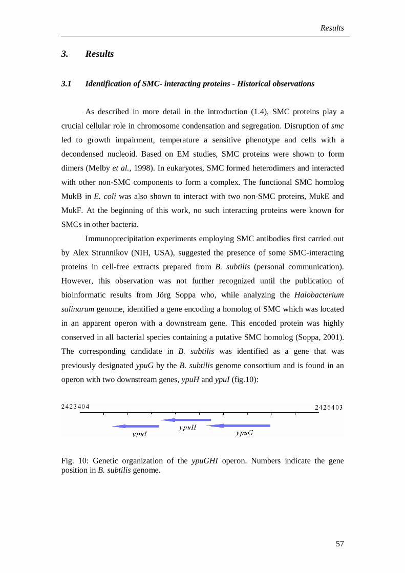
The corresponding candidate in B. subtilis was identified as a gene that was
previously designated ypuG by the B. subtilis genome consortium and is found in an
operon with two downstream genes, ypuH and ypuI (fig.10):
Fig. 10: Genetic organization of the ypuGHI operon. Numbers indicate the geneposition in B. subtilis genome.
Results
58
The importance of ypuG and ypuH was initially noted during sequencing of
the B. subtilis genome. While ypuI could be disrupted by single crossover integration
of an appropriately constructed plasmid, conditional shut-down of ypuG or ypuH
expression abolished growth of the respective mutants at 37°C which led to the
assumption that both of these genes are essential in B. subtilis (Vagner et al., 1998).
However no additional studies were carried out at that time.
3.2 Phenotypic analysis of ypuG and ypuH
In order to understand the function of proteins coded in the ypu operon by the
genes ypuG, ypuH, and ypuI, a detailed phenotype analysis of the three individual
gene knock-out mutants was initiated. A strategy for systematic deletion of each of
the three genes as well as for the construction of a combined ypuGH deletion strain
was developed. Early attempts to create conditional single-crossover disruption
mutants using the pMutin2 vector system (Vagner et al., 1998) resulted in rapid
mutant to wild type reversions. Therefore, stable deletions were constructed by
replacing 70-80% of the gene of interest with a tetracycline resistance cassette which
was integrated into the chromosome by double-crossover using a PCR knockout
method (Kuwayama et al., 2002) (see Materials and Methods 2.4.10). The constructed
deletion strains were confirmed by PCR using primers locating up and downstream to
the genetically modified region (data not shown).
While the ypuI null mutant PG31 (ypuI::tet) grew indistinguishable from wild
type cells, the ypuG and ypuH null mutants JM11 (ypuG::tet) and JM12 (ypuH::tet),
showed a phenotype quite similar to a smc null mutant. This phenotype included
temperature sensitivity, i e., cells did not grow above 23°C, and displayed 2-2.5 fold
reduced cell doubling times compared to the wild type (table13). In addition to this,
both strains grown at 23°C in LB medium contained decondensed nucleoids and
formed 12-15% anucleate cells:
Results
59
Fig. 11: Fluorescence microscopic images of (A) wild type, (B) JM11 (ypuG::tet),(C) PG32 (ypuH::tet) and (D) JM12 (ypuGH::tet) cells stained with DAPI and FM4-64 stain to view DNA and membrane respectively. Images B and D are DNA stainedvisualized in Nomarski.
To rule out that the phenotype of deletion of ypuG is due to a polar effect on
ypuH. The strain JM10 (Pxyl ypuH-cfp at amyE locus) was constructed, in which a
GFP tagged version of YpuH was expressed under the control of the xylose promoter
at the amylase (amyE) locus that fully complemented the deletion of the ypuH gene
when grown in xylose containing medium. Deletion of ypuG in JM10 in the presence
of xylose still led to a segregation and condensation defect and temperature sensitive
slow growth phenotype similar to that of JM11 cells. Showing that both YpuG and
YpuH are essential for proper chromosome condensation and segregation.
The combined deletion mutant strain JM13 (ypuGH::tet), in which both ypuG
and ypuH had been replaced by the tetracycline resistance gene did not further
exacerbate the phenotype observed for the single deletion mutations. When the two
single deletions, ypuG and ypuH, were separately combined with a conditional smc
deletion strain EP58 (smc::kan, Pspac-smc: amyE), the strains JM19 (ypuG::tet,
smc::kan, amyE::Pspac-smc) and PG43 (ypuH::tet, smc::kan, Pspac-smc::amyE) grew
similar to the smc deletion strain as long as IPTG was absent. These observations
show that like SMC, YpuG and YpuH are involved in chromosome condensation.
A B
C D
DNA membrane Nomarski/DNA
Results
60
It has been reported that combination of spo0J (whose deletion results in
formation of ~1 % anucleate cells but wildtype like growth) and the smc mutant
exacerbates the phenotype of the smc deletion mutants (Britton et al., 1998). To see if
ypuH has a similar effect, ypuH mutant was combined with spo0J in PG39 strain. The
cells showed 25-35% increase in anucleate cell formation as well as reduction in
growth rate when compared with ypuH mutant alone (table 13), suggesting the
possibility of partial functional overlap between smc and ypuH.
It has also been reported that smc mutants are synthetically lethal with a
spoIIIE deletion (Britton and Grossman, 1999). When ypuG or ypuH mutants where
combined with spoIIIE mutant, the double mutants of ypuG or ypuH with spoIIIE
were temperature sensitive and grew much slower than their single mutants and smc
mutant (table 13):strain doubling time (min) anucleate cells (%)
PY79 (wt) 92 < 0.01
PG31 (ypuI) 98 < 0.01
JM11 (ypuG) 196 11
JM12 (ypuH) 224 12
JM13 (ypuGH) 228 11
PG 388 (smc) 386 12
JM19 (smc, ypuG) 385 11
PG43 (smc, ypuH) 388 13
PG39 (ypuH, spo0J) 267 28
PG36(ypuI, spoIIIE) 96 < 0.01
PG37 (ypuG, spoIIIE) 463 < 1
PG38 (ypuH, spoIIIE) 448 1
Table 13: Doubling times of wild type PY79, smc null mutant, null mutants fromypuG, ypuH, ypuI, ypuGH and combinations with smc, spo0J, and spoIIIE nullmutants grown at 23°C in LB medium.
In conclusion, all the above results suggest that the gene products of ypuG and
ypuH are involved in a similar cellular function as smc and belong to the same
epistatic group. However, the influence of SMC seems to be more prominent than
YpuG and YpuH.
Results
61
So far, the ‘y’-genes ypuG and ypuH were of unknown function, based on the
observations described above and their apparent role in chromosome condensation
and segregation, they were renamed as ScpA and ScpB respectively. ‘Scp’ stands for
‘segregation and condensation protein’ or proposed ‘SMC complex protein’.
3.3 ScpA and ScpB - A new family of conserved proteins
To analyze the relationship of ScpA (MW: 29.5kDa; pI: 4.8) and ScpB (MW:
22kDa; pI: 4.3) and their occurrence in other organisms, the protein sequences were
used to perform a similarity search using a BLASTP internet server at NCBI (Altschul
et al., 1997). The results of the BLAST analysis showed that both of the sequences are
conserved among bacteria and archaea. ScpA was identified in all bacterial organisms
possessing SMC, while ScpB, if present, was found in all organisms possessing ScpA
and SMC-like sequences.
The ScpA sequence showed acid-rich sequences between residues 80 to 120
(fig. 12, shown in black bar), and is conserved only in the closely related Bacillus
species and in archaea which indicate that ScpA might be involved in interaction with
other protein or DNA. The C-terminal region of ScpA showed 40-46% similarity to
eukaryotic Rad21, Rec8, and Scc1 families, a subunit of cohesin complex.
ScpB: 60 LI EYADTYMLS TKKDFAPYLKKL I EVPSK- GLSQASLEVLAIVSYKQPITRAEIEEIRGV 118 CD: 59 LVEVAEGWRLQTKQEYAEYLEKLQEQRPKRELSRAALETLAIIAYKQPVTRSEIEEIRGV 118
ScpB: 119 KSERILHS LVAKALLCEVGRA DGPGRA ILYGTTP TFLEQ FGLKTLDELPPLPENAEEDVL 178 CD: 119 AVSQVISTLLE R GLI REVGRR DTPGRPYLYGTTE KFLDYFGLDSLDELPDLEELKDAGLL 178
ScpB: 179 QEEADL 184 CD: 179 SEEDLL 184
Fig13B: Sequence alignment of ScpB with the predicted transcriptional regulatorcontaining the HTH domain. Conserved residues are with black background.
The conserved domain (CD) search of ScpB using CD-Database showed a
sequence similar to the helix-turn-helix (HTH) motif found in other DNA-binding
proteins that are conserved among bacteria (fig. 13B). Proteins with HTH belong to
the LuxR-FixJ family that constitutes transcriptional activator proteins (Crater and
Moran, 2001). The DNA-binding HTH structural motif is composed of an alpha helix,
Results
64
a turn region, and then a second alpha helix. The second or C-terminal alpha helix of
the motif is involved in sequence-specific DNA base interactions and is termed as
recognition helix (Pabo and Sauer, 1992).
3.4 Subcellular localization pattern of ScpA, ScpB and SMC
In order to visualize proteins in the living cell, ScpA, ScpB, and SMC were C-
terminally tagged to the N-terminus of a fluorescent protein (YFP). To construct the
required fusions, the C-terminal region of the scpA and scpB genes were amplified by
PCR and cloned into a plasmid carrying a downstream yfp gene. To ensure that
tagging of the YFP to ScpA caused no disturbance of transcription of ScpB, an IPTG-
inducible Pspac promoter was cloned into the plasmid upstream of ScpA-YFP. The
resulting plasmids were then transformed into PY79, where they integrated at the
original gene locus by single-crossover integration.
The strains JM8 (scpA-yfp) and JM9 (scpB-yfp) were PCR-tested for
successful integration of the transformed plasmid into the chromosome and the
expression of the respective fusion protein was confirmed by western blot analyses
using antibodies against ScpB (fig 14B) and against GFP. Both strains grew in a
manner comparable to the wild type, i.e. there were no anucleate cells and growth rate
was indistinguishable to the wildtype suggesting that the protein fusions served as
functional replacements of their wild type counterparts. Strain JM8 was able to grow
in a similar manner with and without IPTG, inferring that tagging of ScpA to a FP did
not affect ScpB transcription. Likewise, SMC was tagged to a fluorescent protein in a
similar way, except that in this case a glycine linker was introduced between SMC
and the FP according to a technique developed in Hiraga’s lab for fusing E. coli’s
functional SMC homolog MukB to GFP (Ohsumi et al., 2001). This method was
designed to ensure that the folding of the GFP tag does not interfere with the folding
of its N-terminally tagged protein. Integration of this construct into the smc locus
resulted in strain JM20 (smc-gfp) which were temperature sensitive and grew at 25°C
but not at 37°C. When the JM20 cells grown at 25°C were examined by microscopy,
their nucleoids appeared normal, suggesting that the fusion was partially functional.
The observed temperature sensitive phenotype might be due to the process of slowing
down folding of the respective proteins or the disruption in transcription of
downstream gene due to plasmid integration. To construct a fully functional strain, a
Results
65
pMutin-YFP vector carrying an IPTG-inducible spac promoter designed to drive
expression of the downstream gene was used to clone in the smc region (Kaltwasser et
al., 2002). The resulting strain JM25 (smc-yfp, Pspac) was able to grow at 37°C in the
presence but not in the absence of IPTG, indicating that tagging of GFP to SMC
abolished the continued transcription of a gene downstream of smc, ftsY, a signal
recognition particle receptor, which serves an important function during the growth
(Oguro et al., 1995).
The strains JM8 (scpA-yfp), JM9 (scpB-yfp), JM20 (smc-gfp), and JM25 (smc-
yfp, Pspac) showed a similar pattern of fluorescent foci at 25°C in S750 minimal
medium. (fig. 15 A, B, C). They localized in a cell cycle-dependent manner where
one or two fluorescent foci were present in the middle of small cells, while in larger
cells (and thus later in the cell cycle) one or two fluorescent foci were present close to
each cell pole of future daughter cell (fig.15). Fluorescence of cells outside of the
protein-YFP foci was similar to that seen in cells that did not carry the fusion,
indicating that most of the protein molecules are present within the foci. The
fluorescence intensity of the foci was brighter in JM8 and JM9 cells when compared
to strain JM25. The similar pattern of localization of all the three proteins and the
genetic analyses showing similar phenotype upon deletion suggest that the three
proteins ScpA, ScpB, and SMC might be involved in a same function and might work
together as a complex.
Results
66
Fig. 15: Fluorescence microscopy. (A) JM8 (scpA-yfp), (B) JM9 (scpB-yfp), (Ca)JM20 (smc-yfp) and (Cb) overlay of JM26 with DAPI stain.
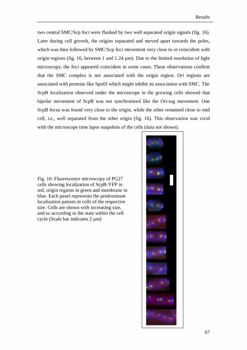
3.5 Dynamic localization of SMC, ScpA, and ScpB
The localization pattern of ScpA, ScpB or SMC proteins seem to differ during
the cell growth. The smaller cells showed 1-2 foci at the mid cell and the larger cells
had 2-4 bipolar foci. To compare the relative positions of the foci during the cell
growth, the location of ScpB was monitored relative to the position of origins of
replication during the cell cycle. A strain PG27 which expressed ScpB-YFP and
possessed LacI-CFP bound to the tandem repeats of lacO cassette near the origin at
359° was used. Cells from this strain showed characteristic bipolar foci of the Ori s
coloured in green (fig. 16). In most cases, ScpB localized close to the origin regions,
often with 2 foci (in red) flanking each origin (fig. 16). In small cells (<1 µm), one or
(A) (B)
(Ca) (Cb)
membrane sta in
YFP tagge d prote in
DNA stain
Results
67
two central SMC/Scp foci were flanked by two well separated origin signals (fig. 16).
Later during cell growth, the origins separated and moved apart towards the poles,
which was then followed by SMC/Scp foci movement very close to or coincident with
origin regions (fig. 16, between 1 and 1.24 µm). Due to the limited resolution of light
microscopy, the foci appeared coincident in some cases. These observations confirm
that the SMC complex is not associated with the origin region. Ori regions are
associated with proteins like Spo0J which might inhibit its association with SMC. The
ScpB localization observed under the microscope in the growing cells showed that
bipolar movement of ScpB was not synchronized like the Ori-tag movement. One
ScpB focus was found very close to the origin, while the other remained close to mid
cell, i.e., well separated from the other origin (fig. 16). This observation was vivid
with the microscope time lapse snapshots of the cells (data not shown).
Fig. 16: Fluorescence microscopy of PG27cells showing localization of ScpB-YFP inred, origin regions in green and membrane inblue. Each panel represents the predominantlocalization pattern in cells of the respectivesize. Cells are shown with increasing size,and so according to the state within the cellcycle (Scale bar indicates 2 µm)
Results
68
After the origins reached the poles, the Scp foci moved away from the origins
towards the center of each cells halves (fig. 16). This was visible in the larger cells
that had four bipolar origins, while SMC/Scp foci were located towards quarter sites
corresponding to the future middle of newborn cells after cell division and flanked by
the origin foci which are indicative of new rounds of chromosome replication before
cell division (fig. 16). Thus, the mobility of condensation centres appears to be
associated with replication process.
To investigate the association of SMC complex with the replication
machinery, SMC-YFP encoding plasmid was transformed into strain PG28, carrying a
CFP tagged to the C-terminus of the τ subunit of DNA Polymerase III (Lemon and
Grossman, 1998). The resulting strains JM27 (smc-yfp, dnaX-cfp) showed a centrally
located DNA polymerase. SMC foci were adjacent but not coinciding with the DNA
polymerase in smaller cells but the foci were not coincident in larger cells (fig. 17):
Fig 17: Fluorescence microscopy of JM27 (smc-yfp, dnaX-cfp) cells. SMC-YFP is ingreen (white arrows) and DnaX-CFP in red (grey arrows).
These observations indicate that in smaller cells, the SMC complex might be
transiently associated with the replication machinery while organizing the
chromosomes for the replication process. Once the chromosomes have started
replicating, the SMC foci might be involved in organizing the newly replicated
chromosomes. Similar pattern of localization was also observed with ScpB-YFP and
DNA Pol III.
Results
69
3.6 SMC, ScpA, ScpB are associated with DNA
Biochemically, it has been shown that SMC binds to single-stranded (Hirano
and Hirano, 1998) as well as double stranded DNA (Volkov et al., 2003). SMC,
ScpA, and ScpB were always observed to be present in the nucleoid (fig. 15Cb). To
prove the association of SMC and its complex partners with DNA in vivo, the
chromosomal DNA from JM8 (scpA-yfp), JM9 (scpB-yfp) or JM25 (smc-yfp, Pspac)
were transformed in a spo0J mutant strain (spo0J::spec) and examined for protein
localization in anucleate cells formed as a result of the spo0J deficiency (Ireton et al.,
1994). SMC, ScpA, and ScpB localized as bipolar foci in cells with DNA but did not
show any fluorescence in anucleate cells as seen in fig. 18:
Fig. 18: Fluorescence microscopy of PG39 (scpB-yfp, spo0J-) cells. Arrows show theanucleate cells.
This observation demonstrates that SMC, ScpA, and ScpB are associated with cellular
DNA.
ScpB-YFP DNA membrane
Results
70
3.7 Colocalization of ScpA, ScpB, and SMC
In order to examine whether proteins possess the same subcellular address and
colocalize with each other, they have to be viewed simultaneously in a cell. To
accomplish this, one of the proteins under investigation was tagged to CFP and
combined with strains expressing its potential interaction partner fused to YFP (the
emission spectra of YFP and CFP do not interfere, which allows the observation of
both of the proteins in the cell as long as appropriately selected filters are used). For
the construction of N-terminal fusion of ScpB to CFP, scpB was cloned in a plasmid
that integrated at amyE locus and expressed the fusion protein from a xylose-inducible
promoter. This strain JM10 (amy::cfp-scpB, Pxyl) was able to complement the scpB
mutant and also localized in a similar manner as in JM9 cells but the CFP
fluorescence was weaker when compared to YFP. The strain JM10 was combined
with JM8 (scpA-yfp) and JM25 (smc-yfp) to yield JM14 (scpA-yfp, amy::cfp-scpB,
Pxyl) and PG44 (smc-yfp, amy::cfp-scpB, Pxyl), respectively. When analyzed under
microscope, JM14 cells showed foci for both YFP and CFP that were visible in both
of the filters. Upon overlapping the images, these foci were coincident:
Fig: 19a: Colocalization of ScpA and ScpB in JM14 cells.
Results
71
Fig: 19b: Colocalization of ScpB and SMC in JM30 cells.
To colocalize SMC and ScpA, SMC was tagged to CFP in strain JM26 (smc-
cfp) which showed very faint fluorescence foci in the cell. When combined with
ScpA-YFP or ScpB-YFP to yield strains JM29 (scpA-yfp, smc-cfp) and JM30 (scpB-
yfp, smc-cfp) respectively. It was difficult to localize a SMC-CFP fusion in JM29 and
in JM30 cells (fig. 19b). In the rare case where clear CFP and YFP foci were visible,
they were coincident. Moreover, growth was severely impaired in strain JM29 when
compared to JM30. The observation that ScpA and ScpB colocalize with each other
and with SMC supports the idea that they function together in a complex. The rather
poor visibility of fluorescence foci in PG44, JM29, and JM30 cells might be either
due to an interference with proper protein folding or a negative effect mediated by the
fluorescent proteins in complex formation, providing a clue that ScpA and ScpB binds
at the SMC head region, comprising the N- and the C- terminus of SMC.
3.8 Interaction of ScpA, ScpB, and SMC in vivo
In order to verify whether colocalization is equal to a true interaction of ScpA,
ScpB and SMC in vivo, a technique called FRET was employed. The FRET effect
(fluorescence resonance energy transfer) is distance-dependent and requires
interaction between YFP and CFP such that the emission energy of a previously
excited CFP (the donor) is transferred and absorbed to excite YFP (the acceptor).
Under optimal conditions no emission energy spectra from the donor molecule is seen
when examined under the microscope. An efficient energy transfer between YFP and
ScpB-CFP
SMC-YFP
ove r lay
Results
72
CFP can occur only if both are at close proximity within 50Å, which requires
interaction of the proteins.
The strains PG41 (scpB::tet, Pxyl-scpB-cfp, scpA-yfp) and PG44 (amy:: Pxyl-
scpB-cfp, smc-yfp) were observed through a special FRET filter that specifically
excited with a CFP wavelength and allowed the observation of the YFP emission,
such that fluorescence was visible only if FRET occurred. When compared to the
control strains PG40 (scpB::tet, Pxyl-scpB-cfp) and JM8 (scpA-yfp) that did not show
any FRET fluorescence (fig. 20 A and B), cells of the combined strain PG41 showed
FRET fluorescence which was seen as bipolar foci, confirming the interaction of
ScpA and ScpB in vivo:
Fig. 20: Fluorescence resonance energy transfer (FRET) analysis.Fluorescence microscopy of cells grown in the presence of xylose (A) PG40(scpB::tet, Pxyl-scpB-cfp at amyE locus), (B) JM8 (scpA-yfp), and (C) PG41(scpB::tet, Pxyl-scpB-cfp at Amy locus, scpA-yfp). Cells were observed in FRET andYFP filters. Arrows indicate the ScpA-YFP foci. Short white bars show the ends ofcells. Scale bar 2µm.
Results
73
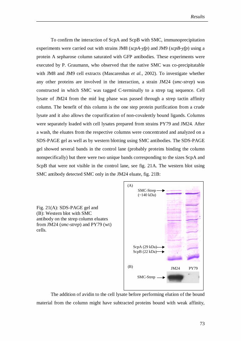
To confirm the interaction of ScpA and ScpB with SMC, immunoprecipitation
experiments were carried out with strains JM8 (scpA-yfp) and JM9 (scpB-yfp) using a
protein A sepharose column saturated with GFP antibodies. These experiments were
executed by P. Graumann, who observed that the native SMC was co-precipitatable
with JM8 and JM9 cell extracts (Mascarenhas et al., 2002). To investigate whether
any other proteins are involved in the interaction, a strain JM24 (smc-strep) was
constructed in which SMC was tagged C-terminally to a strep tag sequence. Cell
lysate of JM24 from the mid log phase was passed through a strep tactin affinity
column. The benefit of this column is the one step protein purification from a crude
lysate and it also allows the copurification of non-covalently bound ligands. Columns
were separately loaded with cell lysates prepared from strains PY79 and JM24. After
a wash, the eluates from the respective columns were concentrated and analyzed on a
SDS-PAGE gel as well as by western blotting using SMC antibodies. The SDS-PAGE
gel showed several bands in the control lane (probably proteins binding the column
nonspecifically) but there were two unique bands corresponding to the sizes ScpA and
ScpB that were not visible in the control lane, see fig. 21A. The western blot using
SMC antibody detected SMC only in the JM24 eluate, fig. 21B:
Fig. 21(A): SDS-PAGE gel and(B): Western blot with SMCantibody on the strep column eluatesfrom JM24 (smc-strep) and PY79 (wt)cells.
The addition of avidin to the cell lysate before performing elution of the bound
material from the column might have subtracted proteins bound with weak affinity,
SMC-Strep(~140 kDa)
ScpA (29 kDa)ScpB (22 kDa)
SMC-Strep
(A)
(B) JM24 PY79
Results
74
but this was not performed due to the paucity of the material and time. The results
obtained so far from FRET studies and immunoprecipitation experiments confirm that
SMC has ScpA and ScpB as its direct interacting partners.
3.9 Specific localization depends on all three proteins of the complex
To investigate the interdependence of SMC, ScpA, and ScpB localization, one
of the proteins was viewed in the absence of the others. To do so, the chromosomal
DNA from JM8 (scpA-yfp) and JM9 (scpB-yfp) were transformed into JM16
(smc::kan, amy::Pspac-smc, cm::tet) strain that carried SMC under the control of the
IPTG-inducible Pspac promoter. This strain was derived from EP58 (smc::kan,
amy::Pspac smc) by exchanging the resistance cassette from cm to tet in order to make
it compatible with JM8 or JM9 strain that also carried the cm resistance. The
corresponding strains JM17 (scpA-yfp, smc::kan, amy::Pspac-smc) and PG33 (scpB-
yfp, smc::kan, amy::Pspac-smc) were grown in the presence and in the absence of
IPTG. The strains grown with IPTG behaved like wild type and showed the bipolar
foci formation of the ScpA and ScpB proteins, as seen in fig. 22. In the absence of
IPTG the cells had decondensed nucleoids and the SMC was not seen in bipolar foci
fluorescence but was distributed throughout the cell:
Fig. 22: Fluorescence microscopy of PG33 cells grown (A) with and (B) withoutIPTG.
(A) PG33 cells grownwith IPT G
(B) PG33 ce lls grownwithout IPT G
Results
75
To investigate whether SMC can form foci in the absence of ScpA and/or
ScpB, strains were derived from JM25 (smc-yfp) in which scpA, scpB or both genes
were deleted. A control strain JM33 (smc-yfp ypuI::tet) was constructed in which the
ypuI gene downstream of scpA and scpB was deleted. As seen in fig. 23A, the control
strain JM33 (smc-yfp ypuI:: tet) grew like the wild type and showed SMC as bipolar
foci. In strains JM31 (smc-yfp, scpA::tet), JM32 (smc-yfp, scpB::tet) and JM34
(scpAB::tet, smc-yfp), SMC was no longer visible as foci but was distributed
throughout the cell:
Fig. 23: Fluorescence microscopy of B. subtilis cells growing at mid-exponentialphase. Left side panel shows fluorescence of SMC-YFP and the right panel, DNAstained by DAPI. A: JM33 (smc-yfp, ypuI::tet), B: JM 31(smc-yfp, scpA::tet), C:JM32 (smc-yfp, scpB::tet).
In most cases, fluorescence was seen distributed throughout the cell (fig. 23B
and C), however, some cells had foci that were located on the nucleoids but in an
aberrant fashion. In few cases, fluorescence was seen to accumulate near the
membrane. Interestingly, some cells showed fluorescence even in anucleate cells (fig.
23B and 23C, indicated by arrows), which was in contrast to anucleate cells
containing ScpA and ScpB that carried a spo0J mutation. These observations show
that the foci represent the active SMC complex. While SMC can still form foci on
Results
76
DNA, both ScpA and ScpB are required for proper localization on the nucleoids.
From this, it can be inferred that ScpA and ScpB play an essential role for the
formation of foci which make the condensation centers supporting the ternary
complex formation by proteins in vivo.
3.10 The SMC complex requires active replication for its bipolar foci segregation
In order to probe the dynamic bipolar movement of the SMC complex foci, the
ScpA-YFP fusion was viewed in a strain KL210, which carries a temperature
sensitive mutation in dnaB that blocks the initiation of replication process upon
temperature upshift. JM46 (scpA-yfp, dnaBts) cells were monitored for ScpA
localization by shifting the growing cultures from 25°C to 45°C for an hour which
arrested the replication process and then back to 25°C to resume replication. At the
time of temperature upshift all ongoing replication processes were completed but no
new process was initiated. Under the microscope, cells at this stage appeared
filamentous and showed a single compact nucleoid (fig 24A). One to three ScpA foci
were seen aggregated at the cell center on the nucleoid:
Fig. 24A: Fluorescence microscopy of JM46 cells after 60 min at 45°C (temperatureupshift). Left panel showing the ScpA-YFP and the right panel DNA, stained withDAPI. White bars represent the ends of the cell.
ScpA- YFP DNA
Results
77
When the cultures were shifted from 42°C back to 25°C and grown for an
hour, the cells had resumed their replication process, which was visible by the
presence of two nucleoids per cell. The cells were no longer filamentous and ScpA
was seen as separated bipolar foci, shown in fig. 24B below:
ScpA-YFP DNA
Fig. 24B: Fluorescence microscopy of JM46 cells shifted from 45°C to 25°C(temperature downshift). Left image showing the fluorescence of ScpA-YFP and theright image, the DAPI stained DNA. White lines mark the ends of the cells.
Similar observations were seen even with SMC-YFP strain. These
observations show that the active segregation of the replicated chromosomes depends
on SMC complex and in turn on active replication process. The SMC complex is
initially loaded at the replication center and active replication causes the foci to move
apart bidirectionally resulting in condensation and segregation of replicated
chromosomes.
3.11 SMC localization depends on DNA topology
Studies have shown that B. subtilis SMC affects plasmid topology in vivo
(Lindow et al., 2002a). Moreover, SMC mutants showed decondensed nucleoids and
were hypersensitive to DNA gyrase inhibitors (Lindow et al., 2002a). Depletion of
topoisomerase I suppressed the chromosome-partitioning defect of the smc null
mutant. Thus, SMC seems to function in concert with other topoisomerases as a
chromosome organizer. In order to look for the localization behaviour of SMC in the
absence of active DNA topology-modulating proteins like gyrase and topoisomerase
Results
78
IV, JM25 (smc-yfp) and JM8 (scpA-yfp) cells were treated with drugs that inhibited
gyrase activity. Novobiocin belongs to the coumarin drug family that contains natural
products originally isolated form Streptomyces species. Coumarins inhibit
supercoiling and enzyme turnover by preventing the binding and hydrolysis of ATP
(Ali et al., 1993). Nalidixic acid is a member of the quinolones drug family that are
synthetic drugs that act by forming an inhibiting ternary complex with the
topoisomerases in the presence of DNA (Grompone et al., 2003; Kato et al., 1990).
JM8 and JM25 cells were treated with novobiocin (10 µg/ml) or nalidixic acid (200
ng/ml) for one hour and examined for changes in the nucleoid structure using DAPI
stain and foci localization pattern analyses. The treated cells showed decondensed
nucleoids, some of the cells showed a much denser DNA region at the cell center:
ScpA-YFP DNA
Fig. 25: Fluorescence microscopy of JM8 cells treated with nalidixic acid (white barsshow the ends of the cells). Arrows indicate dense DNA regions, where a ScpA-YFPfocus is formed.
The SMC/ScpA proteins were no longer seen as foci in the cells with
decondensed nucleoid, in smaller cells foci were seen at the cell center (fig. 25) which
also corresponded to the dense DNA region visualized by DAPI staining. The
presence of the central dense DNA region might be due to the secondary effect of
gyrase inhibition on replication inhibition. Inhibited gyrase increases the local
accumulation of positive supercoils downstream of replication forks and this effect
slows down the replication fork progression (to one third) (Khodursky et al., 2000).
These observations suggest that SMC requires a defined DNA topology for proper
Results
79
function. Disturbances exerted by action of the antibiotics novobiocin or nalidixic
acid might interfere with proper recognition of the DNA template by SMC and
weaken or even abolish SMC-mediated DNA compaction. One of the possible
conclusions could be that SMC binds to specific DNA structures and is necessary to
maintain the DNA topology originally established by the topoisomerases.
3.12 SMC - A bacterial condensin protein
3.12.1 Effects of overexpression of SMC
To purify SMC from B. subtilis for biochemical studies, smc was cloned into a
plasmid pMW6 that could overexpress the protein as a C-terminal RGS-His tag from
the amyE locus upon induction with xylose. The resulting strain JM6 (amy::Pxyl-smc-
RGS-His6), was able to overproduce SMC-RGS-His6 upon induction (compare lane 1
and 2 in fig. 26), but the western blot with anti-SMC and anti-His antibodies revealed
in addition to a band of an expected size, the presence of a second band approximately
half of the size of SMC which was seen only in the xylose induced strain (fig. 26,
lanes 2). The strain JM6 was not used for further analyses because it also failed to
rescue the smc null mutant. Instead, another strain CAS4 (amy::Phyperspank-smc-
His6), was used in which SMC could be overproduced using expression driven by an
IPTG-inducible hyperspank promoter (Volkov et al., 2003). SMC expressed by this
system also complemented the smc null mutant. Upon induction of SMC with 1 mM
IPTG, the nucleoids were more condensed and also formed up to 5% anucleate cells
(Volkov et al., 2003). Similar experiments were carried out with overexpression of
ScpA or ScpB, which did not exhibit any detectable effect on the cellular
chromosome structure suggesting that SMC is the key player in chromosome
condensation and the Scp’s appear to function only in conjunction with SMC.
Results
80
SMC (~135 kDa)
Fig. 26: Western blot analysis using SMC antibody.Lanes 1 and 2: JM6 cells grown without and with xylose, respectively.Lanes 3 and 4: CAS4 cells grown without and with IPTG, respectively.
3.12.2 SMC condenses DNA from a single position on the nucleoid
To visualize whether SMC retained its localization pattern upon
overproduction, chromosomal DNA from the strain JM25 carrying a functional C-
terminal YFP fusion of SMC was transformed into the SMC overexpression strain
CAS5 (Phyperspac-smc) where smc was placed under the control of a hyperspac
promoter at its original locus. The resulting strain JM35 (smc:: Phyperspac-smc-yfp),
grown with 0.025 mM IPTG, showed similar nucleoid morphology from that of wild
type cells and SMC-YFP localized in a similar bipolar manner as in the parent strain.
With 1 mM IPTG, chromosomes were highly compacted, the SMC-YFP were much
brighter but still largely retained in the foci (fig. 27A). Similarly, ScpB-YFP was also
retained as foci at 1mM IPTG in JM36 (scpB-yfp, Phyperspac-smc) cells:
Results
81
Fig. 27: Fluorescence images showing the effect of SMC overproduction on
localization of (A) SMC-YFP in JM35 cells and (B) ScpB-YFP in JM36 cells.
Similar observations were made when SMC-His6 was overproduced from an
ectopic location on the chromosome, while a SMC-YFP fusion was driven by the
original promoter at the smc locus in PG45 (smc-yfp, Phyperspank, smc-His6 at
amyE). These foci represent the condensed state of the nucleoid and also indicates that
the whole SMC complex are retained at its specific location even upon
overproduction and brings about global chromosome compaction from a defined
position on the nucleoid.
3.13 Regulation of SMC
The rate of gene expression can be modulated in several ways, either through
post-transcriptional mechanisms that affect mRNA half lives, translation initiation
and/or translation progression (McAdams and Shapiro, 2003) or through translational
or post-translational processes operating at the protein level, e.g. by action of
proteases. SMC is found in an operon flanked by essential genes with rncS located
upstream, which is involved in RNA processing (Herskovitz and Bechhofer, 2000)
and ftsY located downstream and serving as the anchor for the bacterial signal
Results
82
recognition particle (SRP). FtsY is not only involved in secretion of extracellular
proteins, but also participates in proper localization of spore-forming proteins
(Kakeshita et al., 2000). In many cases, genes that are co-transcribed are involved in a
common functional pathway, which however does not seem to be the case with smc.
Within the rncS operon, all three genes have been shown to be regulated by a σA
promoter situated upstream of rnc (Oguro et al., 1996). In addition, a σk promoter
sequence was mapped near the 3’ end of smc that regulates the late stationary phase
expression of FtsY (Kakeshita et al., 2000).
Immunoblot and Immunofluorescence studies employing SMC antibodies
have demonstrated the absence of SMC in stationary phase cells (Graumann et al.,
1998). In order to investigate if SMC is regulated at the transcriptional level, primer
extension studies were carried out using total RNA extracts prepared from different
growth stages of PY79 cultures (fig. 28B), employing a primer specifically located
150 bp downstream from the start codon of smc (fig. 28A). The results of primer
extension experiments revealed the presence of a very strong extension signal
corresponding to a transcript which was identified in all the growth stages (fig 28C).
The size of this transcript corresponds to approx. 480 bp (distance of transcript signal
to primer location) and the sequence upstream to the corresponding transcript signal
possesses a weak similarity to the consensus of a typical σH recognition sequence
(AGGA-15 bp-GAAT), although more experiments are required to confirm the
existence an independent promoter for smc expression. It can of course not be
excluded that the transcript points to a post-transcriptional modification site of the
mRNA transcribed from the σA promoter upstream of rncS. The experiment showed
the presence of a smc transcript in all the growth phases, which strongly suggests that
SMC might be regulated at the stage of protein synthesis.
Results
83
Fig. 28A: Schematic representation of rnc operon. The expression of rncS, smc, andftsY are controlled by PA ( A promoter).The expression of ftsY in the stationary phaseis regulated by the PK ( K promoter). PH is the putative promoter site mapped in thisexperiment from the primer extension analysis.B: Growth curve of PY79 cells starting from mid exponential phase. The blue dotsrepresent the time points when samples were withdrawn for analyses.C: Primer extension analysis from total RNA extracts of samples from mid log(lane1), late exponential (lane 2), early stationary (lane 3) and late stationary phasecells (lane 4) (fig.28B). The DNA sequencing ladder to compare the size of thetranscript signal (left 4 lanes reading CTGA). Arrow showing the strong transcriptsignal for smc. The sequence region of the rnc and smc in fig. 28A shows the primer‘p’ used to map the smc transcript and the transcriptional initiation site ‘t’, leading tothe possible promoter sequence PH (black, underlined).
3.13.1 Growth phase dependent expression of SMC and ScpB
To monitor the SMC levels during different growth phases, cell extracts taken
at various time points from the growing cultures (fig. 28B) were analyzed by western
blotting utilizing SMC antibodies. The results showed the presence of SMC at the mid
log phase, a gradually diminishing SMC amount at the onset and progression of the
stationary phase, and its absence in the late stationary phase (fig. 29B, lanes 1- 4). The
ScpB expression profile was also analyzed by using ScpB polyclonal antibodies (fig.
29C). Interestingly, the ScpB protein profile closely resembled that of SMC which
further indicate that both proteins function together and thus might be regulated in a
similar manner. Surprisingly, a second band of significantly higher size (~48kDa)
Results
84
compared to that of ScpB was detected when the ScpB band began to disappear at the
entry into the stationary phase. This band might be explained by a cross reaction of
ScpB antibody with another protein synthesized at the later stage of growth.
Alternatively, although unlikely, it cannot be excluded that this band might
correspond to a modified variant of ScpB itself.
1 2 3 4 1 2 3 4A B
C
135 kDa
22 kDa
Fig. 29A: SDS-PAGE of the crude cell extracts showing the protein levels. Cellstaken from various time points (as in fig. 28B), from mid-exponential phase tostationary phase. B: Western blot with SMC, and C: ScpB antibodies.
3.13.2 Stability of SMC
In order to check the stability of SMC during different growth phases, cultures
were treated with chloramphenicol (Cm) to inhibit protein synthesis and the levels of
SMC was compared to that of the untreated cultures. Cm (90 µg/mL final
concentration) was added to growing cultures at the mid-exponential phase (OD600nm=
0.45) and at the onset of the stationary phase (OD600nm= 2.0). For determining the
relative abundance of SMC during the cell cycle, samples were normalized, such that
almost equal amounts of total protein were loaded in all lanes and the cellular SMC
levels were measured by western analysis employing SMC antibodies. As shown in
fig. 30 (lanes 1-4), in untreated cells the levels of SMC decreased during stationary
growth. In cultures where Cm was added at mid-exponential growth phase, shown in
fig. 30 (lanes 5-7), SMC was present even in the stationary phase cells, even though
there were some decrease in their levels, it was not as significant compared to the
untreated. In cultures where Cm was added at a later stage of growth, shown in fig. 30
(lanes 8 and 9), SMC levels were similar to the corresponding untreated cultures:
Results
85
1 2 3 4 5 6 7 8 9
Fig. 30: Stability of SMC: Western blot analyses of cell extracts with SMC antibodiesLanes 1-4: SMC profile of untreated cells, samples taken at time points similar toprevious experiment (fig. 28B)Lanes 5-6: SMC profile of cells treated with Cm at mid-exponential phaseLane 5: 2 h after Cm treatment (OD600 0.75)Lane 6: 4 h after Cm treatment (OD600 0.9)Lane 7: 7 h after Cm treatment (OD600 1.0)Lanes 8 and 9: SMC profile of cells treated with Cm at late exponential phaseLane 8: 1 h after Cm treatment (OD600 2.5)Lane 9: 5 h after Cm treatment (OD600 3.2)
Together with the experiments described above, these results demonstrate that
SMC is specifically degraded in a post-translational manner by a dedicated protease
that is expressed exclusively at the transition to the stationary growth phase. In order
to identify this protease, the experiments presented here could be repeated with
available protease null mutants.
3.14 Involvement of SMC complex in repair
It is known that in eukaryotes SMC proteins like SMC5, SMC6, and Rad50, a
member of SMC family play an important role in DNA repair (Introduction 1.4.2). In
order to interrogate the involvement of B. subtilis SMC in repair, smc null mutants
were tested for sensitivity to mitomycin C (MMC). MMC, is an antitumor agent
isolated from Streptomyces cultures which is used in chemotherapy (Paz et al., 1999).
MMC acts as a bifunctional or trifunctional alkylating agent which cross-links DNA
by formation of adducts to an extent proportional to its content of guanine and
cytosine. The excision of adducts triggers the SOS response, a concerted induction of
several DNA repair and recombination activities that has been studied in E. coli,
where the response is controlled by the lexA-specified repressor (Wei et al., 2001).
Results
86
The strains PG∆388, JM11, and JM13 carrying deletion of smc, scpA, and
scpB genes, respectively, and the wild type PY79, were streaked on LB plates
containing 0, 50, 100 and 200 ng/ml of MMC and were incubated at 23°C because of
the temperature sensitivity of the SMC and ScpAB mutants. All four strains grew on
LB plates without MMC. While the wild type cells grew on plates containing 50 and
100 ng/ml MMC, the deletion strains of smc and also its interaction partners scpA and
scpB did not show any growth even on a plate with 50 ng/ml MMC. The sensitivity of
the mutants towards MMC suggests a direct or an indirect involvement of SMC,
ScpA, and ScpB in DNA repair. Furthermore, the localization patterns of SMC and
ScpB were examined after the treatment of the cells with MMC (100 ng/ml) for 30
minutes. The MMC treated cells possessed a slightly decondensed nucleoid and
anamalous foci:
ScpB-YFP
SMC-YFP
Fig. 31: Fluorescence microscopic images showing the effect of mitomycin onlocalization of SMC and ScpB in JM25 (smc-yfp) and JM9 (scpB-yfp) cellsrespectively. White bars represent the ends of the cell.
Under normal conditions there are 2-4 foci in the cell occupying the polar
positions, whereas MMC-treated cells (200 ng/ml) showed foci distribution
throughout the cellular space. Some cells contained two foci at the cell center rather
than at the poles (fig. 31). These foci rearrangements after the MMC treatment might
Results
87
indicate an incomplete process of recombination where the SMC/ScpA/ScpB complex
might play a crucial role mediating the repair process.
3.15 Identification and examination of other SMC-like proteins in Bacillussubtilis
Since eukaryotic cells possess more than just one SMC-like protein, it was
interesting to investigate for other SMC homologs in B. subtilis. A BLASTP search
performed at the SubtiList server using SMC from B. subtilis as a query sequence
revealed a number of homologs with the two most significant having E-values of
3·10-12 and 6·10-7. These corresponded to YirY (1130 a.a; MW 128.69 kDa) which
was identified in the course of genome sequencing project as an SbcC homolog
(Medina et al., 1997) and RecN (576 a.a; MW 64.3 kDa) which is involved in DNA
repair and genetic recombination (Alonso et al., 1993).
In B. subtilis, YirY/SbcC is located in an operon flanked by addA, addB
(ATP-dependent DNAses and analogs of RecBCD in E. coli), and sbcD genes
upstream and a functionally unknown yisB downstream of it:
Fig. 32: Organization of add operon in B. subtilis genome.
The proteins AddAB and SbcCD are DNA associated and homologs of these
proteins have been shown to play a role in recombination and repair in various
organisms (Alonso et al., 1993; Cromie et al., 2001). The SbcC-SbcD complex in E.
coli are similar to the eukaryotic Rad50-Mre11 complex involved in double strand
break repair (Cromie et al., 2001). Rad50 is a member of the SMC protein family
possessing the conserved ABC ATPases and a N-terminal ‘FKS’ (or FRS) motif
located near the Walker A site, conserved in most of the SMC proteins and also in
SbcC and RecN (Cobbe and Heck, 2003).
Results
88
3.15.1 Analysis of YirY/SbcC function
In order to experimentally probe the SbcC (YirY) function and its proposed
involvement in DNA repair, a sbcC disruption strain JM42 (sbcC::pJQ43) was
constructed by a Campbell integration of a plasmid carrying an internal fragment (~1
kb) of sbcC. The strain JM42 did not show any obvious phenotype and grew like the
wild type under optimal growth conditions. To test for the role of SbcC in DNA
repair, the strain JM42 was streaked on LB plates supplemented with MMC (that
induces double stranded breaks). When compared to wild type cells that survived on
the plate containing 50 and 100 ng/ml MMC, JM42 grew only poorly in the presence
of 50 ng/ml of MMC and did not show any growth on a plate with 100 ng/ml of
MMC confirming its role in the DNA damage related stress response.
To localize SbcC in cells and to observe the effect of DNA damage stress,
SbcC was tagged to a fluorescent protein in a plasmid with a xylose promoter that
assured the transcription of the down stream genes. The resulting strain JN6 (sbcC-
yfp, Pxyl) grew only in the presence of xylose but slowed down its growth when
xylose was removed from the medium, indicating an important cellular role for yisB.
When analyzed under the microscope, the JN6 cells showed low level fluorescence
visible throughout the cell. 30 min after addition of MMC, nucleoids appeared slightly
condensed and SbcC-YFP foci were detectable in 5% of the cells. After 2 h of
induction with mitomycin, 1 or 2 foci were observed in 45% of the cells (fig. 33).
These foci might be interpreted as locations where DNA repair takes place. In contrast
to the foci formation of SbcC, SbcD-YFP localized through out the cell (data not
shown).
Results
89
SbcC-YFP DNA membrane overlay
Fig. 33: Fluorescence microscopy of JN6 cells 2 h after addition of MMC. Cells werestained for DNA and membrane. Image to the extreme right shows the overlay of theimages with membrane in red, DNA in blue and the SbcC-YFP foci in green showedby arrows.
RecN, the second SMC-like protein in B. subtilis that functions in association
with RecF, RecL, RecO, RecR, and RecA proteins in homologous recombination
during transformation (Alonso et al., 1993), also showed foci formation upon
induction with MMC. The RecN disruption mutant behaved similar to sbcC mutant on
MMC plates (Kidane et al, in press).
3.15.2 Localization of AddAB
The addA and addB genes encode different subunits of the nuclease-helicase
AddAB (also termed exonuclease V or RecBCD in E. coli) involved in initial stages of
recombination (Haijema et al., 1996). AddB was discussed as an interacting protein of
ScpA, a subunit of SMC complex (Noirot P, 2003). If AddB and ScpA would interact,
one would also expect them to colocalize. To verify this idea, the proteins AddA and
AddB were C-terminally tagged to fluorescence proteins in plasmids with inducible
promoters that assured the transcription of downstream genes upon integration into
operon. Fluorescence microscopic investigations of strains JN7 (addA-gfp, Pxyl) or
JM41 (addB-yfp, Pspac) did not show any foci formation like ScpA, instead a weak
fluorescence was uniformly distributed through out the cell. The observed
fluorescence increased between 30 min and 2 h after addition of MMC:
Results
90
Fig. 34: Fluorescence microscopy of JN7 (addA-gfp) cells after MMC induction. A:
untreated, B: after 1 h, C: after 2 h. White bars show the ends of the cell.
These observations show that proteins acting at specific sites on the DNA (i.e.
DNA breaks) do not necessarily form foci after induction of breaks or are not
necessarily associated with the nucleoid.
3.16 Topoisomerase IV - A chromosome segregator
So far, SMC has been thought to play a key role in chromosome segregation,
however, the studies presented in this work show that SMC is more a condensing than
a segregation factor. Other proteins that are also involved in chromosome segregation
are topoisomerase IV (Huang et al., 1998), CodV and RipX (Sciochetti et al., 1999).
Topoisomerase IV is primarily responsible for unlinking the catenates that are
generated at the end of replication when the two replication forks converge
(Zechiedrich and Cozzarelli, 1995). Topoisomerase IV is a heterodimeric enzyme
belonging to the type II topoisomerase family and is formed of two subunits- ParC
and ParE, similar to the ParC and ParE of E. coli. Temperature sensitive mutants of
topoisomerase IV in E. coli and Salmonella typhimurium formed long filamentous
cells defective in nucleoid segregation that divided frequently to produce anucleate
cells (Luttinger et al., 1991). B. subtilis also showed a similar phenotype at the non-
permissive temperature (Huang et al., 1998). Topoisomerase IV mutants of C.
crescentus are highly pinched at multiple sites, a typical cell separation phenotype,
Results
91
but the cells do not divide to produce cells lacking DNA (Ward and Newton, 1997).
Similar to overproduction of gyrase, ParC and ParE showed enhanced relaxation
activity of supercoiled plasmid and were able to suppress a topA mutation (Kato et al.,
1990).
In E. coli, ParC is associated with the replication machinery and was seen to
form foci that colocalized with DNA polymerase III while ParE was not present at the
replication factory but localized as foci at the poles and at the cell center (Espeli et al.,
2003). In B. subtilis, ParE-GFP was shown to localize uniformly throughout the cell
in actively growing cells, while the ParC-GFP fusion protein showed bipolar
localization (Huang et al., 1998) in a manner which looked similar to SMC.
In order to verify whether ParC and SMC colocalize, the protein ParC was
tagged to YFP to yield the strain JM43 (parC-yfp). Contrary to what was reported
previously (Huang et al., 1998), ParC-YFP localized throughout the nucleoids and the
fluorescence distribution was similar to the DAPI stain:
Fig. 35: Fluorescence microscopy of JM43 cells (A) ParC-YFP (B) DNA stained withDAPI, (C) overlay of image (A) and (B).
Thus, ParC unlike SMC does not act on a specific site but bind throughout the
nucleoids. In the meantime, another work group presented results demonstrating that
ParE-GFP fusion was present on the nucleoid similar to that of ParC (Meile C.J,
2003), suggesting that they function together in maintaining the DNA topology.
Results
92
In E. coli, a topA mutant was found to be one of the suppressors of mukB
deletion strain (Sawitzke and Austin, 2000). It was also reported that mutations in
topA could be rescued by overproduction of ParC and ParE or the gyrases (Kato et al.,
1990). In B. subtilis, the partitioning defect of the smc null mutant was significantly
suppressed by the depletion of topA (Lindow et al., 2002a). These observations led to
the question whether the depletion of topoisomerase IV could rescue the segregation
defect of smc null mutant. To answer this, strain JM45 (Phyperspank-parE) was
constructed in which ParE and ParC expression was controlled by an IPTG-inducible
hyperspac promoter. Initial experiments with JM45 cells upon induction with 1 mM
IPTG showed more condensed nucleoids. Further analyses of this strain could not be
carried out due to the paucity of time. However, the effect of ParC and ParE
overproduction or depletion on smc phenotype could give us important insight
towards a better understanding of the networked action of proteins involved in
chromosome organization.
Discussion
93
4. Discussion
In this work, a novel protein complex termed the SMC complex has been
identified and characterized. The evolutionary conserved SMC protein forms a
complex with two proteins, ScpA and ScpB, formerly known as YpuG and YpuH
respectively, which have been analyzed and assigned a new role in chromosome
condensation. The deletion/disruption of these genes was demonstrated to affect
chromosome organization and each null mutant showed a phenotype similar to that of
a smc deletion strain, with temperature sensitive slow growth and decondensed
nucleoids showing a strong segregation defect (Britton et al., 1998; Graumann et al.,
1998; Moriya et al., 1998). Depletion of smc in the scpA or scpB deletion strains, or
deletion of both scpA and scpB, also resulted in the same phenotype, indicating that
SMC, ScpA, and ScpB function in the same pathway for chromosome condensation
and thus belong to the same epistatic group. ScpA and ScpB form a new family of
proteins that is conserved in most of the eubacterial and archaeal species that possess
a SMC homolog. ScpA also shows homology to the eukaryotic SMC-interacting
protein Scc1 and has lately been assigned as a member of the kleisin protein family
(Schleiffer et al., 2003). The resemblance of ScpA to Scc1 and the presence of a HTH
motif in ScpB provides us with some insights about how these proteins might be
associated to function with SMC. Because biochemical and structural studies of these
proteins are not available, at the moment, several possible roles can be assigned to
them ranging from a cofactor in the mechanical functioning of the SMC molecule to a
regulator of SMC protein that directs the SMC complex to bind DNA in a specific
manner by mediating its interaction with other proteins.
A key observation in this work has been the finding that SMC, ScpA, and
ScpB form discrete foci on the DNA, unlike other chromosome-associated proteins
that are bound throughout the nucleoids. This striking finding is a novel pattern of
localization and suggests that SMC, ScpA, and ScpB affect chromosome compaction
and segregation from a defined region on the nucleoids and the foci represented the
functional SMC complex and thus the condensed state of the nucleoid. Attempts to
localize SMC was previously made by (Britton et al., 1998), who observed discrete
foci with partially functional SMC-GFP fusion. Immunolabeling with SMC
antibodies also showed bipolar foci which were independent of nucleoid as reported
Discussion
94
by (Graumann et al., 1998). Similar patterns for SMC and ScpB were later reported
by (Lindow et al., 2002b) which was consistent with the observations described here.
The SMC counterpart in E. coli, MukB also showed foci formation (den Blaauwen et
al., 2001). In Caulobacter, SMC showed 2-3 foci in swarmer cells that do not
replicate their DNA, while stalked cells which actively replicate DNA possessed 3-4
foci randomly distributed in the cell (Jensen and Shapiro, 2003). The observed
localization pattern of the SMC/ScpA/ScpB complex in form of foci within the
nucleoid might indicate a recruitment of the proteins to a specific nucleoid structure.
In the eukaryote Saccharomyces cerevisiae, the SMC1 and SMC2 was demonstrated
to bind to secondary structures, preferentially to AT rich sequences (Akhmedov et al.,
1998; Gregson et al., 2002; Laloraya et al., 2000) and SMC4p-GFP localized to
rDNA regions (Freeman et al., 2000). Surprisingly, B. subtilis SMC has been shown
to bind to DNA in a non-specific manner in vivo (Lindow et al., 2002b) and in vitro
(Volkov et al., 2003). In bacteria, it is known that chromosomal DNA is divided into
topologically independent domains (Sinden and Pettijohn, 1981); (Drlica, 1986).
Therefore, it can be speculated that the presence of repetitive patches of T/AT-rich
sequences within the B. subtilis genome might be of some significance in providing a
defined chromosomal structure that could represent a binding location for the SMC
complex. In fact, as shown in this work, the observed specific subcellular localization
of the SMC complex requires a well-defined chromosome topology since SMC foci
formation was abolished upon addition of the gyrase inhibitor novobiocin, which
disturbs the DNA supercoiling status.
The bipolar localization pattern of SMC, ScpA, and ScpB suggests that all
three proteins function together in a complex, especially since the interaction was also
confirmed in vivo using FRET technique. The SMC strep tag affinity purification and
immunoprecipitation experiments also confirmed that SMC appears to have ScpA and
ScpB as interacting partners. The formation of a ternary complex of SMC/ScpA/ScpB
has also been biochemically confirmed using surface plasmon resonance technique
with purified proteins. ScpA was able to bind to SMC head domains only in the
presence of ScpB (Volkov et al., 2003), although the binding was not influenced by
DNA in this case. In E. coli, the subunits of MukB complex, MukE and MukF, were
shown to bind to the C-terminus of MukB (Yamazoe et al., 1999). In yeast, it has
been shown that a cohesins non-SMC subunit Scc1 binds to the SMC head domain
(Yoshimura et al., 2002) by bridging the head regions of a SMC3 and SMC1
Discussion
95
heterodimer (Gruber et al., 2003). The interactions between SMC, ScpA, and ScpB
were also independently confirmed by yeast two hybrid screenings reported by D.
Ehrlich’s lab (Soppa et al., 2002).
SMC appears to act as a true condensing factor, since the overproduction of
SMC alone led to more condensed nucleoids (Volkov et al., 2003), while
overproduction of ScpA or ScpB did not affect the nucleoid structure (unpublished
data). This infers that SMC is the major condensing factor and that ScpA/ScpB act as
cofactors in SMC function. The overproduction of SMC did not disturb the bipolar
localization pattern of the SMC complex, but the fluorescence was retained as
brighter foci in the cell. These findings support the idea that the foci represent
chromosome condensation centers, where SMC actively condenses the nucleoid from
a single specific position.
So far, a few biochemical studies have tried to explain how SMC might
function at the mechanistic level. Hirano et al., studied biochemical properties of
SMC by constructing various mutations in the hinge domain of the SMC molecule as
well as in its ATPase cassette. More specifically, in their studies focusing on the
ATPase function of SMC, they introduced point mutations in (i) the Walker A motif
changing the lysine at position 37 to isoleucine (K37I), (ii) the Walker B motif
changing aspartic acid 1117 to alanine (D1117A), and (iii) the signature C motif
where the conserved serine 1090 residue was mutated to arginine (S1090R). While
the Walker A and Walker B mutants were unable to bind ATP, the C motif mutant
could bind ATP but was unable to hydrolyse it. All the three mutants showed no DNA
binding activity, suggesting the importance of the ATPase region in the DNA binding
function (Hirano et al., 2001). Attempts to study the behaviour of ATPase mutants in
vivo were initiated during this work. Point mutations identical to those described
above on an YFP plasmid carrying a C-terminal or N-terminal SMC fragment. The
constructed plasmids were sequenced for confirmation of the presence of the desired
mutation and were then transformed into PY79 cells to yield JM37 (K37I,
Phyperspac-smc-yfp), JM38 (D1155G, smc-yfp), and JM39 (S1090R, smc-yfp).
Surprisingly, all the three mutants did not show any growth defect and grew similar to
wildtype cells. Microscopic observations, however, revealed 10-15% of the cells with
anamalous nucleoids where the fluorescence was distributed through out the cell.
Although the expression of SMC as mutant protein in vivo needs to be confirmed,
these preliminary observations suggest that the specific foci formation is strictly
Discussion
96
dependent on DNA binding, which in turn is facilitated by ATPase activity. However,
it cannot be ruled out that the ATPase activity might also be necessary for some sort
of mechanical action of SMC during its role in chromosome condensation.
Furthermore, it remains to be explained why strains harbouring mutated ATPase SMC
variants grow like the wild-type. This discovery might be a first clue to that the
observed growth defect and the aberrant nucleoid structure of smc null mutants might
be a result of at least two different, i.e. independent effects of SMC’s cellular
functions.
Several studies have tried to address the role of ATP binding and hydrolysis in
SMC proteins. In Rad50, a member of SMC family, ATP binding and hydrolysis are
connected to DNA binding and end processing by Mre11 (Hopfner et al., 2000). In B.
subtilis SMC, the addition of DNA stimulated the ATPase activity by 2- to 4-fold and
also its DNA binding (Hirano et al., 2001; Yoshimura et al., 2002). Studies using
atomic force microscopy (AFM) have shown that a single condensin complex is able
to trap two positive supercoils of DNA in an ATP hydrolysis-dependent manner
(Yoshimura et al., 2002). Based on biochemical results, Haering et al proposed that
cohesin holds the two sister chromatids together by ‘embracing’ them within its arms
(Haering et al., 2002) by forming a closed ring-like structure. Biochemical studies
with SMC proteins and DNA using surface plasmon resonance supported this model
and showed that SMC does not bind to DNA through its head domains alone, but
requires the coiled coil regions, therefore the entire SMC molecule takes part in DNA
binding (Hirano and Hirano, 2002; Volkov et al., 2003). It was speculated that
ATPase activity is required for initial loading of SMC on DNA, whereby ATP
binding either induces the formation of the closed loops or is required for temporary
opening of the rings in the loading process (Hopfner, 2003). Recent reports on
eukaryotic cohesin (SMC1 and SMC3) have shown that ATP hydrolysis is required
for its stable association with chromosomes (Arumugam et al., 2003).
Nucleoids undergo several conformational changes during cell growth. At the
onset of stationary phase and of sporulation, the compact central nucleoid transforms
to an elongated axial filament and the chromosome is stored as a condensed donut
structure in spores (Pogliano et al., 1995). The SMC complex is responsible for the
compact chromosome organization in rapidly replicating cells. As demonstrated in
this work, at the onset of stationary phase, B. subtilis cells contain progressingly
reduced amounts of SMC which is reflected by a decondensed nucleoid. In spores, the
Discussion
97
chromosome has to be accommodated into a much smaller space. This extreme
compaction/conformational change is brought about by HBsu and SASPs that are
nucleoid-binding proteins abundantly expressed in spores (Ross and Setlow, 2000).
Even in E. coli, during the early stationary growth phase, average fluorescence of
MukB-GFP foci decreased (Ohsumi et al., 2001). Moreover, the levels of major
nucleoid associated proteins, Fis, Hfq, StpA, H-NS, HU, and DnaA are reduced in the
stationary phase, while there is an increased level of IHF, Dps and other DNA-
binding proteins (Ali Azam et al., 1999). Such variation in the levels of DNA binding
proteins affect the DNA topology, and in turn change the transcriptional profile,
which is required for adaptation of the bacterium to the changed stationary phase
environment.
During the transition state of exponential to stationary phase, B. subtilis
changes its protein expression profile and induces various extracellular proteases and
other degradative enzymes to maximize utilization of nutrients and also by altering its
metabolic pathways (Phillips and Strauch, 2002). SMC might be regulated at the
transcriptional or at the protein level due to the effect of stationary phase-induced
proteases, since the primer extension studies showed the presence of smc transcript
throughout the various growth phases. Instability of SMC at stationary phase could
also explain the observation that overproduction of SMC during the stationary phase
had no effect on chromosome condensation (P. Graumann, personal communication).
Functionally, SMC might act as a coupling factor between replication and
chromosomal condensation. SMC stability assays showed that SMC activity is
essential only in the actively replicating/growing cells. Also the movement of the
SMC foci was dependent on active replication. SMC is necessary for the proper
alignment of chromosomes in the cell, since the smc mutants showed aberrant
location of origin regions (Graumann, 2000). The process of sporulation is initiated
when the origins are at the poles and chromosomes extend into an axial filament. A
proper positioning of the Ori region is disturbed in the absence of SMC which is
reflected in its spore forming deficiency (only 0.4% cells sporulate in smc null strain
when compared to 70% in wildtype cells). Recent observations with MreB and Mbl
mutants showed disturbances in chromosome alignment and mislocalization of the
SMC complex foci (Soufo and Graumann, 2003). MreB and Mbl are actin-like
skeletal proteins in B. subtilis, involved in cell shape determination and localize as
helical filaments around the circumference of the rod-shaped cells in vivo (Jones et
Discussion
98
al., 2001). MreB and Mbl could mediate the interaction with certain regions of
chromosomal DNA to the membrane, thus orienting the chromosome in the cell.
These observations support the long-standing notion of DNA being attached to the
membrane, in this case mediated by MreB and Mbl. One of the future aspects would
be to see if MreB and Mbl mediate the interaction of SMC to DNA.
The hypersensitivity of SMC or ScpA/B mutants to Mitomycin C (MMC) and
the disturbance of the localization pattern in response to MMC treatment
demonstrates their role in DNA repair, either by acting as a cohesion factor through
bringing together the sister chromosomes to repair the damaged strand by
homologous recombination or making the damaged strands accessible for the repair
proteins. Other proteins that showed similarity to SMC, SbcC and the RecN in B.
subtilis are also crucial for cell function during DNA damage stress. The localization
as ‘foci’ in these proteins infers that they have common mode of DNA binding while
performing their respective functions. So far, no data have been available on the in
vivo role of proteins involved in DSB repair, and the findings in this work provide a
base to answer further questions. It will be interesting to attempt colocalization of
SMC and RecN or SbcC, which form repair centers (Kidane et al, in press), and
attempt to co-immunoprecipitate to detect any possible protein/protein interactions.
Also in this work, the discovery that yisB is important for growth has opened a new
prospects for further investigation and our lab has already discovered that YisB
possesses a HNH motif typical for endonulceases and indeed shows DNA
endonuclease activity in vitro (V. Kaiser, personal communication).
Model for SMC function
The results acquired during this study could be combined into a model
schematically shown in fig.37. The SMC complex can be imagined to possess a ring
conformation that can open and close. The closed form mediated by the binding of
ScpA/ScpB (fig37A) and the ring opening caused by ATP hydrolysis, DNA binding
or proteolysis. The cycle of opening and closing of the ring facilitates the traping of
more and more DNA, leading to the DNA condensation process. The foci formation
of SMC complex proteins can be explained by the aggregation of the SMC proteins in
the form of a ‘rosette’ by protein-protein interactions mediated by the ScpA/B
Discussion
99
proteins and DNA topology (fig. 37B). The condensation foci representing several
SMC complexes were seen close to the replication foci in smaller cells. The
bidirectional movement of the condensing foci followed after the separation of the
origins towards the poles. It was reported that SMC does not influence the bipolar
separation of origins (Graumann, 2000), so one can surmise the involvement of an
unknown motor-like protein in the bidirectional movement of the origins and the
condensing foci, one possible candidate would be MreB. In young cells, the SMC
complex is initially loaded at the cell center near the replication machinery, which is
supported by the observation that SMC foci were at the cell center upon inhibition of
replication. When replication commences, the SMC complex brings together loops of
the newly replicated chromosomes and compacts them. As replication progresses, the
extrusion force of replicating chromosome or an additional mechanism that directs the
replicated origins also drives the condensing foci outwards close to the poles. Thus
the progressive condensation of chromosomes by the SMC complex further eases the
segregation of chromosomes.
Fig 37: Models representing, A: SMC complex, B: Aggregation of SMC complex inthe replicating cell. DNA polymerase (grey triangle) replicating DNA (grey lines) andC: Mode of DNA binding by SMC complex.
Discussion
100
The existence of SMC and SMC-like proteins in prokaryotes and eukaryotes
shows that SMC proteins have an ancient origin, reflecting their fundamental role in
chromosome dynamics. The genetic and microscopic studies in B. subtilis SMC has
given an insight to the in vivo functioning of prokaryotic SMC. Its mechanism of
action and its coordination with various proteins, involved in DNA topology,
replication and repair needs to be addressed in the future. Investigation of suppressors
to SMC mutant might unravel the whole network of regulatory genes involved in
chromosome organization. In eukaryotes, the SMC protein family has been of special
relevance, since its members play a central role in several cell cycle regulatory
networks, and hence they have been proposed as a promising target for therapeutic
drugs affecting cell proliferation (Strunnikov, 2003). Consequently, even in bacteria,
disruption of the SMC complex formation might represent a promising drug target,
especially since ScpA and ScpB are not present in eukaryotic cells, but vital for the
function of the complex.
4.1 Fluorescence microscopy – changing the view of prokaryotes
Unlike eukaryotes that contain separated compartments designed to fulfil
specialized cellular functions, bacterial cells have long been thought of resembling a
more or less simple bag in which the proteins freely diffuse. However, in recent years,
with the help of advanced microscopic techniques and the knowledge of entire
genome sequences and characterization of unknown proteins, it has become clear that
bacteria are precursors of highly evolved eukaryotes and therefore possess a
compartmentalized cell architecture as well, in which even cytosolic proteins carry
out their function(s) at well-defined places by interactions with specific partners.
Intracellular distribution of proteins
Fluorescence microscopy has proved as a valuable in vivo tool to spot proteins
and provide us with a better knowledge about the probable functioning of these
proteins in the cell.
Discussion
101
Proteins associated with the nucleoid that has been observed so far, showed
two kinds of localization patterns, either they are uniformly distributed throughout the
nucleoids or they form specific foci on the DNA. The major DNA-binding protein
HBsu and the type II topoisomerase proteins GyrA and GyrB localized all over on the
nucleoid (Huang et al., 1998), similar pattern was observed for ParC (this work) and
ParE (Meile C.J, 2003). DnaB and Spo0J are the proteins that form foci, bind to the
origin region (Lewis and Errington, 1997). SMC, ScpA, ScpB proteins that also
localized as bipolar foci, might bind to specific topological structures (this work).
Various DNA repair proteins like the SbcC, RecA, RecN, RecO, RecF formed foci
upon DNA damage induction (Kidane et al, in press and submitted). These repair foci
might represent the repair centers, where the damaged DNA might be recruited for
repair. UvrA, a base excitation repair protein, localizes throughout the nucleoid,
indicating that the protein probably constantly scans the genome searching for lesions
in the DNA (Smith et al., 2002). PolA, (DNA polymerase I involved in replication
and repair) localized throughout the nucleoid (Kidane et al., submitted), while
AddAB, required for homologous recombination, did not show any specific
localization but was present throughout the cell (this work). Several different proteins
constituting the DNA replication machinery localized in a single focus located at
about the mid-cell in actively growing B. subtilis cells.
The septal protein FtsZ, assembles as a ring at the mid cell in actively growing
cells and repositions itself near the pole through a spiral intermediate during
sporulation (Lutkenhaus, 2002), thus assigning its role as a major divisome protein.
Other division proteins DivIVA, MinC and MinD localize to the poles (Marston and
Errington, 1999b), whereas the Min proteins in E. coli (MinC, D, E), are organized
into extended membrane-associated spiral structures that wind around the cell
between the two poles, preventing the formation of the division septum by FtsZ at the
poles (Shih et al., 2003). Soj, which is distantly related to MinD but is involved in
chromosome segregation and transcriptional regulation, also undergoes dynamic, co-
operative movement (Marston and Errington, 1999a). Bacterial cells also possess
cytoskeletal proteins- MreB and Mbl that are homologs of actin and essential for cell
shape in bacilli. MreB and Mbl were shown to localize as spirals or helical filaments
along the long axis of the cell in E. coli and B. subtilis (Jones et al., 2001). It is still
unclear how most of the above mentioned proteins achieve their specific localization
within the cell.
Discussion
102
The localization of proteins associated with ribosomes and RNA provided
evidence of existence of transcription and translational compartmentalization in
bacteria. The RNA polymerase localized in concentrated regions on the nucleoid
called transcription foci corresponding to the sites of rRNA synthesis, and ribosomes
were present in the cytoplasmic spaces surrounding the nucleoid (Davies and Lewis,
2003; Lewis et al., 2000). Cold shock proteins (CSP) that are induced in the cells
upon temperature downshift are thought to bind to the nascent mRNA preventing the
secondary structure formation (Graumann et al., 1997) also colocalized with
ribosomes (Weber et al., 2001). A part of this work has dealt with this aspect (see
appendix, Pg. 103). Observing the localization of ribosomes, CspB, and DNA
simultaneously in the cell, has shown that CspB might mediate coupling of
transcription and translation, because it was seen to overlap between the peripheries
of nucleoid and ribosomes.
In conclusion, the era of fluorescence microscopic technique has given a new
approach to characterize cellular proteins in three dimensional context of the cell. The
release of the Bacillus genome sequence in 1998 showed that out of 4225 coding
genes, approx. 2000 represent yet uncharacterized genes encoding proteins of
unknown function (called ‘y’ genes). The systematic characterization of these ‘y’
genes will surely provide a better knowledge of the organism. Together with genetic,
biochemical and fluorescence microscopic techniques and with the anticipated further
advances of these techniques one can expect additional important applications and
discoveries that will give us a more detailed insight into the microbial cell.
Appendix
103
5. Appendix
Specific polar localization of ribosomes in Bacillus subtilis depends on active transcription
Adachi, S., and Hiraga, S. (2003) Mutants suppressing novobiocin hypersensitivity ofa mukB null mutation. J Bacteriol 185: 3690-3695.
Akhmedov, A.T., Frei, C., Tsai-Pflugfelder, M., Kemper, B., Gasser, S.M., andJessberger, R. (1998) Structural maintenance of chromosomes protein C-terminal domains bind preferentially to DNA with secondary structure. J BiolChem 273: 24088-24094.
Ali Azam, T., Iwata, A., Nishimura, A., Ueda, S., and Ishihama, A. (1999) Growthphase-dependent variation in protein composition of the Escherichia colinucleoid. J Bacteriol 181: 6361-6370.
Ali, J.A., Jackson, A.P., Howells, A.J., and Maxwell, A. (1993) The 43-kilodalton N-terminal fragment of the DNA gyrase B protein hydrolyzes ATP and bindscoumarin drugs. Biochemistry 32: 2717-2724.
Alonso, J.C., Stiege, A.C., and Luder, G. (1993) Genetic recombination in Bacillussubtilis 168: effect of recN, recF, recH and addAB mutations on DNA repairand recombination. Mol Gen Genet 239: 129-136.
Alonso, J.C., Gutierrez, C., and Rojo, F. (1995) The role of chromatin-associatedprotein Hbsu in beta-mediated DNA recombination is to facilitate the joiningof distant recombination sites. Mol Microbiol 18: 471-478.
Altschul, S.F., Madden, T.L., Schaffer, A.A., Zhang, J., Zhang, Z., Miller, W., andLipman, D.J. (1997) Gapped BLAST and PSI-BLAST: a new generation ofprotein database search programs. Nucleic Acids Res 25: 3389-3402.
Arumugam, P., Gruber, S., Tanaka, K., Haering, C.H., Mechtler, K., and Nasmyth, K.(2003) ATP hydrolysis is required for cohesin's association withchromosomes. Curr Biol 13: 1941-1953.
Ben-Yehuda, S., Rudner, D.Z., and Losick, R. (2003) RacA, a bacterial protein thatanchors chromosomes to the cell poles. Science 299: 532-536.
Bignell, C., and Thomas, C.M. (2001) The bacterial ParA-ParB partitioning proteins.J Biotechnol 91: 1-34.
Bohrmann, B., Villiger, W., Johansen, R., and Kellenberger, E. (1991) Corallineshape of the bacterial nucleoid after cryofixation. J Bacteriol 173: 3149-3158.
Bouet, J.Y., and Funnell, B.E. (1999) P1 ParA interacts with the P1 partition complexat parS and an ATP-ADP switch controls ParA activities. EMBO J 18: 1415-1424.
Brill, S.J., DiNardo, S., Voelkel-Meiman, K., and Sternglanz, R. (1987) DNAtopoisomerase activity is required as a swivel for DNA replication and forribosomal RNA transcription. NCI Monogr: 11-15.
Britton, R.A., Lin, D.C., and Grossman, A.D. (1998) Characterization of a prokaryoticSMC protein involved in chromosome partitioning. Genes Dev 12: 1254-1259.
Britton, R.A., and Grossman, A.D. (1999) Synthetic lethal phenotypes caused bymutations affecting chromosome partitioning in Bacillus subtilis. J Bacteriol181: 5860-5864.
Broccoli, S., Phoenix, P., and Drolet, M. (2000) Isolation of the topB gene encodingDNA topoisomerase III as a multicopy suppressor of topA null mutations inEscherichia coli. Mol Microbiol 35: 58-68.
Bussiere, D.E., and Bastia, D. (1999) Termination of DNA replication of bacterial andplasmid chromosomes. Mol Microbiol 31: 1611-1618.
References
118
Bylund, J.E., Haines, M.A., Piggot, P.J., and Higgins, M.L. (1993) Axial filamentformation in Bacillus subtilis: induction of nucleoids of increasing length afteraddition of chloramphenicol to exponential-phase cultures approachingstationary phase. J Bacteriol 175: 1886-1890.
Cha, J.H., and Stewart, G.C. (1997) The divIVA minicell locus of Bacillus subtilis. JBacteriol 179: 1671-1683.
Cobbe, N., and Heck, M.M. (2000) Review: SMCs in the world of chromosomebiology- from prokaryotes to higher eukaryotes. J Struct Biol 129: 123-143.
Cobbe, N., and Heck, M.M. (2003) The Evolution of SMC Proteins: PhylogeneticAnalysis and Structural Implications. Mol Biol Evol.
Crater, D.L., and Moran, C.P., Jr. (2001) Identification of a DNA binding region inGerE from Bacillus subtilis. J Bacteriol 183: 4183-4189.
Cromie, G.A., Connelly, J.C., and Leach, D.R. (2001) Recombination at double-strand breaks and DNA ends: conserved mechanisms from phage to humans.Mol Cell 8: 1163-1174.
Davies, K.M., and Lewis, P.J. (2003) Localization of rRNA synthesis in Bacillussubtilis: characterization of loci involved in transcription focus formation. JBacteriol 185: 2346-2353.
Declais, A.C., de La Tour, C.B., and Duguet, M. (2001) Reverse gyrases frombacteria and archaea. Methods Enzymol 334: 146-162.
den Blaauwen, T., Lindqvist, A., Lowe, J., and Nanninga, N. (2001) Distribution ofthe Escherichia coli structural maintenance of chromosomes (SMC)-likeprotein MukB in the cell. Mol Microbiol 42: 1179-1188.
Draper, G.C., and Gober, J.W. (2002) Bacterial chromosome segregation. Annu RevMicrobiol 56: 567-597.
Dubnau, D. (1991) Genetic competence in Bacillus subtilis. Microbiol Rev 55: 395-424.
Dworkin, J. (2003) Transient genetic asymmetry and cell fate in a bacterium. TrendsGenet 19: 107-112.
Edwards, D.H., and Errington, J. (1997) The Bacillus subtilis DivIVA protein targetsto the division septum and controls the site specificity of cell division. MolMicrobiol 24: 905-915.
Emptage, N.J. (2001) Fluorescent imaging in living systems. Curr Opin Pharmacol 1:521-525.
Errington, J. (2001) Septation and chromosome segregation during sporulation inBacillus subtilis. Curr Opin Microbiol 4: 660-666.
Errington, J., Bath, J., and Wu, L.J. (2001) DNA transport in bacteria. Nat Rev MolCell Biol 2: 538-545.
Espeli, O., Levine, C., Hassing, H., and Marians, K.J. (2003) Temporal regulation oftopoisomerase IV activity in E. coli. Mol Cell 11: 189-201.
Fernandez, S., Sorokin, A., and Alonso, J.C. (1998) Genetic recombination in Bacillussubtilis 168: effects of recU and recS mutations on DNA repair andhomologous recombination. J Bacteriol 180: 3405-3409.
Feucht, A., and Lewis, P.J. (2001) Improved plasmid vectors for the production ofmultiple fluorescent protein fusions in Bacillus subtilis. Gene 264: 289-297.
Freeman, L., Aragon-Alcaide, L., and Strunnikov, A. (2000) The condensin complexgoverns chromosome condensation and mitotic transmission of rDNA. J CellBiol 149: 811-824.
Fujioka, Y., Kimata, Y., Nomaguchi, K., Watanabe, K., and Kohno, K. (2002)Identification of a novel non-structural maintenance of chromosomes (SMC)
References
119
component of the SMC5-SMC6 complex involved in DNA repair. J BiolChem 277: 21585-21591.
Gordon, G.S., and Wright, A. (2000) DNA segregation in bacteria. Annu RevMicrobiol 54: 681-708.
Graumann, P., Wendrich, T.M., Weber, M.H., Schroder, K., and Marahiel, M.A.(1997) A family of cold shock proteins in Bacillus subtilis is essential forcellular growth and for efficient protein synthesis at optimal and lowtemperatures. Mol Microbiol 25: 741-756.
Graumann, P.L., Losick, R., and Strunnikov, A.V. (1998) Subcellular localization ofBacillus subtilis SMC, a protein involved in chromosome condensation andsegregation. J Bacteriol 180: 5749-5755.
Graumann, P.L. (2000) Bacillus subtilis SMC is required for proper arrangement ofthe chromosome and for efficient segregation of replication termini but not forbipolar movement of newly duplicated origin regions. J Bacteriol 182: 6463-6471.
Gregson, H.C., Van Hooser, A.A., Ball, A.R., Jr., Brinkley, B.R., and Yokomori, K.(2002) Localization of human SMC1 protein at kinetochores. ChromosomeRes 10: 267-277.
Grompone, G., Ehrlich, S.D., and Michel, B. (2003) Replication restart in gyrBEscherichia coli mutants. Mol Microbiol 48: 845-854.
Gruber, S., Haering, C.H., and Nasmyth, K. (2003) Chromosomal cohesin forms aring. Cell 112: 765-777.
Guerout-Fleury, A.M., Shazand, K., Frandsen, N., and Stragier, P. (1995) Antibiotic-resistance cassettes for Bacillus subtilis. Gene 167: 335-336.
Guillen, N., and Bohin, J.P. (1986) Folded chromosome in Bacillus subtilis afterrifamycin treatment. Biol Cell 56: 41-48.
Haering, C.H., Lowe, J., Hochwagen, A., and Nasmyth, K. (2002) Moleculararchitecture of SMC proteins and the yeast cohesin complex. Mol Cell 9: 773-788.
Hagstrom, K.A., and Meyer, B.J. (2003) Condensin and cohesin: more thanchromosome compactor and glue. Nat Rev Genet 4: 520-534.
Haijema, B.J., Venema, G., and Kooistra, J. (1996) The C terminus of the AddAsubunit of the Bacillus subtilis ATP-dependent DNase is required for the ATP-dependent exonuclease activity but not for the helicase activity. J Bacteriol178: 5086-5091.
Harmon, J.M., and Taber, H.W. (1977) Some properties of a membrane-deoxyribonucleic acid complex isolated from Bacillus subtilis. J Bacteriol129: 789-795.
Harvey, S.H., Krien, M.J., and O'Connell, M.J. (2002) Structural maintenance ofchromosomes (SMC) proteins, a family of conserved ATPases. Genome Biol3: REVIEWS3003.
Herskovitz, M.A., and Bechhofer, D.H. (2000) Endoribonuclease RNase III isessential in Bacillus subtilis. Mol Microbiol 38: 1027-1033.
Hiraga, S., Niki, H., Imamura, R., Ogura, T., Yamanaka, K., Feng, J., Ezaki, B., andJaffe, A. (1991) Mutants defective in chromosome partitioning in E. coli. ResMicrobiol 142: 189-194.
Hiraga, S. (1992) Chromosome and plasmid partition in Escherichia coli. Annu RevBiochem 61: 283-306.
Hiraga, S. (2000) Dynamic localization of bacterial and plasmid chromosomes. AnnuRev Genet 34: 21-59.
References
120
Hirano, M., and Hirano, T. (1998) ATP-dependent aggregation of single-strandedDNA by a bacterial SMC homodimer. EMBO J 17: 7139-7148.
Hirano, M., Anderson, D.E., Erickson, H.P., and Hirano, T. (2001) Bimodal activationof SMC ATPase by intra- and inter-molecular interactions. EMBO J 20: 3238-3250.
Hirano, M., and Hirano, T. (2002) Hinge-mediated dimerization of SMC protein isessential for its dynamic interaction with DNA. EMBO J 21: 5733-5744.
Hirano, T., Kobayashi, R., and Hirano, M. (1997) Condensins, chromosomecondensation protein complexes containing XCAP-C, XCAP-E and a Xenopushomolog of the Drosophila Barren protein. Cell 89: 511-521.
Hirano, T. (2002) The ABCs of SMC proteins: two-armed ATPases for chromosomecondensation, cohesion, and repair. Genes Dev 16: 399-414.
Hobot, J.A., Villiger, W., Escaig, J., Maeder, M., Ryter, A., and Kellenberger, E.(1985) Shape and fine structure of nucleoids observed on sections ofultrarapidly frozen and cryosubstituted bacteria. J Bacteriol 162: 960-971.
Hoffmann, T., Troup, B., Szabo, A., Hungerer, C., and Jahn, D. (1995) The anaerobiclife of Bacillus subtilis: cloning of the genes encoding the respiratory nitratereductase system. FEMS Microbiol Lett 131: 219-225.
Hopfner, K.P., Karcher, A., Shin, D.S., Craig, L., Arthur, L.M., Carney, J.P., andTainer, J.A. (2000) Structural biology of Rad50 ATPase: ATP-drivenconformational control in DNA double-strand break repair and the ABC-ATPase superfamily. Cell 101: 789-800.
Hsieh, T., and Brutlag, D. (1980) ATP-dependent DNA topoisonmerase from D.melanogaster reversibly catenates duplex DNA rings. Cell 21: 115-125.
Huang, W.M., Libbey, J.L., van der Hoeven, P., and Yu, S.X. (1998) Bipolarlocalization of Bacillus subtilis topoisomerase IV, an enzyme required forchromosome segregation. Proc Natl Acad Sci U S A 95: 4652-4657.
Imai, Y., Ogasawara, N., Ishigo-Oka, D., Kadoya, R., Daito, T., and Moriya, S.(2000) Subcellular localization of Dna-initiation proteins of Bacillus subtilis:evidence that chromosome replication begins at either edge of the nucleoids.Mol Microbiol 36: 1037-1048.
Ireton, K., Gunther, N.W.t., and Grossman, A.D. (1994) spo0J is required for normalchromosome segregation as well as the initiation of sporulation in Bacillussubtilis. J Bacteriol 176: 5320-5329.
Jensen, R.B., and Shapiro, L. (1999) The Caulobacter crescentus smc gene is requiredfor cell cycle progression and chromosome segregation. Proc Natl Acad Sci US A 96: 10661-10666.
Jensen, R.B., Wang, S.C., and Shapiro, L. (2002) Dynamic localization of proteinsand DNA during a bacterial cell cycle. Nat Rev Mol Cell Biol 3: 167-176.
Jensen, R.B., and Shapiro, L. (2003) Cell-cycle-regulated expression and subcellularlocalization of the Caulobacter crescentus SMC chromosome structuralprotein. J Bacteriol 185: 3068-3075.
Jinno, S., Suto, K., Nagata, A., Igarashi, M., Kanaoka, Y., Nojima, H., and Okayama,H. (1994) Cdc25A is a novel phosphatase functioning early in the cell cycle.EMBO J 13: 1549-1556.
Jones, L.J., Carballido-Lopez, R., and Errington, J. (2001) Control of cell shape inbacteria: helical, actin-like filaments in Bacillus subtilis. Cell 104: 913-922.
Kakeshita, H., Oguro, A., Amikura, R., Nakamura, K., and Yamane, K. (2000)Expression of the ftsY gene, encoding a homologue of the alpha subunit ofmammalian signal recognition particle receptor, is controlled by different
References
121
promoters in vegetative and sporulating cells of Bacillus subtilis.Microbiology 146 ( Pt 10): 2595-2603.
Kaltwasser, M., Wiegert, T., and Schumann, W. (2002) Construction and applicationof epitope- and green fluorescent protein-tagging integration vectors forBacillus subtilis. Appl Environ Microbiol 68: 2624-2628.
Kato, J., Nishimura, Y., Imamura, R., Niki, H., Hiraga, S., and Suzuki, H. (1990) Newtopoisomerase essential for chromosome segregation in E. coli. Cell 63: 393-404.
Kellenberger, E. (1991) Functional consequences of improved structural informationon bacterial nucleoids. Res Microbiol 142: 229-238.
Khodursky, A.B., Peter, B.J., Schmid, M.B., DeRisi, J., Botstein, D., Brown, P.O.,and Cozzarelli, N.R. (2000) Analysis of topoisomerase function in bacterialreplication fork movement: use of DNA microarrays. Proc Natl Acad Sci U SA 97: 9419-9424.
Kido, M., Yamanaka, K., Mitani, T., Niki, H., Ogura, T., and Hiraga, S. (1996) RNaseE polypeptides lacking a carboxyl-terminal half suppress a mukB mutation inEscherichia coli. J Bacteriol 178: 3917-3925.
Kimura, K., and Hirano, T. (1997) ATP-dependent positive supercoiling of DNA by13S condensin: a biochemical implication for chromosome condensation. Cell90: 625-634.
Klein, W., and Marahiel, M.A. (2002) Structure-function relationship and regulationof two Bacillus subtilis DNA-binding proteins, HBsu and AbrB. J MolMicrobiol Biotechnol 4: 323-329.
Kohler, P., and Marahiel, M.A. (1997) Association of the histone-like protein HBsuwith the nucleoid of Bacillus subtilis. J Bacteriol 179: 2060-2064.
Koshland, D., and Strunnikov, A. (1996) Mitotic chromosome condensation. AnnuRev Cell Dev Biol 12: 305-333.
Kunst, F., Ogasawara, N., Moszer, I., Albertini, A.M., Alloni, G., Azevedo, V.,Bertero, M.G., Bessieres, P., Bolotin, A., Borchert, S., Borriss, R., Boursier,L., Brans, A., Braun, M., Brignell, S.C., Bron, S., Brouillet, S., Bruschi, C.V.,Caldwell, B., Capuano, V., Carter, N.M., Choi, S.K., Codani, J.J., Connerton,I.F., Danchin, A., and et al. (1997) The complete genome sequence of thegram-positive bacterium Bacillus subtilis. Nature 390: 249-256.
Kuwayama, H., Obara, S., Morio, T., Katoh, M., Urushihara, H., and Tanaka, Y.(2002) PCR-mediated generation of a gene disruption construct without theuse of DNA ligase and plasmid vectors. Nucleic Acids Res 30: E2.
Laloraya, S., Guacci, V., and Koshland, D. (2000) Chromosomal addresses of thecohesin component Mcd1p. J Cell Biol 151: 1047-1056.
Larionov, V.L., and Strunnikov, A.V. (1987) [Accumulation of artificialminichromosomes in a Saccharomyces cerevisiae mutant with impairedsegregation process]. Dokl Akad Nauk SSSR 294: 1493-1497.
Laurent, S.J., and Vannier, F.S. (1973) Temperature-sensitive initiation ofchromosome replication in a mutant of Bacillus subtilis. J Bacteriol 114: 474-484.
Le Hegarat, F., Salti-Montesanto, V., Hauck, Y., and Hirschbein, L. (1993)Purification and characterization of the HU-like protein HPB9 from theBacillus subtilis nucleoid. Biochim Biophys Acta 1172: 101-107.
Lemon, K.P., and Grossman, A.D. (1998) Localization of bacterial DNA polymerase:evidence for a factory model of replication. Science 282: 1516-1519.
References
122
Lemon, K.P., and Grossman, A.D. (2001) The extrusion-capture model forchromosome partitioning in bacteria. Genes Dev 15: 2031-2041.
Levin, P.A., and Losick, R. (1996) Transcription factor Spo0A switches thelocalization of the cell division protein FtsZ from a medial to a bipolar patternin Bacillus subtilis. Genes Dev 10: 478-488.
Levin, P.A., and Grossman, A.D. (1998) Cell cycle: the bacterial approach tocoordination. Curr Biol 8: R28-31.
Lewis, P.J., and Errington, J. (1997) Direct evidence for active segregation of oriCregions of the Bacillus subtilis chromosome and co-localization with theSpoOJ partitioning protein. Mol Microbiol 25: 945-954.
Lewis, P.J., and Marston, A.L. (1999) GFP vectors for controlled expression and duallabelling of protein fusions in Bacillus subtilis. Gene 227: 101-110.
Lewis, P.J., Thaker, S.D., and Errington, J. (2000) Compartmentalization oftranscription and translation in Bacillus subtilis. EMBO J 19: 710-718.
Li, Y., and Austin, S. (2002) The P1 plasmid in action: time-lapse photomicroscopyreveals some unexpected aspects of plasmid partition. Plasmid 48: 174-178.
Lin, D.C., and Grossman, A.D. (1998) Identification and characterization of abacterial chromosome partitioning site. Cell 92: 675-685.
Lindow, J.C., Britton, R.A., and Grossman, A.D. (2002a) Structural maintenance ofchromosomes protein of Bacillus subtilis affects supercoiling in vivo. JBacteriol 184: 5317-5322.
Lindow, J.C., Kuwano, M., Moriya, S., and Grossman, A.D. (2002b) Subcellularlocalization of the Bacillus subtilis structural maintenance of chromosomes(SMC) protein. Mol Microbiol 46: 997-1009.
Lopez-Garcia, P., and Forterre, P. (1999) Control of DNA topology during thermalstress in hyperthermophilic archaea: DNA topoisomerase levels, activities andinduced thermotolerance during heat and cold shock in Sulfolobus. MolMicrobiol 33: 766-777.
Losick, R., and Shapiro, L. (1999) Changing views on the nature of the bacterial cell:from biochemistry to cytology. J Bacteriol 181: 4143-4145.
Lowe, J., Cordell, S.C., and van den Ent, F. (2001) Crystal structure of the SMC headdomain: an ABC ATPase with 900 residues antiparallel coiled-coil inserted. JMol Biol 306: 25-35.
Lutkenhaus, J. (2002) Unexpected twist to the Z ring. Dev Cell 2: 519-521.Luttinger, A.L., Springer, A.L., and Schmid, M.B. (1991) A cluster of genes that
affects nucleoid segregation in Salmonella typhimurium. New Biol 3: 687-697.Marczynski, G.T., and Shapiro, L. (2002) Control of chromosome replication in
caulobacter crescentus. Annu Rev Microbiol 56: 625-656.Marston, A.L., Thomaides, H.B., Edwards, D.H., Sharpe, M.E., and Errington, J.
(1998) Polar localization of the MinD protein of Bacillus subtilis and its rolein selection of the mid-cell division site. Genes Dev 12: 3419-3430.
Marston, A.L., and Errington, J. (1999a) Dynamic movement of the ParA-like Sojprotein of B. subtilis and its dual role in nucleoid organization anddevelopmental regulation. Mol Cell 4: 673-682.
Marston, A.L., and Errington, J. (1999b) Selection of the midcell division site inBacillus subtilis through MinD-dependent polar localization and activation ofMinC. Mol Microbiol 33: 84-96.
Mascarenhas, J., Soppa, J., Strunnikov, A.V., and Graumann, P.L. (2002) Cell cycle-dependent localization of two novel prokaryotic chromosome segregation and
References
123
condensation proteins in Bacillus subtilis that interact with SMC protein.EMBO J 21: 3108-3118.
Mason, J.M., and Setlow, P. (1987) Different small, acid-soluble proteins of thealpha/beta type have interchangeable roles in the heat and UV radiationresistance of Bacillus subtilis spores. J Bacteriol 169: 3633-3637.
McAdams, H.H., and Shapiro, L. (2003) A bacterial cell-cycle regulatory networkoperating in time and space. Science 301: 1874-1877.
Medina, N., Vannier, F., Roche, B., Autret, S., Levine, A., and Seror, S.J. (1997)Sequencing of regions downstream of addA (98 degrees) and citG (289degrees) in Bacillus subtilis. Microbiology 143 ( Pt 10): 3305-3308.
Meima, R., Haan, G.J., Venema, G., Bron, S., and de Jong, S. (1998) Sequencespecificity of illegitimate plasmid recombination in Bacillus subtilis: possiblerecognition sites for DNA topoisomerase I. Nucleic Acids Res 26: 2366-2373.
Melby, T.E., Ciampaglio, C.N., Briscoe, G., and Erickson, H.P. (1998) Thesymmetrical structure of structural maintenance of chromosomes (SMC) andMukB proteins: long, antiparallel coiled coils, folded at a flexible hinge. J CellBiol 142: 1595-1604.
Mengiste, T., Revenkova, E., Bechtold, N., and Paszkowski, J. (1999) An SMC-likeprotein is required for efficient homologous recombination in Arabidopsis.EMBO J 18: 4505-4512.
Micka, B., and Marahiel, M.A. (1992) The DNA-binding protein HBsu is essential fornormal growth and development in Bacillus subtilis. Biochimie 74: 641-650.
Mohr, S.C., Sokolov, N.V., He, C.M., and Setlow, P. (1991) Binding of small acid-soluble spore proteins from Bacillus subtilis changes the conformation ofDNA from B to A. Proc Natl Acad Sci U S A 88: 77-81.
Moriya, S., Fukuoka, T., Ogasawara, N., and Yoshikawa, H. (1988) Regulation ofinitiation of the chromosomal replication by DnaA-boxes in the origin regionof the Bacillus subtilis chromosome. EMBO J 7: 2911-2917.
Moriya, S., Tsujikawa, E., Hassan, A.K., Asai, K., Kodama, T., and Ogasawara, N.(1998) A Bacillus subtilis gene-encoding protein homologous to eukaryoticSMC motor protein is necessary for chromosome partition. Mol Microbiol 29:179-187.
Nakamura, K., Yahagi, S., Yamazaki, T., and Yamane, K. (1999) Bacillus subtilishistone-like protein, HBsu, is an integral component of a SRP-like particle thatcan bind the Alu domain of small cytoplasmic RNA. J Biol Chem 274: 13569-13576.
Niki, H., Jaffe, A., Imamura, R., Ogura, T., and Hiraga, S. (1991) The new genemukB codes for a 177 kd protein with coiled-coil domains involved inchromosome partitioning of E. coli. EMBO J 10: 183-193.
Oguro, A., Kakeshita, H., Honda, K., Takamatsu, H., Nakamura, K., and Yamane, K.(1995) srb: a Bacillus subtilis gene encoding a homologue of the alpha-subunitof the mammalian signal recognition particle receptor. DNA Res 2: 95-100.
Oguro, A., Kakeshita, H., Takamatsu, H., Nakamura, K., and Yamane, K. (1996) Theeffect of Srb, a homologue of the mammalian SRP receptor alpha-subunit, onBacillus subtilis growth and protein translocation. Gene 172: 17-24.
Ohsumi, K., Yamazoe, M., and Hiraga, S. (2001) Different localization of SeqA-bound nascent DNA clusters and MukF-MukE-MukB complex in Escherichiacoli cells. Mol Microbiol 40: 835-845.
Pabo, C.O., and Sauer, R.T. (1992) Transcription factors: structural families andprinciples of DNA recognition. Annu Rev Biochem 61: 1053-1095.
References
124
Paz, M.M., Das, T.A., and Tomasz, M. (1999) Mitomycin C linked to DNA minorgroove binding agents: synthesis, reductive activation, DNA binding andcross-linking properties and in vitro antitumor activity. Bioorg Med Chem 7:2713-2726.
Pedersen, L.B., and Setlow, P. (2000) Penicillin-binding protein-related factor A isrequired for proper chromosome segregation in Bacillus subtilis. J Bacteriol182: 1650-1658.
Phillips, Z.E., and Strauch, M.A. (2002) Bacillus subtilis sporulation and stationaryphase gene expression. Cell Mol Life Sci 59: 392-402.
Pogliano, K., Harry, E., and Losick, R. (1995) Visualization of the subcellularlocation of sporulation proteins in Bacillus subtilis using immunofluorescencemicroscopy. Mol Microbiol 18: 459-470.
Ragkousi, K., Cowan, A.E., Ross, M.A., and Setlow, P. (2000) Analysis of nucleoidmorphology during germination and outgrowth of spores of Bacillus species. JBacteriol 182: 5556-5562.
Rigden, D.J., Setlow, P., Setlow, B., Bagyan, I., Stein, R.A., and Jedrzejas, M.J.(2002) PrfA protein of Bacillus species: prediction and demonstration ofendonuclease activity on DNA. Protein Sci 11: 2370-2381.
Robinson, K.M., and Kadonaga, J.T. (1998) The use of chromatin templates torecreate transcriptional regulatory phenomena in vitro. Biochim Biophys Acta1378: M1-6.
Ross, M.A., and Setlow, P. (2000) The Bacillus subtilis HBsu protein modifies theeffects of alpha/beta-type, small acid-soluble spore proteins on DNA. JBacteriol 182: 1942-1948.
Rouviere-Yaniv, J., Yaniv, M., and Germond, J.E. (1979) E. coli DNA bindingprotein HU forms nucleosomelike structure with circular double-strandedDNA. Cell 17: 265-274.
Ryter, A., and Chang, A. (1975) Localization of transcribing genes in the bacterialcell by means of high resolution autoradiography. J Mol Biol 98: 797-810.
Saitoh, N., Goldberg, I.G., Wood, E.R., and Earnshaw, W.C. (1994) ScII: an abundantchromosome scaffold protein is a member of a family of putative ATPaseswith an unusual predicted tertiary structure. J Cell Biol 127: 303-318.
Sawitzke, J., and Austin, S. (2001) An analysis of the factory model for chromosomereplication and segregation in bacteria. Mol Microbiol 40: 786-794.
Sawitzke, J.A., and Austin, S. (2000) Suppression of chromosome segregation defectsof Escherichia coli muk mutants by mutations in topoisomerase I. Proc NatlAcad Sci U S A 97: 1671-1676.
Schleiffer, A., Kaitna, S., Maurer-Stroh, S., Glotzer, M., Nasmyth, K., andEisenhaber, F. (2003) Kleisins: a superfamily of bacterial and eukaryotic SMCprotein partners. Mol Cell 11: 571-575.
Sciochetti, S.A., Piggot, P.J., Sherratt, D.J., and Blakely, G. (1999) The ripX locus ofBacillus subtilis encodes a site-specific recombinase involved in properchromosome partitioning. J Bacteriol 181: 6053-6062.
Sciochetti, S.A., and Piggot, P.J. (2000) A tale of two genomes: resolution of dimericchromosomes in Escherichia coli and Bacillus subtilis. Res Microbiol 151:503-511.
Setlow, B., and Setlow, P. (1995) Small, acid-soluble proteins bound to DNA protectBacillus subtilis spores from killing by dry heat. Appl Environ Microbiol 61:2787-2790.
References
125
Setlow, P. (1988) Small, acid-soluble spore proteins of Bacillus species: structure,synthesis, genetics, function, and degradation. Annu Rev Microbiol 42: 319-338.
Shapiro, L., and Losick, R. (2000) Dynamic spatial regulation in the bacterial cell.Cell 100: 89-98.
Shih, Y.L., Le, T., and Rothfield, L. (2003) Division site selection in Escherichia coliinvolves dynamic redistribution of Min proteins within coiled structures thatextend between the two cell poles. Proc Natl Acad Sci U S A 100: 7865-7870.
Sikora-Borgula, A., Slominska, M., Trzonkowski, P., Zielke, R., Mysliwski, A.,Wegrzyn, G., and Czyz, A. (2002) A role for the common GTP-bindingprotein in coupling of chromosome replication to cell growth and cell division.Biochem Biophys Res Commun 292: 333-338.
Sinden, R.R., and Pettijohn, D.E. (1981) Chromosomes in living Escherichia coli cellsare segregated into domains of supercoiling. Proc Natl Acad Sci U S A 78:224-228.
Smith, B.T., Grossman, A.D., and Walker, G.C. (2002) Localization of UvrA andeffect of DNA damage on the chromosome of Bacillus subtilis. J Bacteriol184: 488-493.
Smith, T. (2002) Picture story. Double, double coils or trouble. Nat Struct Biol 9: 645.Soppa, J. (2001) Prokaryotic structural maintenance of chromosomes (SMC) proteins:
distribution, phylogeny, and comparison with MukBs and additionalprokaryotic and eukaryotic coiled-coil proteins. Gene 278: 253-264.
Soppa, J., Kobayashi, K., Noirot-Gros, M.F., Oesterhelt, D., Ehrlich, S.D., Dervyn,E., Ogasawara, N., and Moriya, S. (2002) Discovery of two novel families ofproteins that are proposed to interact with prokaryotic SMC proteins, andcharacterization of the Bacillus subtilis family members ScpA and ScpB. MolMicrobiol 45: 59-71.
Soufo, H.J., and Graumann, P.L. (2003) Actin-like Proteins MreB and Mbl fromBacillus subtilis Are Required for Bipolar Positioning of Replication Origins.Curr Biol 13: 1916-1920.
Steinmetz, M., and Richter, R. (1994) Plasmids designed to alter the antibioticresistance expressed by insertion mutations in Bacillus subtilis, through invivo recombination. Gene 142: 79-83.
Stragier, P., and Losick, R. (1996) Molecular genetics of sporulation in Bacillussubtilis. Annu Rev Genet 30: 297-241.
Strunnikov, A.V., Larionov, V.L., and Koshland, D. (1993) SMC1: an essential yeastgene encoding a putative head-rod-tail protein is required for nuclear divisionand defines a new ubiquitous protein family. J Cell Biol 123: 1635-1648.
Strunnikov, A.V. (2003) Condensin and biological role of chromosome condensation.Prog Cell Cycle Res 5: 361-367.
Taylor, E.M., Moghraby, J.S., Lees, J.H., Smit, B., Moens, P.B., and Lehmann, A.R.(2001) Characterization of a novel human SMC heterodimer homologous tothe Schizosaccharomyces pombe Rad18/Spr18 complex. Mol Biol Cell 12:1583-1594.
Teleman, A.A., Graumann, P.L., Lin, D.C., Grossman, A.D., and Losick, R. (1998)Chromosome arrangement within a bacterium. Curr Biol 8: 1102-1109.
Vagner, V., Dervyn, E., and Ehrlich, S.D. (1998) A vector for systematic geneinactivation in Bacillus subtilis. Microbiology 144 ( Pt 11): 3097-3104.
References
126
van den Ent, F., Lockhart, A., Kendrick-Jones, J., and Lowe, J. (1999) Crystalstructure of the N-terminal domain of MukB: a protein involved inchromosome partitioning. Structure Fold Des 7: 1181-1187.
van den Ent, F., Moller-Jensen, J., Amos, L.A., Gerdes, K., and Lowe, J. (2002) F-actin-like filaments formed by plasmid segregation protein ParM. EMBO J 21:6935-6943.
Volkov, A., Mascarenhas, J., Andrei-Selmer, C., Ulrich, H.D., and Graumann, P.L.(2003) A prokaryotic condensin/cohesin-like complex can actively compactchromosomes from a single position on the nucleoid and binds to DNA as aring-like structure. Mol Cell Biol 23: 5638-5650.
Walker, J.E., Saraste, M., Runswick, M.J., and Gay, N.J. (1982) Distantly relatedsequences in the alpha- and beta-subunits of ATP synthase, myosin, kinasesand other ATP-requiring enzymes and a common nucleotide binding fold.EMBO J 1: 945-951.
Ward, D., and Newton, A. (1997) Requirement of topoisomerase IV parC and parEgenes for cell cycle progression and developmental regulation in Caulobactercrescentus. Mol Microbiol 26: 897-910.
Webb, C.D., Teleman, A., Gordon, S., Straight, A., Belmont, A., Lin, D.C.,Grossman, A.D., Wright, A., and Losick, R. (1997) Bipolar localization of thereplication origin regions of chromosomes in vegetative and sporulating cellsof B. subtilis. Cell 88: 667-674.
Weber, M.H., Volkov, A.V., Fricke, I., Marahiel, M.A., and Graumann, P.L. (2001)Localization of cold shock proteins to cytosolic spaces surrounding nucleoidsin Bacillus subtilis depends on active transcription. J Bacteriol 183: 6435-6443.
Weber, M.H., and Marahiel, M.A. (2003) Bacterial cold shock responses. Sci Prog86: 9-75.
Wei, Y., Vollmer, A.C., and LaRossa, R.A. (2001) In vivo titration of mitomycin Caction by four Escherichia coli genomic regions on multicopy plasmids. JBacteriol 183: 2259-2264.
Weitao, T., Nordstrom, K., and Dasgupta, S. (1999) Mutual suppression of mukB andseqA phenotypes might arise from their opposing influences on theEscherichia coli nucleoid structure. Mol Microbiol 34: 157-168.
Weitao, T., Dasgupta, S., and Nordstrom, K. (2000) Plasmid R1 is present as clustersin the cells of Escherichia coli. Plasmid 43: 200-204.
Welch, T.J., Farewell, A., Neidhardt, F.C., and Bartlett, D.H. (1993) Stress responseof Escherichia coli to elevated hydrostatic pressure. J Bacteriol 175: 7170-7177.
Wheeler, R.T., Gober, J.W., and Shapiro, L. (1998) Protein localization during theCaulobacter crescentus cell cycle. Curr Opin Microbiol 1: 636-642.
Wu, L.J., and Errington, J. (1997) Septal localization of the SpoIIIE chromosomepartitioning protein in Bacillus subtilis. EMBO J 16: 2161-2169.
Yamaichi, Y., and Niki, H. (2000) Active segregation by the Bacillus subtilispartitioning system in Escherichia coli. Proc Natl Acad Sci U S A 97: 14656-14661.
Yamanaka, K., Ogura, T., Niki, H., and Hiraga, S. (1992) Identification andcharacterization of the smbA gene, a suppressor of the mukB null mutant ofEscherichia coli. J Bacteriol 174: 7517-7526.
References
127
Yamanaka, K., Mitani, T., Ogura, T., Niki, H., and Hiraga, S. (1994) Cloning,sequencing, and characterization of multicopy suppressors of a mukBmutation in Escherichia coli. Mol Microbiol 13: 301-312.
Yamanaka, K., Ogura, T., Niki, H., and Hiraga, S. (1996) Identification of two newgenes, mukE and mukF, involved in chromosome partitioning in Escherichiacoli. Mol Gen Genet 250: 241-251.
Yamazoe, M., Onogi, T., Sunako, Y., Niki, H., Yamanaka, K., Ichimura, T., andHiraga, S. (1999) Complex formation of MukB, MukE and MukF proteinsinvolved in chromosome partitioning in Escherichia coli. EMBO J 18: 5873-5884.
Yang, F., Moss, L.G., and Phillips, G.N., Jr. (1996) The molecular structure of greenfluorescent protein. Nat Biotechnol 14: 1246-1251.
Yanisch-Perron, C., Vieira, J., and Messing, J. (1985) Improved M13 phage cloningvectors and host strains: nucleotide sequences of the M13mp18 and pUC19vectors. Gene 33: 103-119.
Yoshimura, S.H., Hizume, K., Murakami, A., Sutani, T., Takeyasu, K., and Yanagida,M. (2002) Condensin architecture and interaction with DNA: regulatory non-SMC subunits bind to the head of SMC heterodimer. Curr Biol 12: 508-513.
Zechiedrich, E.L., and Cozzarelli, N.R. (1995) Roles of topoisomerase IV and DNAgyrase in DNA unlinking during replication in Escherichia coli. Genes Dev 9:2859-2869.
Zhu, Q., Pongpech, P., and DiGate, R.J. (2001) Type I topoisomerase activity isrequired for proper chromosomal segregation in Escherichia coli. Proc NatlAcad Sci U S A 98: 9766-9771.
Zimmerman, S.B. (2003) Underlying regularity in the shapes of nucleoids ofEscherichia coli: Implications for nucleoid organization and partition. J StructBiol 142: 256-265.
Books and conferences
Drlica, K. (1996)” The Nucleoid” In Escherichia coli and Salmonella”. Cellular andMolecular Biology, American Society for Microbiology, Washington, DC. pp91-103
Harwood, R.C and Cutting, M.S (1990) Molecular Biological Methods for Bacillus. A Wiley- Interscience Publication.Meile, C.J. (2003). Oral presentation at ‘Functional Genomics of Gram-positive
Microorganisms, .12th international conference on Bacilli’.Messer, W., and Weigel, C. (1996) Initiation of chromosome replication. In
Escherichia coli and Salmonella. Cellular and Molecular Biology, AmericanSociety for Microbiology, Washington, DC. pp. 1579–1601.
Neidhard, F. C., Curtiss, R., III, Ingraham, J. L., Lin, E. C. C., Low, K. B.,Magasanik, B., Reznikoff, W. S., Riley, M., Schaechter, M., and Umbarger, H.E., Eds. (1996) “Timing of synthetic activities in the cell cycle. In Escherichiacoli and Salmonella”. Cellular and Molecular Biology, American Society forMicrobiology, Washington, DC. pp. 1627–1639.
Noirot, P (2003). Oral presentation at ‘Functional Genomics of Gram-positiveMicroorganisms, .12th international conference on Bacilli’.
References
130
Papers published or submitted in the course of this work:
Mascarenhas, J., Weber, M.H.W, and Graumann, P.L. (2001) Specific polarlocalization of ribosomes in Bacillus subtilis depends on active transcription. EMBORep 2: 685-689.
Mascarenhas, J., Soppa, J., Strunnikov, A.V., and Graumann, P.L. (2002) Cell cycle-dependent localization of two novel prokaryotic chromosome segregation andcondensation proteins in Bacillus subtilis that interact with SMC protein. EMBO J 21:3108-3118.
Volkov, A., Mascarenhas, J., Andrei-Selmer, C., Ulrich, H.D., and Graumann, P.L.(2003) A prokaryotic condensin/cohesin-like complex can actively compactchromosomes from a single position on the nucleoid and binds to DNA as a ring-likestructure. Mol Cell Biol 23: 5638-5650.
Kaiser, V. , J. Mascarenhas, and P. L. Graumann (2003) Bacillus subtilis yisB encodesa stand alone HNH-like nuclease, HlpB, that is associated with stress inducedchromosome degradation. Submitted.
Kidane, D., J. Mascarenhas, and P. L. Graumann (2003) Differential localization ofBacillus subtilis Rad50, SMC and DNA polymerase I after induction of DNA doublestrand breaks. Submitted.
131
Erklärung
Ich versichere, daß ich meine Dissertation ‘Chromosome dynamics in Bacillus
subtilis – Characterization of the Structural Maintenance of Chromosomes
(SMC) complex’ selbständig, ohne unterlaubte Hilfe angefertigt und mich dabei
keiner anderen als der von mir ausdrüklich bezeichneten Quellen und Hilfen bedient
habe.
Die Dissertation wurde in der jetzeigen oder einer ähnlichen Form noch bei keiner
anderen Hochschule eingereicht und hat noch keinen sonstigen Prüfungszwecken
gedient.
Marburg, den
132
Acknowledgements
First of all I would like to thank my supervisor, Dr. Peter Graumann, for
offering me a Ph.D. position and steering me through the project. I would like to
thank him for his expert help with microscopy, his patience, constructive comments
and encouragement with my work and constant concern and support for my personal
needs.
I wish to thank Prof. Dr. E. Bremer, Prof. Dr. A. Batschauer and Prof. Dr. M.
Bölker for their participation in the examination commission.
I am grateful to Prof. Dr. M. Marahiel, Dr. T. Stachelhaus, their lab members
and all my colleagues for their helpful discussions and readiness to help me especially
during the initial stages of my work and for providing a wonderful work atmosphere. I
thank Michael and Richard for their critical corrections of my thesis.
I am thankful to Prof. Georgi Muskhelishvili erstwhile at MPI, Marburg, for
allowing me to work in his lab and Ramesh for all the help with my RNA and primer
extension project.
Many thanks to Prof Umesh Varshney and his lab members at IISc, Bangalore,
for giving me a base to start up my profession in research.
I would like to heartily thank my dear friends, Suresh, Su-Mei, Nicole,
Lalmohan, Naganand and all ‘Desi’ people here and at home for being there for me
and encouraging me at all times.
Special thanks to Michael for giving me a positive attitude, for all the help
with techniques, for helpful discussions and corrections, and for being a great
companion during my stay in Marburg.
Most deeply, I thank my families for their love, support and encouragement in
doing what I have always wanted to do.
133
Curriculum vitae
Judita Mascarenhas
Born on 26th October 1975 in Bhadravathi, India.
Education:
1980-1991 Primary and high school,St. Charles High School, Bhadravathi, India.
1991-1993 Pre-university examination in science,Silver Jubilee Government College, Bhadravathi, India.
1993-1996 Bachelors of science in chemistry, botany and zoology,Kuvempu University, Shimoga, India.
1996-1998 Master of science in biotechnology,Mysore University, Mysore, India.
1997 M.Sc project under the guidance of Prof. M. S. Shaila in theDepartment of Microbiology and Cell Biology, Indian Institute ofScience, Bangalore, India.
1998-2000 Project assistant at Prof. Umesh Varshney in the Department ofMicrobiology and Cell Biology,Indian Institute of Science, Bangalore, India.
2000 -2004 Ph.D thesis in ‘Characterization of the Structural Maintenance ofChromosomes (SMC) complex’ under the guidance of Dr. PeterGraumann, Fachberiech Chemie, Philipps Universität Marburg,Germany.