Zurich Open Repository and Archive University of Zurich Main Library Strickhofstrasse 39 CH-8057 Zurich www.zora.uzh.ch Year: 2016 The crossroads of tissue growth and metabolism in liver regeneration Kachaylo, Ekaterina Posted at the Zurich Open Repository and Archive, University of Zurich ZORA URL: https://doi.org/10.5167/uzh-132885 Dissertation Originally published at: Kachaylo, Ekaterina. The crossroads of tissue growth and metabolism in liver regeneration. 2016, University of Zurich, Faculty of Science.
Transcript
Zurich Open Repository andArchiveUniversity of ZurichMain LibraryStrickhofstrasse 39CH-8057 Zurichwww.zora.uzh.ch
Year: 2016
The crossroads of tissue growth and metabolism in liver regeneration
Kachaylo, Ekaterina
Posted at the Zurich Open Repository and Archive, University of ZurichZORA URL: https://doi.org/10.5167/uzh-132885Dissertation
Originally published at:Kachaylo, Ekaterina. The crossroads of tissue growth and metabolism in liver regeneration. 2016,University of Zurich, Faculty of Science.
HEPs proliferation after mitotic peak [36]. Among BM SPCs, resident progenitor cells or LSECs, BM
SPCs are considered to be major contributors to the restoration of sinusoidal vasculature as
progenitors of LSECs [40, 41]. Complex angiogenic process starts at day 4 after hepatectomy in mice
and will be completed within next 3-4 days [9]. Crosstalk between LSECs and HEPs described here is a
notable example of mutual intercellular coordination, which assures highly ordered and efficient
process of LR (Fig.2).
Termination. Termination of liver regeneration is the least understood phase. Upregulation of TGFb1
clearly is an important signal for HEPs to cease their proliferation. The remodeling of the extracellular
matrix occurring early after sHx contributes to TGFb1 regulation as it releases ECM (extracellular
matrix) bound active TGFb1 into plasma, where it gets inactivated by absorption through alpha-2-
macroglobulin [42-44]. Over time, when the wave of pro-regenerative factors is declining, and when
sinusoidal repopulation enables the reconstruction of a more ordered liver architecture, the ECM is
being rebuilt by stellate cells. Ongoing with rebuilding, more and more of newly synthesized TGFb1
gets rebound to the ECM, re-establishing a state of hepatocellular quiescence as it occurs in resting
liver [45-47]. Besides TGFb1, the reconstruction of the ECM also relocates in close proximity to HEPs
decorin, which counteracts the key mitogenic pathways downstream of MET and EGFR [48-51].
Moreover, ECM ties down free HGF to inactivate it. Finally, ECM remodeling leads to upregulation of
ILK and GCP3, which - through ill-defined paths likely involving Yap signaling contribute to termination
of LR [52, 53].
There is evidence indicating some exaggeration of the regenerative response leading to overgrowth.
Eventually, the number of HEPs is larger than in the original liver. This number is re-adjusted by a small
wave of apoptosis eliminating excess HEPs. This process perhaps is regulated by dynamic contributions
of MST1/2-YAP1 pathway, famous for its function in organ-size control [54, 55]. Nuclear YAP1
promotes proliferation, anti-apoptosis and “stemness” [56]. It is negatively controlled by MST1/2
kinase which targets YAP1 for degradation [57]. By day 1 after hepatectomy the activity of MST1/2
kinase is attenuated, resulting in elevated YAP1 activity and its downstream targets. When the original
liver-to-body weight ratio is restored, MST1/2 activity rises back to the baseline level and stops YAP1
action [54]. In common agreement, the downregulation of YAP1 is considered to be an endpoint event
in the termination of regeneration also through the induction of the apoptotic wave.
15
Figure 2. Liver regeneration from three different perspectives. (A) Structure of the liver lobule. Zone 1 hepatocytes (periportal; closest to portal vein) proliferate first after sHx, whereas zone 2 hepatocytes contribute to the second and third proliferative peaks. (B) Key intracellular signaling pathways in hepatocytes after PH. Adapted from [58] with modifications . (C) In the intact liver, HEPs are mitotically quiescent. LSECs secrete TGFb1, which acts as a proliferation brake on HEPs. Following sHx, LSECs downregulate ANG2 and TGFb1 during the early phase of regeneration, though ANG2 is expressed in the later angiogenic phase, during which it activates VEGFR2 and TIE2 signaling. Endothelial cells provide HGF and WNT2 and the cytokines CXCR7 and CXCR4, stellate cells - HGF and Kuppfer cells - IL6. These factors act with circulating factors, e.g EGF to stimulate HEP proliferation. Proliferation of LSECs, hepatic macrophages and stellate cells occurs later. Adapted from [20] with modifications.
16
However, akin to the signals that are needed for the initiation of LR, a combination of various events is
likely needed for a proper termination of liver regrowth. The decline in pro-mitogenic factors will add,
but signals of a broader nature are likely to contribute, such as systemic responses that sense the re-
installation of normal hepatic capacity. Recent evidence indicates regulation of termination through
the bodily bile acid pool that liver must circulate and which obviously will relate to the actual size of
liver, reflecting the state of hepatostat [59].
3.3 Regeneration, liver surgery and the associated limits
The capacity of liver to regenerate is fundamental to liver surgery, because it enables the removal of
large parts of liver such as for the treatment of liver tumors.
Small-for-Size-Syndrom
Despite major efforts in improving the management of liver tumors, surgery continues to offer the
best chance for a complete cure. While the application of transplantation is restricted due to the
ongoing shortage in donor organs, the resection of diseased liver parts is the most frequent
intervention against liver malignancies. However, resection has its own limitations, with the most
important being the extent of hepatectomy required for a complete removal of all cancerous parts. In
healthy humans, up to 75% of liver mass can be removed without major risks [60]. Above this
threshold, the liver remnant fails to recover. The small liver mass is unable to meet the functional
demands of the body, with the developing liver failure putting patients at serious risks. In the clinic,
this entity is known as Small-for-Size Syndrome [SFSS] and is the most frequent cause of death due to
liver surgery. Therefore, SFSS is a serious factor limiting the application of liver surgery for the cure of
malignancy.
Deficient regeneration as a cause of the SFSS
Why small liver remnants fail to recover is unclarified. It has been suggested that the massive increase
in portal flow after extended hepatectomy may damage the sinusoidal lining, with persistent injury at
the LSEC-HEP interface compromising liver function. In clinical studies, however, no clear association
between portal pressure and the SFSS risk could be established [61, 62]. The contributions of portal
pressure and liver injury to the SFSS hence are still under debate.
To better understand the pathophysiology behind resection-induced liver failure, our lab has designed
a new mouse model of the SFSS. The model is based on a modified extended 86% hepatectomy [eHx]
designed to protect main hepatic vessels and ducts during surgery. Although mice after eHx do not
display hepatic injury as assessed through multiple parameters, they present with features typically
observed in human SFSS, including persistent steatosis, a reduced metabolic liver capacity, and
increased mortality [63]. These findings therefore argue against liver damage as a requirement for the
17
SFSS to develop.
Instead, the findings from this new SFSS model point to the simple failure of regeneration as a cause
of the SFSS. Comparing the sHx and eHx models, no major differences were observed for the entry of
HEPs into the cell cycle (e.g. via Ki67 and cyclin E/D levels). Following eHx, however, HEPs displayed a
deficient progression through the S and particularly the M cell cycle phase, accompanied by an
upregulation of the cell cycle inhibitor P21. Ablation of P21 corrected these cell cycle deficits after
eHx, enhanced the liver weight regain, and improved both the metabolic SFSS features and survival
[63]. Therefore, means that improve the regenerative capacity of the liver may be of clinical value with
regards to the SFSS. For example, the forkhead transcription factor FOXM1 is a key promoter of the S
and M phases in regenerating hepatocytes, amongst other owing to its ability to promote S/M phase
cyclins and to repress P21 [32]. Indeed, our lab could demonstrate a deficient induction of the Foxm1
gene after eHx [32, 63], perhaps suggesting a crucial role of timely FOXM1 upregulation for the
outcome of extended liver resection.
Management of the SFSS
No recommended treatment currently exists for the management of the SFSS. A novel strategy relies
on functional liver support employing a type of hepatic dialysis. This bio-artificial liver device [BAL] is a
bioreactor consisting of HEPs that are separated from blood with a semipermeable membrane. The
membrane shall mimic fenestrated LSECs, giving HEPs access to blood-derived toxins and proteins but
no larger objects. In this way, HEPs can perform normal liver function tasks without provoking
immunological responses [64]. Obviously, BAL could only serve as a temporary support, either
extending the time window for small liver remnants to regenerate, or to take over liver function until a
transplant is available.
On the other hand, strategies that aim at improving the regenerative capacity of liver might be better
suited for an SFSS management. Such an approach may prevent or treat the SFSS directly, not only
improving the outcome of patients undergoing extended hepatectomy, but extending the application
of surgery previously deemed unresectable due to a risk of SFSS [65].
Experimental strategies in such a direction may come from the work of Katagiri et al [66, 67]. These
researchers observed that a distinct fraction of bone marrow-derived mesenchymal stem cells named
Muse differentiate into liver-lineage cells (HEPs, LSECs and Kupffer cells) and contribute to tissue
repair [66, 67]. Thus, stem cell based therapies may aid the management of the SFSS.
Currently, the main approach is to prevent the SFSS altogether by surgical enlargement of the future
liver remnant. The most important development here are the so-called two-staged hepatectomies,
where in a first step healthy liver mass is enlarged (such as through the ligation of diseased liver parts,
which then provokes compensatory liver growth of unligated parts). Following successful liver growth,
18
the second step, i.e. the resection, is then performed on the enlarged liver, leaving a remnant big
enough to prevent SFSS development. However, these approaches require repeated intervention,
which may affect the progression of background disease, and have a limited time window for
application. Perhaps the most promising procedure here is ALPPS, a two-staged hepatectomy where
the first step combines portal vein ligation with a parenchymal transection. The ligation-transection
combination leads to a markedly accelerated volume gain, enabling the application of the second step
within a much shorter time period [68]. Hopefully, the investigation of mechanisms underlying the
ALPPS effects may identify new pharmacological targets that might enable the postoperative
treatment of acute liver failure as well [69].
Importantly however, a few molecules with the potential as therapeutic SFSS candidates are already
known. The basic principle behind these molecules is that they trigger spontaneous hepatomegaly in
the absence of resection upon activation, suggesting they might be used for a pre/peri-operative
enlargement of liver size to prevent/treat the SFSS. For example, activation of the nuclear receptors
FXR and CAR does trigger hepatomegaly [26, 70]. A growing pool of evidence indicates that these and
related molecules have pro-regenerative functions and could have curative potential in the settings of
marginal liver remnants. These promising therapeutic targets include such molecules as LXR, PPARs,
PPARG, PXR or estrogen receptors (reviewed in [71]). Intriguingly, these molecules are not cytokines
or growth factors classically implicated in LR, but nuclear receptors that are deeply implicated in the
coordination of proliferative responses with alterations in metabolic needs.
3.4 Metabolic control of liver regeneration
Liver regeneration can be viewed as an adaptation to metabolic insufficiencies after tissue loss.
Removal of 70% of the liver results in the tripling of portal flow through the hepatic remnant. This
implies a triple exposure of the remnant not only to growth factors, but also to metabolites and toxins.
To sense these changes, hepatocytes are equipped with an array of nuclear receptors that become
activated upon binding to endo- and xenobiotics. When active, the receptors translocate from the
cytoplasm to the nucleus where they act a transcription factors to induce gene expression changes
aimed at e.g. neutralizing harmful substances.
It is thought that these nuclear receptors also guard body homeostasis during LR. Intriguingly, many of
these receptors appear to have a dual function: besides controlling metabolic circuits, nuclear
receptors also regulate proliferative pathways, seemingly by translating metabolic insufficiency into
mitotic signals.
One of the best studied nuclear receptors is the Farnesoid X Receptor (FXR). Being the primary sensor
of bile acids, FXR regulates genes involved in bile acids synthesis, secretion, transportation,
conjugation and detoxification. Bile acids are produced and secreted by the liver to aid the digestive
19
function of the intestines. 95% of the secreted bile acids are reabsorbed by the liver. Following
hepatectomy, the hepatic bile acid influx proportionally increases with the lost tissue mass, quickly
overloading the liver with potentially toxic bile acids. The subsequent activation of FXR triggers a
metabolic response aimed at re-installing bile acid homeostasis. Simultaneously, FXR accelerates
hepatocellular proliferation via feedback up-regulation of FOXM1b, the hepatic key promotor of cell
cycle progression [16, 72]. In this way, liver size is being adapted to the changed metabolic conditions
after tissue loss. Other nuclear receptors such as PXR, CAR and RXRa (the heterodimeric partner of
most nuclear receptors) appear to function in a similar fashion, and their roles in the regulation of LR
are being described. An interesting member of these nuclear receptors is CAR, known for its role in
regulating xenobiotic responses and bilirubin clearance [73, 74]. Its activation can have a massive
effect on liver and can lead to the spontaneous doubling of liver weight in mice [75]. Therefore, CAR is
an intriguing candidate for the improvement of outcomes after extended resection.
3.5 Liver regeneration and energy metabolism
The extraordinary capacity of liver to regenerate after tissue loss requires an adequate energy supply.
Moreover, liver is a key provider of glucose. Therefore, liver resection is a formidable challenge to
energy homeostasis. Hepatectomy immediately causes systemic hypoglycemia and the associated
depletion of hepatic glycogen stores. These profound changes appear to trigger a systemic response,
that is the mobilization of adipose lipid stores and their redistribution from the periphery into the liver
[76-79]. Lipogenesis seems to play little, if any, role in the formation of RAS. Indeed, FASKOL mice
(liver-specific fatty acid synthase knockouts) exhibit normal RAS accumulation and LR after sHx [80].
Regeneration associated steatosis as an obligate component of liver regeneration.
As mentioned above, regeneration associated steatosis [RAS] peaks at 16h post sHx in mice, thus
before the major wave of parenchymal growth. If LR proceeds normally, steatosis will decline and
disappear somewhen around 48h to 72h after resection. When regeneration is impaired, RAS seems
to persist, such as in SFSS liver featuring impaired HEP cell cycle progression [63]. One possibility is
that RAS persists because no functional liver mass for the processing of lipids is provided as a
consequence to failed regeneration. Yet experimental evidences suggest that hypoglycemia and
formation of RAS are required for successful liver regeneration to occur [81-83]. For example, dextrose
supplementation counteracts hypoglycemia and suppresses hepatectomy-induced LR via failed
induction of FOXM1 and upregulation of P21 [83]. Vice versa, calorie restriction has the opposite
effects and promotes the regenerative process [84]. These findings nicely illustrate the
interconnection between metabolic parameters and transcriptional signaling regulating liver growth
[83].
On the other hand, pharmacological or genetic strategies that abolish RAS accumulation have an anti-
20
regenerative effect and in some instances increase apoptotic rates [76, 85-87]. In support of a RAS as
an obligate component for successful LR, HEPs undergo remarkable changes in their expression
patterns during the first few hours after sHx. These changes include expression of “adipogenic
phenotype” markers, suggesting the existence of a conserved transcriptional program leading to
adipocytic transdifferentiation of hepatocytes specifically for the formation of RAS [87, 88]. However,
in some models of deficient RAS formation (e.g. L-Fabp-null and MTP-IKO mice) LR appeared not to be
strongly affected [80]. Perhaps RAS was not sufficiently inhibited in these models, or it was
compensated for through adaptive lipogenesis [89] that was upregulated in these constitutive
knockout models, possibly in analogy to the redundant signaling pathways that ensure LR under
suboptimal conditions. [80, 89]. In any case, the weight of evidence clearly favors RAS as a required
component of LR [76, 86, 87].
Putative functions of RAS.
If RAS indeed is required for LR, it should have a function. Anecdotal evidence has pointed to β-
oxidation of lipids as the predominant ATP source in regenerating liver early after sHx, with the
inhibition of β-oxidation lowering DNA synthesis [90]. Vice versa, infusion of lipids along with carnitine
(facilitates β-oxidation via transfer of long-chain fatty acids across the mitochondrial membrane)
accelerated the onset of HEP proliferation after sHx [91, 92]. It is hence plausible to speculate that the
accumulation of lipids in HEPs after tissue loss serves to satisfy the energy demands of the growing
parenchyme. Other observations support the view of RAS as an energy provider. The administration of
adiponectin at hepatectomy promotes both β-oxidation and LR, while leptin has the contrary effect
[87, 93]. Indeed, experimental manipulations that lead to a suppression of β-oxidation consistently
inhibit LR but also cause the persistence of RAS [93-95], implying RAS provides the lipids that fuel
regeneration.
Putative signaling axes that may contribute to RAS regulation and/or turnover.
Very little is known about molecular signaling cascades that are associated with RAS and its turnover.
An important pathway implicated in the regulation of metabolism and cell/tissue growth is the AKT-
mTOR axis. In the heart, in skeletal muscles, and in several cancers, mTOR activity has been reported
to promote mitochondrial fatty acid metabolism and oxidative phosphorylation [96-100]. Kenerson
and colleagues recently demonstrated that persistent activation of mTORC1 (one of the two mTOR
complexes) in liver speeds up β-oxidation via upregulation of CPT1A (Carnitine palmitoyltransferase 1a
- a rate-limiting enzyme for mitochondrial lipid oxidation). By promoting lipid catabolism, mTORC1
opposes the lipogenic effects of its upstream activating kinase AKT, thereby protecting liver from high
fat diet-induced steatosis [101]. Notably, AKT has other metabolically active targets. AKT for example
inhibits FOXO1, a transcription factor that is central to the regulation of gluconeogenesis,
Introduction The unique ability of liver to regenerate after tissue loss has permitted the surgical removal of large liver parts and the transplantation of partial liver grafts. The capacity of liver to regain function following tissue loss however is limited. In mice, standard hepatectomy (sHx, removal of 70% volume) leads to complete recovery within a week[1], whereas extreme resection (91% removed) induces liver failure and death within 48h[2]. Therefore, remnant volume is a key determinant for successful recovery after tissue loss. The requirement for a sufficient liver volume is a factor significantly limiting the application of liver surgery. The transplantation of marginal liver grafts puts recipients at risk of developing liver failure, a clinical entity known as the Small-for-Size syndrome (SFSS)[3, 4]. Likewise, a congruent entity can be observed following extended hepatectomy, the most frequent intervention against highly prevalent liver tumors. In both cases, patients present with metabolic liver dysfunction (e.g. hypoalbuminemia, hyperbilirubinemia), persistent hepatostatosis, and an elevated mortality. Indeed, SFSS following liver resection or transplantation represents the most frequent cause of death due to liver surgery[3, 4]. Why small liver remnants/grafts fail to recover is incompletely understood. Following tissue loss, portal blood flow into remnants/grafts increases; an excessive elevation in portal pressure may damage the sinusoidal endothelium, eventually causing parenchymal injury, but its role in the SFSS remains controversial[5, 6]. Liver surgery often is performed with clamping of hepatic blood supply; the resulting ischemic insult (which is unavoidable in transplantation) is known to counteract liver recovery and certainly will impact marginal remnants[7]. Likewise, the accrual of injury has repeatedly been proposed to account for resection-induced liver failure in the absence of ischemia[8-10]. However, hepatectomies in mice are technically challenging and per se may augment liver
injury[11]. To avoid confounding by surgical damage, we have introduced in mice a modified version of extended hepatectomy (eHx, 86% removed) that induces little injury as assessed by diverse parameters[1]. Despite the absence of significant injury, mice following eHx display metabolic liver dysfunction, hepatosteatosis and an elevated mortality akin to human SFSS[1]. Therefore, injury is not required for liver failure to develop after extended tissue loss in mice. Our experimental SFSS model further was associated with delayed regeneration due to arrest at the S and particularly M phase of the hepatocyte cell cycle. When repeating eHx in mice lacking the generic cell cycle inhibitor p21, liver regeneration was restored and most metabolic SFSS features were ameliorated, as was survival[1]. These improvements suggest an impaired regenerative capacity may suffice to cause SFSS. Regenerative deficits indeed are a consistent finding in models of resection- or transplantation-induced SFSS[8-10, 12]. The road to impaired hepatocyte proliferation however remains ill-understood, and no clear-cut molecular defects are known for human SFSS. The notion that impaired regeneration and metabolic dysfunction go hand in hand with a marginal liver volume may hint to a pathway that coordinates hepatocyte proliferation with the liver's metabolic tasks. Nr1i3 (constitutive androstane receptor, Car) is a nuclear receptor that regulates P450 cytochromes and has diverse metabolic functions[13], including the clearance of xeno/endobiotics such as toxic bilirubin[14]. Notably, Car activation through phenobarbital-like agents induces spontaneous hepatomegaly[15]. Likewise, Car appears to be required for liver regeneration after hepatectomy[16]. To this end, we investigated (i) whether disturbed Car-dependent signaling is associated with the development of liver failure after tissue loss in mice, (ii) whether putative deficits are relevant for human SFSS, and (iii) whether Car modulation may be exploited for the clinical management of SFSS.
31
Materials & Methods Animals Animals aged 8-10 weeks were kept on a 12-hour day/night cycle with free access to food and water. Male wild type mice (C57BL6, Harlan) were used unless otherwise stated. CAR knockout animals (9103-M, C57BL/6-Nr1i3tm1.1Arte) and corresponding wild type controls were obtained from Taconic Laboratories, as were humanized CAR mice (9101-M, C57BL/6-Nr1i3tm1(NR1I3)Arte). Due to local requirements, breeding was started with offspring from in-house C57BL6 following embryonic transfer. Animal Surgery Standard hepatectomies (sHx, 70%, fully regenerating, 100% survival) and extended hepatectomies (eHx, 86%, regenerative delay, >75% survival, SFSS model) were performed as reported[1]. The same surgical technique was applied for extreme hepatectomy (91%, 0% survival within 48h), except that all segmental portal vessels of the right, left, and middle lobes were ligated. Sham operation consisted of cholecystectomy. SFSS orthotopic partial liver transplantations (using 30% (v/v) grafts) were performed according to Tian et al.[17]. The gain in liver weight, a physical measure of liver regeneration, was expressed through the ratio of liver weight to body weight (LW/BW). Activation of mouse Car and human CAR BL6 and Car knockout mice were treated with the murine Car agonist TCP (1,4-bis(2-(3,5-dichloropyridyloxy))benzene, Sigma Aldrich) directly prior to surgery or as indicated (see Supplementary Figure 1A for TCP effects in the absence of surgery). TCP was dissolved in DMSO (5mg/ml), mixed with prewarmed PBS (final vol. 100µl) and given by oral gavage (1-3mg/kg). The human CAR agonist CITCO (6-(4-Chlorophenyl)imidazo[2,1-b][1,3]thiazole-5-carbaldehyde O-(3,4-dichloro-benzyl)oxime, Sigma Aldrich, C6240, dissolved in DMSO at 5mg/ml) was i.p. injected with prewarmed PBS (final vol. 100µl) into humanized CAR (huCAR) mice at 50mg/kg directly before hepatectomy and then daily until harvest. For ex vivo liver slice cultures, 250nM TCP and 1 or 100µM
CITCO were added to mouse and human media, respectively. Foxm1 knockdown siRNAs targeting Foxm1 and the controls Aha1and Luciferase were designed by Axolabs Gmbh (Kulmbach, D) and packed into company-owned formulations designed to preferentially target murine hepatocytes. Formulations were injected into the tail vein 48 hours before hepatectomy. The lack of significant toxicity was ascertained through the assessment of liver injury markers. Immunochemistry and tissue microarray These techniques were performed according to standard protocols and are described in the Supplements, including a description of human biopsy material. Western Blotting The procedure was performed as reported[1]. Antibodies are described in the Supplements. Quantitative Real-Time Polymerase Chain Reaction Sequence amplification and data analysis were performed on the ABI Prism 7000 Sequence Detector System (PE Applied Biosystems) as detailed in the Supplements. If not otherwise stated, expression values were normalized to time-matched samples from sham-operated mice. Ex vivo culturing of liver slices Ex vivo cultures of liver slices were prepared as described by de Graaf et al.[18] with slight modifications. Liver biopsies were obtained from three mice (C57BL6) and one human subject (diagnosed with colorectal liver metastasis, tissue from an unaffected lobe). Biopsies were embedded in 10ml liquid (2%wt/vol) ultralow-melting-point agarose dissolved in Krebs-Henseleit buffer (KHB, 5mM NaCl, 118mM KCl, 1.1mM MgSO4·7H2O, 1.2mM KH2PO4, 25mM NaHCO3, 2.5mM CaCl2·2H2O, 25mM D-Glucose, 9mM HEPES in ultrapure water), cut with a vibratome into 200µm thick slices and kept in KHB. Five to six slices were then plated on 0.4uM inserts (30mm diameter, Millicell) and the residual buffer was removed before placing the inserts into a cell culture plate containing culturing medium (Williams E medium (+L-glutamine)
32
supplemented with antibiotics and 14mM D-glucose (Sigma), 30nM insulin (Gibco Life Technologies), 100nM glucagon (Sigma), 1nM corticosterone (Sigma), 1nM Egf (Sigma) plus 5% FCS. The tissue cultures were kept in a standard cell incubator (37°C, 5% CO2 and 95% humidity) and the medium was changed daily. Integrity of explants was assessed by HE staining (i.e. the presence of a nucleus and a normal cell structure on histology). Experiments were limited to 24/48h cultures due to inconsistent tissue integrity at later times. Experiments with liver biopsies from three mice and one human subject were run in triplicates. Ethical approval for the use of human biopsy tissue was granted by the local ethics committee from Zürich (KEK-ZH-Nr. 2012-01 08). Statistical analysis Data are presented as mean ±SD. Differences between the groups were assessed by a two-tailed t-test assuming unequal variance. In general, at least 5 mice/group were analyzed. For survival after sHx/eHx in wt/ Car-/- mice and Foxm1 knockdown, 10 animals/group were included. For the molecular analyzes following siRNA knockdown, at least three mice/group were used. Differences were considered significant at P<0.05 and indicated in figures by an asterisk (*). Statistical analyzes were performed using Prism 6.0 (GraphPad). Study approval All animal experiments were in accordance with Swiss Federal Animal Regulations and approved by the Veterinary Office of Zurich. Ethical approval for the human sections was granted by the regional ethics committee (KEK-ZH -Nr. 2012-01 08). Written consent to study tissue for research purposes was received from the patient prior to inclusion in the study. Results Car activation after eHx is defective. To assess Car activity following resection, we measured its mRNA levels, protein levels/localization and downstream targets following resection. Compared to sHx, Car mRNA induction and nuclear localization were impaired after eHx (Figure 1A, Supplementary Figure 1B).
Likewise, the prototypical Car target Cyp2b10[13] and the proliferation-related downstream molecule Foxm1[19] were hardly induced (Figure 1A, Supplementary Figure 1C), indicating defective Car activation following eHx.
Car deficiency causes an SFSS phenotype after sHx. Car is thought to be needed for efficient liver regeneration[16]. To detail its function in regeneration, we analyzed Car-/- mice following resection and compared to wild type (wt) mice post sHx/eHx. Although usually at 100%, survival after sHx in Car-/- mice was reduced to levels comparable to eHx in wt mice (Figure 1B). Liver weight gain, pH3 staining, mitoses, and Cyp2b10 expression were decreased compared to wt sHx. Similarly, Foxm1 and its cyclin targets Ccna2/b2 were reduced, whereas Cdkn1a (p21, repressed by Foxm1)[20] was upregulated; the SFSS-associated hepatosteatosis (see also Supplementary Figure 1D) and metabolic liver dysfunction (hypoalbuminemia, hyperbilirubinemia) were present (Figure 1B). Therefore, sHx in Car-/- mice induces a phenotype akin to eHx in wt mice. Together with impaired Car activity post eHx, these results identify Car deficiency as a cause of experimental SFSS.
Car re-activation rescues from SFSS. Car ligands such as phenobarbital-like compounds induce Car activity[15]; TCP (1,4-bis(2-(3,5-dichloropyridyloxy))benzene) is the most potent agent of this class, with one TCP gavage nearly doubling mouse liver weight within nine days[21]. We found TCP-induced hepatomegaly was accompanied by Car nuclear accumulation/downstream gene induction (Supplementary Figure 1A). To determine whether TCP is able to re-activate Car after eHx, TCP was given to mice concomitant with eHx. One gavage was sufficient to restore Car nuclear translocation (see also Supplementary Figure 1B) and downstream gene induction. Moreover, TCP suppressed p21, re-elevated pH3 and mitotic counts, accelerated liver weight gain, and normalized metabolic SFSS features after eHx (Figure 1C). TCP lost these effects in Car-/- mice, confirming dependency on Car
33
(Supplementary Figure 2). Next, we assessed the impact of TCP on survival. Given that most mice (>75%) survive eHx-induced SFSS, TCP was tested in an alternative SFSS model featuring 0% survival (91% hepatectomy). One-week-survival was assessed, as this is the critical period after hepatectomy. TCP rescued 40% of mice after 91% hepatectomy. When TCP was tested in another lethal SFSS model (transplantation of 30% (v/v) SFSS grafts)[17], it again raised survival to 40% (Figure 1D). These findings demonstrate that Car reactivation through TCP leads to a functional recovery of marginal liver remnants and grafts.
Foxm1 mediates Car effects in experimental SFSS. TCP-induced hepatomegaly (Supplementary Figure 1) is paralleled by Foxm1 induction[19], however the dependency of Car effects on Foxm1 remains unexplored. On the other hand, sHx in Foxm1HEP-/- mice induces delayed progression through the S- and M-cell cycle phases, akin to eHx in wt mice[1, 20]. To determine whether Foxm1 may be associated with SFSS, we mimicked its deficiency by αFoxm1-siRNA-mediated knockdown before sHx. Knockdown was observed during the S and M phase peaks (32h and 48h, respectively, Figure 2A, Supplementary Figure 1E) when Foxm1 is maximally induced after sHx (Figure 1A, Supplementary Figure 1C). Foxm1 knockdown reduced proliferative parameters, diminished liver weight gain, and caused hypoalbuminemia, hyperbilirubinemia and hepatosteatosis (see also Supplementary Figure 1D). Together with the compromised survival following knockdown and sHx (Figure 2A), these findings indicate a crucial contribution of Foxm1 deficiency to the development of SFSS. When knocking down Foxm1 before eHx, TCP lost its effects, with liver remnants remaining small and steatotic (Figure 2B, Supplementary Figure 1D). We conclude that Car activation via TCP requires signaling through Foxm1 to prevent the development of experimental SFSS.
Human SFSS displays pathobiological changes akin to mouse SFSS. Human SFSS has not yet been investigated at a molecular level. We analyzed liver tissue from SFSS patients and
those without complications after resection. Regenerating human livers, but not SFSS livers, were positive for nuclear CAR and FOXM1 (Supplementary Figure 3). Both regenerating and SFSS livers expressed KI67, indicating hepatocytes have entered the cell cycle[1]. In contrast, p21 was induced whilst pH3 was hardly detectable in SFSS livers, consistent with deficient cell cycle progression. Human and mouse SFSS thus seem to share basic pathophysiological mechanisms, implying the activation of human CAR might prevent SFSS in the clinic.
CAR activation for human SFSS. Because TCP has little activity towards human CAR[22], we examined the human CAR agonist CITCO (6-(4-chlorophenyl)imidazo[2,1-b][1,3]thiazole-5-carbaldehydeO-(3,4-dichlorobenzyl)oxime)[23] for its ability to prevent experimental SFSS in transgenic mice bearing a human CAR (huCAR mice, where mouse Car has been replaced with human CAR)[14]. In response to CITCO alone, huCAR mice developed spontaneous hepatomegaly (Figure 3A), albeit less pronounced than wt mice on TCP. In fact, huCAR mice following sHx displayed impaired regeneration accompanied by metabolic SFSS features and increased mortality, indicating that huCAR mice do not retain the full regenerative capacity of wt mice (Figure 3B). Nonetheless, CITCO improved liver weight gain, pH3 counts, steatosis and hyperbilirubinemia in huCAR mice after eHx (Figure 3C). To provide further evidence for the clinical benefit of CAR activation, we treated ex vivo cultures of liver slices from a patient biopsy. Similar to mouse liver slices on TCP (Figure 4A), CITCO improved histology (see also Supplementary Figure 4) and increased pH3 counts in human slices (Figure 4B). Importantly, CITCO at high doses also improved viability (AST, Hmgb1, Casp3), but was less potent than TCP. Therefore, CITCO mitigates liver failure in huCAR mice and exerts proregenerative and protective effects on human liver slices, suggesting CAR activation may be effective against human SFSS.
34
Discussion In this study, we demonstrate a vital role for the nuclear receptor Car in the development of liver failure following tissue loss. Unlike in normally regenerating liver, Car is not activated following extended resection of the liver. Standard hepatectomy in mice lacking Car provokes a phenotype akin to that seen in our SFSS model after eHx. A key consequence of Car deficiency is the failed induction of the cell cycle promoter Foxm1, knockdown of which is sufficient to induce most SFSS features following sHx. Re-activation of Car through its species-specific ligand TCP normalizes all of these features, illustrating the importance of Car activity for the prevention of experimental SFSS. The association between Foxm1-dependent cell cycle deficits and the SFSS phenotype suggests delayed liver regeneration is the underlying cause of resection-induced liver failure. Foxm1 is considered a proliferation-specific transcription factor also in hepatocytes[24][20]. Foxm1 knockdown not only induced an SFSS-like phenotype, but also blunted the rescuing effects of TCP. As a limitation, only one siRNA against Foxm1 was used, and off-target effects hence cannot be excluded. However, Foxm1 knockdown before sHx causes cell cycle defects similar to sHx in hepatocyte-specific Foxm1 knockouts[20], sHx in Car-/- mice, or eHx in wt mice[1]. We therefore propose that delayed progression through the S and particularly M cell cycle phase is a basic cause of liver failure. Besides regulating Foxm1, the functions of Car in hepatic metabolism will add to the full prevention of an SFSS phenotype. Simply due to volume loss, marginal remnants may be overwhelmed by the metabolic needs posed on liver. Acting as a xeno/endobiotic sensor[13], Car may react via Foxm1 to augment liver mass, but also by inducing a panel of metabolizing enzymes, including those needed for the clearance of elevated bilirubin levels[14]. The unusual ability of Car to modulate organ size restraints while controlling hepatocyte proliferation and function[25] implies its main task is the coordination between liver volume, its metabolic capacity, and the current metabolic
demands. These qualities, and the possibility to induce its activity by exogenous ligands, render Car an attractive candidate for the mitigation of SFSS in the clinic. For Car to be a clinically viable target, (i) human SFSS should display functional deficits in the Car axis, (ii) activation of human CAR should lead to similar outcomes as with mouse Car, and (iii) clinical situations should be amenable to CAR-based strategies. The alterations we observed in human SFSS liver were consistent with a deficient CAR-FOXM1 axis along with defective cell cycle progression. As for the activation of human CAR, the human agonist CITCO was tested in two different systems, huCAR mice and ex vivo human liver slices. Although huCAR mice have been reported to efficiently induce Car-dependent metabolic enzymes[14], the CITCO responses we observed (i.e. the development of mild SFSS following sHx, volatile CAR downstream gene expression patterns) suggest a subpar communication between human CAR and its downstream mouse partners, perhaps rooting in the protein structure considerably differing between man and mouse (huCAR/mCar size ratio=1.66, with 70% identity only in the common sequence (http://www.uniprot.org). These observations however also emphasize the need of full Car activity for the prevention of liver failure. Despite the above inadequacies, CITCO did mitigate SFSS in huCAR mice and efficiently improved liver weight gain, likely owing to the induction of Foxm1. Similar to TCP, CITCO also was able to improve the integrity of human liver slice cultures and promote their proliferation. Again, CITCO was less effective than TCP; unlike TCP, repeated injection of CITCO is required to induce a response in vivo[26], indicating a relatively low efficacy of the ligand. Novel CAR ligands with improved potency/stability will likely be needed to achieve full activation of human CAR. The effects of CITCO on huCAR mice and human liver slices however provide a proof-of-concept for the potential clinical utility of CAR activation. Apart from remnant/graft volume and the presence of pre-existing liver disease[3, 4], no clinical predictors of SFSS currently exist,
placing treatment over prevention. To estimate the therapeutic potential of Car activation, we applied TCP to our lethal SFSS model in a delayed mode (i.e. after surgery). Indeed, TCP maintained its beneficial effects and again rescued 40% of mice (Supplementary Figure 5), implying a therapeutic window exists for the rescue from SFSS. Another obstacle to clinical translation is the malignant potential associated with CAR activation. The prime indication for extended hepatectomies is liver malignancy, and Car ligands are non-genotoxic promoters of rodent liver tumors[27]. CAR activation hence might potentially increase the risk of recurrence, however both TCP and Car activation are associated with malignancy only in chronic settings, suggesting a single application bears little risk [27, 28]. No according data is available for human CAR, however phenobarbital treatment seems not to increase liver cancer incidence in patients[29]. To minimize risks, putative trails might focus on hepatectomies for liver cancer displaying CAR downregulation (Supplementary Figure 6). Given that TCP was likewise efficient in a model of SFSS transplantation, live liver donors may safely benefit from CAR activation, i.e. for the treatment of SFSS developing in donors following partial graft retrieval, or for a pre-operative enlargement of donor liver size to enable the riskless removal of sufficient volume for transplantation. Finally, unlike human liver, mouse liver is composed of distinct lobes that can be resected for hepatectomy. The more compacted architecture of human liver however usually requires parenchymal transection for hepatectomy, causing liver injury. Although injury is not necessary for SFSS to develop, it will increase the SFSS risk. Therefore, effective clinical strategies could be based on a combined approach to promote the regenerative capacity (i.e. CAR) and to prevent injury. CAR activation itself has some protective effects, however adding compounds specifically targeting injury may be more effective[10]. Taken together, our study identifies Car deficiency as a key mechanism underlying the development of liver failure following extended tissue loss, and provides the means to correct
these deficits. The function of Car in hepatic regeneration points to the translational potential of CAR activation, creating demand for novel, powerful agonists of human CAR. Moreover, the dependency of the beneficial Car effects on cell cycle-associated molecules imply that liver failure, including many of its metabolic features, arise from an insufficient capacity of marginal remnants to regenerate, illustrating the intimate nexus between proliferative pathways and the metabolic function of liver. Funding The study was supported by grants from the Liver and Gastrointestinal Disease Foundation and the Candoc Forschungskredit (University of Zürich), the MD-PhD Fellowship (Swiss National Science Foundation), the Bridge to Academic Career (Swiss Surgical Society), the Association for Research in Surgery (Switzerland), the United European Gastroenterology Federation Research Prize 2011 to PAC, the Clinical Research Priority Program of the University Zürich, and the Swiss National Science Foundation (S-87002-09-01, CRSII3_141798/1). Author’s contributions CT, BH and RG designed the experiments. CT, EK, LP, DAR, and ML performed molecular analyses. KG performed immunohistochemistry. YT performed mouse liver transplantations. UH provided expertise for the ex vivo experiments. AW evaluated human liver samples and contributed to tissue array studies. AC provided intellectual input and critically revised the manuscript. BH designed the study. BH, CT, RG and PAC wrote the manuscript. BH, RG and PAC supervised the study. The authors have no conflicts of interest. Acknowledgements We would like to thank Udo Ungethüm, Pia Fuchs, Marta Bain-Stucki, Eleonora Maurizio, Nadja Bain, André Fitsche, Martina Storz and
36
Susanne Dettwiler for their excellent technical help, and Nathalie Borgeaud, Jae-Hwi Jang, Patryk Kambakamba, Philipp Kron, Kuno Lehmann, Theresia Reding Graf, Enrica Saponara, Andrea Schlegel, Andrea Wirsching and Song Zhuolun for their continuous support throughout this study. We would also like to thank Adriano Aguzzi who kindly shared knowledge and materials for the ex vivo experiments and Christopher Soll who constructed the tissue array. References 1 Lehmann K, Tschuor C, Rickenbacher A, Jang JH, Oberkofler CE, Tschopp O, et al. Liver failure after extended hepatectomy in mice is mediated by a p21-dependent barrier to liver regeneration. Gastroenterology 2012;143:1609-1619 e1604. 2 Makino H, Togo S, Kubota T, Morioka D, Morita T, Kobayashi T, et al. A good model of hepatic failure after excessive hepatectomy in mice. The Journal of Surgical Rresearch 2005;127:171-176. 3 Clavien PA, Oberkofler CE, Raptis DA, Lehmann K, Rickenbacher A, El-Badry AM. What is critical for liver surgery and partial liver transplantation: size or quality? Hepatology 2010;52:715-729. 4 Clavien PA, Petrowsky H, DeOliveira ML, Graf R. Strategies for safer liver surgery and partial liver transplantation. The New England Journal of Medicine 2007;356:1545-1559. 5 Man K, Fan ST, Lo CM, Liu CL, Fung PC, Liang TB, et al. Graft injury in relation to graft size in right lobe live donor liver transplantation: a study of hepatic sinusoidal injury in correlation with portal hemodynamics and intragraft gene expression. Annals of Surgery 2003;237:256-264. 6 Ishizaki Y, Kawasaki S, Sugo H, Yoshimoto J, Fujiwara N, Imamura H. Left lobe adult-to-adult living donor liver transplantation: Should portal inflow modulation be added? Liver Transplantation 2012;18:305-314. 7 Selzner M, Camargo CA, Clavien PA. Ischemia impairs liver regeneration after major tissue loss in rodents: protective effects of interleukin-6. Hepatology 1999;30:469-475. 8 Cataldegirmen G, Zeng S, Feirt N, Ippagunta N, Dun H, Qu W, et al. RAGE limits regeneration after massive liver injury by
coordinated suppression of TNF-alpha and NF-kappaB. The Journal of Experimental Medicine 2005;201:473-484. 9 Jin X, Zhang Z, Beer-Stolz D, Zimmers TA, Koniaris LG. Interleukin-6 inhibits oxidative injury and necrosis after extreme liver resection. Hepatology 2007;46:802-812. 10 Marshall KM, He S, Zhong Z, Atkinson C, Tomlinson S. Dissecting the complement pathway in hepatic injury and regeneration with a novel protective strategy. The Journal of Experimental Medicine 2014;211:1793-1805. 11 Martins PN, Theruvath TP, Neuhaus P. Rodent models of partial hepatectomies. Liver International 2008;28:3-11. 12 Tian Y, Jochum W, Georgiev P, Moritz W, Graf R, Clavien PA. Kupffer cell-dependent TNF-alpha signaling mediates injury in the arterialized small-for-size liver transplantation in the mouse. Proceedings of the National Academy of Sciences of the United States of America 2006;103:4598-4603. 13 Wei P, Zhang J, Egan-Hafley M, Liang S, Moore DD. The nuclear receptor CAR mediates specific xenobiotic induction of drug metabolism. Nature 2000;407:920-923. 14 Huang W, Zhang J, Chua SS, Qatanani M, Han Y, Granata R, et al. Induction of bilirubin clearance by the constitutive androstane receptor (CAR). Proceedings of the National Academy of Sciences of the United States of America 2003;100:4156-4161. 15 Columbano A, Ledda-Columbano GM, Pibiri M, Piga R, Shinozuka H, De Luca V, et al. Increased expression of c-fos, c-jun and LRF-1 is not required for in vivo priming of hepatocytes by the mitogen TCPOBOP. Oncogene 1997;14:857-863. 16 Huang W, Ma K, Zhang J, Qatanani M, Cuvillier J, Liu J, et al. Nuclear receptor-dependent bile acid signaling is required for normal liver regeneration. Science 2006;312:233-236. 17 Tian Y, Graf R, Jochum W, Clavien PA. Arterialized partial orthotopic liver transplantation in the mouse: a new model and evaluation of the critical liver mass. Liver Transplantation 2003;9:789-795. 18 de Graaf IA, Olinga P, de Jager MH, Merema MT, de Kanter R, van de Kerkhof EG, et al. Preparation and incubation of precision-cut liver and intestinal slices for application in drug metabolism and toxicity studies. Nature Protocols 2010;5:1540-1551. 19 Blanco-Bose WE, Murphy MJ, Ehninger A, Offner S, Dubey C, Huang W, et al. C-Myc
37
and its target FoxM1 are critical downstream effectors of constitutive androstane receptor (CAR) mediated direct liver hyperplasia. Hepatology 2008;48:1302-1311. 20 Wang X, Kiyokawa H, Dennewitz MB, Costa RH. The Forkhead Box m1b transcription factor is essential for hepatocyte DNA replication and mitosis during mouse liver regeneration. Proceedings of the National Academy of Sciences of the United States of America 2002;99:16881-16886. 21 Tzameli I, Pissios P, Schuetz EG, Moore DD. The xenobiotic compound 1,4-bis[2-(3,5-dichloropyridyloxy)]benzene is an agonist ligand for the nuclear receptor CAR. Molecular and Cellular Biology 2000;20:2951-2958. 22 Moore LB, Parks DJ, Jones SA, Bledsoe RK, Consler TG, Stimmel JB, et al. Orphan nuclear receptors constitutive androstane receptor and pregnane X receptor share xenobiotic and steroid ligands. The Journal of Biological Chemistry 2000;275:15122-15127. 23 Maglich JM, Parks DJ, Moore LB, Collins JL, Goodwin B, Billin AN, et al. Identification of a novel human constitutive androstane receptor (CAR) agonist and its use in the identification of CAR target genes. The Journal of Biological Chemistry 2003;278:17277-17283. 24 Kalin TV, Ustiyan V, Kalinichenko VV. Multiple faces of FoxM1 transcription factor: lessons from transgenic mouse models. Cell Cycle 2011;10:396-405. 25 Chen F, Zamule SM, Coslo DM, Chen T, Omiecinski CJ. The human constitutive androstane receptor promotes the differentiation and maturation of hepatic-like cells. Developmental Biology 2013;384:155-165. 26 Chakraborty S, Kanakasabai S, Bright JJ. Constitutive androstane receptor agonist CITCO inhibits growth and expansion of brain tumour stem cells. British Journal of Cancer 2011;104:448-459. 27 Diwan BA, Lubet RA, Ward JM, Hrabie JA, Rice JM. Tumor-promoting and hepatocarcinogenic effects of 1,4-bis[2-(3,5-dichloropyridyloxy)]benzene (TCPOBOP) in DBA/2NCr and C57BL/6NCr mice and an apparent promoting effect on nasal cavity tumors but not on hepatocellular tumors in F344/NCr rats initiated with N-nitrosodiethylamine. Carcinogenesis 1992;13:1893-1901. 28 Huang W, Zhang J, Washington M, Liu J, Parant JM, Lozano G, et al. Xenobiotic stress induces hepatomegaly and liver tumors via the nuclear receptor constitutive androstane
receptor. Molecular Endocrinology 2005;19:1646-1653. 29 Lamminpaa A, Pukkala E, Teppo L, Neuvonen PJ. Cancer incidence among patients using antiepileptic drugs: a long-term follow-up of 28,000 patients. European Journal of Clinical Pharmacology 2002;58:137-141.
38
Figure legends Fig.1. Car deficiency underlies liver failure after eHx. (A) Car gene/protein expression and downstream gene expression after eHx. Despite similar protein levels, Car nuclear accumulation is impaired after eHx. (B) sHx in Car-/- mice induces an SFSS phenotype, as evinced through the assessment of relevant SFSS parameters (at 48h post sHx/eHx for wt mice and post sHx for Car-/- mice). Note that LW/BW (liver-to-body-weight ratio) cannot be compared between sHx and eHx due to a different starting value. For survival, 10 mice/group were used. (C) TCP reactivates Car and normalizes SFSS features (shown for 48h post Hx) after eHx. TCP-induced Foxm1 re-elevation at 32/48h post eHx is illustrated in a magnified square. (D) TCP improves survival in lethal SFSS models. N=5/group unless otherwise stated; *P<.05. Fig. 2. The beneficial effects of TCP in SFSS rely on signaling through Foxm1. (A) Foxm1 knockdown before sHx provokes SFSS-like features. Control siRNAs against Luc and Aha1 (shown for 48h only) were used. Note the comparison to plain sHx/eHx for LW/BW, albumin and bilirubin. Survival following sHx was reduced by Foxm1-siRNA, reflecting compromised liver function. (B) Foxm1 knockdown prior to eHx abrogates the beneficial effects of TCP. In the siRNA-treated samples, (-) indicates a control without siRNA. Note the small size and the pale complexion (typifying steatosis) of liver remnants after eHx, or eHx plus TCP following Foxm1-siRNA-pretreatment. N≥3/group for molecular analyses, n=10/group for survival, otherwise n=5/group. Fig. 3. CITCO induces spontaneous hepatomegaly and mitigates most SFSS features in huCAR mice. (A) Spontaneous hepatomegaly in huCAR mice through CAR activation via CITCO as evinced through the assessment of SFSS parameters. Note upregulation of Cdkn1a, and the marginal elevation in Ccna2/b2. (B) sHx in huCAR mice induces a mild SFSS phenotype. Note the reduced LW/BW and survival, the high gene expression, and elevated steatosis/bilirubin in huCAR mice. (C) CITCO mitigates liver failure in huCAR mice after eHx. Note the improvements in LW/BW, Foxm1, Cdkn1a, Ccna2/b2, pH3 and bilirubin, and the lack of significant effects on steatosis and albumin through CITCO. N=5/group. Fig. 4. CAR activation promotes the proliferative state and the integrity of ex vivo liver slice cultures. (A) TCP effects in mouse liver slices cultured for 24h or 48h. TCP added to media improves liver histology (HE: appearance of nuclei and regular cell structure, see also Supplementary Figure 4), promotes nuclear pH3 positivity, accompanied by reduced supernatant levels of injury markers AST/Hmgb1. Staurosporin served as positive injury control. (B) CITCO effects in human liver slices. At 1µM, CITCO induces modest improvements in histology and proliferative markers. At 100µM, these effects are stronger, along with reduced apoptotic counts (Casp3) and Hmgb1 supernatant levels. For ex vivo culture, three slices from each three mice, and five slices from one human liver biopsy were analyzed.
39
Figures
40
41
42
43
Supplement
- Supplementary Materials & Methods
- Supplementary Figures 1-6
Materials & Methods
Immunochemistry and tissue arrays
Immune stainings were performed on 3 µm formalin-fixed, paraffin-embedded liver sections.
Antigenes were retrieved by boiling in citrate buffer. The following primary antibodies were used:
(Mm00514924_m1) and 18S rRNA internal control (TaqMan ribosomal RNA control reagents) were
from PE Applied Biosystems; PE Applied Biosystems). The results shown represent fold induction of
mRNA expression ± SD.
AST, albumin, bilirubin and Hmgb1 levels
Serum samples were obtained from the inferior vena cava before organ harvesting. AST, albumin
and bilirubin levels were measured using a serum multiple biochemical analyzer (Dri-Chem 4000i,
Fujifilm). Hmgb1-ELISA was from Shino Test.
45
Supplementary Figure legends Supplementary Figure 1. Supplementary information for figures 1 and 2. (A) Car activation via TCP induces spontaneous hepatomegaly. A single TCP administration leads to liver growth (liver-to-body weight ratio) and nuclear accumulation of Car accompanied by increases in pH3 counts. The elevation in Car activity through TCP is reflected in the induction of its downstream genes Cyp2b10 and Foxm1 (expression normalized to vehicle-treated, time-matched samples). N=3/group; *P<.05. (B) Quantification of Car deficiency after eHx and its correction through TCP. N=5/group. Control immunohistochemistry shows Car staining in kidney (where expression is very low) and a negative liver control without primary antibody. (C) Foxm1 deficiency after eHx. Immunohistochemistry confirms deficient induction of nuclear Foxm1 protein after eHx. Three mice/group were analyzed. (D) Plin2 staining for confirmation of steatotic changes. Staining for Plin2, a membrane protein required for the formation of lipid vesicles, marks fat droplets in liver and corroborates the steatotic alterations seen on histology (Figs. 1&2) following sHx, eHx, and eHx plus TCP. Three mice/group were analyzed. (E) Efficacy and tissue-specificity of Foxm1 knockdown at the protein level. Knockdown of Foxm1, but not Aha1 or Luc, causes nuclear Foxm1 depletion in regenerating liver. Note the absence of nuclear Foxm1 expression in non-parenchymal liver cells (see also C). Although Foxm1 was also expressed at moderate levels in the colon and lung of mice after Hx, no expression differences were noted for these tissues following Aha1, Luc, or Foxm1 knockdown, suggesting that the siRNA formulation preferentially targets hepatocytes. Three mice/group were analyzed. Supplementary Figure 2. TCP remains without effect in Car-/- mice. (A) TCP does not induce spontaneous hepatomegaly in Car-/- mice. Liver weight (LW/BW) and Car nuclear expression are shown for day 3 following TCP (3mg/ml) gavage. Car downstream gene expression likewise is not induced at day 3 after TCP injection in both naive and sham-operated liver of Car-/- mice. (B) TCP does not improve SFSS-related parameters in Car-/- mice. LW/BW, Car immunohistochemistry, downstream gene expression, pH3 staining, cell cycle-associated gene expression, and metabolic parameters are shown for 48h post eHx. Hepatectomized wt mice with or without TCP treatment are included for comparison. Note the lack of differences between TCP-treated and untreated Car-/- mice. N=5/group; *P<.05. Supplementary Figure 3. Pathobiology of human SFSS. (A) CAR activation is deficient in human SFSS. CAR immunohistochemistry for normal (non-regenerating), regenerating (after hepatectomy) and SFSS (after extended hepatectomy) is shown. CAR nuclear positivity counts were 5±3/HPF in normal, 318±47/HPF in regenerating, and 54±33 in SFSS liver. Note the presence of lipid droplets in SFSS liver. (B) CAR-dependent processes are defective in human SFSS. FOXM1, P21, pH3 and KI67 immunostainings are shown for regenerating and SFSS liver. Note that visualization of P21 was done using an alkaline-phosphatase-based red chromogenic substrate. Five and seven patients with regenerating and SFSS liver, respectively, were analyzed (see Supplementary Methods for patient details). Stainings for the same set of molecules in mouse liver after sHx (normally regenerating, n=5) or eHx (experimental SFSS, n=5) are shown below for comparison. Supplementary Figure 4. Histology of ex vivo human liver slice cultures treated or not with CITCO. Upper images: HE stains of vehicle-treated control slices show examples of liver with well-preserved histology in the absence of treatment. Note the absence of regular hepatocyte architecture and/or the paucity of well-formed nuclei. Lower images: HE stains of CITCO-treated liver slices show one example of liver with ill-preserved histology (left) and one with well-preserved histology (right). Note the more regular liver architecture and hepatocyte nuclear structure in CITCO-treated versus vehicle-
46
treated slices. Further note that unlike for CITCO-treated liver, ill-preserved histology dominated over well-preserved histology in vehicle-treated slices. Supplementary Figure 5. Delayed TCP injection (after eHx) maintains the beneficial effects of concomitant injection (with eHx). Survival is shown after extreme (91%) hepatectomy normally leading to 100% mortality. TCP injection 3h or 9h following hepatectomy rescues 40% of mice, akin to concomitant injection (Fig. 1D). N=5/group; *P<.05. Supplementary Figure 6. CAR is downregulated in the majority of human HCC. CAR immunohistochemistry was performed on tissue arrays including formalin-fixed paraffin-embedded biopsies from 100 HCC and 90 non-malignant liver controls. Representative stainings are shown to the left. Less than 40% of HCC examined displayed nuclear or cytoplasmic CAR expression. CAR activation for the treatment of SFSS may be amenable to patients undergoing hepatectomies for HCC with downregulated CAR.
47
Supplementary Figures Supplementary Figure 1
48
Supplementary Figure 2
49
Supplementary Figure 3
50
Supplementary Figure 4
Supplementary Figure 5
51
Supplementary Figure 6
52
5. Manuscript B
PTEN downregulation promotes β-oxidation to fuel hypertrophic liver
growth after hepatectomy in mice
Ekaterina Kachaylo1, Christoph Tschuor1, Nicolas Calo2, Nathalie Borgeaud1, Perparim
Liver regrowth after tissue loss requires the orchestrated regeneration of all liver-resident cells. The functional units of liver, the hepatocytes, are regenerated first via both hyperplastic and hypertrophic mechanisms, followed by the reconstitution of non-parenchymal cells such as the liver sinusoids.1,2 Regeneration is highly efficient, with removal of 70% of the liver leading to complete regrowth within a week in mice.3 The enormous growth rate points to the need for suitable energy sources that fuel liver regeneration (LR). Systemic metabolic changes after partial hepatectomy (PH) are thought to provide regenerative triggers, but might also serve to satisfy energy demands. Liver is the major glucose provider, and hypoglycemia inevitably develops when liver mass is lost. Indeed, hypoglycemia is an essential stimulus to induce regeneration.1 Moreover, hypoglycemia is thought to trigger a systemic response leading to a redistribution of lipids from the periphery into the regenerating liver.4 Unlike pathological steatosis (i.e. fatty liver disease), hepatocellular lipid accumulation after hepatectomy is a universal, physiological process that accompanies successful LR.4 In mouse, regeneration-associated steatosis (RAS) peaks 16h after PH and then gradually declines to lean values by 48-72h, thus around the major wave of parenchymal growth.3 RAS is needed for regeneration, because its disruption - be it through inhibition of peripheral fat mobilization or of fat droplet formation - impairs liver recovery.5-7 Although first described more than 60 years ago,8 the function of RAS remains unknown. The tumor suppressor PTEN is a key inhibitor of the growth-promoting PI3K-AKT-mTOR axis. Specifically, PTEN opposes the phosphoinositide-dependent activation of AKT-mTOR through PI3K. Signaling through this pathway has profound impact on fundamental biological processes. The AKT-mTOR axis is considered a key regulator of cell-autonomous and systemic metabolism, orchestrating the metabolic and energetic needs with cellular
growth also in response to altered states.9 By opposing AKT-mTOR activation, PTEN holds a powerful position, and even minor reductions in its activity can have serious consequences.10 The loss of PTEN in hepatocytes promotes lipid import and lipogenesis, rapidly leading to pathological steatosis, which then progresses to hepatitis and eventually cancer.11-13 The PI3K-AKT-mTOR pathway does participate in LR, with interruption of this axis compromising recovery after hepatectomy. Intriguingly, PI3K seems to promote hyperplasia by phosphorylating STAT3, while the phosphoinositide-dependent AKT-mTOR axis stimulates hypertrophic growth.1,14 Notably, AKT signaling regulates many of the metabolic adaptations associated with regeneration after tissue loss.15 No direct evidence supports a role for PTEN in liver regeneration thus far; however the pro-regenerative functions of several miRNAs have been associated with the suppression of PTEN after resection.16-18 The downregulation of PTEN16-18 suggests the phosphatase is liberating PI3K-mediated AKT-mTOR activities during LR. As such, PTEN reductions might regulate not only parenchymal growth, but also adaptations to altered metabolic demands following tissue loss. Given the steatotic phenotype of resting liver lacking PTEN,11,12 RAS observed in regenerating liver might be related to the reductions in this tumor suppressor after hepatectomy. Furthermore, recent evidence indicates also catabolic roles for AKT-mTOR activities in hepatic lipid metabolism,19 perhaps implying that PTEN contributes to the turnover of RAS. Therefore, downregulation of PTEN after hepatectomy might serve to orchestrate tissue growth with the resulting energy demands. To this end, we explored PTEN and associated changes in regenerating liver. More specifically, we aimed at identifying the role of PTEN in liver growth and RAS, with the goal to shed light onto the function of RAS after tissue loss.
56
Materials & Methods Animals Animals aged 8–10 weeks were kept on a 12h-day/night cycle with free food/water access. Male wild-type (wt) mice (C57BL/6) were from (Envigo, Horst, NL). Hepatocyte-specific inducible Pten knockout (PtenKO) animals (AlbCre-ERT2Tg/+Ptenfl/fl) and corresponding controls (AlbCre-ERT2+/+Ptenfl/fl, PtenC) were kindly provided by M. Foti (University of Geneva). PtenKO/PtenC breeding was started with offspring from in-house C57BL/6 following embryo transfer. Knockout was induced by tamoxifen (Sigma, 100ul, 20mg/ml in corn oil i.p. once a day for 5 consequent days) and animals were operated 4 days later. All animal experiments were in accordance with Swiss Federal Animal Regulations and approved by the Veterinary Office of Zürich. Animal surgery and substances Partial hepatectomy (68%, PH) and 91% hepatectomy (a model of lethal liver failure) were performed as reported.20 Sham operation consisted of cholecystectomy. The gain in liver weight was expressed through the liver-weight-to-body-weight ratio (Lw/Bw). Wortmannin (in 10%DMSO), etomoxir (in H20) and rapamycin (10%DMSO) were from Sigma (Buchs, Switzerland), while bpV (in saline) was from Merck Millipore (Darmstadt, Deutschland). Substances were i.p. injected in 100µl volume at doses and times indicated in the results. Histological staining H&E, PAS and immunochemical stainings were performed on 3µm archived liver sections and Oil Red O on cryosections. Antibodies used are listed in Supplementary Table 1; immunochemistry was performed with a Dako Autostainer Link48 Instrument and the iView DAB kit (Dako Glostrup, Denmark).3,20 Quantification of Ki67- and pH3-positive hepatocytes was done by blinded manual counting in 10 random visual fields (20x). Hepatocyte size The ratio of cytoplasmic area/number of hepatocyte nuclei was histologically assessed on H&E images taken randomly at 40x magnification (5 images/sample) in a blinded way. Threshold was applied for area
quantification (ImageJ NIH software) to exclude vessels and lipid droplets. Forward scatter was used to measure cell size in a flow cytometer BD FACSCantoII (BD Biosciences, Eysins, Switzerland) via FACSDiva v6.1.2 software. Hepatocytes were isolated from liver following perfusion and density centrifugation as described (http://munin.uit.no/bitstream/handle/10037/4575/article.pdf?sequence=1). Western blotting The procedure was performed as reported3,20 with antibodies listed in Supplementary Table 1. Quantitative real-time polymerase chain reaction Total RNA was extracted from 50 mg of tissue using Trizol reagent (Invitrogen, Basel, Switzerland). qPCR was performed on cDNA (Thermo Script reverse transcription PCR System, Invitrogen) using the ABI Prism 7500 Sequence Detector System (PE Applied Biosystems, Rotkreuz, Switzerland) as described.3,20 18S rRNA-normalized expression values were presented as fold induction (2-ΔCt) relative to time-matched sham samples, and relative to uninduced liver for PtenKO/C. Taqman gene expression assays (Applied Biosytems) are listed in Supplementary Table 2. Indirect calorimetry and Echo-MRI Indirect calorimetry experiments were conducted at the Small Animal Metabolic Phenotyping core facility (University of Geneva), and approved by the Geneva Health Head Office. Energy expenditure and the respiratory exchange ratio (RER) were derived from O2 consumption and CO2 production; RER differences between PtenKO and PtenC were assessed through AUC (area-under-the-curve) analysis. Locomotor activity was recorded by an infrared frame, and food/fluid intake were measured by respective sensors. Parameters were recorded in mice individually housed in Labmaster metabolic cages (TSE, Bad Homburg, Germany) after 5 days of adaptation. After recording day 2, mice were operated, underwent Echo-MRI and were returned to metabolic cages for another 3 recording days. An EchoMRI-700 quantitative nuclear magnetic resonance analyzer (Echo Medical Systems, Houston, TX) was used to measure total fat and lean mass.
Lipid and glycogen measurements Quantitation kits were used to measure triglycerides (Abcam, Cambridge, UK; ab65336). HDL (Sigma, Buchs, Switzerland; MAK045-1KT), and glycogen (Sigma; MAK016-1KT). Statistical analysis Data are presented as mean ±SD unless stated otherwise. Differences between groups were assessed by a two-tailed t test assuming unequal variance. In general, n≥5 mice/group were analyzed. For survival after 91%-hepatectomy, 10 animals/group were included. Differences were considered significant at p<0.05 and indicated by an asterisk (∗). Statistical analyzes were performed using Prism 6.0 (GraphPad).
Results
PTEN is associated with RAS turnover, weight gain and hypertrophy in regenerating liver To explore the roles of PTEN and RAS, we assessed lipid-associated parameters and PTEN levels following standard (68%) hepatectomy (PH) in wt mice. Histology confirmed the RAS peak at 16h and its gradual disappearance around the times of major parenchymal growth.3,8 The steatotic peak was preceded by the induction of Plin2, which promotes hepatocyte-adipocyte transdifferentiation and is required for lipid droplet formation.21 Around the steatotic peak, Cd36 (fatty acid translocase) was upregulated, consistent with peripheral import of fat.6 Further, β-oxidation genes (Cpt1a, Hadha/b) were elevated at the expense of lipogenic genes (Scd1a, Acc, Fasn) (Fig. 1B), suggesting fat is being accumulated for energetic needs. Significant PTEN protein downregulation (Fig. 1C) occurred at the RAS peak, but after Plin2 induction, and persisted during lipid disappearance. PTEN reduction may hence be associated with RAS turnover but not with its accumulation. To estimate PTEN's function during LR, we simulated elevated PTEN activity through the inhibition of PI3K. Injection of wortmannin (0.75mg/kg) at 13h post PH led to reduced mitoses, a reduced hepatocyte size (however
only if lipid vesicles were excluded from hepatocyte area), but an increased liver-to-body-weight-ratio (Lw/Bw) at 48h. The latter was likely due to lipid accumulation, which was strongly elevated compared to vehicle controls (Fig. 1D). When PTEN was inhibited by bpV (3.3mg/kg) at 13h post PH, Lw/Bw and hepatocyte size were increased, while RAS was diminished (Fig. 1E). Notably, mitotic counts were also reduced through bpV, suggesting that PTEN inhibition during LR specifically affects the phosphoinositide-dependent AKT-mTOR axis, which promotes hypertrophy at the expense of hyperplasia.14 In contrast, PI3K inhibition additionally affects phosphoinositide-independent STAT3 activation, hence impacting on both hyperplasia and hypertrophy.14 Taken together, these findings indicate PTEN downregulation is associated with RAS turnover, weight regain, and hypertrophy in regenerating liver.
Hepatocyte-specific Pten deficiency accelerates functional liver recovery via hypertrophy PTEN inhibition after hepatectomy promotes liver weight recovery, however bpV acts systemically and may exert unspecific effects. We therefore used inducible hepatocyte-specific Pten knockout mice to define the impact of PTEN deficiency on LR. Knockout was induced by TAM in AlbCreERT2tg/+-Ptenfl/fl (PtenKO) 4d prior to hepatectomy to avoid pre-existing fatty liver that may impair regenerative capacity.22 AlbCreERT2+/+-Ptenfl/fl lacking Cre served as controls (PtenC). Pten expression was not significantly altered after PH in PtenC (Fig. 2A), suggesting PTEN downregulation (Fig. 1C) is regulated posttranscriptionally following resection. Regenerated livers in PtenKO mice remained Pten deficient, reflecting liver reconstitution from differentiated (albumin-positive) hepatocytes (Fig. 2A). At PH, the starting weight of the liver remnant was slightly increased in PtenKO relative to PtenC. After PH, the difference in liver weight became more pronounced over time, leading to hepatomegaly in PtenKO after a week (Fig. 2A). Notably, accelerated weight gain was not
58
associated with proliferation (Fig. 2B; Suppl. Fig. 1), but with enhanced hepatocellular hypertrophy (Fig. 2C). To determine whether accelerated weight gain leads to improved liver recovery, we performed 91%-hepatectomy, which causes lethal liver failure in wt mice.20 The best measure to assess recovery of liver function is seven-day-survival, the critical period after liver loss. Remarkably, survival was raised to 40% after 91%-resection in PtenKO mice (Fig. 2D), indicating that the surplus hypertrophic liver mass generated by PTEN deficiency is functional. Given the significant hypertrophy at 72h post PH in PtenKO, we investigated AKT-mTOR-S6K signaling, known to promote a hyperplasia-to-hypertrophy switch.14 Activating AKT phosphorylation was markedly increased by PTEN deficiency (Fig. 2E). Likewise, enhanced S6K phosphorylation indicated elevated mTORC1 activity. Increased phosphorylation of AKT and S6K was also evident at 32h (Suppl. Fig. 2), consistent with AKT-mTORC1-S6K signaling as a hypertrophic driver in regenerating liver from PtenKO.
Pten deficiency promotes glucose storage and lipid metabolism in regenerating liver Acceleration of liver regeneration in PtenKO is expected to rely on additional energy supply. We hence assessed hepatic energy stores, mainly consisting of triglycerides (TGs) and glycogen. Following hepatectomy, both PtenKO and PtenC displayed early drops in liver glycogen and serum glucose (Fig. 3A), a reported signal required for the initiation of regeneration.4 Notably, glycogen stores recovered faster in PtenKO relative to PtenC, accompanied by downregulation of gluconeogenic expression (Ppargc1a, Pck1, G6pc; Fig. 3A). Given the similar serum glucose levels, these findings indicate PTEN deficiency counteracts glucose usage in regenerating liver. Hepatic TG content was elevated in PtenKO at hepatectomy and peaked at 16h akin to PtenC (Fig. 3B; see Supplementary Fig. 3 for chemical fat analysis). The subsequent decline in TG levels was delayed in PtenKO relative to PtenC. Therefore, the loss of PTEN prior to
hepatectomy leads to an expanded RAS period. In contrast, inhibition of PTEN at the RAS peak shortened this period (Fig. 1E), suggesting PTEN deficiency does not directly enhance RAS formation during LR. An expanded RAS period may hence be secondary to the increased hepatoperipheral lipid shuttle pre-existing in PtenKO before PH.11 Accordingly, serum TGs were elevated in PtenKO at PH, but dropped during the RAS peak similar to PtenC (Fig. 3B). Likewise, serum levels of HDL - transporting TGs into liver - were increased in PtenKO versus PtenC during LR (Fig. 3B). Moreover, the expression of Lipe and Lpl - lipases that free fatty acids from TGs23,24 - was elevated in PtenKO (Fig. 3B). On the other hand, Plin2 and Cd36 expression was unaffected by PTEN knockout (Supplementary Fig. 3), again suggesting PTEN deficiency does not directly promote RAS formation in regenerating liver (Fig. 1A/C). Accordingly, expression of Fabp4 - required for hepatocyte-adipocyte transdifferentiation and upregulated in resting PtenKO liver12 - was not enhanced and even downregulated by PTEN loss during the RAS period (Supplementary Fig. 3). The changes in lipid content further suggest that hypertrophy in PtenKO is not due to fat elevations, because at 72h the gains in hypertrophy and Lw/Bw were marked (Fig. 2A/C) despite a minimal lipid content (Fig. 3B, Supplementary Fig. 3). Hypertrophy may hence relate to the elevated glycogen content (Fig. 3A) in regenerating PtenKO liver. Besides peripheral fat import, resting PtenKO liver is known to accumulate fat also via increased lipid synthesis.11 Accordingly, lipogenic molecules (SREBP1 and its transcriptional targets Scd1, Fasn) were upregulated at hepatectomy in PtenKO liver remnants (Fig. 3C). After hepatectomy, however, the lipogenic program was suppressed (Fig. 3C), indicating little contribution of hepatic lipogenesis to RAS. Finally, we assessed the expression of Fgf21, a catabolic molecule shown in the liver to inhibit lipogenesis but promote glycogenesis and lipid oxidation.25,26 In both PtenKO and PtenC, Fgf21 expression peaked with RAS. In PtenKO, a
59
second peak was present around 48h post PH (Fig. 3D), implying a prolonged catabolic phase perhaps related to the extended period of RAS and its disappearance at 72h in the mutant animals. Altogether, these results suggest that the expanded RAS period in PtenKO results neither from increased lipogenesis nor from elevations in active lipid import. Rather, the systemic redistribution of lipids into liver after tissue loss4,27 may be enhanced due to an elevated mobilization of peripheral fats pre-existing in PtenKO liver before PH,11,12 and perhaps because of a higher catabolism of lipids within the liver.
Pten deficiency promotes lipid oxidation as an energy source after hepatectomy To gain insight into concrete metabolic outputs during LR and the effects of PTEN loss thereupon, we measured RER (respiratory exchange ratio CO2/O2) after PH via indirect calorimetry. The typical diurnal shift towards lipid oxidation was recorded for both PtenKO and PtenC (Fig. 4; see Supplementary Fig. 4 for control data). On resection, RER markedly dropped during the first 32h, identifying lipid oxidation as the favored energy source during the RAS period of regenerating liver. Unlike for PtenC, RER around the RAS peak remained close to 0.7 in PtenKO, indicating pronounced fat catabolism when PTEN is low. From 32h onwards, RER began to rise towards pre-hepatectomy levels. Another reduction in RER was recorded in the mutant animals towards 72h, thus before TGs were disappearing in PtenKO liver. Therefore, LR leads to a profound increase in global lipid oxidation, which is enhanced through hepatic PTEN downregulation and correlates with the disappearance of liver fat.
Pten deficiency fuels hypertrophic LR via β-oxidation of RAS-derived lipids The increases in lipid oxidation seen in PtenKO animals after hepatectomy may originate from the metabolism of RAS lipids to fuel the regenerative process. When assessing Fabp2, needed for import of fatty acids into and their oxidation within mitochondria, its expression was consistently increased in regenerating
PtenKO compared to PtenC liver. Moreover, CPT1A, the key β-oxidation enzyme, was upregulated at the RAS peak in PtenKO liver (Fig. 5A). If RAS lipids are oxidized in mitochondria to provide energy for liver growth, inhibition of β-oxidation should result in persisting steatosis and a diminished liver weight after hepatectomy. We first tested siRNA knockdown of Cpt1a using a formulation preferentially targeting hepatocytes.20 While efficient Cpt1a knockdown was achieved in liver, CPT1A protein was not affected (data not shown), perhaps owing to a long half-life ongoing with its vital function.28 Instead we treated animals with the CPT1 inhibitor etomoxir. High doses (20mg/kg) given 16h after resection led to rapid death of animals, likely due to the systemic action of etomoxir on all CPT1 isoforms in tissues such as the heart. When applying lower doses (10mg/kg) every 12h starting at 16h post PH, mice survived and tissue could be analyzed. At 72h after PH - when liver is lean and PtenKO display hypertrophy with elevated liver weight - etomoxir did not significantly alter Lw/Bw or hepatocyte size in PtenC relative to vehicle controls, despite a mild increase in hepatic lipids (Fig. 5B). The lack of distinct effects may not only relate to the low dose but also to the fact that etomoxir will not inhibit the oxidation of medium and short chain fatty acids. In PtenKO however, etomoxir reduced liver weight while markedly increasing the hepatic TG content. Moreover, hepatocyte size of PtenKO was diminished by etomoxir and no more different from that of PtenC hepatocytes (Fig. 5B). Therefore, mild inhibition of β-oxidation does counteract the hypertrophic parenchymal growth driven by PTEN loss, however it appears insufficient to impact on the general regeneration process. We conclude that RAS provides lipids for β-oxidation to fuel hypertrophic LR, a process promoted by PTEN downregulation.
mTOR mediates PTEN loss-driven hypertrophy in regenerating liver Both AKT and mTORC1-S6K have been implicated in the regulation of hepatocellular hypertrophy.4 The activities of both AKT and
60
mTORC1-S6K were elevated in PtenKO relative to controls (Fig. 2E, Supplementary Fig. 2). To reveal contributions of these molecules to the processes driven by PTEN loss, we treated PtenKO and PtenC with moderate doses of the specific mTORC1 inhibitor rapamycin, injecting 1mg/kg at 13h post PH and subsequently every 24h until harvest. In PtenC at 72h post resection, rapamycin had little effect on liver weight, the reduction of hepatocyte size, and the hepatic TG content relative to vehicle controls (Fig. 6). In PtenKO, rapamycin significantly reduced liver weight and hepatocyte size, but did not significantly impact on TG content. These findings portray mTOR is a major promoter of hypertrophy in Pten loss-driven regeneration, while lipid metabolism may preferably be regulated also via mTOR-independent paths in regenerating liver.
Discussion Transient steatosis occurs in every regenerating liver and meanwhile is recognized as an essential component of successful recovery after tissue loss. The precise function of RAS has remained unknown thus far, however the delivery of energy for the regenerative process provides a conceivable explanation. Here, we show that the tumor suppressor PTEN participates in liver regeneration (LR), with its downregulation promoting liver growth fueled by RAS. More specifically, we demonstrate that (i) RAS turnover is associated with the downregulation of PTEN after PH; (ii) PTEN deficiency promotes AKT-mTORC1 signaling and hypertrophic liver growth; and (iii) β-oxidation of RAS-derived lipids is required for hypertrophy driven by PTEN deficiency. Moreover, our data indicate a general shift to lipid oxidation during LR, consistent with the view that the burning of fat is a main energy source for the regenerative process. Following PH, PTEN is downregulated at a time when lipid accumulation peaks in regenerating liver. Our initial experiments using wortmannin and bpV suggested PTEN regulates hypertrophic liver growth in association with RAS turnover. To better appreciate the function of PTEN
downregulation, we explored regeneration in liver with hepatocyte-specific PTEN loss. In quiescent liver, the loss of PTEN causes pathological steatosis developing from elevated lipogenesis and an increased import of peripheral lipids, likely related to exaggerated insulin-PI3K-AKT signaling.11-13 In regenerating liver, Pten loss had a different outcome. Although Pten loss was associated with an expanded RAS period, it had no impact on molecules that are upregulated in resting PtenKO liver to promote lipid accumulation (i.e. import, vesicle formation, lipogenesis) - in keeping with PTEN's role in normal regenerating liver, where RAS formation occurred prior to PTEN downregulation. These observations are remarkable inasmuch as they illustrate how after tissue loss the regenerative program dominates over other processes, adapting the function of PTEN (and likely other molecules) for its own purposes. While PTEN deficiency in resting liver mainly promotes energy storage, our studies on regenerating liver reveal a catabolic function for PTEN downregulation. Calorimetric measurements demonstrated a clear shift towards lipid consumption during the RAS period in PtenC. This shift was accentuated in PtenKO and preceded the disappearance of hepatic TGs after PH. Lipid oxidation was accompanied by an elevation in β-oxidation molecules and a reduction in glucose utilization in regenerating PtenKO liver, emphasizing fat as a preferred energy source. Therefore, PTEN downregulation after hepatectomy appears to foster a unique phenotype in that it promotes lipid oxidation while enhancing glucose storage. Although mild inhibition of long chain fatty acid oxidation had no effect on liver growth in wt mice, the marked calorimetric shift towards lipid oxidation after PH also in PtenC - together with the upregulation of β-oxidation genes in wt mice - strongly suggests that RAS serves as a general provider of energy for regenerating liver. Previous research is fully consistent with this view; in 2004, Shteyer et al. showed that the inhibition of RAS through either leptin or the deletion of hepatic glucocorticoid receptors suppresses regeneration after PH.5 Although the
61
relevance of RAS was questioned in a subsequent study,29 mounting evidence from various approaches consistently points to RAS as crucial for successful regeneration. Accordingly, counteracting peripheral fat mobilization prior to PH is sufficient to prevent RAS and to inhibit regeneration, with depletion of peripheral fat stores being proportional to the lost volume.6,7 Besides interfering with peripheral fat import, deficient regeneration is likewise induced through a variety of other measures that counteract RAS formation, including β-glucosylceramide pretreatment,30 knockout of Plin2 (impaired lipid vesicle formation),21 or Tm7sf2 deletion (defective cholesterol biosynthesis).31 Additionally, animal models associated with deficient β-oxidation - such as due to knockout of adiponectin or other molecules regulating energy usage - present with deficient regeneration and persisting RAS.32-35 Finally, earlier experiments with the β-oxidation inhibitor octanoylcarnitine pointed to lipids as the dominant energy source early after PH.36 Importantly, in both models of PTEN deficiency, liver weight and hepatocyte size were increased at times when TG content was close to nil (i.e. bpV 48h, PtenKO 72h), linking PTEN deficiency, RAS turnover and hypertrophic liver growth. When β-oxidation was mildly inhibited from the RAS peak onwards, hepatic TGs remained elevated at 72h in PtenKO, ongoing with a reduction in liver weight and hepatocyte size. Overall, these findings identify RAS as a source of lipids that are being oxidized to fuel hypertrophic liver growth in a PTEN-dependent manner (Fig. 7). Our observation of PTEN downregulation as a promoter of hypertrophic LR is not unexpected. PTEN deficiency occurs in various hypertrophic pathologies, and cell type-specific Pten loss may be etiological in diseases such as cerebellar hypertrophy,37 macrocephaly,38 or hypertrophic cardiomyopathy.39 Notably, the hypertrophic changes in animal models of above diseases can be corrected by the inhibition of mTORC1.37-39 Similarly, moderate dose rapamycin treatment of PtenKO during the RAS peak inhibited both hepatocellular hypertrophy and the excess liver weight gain, consistent with the PDK1-AKT-
mTORC1 axis as a promoter of hypertrophic regeneration.14 Whether rapamycin may specifically affect mTORC1 or also mTORC2 in liver is a matter of debate.40,41 In any case, our observation is consistent with mTOR as a central regulator of cell size.42 Recent findings indicate that mTORC1-S6K can promote Cpt1a expression and lipid oxidation in resting liver.19 However, rapamycin did not increase TG content in regenerating liver, suggesting either incomplete inhibition or a metabolism of RAS-lipids by other molecules regulated through PTEN. AKT is known to directly regulate hepatic lipid metabolism, however the outcomes appear to be context-dependent. Established is AKT's role in fostering lipid accumulation, via promoting SREBP-dependent lipogenesis43 and via suppression of β-oxidation.44,45 Conversely, AKT seems to promote lipid turnover in other situations, such as in ACE2-mediated amelioration of fatty liver disease.46 The antisteatotic properties of exercise have also been related to the upregulation of AKT.47 Perhaps most fascinating is the association of AKT activity with increased hepatic lipid oxidation and oxidative capacity triggered by calorie restriction.48 Fasting provokes hypoglycemia, glycogen depletion, mobilization of peripheral fats and a switch to lipid usage,49 thus processes that we also observe in regenerating liver. Intriguingly, hepatocyte-specific deletion of Sirt1, a key energy sensor activated through nutritional deprivation, causes deficient liver regeneration accompanied by persisting RAS and a failure to upregulate genes needed for lipid oxidation.35 More so, insulin levels drop upon fasting, while in regenerating liver insulin responsiveness appears to be dampened.50 In terms of energy changes, the instant events occurring after hepatic tissue loss may hence be comparable to the early response towards acute fasting, albeit coupled to the specific demands of a growing tissue. The reduced insulin responsiveness further suggests AKT activation after PH occurs independent of insulin, perhaps explaining the divergent outcomes (i.e. fat storage versus usage) of AKT-mTOR signaling in resting (with overactive
62
insulin signaling) versus regenerating PtenKO liver. The contribution of hypertrophy to liver regeneration is being increasingly appreciated.51,52 Indeed, the promotion of hypertrophic liver growth through PTEN downregulation might be of clinical relevance. The most frequent cause of death due to liver surgery is the small-for-size syndrome (SFSS). This clinical entity can develop following too extended resection, leaving behind marginal remnants that fail to recover due to deficient regeneration.3,20 Intriguingly, a consistent feature of SFSS livers is persisting hypersteatosis, illustrating the intimate relationship between RAS and successful recovery. No treatment currently exists for this entity, however experimental approaches that prevent the SFSS also normalize RAS.20 Indeed, PTEN knockout was able to rescue 40% of mice after 91% hepatectomy, a lethal mouse model of the SFSS.20 Therefore, transient inhibition of PTEN - such as through bpV - may offer new avenues to prevent or treat the SFSS. Moreover, the promotion of lipid oxidation may aid such measures, as suggested by the impact of lipid-plus-carnitine infusion on regeneration in rats.53 In summary, our study highlights the function of PTEN in compensatory liver hypertrophy and emphasizes the importance of RAS for the regenerative process. Following PH, the reductions in PTEN have two major consequences, that is the promotion of cellular hypertrophy via mTOR along with the enhancement of β-oxidation - a process that appears to be independent of mTOR but provides the fuel for the hypertrophic expansion of functional parenchyme. The catabolic properties of PTEN downregulation enhance the turnover of RAS, which forms as a part of the metabolic adaptations imposed by the loss of liver tissue. More broadly, our findings affirm RAS as an obligate component of successful liver regeneration, with fat as the prime regenerative fuel during the periods of low hepatic capacity. The appreciation of lipid catabolism in liver regeneration may point to novel options for the management of
regenerative deficiencies in the clinic, such as experienced in surgery-induced liver failure.
Funding This study was funded by Synergia grants CRSII3_141798/1 and CRSII3_160717/1 from the Swiss National Foundation, and from the Clinical Research Priority Program (“Liver Tumors”) of the University Hospital Zürich.
Author’s contribution
Study concept & design: BH. Experimental design: EK, BH, MF. Data acquisition and analyses: EK, CT, NC, NB, PL, BH. Data interpretation: EK, BH, NC, MF, RG. Provision of preliminary data: ACP, JFD. Provision of transgenic mice: MF. Manuscript writing: BH, EK. Critical revision: BH, EK, RG, MF, JFD, PAC. Funding: BH, PAC. Disclosures and conflict of interest statement: All authors contributing to this manuscript declare that they have nothing to disclose. They do not have a conflict of interest with respect to this manuscript.
Acknowledgements
We wish to thank Udo Ungethüm, Eleonora Maurizio, Pia Fuchs and Dr. Ursula Süss for their excellent technical support.
References
1. Miyaoka Y, Miyajima A. To divide or not to
divide: revisiting liver regeneration. Cell Div 2013;8:8.
3. Lehmann K, Tschuor C, Rickenbacher A, et al. Liver failure after extended hepatectomy in mice is mediated by a p21-dependent barrier to liver regeneration. Gastroenterology 2012;143:1609-1619.
4. Rudnick DA, Davidson NO. Functional Relationships between Lipid Metabolism and Liver Regeneration. Int J Hepatol 2012;2012:549241.
5. Shteyer E, Liao YJ, Muglia LJ, et al. Disruption of hepatic adipogenesis is associated with impaired liver regeneration in mice. Hepatology 2004;40:1322-1332.
63
6. Walldorf J, Hillebrand C, Aurich H, et al. Propranolol impairs liver regeneration after partial hepatectomy in C57Bl/6-mice by transient attenuation of hepatic lipid accumulation and increased apoptosis. Scand J Gastroenterol 2010;45:468-476.
7. Gazit V, Weymann A, Hartman E, et al. Liver Regeneration is Impaired in Lipodystrophic Fatty Liver Dystrophy Mice. Hepatology 2010;52:2109-2117.
8. Trotter NL. A Fine Structure Study of Lipid in Mouse Liver Regenerating after Partial Hepatectomy. J Cell Biol 1964;21:233-244.
9. Zoncu R, Efeyan A, Sabatini DM. mTOR: from growth signal integration to cancer, diabetes and ageing. Nat Rev Mol Cell Biol 2011;12:21-35.
10. Carracedo A, Alimonti A, Pandolfi PP. PTEN level in tumor suppression: how much is too little? Cancer Res 2011;71:629-633.
11. Stiles B, Wang Y, Stahl A, et al. Liver-specific deletion of negative regulator Pten results in fatty liver and insulin hypersensitivity. Proc Natl Acad Sci U S A 2004;101:2082-2087.
12. Horie Y, Suzuki A, Kataoka E, et al. Hepatocyte-specific Pten deficiency results in steatohepatitis and hepatocellular carcinomas. J Clin Invest 2004;113:1774-1783.
13. Peyrou M, Bourgoin L, Poher AL, et al. Hepatic PTEN deficiency improves muscle insulin sensitivity and decreases adiposity in mice. J Hepatol 2015;62:421-429.
14. Haga S, Ozaki M, Inoue H, et al. The survival pathways phosphatidylinositol-3 kinase (PI3-K)/phosphoinositide-dependent protein kinase 1 (PDK1)/Akt modulate liver regeneration through hepatocyte size rather than proliferation. Hepatology 2009;49:204-214.
15. Pauta M, Rotllan N, Fernandez-Hernando A, et al. Akt-mediated foxo1 inhibition is required for liver regeneration. Hepatology 2016;63:1660-1674.
16. Zhou Y, Zhang L, Ji H, et al. MiR-17~92 ablation impairs liver regeneration in an estrogen-dependent manner. J Cell Mol Med 2016;20:939-948.
17. Chen X, Song M, Chen W, et al. MicroRNA-21 Contributes to Liver Regeneration by Targeting PTEN. Med Sci Monit 2016;22:83-91.
18. Bei Y, Song Y, Wang F, et al. miR-382 targeting PTEN-Akt axis promotes liver regeneration. Oncotarget 2016;7:1584-1597.
19. Kenerson HL, Subramanian S, McIntyre R, et al. Livers with constitutive mTORC1 activity resist steatosis independent of feedback suppression of Akt. PLoS One 2015;10:e0117000.
20. Tschuor C, Kachaylo E, Limani P, et al. Constitutive androstane receptor (Car)-driven regeneration protects liver from failure following tissue loss. J Hepatol 2016;65:66-74.
21. Kohjima M, Tsai TH, Tackett BC, et al. Delayed liver regeneration after partial hepatectomy in adipose differentiation related protein-null mice. J Hepatol 2013;59:1246-1254.
22. Vetelainen R, van Vliet AK, van Gulik TM. Severe steatosis increases hepatocellular injury and impairs liver regeneration in a rat model of partial hepatectomy. Ann Surg 2007;245:44-50.
23. Pinent M, Hackl H, Burkard TR, et al. Differential transcriptional modulation of biological processes in adipocyte triglyceride lipase and hormone-sensitive lipase-deficient mice. Genomics 2008;92:26-32.
24. Liu G, Xu JN, Liu D, et al. Regulation of plasma lipid homeostasis by hepatic lipoprotein lipase in adult mice. J Lipid Res 2016;57:1155-1161.
25. Xu J, Lloyd DJ, Hale C, et al. Fibroblast growth factor 21 reverses hepatic steatosis, increases energy expenditure, and improves insulin sensitivity in diet-induced obese mice. Diabetes 2009;58:250-259.
26. Owen BM, Mangelsdorf DJ, Kliewer SA. Tissue-specific actions of the metabolic hormones FGF15/19 and FGF21. Trends Endocrinol Metab 2015;26:22-29.
27. Huang J, Schriefer AE, Cliften PF, et al. Postponing the Hypoglycemic Response to Partial Hepatectomy Delays Mouse Liver Regeneration. Am J Pathol 2016;186:587-599.
28. Gobin S, Thuillier L, Jogl G, et al. Functional and structural basis of carnitine palmitoyltransferase 1A deficiency. J Biol Chem 2003;278:50428-50434.
29. Newberry EP, Kennedy SM, Xie Y, et al. Altered hepatic triglyceride content after partial hepatectomy without impaired liver regeneration in multiple murine genetic models. Hepatology 2008;48:1097-1105.
30. Ben Ya'acov A, Lalazar G, Zolotaryova L, et al. Impaired liver regeneration by beta-glucosylceramide is associated with decreased fat accumulation. J Dig Dis 2013;14:425-432.
31. Bartoli D, Piobbico D, Bellet MM, et al. Impaired cell proliferation in regenerating liver of 3 beta-hydroxysterol Delta14-reductase (TM7SF2) knock-out mice. Cell Cycle 2016;15:2164-2173.
32. Ezaki H, Yoshida Y, Saji Y, et al. Delayed liver regeneration after partial hepatectomy in adiponectin knockout mice. Biochem Biophys Res Commun 2009;378:68-72.
33. Anderson SP, Yoon L, Richard EB, et al. Delayed liver regeneration in peroxisome proliferator-activated receptor-alpha-null mice. Hepatology 2002;36:544-554.
34. Wheeler MD, Smutney OM, Check JF, et al. Impaired Ras membrane association and activation in PPARalpha knockout mice after partial hepatectomy. Am J Physiol Gastrointest Liver Physiol 2003;284:G302-G312.
35. Bellet MM, Masri S, Astarita G, et al. Histone Deacetylase SIRT1 Controls Proliferation, Circadian Rhythm and Lipid Metabolism during Liver Regeneration in Mice. J Biol Chem 2016.
36. Nakatani T, Ozawa K, Asano M, et al. Differences in predominant energy substrate in relation to the resected hepatic mass in the phase immediately after hepatectomy. J Lab Clin Med 1981;97:887-898.
37. Kwon CH, Zhu X, Zhang J, et al. mTor is required for hypertrophy of Pten-deficient neuronal soma in vivo. Proc Natl Acad Sci U S A 2003;100:12923-12928.
38. Zhou J, Blundell J, Ogawa S, et al. Pharmacological inhibition of mTORC1 suppresses
64
anatomical, cellular, and behavioral abnormalities in neural-specific Pten knock-out mice. J Neurosci 2009;29:1773-1783.
39. Xu X, Roe ND, Weiser-Evans MC, et al. Inhibition of mammalian target of rapamycin with rapamycin reverses hypertrophic cardiomyopathy in mice with cardiomyocyte-specific knockout of PTEN. Hypertension 2014;63:729-739.
40. Sarbassov DD, Ali SM, Sengupta S, et al. Prolonged rapamycin treatment inhibits mTORC2 assembly and Akt/PKB. Mol Cell 2006;22:159-168.
41. Wang Y, He Z, Li X, et al. Chronic rapamycin treatment exacerbates metabolism and does not down-regulate mTORC2/Akt signaling in diabetic mice induced by high-fat diet and streptozotocin. Pharmazie 2015;70:604-609.
42. Wullschleger S, Loewith R, Hall MN. TOR signaling in growth and metabolism. Cell 2006;124:471-484.
43. Porstmann T, Santos CR, Griffiths B, et al. SREBP activity is regulated by mTORC1 and contributes to Akt-dependent cell growth. Cell Metab 2008;8:224-236.
44. Li X, Monks B, Ge Q, et al. Akt/PKB regulates hepatic metabolism by directly inhibiting PGC-1alpha transcription coactivator. Nature 2007;447:1012-1016.
45. von Meyenn F, Porstmann T, Gasser E, et al. Glucagon-induced acetylation of Foxa2 regulates hepatic lipid metabolism. Cell Metab 2013;17:436-447.
46. Cao X, Yang F, Shi T, et al. Angiotensin-converting enzyme 2/angiotensin-(1-7)/Mas axis activates Akt signaling to ameliorate hepatic steatosis. Sci Rep 2016;6:21592.
47. Wu H, Jin M, Han D, et al. Protective effects of aerobic swimming training on high-fat diet induced nonalcoholic fatty liver disease: regulation of lipid metabolism via PANDER-AKT pathway. Biochem Biophys Res Commun 2015;458:862-868.
48. Barazzoni R, Zanetti M, Bosutti A, et al. Moderate caloric restriction, but not physiological hyperleptinemia per se, enhances mitochondria oxidative capacity in rat liver and skeletal muscle—tissue-specific impact on tissue triglyceride content and AKT activation. Endocrinology 2005;146:2098-2106.
49. Rui L. Energy metabolism in the liver. Compr Physiol 2014;4:177-197.
50. Macho L, Fickova M, Zorad S, et al. Changes of insulin and glucagon binding to receptors in hepatocytes during liver regeneration. Physiol Res 1994;43:281-287.
51. Nagy P, Teramoto T, Factor VM, et al. Reconstitution of liver mass via cellular hypertrophy in the rat. Hepatology 2001;33:339-345.
52. Paranjpe S, Bowen WC, Mars WM, et al. Combined systemic elimination of MET and EGFR signaling completely abolishes liver regeneration and leads to liver decompensation. Hepatology 2016.
53. Blaha V, Simek J, Zadak Z. Liver regeneration in partially hepatectomized rats infused with carnitine and lipids. Exp Toxicol Pathol 1992;44:165-168.
65
Figures
Figure 1. Association between RAS, PTEN and liver regeneration in wt mice. (A) Lipid accumulation (HE and Oil RedO stains) after sham surgery or resection. Hepatic Plin2 expression is shown to the right. (B) Hepatic gene expression for Cd36 (lipid import), Cpt1a, Hadha, Hadhb (β-oxidation), and Scd1, Acaca, Fasn (lipogenesis). (C) Quantified immunoblots for PTEN levels after surgery. (D) Wortmannin ("high" PTEN activity) and (E) bpV ("low" PTEN activity) effects on histological RAS, hepatic TGs, LWBW regain, mitoses and hepatocyte size at 48h after PH. n≥5/group for mean±SD; *P<0.05.
66
Figure 2. Promotion of liver regeneration through hepatic Pten loss. (A) Pten expression and Lw/Bw following tamoxifen-treatment and PH in PtenKO and PtenC (control) mice. (B) Mitotic counts and cyclin B2 gene expression. Ki67 and pH3 stains of PtenKO/PtenC liver post PH are shown to the right. See also Supplementary Fig. 1. (C) Hepatocyte size after resection on histology (left) and by cytometry (right). (D) Seven-day survival after lethal 91% PH in PtenKO and controls. (E) AKT-mTOR signaling in PtenKO/C assessed by immunoblots 72h after PH. n≥5/group for mean±SD; *P<0.05.
67
Figure 3. Glucose and lipid metabolism in PtenKO and PtenC after PH. (A) Hepatic glycogen content, serum glucose and hepatic expression of gluconeogenic genes (Ppargc1a, Pck1, G6pc). (B) Hepatic and serum TG levels, serum HDL, and hepatic expression of the genes encoding lipases LIPE and LPL. (C) Immunochemistry for the transcription factor SREBP1 and gene expression of its lipogenic targets Scd1 and Fasn. (D) Hepatic gene expression of Fgf21. n≥5/group for mean±SD; *P<0.05.
68
Figure 4. Bodily substrate usage in regenerating PtenKO and PtenC mice. Indirect calorimetry via metabolic cage measurements of RER before and after PH. n=5/group for mean ±SEM; *P<0.05.
Figure 5. β-oxidation associates hypertrophy with RAS in regenerating PtenKO liver. (A) Fabp2 gene expression and CPT1A protein expression in PtenKO/C at the RAS peak. (B) Impact of low dose etomoxir on Lw/Bw, hepatocyte size and RAS (histology, TG content) in PtenKO/C at 72h port PH. n≥5/group for mean±SD; *P<0.05.
69
Figure 6. Dependency of hypertrophy but not lipid oxidation on mTOR in PtenKO. Effects of rapamycin treatment during RAS on Lw/Bw, hepatocyte size, and hepatic TG content in PtenKO and PtenC at 72h after PH. n≥5/group for mean±SD; *P<0.05.
Figure 7. Model for the function of PTEN downregulation after hepatectomy. The hypoglycemia developing with resection triggers the mobilization of adipose stores to redistribute fats from the periphery into the liver. PTEN downregulation occurs with the peak of hepatocellular lipid accumulation. The resulting enhancement of AKT-mTOR signaling promotes β-oxidation and cellular hypertrophy, which is fed through the catabolism of RAS-derived lipids. To facilitate lipid usage, PTEN downregulation enhances the expression of lipases to free fatty acids (FFA) from triglycerides (TG) and increases the levels of molecules needed for the transport of FFAs into mitochondria.
70
Supplementary Materials
Supplementary methods Water content
Weighed livers (approx.100mg) were incubated at 1000C. After 24 hours, dry liver remnant weight
was recorded and the ratio (% dry/total weight) was calculated.
Protein content
The DCTM Protein Assay (Biorad, Hercules, CA) was used to quantify protein content (mg/g tissue) of
liver samples.
Chemical lipid content
Liver fat was chemically quantified by the Vanillin method according to Van Handel E. (J Am Mosq
Control Assoc. 1985;1:302–304)
Supplementary Tables
Table S1. Antibodies for immunoblots and immunohistochemistry
Figure S1. Proliferative parameters in PtenKO and PtenC after PH. A trend towards reduced Ki67 counts was observed in PtenKO from 32h (S phase peak) onwards, suggesting reduced progression to and from the S phase. Total pH3 counts (marking G2 or M phase cells) were reduced already around the S phase peak, and bold pH3 counts (M phase cells) are reduced around the mitotic peak, again suggesting reduced cell cycle progression. Entry into but reduced progression through the cell cycle is consistent with an elevation of cellular hypertrophy in PtenKO. At 72h after PH (when TG content is close to nil), hepatic water content was similar in both genotypes.
73
Figure S2. AKT-MTOR signaling in PtenKO/PtenC assessed by immunoblots 32h after PH. PtenKO liver displayed increased phosphorylation of AKT and S6K, indicating upregulated AKT-MTOR signaling at 32h after PH.
74
Figure S3. Lipid content, Cd36 expression and Plin2 expression in PtenKO and PtenC liver after PH. The assessment of lipid content via the vanillin methodology mirrored the changes in hepatic TGs after PH. Plin2 and Cd36 expression were similar in PtenKO and PtenC during the regenerative phase, suggesting PtenKO has little impact on lipid import and vesicle formation. Towards the end of regeneration (1 week) however, lipid content and Cd36 expression began to rise (with Plin2 displaying an nonsignificant increase) in knockouts, consistent with the phenotype reported for resting PtenKO liver.
75
Figure S4. Assessment of food intake, heat production, movement and body composition (fat versus lean mass) in PtenKO and PtenC mice after PH. Data was retrieved from individual mice kept in metabolic cages. No significant differences between the two genotypes were observed.
76
6. Discussion
The metabolic adaptations that occur immediately after hepatic resection were among the first
changes reported for the regenerating liver.
Initially, hypoglycemia and the subsequent accumulation of hepatic lipids were not considered to be of
functional relevance, but rather a consequence of deficient liver function. However, growing piles of
experimental evidence point to the contrary. Eventually, Rudnick and Davidson summarized these
novel insights in a review entitled “Metabolic theory of liver regeneration.” In its essence, their
message was that the metabolic insufficiencies caused by acute tissue loss have a role in the initiation
of liver regeneration [LR], which is equal to that of growth factors, cytokines and other pro-
regenerative pathways [89].
During the past few years my main interest was directed towards LR-associated metabolism. For my
thesis, I wished to explore (i) how diminished metabolic function of the liver is translated into
regenerative signals; and (ii) whether regeneration associated steatosis [RAS] as a metabolic response
to hepatic insufficiency is providing energy for tissue recovery.
In brief, my research revealed the following main findings:
- CAR after resection promotes liver function through its metabolic activities (such as bilirubin
clearance) and the parallel induction of pro-regenerative molecules, in particular FOXM1. In resection-
induced liver failure, CAR activation is deficient and associated with both metabolic and proliferative
deficiencies. Reactivation of CAR normalizes all deficiencies in a way dependent on FOXM1,
emphasizing that regrowth of liver mass is essential to maintain metabolic capacity.
- After 70% hepatectomy, PTEN is downregulated to promote beta-oxidation of RAS-derived
lipids, thereby providing energy for AKT-mTORC1-driven hypertrophy.
Further to the elucidations in our manuscripts, I would like to discuss some more aspects of my work I
deem relevant.
6.1 Multiple aspects of CAR action during liver regeneration
The clinical potential of CAR activators was extensively discussed in “Manuscript B”. Here I would like
to concentrate on the diverse molecular mechanisms which are induced through CAR and result in
accelerated LR or even can rescue from the small-for-size syndrome [SFSS].
We demonstrated that FOXM1 is necessary for CAR to exhibit its beneficial effect on LR. It is known
that CAR can activate FOXM1 via MYC [75], however it also was shown that FOXM1 on its own is
essential for LR itself [32]. Therefore, its knockdown might have obscured FOXM1-independent effects
of CAR. Moreover, Foxm1 knockdown did not completely abrogate all effects of CAR activation,
providing alternate evidence for FOXM1-independent pathways regulated through CAR during LR.
77
As already mentioned in the thesis introduction, CAR is known to affect liver growth also through
molecules other than MYC/FOXM1 (see Fig. 3 in the introduction). For example, CAR can stimulate the
expression of cyclin D directly, thereby promoting also cell cycle entry. Notably, livers following
extended hepatectomy exhibit higher expression of cyclin D vs 70% hepactectomy ([63] and data not
shown). However, the induction of cyclin D is unlikely to account for the preventive and/or rescuing
effects CAR activation had in our SFSS mouse model. The prime defect behind SFSS development is a
delay in the progression through the S and M phases; accordingly, we observe reduced cyclin A and B
expression after extended hepatectomy relative to standard 70% hepatectomy. Indeed, cyclin D
expression is increased after extended hepatectomy, and so are Ki67 counts (i.e. counts of
hepatocytes that have entered the cell cycle after resection). It therefore seems that cyclin D is more
important for CAR-induced spontaneous hepatomegaly than for the prevention of the SFSS.
On the other hand, CAR has been shown to increase YAP1 activities in the liver [114]. Intriguingly, our
preliminary data indicate a deficient induction of YAP1 in the SFSS, which can be normalized through
CAR re-activation. Further to this, our data suggest that YAP1 is important for the entry of hepatocytes
into the cycle, but also for their further progression to the S phase. We hence believe that the benefits
of CAR activation in the SFSS may to some part also rely on the promotion of YAP1 activity after
resection.
Finally, CAR can repress P21 not only via FOXM1 or YAP1-dependent mechanisms, but also through
the direct inhibition of the Cdkn1a transcriptional promoter FOXO1 [112]. Given that deletion of P21
mitigates most abnormalities associated with the SFSS [63], the direct effects of CAR on P21 are
expected to add to the promotion of regeneration in resection-induced liver failure.
6.2 PTEN and steatosis in liver surgery
In the “Manuscript A” covering PTEN's function in LR we discussed potential molecular mechanisms
that may mediate the effects elicited through PTEN downregulation. Further to this, I would like to
discuss the broader relevance of these findings in clinical settings, particularly with regards to
pathological steatosis.
Severe pre-existing steatosis has been identified as a significant risk factor for postoperative
complications including liver failure. In both cadaveric and living donor liver transplantation, grafts
with severe or moderate steatosis (>30%) are excluded [135]. Likewise, liver resection is considered to
be safe only if the degree of steatosis does not exceed 30% [136].
Steatotic liver is highly sensitive towards ischemic injury (often an obligate component of liver surgery)
and has a diminished capacity to regenerate. The reasons behind these faults are not completely
clarified, but endothelial dysfunction along with altered mitochondrial capacity and oxidative stress
are conceivable and accepted causes [137-139]. The options to manage fatty liver disease are rather
78
limited. Apart from exercise, pre-operative steatosis can be improved within a few weeks via a diet low
in fat but rich in protein [140]. Another promising approach is based on omega-3 fatty acids and is
currently being trialed in our clinic (NCT01884948). In mouse models, our lab could show that
treatment of steatotic liver with omega-3 fatty acids ameliorates hepatic ischemia reperfusion injury
and accelerates regeneration via both antisteatotic and steatosis-independent effects [141].
Intriguingly however, others have reported that mild pre-existing steatosis has little or even a
beneficial impact on liver regeneration [142, 143]. Such findings suggest that steatosis may be harmful
only if above a certain threshold. Additional research is hence needed to define the degree of
steatosis, but also the extent of associated changes (e.g. inflammation, lipotoxicity, microcirculation)
below which surgery is safe. On the other hand, our findings together with those from several previous
studies indicate that the acute development of steatosis after resection is a phase essential for the
regenerative process [76, 86, 87]. Our calorimetric measurements demonstrated a clear shift towards
the use of lipids as a primary energy source in the initial stage of regeneration. The shift to lipid usage
correlated with PTEN downregulation after hepatectomy and was pronounced in our Pten knockout
animals. The requirement of fat for liver to regenerate hence explains why mild pre-existing steatosis
can promote regeneration. If steatosis however is marked, it starts to impair sinusoidal function and
perfusion, which have key roles in the recovery of liver mass.
The loss of PTEN in liver is known to cause steatosis [123, 124]. We induced liver-specific Pten
knockout a few days before hepatectomy to avoid the development of significant, pre-existing fatty
liver. Despite this short interval, PtenKO mice presented with very mild steatosis already at
hepatectomy. Following hepatectomy, PtenKO caused an accelerated liver weight regain. While our
experiments using the inhibitor etomoxir showed that the promotion of beta-oxidation through PTEN
loss is the main driver behind enhanced regeneration, it is well possible that the mild pre-existing
steatosis in PtenKO has aided in this effect.
Along these lines, another noteworthy observation in our PTEN study is the divergent action of this
molecule in resting versus regenerating liver. The loss of PTEN in resting liver upregulates lipogenesis,
fat import (e.g. Cd36), and induces a mild transdifferentiation of hepatocytes towards adipocytes (as
evinced through the upregulation of adipocyte markers such as Plin2 and Fabp4). In contrast,
lipogenesis and the expression of Cd36, Plin2 and Fabp4 were similar in PtenKO and PtenC after
hepatectomy, indicating little effect of the knockout on these parameters in regenerating liver. These
observations imply that the liver regeneration program is highly dominant over the usual processes
occurring in resting liver. Thus, the regeneration program appears to adapt PTEN for functions this
molecule normally does not exert in resting liver. Indeed - at least to our knowledge - this is the first
study that has associated PTEN downregulation with the catabolism of lipids, in stark contrast to many
other reports linking PTEN deficiency with the accumulation of lipids [123, 124, 144]. Therefore, the
79
unique role of PTEN in liver regeneration also highlights the biological plasticity that has evolved to
adapt an organism to acute changes.
By fostering the beta-oxidation of RAS-derived lipids, PTEN downregulation promotes hypertrophic
liver growth. In PtenKO, the hypertrophic response is enhanced and leads to hepatomegaly.
Importantly, this additional liver mass is functional, as demonstrated by the survival raised from 0% in
controls to 40% in PtenKO after 91%-hepatectomy (a lethal model of resection-induced liver failure
mimicking the small-for-size syndrome). These results suggest that the transient inhibition of PTEN at
or after extended resection may be a strategy to improve outcomes of hepatic surgery. Given that
cancer is the prime indication for extended resection, the risks associated with PTEN inhibition are
obvious. One of the most mutated tumor suppressors in human cancer [145], the loss of PTEN in
resting mouse liver leads to the development of steatosis-associated hepatocellular carcinoma over
time [124]. However, it is currently unclear whether a short-lived, transient inhibition of PTEN is
sufficient to actually promote residual/occult disease in liver. Moreover, inhibition would occur in
regenerating liver, where PTEN seems to assume functions different from those in resting liver.
Clearly, such risks need a careful assessment, but also have to be outweighed against the potential
benefit of PTEN inhibition, i.e. the rescue from sudden death due to postoperative liver failure - still
the most frequent cause of death due to liver surgery. Finally, our findings imply that a perioperative
supply of lipids together with promoters of beta-oxidation (such as carnitine) might aid the successful
completion of regeneration after extended hepatectomy.
Concluding remarks: At the end of the discussion, I would like to draw attention to the similarities that
seem to exist for CAR and PTEN in the regenerating liver. PTEN downregulation after hepatectomy
promotes liver growth in relation to the suppression of lipogenesis, the oxidation of lipids, and a shift
from glucose usage to storage. CAR activation after hepatectomy likewise promotes liver growth;
besides its detoxifying and clearance activities, CAR has further been shown to enhance insulin
sensitivity and to suppress lipogenesis as well as gluconeogenesis [146, 147]. Whether the latter
occurs in a CAR-dependent way also in regenerating liver is not known. Nonetheless, the features
associated with PTEN downregulation and CAR activation point to interesting parallels, that is the
coupling of growth promoting abilities with the capacity to modulate energy metabolism - features
that well might predestine these two molecules to play crucial roles in the regrowth of liver.
Taken together, these two proteins nicely illustrate the reciprocal regulation that occurs between
tissue growth and its metabolic performance during liver regeneration: PTEN regulates catabolic
metabolism to promote tissue growth, while CAR accelerates liver growth to promote metabolic
function.
80
7. Future directions
YAP1 as a mediator of CAR activities in liver growth
In manuscript B, we demonstrated that the pro-regenerative effects of CAR are implemented through
FOXM1. Our results further suggest that other downstream molecules must act to convey the full
impact of CAR activation on regenerating liver. Our recent, preliminary data indicate that YAP1 as well
is an effector downstream of activated CAR following hepatectomy. Using a siRNA knockdown
approach, we are currently working to confirm the role YAP1 appears to have in the CAR-dependent
network after resection (Fig. 5). We expect that the establishment of YAP1-regulated effects will help
to cement the central position of CAR in the coordination of different stages during liver recovery.
Figure 5. CAR-dependent pathways in the prevention of liver failure following tissue loss. After extended hepatectomy and TCPOBOP (TCP) administration, CAR promotes cell cycle progression through YAP1 and particularly FOXM1. YAP1 mainly stimulates progression through G1 and S, associated with improvements in albumin production and the exaggerated RAS seen in the SFSS. FOXM1 accelerates progression through S and M, improving hypoalbuminemia, persistent steatosis and hyperbilirubinemia. Hyperbilirubinemia likely is reduced also through direct induction of the bilirubin clearance pathway by CAR. (Dr. Humar, unpublished)
Role of PTEN alterations in liver failure
Persistent steatosis and deficient regeneration are two of the characteristic features of SFSS liver. The
generic observation that RAS seems to persist after hepatectomy when liver fails to recover suggests
that the regenerative delay in the SFSS results in a diminished turnover (e.g. oxidation) of RAS-derived
lipids and hence to the persistence of RAS. On the other hand, the tenacity of RAS may itself impair the
regenerative capacity of liver.
To address this question, the timing/magnitude of RAS formation and disappearance should be
documented at high resolution for both standard and extended resection, and then compared to the
onset of regenerative deficits developing with liver failure.
Furthermore, our current data on PTEN downregulation and associated AKT activation after extended
hepatectomy are puzzling. Compared to standard hepatectomy, PTEN is equally or even more strongly
downregulated, yet activating AKT phosphorylation is reduced. Given that the deletion of PTEN before
hepatectomy accelerates regeneration, the prolonged PTEN downregulation seen in the SFSS is
81
unlikely to lead to a negative feedback that would dampen AKT activities. On the other hand, PTEN is
normally downregulated at the peak of RAS; its prolonged downregulation in the SFSS may hence
relate to the persistence of RAS (e.g. in the sense that as long there are lipids to be burned, PTEN will
remain downregulated to promote their oxidation). Finally, we observed a peculiar downregulation of
PTEN one hour (but re-elevation again at 4h and 8h followed by the expected drop at 16h, the RAS
peak) after extended but not standard hepatectomy. Again, a fine-tuned time course will be needed to
define whether this one-hour-drop is unique to the SFSS or merely shifted in time (e.g. observed 30
min. or 2h after standard hepatectomy).
The information about RAS dynamics and corresponding PTEN changes should provide important
information to define potential time windows for a therapeutic exploitation of transient PTEN
inhibition against the SFSS.
Figure 6. miR-21 expression profile following standard (sHx) and extended (eHx) resection
Molecular mechanisms underlying the function of PTEN in liver regeneration
Upstream of PTEN: What leads to PTEN downregulation after hepatectomy remains unclear. Several
reports have proposed regulation of PTEN by microRNAs. For example, the Inhibition of the miR-17~92
cluster in vivo leads to elevated PTEN levels after hepatectomy. However the cluster itself is
downregulated after resection and hence cannot explain the reductions in PTEN levels [131]. PTEN
downregulation through miR-382 has likewise been proposed, but was only documented in
hepatocellular cell lines [133]. Moreover, PTEN downregulation in regenerating liver was associated
with miR-21 induction [132]. Although such an association could be documented in vitro [148], we
could not substantiate a miR-21-PTEN relationship after hepatectomy. We tested this possibility in two
systems: (i) we performed hepatectomy in mice with a liver-specific deletion of miR-21, but saw little
impact on PTEN levels compared to mice with wildtype miR-21; (ii) we measured miR-21 expression
after standard and extended hepatectomy in wildtype mice, but did not observe an inverse correlation
between miR-21 and PTEN levels. Indeed, PTEN downregulation was prolonged after extended
resection, but miR-21 induction was deficient (Fig. 6), disqualifying this microRNA as a PTEN regulator
during LR. However, we plan to follow up the interesting finding of deficient miR-21 expression in the
82
SFSS, particularly as miR-21 has been assigned a pro-regenerative function in regeneration [149]. The
identification of non-coding RNAs that controls Pten expression during regeneration would offer new
options (e.g. miRNA-mimics) to modulate PTEN levels after resection.
A close relative of miR-21 - namely miR-22 - has been reported to target Pten and to promote
hypertrophy. Moreover, the analysis of constitutive knockout mice has linked miR-22 deficiency to
metabolic changes that overlap with the PtenKO phenotype seen in resting liver. To define whether
miR-22 is a potential PTEN regulator after resection; its expression should be assessed during the
times of PTEN downregulation after both standard and extended hepatectomy. We furthermore plan
to assess PTEN levels in liver of hepatocyte-specific miR-22 knockout mice and their controls to obtain
functional in vivo evidence for the association between miR-22 and PTEN in regenerating liver.
PTEN-interacting molecules
The AKT-mTOR axis is considered as the main pathway under the control of PTEN. However, PTEN
meanwhile is known to exert many other functions and to directly interact with a variety of proteins
that may mediate some of its actions. Within the frame of our Sinergia collaboration with the group of
Michelangelo Foti (University of Geneve), we are currently defining proteins that might interact with
PTEN in regenerating liver. Following their screens in resting, steatotic and cancerous liver, Foti's
group has identified several proteins the expression of which is strictly correlated to PTEN alterations
Most of these proteins have been assigned tumor suppressor or oncogenic function. Currently,
samples from our regenerating livers are being analysed to reveal whether PTEN may interact with
these proteins also after tissue loss. Pending on outcomes, functional studies may follow to assign
functions to candidate proteins. A desired result would be the dissection LR-associated PTEN functions
to specific interaction partners. Such knowledge might enable us to manipulate liver regeneration
after extended resection in a more targeted way. For example, one might expect a lowering of
tumorigenic risks if cellular growth could be enhanced indirectly, such as the targeted promotion of
PTEN-dependent pathways associated with the catabolism of RAS. However, I have learned in my PhD
that the road leading to these goals will be long and stony.
83
8. Bibliography
1. Adams, D.H. and B. Eksteen, Aberrant homing of mucosal T cells and extra-intestinal manifestations of inflammatory bowel disease. Nat Rev Immunol, 2006. 6(3): p. 244-51.
2. Higgins, G.M. and R.M. Anderson, Experimental pathology of the liver I Restoration of the liver of the white rat following partial surgical removal. Archives of Pathology, 1931. 12(2): p. 186-202.
3. Evarts, R.P., et al., A Precursor Product Relationship Exists between Oval Cells and Hepatocytes in Rat-Liver. Carcinogenesis, 1987. 8(11): p. 1737-1740.
4. Michalopoulos, G.K., Liver regeneration. J Cell Physiol, 2007. 213(2): p. 286-300. 5. Michalopoulos, G.K., Principles of liver regeneration and growth homeostasis.
Compr Physiol, 2013. 3(1): p. 485-513. 6. Fausto, N., Liver regeneration. J Hepatol, 2000. 32(1 Suppl): p. 19-31. 7. Kron, P., et al., Hypoxia-driven Hif2a coordinates mouse liver regeneration by
coupling parenchymal growth to vascular expansion. Hepatology, 2016. 8. Gracia-Sancho, J., et al., Endothelial expression of transcription factor Kruppel-like
factor 2 and its vasoprotective target genes in the normal and cirrhotic rat liver. Gut, 2011. 60(4): p. 517-24.
9. Poisson, J., et al., Liver sinusoidal endothelial cells: physiology and role in liver diseases. J Hepatol, 2016.
10. Mei, Y. and S. Thevananther, Endothelial nitric oxide synthase is a key mediator of hepatocyte proliferation in response to partial hepatectomy in mice. Hepatology, 2011. 54(5): p. 1777-89.
11. Ding, B.S., et al., Inductive angiocrine signals from sinusoidal endothelium are required for liver regeneration. Nature, 2010. 468(7321): p. 310-5.
12. Tan, X., et al., Conditional deletion of beta-catenin reveals its role in liver growth and regeneration. Gastroenterology, 2006. 131(5): p. 1561-72.
13. Sodhi, D., et al., Morpholino oligonucleotide-triggered beta-catenin knockdown compromises normal liver regeneration. J Hepatol, 2005. 43(1): p. 132-41.
14. Burr, A.W., et al., Anti-hepatocyte growth factor antibody inhibits hepatocyte proliferation during liver regeneration. J Pathol, 1998. 185(3): p. 298-302.
15. Nakamura, T., K. Nawa, and A. Ichihara, Partial purification and characterization of hepatocyte growth factor from serum of hepatectomized rats. Biochem Biophys Res Commun, 1984. 122(3): p. 1450-9.
16. Mars, W.M., R. Zarnegar, and G.K. Michalopoulos, Activation of hepatocyte growth factor by the plasminogen activators uPA and tPA. Am J Pathol, 1993. 143(3): p. 949-58.
17. Masumoto, A. and N. Yamamoto, Stimulation of DNA synthesis in hepatocytes by hepatocyte growth factor bound to extracellular matrix. Biochem Biophys Res Commun, 1993. 191(3): p. 1218-23.
18. Schirmacher, P., et al., Hepatocyte growth factor/hepatopoietin A is expressed in fat-storing cells from rat liver but not myofibroblast-like cells derived from fat-storing cells. Hepatology, 1992. 15(1): p. 5-11.
19. Kan, M., et al., Hepatocyte growth factor/hepatopoietin A stimulates the growth of rat kidney proximal tubule epithelial cells (RPTE), rat nonparenchymal liver cells, human melanoma cells, mouse keratinocytes and stimulates anchorage-independent growth of SV-40 transformed RPTE. Biochem Biophys Res Commun, 1991. 174(1): p. 331-7.
84
20. Forbes, S.J. and N. Rosenthal, Preparing the ground for tissue regeneration: from mechanism to therapy. Nat Med, 2014. 20(8): p. 857-69.
21. Kocabayoglu, P., et al., Induction and contribution of beta platelet-derived growth factor signalling by hepatic stellate cells to liver regeneration after partial hepatectomy in mice. Liver Int, 2016. 36(6): p. 874-82.
22. Amaya, M.J., et al., The insulin receptor translocates to the nucleus to regulate cell proliferation in liver. Hepatology, 2014. 59(1): p. 274-83.
23. Okabayashi, T., et al., Effect of perioperative intensive insulin therapy for liver dysfunction after hepatic resection. World J Surg, 2011. 35(12): p. 2773-8.
24. Cruise, J.L., K.A. Houck, and G.K. Michalopoulos, Induction of DNA-Synthesis in Cultured Rat Hepatocytes through Stimulation of Alpha-1-Adrenoreceptor by Norepinephrine. Science, 1985. 227(4688): p. 749-751.
25. Lesurtel, M., et al., Platelet-derived serotonin mediates liver regeneration. Science, 2006. 312(5770): p. 104-7.
26. Huang, W.D., et al., Nuclear receptor-dependent bile acid signaling is required for normal liver regeneration. Science, 2006. 312(5771): p. 233-236.
27. Paranjpe, S., et al., Combined systemic elimination of MET and epidermal growth factor receptor signaling completely abolishes liver regeneration and leads to liver decompensation. Hepatology, 2016. 64(5): p. 1711-1724.
28. Yamada, Y., et al., Analysis of liver regeneration in mice lacking type 1 or type 2 tumor necrosis factor receptor: Requirement for type 1 but not type 2 receptor. Hepatology, 1998. 28(4): p. 959-970.
29. Wen, Y.K., et al., Defective Initiation of Liver Regeneration in Osteopontin-Deficient Mice after Partial Hepatectomy due to Insufficient Activation of IL-6/Stat3 Pathway. International Journal of Biological Sciences, 2015. 11(10): p. 1236-1247.
30. Luo, H.Y., et al., Effects of Kupffer cell inactivation on graft survival and liver regeneration after partial liver transplantation in rats. Hepatobiliary & Pancreatic Diseases International, 2015. 14(1): p. 56-62.
31. Seki, E., et al., Demonstration of cooperative contribution of MET- and EGFR-mediated STAT3 phosphorylation to liver regeneration by exogenous suppressor of cytokine signalings. Journal of Hepatology, 2008. 48(2): p. 237-245.
32. Wang, X.H., et al., The Forkhead Box m1b transcription factor is essential for hepatocyte DNA replication and mitosis during mouse liver regeneration. Proceedings of the National Academy of Sciences of the United States of America, 2002. 99(26): p. 16881-16886.
33. Wu, Y., et al., Triple labeling with three thymidine analogs reveals a well-orchestrated regulation of hepatocyte proliferation during liver regeneration. Hepatol Res, 2011. 41(12): p. 1230-9.
34. Haga, S., et al., The survival pathways phosphatidylinositol-3 kinase (PI3-K)/phosphoinositide-dependent protein kinase 1 (PDK1)/Akt modulate liver regeneration through hepatocyte size rather than proliferation. Hepatology, 2009. 49(1): p. 204-14.
35. Miyaoka, Y. and A. Miyajima, To divide or not to divide: revisiting liver regeneration. Cell Div, 2013. 8(1): p. 8.
36. Hu, J.H., et al., Endothelial Cell-Derived Angiopoietin-2 Controls Liver Regeneration as a Spatiotemporal Rheostat. Science, 2014. 343(6169): p. 416-419.
37. LeCouter, J., et al., Angiogenesis-independent endothelial protection of liver: role of VEGFR-1. Science, 2003. 299(5608): p. 890-3.
85
38. DeLeve, L.D., X. Wang, and L. Wang, VEGF-sdf1 recruitment of CXCR7+ bone marrow progenitors of liver sinusoidal endothelial cells promotes rat liver regeneration. Am J Physiol Gastrointest Liver Physiol, 2016. 310(9): p. G739-46.
39. DeLeve, L.D., Liver sinusoidal endothelial cells and liver regeneration. J Clin Invest, 2013. 123(5): p. 1861-6.
40. Fujii, H., et al., Contribution of bone marrow cells to liver regeneration after partial hepatectomy in mice. J Hepatol, 2002. 36(5): p. 653-9.
41. Wang, L., et al., Liver sinusoidal endothelial cell progenitor cells promote liver regeneration in rats. J Clin Invest, 2012. 122(4): p. 1567-73.
42. LaMarre, J., et al., An alpha 2-macroglobulin receptor-dependent mechanism for the plasma clearance of transforming growth factor-beta 1 in mice. J Clin Invest, 1991. 87(1): p. 39-44.
43. Trautwein, C., et al., Acute-phase response factor, increased binding, and target gene transcription during liver regeneration. Gastroenterology, 1996. 110(6): p. 1854-62.
44. Webb, D.J., et al., A 16-amino acid peptide from human alpha2-macroglobulin binds transforming growth factor-beta and platelet-derived growth factor-BB. Protein Sci, 2000. 9(10): p. 1986-92.
45. Bissell, D.M., et al., Cell-specific expression of transforming growth factor-beta in rat liver. Evidence for autocrine regulation of hepatocyte proliferation. J Clin Invest, 1995. 96(1): p. 447-55.
46. Nakamura, T., et al., Inhibitory effect of transforming growth factor-beta on DNA synthesis of adult rat hepatocytes in primary culture. Biochem Biophys Res Commun, 1985. 133(3): p. 1042-50.
47. Ikeda, H., et al., Activated rat stellate cells express c-met and respond to hepatocyte growth factor to enhance transforming growth factor beta1 expression and DNA synthesis. Biochem Biophys Res Commun, 1998. 250(3): p. 769-75.
48. Dudas, J., et al., Expression of decorin, transforming growth factor-beta 1, tissue inhibitor metalloproteinase 1 and 2, and type IV collagenases in chronic hepatitis. Am J Clin Pathol, 2001. 115(5): p. 725-35.
49. Zhu, J.X., et al., Decorin evokes protracted internalization and degradation of the epidermal growth factor receptor via caveolar endocytosis. J Biol Chem, 2005. 280(37): p. 32468-79.
50. Neill, T., et al., Decorin Antagonizes the Angiogenic Network CONCURRENT INHIBITION OF MET, HYPOXIA INDUCIBLE FACTOR 1 alpha, VASCULAR ENDOTHELIAL GROWTH FACTOR A, AND INDUCTION OF THROMBOSPONDIN-1 AND TIMP3. Journal of Biological Chemistry, 2012. 287(8): p. 5492-5506.
51. Buraschi, S., et al., Decorin antagonizes Met receptor activity and down-regulates {beta}-catenin and Myc levels. J Biol Chem, 2010. 285(53): p. 42075-85.
52. Liu, B., et al., Suppression of liver regeneration and hepatocyte proliferation in hepatocyte-targeted glypican 3 transgenic mice. Hepatology, 2010. 52(3): p. 1060-7.
53. Donthamsetty, S., et al., Liver-specific ablation of integrin-linked kinase in mice results in enhanced and prolonged cell proliferation and hepatomegaly after phenobarbital administration. Toxicol Sci, 2010. 113(2): p. 358-66.
54. Grijalva, J.L., et al., Dynamic alterations in Hippo signaling pathway and YAP activation during liver regeneration. Am J Physiol Gastrointest Liver Physiol, 2014. 307(2): p. G196-204.
86
55. Lu, L., et al., Hippo signaling is a potent in vivo growth and tumor suppressor pathway in the mammalian liver. Proc Natl Acad Sci U S A, 2010. 107(4): p. 1437-42.
56. Yimlamai, D., B.H. Fowl, and F.D. Camargo, Emerging evidence on the role of the Hippo/YAP pathway in liver physiology and cancer. J Hepatol, 2015. 63(6): p. 1491-501.
57. Zhou, D., et al., Mst1 and Mst2 maintain hepatocyte quiescence and suppress hepatocellular carcinoma development through inactivation of the Yap1 oncogene. Cancer Cell, 2009. 16(5): p. 425-38.
58. Fausto, N., J.S. Campbell, and K.J. Riehle, Liver regeneration. J Hepatol, 2012. 57(3): p. 692-4.
59. Naugler, W.E., et al., Fibroblast Growth Factor Signaling Controls Liver Size in Mice With Humanized Livers. Gastroenterology, 2015. 149(3): p. 728-40 e15.
60. Eshkenazy, R., et al., Small for size liver remnant following resection: prevention and management. Hepatobiliary Surg Nutr, 2014. 3(5): p. 303-12.
61. Ishizaki, Y., et al., Left lobe adult-to-adult living donor liver transplantation: Should portal inflow modulation be added? Liver Transpl, 2012. 18(3): p. 305-14.
62. Man, K., et al., Graft injury in relation to graft size in right lobe live donor liver transplantation: a study of hepatic sinusoidal injury in correlation with portal hemodynamics and intragraft gene expression. Ann Surg, 2003. 237(2): p. 256-64.
63. Lehmann, K., et al., Liver failure after extended hepatectomy in mice is mediated by a p21-dependent barrier to liver regeneration. Gastroenterology, 2012. 143(6): p. 1609-1619 e4.
64. van Wenum, M., et al., Bioartificial livers in vitro and in vivo: tailoring biocomponents to the expanding variety of applications. Expert Opinion on Biological Therapy, 2014. 14(12): p. 1745-1760.
65. van Mierlo, K.M., et al., Liver resection for cancer: new developments in prediction, prevention and management of postresectional liver failure. J Hepatol, 2016.
66. Katagiri, H., et al., A Distinct Subpopulation of Bone Marrow Mesenchymal Stem Cells, Muse Cells, Directly Commit to the Replacement of Liver Components. Am J Transplant, 2016. 16(2): p. 468-83.
67. Marrone, G., V.H. Shah, and J. Gracia-Sancho, Sinusoidal communication in liver fibrosis and regeneration. J Hepatol, 2016.
68. Schlegel, A., et al., ALPPS: from human to mice highlighting accelerated and novel mechanisms of liver regeneration. Ann Surg, 2014. 260(5): p. 839-46; discussion 846-7.
69. Langiewicz, M., et al., Hedgehog pathway mediates early acceleration of liver regeneration induced by a novel two-staged hepatectomy in mice. J Hepatol, 2016.
70. Huang, W., et al., Xenobiotic stress induces hepatomegaly and liver tumors via the nuclear receptor constitutive androstane receptor. Mol Endocrinol, 2005. 19(6): p. 1646-53.
71. Vacca, M., et al., Nuclear receptors in regenerating liver and hepatocellular carcinoma. Mol Cell Endocrinol, 2013. 368(1-2): p. 108-19.
72. Chen, W.D., et al., Farnesoid X receptor alleviates age-related proliferation defects in regenerating mouse livers by activating forkhead box m1b transcription. Hepatology, 2010. 51(3): p. 953-62.
73. Wei, P., et al., The nuclear receptor CAR mediates specific xenobiotic induction of drug metabolism. Nature, 2000. 407(6806): p. 920-3.
74. Huang, W., et al., Induction of bilirubin clearance by the constitutive androstane receptor (CAR). Proc Natl Acad Sci U S A, 2003. 100(7): p. 4156-61.
87
75. Blanco-Bose, W.E., et al., C-Myc and its target FoxM1 are critical downstream effectors of constitutive androstane receptor (CAR) mediated direct liver hyperplasia. Hepatology, 2008. 48(4): p. 1302-11.
76. Gazit, V., et al., Liver Regeneration is Impaired in Lipodystrophic Fatty Liver Dystrophy Mice. Hepatology, 2010. 52(6): p. 2109-2117.
77. Bengmark S, O.R., Svanborg A, The influence of glucose supply on liver steatosis and regeneration rate after partial hepatectomy. Acta Chirurgica Scandinavica, 1965. 130: p. 216-223.
78. Trotter, N.L., A Fine Structure Study of Lipid in Mouse Liver Regenerating after Partial Hepatectomy. J Cell Biol, 1964. 21: p. 233-44.
79. Trotter, N.L., Electron-opaque, lipid-containing bodies in mouse liver at early intervals after partial hepatectomy and sham operation. J Cell Biol, 1965. 25(3): p. Suppl:41-52.
80. Newberry, E.P., et al., Altered hepatic triglyceride content after partial hepatectomy without impaired liver regeneration in multiple murine genetic models. Hepatology, 2008. 48(4): p. 1097-105.
81. Caruana, J.A., et al., Paradoxical effects of glucose feeding on liver regeneration and survival after partial hepatectomy. Endocr Res, 1986. 12(2): p. 147-56.
82. Holecek, M., Nutritional modulation of liver regeneration by carbohydrates, lipids, and amino acids: a review. Nutrition, 1999. 15(10): p. 784-8.
83. Weymann, A., et al., p21 is required for dextrose-mediated inhibition of mouse liver regeneration. Hepatology, 2009. 50(1): p. 207-15.
84. Cuenca, A.G., et al., Calorie restriction influences cell cycle protein expression and DNA synthesis during liver regeneration. Exp Biol Med (Maywood), 2001. 226(11): p. 1061-7.
85. Srinivasan, S.R., C.K. Chow, and H.P. Glauert, Effect of the peroxisome proliferator ciprofibrate on hepatic DNA synthesis and hepatic composition following partial hepatectomy in rats. Toxicology, 1990. 62(3): p. 321-32.
86. Walldorf, J., et al., Propranolol impairs liver regeneration after partial hepatectomy in C57Bl/6-mice by transient attenuation of hepatic lipid accumulation and increased apoptosis. Scand J Gastroenterol, 2010. 45(4): p. 468-76.
87. Shteyer, E., et al., Disruption of hepatic adipogenesis is associated with impaired liver regeneration in mice. Hepatology, 2004. 40(6): p. 1322-1332.
88. Yu, S.T., et al., Adipocyte-specific gene expression and adipogenic steatosis in the mouse liver due to peroxisome proliferator-activated receptor gamma 1 (PPAR gamma 1) overexpression. Journal of Biological Chemistry, 2003. 278(1): p. 498-505.
89. Rudnick, D.A. and N.O. Davidson, Functional Relationships between Lipid Metabolism and Liver Regeneration. Int J Hepatol, 2012. 2012: p. 549241.
90. Nakatani, T., et al., Differences in predominant energy substrate in relation to the resected hepatic mass in the phase immediately after hepatectomy. J Lab Clin Med, 1981. 97(6): p. 887-98.
91. Holecek, M., et al., Acceleration of the onset of liver regeneration by carnitine in partially hepatectomized rats. Physiol Bohemoslov, 1989. 38(6): p. 503-8.
92. Blaha, V., J. Simek, and Z. Zadak, Liver regeneration in partially hepatectomized rats infused with carnitine and lipids. Exp Toxicol Pathol, 1992. 44(3): p. 165-8.
93. Ezaki, H., et al., Delayed liver regeneration after partial hepatectomy in adiponectin knockout mice. Biochem Biophys Res Commun, 2009. 378(1): p. 68-72.
88
94. Bellet, M.M., et al., Histone Deacetylase SIRT1 Controls Proliferation, Circadian Rhythm and Lipid Metabolism during Liver Regeneration in Mice. J Biol Chem, 2016.
95. Anderson, S.P., et al., Delayed liver regeneration in peroxisome proliferator-activated receptor-alpha-null mice. Hepatology, 2002. 36(3): p. 544-54.
96. Gopal, Y.N., et al., Inhibition of mTORC1/2 overcomes resistance to MAPK pathway inhibitors mediated by PGC1alpha and oxidative phosphorylation in melanoma. Cancer Res, 2014. 74(23): p. 7037-47.
97. Fumarola, C., et al., Effects of sorafenib on energy metabolism in breast cancer cells: role of AMPK-mTORC1 signaling. Breast Cancer Res Treat, 2013. 141(1): p. 67-78.
98. Bentzinger, C.F., et al., Differential response of skeletal muscles to mTORC1 signaling during atrophy and hypertrophy. Skelet Muscle, 2013. 3(1): p. 6.
99. Grevengoed, T.J., et al., Loss of long-chain acyl-CoA synthetase isoform 1 impairs cardiac autophagy and mitochondrial structure through mechanistic target of rapamycin complex 1 activation. FASEB J, 2015. 29(11): p. 4641-53.
100. Darzynkiewicz, Z., et al., In search of antiaging modalities: evaluation of mTOR- and ROS/DNA damage-signaling by cytometry. Cytometry A, 2014. 85(5): p. 386-99.
101. Kenerson, H.L., et al., Livers with constitutive mTORC1 activity resist steatosis independent of feedback suppression of Akt. PLoS One, 2015. 10(2): p. e0117000.
102. Pauta, M., et al., Akt-mediated foxo1 inhibition is required for liver regeneration. Hepatology, 2016. 63(5): p. 1660-1674.
103. Gielchinsky, Y., et al., Pregnancy restores the regenerative capacity of the aged liver via activation of an mTORC1-controlled hyperplasia/hypertrophy switch. Genes Dev, 2010. 24(6): p. 543-8.
104. Milella, M., et al., PTEN: Multiple Functions in Human Malignant Tumors. Front Oncol, 2015. 5: p. 24.
105. Olinga, P., et al., Coordinated induction of drug transporters and phase I and II metabolism in human liver slices. Eur J Pharm Sci, 2008. 33(4-5): p. 380-9.
106. Maglich, J.M., et al., Nuclear pregnane x receptor and constitutive androstane receptor regulate overlapping but distinct sets of genes involved in xenobiotic detoxification. Mol Pharmacol, 2002. 62(3): p. 638-46.
107. Tian, J.M., et al., Gadd45 beta is an inducible coactivator of transcription that facilitates rapid liver growth in mice. Journal of Clinical Investigation, 2011. 121(11): p. 4491-4502.
108. Ledda-Columbano, G.M., et al., Early increase in cyclin-D1 expression and accelerated entry of mouse hepatocytes into S phase after administration of the mitogen 1, 4-Bis[2-(3,5-Dichloropyridyloxy)] benzene. Am J Pathol, 2000. 156(1): p. 91-7.
109. Dong, B., et al., Activating CAR and beta-catenin induces uncontrolled liver growth and tumorigenesis. Nat Commun, 2015. 6: p. 5944.
110. Wang, I.C., et al., Forkhead box M1 regulates the transcriptional network of genes essential for mitotic progression and genes encoding the SCF (Skp2-Cks1) ubiquitin ligase. Mol Cell Biol, 2005. 25(24): p. 10875-94.
111. Bornstein, G., et al., Role of the SCFSkp2 ubiquitin ligase in the degradation of p21Cip1 in S phase. J Biol Chem, 2003. 278(28): p. 25752-7.
112. Kazantseva, Y.A., A.A. Yarushkin, and V.O. Pustylnyak, CAR-mediated repression of Foxo1 transcriptional activity regulates the cell cycle inhibitor p21 in mouse livers. Toxicology, 2014. 321: p. 73-9.
89
113. Kazantseva, Y.A., et al., Xenosensor CAR mediates down-regulation of miR-122 and up-regulation of miR-122 targets in the liver. Toxicol Appl Pharmacol, 2015. 288(1): p. 26-32.
114. Kowalik, M.A., et al., Yes-associated protein regulation of adaptive liver enlargement and hepatocellular carcinoma development in mice. Hepatology, 2011. 53(6): p. 2086-96.
115. Columbano, A., et al., Gadd45beta is induced through a CAR-dependent, TNF-independent pathway in murine liver hyperplasia. Hepatology, 2005. 42(5): p. 1118-26.
116. Yamamoto, Y., et al., Nuclear receptor CAR represses TNFalpha-induced cell death by interacting with the anti-apoptotic GADD45B. PLoS One, 2010. 5(4): p. e10121.
117. Miao, J., et al., Functional inhibitory cross-talk between constitutive androstane receptor and hepatic nuclear factor-4 in hepatic lipid/glucose metabolism is mediated by competition for binding to the DR1 motif and to the common coactivators, GRIP-1 and PGC-1alpha. J Biol Chem, 2006. 281(21): p. 14537-46.
118. Kodama, S., et al., Nuclear receptors CAR and PXR cross talk with FOXO1 to regulate genes that encode drug-metabolizing and gluconeogenic enzymes. Mol Cell Biol, 2004. 24(18): p. 7931-40.
119. Kassam, A., et al., The peroxisome proliferator response element of the gene encoding the peroxisomal beta-oxidation enzyme enoyl-CoA hydratase/3-hydroxyacyl-CoA dehydrogenase is a target for constitutive androstane receptor beta/9-cis-retinoic acid receptor-mediated transactivation. J Biol Chem, 2000. 275(6): p. 4345-50.
120. Kazantseva, Y.A., Y.A. Pustylnyak, and V.O. Pustylnyak, Role of Nuclear Constitutive Androstane Receptor in Regulation of Hepatocyte Proliferation and Hepatocarcinogenesis. Biochemistry (Mosc), 2016. 81(4): p. 338-47.
121. Stambolic, V., et al., Negative regulation of PKB/Akt-dependent cell survival by the tumor suppressor PTEN. Cell, 1998. 95(1): p. 29-39.
122. Worby, C.A. and J.E. Dixon, Pten. Annu Rev Biochem, 2014. 83: p. 641-69. 123. Stiles, B., et al., Liver-specific deletion of negative regulator Pten results in fatty
liver and insulin hypersensitivity [corrected]. Proc Natl Acad Sci U S A, 2004. 101(7): p. 2082-7.
124. Horie, Y., et al., Hepatocyte-specific Pten deficiency results in steatohepatitis and hepatocellular carcinomas. J Clin Invest, 2004. 113(12): p. 1774-83.
125. Palian, B.M., et al., Maf1 is a novel target of PTEN and PI3K signaling that negatively regulates oncogenesis and lipid metabolism. PLoS Genet, 2014. 10(12): p. e1004789.
126. Di Cristofano, A., et al., Pten is essential for embryonic development and tumour suppression. Nat Genet, 1998. 19(4): p. 348-55.
127. Carracedo, A., A. Alimonti, and P.P. Pandolfi, PTEN level in tumor suppression: how much is too little? Cancer Res, 2011. 71(3): p. 629-33.
128. Song, M.S., L. Salmena, and P.P. Pandolfi, The functions and regulation of the PTEN tumour suppressor. Nat Rev Mol Cell Biol, 2012. 13(5): p. 283-96.
129. Park, K.K., et al., Promoting axon regeneration in the adult CNS by modulation of the PTEN/mTOR pathway. Science, 2008. 322(5903): p. 963-6.
130. Stiles, B.L., et al., Selective deletion of Pten in pancreatic beta cells leads to increased islet mass and resistance to STZ-induced diabetes. Mol Cell Biol, 2006. 26(7): p. 2772-81.
131. Zhou, Y., et al., MiR-17~92 ablation impairs liver regeneration in an estrogen-dependent manner. J Cell Mol Med, 2016. 20(5): p. 939-48.
90
132. Chen, X., et al., MicroRNA-21 Contributes to Liver Regeneration by Targeting PTEN. Med Sci Monit, 2016. 22: p. 83-91.
133. Bei, Y., et al., miR-382 targeting PTEN-Akt axis promotes liver regeneration. Oncotarget, 2016. 7(2): p. 1584-97.
134. Pauta, M., et al., Akt-mediated foxo1 inhibition is required for liver regeneration. Hepatology, 2016. 63(5): p. 1660-74.
135. Selzner, M. and P.A. Clavien, Fatty liver in liver transplantation and surgery. Semin Liver Dis, 2001. 21(1): p. 105-13.
136. Clavien, P.A., et al., Strategies for safer liver surgery and partial liver transplantation. N Engl J Med, 2007. 356(15): p. 1545-59.
137. El-Badry, A.M., et al., Chemical composition of hepatic lipids mediates reperfusion injury of the macrosteatotic mouse liver through thromboxane A(2). J Hepatol, 2011. 55(6): p. 1291-9.
138. Sumida, Y., et al., Involvement of free radicals and oxidative stress in NAFLD/NASH. Free Radic Res, 2013. 47(11): p. 869-80.
139. Mantena, S.K., et al., High fat diet induces dysregulation of hepatic oxygen gradients and mitochondrial function in vivo. Biochem J, 2009. 417(1): p. 183-93.
140. Nakamuta, M., et al., Short-term intensive treatment for donors with hepatic steatosis in living-donor liver transplantation. Transplantation, 2005. 80(5): p. 608-12.
141. Linecker, M., et al., Omega-3 Fatty Acids Protect Fatty and Lean Mouse Livers After Major Hepatectomy. Ann Surg, 2016.
142. Cho, J.Y., et al., Mild hepatic steatosis is not a major risk factor for hepatectomy and regenerative power is not impaired. Surgery, 2006. 139(4): p. 508-15.
143. Sydor, S., et al., Steatosis does not impair liver regeneration after partial hepatectomy. Lab Invest, 2013. 93(1): p. 20-30.
144. Peyrou, M., et al., Hepatic PTEN deficiency improves muscle insulin sensitivity and decreases adiposity in mice. J Hepatol, 2015. 62(2): p. 421-9.
145. Hollander, M.C., G.M. Blumenthal, and P.A. Dennis, PTEN loss in the continuum of common cancers, rare syndromes and mouse models. Nat Rev Cancer, 2011. 11(4): p. 289-301.
146. Gao, J., et al., The constitutive androstane receptor is an anti-obesity nuclear receptor that improves insulin sensitivity. J Biol Chem, 2009. 284(38): p. 25984-92.
147. Yan, J., et al., Deciphering the roles of the constitutive androstane receptor in energy metabolism. Acta Pharmacol Sin, 2015. 36(1): p. 62-70.
148. Yan-nan, B., et al., MicroRNA-21 accelerates hepatocyte proliferation in vitro via PI3K/Akt signaling by targeting PTEN. Biochem Biophys Res Commun, 2014. 443(3): p. 802-7.
149. Ng, R., et al., A microRNA-21 surge facilitates rapid cyclin D1 translation and cell cycle progression in mouse liver regeneration. J Clin Invest, 2012. 122(3): p. 1097-108.
91
9. Acknowledgement
I wish to express my gratitude to Prof. Pierre-Alain Clavien for the opportunity to be a member of his unique team where science and medicine work hand in hand. His hardworking example gave me the motivation to proceed and persist when things didn’t work. I am thankful to Prof. Rolf Graf for looking after me and making sure that I have everything to turn my plans into reality, scientific or personal. He also taught me patience while moving towards my goals: “Wax on, right hand. Wax off, left hand. Breathe in through nose, out the mouse. Don’t forget to breathe, very important”. I want to sincerely thank my supervisor with a brilliant mind Dr Bostjan Humar. He taught me to be independent and, more important for scientist, to be able to connect the dots into a bigger picture. Due to his deep belief in my abilities and his continuous guidance, my “scientific maturity shines through” now. Still, I have many things to learn from him and, of course my “English needs to be improved”. Thank you for always being understanding and respecting my rhythm. I am grateful to ALL the colleagues with whom I worked for the last 5 years. Each one of you taught me important lessons in life. Particularly, I wish to thank Christoph for all the good laughs and “Don’t try to change the system, try to fit” – which I still don’t agree with by the way. As a young professional I’ve learnt many important things, such as being accurate and skilled from Theresia and Udo. But I also learnt that sometimes it is necessary to be chaotic and relaxed (secret sources). Thanks to each person from the “pancreas” group for helping to establish scientific balance in our mixed environment. Also, I appreciate support and advice from my mates Mags and Nathalie with whom I shared every day in the lab. My deepest gratitude goes to my BFFs Lara and Enrica for being there in the bright times, but more importantly in the catastrophic ones. Some of those moments I will never forget. Thank you, Lara, for encouraging and helping me 5 years ago to begin this adventure. There is one special person without whom I would probably have dropped my studies somewhere in the middle. He showed me my strength and made me believe that most obstacles are surmountable. To my husband Gil I owe balance and joy. Finally, I wish to dedicate this thesis to my mother and sister who never stopped believing in me and made certain sacrifices for my success. I hope you are proud of me.
92
10. Curriculum vitae
Last name: KACHAYLO
First name: Ekaterina
Date of birth: 30 October 1989
Nationality: Russian Federation
Education
2011 – Present Doctoral studies at Zurich Center for Integrative Human Physiology (ZIHP),
PhD program in Integrative Molecular Medicine (imMed), University of Zurich,
Switzerland
PhD thesis title: The Crossroads of Tissue Growth and Metabolism in Liver
2006 – 2011 Pursuing a Specialist Degree in Biology at Novosibirsk State University, Russia
Major: Molecular Biology
Thesis title: The effects of 2,4,6-triphenyldioxane-1,3 analogs on the constitutive
androstane receptor and gluconeogenesis enzymes
PI: Gulyaeva Lyudmila, Dr Vladimir Pustylnyak
2005 – 2006 Specialized Educational Center of Novosibirsk State University, Novosibirsk, Russia
2002 – 2005 Gymnasium No 69, Specialized Chemical-Biological Class, Omsk, Russia
1996 – 2002 General Education School No 48, Omsk, Russia
Additional training
2015 Couse in leadership competencies, UZH, Zurich
2014 Parallels between tissue repair and cancer, ETH, Zurich
2013 Mouse physiology and pathophysiology, UZH, Zurich
2013 Science and grant writing, USZ, Brienz, Switzerland
2013 Scientific writing in sciences and medicine, UZH, Zurich
2012 Course in laboratory animal sciences, FELASA, Zurich
Scholarships
• Novosibirsk State University Student Scholarship 2007 (from 2nd semester); continuously awarded
for the remaining study time based on excellent performance
93
Publications
1. Tschuor C, Kachaylo E, Perparim L, Raptis DA, Linecker M, Tian Y, Herrmann U, Grabliauskaite K, Weber A, Columbano A, Graf R, Humar B, Clavien PA. Car-driven regeneration protects liver from failure following tissue loss and bears therapeutic potential. J Hepatol. 2016 Jul;65(1):66-74 - with Editorial
2. Limani P, Linecker M, Kachaylo E, Tschuor C, Kron P, Schlegel A, Ungethuem U, Jang JH, Georgiopoulou S, Nicolau C, Lehn JM, Graf R, Humar B, Clavien PA. Antihypoxic Potentiation of Standard Therapy for Experimental Colorectal Liver Metastasis through Myo-Inositol Trispyrophosphate. Clin Cancer Res. 2016, in press
3. Limani P, Borgeaud N, Linecker M, Tschuor C, Kachaylo E, Schlegel A, Jang JH, Ungethüm U, Montani M, Graf R, Humar B, Clavien PA. Selective portal vein injection for the design of syngeneic models of liver malignancy. Am J Physiol Gastrointest Liver Physiol. 2016 May 1;310(9):G682-8
[(4R,6R)-4,6-diphenyl-1,3-dioxan-2-yl]-N,N-dimethylaniline inhibits the gluconeogenic genes PEPCK and G6Pase through the suppression of HNF4α and FOXO1 transcriptional activity. Br J Pharmacol 2013, 168:1923-32
5. Kachaylo EM, Yarushkin AA, Pustylnyak VO. Constitutive androstane receptor activation by 2,4,6-triphenyldioxane-1,3 suppresses the expression of the gluconeogenic genes. Eur J Pharmacol 2012, 679:139-43
(CAR) is a xenosensor and target for therapy. Review. Biochemistry (Mosc) 2011, 76:1087-97
7. Pustylnyak V, Yarushkin A, Kachaylo E, Slynko N, Lyakhovich V, Gulyaeva L. Effect of several analogs of 2,4,6 triphenyldioxane-1,3 on constitutive androstane receptor activation. Chem Biol Interact 2011 192:177-83
Submitted manuscripts
• Kachaylo E, Tschuor Ch, Calo N, Borgeaud N, Limani P, Piguet AC, Dufour JF, Foti M, Graf R, Clavien PA and Humar B. "Pten downregulation promotes beta-oxidation to fuel hypertrophic liver growth after hepatectomy". Submitted to Gastroenterology, October 2016


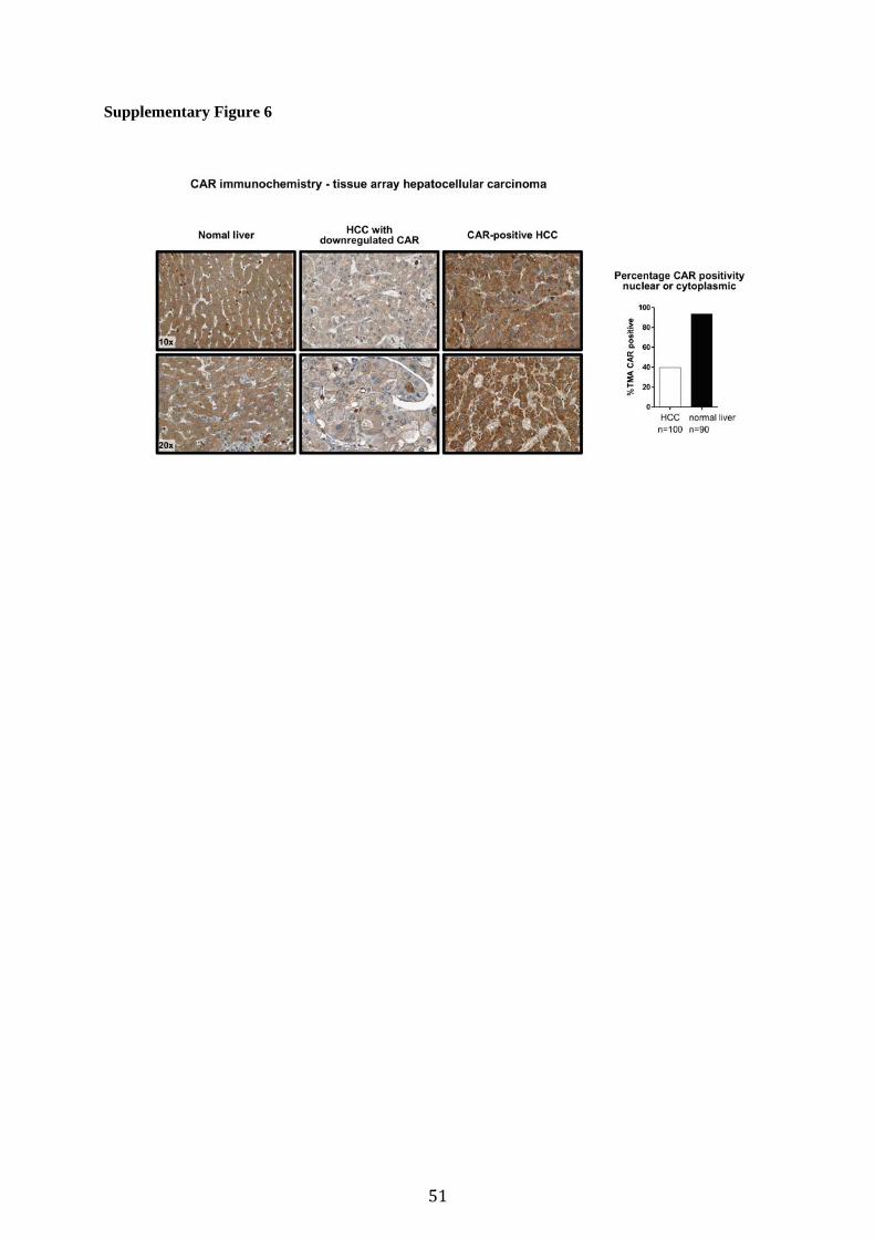


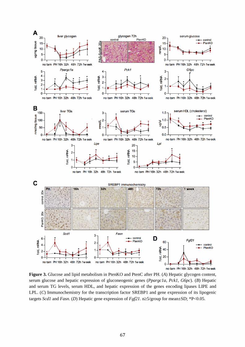
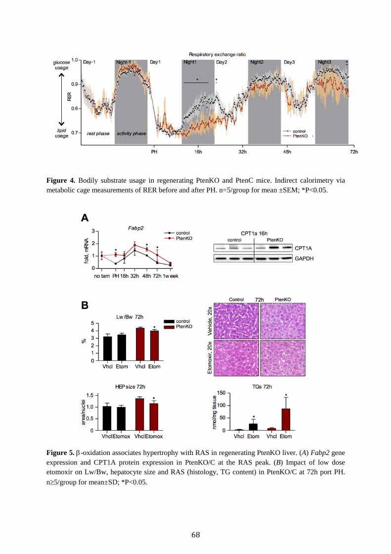


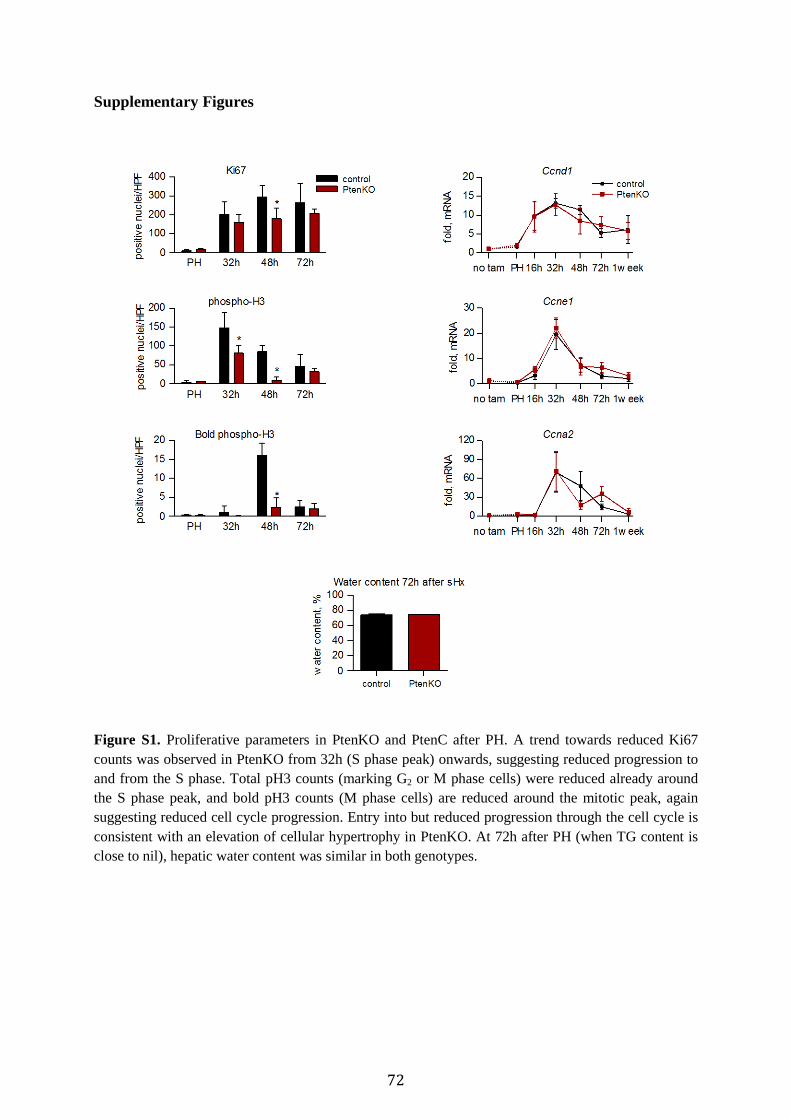


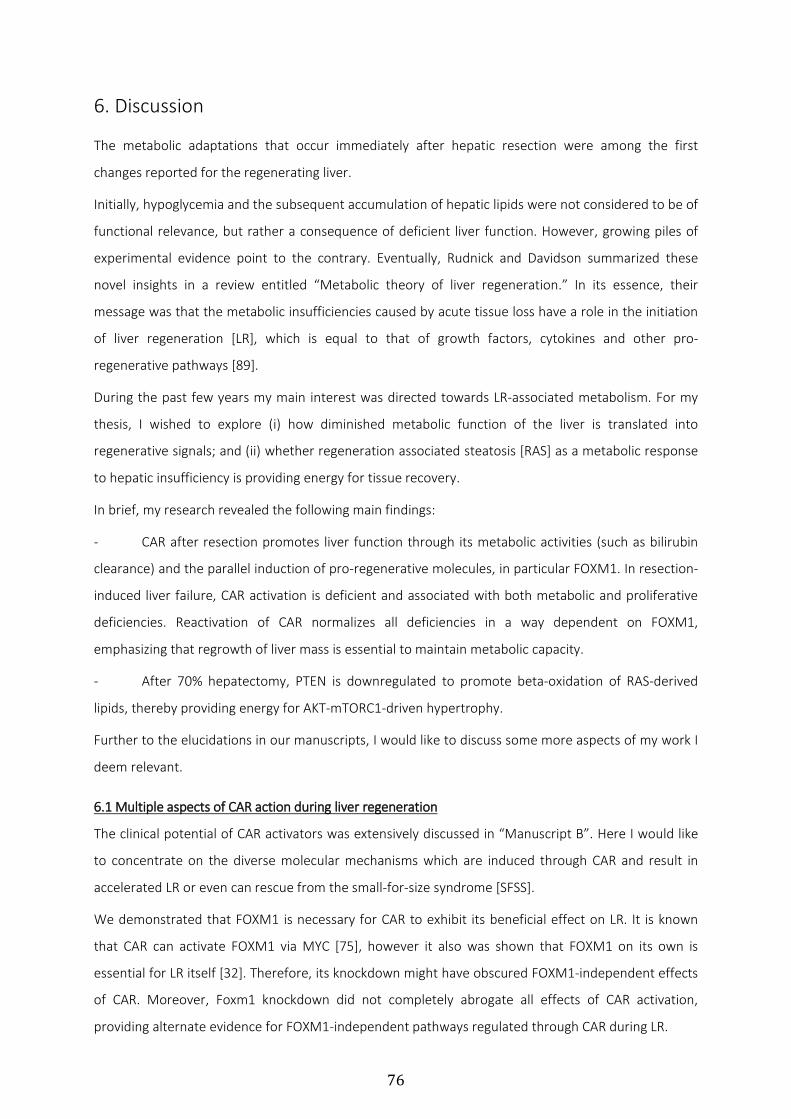


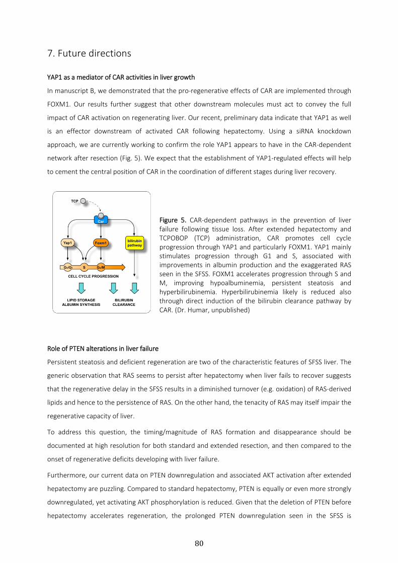


![Ekaterina Bazyka Market Update [SF] MIAMI](https://static.documents.pub/doc/80x56/577d25ef1a28ab4e1e9fecbe/ekaterina-bazyka-market-update-sf-miami.jpg)


![Ekaterina Bazyka Market Update [Condo] MIAMI BEACH](https://static.documents.pub/doc/80x56/577d25ef1a28ab4e1e9fecb5/ekaterina-bazyka-market-update-condo-miami-beach.jpg)


![Ekaterina Bazyka Market Update [SF] MIAMI BEACH](https://static.documents.pub/doc/80x56/577d25ef1a28ab4e1e9fecbd/ekaterina-bazyka-market-update-sf-miami-beach.jpg)


