Transcriptomic analyses of primary astrocytes under TNFα treatment Cindy Birck, Eric Koncina, Tony Heurtaux, Enrico Glaab, Alessandro Michelucci, Paul Heuschling, Luc Grandbarbe PII: S2213-5960(15)30074-X DOI: doi: 10.1016/j.gdata.2015.11.005 Reference: GDATA 398 To appear in: Genomics Data Received date: 28 October 2015 Accepted date: 6 November 2015 Please cite this article as: Cindy Birck, Eric Koncina, Tony Heurtaux, Enrico Glaab, Alessandro Michelucci, Paul Heuschling, Luc Grandbarbe, Transcriptomic anal- yses of primary astrocytes under TNFα treatment, Genomics Data (2015), doi: 10.1016/j.gdata.2015.11.005 This is a PDF file of an unedited manuscript that has been accepted for publication. As a service to our customers we are providing this early version of the manuscript. The manuscript will undergo copyediting, typesetting, and review of the resulting proof before it is published in its final form. Please note that during the production process errors may be discovered which could affect the content, and all legal disclaimers that apply to the journal pertain.
Transcript
�������� ����� ��
Transcriptomic analyses of primary astrocytes under TNFα treatment
Cindy Birck, Eric Koncina, Tony Heurtaux, Enrico Glaab, AlessandroMichelucci, Paul Heuschling, Luc Grandbarbe
Received date: 28 October 2015Accepted date: 6 November 2015
Please cite this article as: Cindy Birck, Eric Koncina, Tony Heurtaux, EnricoGlaab, Alessandro Michelucci, Paul Heuschling, Luc Grandbarbe, Transcriptomic anal-yses of primary astrocytes under TNFα treatment, Genomics Data (2015), doi:10.1016/j.gdata.2015.11.005
This is a PDF file of an unedited manuscript that has been accepted for publication.As a service to our customers we are providing this early version of the manuscript.The manuscript will undergo copyediting, typesetting, and review of the resulting proofbefore it is published in its final form. Please note that during the production processerrors may be discovered which could affect the content, and all legal disclaimers thatapply to the journal pertain.
Sequencer or array type Affymetrix GeneChip Mouse Gene 1.0 ST arrays
Data format CEL files
Experimental factors Primary astrocytes were treated with TNFα (50 ng/ml) during 24 h and
compared to untreated cells
Experimental features Total RNA was extracted to study gene expression changes. Three replicates
were used for each experimental condition.
Consent N/A
Sample source location N/A
1. Direct link to deposited data Deposited data can be found at: http://www.ncbi.nlm.nih.gov/geo/query/acc.cgi?acc=GSE73022.
2. Experimental Design, Materials and Methods
2.1. Cell culture and experimental design
Primary mouse astrocytes cultures were prepared from newborn C57BL/6JOlaHsd mice brains as
previously described [8]. After removing meninges and large blood vessels, brains were minced in
phosphate-buffered saline solution by mechanical dissociation. Cells were cultivated in Dulbecco’s
Modified Eagle Medium supplemented with 10% fetal bovine serum, 100 U/mL penicillin and 100 μg/mL
streptomycin at 37°C in a humidified atmosphere containing 5% CO2. The culture medium was changed
after three days, and cultures reached confluence after 10-14 days. Then, glial cells were separated by a
magnetic cell sorting (MACS) method according to the manufacturer’s protocol (Miltenyi Biotec, The
Netherlands). Briefly, glial cultures were trypsinized and microglia, the CD11b-positive cells present in
the astrocyte monolayer, were collected by a positive selection. Simultaneously, astrocytes were
negatively sorted as previously described [8, 9]. Astrocyte-enriched cultures were obtained by platting
the cells in 75 cm2 flasks. After 3 days, the culture medium was replaced and after 7 days, when cultures
reached confluence, the MACS procedure was repeated in order to reduce the residual microglial
contamination in our astrocyte population.
After additional 7 days, cultures of primary mou
Systems, United Kingdom) during 24 h. Total RNA was extracted using RNA NOW reagent (OZYME,
ACC
EPTE
D M
ANU
SCR
IPT
ACCEPTED MANUSCRIPT
France) according to the manufacturer’s instructions.
2.2 Microarrays experiments, quality control and data analysis
To determine the effects of TNFα on primary astrocytes, mRNA samples were analyzed by Affymetrix
GeneChip Mouse Gene 1.0 ST arrays. All samples were of high purity and integrity and were assessed by
the Agilent 2100 Bioanalyzer and RNA 6000 Nano LabChip kits (Agilent Technologies). Data from three
biological replicates were analyzed for each experimental condition.
Microarray gene expression data was normalized using the GC-RMA procedure with default parameters
for background correction, quantile normalization, and probe replicate summarization [10].
Differentially expressed genes between control and TNFα conditions were then determined using the
empirical Bayes moderated t-statistic (eBayes) [11]. P-value significance scores for these genes were
adjusted for multiple hypothesis testing according to the Benjamini–Hochberg procedure [12].
A heat map and dendrogram cluster visualization for the top 150 most significant known genes (Fig. 1)
was obtained using standard hierarchical average linkage clustering with a Euclidean distance metric.
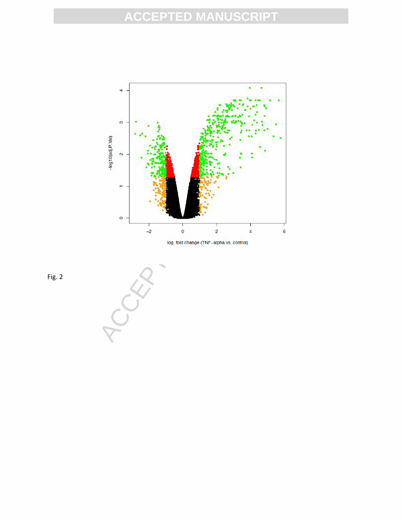
A volcano plot for the analysis of differential gene expression between TNFα and control samples was
obtained. For each transcript, the negative decadic logarithm of the adjusted p-value significance score
was plotted against the logarithm of the fold change. Several genes (green dots) are significantly altered
(adjusted p < 0.05) and display an absolute log fold change above 1 in expression (Fig. 2).
Alterations in known cellular pathways and processes were identified and visualized by applying the
MetaCoreTM GeneGo software onto the differential expression statistics obtained from the eBayes
analysis [11]. The genes were pre-filtered using a significance threshold (adjusted p value <0.05) before
applying the default GeneGO pathway analysis. Pathway analysis with GeneGO revealed that pathways
related to glial differentiation, immune response and apoptosis were modulated (Fig. 3).
3. Conclusion
Herein we describe the transcriptional analysis of primary astrocytes following a TNFα exposure. These
expression data could be useful to describe the effect of the NFκB activation on primary astrocyte
cultures devoid of microglia. Taking advantage of the MACS technology, in contrast to the main studies
reported in the literature, we were able to characterize pure populations of astrocytes under
inflammatory conditions.
We show that TNFα increases the expression of genes associated with the NFκB pathway and induces
the re-expression of genes implicated in glial developmental processes.
These data highlight the importance of the NFκB pathway during the conversion of astrocytes into
reactive cells and, particularly, its active role in the dedifferentiation process [13].
Conflict of interest
The authors declare that there are no conflicts of interests.
Acknowledgments
This work was supported by the University of Luxembourg. Cindy Birck is thankful to the doctoral school
ACC
EPTE
D M
ANU
SCR
IPT
ACCEPTED MANUSCRIPT
in systems and molecular biomedicine of the University of Luxembourg for providing a bench fee grant
to realize microarrays analyses. Enrico Glaab acknowledges support by grants from the Luxembourgish
Fonds Nationale de la Recherche (C13/BM/5782168 and NCER-PD I1R-BIC-PFN-15NCER).
References
[1] Barres BA The Mystery and Magic of Glia: A Perspective on Their Roles in Health and Disease Neuron, 2008, 60(3), pp. 430-440 [2] Zhang Y and Barres BA Astrocyte heterogeneity: an underappreciated topic in neurobiology Curr. Opin. Neurobiol., 2010, 20(5), pp. 588-594 [3] Sofroniew MV Molecular dissection of reactive astrogliosis and glial scar formation Trends Neurosci., 2009, 32(12), pp. 638-647 [4] Yang H, Cheng XP, LI JW, Yao Q, Ju G De-differentiation response of cultured astrocytes to injury induced by scratch or conditioned culture medium of scratch-insulted astrocytes Cell. Mol. Neurobiol., 2009, 29(4), pp. 455-473 [5] Buffo A, Rite I, Tripathi P, Lepier A, Colak D, Horn AP, Mori T, Gotz M Origin and progeny of reactive gliosis: a source of multipotent cells in the injured brain Proc Natl Acad Sci U S A, 2008, 105(9), pp. 3581–3586 [6] Sirko S, Behrendt G, Johansson PA, Tripathi P, Costa M, Bek S, Heinrich C, Tiedt S et al Reactive glia in the injured brain acquire stem cell properties in response to sonic hedgehog Cell Stem Cell, 2013, 12 (4), pp. 426–439 [7] Michelucci A, Bithell A, Burney MJ, Johnston CE, Wong KY, Teng SW, Desai J, Gumbleton N, Anderson G, Stanton LW, Williams BP, Buckley NJ The Neurogenic Potential of Astrocytes Is Regulated by Inflammatory Signals Mol Neurobiol., (2015) [8] Losciuto S, Dorban G, Gabel S, Gustin A, Hoenen C, Grandbarbe L, Heuschling P, Heurtaux T An efficient method to limit microglia-dependent effects in astroglial cultures J. Neurosci. Methods, 2012, 207(1), pp. 59–71. [9] Marek R, Caruso M, Rostami A, Grinspan JB, Das Sarma J Magnetic cell sorting: a fast and effective method of concurrent isolation of high purity viable astrocytes and microglia from neonatal mouse brain tissue J. Neurosci. Methods, 2008, 175(1), pp. 108–118 [10] Wu L, Thompson DK, Liu X, FieldsMW, Bagwell CE, Tiedje JM, Zhou J Development and evaluation of microarray-based whole-genome hybridization for detection of
ACC
EPTE
D M
ANU
SCR
IPT
ACCEPTED MANUSCRIPT
microorganisms within the context of environmental applications Environ Sci Technol, 2004, 38(24), pp. 6775–6782 [11] Smyth GK Linear models and empirical Bayes methods for assessing differential expression in microarray experiments Statistical applications in genetics and molecular biology, 2004 [12] Hochberg Y, Benjamini Y More powerful procedures for multiple significance testing Stat Med, 1990, 9(7) pp. 811–818
13 Gabel S, Koncina E, Dorban G, Heurtaux T, Birck C, Glaab E, Michelucci A, Heuschling P, Grandbarbe L Inflammation promotes conversion of astrocytes into neural progenitor cells via NFκB activation Mol Neurobiol., (2015)
Figures legends
Figure 1. Heat map visualization of the normalized gene expression levels for the top 150 most
significant known genes with differential expression between control and TNF samples according to the
empirical Bayes moderated t statistics.
Figure 2. Volcano plot for the analysis of differential gene expression between TNF and control samples.
For each transcript, the negative decadic logarithm of the adjusted p-value significance score is plotted
against the logarithm of the fold change. To highlight the transcripts with highest effect size and
significance, data points are colored red if the adjusted p-value is below 0.05, orange if the absolute
value of the log fold change is greater than 1, and green if both of these criteria are fulfilled.
Figure 3. Cellular pathways enriched in significantly differentially expressed genes between TNF and
control sample. These pathways were identified using the GeneGO pathway analysis software.
ACC
EPTE
D M
ANU
SCR
IPT
ACCEPTED MANUSCRIPT
Fig. 1
ACC
EPTE
D M
ANU
SCR
IPT
ACCEPTED MANUSCRIPT
Fig. 2
ACC
EPTE
D M
ANU
SCR
IPT
ACCEPTED MANUSCRIPT
Fig. 3
ACC
EPTE
D M
ANU
SCR
IPT
ACCEPTED MANUSCRIPT
Abstract
Astrocytes, the most abundant glial cell population in the central nervous system, have important
functional roles in the brain as blood brain barrier maintenance, synaptic transmission or intercellular
communications [1, 2]. Numerous studies suggested that astrocytes exhibit a functional and
morphological high degree of plasticity. For example, following any brain injury, astrocytes become
reactive and hypertrophic. This phenomenon, also called reactive gliosis, is characterized by a set of
progressive gene expression and cellular changes [3]. Interestingly, in this context, astrocytes can re-
acquire neurogenic properties. It has been shown that astrocytes can undergo dedifferentiation upon
injury and inflammation, and may re-acquire the potentiality of neural progenitors [4, 5, 6, 7].
To assess the effect of inflammation on astrocytes, primary mouse astrocytes were treated with tumor
necrosis factor α (TNFα), one of the main pro-inflammatory cytokines. The strength of this study is that
pure primary astrocytes were used. As microglia are highly reactive immune cells, we used a magnetic
cell sorting separation (MACS) method to further obtain highly pure astrocyte cultures devoid of
microglia.
Here, we provide details of the microarray data, which have been deposited in the Gene Expression
Omnibus (GEO) under the series accession number GSE73022. The analysis and interpretation of these
data are included in Gabel et al. -
associated genes were induced after a TNFα treatment. We have shown that primary astrocytes devoid
of microglia can respond to a TNFα treatment with the re-expression of genes implicated in the glial cell
development.
ACC
EPTE
D M
ANU
SCR
IPT
ACCEPTED MANUSCRIPT
Data in Brief Title: Transcriptomic analyses of primary astrocytes under TNFα treatment
Authors: Cindy Bircka, Eric Koncinaa, Tony Heurtauxa, Enrico Glaabb, Alessandro Micheluccib,c, Paul