4311 Research Article Introduction Podosomes are specialized plasma-membrane actin-based microdomains consisting of a core of actin filaments associated with the Arp-2/3-based actin polymerization machinery and surrounded by a ring of vinculin, talin, paxillin and integrins. They can be distinguished from other focal adhesions complexes by the presence of ‘podosomal markers’, such as gelsolin, cortactin, dynamin 2 and WASP/NWASP proteins (Gimona et al., 2008; Linder and Aepfelbacher, 2003). Therefore, double staining for F-actin and cortactin is routinely used to identify podosomal structures. In addition, podosomes are enriched with metalloproteases, which endow them with matrix-degradation activities. In physiological settings, podosomes form spontaneously in certain cells such as macrophages and immature dendritic cells, which share the common feature of travelling across tissue boundaries. Podosomes can also be seen in non-myeloid cells, where they are induced upon stimulation with specific receptor ligands and/or second messenger stimulators (Hai et al., 2002; Osiak et al., 2005). Podosome-related structures named invadopodia assemble in cultured cells transformed by the viral src oncogene and in melanoma or carcinoma in response to oncogenic signals (Buccione et al., 2004; Stylli et al., 2008). We have established that TGFstimulates podosome formation in cultured bovine arterial endothelial (BAE) cells (Varon et al., 2006). In this model, podosomes always arise assembled together into large ring-shaped structures. TGFis a multifunctional cytokine that is secreted from cells in a latent form which is unable to bind the TGFreceptor and is inactive (Lawrence et al., 1985). Primary regulation of TGFactivity occurs through factors that control the processing of the latent form into the biologically active molecule. TGFis active on most cells and the specificity of the response is determined by expression levels of the various TGFreceptors and co-receptors, as well as the TGFconcentration. TGFfirst binds to type II receptors (TRII), which then recruit a type I receptor (TRI). Formation of the complex leads to TRI kinase activation, which subsequently propagates the signal into the cell by phosphorylating members of the Smad family of transcription factors (Derynck and Zhang, 2003; ten Dijke and Hill, 2004). Because TGFis deeply involved in the maintenance of vascular homeostasis during adult life (Goumans and Mummery, 2000), in particular in the arterial tree, we wondered whether podosomal structures assemble in vivo. Indeed, podosomes have only been described in cultured cells in vitro and convincing evidence for the existence of such structures in intact tissues is still lacking. Here, we show the presence of rosettes of podosomes in the native endothelium of arterial vessel segments exposed to the inflammatory cytokine TGF, but not in the normal resting endothelium. The structures formed in arterial vessel segments exposed to TGFstrongly resemble those formed in TGF-treated cultured cells and the only difference found was in the topology and architectural organization of proteins in the rosettes, which might be accounted for by differences in terms of rigidity, roughness or molecular Podosomes are specialized plasma-membrane actin-based microdomains that combine adhesive and proteolytic activities to spatially restrict sites of matrix degradation in in vitro assays, but the physiological relevance of these observations remain unknown. Inducible rings of podosomes (podosome rosettes) form in cultured aortic cells exposed to the inflammatory cytokine TGF. In an attempt to prove the existence of podosomes in living tissues, we developed an ex vivo endothelium observation model. This system enabled us to visualize podosome rosettes in the endothelium of native arterial vessel exposed to biologically active TGF. Podosomes induced in the vessel appear similar to those formed in cultured cells in terms of molecular composition, but in contrast to the latter, arrange in a protruding structure that is similar to invadopodia. Local degradation of the basement membrane scaffold protein collagen-IV, is observed underneath the structures. Our results reveal for the first time the presence of podosome rosettes in the native endothelium and provide evidence for their capacity to degrade the basement membrane, opening up new avenues to study their role in vascular pathophysiology. We propose that podosome rosettes are involved in arterial vessel remodeling. Supplementary material available online at http://jcs.biologists.org/cgi/content/full/122/23/4311/DC1 Key words: Podosomes, Endothelial cells, Actin, Transforming growth factor-, Arterial vessel remodeling Summary TGF-induced endothelial podosomes mediate basement membrane collagen degradation in arterial vessels Patricia Rottiers 1,2 , Frédéric Saltel 1,2 , Thomas Daubon 1,2 , Benjamin Chaigne-Delalande 1,2, *, Viviane Tridon 2 , Clotilde Billottet 1,2 , Edith Reuzeau 1,2 and Elisabeth Génot 1,2,‡ 1 INSERM, U889, Université Victor Segalen Bordeaux 2, 146 rue Léo Saignat, Bordeaux, F-33076, France 2 European Institute of Chemistry and Biology, Université de Bordeaux 1, 2 rue Robert Escarpit, Pessac, F-33600, France *Present address: Laboratory of Immunology, National Institute of Allergy and Infectious Diseases, NIH, Bethesda, Maryland, USA ‡ Author for correspondence ([email protected]) Accepted 20 August 2009 Journal of Cell Science 122, 4311-4318 Published by The Company of Biologists 2009 doi:10.1242/jcs.057448 Journal of Cell Science
Transcript
4311Research Article
IntroductionPodosomes are specialized plasma-membrane actin-basedmicrodomains consisting of a core of actin filaments associated withthe Arp-2/3-based actin polymerization machinery and surroundedby a ring of vinculin, talin, paxillin and integrins. They can bedistinguished from other focal adhesions complexes by the presenceof ‘podosomal markers’, such as gelsolin, cortactin, dynamin 2 andWASP/NWASP proteins (Gimona et al., 2008; Linder andAepfelbacher, 2003). Therefore, double staining for F-actin andcortactin is routinely used to identify podosomal structures. Inaddition, podosomes are enriched with metalloproteases, whichendow them with matrix-degradation activities. In physiologicalsettings, podosomes form spontaneously in certain cells such asmacrophages and immature dendritic cells, which share the commonfeature of travelling across tissue boundaries. Podosomes can alsobe seen in non-myeloid cells, where they are induced uponstimulation with specific receptor ligands and/or second messengerstimulators (Hai et al., 2002; Osiak et al., 2005). Podosome-relatedstructures named invadopodia assemble in cultured cellstransformed by the viral src oncogene and in melanoma orcarcinoma in response to oncogenic signals (Buccione et al., 2004;Stylli et al., 2008).
We have established that TGF stimulates podosome formationin cultured bovine arterial endothelial (BAE) cells (Varon et al.,2006). In this model, podosomes always arise assembled togetherinto large ring-shaped structures. TGF is a multifunctional cytokine
that is secreted from cells in a latent form which is unable to bindthe TGF receptor and is inactive (Lawrence et al., 1985). Primaryregulation of TGF activity occurs through factors that control theprocessing of the latent form into the biologically active molecule.TGF is active on most cells and the specificity of the response isdetermined by expression levels of the various TGF receptors andco-receptors, as well as the TGF concentration. TGF first bindsto type II receptors (TRII), which then recruit a type I receptor(TRI). Formation of the complex leads to TRI kinase activation,which subsequently propagates the signal into the cell byphosphorylating members of the Smad family of transcriptionfactors (Derynck and Zhang, 2003; ten Dijke and Hill, 2004).
Because TGF is deeply involved in the maintenance of vascularhomeostasis during adult life (Goumans and Mummery, 2000), inparticular in the arterial tree, we wondered whether podosomalstructures assemble in vivo. Indeed, podosomes have only beendescribed in cultured cells in vitro and convincing evidence for theexistence of such structures in intact tissues is still lacking. Here,we show the presence of rosettes of podosomes in the nativeendothelium of arterial vessel segments exposed to the inflammatorycytokine TGF, but not in the normal resting endothelium. Thestructures formed in arterial vessel segments exposed to TGFstrongly resemble those formed in TGF-treated cultured cells andthe only difference found was in the topology and architecturalorganization of proteins in the rosettes, which might be accountedfor by differences in terms of rigidity, roughness or molecular
Podosomes are specialized plasma-membrane actin-basedmicrodomains that combine adhesive and proteolytic activitiesto spatially restrict sites of matrix degradation in in vitro assays,but the physiological relevance of these observations remainunknown. Inducible rings of podosomes (podosome rosettes)form in cultured aortic cells exposed to the inflammatorycytokine TGF. In an attempt to prove the existence ofpodosomes in living tissues, we developed an ex vivoendothelium observation model. This system enabled us tovisualize podosome rosettes in the endothelium of native arterialvessel exposed to biologically active TGF. Podosomes inducedin the vessel appear similar to those formed in cultured cells interms of molecular composition, but in contrast to the latter,arrange in a protruding structure that is similar to invadopodia.
Local degradation of the basement membrane scaffold proteincollagen-IV, is observed underneath the structures. Our resultsreveal for the first time the presence of podosome rosettes inthe native endothelium and provide evidence for their capacityto degrade the basement membrane, opening up new avenuesto study their role in vascular pathophysiology. We propose thatpodosome rosettes are involved in arterial vessel remodeling.
Supplementary material available online athttp://jcs.biologists.org/cgi/content/full/122/23/4311/DC1
TGF-induced endothelial podosomes mediatebasement membrane collagen degradation in arterialvesselsPatricia Rottiers1,2, Frédéric Saltel1,2, Thomas Daubon1,2, Benjamin Chaigne-Delalande1,2,*, Viviane Tridon2,Clotilde Billottet1,2, Edith Reuzeau1,2 and Elisabeth Génot1,2,‡
1INSERM, U889, Université Victor Segalen Bordeaux 2, 146 rue Léo Saignat, Bordeaux, F-33076, France2European Institute of Chemistry and Biology, Université de Bordeaux 1, 2 rue Robert Escarpit, Pessac, F-33600, France*Present address: Laboratory of Immunology, National Institute of Allergy and Infectious Diseases, NIH, Bethesda, Maryland, USA‡Author for correspondence ([email protected])
Accepted 20 August 2009Journal of Cell Science 122, 4311-4318 Published by The Company of Biologists 2009doi:10.1242/jcs.057448
Jour
nal o
f Cel
l Sci
ence
4312
composition of the substratum. Importantly, in the context of thevessel, TGF-induced podosome rosettes exhibit collagen-directedmatrix-degrading activities. Our results reveal for the first time thepresence of podosome rosettes in the native endothelium, and opennew avenues to study their role in vascular pathophysiology.
ResultsDetection of actin rings in the living endotheliumEndothelial cells in culture do not normally form podosomes.However, BAE cells assemble podosomes rosettes upon TGFexposure (Varon et al., 2006). In vitro studies indicate that TGFis activated upon contact between endothelial and smooth musclecells in vitro (Flaumenhaft et al., 1993), suggesting that localactivation of TGF regulates vessel stability in vivo. Since theendothelium is constantly exposed to TGF present in circulatingblood or trapped in the underlying matrix, we wondered whetherendothelial cells lining the interior surface of the aorta displaypodosomal structures. We developed a protocol to perform suchscreening in the aortic endothelium in mice. Tissues were fixed insitu through intracardiac injection of paraformaldehyde (ivc) inanesthetized animals. Next, vessel segments were harvested, cutalong their long axis and stained for F-actin, cortactin, VE-cadherinor CD31 whilst nuclei were highlighted with Hoechst 33342 stain.The specimens were then subjected to confocal microscopy, withendothelial cells facing upward, for ‘en face’ viewing (Fig. 1).F-actin and Hoechst staining distinguished between endothelial cellswith large round nuclei (Fig. 1A) and vascular smooth muscle cells(vSMCs) with thin and elongated nuclei (Fig. 1B). Along the z-axis,the two cell types were seen separated by the first elastic lamina(Fig. 1C), as illustrated in Fig. 1D. VE-cadherin and CD31 stainingwas detected in the endothelial layer (Fig. 1E,F), but absent in thedeeper layer of smooth muscle cells (Fig. 1B), clearly delimitingthe cellular boundaries and revealing the integrity of the endotheliumin the specimen. F-actin and cortactin were consistently concentratedat cell margins within the endothelium and never showed thecharacteristic arrangement of podosomal structures (Fig. 1G-H).
We therefore reasoned that, under normal physiologicalconditions, the bioavailability of TGF in the endothelium was toolow to induce podosomes. To investigate further the existence ofpodosomes in tissues, we examined the effects of biologically activeTGF concentrations on the aortic endothelium. Live aortic vesselsegments were cultured ex vivo, in the presence or absence ofexogenously added recombinant TGF1 (TGF) under conditionssimilar to those used to induce podosome rosettes in cultured BAEcells (Varon et al., 2006) (Fig. 2A-C). Aortic explants were thenfixed, stained for F-actin and cortactin and prepared for en faceviewing. In untreated samples, whereas some fields of view showedmarker distribution that was similar to that obtained with aorticvessel segments fixed in vivo (supplementary material Fig. S1A-D), other fields of view showed less prominent cortactin and F-actin staining at cell-cell junctions, a pattern which persistedthroughout the organ culture (supplementary material Fig. S1E-H).A similar staining pattern was observed in endothelia of TGF-treated samples (supplementary material Fig. S1I-L). However,careful examination revealed zones where F-actin and cortactindistinctly colocalized in ring-like structures within the endotheliumof TGF-treated vessels (Fig. 2D-F), that were not detected inuntreated specimens (supplementary material Fig. S1E-G). Toassess endothelial cell responsiveness to TGF stimulation, aorticvessel segments were stained for canonical effectors of TGF(Lebrin et al., 2005). A twofold increase in phosphorylated Smad2
was detected in the cell nuclei of TGF-treated endothelial cellsupon TGF exposure, attesting to efficient TGF signaling in thisexperimental setting (Fig. 3).
Molecular characterization of the structures formed inendothelium exposed to TGFFurther characterization of the ring structures revealed the presenceof structural components such as vinculin and paxillin, signalingcomponents such as FAK, phosphotyrosine-containing proteins and3 integrin (which is an ‘activation’ marker for endothelial cells),as well as genuine podosome markers (Linder and Aepfelbacher,2003) Arp3, cortactin and dynamin 2 (Fig. 4A-C and F). We alsodetected MT1-MMP (Fig. 4D), MMP2 and MMP9 (supplementarymaterial Fig. S3A,B) at the rosettes. N-WASp was distributed allalong the structure, including its tip (Fig. 4E). This situationresembles that encountered in invadopodia, podosome-relatedstructures assembled in cultured cells transformed by oncogenes(Baldassarre et al., 2006; Lorenz et al., 2004; Stylli et al., 2008).Interestingly, 1 integrins were distributed along the inner and outerrim of the podosome rosettes in vitro. With respect to integrinlocalization, the resemblance between ring-like actin structures inaortic vessel segments (Fig. 4G) and podosome rosettes in culturedBAE cells (Fig. 4H) is a convincing reason for considering thatthey are one and the same kind of structure.
Journal of Cell Science 122 (23)
Fig. 1. Model for observation of the endothelium from large arterial vessels.(A-B)An aortic vessel segment was harvested from mice after intraventricularcardiac injection (ivc) of paraformaldehyde, prepared forimmunofluorescence, labelled with phalloidin (red), VE cadherin (green) andHoechst 33342 and mounted en face for confocal observation. Viewed face on,VE-cadherin and F-actin staining of the endothelial cell layer delineatesindividual cells with round nuclei (A) and of the underlying layer of vSMCcells with elongated nuclei (B). Scale bar: 15m. (C)Multiple x-y imageswere sectioned in the z direction through the aortic vessel segment until atransverse image was resolved. The endothelium is visualized by VE-cadherin(green) immunostaining, the vSMCs by dense phalloidin staining of F-actinand nuclei by Hoechst 33342 fluorescence. Three autofluorescent (green)elastic bands appear underneath the endothelium monolayer and in betweenmultiple vSMC layers with a wave like appearance. The white dotted linescorrespond to the focal planes shown in A and B. The white dotted linescorrespond to the upper (endothelium) and lower (vSMC) focal planes shownin A and B, respectively. Scale bar: 5m. (D)Schematic side view of the innerarterial wall showing the endothelial monolayer (EC), the basement membrane(BM) in grey, the elastic lamina (IEL) in green, the first smooth muscle celllayer (vSMC) and a second elastic lamina (green). (E-H)Individual staining ofVE-cadherin, CD31, F-actin and cortactin, respectively, representative of theirarrangement in the native endothelium. Scale bar: 10m. Each image isrepresentative of three to five vessel segments.Jo
urna
l of C
ell S
cien
ce
4313Podosomes in the arterial endothelium
Endothelial actin-rich VCAM+ docking structures that mediateadhesion of leukocytes to the endothelium have been described ininflammatory contexts (Barreiro et al., 2002). Here, the actin ringsformed in response to TGF stained negative for endothelial(VCAM) or leukocyte (CD11b) adhesion molecules (data notshown), indicating that such endothelial figures are not generatedby extravasating blood leukocytes undergoing transcellulardiapedesis (Carman and Springer, 2004). Therefore, based on thesecriteria, we conclude that TGF induces genuine podosome rosettesin aortic vessel segments. Similar results were found with murinecarotid artery segments processed in the same way (data not shown).Podosome rosettes were detected in less than 5% of arterialendothelial cells exposed to TGF for 24 hours.
Similarities and differences of podosomes in in vitro andex vivo modelsInterestingly, the endothelium that lines blood vessels represents abi-dimensional space, suggesting that podosome rosettes formed invivo will be found under the same sort of organization as thoseobserved in vitro. Fig. 5A shows a rosette located on the basal sideof the endothelial cell, in close apposition with the underlying
substratum of the endothelium. Along the z-axis, the podosomalstructure was seen to extend downwards, as if pushing in into thevSMC layer (Fig. 5B). The rosette formed in the endotheliumdisplayed prominent cortactin staining at the distal extremity of therosette, protruding through the smooth muscle cells (Fig. 5A,B).This organization of the podosomal components fits with the modelof actin polymerization of podosomes, which is thought to proceedsimilarly to extending lamellipodia, but along the vertical axis(Linder and Aepfelbacher, 2003; Vignjevic and Montagnac, 2008).We conclude that these endothelial integrin-rich structures adhereto the underlying substratum. These findings confirm that thepodosome rosettes depicted here constitute entities distinct frompreviously described actin structures that interact with circulatingcells on the apical surface of the endothelium (Barreiro et al., 2002).
We next explored the spatial organization of podosomal proteinsin endothelial rosettes from TGF-treated aortic vessel segments(Fig. 5C), cultured BAE cells (Fig. 5G) and in cultured BAE cellsseeded onto a vessel where the endothelial monolayer, but not thebasement membrane, had been removed (Fig. 5K). Rosettes formedin the intact endothelium or in BAE cells seeded onto the denudedaortic vessel segments were both found to be more compact andthicker along the z-axis than those formed on glass coverslips [outerdiameter, 7.53±2.43 m, n11 (ex vivo); 9.17±2.67 m, n15 (BAEcells on denuded vessels); 16.13±2.39 m, n10 (in vitro)]. Thesefindings suggest that the molecular composition of the underlyingsubstratum rather than cell-cell interactions in the endotheliumaccount for the differences observed between the in vitro and exvivo characteristics of podosome rosettes.
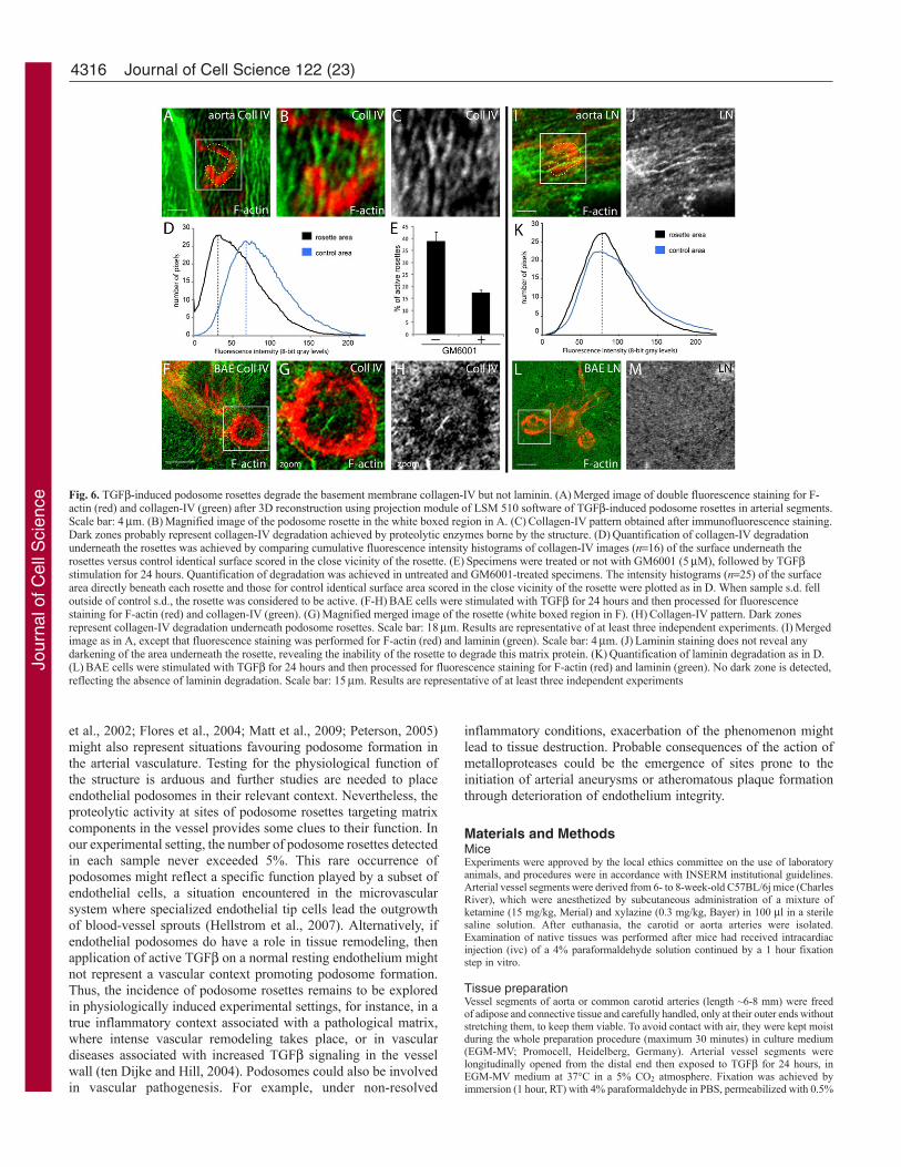
Endothelial podosome rosettes formed in arterial vesselsegments degrade the basement membrane collagen-IVA specific feature of podosomes is their ability to degrade gelatin.Podosome rosettes formed in cultured TGF-treated endothelialcells fulfil this criterion, as measured by an in vitro gelatin-degradation assay (Varon et al., 2006). In vessels, endothelial cellsrest on the basement membrane, which provides physical supportfor the cells and exerts a multitude of regulatory functions on theirgrowth and differentiation state (Adams and Watt, 1993). Basementmembrane consists primarily of laminin and type IV collagenbridged by non-covalent interactions with nidogens and perlecan(Kalluri, 2003; Rowe and Weiss, 2008; Yurchenco et al., 2004). Toexplore the degradative potential of endothelial podosome rosettespresent in vessel segments, samples were stained with antibodiesrecognizing matrix proteins. Non-fluorescent patches were detecteddirectly beneath the podosome rosettes in the endothelium of TGF-treated arterial vessel segments, when staining was performed withanti-collagen-IV antibodies (Fig. 6A-C). This pattern stronglysuggests matrix protein degradation by proteolytic activities atpodosome rosettes, in a manner analogous to that seen in podosomerosettes in BAE cells cultured on a collagen-IV matrix in vitro (Fig.
Fig. 2. Detection of actin rings in TGF-treated aortic vessel segments.(A-C)Representative immunoconfocal images of F-actin and cortactinorganization in ring-like structures in TGF-treated cultured BAE cells (A).The white dotted line delineates the endothelial cell outlines. Scale bar: 10m.A magnified view of the boxed zone in A shows individual cortactin staining(B) and merged F-actin and cortactin (yellow) (C). (D)Representativeimmunoconfocal image of the merged staining for F-actin and cortactinshowing organization and colocalization (yellow) of the two proteins in ring-like structures in TGF-treated aortic vessel segments. The white dotted linedelineates cell outlines of the endothelial cell harbouring the podosome rosette.Scale bar: 20m. (E)Magnified image of individual cortactin staining.(F)Merged staining for F-actin and cortactin of the boxed zone in D. Resultsare representative of several independent experiments.
Fig. 3. Endothelial cells respond to TGF.(A,B) Aortic vesselsegments were either left untreated (A) or stimulated with TGF for24 hours (B), fixed and then processed for fluorescence staining forphosphorylated Smad2. Scale bar: 5m. (C)Quantitative analysis ofthe phosphorylated Smad2 intensity/nuclei ratio shows a twofoldincrease upon TGF exposure, attesting for efficient TGFsignaling in this experimental setting. *P<0.05.
Jour
nal o
f Cel
l Sci
ence
4314
6F-H) or on the basement membrane of denuded vessel segments(supplementary material Fig. S2). Quantitative analysis revealedefficient matrix degradation beneath the rosettes formed in theendothelium exposed to TGF (Fig. 6D). Focal collagen-IVdegradation at podosome sites was inhibited by the broad-spectrumMMP inhibitor GM6001 (Fig. 6E). Under similar conditions,laminin staining remained intact beneath the rosettes (Fig. 6I-K).This finding was confirmed in vitro in the BAE model. TGF-induced endothelial podosomes were unable to detectably alter thederived laminin coating (Fig. 6L,M). Likewise, thorough inspectionof the elastic lamina did not reveal any alteration in its integrity atthis stage (data not shown).
DiscussionPodosomes have been extensively described in the artificialenvironment of cultured cells. Our findings demonstrate thatpodosome rosettes form in the living endothelium of large arterialvessel segments exposed to physiological levels of biologicallyactive TGF. We also reveal their capacity to degrade the basementmembrane. Our data provide the first evidence for podosomes innative tissues and support the concept that podosomes arephysiological structures involved in biological processes in vivo.We believe that these findings represent a major advance in thefield and will support researchers aiming to demonstrate thepresence of invadopodia in cancer cells in vivo.
Imaging podosome rosettes in their native environment enabledus to disclose their distinctive features. Similarities and differencesbetween podosomes and invadopodia, collectively calledinvadosomes (Machesky et al., 2008) are not yet definitive (Weaver,
2008). A major difference often reported between podosomes andinvadopodia is the overall architecture of the microdomain. Whereaspodosomes self-organize within the boundaries of the cell on a softgelatin matrix, protrusive organization is the hallmark ofinvadopodia (Stylli et al., 2008). Our studies reveal the spatialorganization of the components and the protruding architecture ofendothelial podosome rosettes, providing evidence for similaritiesbetween the extensions formed by invadopodia of transformed cells(Baldassarre et al., 2006) and aggregated podosomes in rosettes ofendothelial cells. The arrangement of the podosomal componentsin the structure is dependent on the substratum, suggesting amisguided organization on glass coverslips in in vitro settings. Ourstudy thus describes a situation where the protrusive organizationof invadopodia, rather than the non-protrusive organization ofpodosomes as described in vitro, vanishes in this in vivo context.Our results establish that the cell microenvironment and theexperimental settings impact invadosome architecture.
A common characteristic feature of podosomes and invadopodiais their ability to degrade proteins of the extracellular matrix. Wehave defined the degradation capacities of podosome rosettestowards basement membrane proteins in their native environment.Matrix degradation measurements recorded upon 24 hours ofexposure to TGF revealed collagen-directed matrix-degradingactivities, which is consistent with the recruitment of MT1-MMP,MMP2 and MMP9 at the structures (Guegan et al., 2008; Tatin etal., 2006; Varon et al., 2006). Under these conditions, no alterationin the laminin pattern was detected. However, unlike the collagen-IV network, which is stabilized through covalent links, lamininpolymers are not covalently associated, but are rather assembled
Journal of Cell Science 122 (23)
Fig. 4. Podosomal components are localized within actinrings of arterial endothelium exposed to TGF ex vivo.(A-C)Aortic vessel segments were stimulated with TGFfor 24 hours, fixed and then processed for fluorescencestaining for F-actin (red) and podosomal markers (green)FAK (A), phosphorylated Tyr (B), dynamin 2 (C) or MT1-MMP (D). Confocal sections (0.4m) of F-actin and N-WASP double staining were taken from the top to thebottom of the rosette, and orthogonal z sections from the x-y view are shown (E). Merged images reveal colocalizationof F-actin with N-WASp proteins, shown individually inthe lower panels. Scale bar: 4m. (F)Molecularcharacterization of the structures formed in the endotheliumof arterial vessel segments, using a panel of antibodiesdirected against proteins known to localize at podosomes incultured cells. Results are representative of at least threeindependent experiments. (G,H)1 integrins (green)distribute along the inner and outer rim of the actin rosette(red) formed in the living endothelium stimulated withTGF for 24 hours (G). Similar localization of 1 integrinis seen in BAE cells stimulated with TGF for 24 hours invitro (H). Scale bar: 4m. Results are representative of atleast three independent experiments.
Jour
nal o
f Cel
l Sci
ence
4315Podosomes in the arterial endothelium
together by the collagen-IV network (Yurchenco et al., 2004). It isthe highly ordered and crosslinked nature of the type IV collagennetwork that confers basement membranes with their structuralintegrity. Therefore, disruption of the collagen bonds naturallydisrupts the laminin network, and from there, the laminin-collagen-IV scaffold (Rowe and Weiss, 2008). In our experimental set-up,there is no laminin degradation, and the loosening of the lamininmeshwork structure is not detectable. Since perforation of thebasement membrane invadopodia takes several days (Hotary et al.,2006) and collagen-IV is the most abundant constituent of thebasement membrane (Kalluri, 2003), we assume that ourexperimental setting enables us to detect the early events of a TGF-induced remodeling programme where collagen-IV degradationoccurs at an early stage, loosening the laminin network and resulting
in basement membrane splitting. Our results reveal, for the firsttime, the presence of podosome rosettes in the native endotheliumand open new avenues to study their role in vascularpathophysiology.
Examination of the aortic and carotid arteries specimen fixed insitu revealed that, in the absence of stimulus, the endothelium oflarge vessels is normally devoid of podosome rosettes. Theseobservations in vivo are consistent with the situation in vitro, whereresting aortic endothelial cells show no podosomes. Thus,podosomes are expected to come into play in specific situationsassociated with local release of active TGF, such as those inducedby disturbed blood flow or ischemia (Jones et al., 2009; Khalil,1999; Sho et al., 2002; van Royen et al., 2002). Conditionsassociated with elevated levels of circulating TGF (Derhaschnig
Fig. 5. Architecture of the podosome rosette formed ex vivo. (A)Arterial vessel segments were stimulated with TGF for 24 hours and then processed for doublefluorescence staining for F-actin (red) and cortactin (green). Viewed face on, immunoconfocal F-actin and cortactin staining of the top (up merge) and bottom(down merge) of one of the structures. Scale bar: 9m. (B)Representative immunoconfocal images show the rosette located on the basal surface of the endothelialcells, extending towards the vSMC along the z-axis. The white dotted lines correspond to the upper and lower focal planes shown in A. Scale bars: x-y, 9m; z,3m. Merged images reveal colocalisation of the two proteins (yellow), shown individually in the lower panels. (C)The same structure in its surroundings seen atlower magnification. Merged images of triple fluorescence staining for F-actin (red), cortactin (green) and nuclei (blue). Scale bar: 9m. (D-F)The rosette(cortactin image) and two nuclei (Hoechst image) in adjacent endothelial cells denoting landmarks (dotted line shown in C) were digitally isolated andreconstructed using Imaris Isosurface function. (D,E)3D reconstruction shows the protruding end of the rosette (green) and nuclei (white) (vSMC nuclei are stainedblue) above and below the confocal planes shown in A and B, respectively (magnified images of the boxed area in C). (F)A skewed and a side view of the rosetteafter digital subtraction of the vessel surroundings. (G-J)Merged image of double fluorescence staining for F-actin (red) and cortactin (green) of BAE cellsstimulated with TGF for 24 hours (G) and individual cortactin staining (H). Scale bar: 9m. (I,J)A skewed (I) and a side view (J) of the rosette after 3Dreconstruction of the optical sections of the cortactin image after subtraction of the surroundings shows the flat surface of the rosette. (K-O)Merged image ofdouble fluorescence staining for F-actin (red) and cortactin (green) of BAE cells seeded on an opened up denudated vessel segment and stimulated with TGF for24 hours (K), and individual cortactin staining (L). Scale bar: 9m. A skewed (M) and a side (N) view of the rosette after 3D reconstruction of the optical sectionsof cortactin and subtraction of the surroundings. (O)The focal plane runs through the vSMC layer and shows the rosette protruding downwards.
Jour
nal o
f Cel
l Sci
ence
4316
et al., 2002; Flores et al., 2004; Matt et al., 2009; Peterson, 2005)might also represent situations favouring podosome formation inthe arterial vasculature. Testing for the physiological function ofthe structure is arduous and further studies are needed to placeendothelial podosomes in their relevant context. Nevertheless, theproteolytic activity at sites of podosome rosettes targeting matrixcomponents in the vessel provides some clues to their function. Inour experimental setting, the number of podosome rosettes detectedin each sample never exceeded 5%. This rare occurrence ofpodosomes might reflect a specific function played by a subset ofendothelial cells, a situation encountered in the microvascularsystem where specialized endothelial tip cells lead the outgrowthof blood-vessel sprouts (Hellstrom et al., 2007). Alternatively, ifendothelial podosomes do have a role in tissue remodeling, thenapplication of active TGF on a normal resting endothelium mightnot represent a vascular context promoting podosome formation.Thus, the incidence of podosome rosettes remains to be exploredin physiologically induced experimental settings, for instance, in atrue inflammatory context associated with a pathological matrix,where intense vascular remodeling takes place, or in vasculardiseases associated with increased TGF signaling in the vesselwall (ten Dijke and Hill, 2004). Podosomes could also be involvedin vascular pathogenesis. For example, under non-resolved
inflammatory conditions, exacerbation of the phenomenon mightlead to tissue destruction. Probable consequences of the action ofmetalloproteases could be the emergence of sites prone to theinitiation of arterial aneurysms or atheromatous plaque formationthrough deterioration of endothelium integrity.
Materials and MethodsMiceExperiments were approved by the local ethics committee on the use of laboratoryanimals, and procedures were in accordance with INSERM institutional guidelines.Arterial vessel segments were derived from 6- to 8-week-old C57BL/6j mice (CharlesRiver), which were anesthetized by subcutaneous administration of a mixture ofketamine (15 mg/kg, Merial) and xylazine (0.3 mg/kg, Bayer) in 100 l in a sterilesaline solution. After euthanasia, the carotid or aorta arteries were isolated.Examination of native tissues was performed after mice had received intracardiacinjection (ivc) of a 4% paraformaldehyde solution continued by a 1 hour fixationstep in vitro.
Tissue preparationVessel segments of aorta or common carotid arteries (length ~6-8 mm) were freedof adipose and connective tissue and carefully handled, only at their outer ends withoutstretching them, to keep them viable. To avoid contact with air, they were kept moistduring the whole preparation procedure (maximum 30 minutes) in culture medium(EGM-MV; Promocell, Heidelberg, Germany). Arterial vessel segments werelongitudinally opened from the distal end then exposed to TGF for 24 hours, inEGM-MV medium at 37°C in a 5% CO2 atmosphere. Fixation was achieved byimmersion (1 hour, RT) with 4% paraformaldehyde in PBS, permeabilized with 0.5%
Journal of Cell Science 122 (23)
Fig. 6. TGF-induced podosome rosettes degrade the basement membrane collagen-IV but not laminin. (A)Merged image of double fluorescence staining for F-actin (red) and collagen-IV (green) after 3D reconstruction using projection module of LSM 510 software of TGF-induced podosome rosettes in arterial segments.Scale bar: 4m. (B)Magnified image of the podosome rosette in the white boxed region in A. (C)Collagen-IV pattern obtained after immunofluorescence staining.Dark zones probably represent collagen-IV degradation achieved by proteolytic enzymes borne by the structure. (D)Quantification of collagen-IV degradationunderneath the rosettes was achieved by comparing cumulative fluorescence intensity histograms of collagen-IV images (n16) of the surface underneath therosettes versus control identical surface scored in the close vicinity of the rosette. (E)Specimens were treated or not with GM6001 (5M), followed by TGFstimulation for 24 hours. Quantification of degradation was achieved in untreated and GM6001-treated specimens. The intensity histograms (n25) of the surfacearea directly beneath each rosette and those for control identical surface area scored in the close vicinity of the rosette were plotted as in D. When sample s.d. felloutside of control s.d., the rosette was considered to be active. (F-H)BAE cells were stimulated with TGF for 24 hours and then processed for fluorescencestaining for F-actin (red) and collagen-IV (green). (G)Magnified merged image of the rosette (white boxed region in F). (H)Collagen-IV pattern. Dark zonesrepresent collagen-IV degradation underneath podosome rosettes. Scale bar: 18m. Results are representative of at least three independent experiments. (I)Mergedimage as in A, except that fluorescence staining was performed for F-actin (red) and laminin (green). Scale bar: 4m. (J)Laminin staining does not reveal anydarkening of the area underneath the rosette, revealing the inability of the rosette to degrade this matrix protein. (K)Quantification of laminin degradation as in D.(L)BAE cells were stimulated with TGF for 24 hours and then processed for fluorescence staining for F-actin (red) and laminin (green). No dark zone is detected,reflecting the absence of laminin degradation. Scale bar: 15m. Results are representative of at least three independent experiments
Jour
nal o
f Cel
l Sci
ence
4317Podosomes in the arterial endothelium
Triton X-100 for 1 hour, and blocked with 3% fetal bovine serum for 30 minutes(RT). The specimens were incubated with primary antibodies (overnight, RT), thenwith the appropriate secondary antibodies conjugated to Alexa Fluor-488, -546 or -633(3 hours, RT). F-actin and nuclei were stained at this step with phalloidin conjugatedto Alexa Fluor-546 or -488 and Hoechst 33342, respectively. Arterial vessel segmentswere spread then mounted on microscope slides with Fluoromount mounting medium.Denuded basement membrane arterial segments obtained by endothelial cell lysisupon exposure to hypotonic shock.
CellsBAE cells (Lonza) were maintained in complete endothelial cell growth medium(EGM-MV; Promocell) at 37°C in a 5% CO2 humidified atmosphere and used betweenpassages three and six (Varon et al., 2006).
ReagentsRecombinant human TGF (TGF1, used at 5 ng/ml in all experiments) was obtainedfrom R&D Systems and type IV collagen and laminin were obtained from Sigma.GM6001 was purchased from Calbiochem and Fluoromount mounting medium wasfrom Clinisciences. Antibodies against the following proteins were obtained asindicated: cortactin (clone 4F11), Arp3, phosphorylated Smad2 (Ser465/467) andcollagen-IV were from Millipore; Cdc42, paxillin, FAK, N-WASp from CellSignaling; vinculin and phosphotyrosine (clone 4G10) from Sigma; laminin fromAbcam; 3 integrin was from Emfret and 1 integrin from BD Biosciences; VE-cadherin was from MedSystems. Polyclonal antibodies directed against dynamin 2were from Mark McNiven (Mayo Clinic, Rochester, MA). Anti-MT1-MMP is amonoclonal antibody (mAb-2) obtained from the same fusion as that described recently(Ingvarsen et al., 2008). MMP9 antibodies were from BioMol and those against MMP2were from R&D systems. Alexa Fluor 546-phalloidin, Alexa Fluor 488-labeledsecondary antibodies, fluorescein isothiocyanate (FITC) and Hoechst 33342 werepurchased from Invitrogen.
Immunofluorescence stainingSubconfluent cells grown on glass coverslips were prepared for immunofluorescenceas previously described (Varon et al., 2006). The coverslips were washed in waterand mounted on microscope slides with Fluoromount mounting medium. For matrixdegradation assay, BAE cells were seeded on coverslips coated with collagen-IV (0.2mg/ml) or laminin (20 g/ml), and dark areas and podosome rosettes were visualizedafter staining with the relevant antibodies and Alexa Fluor 546-phalloidin, respectively.
Microscopy and image analysisCells and arterial vessel segments were analyzed by confocal imaging using a ZeissLSM 510 inverted laser-scanning fluorescence microscope equipped with acquisitionsoftware (LSM 510 acquisition software; Zeiss) and a �63 NA 1.4 oil-immersionobjective. Triple-color imaging using Hoechst 33342, Alexa Fluor 488- or AlexaFluor 633-labeled secondary antibodies, and Alexa Fluor 546-phalloidin was obtainedusing selective laser excitation at 350 nm, 488 nm, 633 nm and 543 nm, respectively.Each channel was imaged sequentially using the multitrack recording module beforemerging. Fluorescent images were processed with Adobe Photoshop 7.0. To make3D images, serial optical sections of tissue samples or cells were captured with thesoftware’s automatic scanning mode. Z-stack digital images were collected opticallyat every 0.4 m depth, saved and used for 3D reconstruction using Imaris software(Bitplane).
Measurement of matrix degradation underneath podosomerosettes in arterial vessel segmentsSpecimens were double stained for F-actin and for collagen-IV or laminin to detectpodosome rosettes and basement membrane, respectively. Z-stack digital images wereacquired, running through the rosette and first smooth muscle cell layer. Images wereused for 3D reconstruction analysis using the LSM 510 acquisition softwareprojection module. 3D images were saved and analyzed using ImageJ software afterimages had been converted to an eight-bit gray-scale image. For each rosette, therosette area was delimited manually in the 3D F-actin image and this region waspasted on the same location on the 3D basement membrane image. In this 3D image,intensity profiles of regions of interest (rosette area) were measured and comparedwith intensity profile of regions in the rosette vicinity (control area). This measurementwas repeated on 16 (collagen-IV) or 23 (laminin) podosome rosettes, and an averagegraph was drawn. For quantification of degradation with GM6001, ‘rosette area’ and‘control area’ values were compared for each rosette. s.d. values of intensity profilesbetween rosette area and CT area determine the degradation potential of the rosette.
StatisticsData were compared between groups using Student’s t-test. Differences wereconsidered to be statistically significant at P<0.05.
We are grateful to Signe Ingvarsen and Lars Engelholm whoprovided the anti-MT1-MMP antibodies, and to Mark McNiven forthose recognizing dynamin 2. We thank Steve Weiss, Bob Mecham,
Brant Isakson, Rosemary Akhurst, Ellen Van Obberghen-Schilling, ZiadMallat, Gareth Jones and Wolfgang Schaper for helpful discussions.We also wish to knowledge Alain-Pierre Gadeau and Claude Desgrangesfor expert help in the initial experiments and IJsbrand Kramer for criticalcomments on the manuscript. C.B. is a recipient of grant ANR-06-BLAN-0362 and her research is funded through this route. P.R. issupported by a predoctoral fellowship from the Region Aquitaine andINSERM. This work was supported by INSERM, ARC (3549), LaLigue Nationale contre le Cancer and Fondation de France and EUgrant PITN-GA-2009-237946.
ReferencesAdams, J. C. and Watt, F. M. (1993). Regulation of development and differentiation by
the extracellular matrix. Development 117, 1183-1198.Baldassarre, M., Ayala, I., Beznoussenko, G., Giacchetti, G., Machesky, L. M., Luini,
A. and Buccione, R. (2006). Actin dynamics at sites of extracellular matrix degradation.Eur. J. Cell Biol. 85, 1217-1231.
Barreiro, O., Yanez-Mo, M., Serrador, J. M., Montoya, M. C., Vicente-Manzanares,M., Tejedor, R., Furthmayr, H. and Sanchez-Madrid, F. (2002). Dynamic interactionof VCAM-1 and ICAM-1 with moesin and ezrin in a novel endothelial docking structurefor adherent leukocytes. J. Cell Biol. 157, 1233-1245.
Buccione, R., Orth, J. D. and McNiven, M. A. (2004). Foot and mouth: podosomes,invadopodia and circular dorsal ruffles. Nat. Rev. Mol. Cell. Biol. 5, 647-657.
Carman, C. V. and Springer, T. A. (2004). A transmigratory cup in leukocyte diapedesisboth through individual vascular endothelial cells and between them. J. Cell Biol. 167,377-388.
Derhaschnig, U., Shehata, M., Herkner, H., Bur, A., Woisetschlager, C., Laggner, A.N. and Hirschl, M. M. (2002). Increased levels of transforming growth factor-beta1 inessential hypertension. Am. J. Hypertens 15, 207-211.
Derynck, R. and Zhang, Y. E. (2003). Smad-dependent and Smad-independent pathwaysin TGF-beta family signalling. Nature 425, 577-584.
Flaumenhaft, R., Abe, M., Sato, Y., Miyazono, K., Harpel, J., Heldin, C. H. and Rifkin,D. B. (1993). Role of the latent TGF-beta binding protein in the activation of latentTGF-beta by co-cultures of endothelial and smooth muscle cells. J. Cell Biol. 120, 995-1002.
Flores, L., Naf, S., Hernaez, R., Conget, I., Gomis, R. and Esmatjes, E. (2004).Transforming growth factor beta at clinical onset of Type 1 diabetes mellitus. A pilotstudy. Diabet. Med. 21, 818-822.
Gimona, M., Buccione, R., Courtneidge, S. A. and Linder, S. (2008). Assembly andbiological role of podosomes and invadopodia. Curr. Opin. Cell Biol. 20, 235-241.
Goumans, M. J. and Mummery, C. (2000). Functional analysis of the TGFbetareceptor/Smad pathway through gene ablation in mice. Int. J. Dev. Biol. 44, 253-265.
Guegan, F., Tatin, F., Leste-Lasserre, T., Drutel, G., Genot, E. and Moreau, V. (2008).p190B RhoGAP regulates endothelial-cell-associated proteolysis through MT1-MMPand MMP2. J. Cell Sci. 121, 2054-2061.
Hai, C. M., Hahne, P., Harrington, E. O. and Gimona, M. (2002). Conventional proteinkinase C mediates phorbol-dibutyrate-induced cytoskeletal remodeling in a7r5 smoothmuscle cells. Exp. Cell Res. 280, 64-74.
Hellstrom, M., Phng, L. K., Hofmann, J. J., Wallgard, E., Coultas, L., Lindblom, P.,Alva, J., Nilsson, A. K., Karlsson, L., Gaiano, N. et al. (2007). Dll4 signalling throughNotch1 regulates formation of tip cells during angiogenesis. Nature 445, 776-780.
Hotary, K., Li, X. Y., Allen, E., Stevens, S. L. and Weiss, S. J. (2006). A cancer cellmetalloprotease triad regulates the basement membrane transmigration program. GenesDev. 20, 2673-2686.
Ingvarsen, S., Madsen, D. H., Hillig, T., Lund, L. R., Holmbeck, K., Behrendt, N. andEngelholm, L. H. (2008). Dimerization of endogenous MT1-MMP is a regulatory stepin the activation of the 72-kDa gelatinase MMP-2 on fibroblasts and fibrosarcoma cells.Biol. Chem. 389, 943-953.
Jones, J. A., Spinale, F. G. and Ikonomidis, J. S. (2009). Transforming growth factor-beta signaling in thoracic aortic aneurysm development: a paradox in pathogenesis. J.Vasc. Res. 46, 119-137.
Kalluri, R. (2003). Basement membranes: structure, assembly and role in tumourangiogenesis. Nat. Rev. Cancer 3, 422-433.
Khalil, N. (1999). TGF-beta: from latent to active. Microbes Infect. 1, 1255-1263.Lawrence, D. A., Pircher, R. and Jullien, P. (1985). Conversion of a high molecular
weight latent beta-TGF from chicken embryo fibroblasts into a low molecular weightactive beta-TGF under acidic conditions. Biochem. Biophys. Res. Commun. 133, 1026-1034.
Lebrin, F., Deckers, M., Bertolino, P. and Ten Dijke, P. (2005). TGF-beta receptor functionin the endothelium. Cardiovasc Res. 65, 599-608.
Linder, S. and Aepfelbacher, M. (2003). Podosomes: adhesion hot-spots of invasive cells.Trends Cell Biol. 13, 376-385.
Lorenz, M., Yamaguchi, H., Wang, Y., Singer, R. H. and Condeelis, J. (2004). Imagingsites of N-wasp activity in lamellipodia and invadopodia of carcinoma cells. Curr. Biol.14, 697-703.
Machesky, L., Jurdic, P. and Hinz, B. (2008). Grab, stick, pull and digest: the functionaldiversity of actin-associated matrix-adhesion structures. Workshop on Invadopodia,Podosomes and Focal Adhesions in Tissue Invasion. EMBO Rep. 9, 139-143.
Matt, P., Schoenhoff, F., Habashi, J., Holm, T., Van Erp, C., Loch, D., Carlson, O. D.,Griswold, B. F., Fu, Q., De Backer, J. et al. (2009). Circulating transforming growthfactor-{beta} in Marfan syndrome. Circulation 120, 526-532.
Jour
nal o
f Cel
l Sci
ence
4318
Osiak, A. E., Zenner, G. and Linder, S. (2005). Subconfluent endothelial cells formpodosomes downstream of cytokine and RhoGTPase signaling. Exp. Cell Res. 307, 342-353.
Peterson, M. C. (2005). Circulating transforming growth factor beta-1: a partial molecularexplanation for associations between hypertension, diabetes, obesity, smoking and humandisease involving fibrosis. Med. Sci. Monit. 11, RA229-RA232.
Rowe, R. G. and Weiss, S. J. (2008). Breaching the basement membrane: who, when andhow? Trends Cell Biol. 18, 560-574.
Sho, E., Sho, M., Singh, T. M., Nanjo, H., Komatsu, M., Xu, C., Masuda, H. and Zarins,C. K. (2002). Arterial enlargement in response to high flow requires early expression ofmatrix metalloproteinases to degrade extracellular matrix. Exp. Mol. Pathol. 73, 142-153.
Stylli, S. S., Kaye, A. H. and Lock, P. (2008). Invadopodia: at the cutting edge of tumourinvasion. J. Clin. Neurosci. 15, 725-737.
Tatin, F., Varon, C., Genot, E. and Moreau, V. (2006). A signalling cascade involvingPKC, Src and Cdc42 regulates podosome assembly in cultured endothelial cells inresponse to phorbol ester. J. Cell Sci. 119, 769-781.
ten Dijke, P. and Hill, C. S. (2004). New insights into TGF-beta-Smad signalling. TrendsBiochem. Sci. 29, 265-273.
van Royen, N., Hoefer, I., Buschmann, I., Heil, M., Kostin, S., Deindl, E., Vogel, S.,Korff, T., Augustin, H., Bode, C. et al. (2002). Exogenous application of transforminggrowth factor beta 1 stimulates arteriogenesis in the peripheral circulation. FASEB J.16, 432-434.
Varon, C., Tatin, F., Moreau, V., Van Obberghen-Schilling, E., Fernandez-Sauze, S.,Reuzeau, E., Kramer, I. and Genot, E. (2006). Transforming growth factor beta inducesrosettes of podosomes in primary aortic endothelial cells. Mol. Cell. Biol. 26, 3582-3594.
Vignjevic, D. and Montagnac, G. (2008). Reorganisation of the dendritic actin networkduring cancer cell migration and invasion. Semin. Cancer Biol. 18, 12-22.
Weaver, A. M. (2008). Invadopodia. Curr. Biol. 18, R362-R364.Yurchenco, P. D., Amenta, P. S. and Patton, B. L. (2004). Basement membrane assembly,
stability and activities observed through a developmental lens. Matrix Biol. 22, 521-538.