112
Nerve Cells 1

Nerve Cells
1
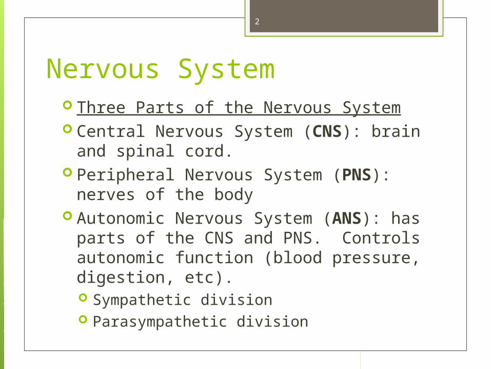
Nervous System Three Parts of the Nervous System Central Nervous System (CNS): brain and
spinal cord. Peripheral Nervous System (PNS): nerves
of the body Autonomic Nervous System (ANS): has
parts of the CNS and PNS. Controls autonomic function (blood pressure, digestion, etc). Sympathetic division Parasympathetic division
2

Figure 12.2
Basic Divisions of the Nervous System
3

Basic Divisions of the Nervous System Central nervous system (CNS)
Brain and spinal cord Integrating and command center
4

Basic Divisions of the Nervous System Peripheral nervous system (PNS)
Outside the CNS Consists of nerves from brain and spinal
cord Cranial nerves Spinal nerves
Peripheral nerves link all regions of the body to the CNS
5
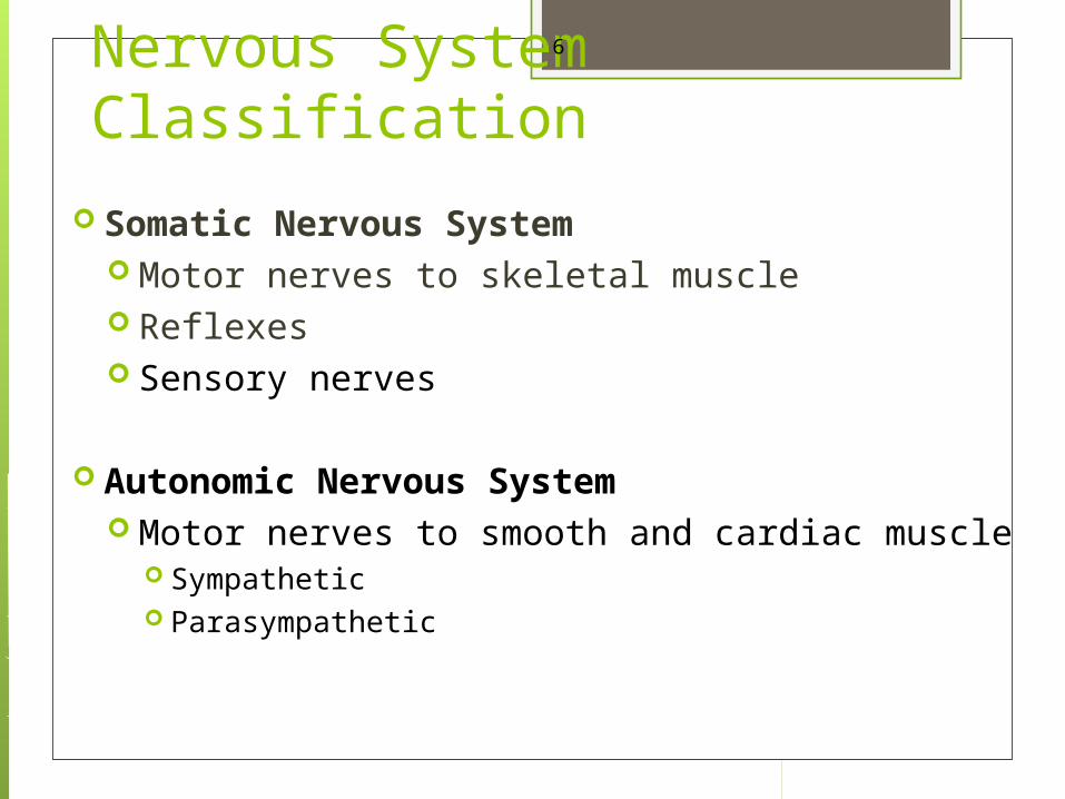
Nervous System Classification
Somatic Nervous System Motor nerves to skeletal muscle Reflexes Sensory nerves
Autonomic Nervous System Motor nerves to smooth and cardiac muscle
Sympathetic Parasympathetic
6

Neurons The nervous system is made up of more
cells than any other system. For instance, the brain has about 100
billion cells. There are also a number of different
cell types, the most important is the neuron.
7
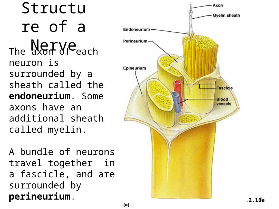
Structure of a Nerve
8Figure 12.16a
The axon of each neuron is surrounded by a sheath called the endoneurium. Some axons have an additional sheath called myelin.
A bundle of neurons travel together in a fascicle, and are surrounded by perineurium.
A bundle of fascicles is surrounded by epineurium

NEURON All neurons do three things: Receive a signal. Can be any type of stimulus
(touch, vibration, light, sound, signal from another neuron, etc).
Transmit a signal to another location. E.g. finger touching something signal to spinal cord or brain.
Stimulate another cell Another neuron transmit signal Muscle contraction Gland secretion Blood vessel constriction
9

The Neuron Other special characteristics
Longevity – can live and function for a lifetime
Do not divide – fetal neurons lose their ability to undergo mitosis because their centrioles no longer function and cannot pull the chromosomes apart; neural stem cells are an exception
High metabolic rate – require abundant oxygen and glucose
10
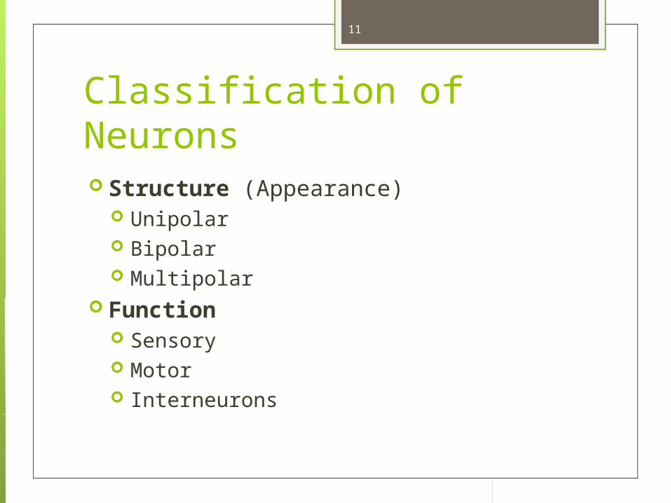
Classification of Neurons Structure (Appearance)
Unipolar Bipolar Multipolar
Function Sensory Motor Interneurons
11

Structural Classification of NeuronsMultipolar neurons
– Most neurons are this type, having many dendrites and one axon.
Bipolar neurons– Have two processes that extend
from opposite sides of the cell body. Some sensory neurons are bipolar.
12
Unipolar neuronsHave one short process emerging from the cell body, which then branches into a “T”. Sensory neurons in the PNS are unipolar.
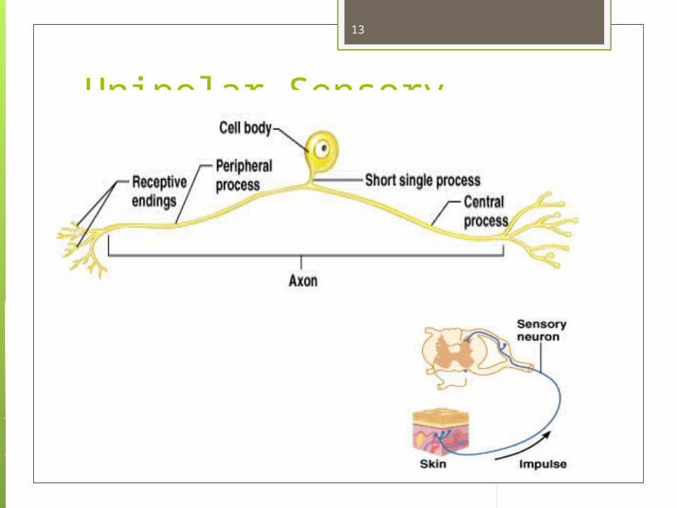
Unipolar Sensory Neuron
13

Functional Classification of Neurons
Neurons are grouped functionally according to the direction the nerve impulse travels relative to the CNS.
Sensoroy Neurons (afferent neurons) transmit impulses toward the CNS. They originate in the PNS and terminate in the CNS.
Motor Neurons (efferent neurons) transmit impulses from the CNS to effector organs (muscles and glands). They originate in the CNS and terminate in the PNS.
Interneurons (association neurons) connect sensory neurons to motor neurons within the spinal cord and brain. They originate and terminate in the CNS, and form complex neuronal pathways. They make up 99.98% of the neurons in the body, reflecting the vast amount of information processed in the CNS.
14

Neurons Classified by Function: Sensory vs. Motor Neurons
15Figure 12.11
Sensory neurons enter the spinal cord. Motor neurons leave the spinal cord. Interneurons connect the sensory and motor neurons.

There are hundreds of different types of neurons, each one is specialized for a particular task
(e.g. sensory neurons receive and transmit sensory information, and there are several different types of them, with receptors for light, smell, pain, light touch, vibration, position in space).
Motor neurons transmit signals for muscle contraction (organ contraction, blood vessel constriction), gland secretion.
They all share certain characteristics. Longevity (can last a lifetime) High metabolic rate Cannot divide to reproduce (they lose their centrioles
during fetal development) Cannot survive without oxygen
16

Neuron Anatomy
17
Axon hillock (trigger zone)
Soma (cell body)
Axon (transmits signals)
Axon terminals (stimulate another cell)
Dendrites (receive signal)
Neuron Anatomy

NEUROLEMMA is the name of the plasma membrane (outermost covering) of a neuron.
DENDRITES function to receive the signal and carry the nerve conduction toward the cell body.
SOMA (cell body) is where the nucleus, ribosomes, and most organelles are located
AXON HILLOCK is the area on the soma where the action potential (electrical charges) of the neuron builds up before it transmits the signal down the axon.
AXON function is to transmit signals. Some cells have more than one axon, some axons are short, and some are long.
AXON TERMINALS (also called boutons or synaptic knobs) contain a neurotransmitter which, when released, stimulates another cell.
A SYNAPSE is where one neuron touches another neuron. Neurons may have a couple of synapses, or hundreds.
AXOPLASMIC TRANSPORT: Movement of nutrients, wastes, and organelles between the cell body and axon terminals
18
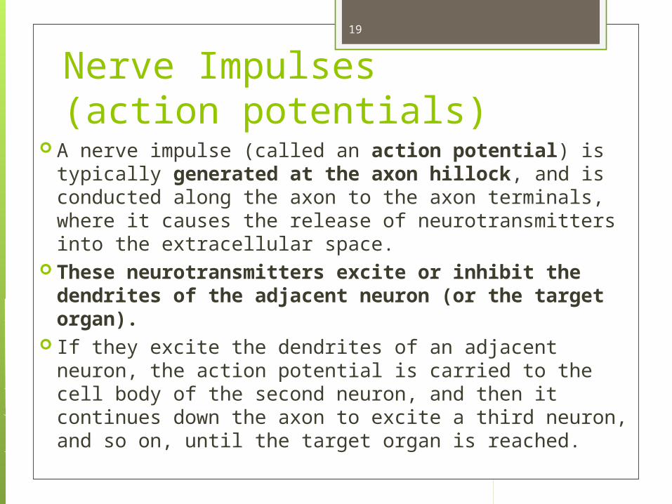
Nerve Impulses(action potentials)
A nerve impulse (called an action potential) is typically generated at the axon hillock, and is conducted along the axon to the axon terminals, where it causes the release of neurotransmitters into the extracellular space.
These neurotransmitters excite or inhibit the dendrites of the adjacent neuron (or the target organ).
If they excite the dendrites of an adjacent neuron, the action potential is carried to the cell body of the second neuron, and then it continues down the axon to excite a third neuron, and so on, until the target organ is reached.
19
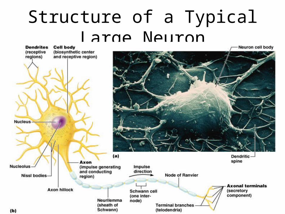
Structure of a Typical Large Neuron
20Figure 12.4

Synapse A synapse is the site at which two neurons
communicate, or a neuron and its target organ. The neuron that conducts the signals towards
the synapse is called the presynaptic neuron. The neuron that transmits the signals away
from the synapse is called the postsynaptic neuron.
Most neurons function as pre-synaptic and post-synaptic neurons.
The neurons don’t physically touch each other at the synapse. The space between them is called the synaptic cleft.
21

Two Neurons Communicating at a Synapse
22Figure 12.6
Know this order: The action potential travels from the Axon of presynaptic neuron SYNAPTIC CLEFT dendrite of post synaptic neuron
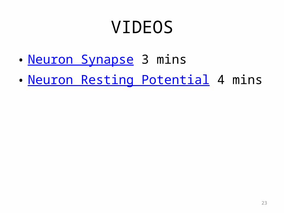
VIDEOS
• Neuron Synapse 3 mins• Neuron Resting Potential 4 mins
23
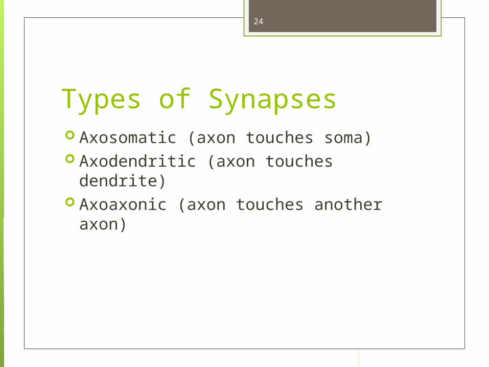
Types of Synapses Axosomatic (axon touches soma) Axodendritic (axon touches dendrite) Axoaxonic (axon touches another axon)
24

Axosomatic Synapse
25Figure 12.7

Axodendritic Synapses
26

How does the signal go through the space? By a chemical transmission.
The axon terminals have vesicles filled with a neurotransmitter that transmits the signal across the synapse.
Each type of neuron uses a particular type of neurotransmitters, so there are many types of neurotransmitters.
Some neurotransmitters excite the adjacent neuron, and some are inhibitory.
27

Structure of a Synapses
28Figure 12.8a, b
Chemical substances released from the presynaptic terminal:
•May inhibit or stimulate an action in the postsynaptic cell
•May be broken down by enzymes or taken back up into the vesicles and recycled.

Fun Fact Children under 3 years of age have
twice as many synapses as adults. That is why they learn languages better.
A child must learn a second language before the age of ten, or they probably will not have the proper native accent of that language.
29

• The axon terminal of the nerve cell rests in indentations in the cell membrane of the muscle fiber.
• The enlarged knob of the axon is called the presynaptic terminal
• The space between the presynaptic terminal and the muscle fiber membrane is the synaptic cleft
• The muscle fiber membrane is the postsynaptic membrane.
• If this neuron innervates skeletal muscle, the vesicles of its axon terminal will contain the neurotransmitter acetylcholine.
30

An action potential causes the release of Ach (acetylcholine; the neurotransmitter at the neuromuscular junction) into the synaptic cleft.
Ach binds to receptor sites on the muscle fiber (muscle cell) membrane and starts an electrical impulse called an action potential, which travels along the length of the muscle fiber and causes it to contract.
The Ach that was released is rapidly broken down by an enzyme, Ach-ase (acetylcholinesterase). This ensures that the action potential will result in only one contraction of each muscle fiber.
A neuron might be temporarily unable to transmit an impulse to another cell if its supply of neurotransmitters is exhausted.
31

32
AchE Blockers Neostigmine Physostigmine Myasthenia Gravis-
ptosis

Myasthenia gravis Myasthenia gravis is an autoimmune disorder in which
antibodies attack and destroy some acetylcholine receptors. Acetylcholine is therefore less likely to stimulate muscle
contraction, resulting in muscle weakness and fatigue. Symptoms usually begin in the eyelid and facial muscles, and
manifests as drooping muscles on half or both sides of the face, drooping eyelids, and slurred speech.
Neostigmine is an anti-cholinesterase drug which reduces the symptoms by inhibiting Ach-ase activity, preventing the breakdown of Ach. Consequently, Ach levels in the synapse remain elevated, so Ach is available to bind to those few functional Ach receptors that are left.
33
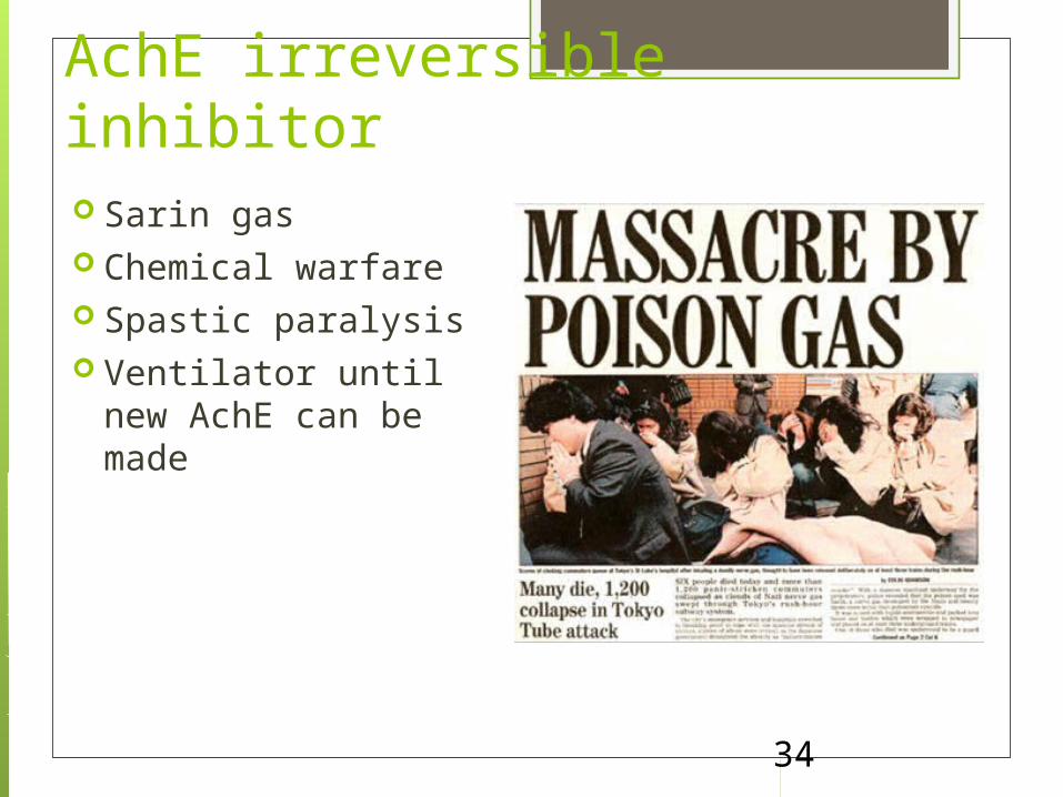
34
AchE irreversible inhibitor Sarin gas Chemical warfare Spastic paralysis Ventilator until new
AchE can be made

Sarin Gas Attack by Syria, 2013
https://www.youtube.com/watch?v=doytZVNltc4
Video
35

Acetylcholine-esterase Blocker
Some INSECTICIDES inhibit acetylcholinesterase, so Ach accumulates in the synaptic cleft and acts as a constant stimulus to the muscle fiber. The insects die because their respiratory muscles contract and cannot relax.
Other poisons, such as CURARE, the poison used by South American Indians in poison arrows, bind to the Ach receptors on the muscle cell membrane and prevent Ach from working. That prevents muscle contraction, resulting in flaccid paralysis.
36
Acetylcholine Antagonists
37
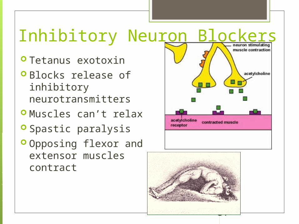
Inhibitory Neuron Blockers Tetanus exotoxin Blocks release of
inhibitory neurotransmitters
Muscles can’t relax Spastic paralysis Opposing flexor and
extensor muscles contract
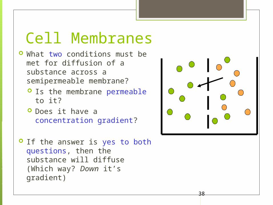
What two conditions must be met for diffusion of a substance across a semipermeable membrane? Is the membrane permeable to
it? Does it have a concentration
gradient?
If the answer is yes to both questions, then the substance will diffuse (Which way? Down it’s gradient)
38
Cell Membranes

• Each cell in our body is surrounded by a cell membrane composed of a phospholipid BI-LAYER.
• That means that our cell membranes have two layers: an outer layer, and an inner layer.
• The inside layer of each cell membrane in the body, (including each neuron) has a charge (usually negative), and the outside layer of each cell membrane has a charge (usually positive).
39

• The reason for the charge difference is that there are many proteins inside of cells, and proteins are made of amino acids, most of which have a negative charge. Because proteins are negatively charged, the inside layer of the cell membrane has a negative charge.
• Outside of the cell, there are many electrolytes, especially sodium (Na+), which have a positive charge. They stay outside of the cell because they cannot get in unless sodium channels in the cell membrane are open, which they usually are not. That’s why the outside of the cell membrane usually has a positive charge.
40

What is a sodium channel? Proteins embedded in the cell membranes form channels which only allow certain ions to cross the cell membrane.
A sodium ion can only get into the cell by way of a sodium channel. A potassium ion can only get in by way of a potassium channel, etc.
Charged ions, such as potassium (K+), sodium (Na+), calcium (Ca++), and chloride (Cl-) are called electrolytes. When they move from one side of the cell membrane to the other (when their channels are open), they carry their electrical charge with them.
This changes the overall charge of the inside and outside of the cell membrane.
41

42
Not just separation of solutes, but charges, too! Inside of the cell is
negative due to : Abundance of proteins,
which have a negative charge
The cell membrane is very permeable (“leaky”) to K+, so it can LEAVE the cell whenever it wants. That leaves the inside of the cell more negative.
_ _ _ __ _ _ __ _ _ _
++++
++++
++++
++++

“Sidedness” of the membrane Sidedness means that the electrical charges on one side of the membrane (positive or negative) are different than on the other side. Why does sidedness exist?The cell membrane has different permeabilities to each ion; for instance the cell is more permeable to K+ than any other ion.Pumps exist which force particular ions into or out of the cellChannels made out of protein selectively allow particular ions into or out of the cell. These channels may be open or closed at any given time.
Sidedness
43

Every cell has a positive charge on the outside of the membrane and a negative charge on the inside of the cell membrane, when the cell is at rest (not being stimulated).
K+ constantly leaks out of the cell because it wants to diffuse down its concentration gradient. That means it wants to go from its area of high concentration (the inside of the cell) to an area where it is in low concentration (the outside of the cell).
44
• Na+ wants to get into the cell, too, but it’s channel is closed.
• Because of this separation of chemicals and electrical charges, every cell has a Resting Membrane “Potential”.
There is a difference in electrical charge across the membrane (a potential difference)
More negative inside; more positive outsideResting membrane potential is minus70 to minus
90mV

As K+ leaves the cell, it takes a positive charge outside with it, so the inside is more negative.
However, as the inside of the cell is becoming more negative, the outside of the cell is becoming more positive, and the positive charges will want to flow back inside of the cell since they are attracted to the negative charges.
This is what keeps K+ from just leaving the cell until it is in equal numbers on both sides of the cell. Before it can reach such an equilibrium, it gets pulled back into the cell because its positive charges are drawn into the inside of the cell, where the charge has become strongly negative (because proteins are on the inside of the cell and they have a negative charge).
Other positively charged ions, like Na+, want to go into the cell also, but they are blocked by protein gates that only K+ can get through.
45

ACTION POTENTIAL The resting membrane potential
Occurs because the cell membrane is more permeable to potassium ions than sodium ions
Occurs because there are negatively charged proteins and ions inside the cell
The action potential occurs if the membrane potential reaches a certain threshold.
46

What would happen to the membrane potential of the cell when this event occurs?
What if a membrane suddenly became MORE PERMEABLE to Na+?????
Even for just a moment in time….. What would Na+ do? (Ask yourself
the 3 questions)
Which way is the electrochemical gradient for Na+?
Electrical: inwardChemical: inwardAnswer:Most definitely INWARDSodium WANTS IN!Can it get in?
47
What if…..
-
+
-70 mV
- - -
+++
Na+
Na+
Cl-
Cl-
Na+
Cl-
Na+
Na+
Cl-
Cl-
Na+
Na+
Cl-
Cl-

Know the following: Direction of impulse Ions involved, charges States: resting potential, depolarization, repolarization
Direction of impulse
Na+
Na+
Na+
Resting potential Depolarization Repolarization
Na+ enters and K+ leaves, so outside of membrane becomes negative charge. This is depolarization
Outside of membrane is positive charge at resting potential.
When stimulus is over, Na+ leaves and K+ lenters the cell, so outside of membrane returns to positive charge. This is repolarization
48

What would happen to the membrane potential of the cell when you open up a sodium channel?
If we instantly increase sodium permeability, sodium will enter the cell, changing the charge of the inside of the cell so that it goes from negative to positive. The outside layer of the cell membrane would then go from positive to negative (the charges flip). This is called DEPOLARIZATION.
However, when this occurs, Na+ will be in higher concentration on the inside of the cell, so it wants to diffuse back out of the cell.
Once it leaves the cell again, the membrane potential of the inside of the cell membrane will return to a negative charge. This is called REPOLARIZATION.
49

Spread of Depolarization
50

+ + + + + + + + + + + + + + + + + + + + + + + + + + + + + + + +
+ + + + + + + + + + + + + + + + + + + + + + + + + + + + + + + +
- - - - - - - - - - - - - - - - - - - - - - - - - - - - - - - - - - - - - - - - - - -
- - - - - - - - - - - - - - - - - - - - - - - - - - - - - - - - - - - - - - - - - - -
Direction of Depol
Resting Cell
51
+ + + + + + + + + + + + + + + + + + + + + + + + + + + + + + + +
+ + + + + + + + + + + + + + + + + + + + + + + + + + + + + + + +
- - - - - - - - - - - - - - - - - - - - - - - - - - - - - - - - - - - - - - - - - - -
- - - - - - - - - - - - - - - - - - - - - - - - - - - - - - - - - - - - - - - - - - -
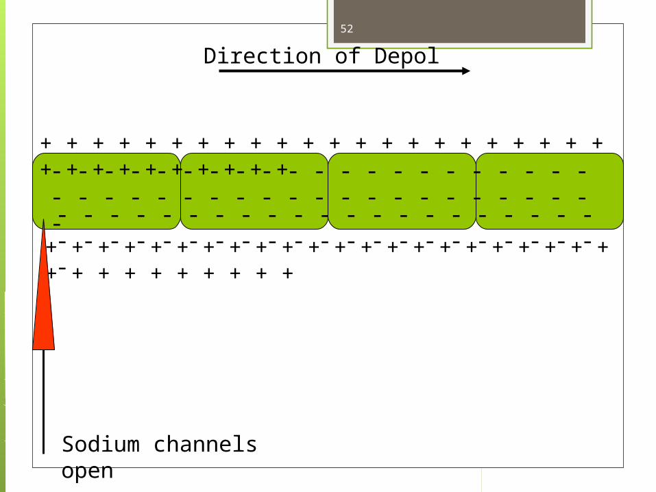
Direction of Depol
Sodium channels open
52

+ + + + + + + + + + + + + + + + + + + + + + + + + + + + + + + +
+ + + + + + + + + + + + + + + + + + + + + + + + + + + + + + + +
- - - - - - - - - - - - - - - - - - - - - - - - - - - - - - - - - - - - - - - - - - -
- - - - - - - - - - - - - - - - - - - - - - - - - - - - - - - - - - - - - - - - - - -
Direction of Depol53

+ + + + + + + + + + + + + + + + + + + + + + + + + + + + + + + +
+ + + + + + + + + + + + + + + + + + + + + + + + + + + + + + + +
- - - - - - - - - - - - - - - - - - - - - - - - - - - - - - - - - - - - - - - - - - -
- - - - - - - - - - - - - - - - - - - - - - - - - - - - - - - - - - - - - - - - - - -
Direction of Depol
+
54
+ + + + + + + + + + + + + + + + + + + + + + + + + + + + + + + +
+ + + + + + + + + + + + + + + + + + + + + + + + + + + + + + + +
- - - - - - - - - - - - - - - - - - - - - - - - - - - - - - - - - - - - - - - - - - -
- - - - - - - - - - - - - - - - - - - - - - - - - - - - - - - - - - - - - - - - - - -
Direction of Depol
++ +
55
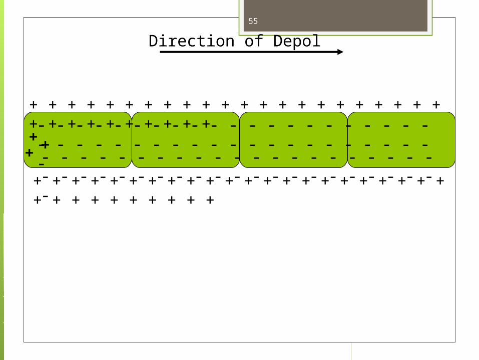
- - - + + + + + + + + + + + + + + + + + + + + + + + + + + + + +
- - - + + + + + + + + + + + + + + + + + + + + + + + + + + + + + +
+ + - - - - - - - - - - - - - - - - - - - - - - - - - - - - - - - - - - - - - - - -
+ + - - - - - - - - - - - - - - - - - - - - - - - - - - - - - - - - - - - - - - - - -
Direction of Depol56

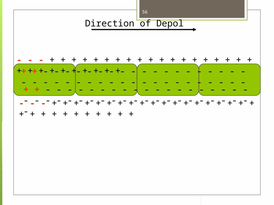
- - - - - - - - + + + + + + + + + + + + + + + + + + + + + + + + +
- - - - - - - + + + + + + + + + + + + + + + + + + + + + + + + + +
+ + + + + - - - - - - - - - - - - - - - - - - - - - - - - - - - - - - - - - - - -
+ + + + + - - - - - - - - - - - - - - - - - - - - - - - - - - - - - - - - - - - - -
Direction of Depol57

- - - - - - - - - - - - - - - - + + + + + + + + + + + + + + + + + + + +
- - - - - - - - - - - - - - - + + + + + + + + + + + + + + + + + + + +
+ + + + + + + + + + - - - - - - - - - - - - - - - - - - - - - - - - - - - - -
+ + + + + + + + + + - - - - - - - - - - - - - - - - - - - - - - - - - - - - - -
Direction of Depol58
- - - - - - - - - - - - - - - - - - - - - + + + + + + + + + + + + + + + +
- - - - - - - - - - - - - - - - - - - - + + + + + + + + + + + + + + + + + +
+ + + + + + + + + + + + + + - - - - - - - - - - - - - - - - - - - - - - -
+ + + + + + + + + + + + + + - - - - - - - - - - - - - - - - - - - - - - - -
Direction of Depol59
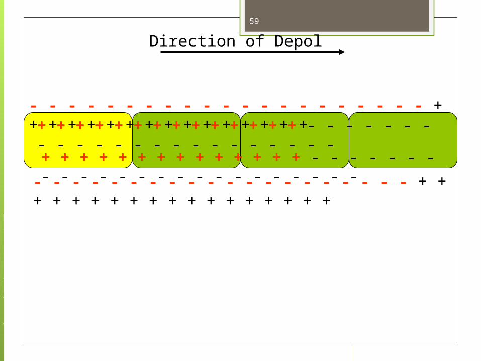
- - - - - - - - - - - - - - - - - - - - - - - - - - + + + + + + + + + + + +
- - - - - - - - - - - - - - - - - - - - - - - - - - + + + + + + + + + + + + +
+ + + + + + + + + + + + + + + + + + + - - - - - - - - - - - - - - - -
+ + + + + + + + + + + + + + + + + + + - - - - - - - - - - - - - - - - -
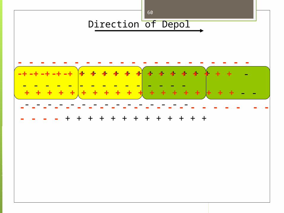
Direction of Depol60

- - - - - - - - - - - - - - - - - - - - - - - - - - - - - - - - - - + + + + + +
- - - - - - - - - - - - - - - - - - - - - - - - - - - - - - - - - - + + + + + + +
+ + + + + + + + + + + + + + + + + + + + + + + + - - - - - - - - -
+ + + + + + + + + + + + + + + + + + + + + + + + - - - - - - - - - -
Direction of Depol61
- - - - - - - - - - - - - - - - - - - - - - - - - - - - - - - - - - - - - - - - - + +
- - - - - - - - - - - - - - - - - - - - - - - - - - - - - - - - - - - - - - - - - + +
+ + + + + + + + + + + + + + + + + + + + + + + + + + + + - - -
+ + + + + + + + + + + + + + + + + + + + + + + + + + + + + - - -
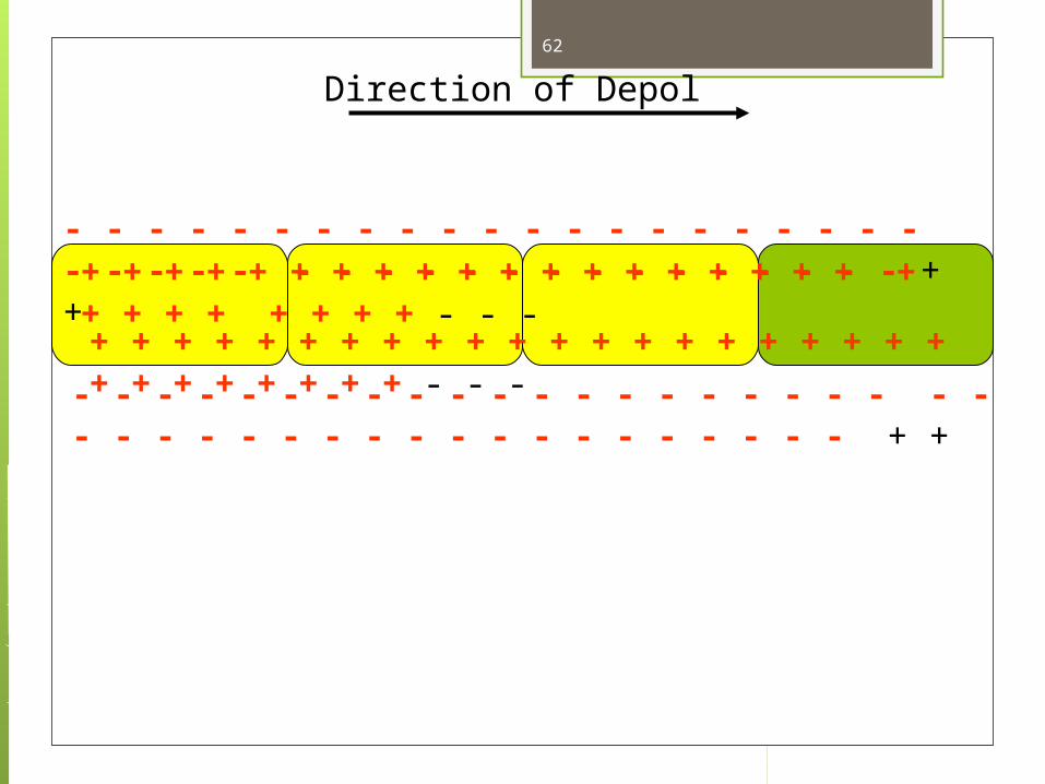
Direction of Depol62

- - - - - - - - - - - - - - - - - - - - - - - - - - - - - - - - - - - - - - - - - - - -
- - - - - - - - - - - - - - - - - - - - - - - - - - - - - - - - - - - - - - - - - - - - -
+ + + + + + + + + + + + + + + + + + + + + + + + + + + + + + +
+ + + + + + + + + + + + + + + + + + + + + + + + + + + + + + +
Direction of Depol63

- - - - - - - - - - - - - - - - - - - - - - - - - - - - - - - - - - - - - - - - - - - -
- - - - - - - - - - - - - - - - - - - - - - - - - - - - - - - - - - - - - - - - - - - - -
+ + + + + + + + + + + + + + + + + + + + + + + + + + + + + + +
+ + + + + + + + + + + + + + + + + + + + + + + + + + + + + + +
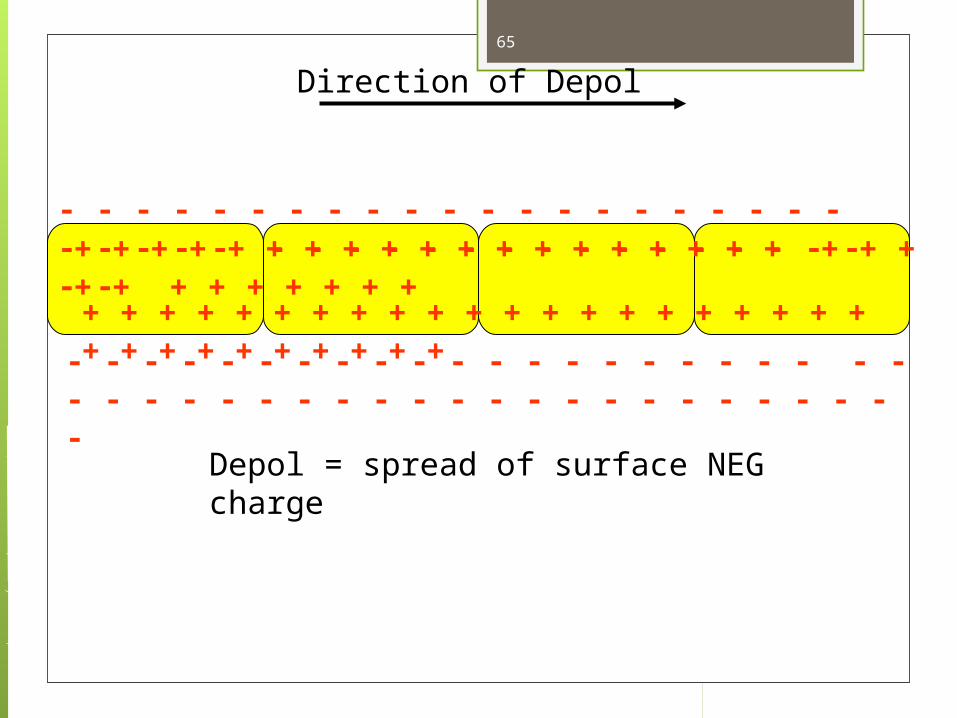
Direction of Depol64
- - - - - - - - - - - - - - - - - - - - - - - - - - - - - - - - - - - - - - - - - - - -
- - - - - - - - - - - - - - - - - - - - - - - - - - - - - - - - - - - - - - - - - - - - -
+ + + + + + + + + + + + + + + + + + + + + + + + + + + + + + +
+ + + + + + + + + + + + + + + + + + + + + + + + + + + + + + +
Direction of Depol
Depol = spread of surface NEG charge
65
- - - - - - - - - - - - - - - - - - - - - - - - - - - - - - - - - - - - - - - - - - - -
- - - - - - - - - - - - - - - - - - - - - - - - - - - - - - - - - - - - - - - - - - - - -
+ + + + + + + + + + + + + + + + + + + + + + + + + + + + + + +
+ + + + + + + + + + + + + + + + + + + + + + + + + + + + + + +
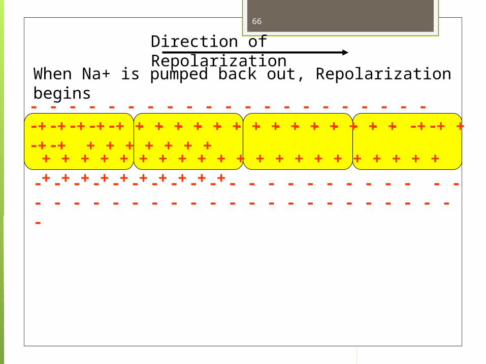
Direction of Repolarization
When Na+ is pumped back out, Repolarization begins
66

- - - - - - - - - - - - - - - - - - - - - - - - - - - - - - - - - - - - - - - - - - - -
- - - - - - - - - - - - - - - - - - - - - - - - - - - - - - - - - - - - - - - - - - - - -
+ + + + + + + + + + + + + + + + + + + + + + + + + + + + + + +
+ + + + + + + + + + + + + + + + + + + + + + + + + + + + + + +
Direction of Repolarization
- - -
67

+ + + + + + - - - - - - - - - - - - - - - - - - - - - - - - - - - - - - - - - - -
+ + + + + - - - - - - - - - - - - - - - - - - - - - - - - - - - - - - - - - - - - -
- - - - - - - + + + + + + + + + + + + + + + + + + + + + + + + + +
- - - - - - + + + + + + + + + + + + + + + + + + + + + + + + + + +
Direction of Repolarization68

+ + + + + + + + + + + + - - - - - - - - - - - - - - - - - - - - - - - - - - -
+ + + + + + + + + + - - - - - - - - - - - - - - - - - - - - - - - - - - - - - -
- - - - - - - - - - - - - + + + + + + + + + + + + + + + + + + + + + +
- - - - - - - - - - - + + + + + + + + + + + + + + + + + + + + + +
Direction of Repolarization69

+ + + + + + + + + + + + + + + + + + - - - - - - - - - - - - - - - - - - -
+ + + + + + + + + + + + + + + + + - - - - - - - - - - - - - - - - - - - -
- - - - - - - - - - - - - - - - - - - - - + + + + + + + + + + + + + + + +
- - - - - - - - - - - - - - - - - - - - - + + + + + + + + + + + + + + +
Direction of Repolarization70
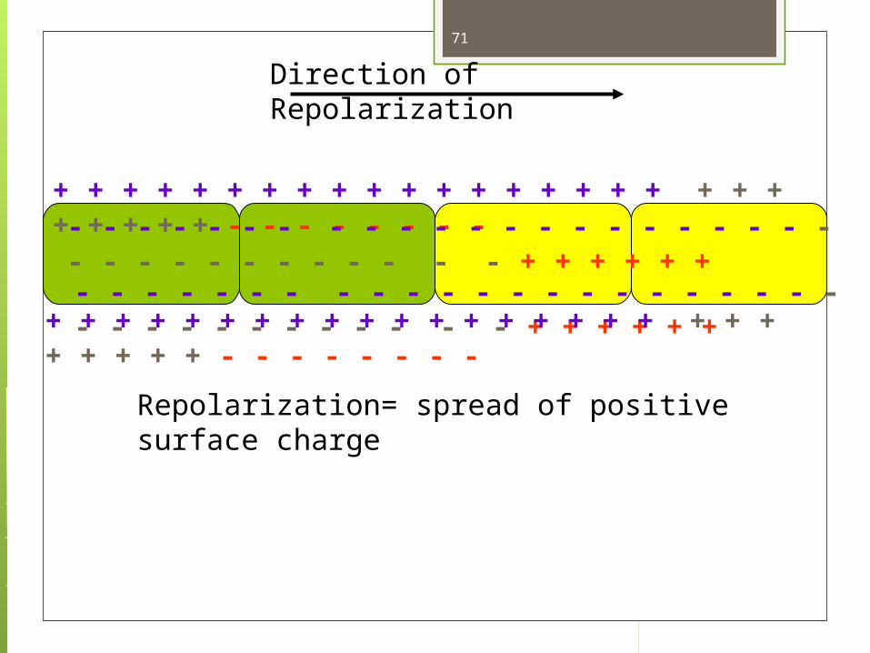
+ + + + + + + + + + + + + + + + + + + + + + + + + + - - - - - - - - - - - - - - - - - - - - - - - - - - - - - - - - - - - - - - - - - - + + + + + +
Direction of Repolarization
- - - - - - - - - - - - - - - - - - - - - - - - - - - - - - - - - - + + + + + +
Repolarization= spread of positive surface charge
+ + + + + + + + + + + + + + + + + + + + + + + + + + - - - - - - - -
71

+ + + + + + + + + + + + + + + + + + + + + + + + + + + + + + + - - - - - - - - - - - - - - - - - - - - - - - - - - - - - - - - - - - - - - - - - - -
Direction of Repolarization
- - - - - - - - - - - - - - - - - - - - - - - - - - - - - - - - - - - - - - - - - - + + + + + + + + + + + + + + + + + + + + + + + + + + + + + ++ +
72

- - - - - - - - - - - - - - - - - - - - - - - - - - - - - - - - - - - - - - - - - - -
Direction of Repolarization
- - - - - - - - - - - - - - - - - - - - - - - - - - - - - - - - - - - - - - - - - - + + + + + + + + + + + + + + + + + + + + + + + + + + + + + ++ +
+ + + + + + + + + + + + + + + + + + + + + + + + + + + + + ++ +
73

Excitable cells (neurons and muscles) are those that want this large electrical current to use for work.
They have proteins embedded in their cell membranes that are sodium channels (allow Na+ to pass into the cell for a short time after stimulation).
Muscle cells use the electrical force to contract, and neurons use it to excite the neurons touching them.
74

Excitable Cells Cells that can experience a momentary change in
membrane voltage are “excitable” cells (muscles and nerve cells)
That temporary change in voltage is due to a momentary change in permeability of Na+
The membrane, for only a moment, becomes more permeable to Na+ than to K+
When the outside and inside of the cell membrane reverse their charges (inside becomes positive and outside becomes negative), and then reverts back to normal, this process (depolarization + repolarization) is called an ACTION POTENTIAL.
The reversal of charges (action potential) is carried like a wave down the length of the cell, and into the next cell touching it, and so on.
75

ACTION POTENTIAL The action potential occurs when the membrane
potential (how negatively charged the inside of the cell is) reaches a certain threshold.
When the Na+ rushes into the cell, the membrane potential becomes less and less negative. Eventually, it reaches zero charge, and as more Na+ enters the cell, the inside of the cell becomes positively charged.
When it becomes positively charged enough (+30 mV), an action potential will sweep down the length of the cell membrane, like a wave of electricity. This is how one neuron stimulates a cell (a muscle cell, gland, or another neuron).
If the neuron stimulates a muscle cell, it contracts. If it stimulates a gland, it secretes. If it stimulates another neuron, the action potential is carried further along the nerve pathway, until it reaches the target organ.
Resting Membrane Potential VIDEO
76
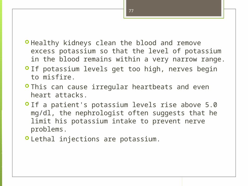
Healthy kidneys clean the blood and remove excess potassium so that the level of potassium in the blood remains within a very narrow range.
If potassium levels get too high, nerves begin to misfire.
This can cause irregular heartbeats and even heart attacks.
If a patient's potassium levels rise above 5.0 mg/dl, the nephrologist often suggests that he limit his potassium intake to prevent nerve problems.
Lethal injections are potassium.
77

The AP is a passive event: ions diffuse down their EC gradients when
gated channels open.A “wave of depolarization” occurs along the neighboring areas. Occurs in one direction along the axon; actually, AP regenerates over and over, at each point by diffusion of incoming Na+ ….WHY? Refractory period (Na+ channels become inactivated).Saltatory Conduction This type of conduction is found with myelinated axons. AP’s only occur at the nodes (Na channels concentrated here!) increased velocity energy conservation
Figure 5-17; Guyton & Hall
78http://www.youtube.com/watch?v=GTHWig1vOnY
Na/K ATPase Pump
http://www.brainviews.com/abFiles/AniSalt.htm
Saltatory Conduction
Other Types of Neurons: Supporting CellsNeurons are only one type of cell; there are others, which
are supporting cells, with a special name: GLIA (neuroglia, meaning “nerve glue”) are the
supporting cells of the nervous system. These are only brain cells that can reproduce. Since cancerous cells are those that reproduce, all brain tumors originate from glial cells.
There are five types of glial cells that we will cover: 1. Oligodendrocytes2. Schwann cells3. Astrocytes4. Microglia
5. Ependymal cells
79

Types of Glial Cells
1. OLIGODENDROCYTES (“few branches”). They are found in the CNS, are very large and complex cells. Oligodendrocytes form MYELIN SHEATHS in the CNS. This sheath is a covering around an axon to speed up the nerve conduction. Myelin is mostly made of lipid (fat).
80
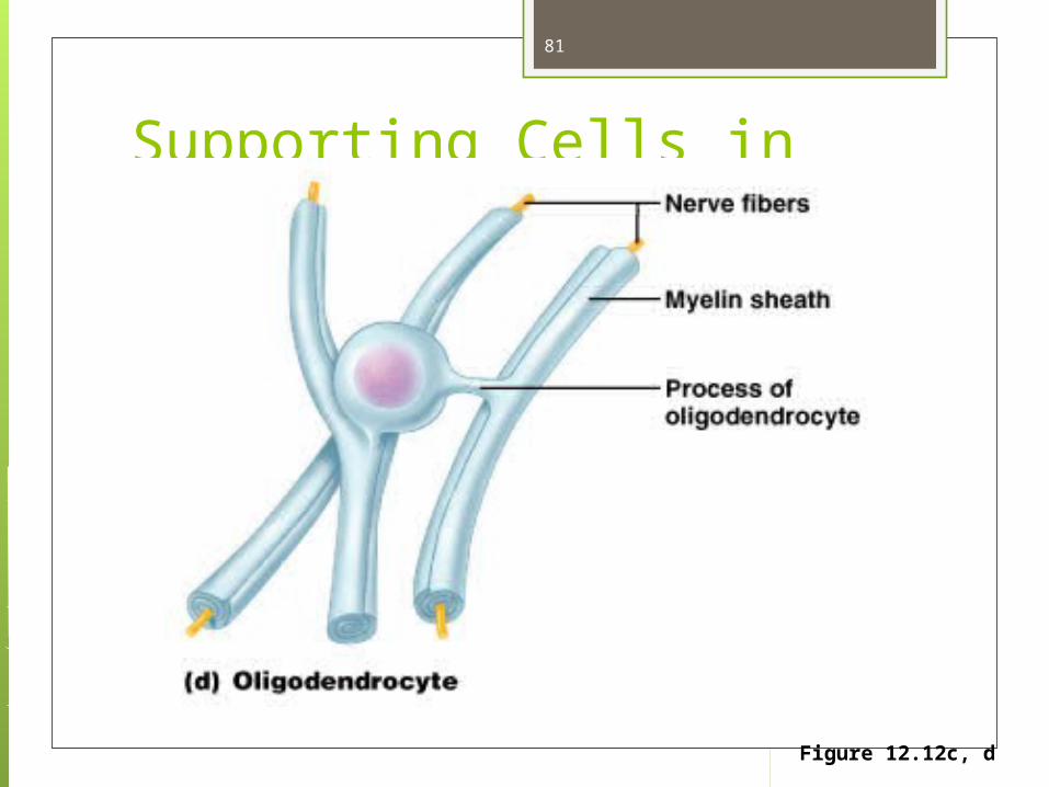
Supporting Cells in the CNS
Figure 12.12c, d
81

A sheet of paper is like an Oligodendrocyte, and it wraps itself around a pencil (axon), so there are many layers.
The myelin sheath is an electrical insulator. BETWEEN the sheaths are nodes = NODES OF RANVIER; these are BARE regions of axonal membranes only found in myelinated axons.
Nodes of Ranvier
82

The action potential jumps from one Node of Ranvier (the bare area) to the next Node, skipping the myelin that is between the bare areas. This speeds up the overall nerve conduction.
Therefore, a myelinated axon conducts impulses faster than an unmyelinated axon.
Why aren’t all neurons myelinated? Isn’t it good for everything to be faster?
No, myelin is a living cell, so it uses nutrients! We’d better save the myelin for where we
need it.
83

It is not necessary for things like digestion to start at 1/10 of a second rather than one second, or start sweating in 1/20 of a second rather than 1 second.
Walking and thinking are things that need to be quick.
84
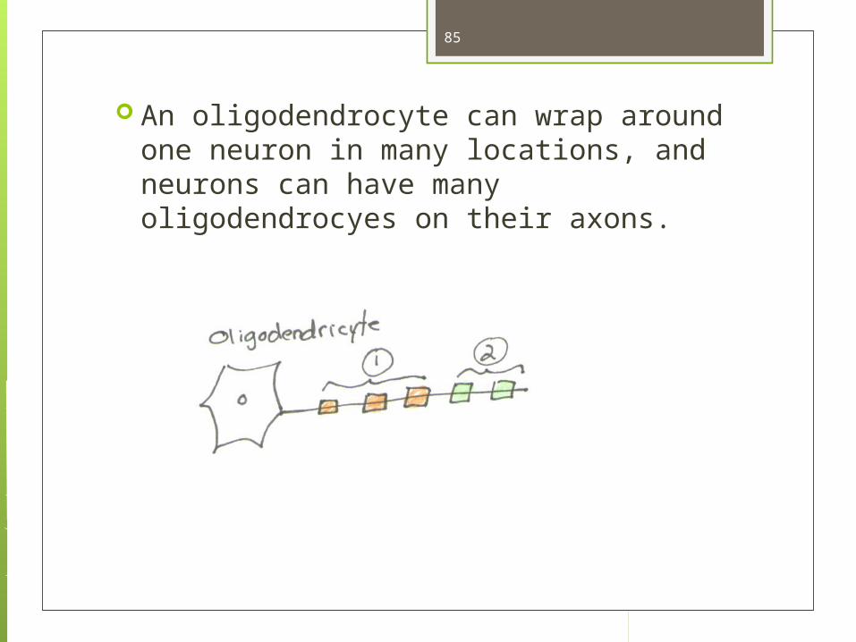
An oligodendrocyte can wrap around one neuron in many locations, and neurons can have many oligodendrocyes on their axons.
85

MULTIPLE SCLEROSIS is an autoimmune disease where the oligodendrocytes (the myelin sheaths) are destroyed (demyelination), interfering with the neuron functions in the CNS. Oligodendrocytes cannot regenerate.
MS is the most common neurological disease of young adults. Starts to manifest in late teens and early 20’s.
It progresses to paralysis and sometimes death. One in 1000 people get it. There are treatments, but
no cure.
86
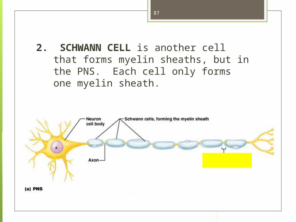
2. SCHWANN CELL is another cell that forms myelin sheaths, but in the PNS. Each cell only forms one myelin sheath.
87
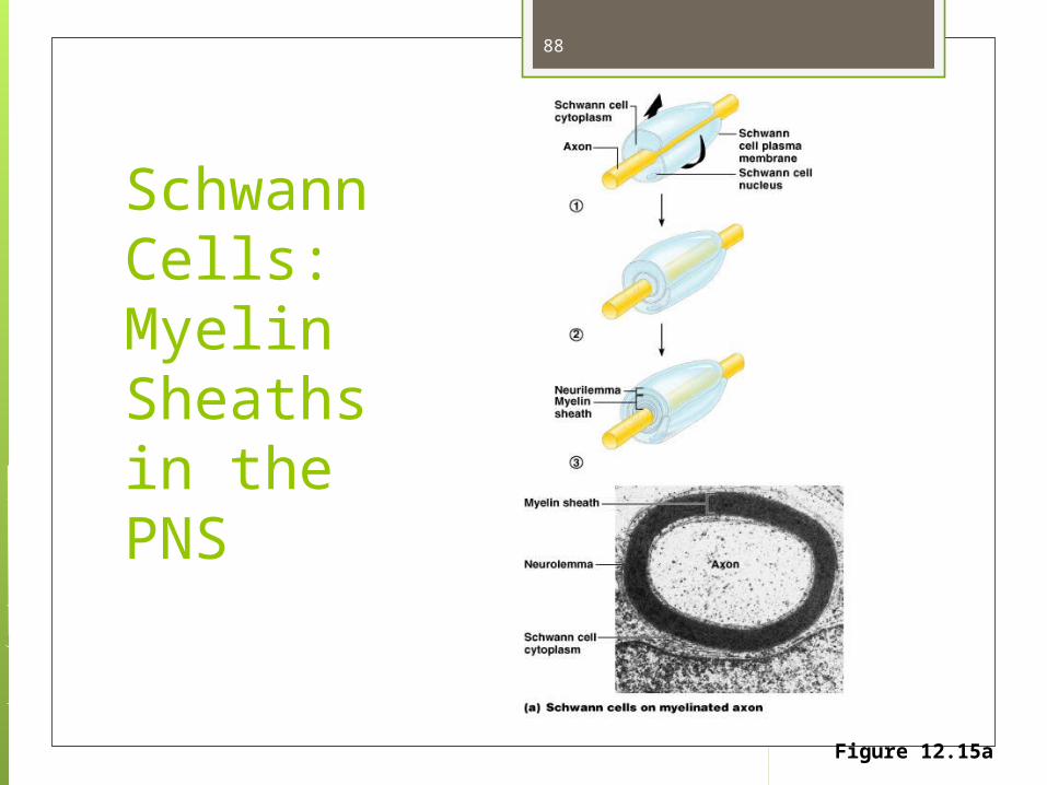
Schwann Cells:Myelin Sheaths in the PNS
Figure 12.15a
88
Neurofibromatosis (NF) Genetically-inherited disorder in which the
nerve tissue grows tumors (neurofibromas) that may be harmless or may cause serious damage by compressing nerves and other tissues.
The disorder affects Schwann cells and melanocytes.
Proliferation causes tumors and abnormal skin pigmentation.
The tumors may cause nothing but bumps under the skin and colored spots, or they may cause skeletal problems, pressure on spinal nerve roots, and other neurological problems.
89
Neurofibromatosis (NF)
Neurofibromatosis affects males and females equally and is dominant (only one copy of the affected gene is needed to get the disorder).
If only one parent has neurofibromatosis, the children have a 50% chance of developing the condition.
In half of cases there is no other affected family member because a new mutation has occurred.
90

Neurofibromatosis (NF)
Diagnostic Criteria Two or more neurofibromas Freckling of the groin or the axilla Six or more Café au lait spots (coffee with milk
colored pigment)
91

Neurofibromatosis (NF)
92
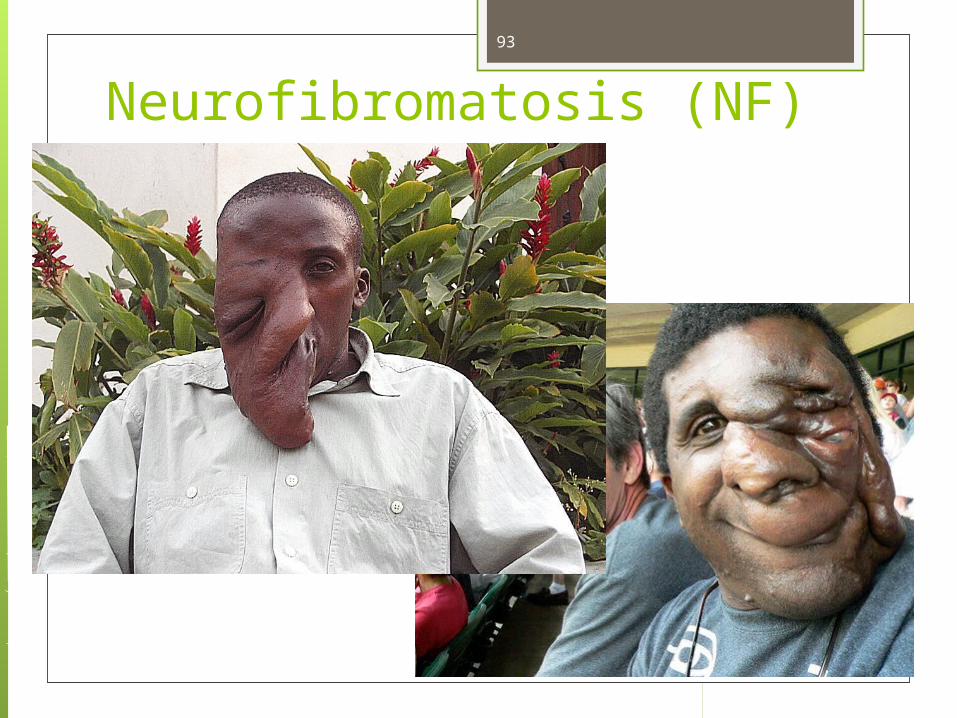
Neurofibromatosis (NF) 93

94

Café au lait spots95

One cell may form 3 bumps, another cell may form 2 bumps
One cell always forms only one bumps.
Difference between oligodendrocytes and Schwann cells
Found in CNS only Found in PNS only
96

3. ASTROCYTE is another very large, complex cell, in the CNS. Its function is to wrap around capillaries while it also is physically supporting and wrapping around neurons. Physically supports the neurons Transmits materials from capillaries to
neurons Forms blood-brain barrier (BBB)
97
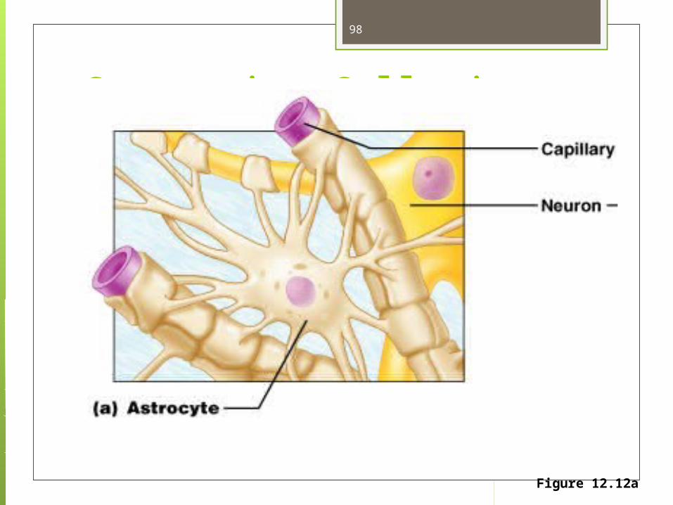
Supporting Cells in the CNS
Figure 12.12a
98
The BBB prevents a lot of certain types of materials from leaving the blood and entering the brain (e.g. hormones, drugs).
The brain still gets its nourishment from the blood, without the toxins.
The capillaries have leakage, but are surrounded by astrocytes, so not everything can leak out.
Certain antibiotics can’t cross the BBB, so they can’t be used for brain infections.
The only function of the blood-brain barrier is to help protect the central nervous system.
Blood-Brain-Barrier
99

Brain Tumors from NeurogliaAstrocytoma: tumor of the astrocytes 80% of Brain tumors are from
astrocytes They are graded from I to IV in severity, with IV
being the worst prognosis. The grades I and II have a good (80%) chance of
survival if treated by surgery, radiation, and chemotherapy. They tend to occur in 1/100,000 young people under age 20.
Grade IV survival rate is 3 months without treatment, and only about 12 months with treatment. They are more common in ages 65-75.
100
Symptoms of an astrocytoma tumor are a result of growing pressure inside the skull.
These symptoms include headache, vomiting, impaired balance, and mental status changes.
Other symptoms may occur, depending on the location of the tumor.
101
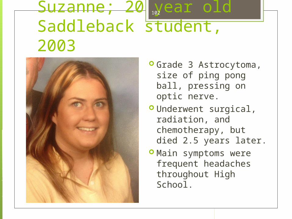
Suzanne; 20 year old Saddleback student, 2003
Grade 3 Astrocytoma, size of ping pong ball, pressing on optic nerve.
Underwent surgical, radiation, and chemotherapy, but died 2.5 years later.
Main symptoms were frequent headaches throughout High School.
102

4. MICROGLIA (one word, two errors!). These are not micro, nor are they glia.
They are macrophages, the same size as everywhere else in the body.
They are called micro because they are much smaller than real glia cells.
They pick up bacteria and dead cell, etc.
103

Supporting Cells in the CNS
Figure 12.12b
104
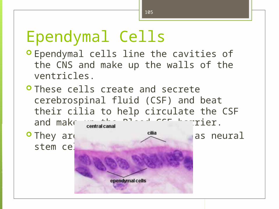
Ependymal Cells Ependymal cells line the cavities of the CNS
and make up the walls of the ventricles. These cells create and secrete cerebrospinal
fluid (CSF) and beat their cilia to help circulate the CSF and make up the Blood-CSF barrier.
They are also thought to act as neural stem cells.
105

TERMINOLOGY
WHITE MATTER: that portion of the CNS with myelinated axons; the myelin makes the area look white.
GREY MATTER: that portion of the CNS that is unmyelinated (cell bodies of neurons, some types of glia (neuroglia), and dendrites)
NERVE: collection of axons in the PNS. No cell bodies, dendrites, or synapses; just axons.
TRACT: collection of axons in the CNS. SYNAPSES: Where information is processed.
Most synapses are in the CNS
106

TERMINOLOGY GANGLION: A collection of cell bodies in the
PNS NUCLEI: A collection of cell bodies in the CNS NERVE PLEXUS: A network of nerves (nerves
don’t run by themselves, they go in groups) WALLERIAN DEGENERATION: process that
results when a nerve fiber is cut or crushed, in which the part of the axon separated from the neuron's cell body degenerates distal to the injury.
107

Ganglion: Collection of cell bodies in the PNS
108Figure 12.11

Nerve Regeneration In humans, axons outside the brain and spinal cord can
regenerate, but not those inside. After injury, axons in the human central nervous system degenerate, resulting in permanent loss of nervous function. This is not so in cold water fish and amphibians, where axon regeneration in the central nervous system does occur.
So far, investigators have identified several proteins that seem to be necessary to axon regeneration in these central nervous system of these animals, but it may be a long time before biochemistry can offer a way to bring about axon regeneration in the human central nervous system.
It is possible though, that one day these proteins will become drugs or that gene therapy might be used to cause humans to produce the same proteins when such injuries occur.
109
Nerve Regeneration
In the meantime, some accident victims are trying other ways to bring about a cure. In 1995, Christopher Reeve, best known for his acting role as Superman, was thrown headfirst from his horse, crushing the spinal cord just below the neck’s top two vertebrae. Immediately, his brain lost almost all communication with the portion of his body below the site of damage and he could not move his arms and lags.
Many years later, Reeve could move his left index finger slightly and could take tiny steps while being held upright in a pool. He had sensation throughout his body and could feel his wife's touch.
110

Nerve Regeneration Reeves improvement was not the result of
cutting edge drugs or gene therapy-- it was due to exercise. Reeves exercised as much as five hours per day, especially using a recombinant bike outfitted with electrodes that made his leg muscles contract and relax.
The bike cost him $16,000. It could cost less if commonly used by spinal cord injury patients in their own homes. Reeves, who was an activist for the disabled, was pleased that insurance would pay for the bike about 50% of the time.
111

Nerve Regeneration It's possible that Reeves advances were the result of
improved strength in bone density, which led to stronger nerve signals.
Normally, nerve cells are constantly signaling one another, but after a spinal cord injury, this signal ceases. Perhaps Reeves’ intensive exercise brought back some of the normal communication between nerves. His physician is convinced that his axons were regenerating. Reeves was convinced that stem cell therapy would one day allow him to be off his ventilator and functioning normally; however, Reeve died in 2004.
So far, researchers have shown that both embryonic stem cells and bone marrow stem cells can differentiate into neurons in the laboratory. Bone marrow stem cells apparently can also become neurons when injected into the body.
112


![CENTRAL NERVOUS SYSTEM [CNS] TUTORIAL DISCUSSION](https://static.documents.pub/doc/80x56/5681368e550346895d9e19c6/central-nervous-system-cns-tutorial-discussion.jpg)


