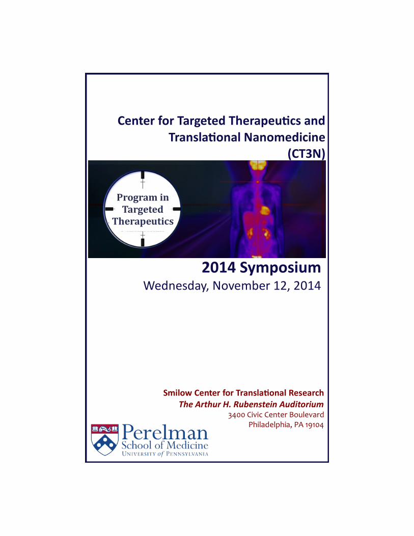
Smilow Center for Translaonal Research The Arthur H. Rubenstein Auditorium 3400 Civic Center Boulevard Philadelphia, PA 19104 Program in Targeted Therapeutics Center for Targeted Therapeucs and Translaonal Nanomedicine (CT3N) 2014 Symposium Wednesday, November 12, 2014
Transcript
Smilow Center for Translational Research The Arthur H. Rubenstein Auditorium
3400 Civic Center Boulevard Philadelphia, PA 19104
Program in Targeted
Therapeutics
Center for Targeted Therapeutics and Translational Nanomedicine
(CT3N)
2014 Symposium Wednesday, November 12, 2014
8:15 AM Registration and Breakfast: SCTR Lobby 8:45 AM Welcome & Introduction: SCTR Auditorium
Vladimir Muzykantov, MD, PhD, Professor of Pharmacology and Medicine, Vice-Chair Department of Pharmacology, Director, Center for Targeted Therapeutics and Translational Nanomedicine, University of Pennsylvania
Session 1: Keynote Lecture
Session Chair: Andrew Tsourkas, Ph.D. Associate Professor of Bioengineering
9:00 AM Opening the Intracellular Universe to Biologic Drugs
Patrick Stayton, PhD, Professor, Department of Bioengineering Director, Molecular Engineering & Sciences Institute, University of Washington Session 2: Drug Delivery for Treatment of Vascular and Metabolic Diseases
Session Chair: David Eckmann, M.D., Ph.D. Professor of Anesthesiology, University of Pennsylvania
10:00 AM Targeted Drug Delivery via Inflammatory Cues Debra Auguste, Ph.D., Associate Professor of Biomedical Engineering, City College of New York 10:30 AM Targeted Drug Delivery by Natural or Artificial Nano- and Microsized Carriers PD Dr. Hans Bäumler, Associate Professor, Head of the Research Department, Institute of Transfusion Medicine, Charité – University Medicine Berlin
11:00 AM “Eat-Me" Imaging of Macrophages in Cardiovascular Disease Andrei Maiseyeu, PhD Assistant Professor of Medicine, University of Maryland, School of Medicine 11:30 AM Novel Supramolecular Materials Designed to Remediate the Cholesterol Metabolism Disorder in Niemann-Pick Type C Disease David Thompson, PhD, Professor, Department of Chemistry and Weldon School of Biomedical Engineering, Purdue University 12:00 PM Lunch: SCTR Lobby
Agenda
Session 3: Keynote Lecture Session Chair: Theresa Busch, Ph.D., Associate Professor of Radiation Oncology, University of Pennsylvania 1:00 PM Therapeutic Polymer Nanoparticles Designed for Treatment of Pulmonary and Urinary Tract Diseases Karen Wooley, PhD, W.T. Doherty-Welch Chair in Chemistry University Distinguished Professor, Texam A&M Univeristy, Director, Laboratory of Synthetc-Biologic Interactions Session 4: New Carriers and Cancer Session Chair: Michael Chorny, Ph.D., Assistant Professor of Pediatrics, Children’s Hospital of Philadelphia 2:00 PM Translational Targeted Oncologial Agents Michelle Bradbury, MD, PhD, Department of Radiology, Memorial Sloan Kettering Cancer Center, Associate Professor of Radiology, Weill Medical College of Cornell University
2:30 PM Nanotechnology Approaches for Personalized Treatment of Cancer Tamara Minko, PhD, Distinguished Professor and Chair, Department of
Pharmaceutic, Ernest Mario School of Pharmacy
3:00 PM Therapeutic Nanomaterials made from Proteins Julie A. Champion, PhD, Assistant Professor, School of Chemical & Biomolecular Engineering, Georgia Institute of Technology 3:30 PM Using DNA to Deliver Drugs: The 3DNA Nanotechnology Platform Robert Getts PhD, Vice President, Research and Development, Genisphere Session 5: Panel “Matching materials with biology” Session Chair: Dennis Discher, Ph.D., Professor of Chemical & Bimolecular Engineering, University of Pennsylvania 4:00 PM All Panel Participants
5:00 PM Reception: SCTR Lobby
Opening the Intracellular Universe to Biologic Drugs Patrick Stayton, PhD, Professor, Department of Bioengineering Director, Molecular Engineering & Sciences Institute, University of Washington
Synthetic polymeric delivery systems for protein drugs have been developed that mimic the highly efficient intracellular delivery systems found in pathogenic viruses and organisms. The carriers possess a hidden functionality that is expressed in the endosomal compartment to increase cytosolic delivery of macromolecules. The ampholytic carriers are designed like these pathogens to activate via protonation events triggered in the endosome. This endosomal-releasing activity is then built into a multi-functional polymer platform that incorporates targeting elements, conjugation or complexation elements, and a “stealth” component to optimize safety and pharmaco-kinetic properties. Controlled polymerization techniques are exploited to streamline bioconjugation of targeting agents and therapeutics, as well as to generate controlled carrier architectures. These drug delivery systems have been applied to protein therapeutics in cancer immune-therapy and RNA/DNA nucleic acid drug development. Targeted Drug Delivery via Inflammatory Cues Debra Auguste, Ph.D., Associate Professor of Biomedical Engineering, City College of New York
Cells sense changes in their environment and respond by altering their behavior. We investigate how cells manipulate their membrane surface chemistry, which has profound effects on disease progression. Cells orchestrate the density and organization of proteins and lipids to govern adhesion and migration. From this knowledge, we can engineer drug delivery vehicles that complement the molecular patterns observed on cells to achieve strong, cooperative binding. We have employed these strategies in a model system of endothelial inflammation and in breast cancer metastasis. Our multi-targeting drug delivery platform may identify new strategies in personalized medicine. Targeted Drug Delivery by Natural or Artificial Nano and Microsized Carriers PD Dr. Hans Bäumler, Associate Professor, Head of the Research Department, Institute of Transfusion Medicine, Charité – University Medicine Berlin
The red blood cells (RBC) represent a potential system to carry drugs to the desired site of therapeutic action. Additionally, RBC provide an extraordinary vehicle for the dissemination of drugs in the circulation. This carrier system is biocompatible, non-immunogenic, has a long life-span and a large capacity. Targeting of organs or tissues by RBC loaded with magnetite NPs can be achieved by magnetic focusing. Moreover, magnetite loaded RBC can be visualized by MRI, offering the opportunity for diagnostic monitoring of the therapy. Further opportunities are offered by conjugation of specific antibodies to the surface of RBC or by combination of magnetic and receptor targeting. In addition, RBC carriers can deliver high dosage of different drugs protecting them from inactivating reactions and minimizing side effects. As model drugs we used 5-fluorouracil (5 FU) and fluorouracil acetate (FUAC).
Another very new challenge consists in triggering the drug release at a desired site with defined time sequence and quantities. Opto-nanoporation has the potential to find application as a new tool for studying stimuli-responsive cross-membrane transport of molecules. We studied the permeability changes of the nanoparticle-functionalized membrane of RBCs upon laser irradiation and report on the laser-assisted controlled release of model drugs encapsulated in the RBC interior. Simultaneous release of two molecules, the dye 5(6)-carboxyfluorescein (5(6)-CF) and a rhodamine-labeled dextran (Rh-dextran), with different molecular masses of 376 and 7500 Da, respectively, was conducted. Practically, our approach is relevant for delivery of a combination of drugs, which is often needed, for example, in the treatment of cancer.
(continued)
Abstracts (Alphabetical order by speaker’s last name)
Last but not least the fabrication of biocompatible micro- and nanoparticles has attracted a widespread interest due to their potential application in biotechnology as tools for catalysis, sensing and separation and in medicine as systems for drug delivery, diagnostics and in vivo imaging. An increasing number of studies has been dealing with multicompartment systems motivated by the need of multifunctional, controllable and triggered carriers that are desired for example in drug delivery or as bioreactors and biosensors. Multi-compartmentalization is envisioned to be the next step of development in the area of drug carriers, due to possibilities of simultaneous delivery of various substances in one vehicle but separated spatially. Here a simple and inexpensive concept for the fabrication of single and multi-compartment particles fully made of biomacromolecules is presented. The technique is based on the process of co-precipitation of biopolymers with an inorganic salt, followed by one or several cross-linking procedures and dissolution of the inorganic support. We prepared particles with several concentrically arranged compartments placing up to three cou-pled enzymes in own compartments Visualizing the first and the last enzyme in a three enzyme reaction by fluorogenic substrates and confocal laser scanning microscopy the influence of the spacing between com-partments containing different enzymes on the reaction kinetics can be demonstrated on the microscopic scale within one microparticle. "Eat-Me" Imaging of Macrophages in Cardiovascular Disease Andrei Maiseyeu, PhD Assistant Professor of Medicine, University of Maryland, School of Medicine
Cardiovascular complications from obesity and type 2 diabetes are the most important cause of disease morbidity and mortality. The main driver of these arises secondary to inflammation in the arterial wall and adipose tissue and is characterized by a stereotypic infiltration of monocytes and T lymphocytes into the vascular wall. It has been postulated that targeting inflammation in conjunction with accurate imaging of detrimental cells would significantly enhance and likely revolutionize treatment, particularly if one agent simultaneously accomplished these goals. We have developed and tested in vivo multifunctional nanoparticle (NP) agents for inflammation imaging and therapy. We present an elegant layer-by-layer NP synthesis methodology that allows for facile fabrication and incorporation of various imaging probes and disease-specific therapeutics. Targeted delivery of NPs to inflammatory macrophages is achieved by means of “eat-me” signals such as phosphatidylserine and oxidized cholesterol derivatives. We show that these signals able to convert any NP vehicle into highly-desirable "meals" for the macrophages at sites of inflammatory disease (e.g. atherosclerotic plaque or inflamed adipose). The “eat-me” signals not only facilitate the engulfment of the NPs, but also permit the identification of plasticity of polarized macrophages (M1 vs M2 subtype). Upon exposure to inflammatory stimuli, these NPs are able to release the therapeutic cargo within the macrophage, resulting in decreased cell activation and release of proinflammatory mediators. These NPs are additionally coupled with gadolinium and fluorochrome probes allowing for their imaging through MRI or fluorescence. Our results suggest that this theranostic (therapy+diagnostic) technology can serve as a platform with application in various inflammatory diseases such as cardiovascular disease, diabetes, obesity and cancer. Novel Supramolecular Materials Designed to Remediate the Cholesterol Metabolism Disorder in Niemann-Pick Type C Disease David Thompson, PhD, Professor, Department of Chemistry and Weldon School of Biomedical Engineering, Purdue University 1‡ Yawo Mondjinou, 1‡ Chris Collins, 2Aditya Kulkarni, and 1,2David H. Thompson* These authors contributed equally to this work. 1Purdue University, Department of Chemistry, Bindley Biosciences Center
(continued)
(continued)
1203 W. State Street, West Lafayette, IN 47907 and 2Aten Biotherapeutics LLC, 230 Spring Valley Lane, West Lafayette, IN 47906 A growing body of evidence suggests that hydroxypropyl-b-cyclodextrin (HP-b-CD) and it’s homologs are capable of inhibiting cholesterol biosynthesis, mobilizing the aberrant pool of lysosomally-associated cholesterol, and effectively reversing the symptoms of the rare disease, Niemann-Pick Type C (NPC), in npc-/- mice. Unfortunately, the in vivo persistence of HP-b-CD derivatives is brief, with >90% clearance within 24 hrs after a 4,000 mg/kg subcutaneous injection in mice. In an attempt to address this limitation, we have prepared a family of high molecular weight, non-covalent HP-b-CD polyrotaxane contructs derived from Pluronic® surfactants and 2-hydroxypropyl-b-cyclodextrin derivatives that produce polyrotaxanes with different relative block lengths and molecular weights ranging between 1.9 - 12.6 kD [1,2]. These compounds carry multiple copies of HP-b-CD (or b-CD) as shown by 1H NMR, GPC-MALS, DOSY, and analytical ultracentrifugation analysis. Filipin staining data suggests that these compounds mobilize aberrantly stored cholesterol in both NPC2-/- and NPC1-/- fibroblasts on an equivalent per mole HP-b-CD basis. Control experiments suggest that the cholesterol mobilization enabled by these materials does not depend on low pH or glutathione activation; we infer from these findings that enzyme activation of the polyrotaxane prodrug occurs within the late endosome/lysosome compartment. To evaluate their potential for long circulation in the blood, Gd3+:DOTA-HP-b-CD:Pluronic polyrotaxanes were synthesized and their blood pool contrast properties characterized. These constructs were found to circulate for more than 30 min and provide substantial vascular enhancement relative to the monomeric Gd3+:DOTA-HP-b-CD control that is rapidly cleared via the kidney [3]. The high r1 relaxivity at 37 °C (23.83 mM-1s-1 at 1.5T), extended blood circulation, and well-known pharmacology of the polyrotaxane precursors make it a highly attractive candidate for biodegradable blood pool contrast agents. ICP-MS findings indicate that a nearly 10% of the injected material remains in circulation 24 h after tail vein injection in normal Balb/c mice in some cases, although this is dependent on polyrotaxane threading extent and average molecular weight. References [1] Y. A. Mondjinou, L. McCauliff, A. Kulkarni, L. Paul, S.-H. Hyun, Z. Zhang, Z. Wu, M. Wirth, J. Storch, D. H. Thompson, “Synthesis of 2-Hydroxypropyl-b-Cyclodextrin and Pluronic Based Polyrotaxanes in Heterogeneous Reactions for Niemann-Pick Type C Therapy”, Biomacromolecules 2013 14, 4189-4197. [2] C. J. Collins, L. McCauliff, S.-H. Hyun, Z. Zhang, L. N. Paul, A. Kulkarni, K. Zick, M. Wirth, J. Storch, D. H. Thompson, “Synthesis, Characterization, and Evaluation of Pluronic-based b-Cyclodextrin Polyrotaxanes for Mobilization of Accumulated Cholesterol from Niemann-Pick Type C Fibroblasts”, Biochemistry 2013 52, 3242-3253. [3] A. Kulkarni, Z. Zhou, Y. Mondjinou, C. Collins, V. Badwaik, C. K. Yerneni, J. Garst, M. J. Wirth, Z.-R. Lu, D. H. Thompson, “Gd3+-DOTA-2-Hydroxypropyl-b-Cyclodextrin:Pluronic Polyrotaxane as a Long Circulating, Degradable High Relaxivity MRI Contrast Agent”, submitted. Translational Targeted Oncologial Agents Michelle Bradbury, MD, PhD, Department of Radiology, Memorial Sloan Kettering Cancer Center, Associate Professor of Radiology, Weill Medical College of Cornell University
Despite recent advances in imaging probe development for biomedicine, the translation of targeted diagnos-tic platforms remains challenging. Nanomaterials platforms currently under evaluation in oncology clinical trials are largely non-targeted drug delivery vehicles or devices to thermally treat tissue; these are not typi-cally surface modified for direct detection by clinical imaging tools. New tumor -selective platforms need to satisfy critical safety benchmarks, in addition to assaying targeted interactions with the microenvironment and their effects on biological systems. Metabolic imaging and analysis tools, such as PET, are essential for providing complete and quantitatively accurate data sets for whole body distributions, targeting kinetics, and clearance profiles of new diagnostic platforms undergoing preclinical testing or
Abstracts (continued)
(continued)
transitioning into early-phase clinical trials. We have applied these methods to a previously described novel class of renally-cleared, fluorescent silica-based core-shell nanoparticles, Cornell dots (C dots). Surface-modification of dye-encapsulated C dots with radiolabels and integrin-binding ligands has resulted in a selective platform5 for cancer detection, staging, and targeted therapy using dual-modality imaging. The results of these studies have led to a first-in-human clinical trial, which is nearing completion. We have extended these approaches to therapeutic particle tracer preparations, and propose that PET imaging can be used to serially monitor drug delivery, localize lesions, and extract key biologic properties needed to establish treatment planning protocols for individual tumors. Nanotechnology Approaches for Personalized Treatment of Cancer Tamara Minko, PhD, Distinguished Professor and Chair, Department of Pharmaceutic, Ernest Mario School of Pharmacy
Authors: Justin Sapiezynski1, Peter Zhou1, Oleh Taratula1, Vatsal Shah1, Min Zhang1, Jue Gong1, Shali John1, Olga B. Garbuzenko1, Lorna Rodriguez-Rodriguez2,3 and Tamara Minko1,2,4
Affiliations: 1Department of Pharmaceutics, Ernest Mario School of Pharmacy, Rutgers, The State University of New Jersey, Piscataway, NJ 08854; 2Rutgers Cancer Institute of New Jersey, New Brunswick, NJ 08903; 3Department of Obstetrics and Gynecology, Robert Wood Johnson Medical School, Rutgers, The State University of New Jersey, Piscataway, NJ 08901; 4Environmental and Occupational Health Sciences Institute, Piscataway, NJ 08854. Purpose: To study the expression of genes involved in drug resistance, growth, signaling pathways and development of metastasis in sensitive and resistant ovarian cancer cells and select targets for the personalized selection of nanotherapeutics for cancer treatment. Methods: The expression of genes from three commercially available gene expression panels was analyzed (total 191 distinct genes) by the quantitative reverse transcription polymerase chain reaction (qPCR). The expressions of the genes were compared between cells isolated from normal human ovaries, from primary and metastatic ovarian tumors isolated from tissues of patients with ovarian carcinoma, and in drug sensitive A2780 and multidrug resistant A2780/AD human ovarian carcinoma cells. Based on the results of the measurements, several genes were selected as siRNA targets for in vitro and in vivo studies. siRNAs were delivered by liposomes or dendrimers together with anticancer drugs (doxorubicin, paclitaxel and/or cisplatin). Results: Based on the results obtained, 83 candidate genes (the size of a qPCR plate) were selected for future analysis. The significance of genes selected in A2780 cells obtained from ATCC was confirmed by the measurement of gene expression in cells isolated from primary tumors and malignant ascites obtained from a patient with advanced metastatic ovarian carcinoma. Genes from nine functional groups responsible for angiogenesis, apoptosis (inducers of cell death and antiapoptotic cellular defense), cell cycle regulation, DNA damage, drug resistance, detoxification of anticancer drugs, signal transduction, and transcription factors were analyzed. Gene expression in each group in drug sensitive and resistant human ovarian carcinoma cells was compared in order to determine genes mainly responsible for the development of drug resistance and tumor progression. Based on such comparison, nine genes were selected as prospective targets for the future suppression of drug resistance and enhancement the efficacy of treatment. Liposomal and dendrimer-base delivery systems were constructed and evaluated. It was found that simultaneous suppression of targeted mRNAs and induction of cell death by anticancer agents significantly enhanced the efficacy of treatment of primary tumor and intraperitoneal metastases. Conclusions: The data obtained allowed for identifying major proteins responsible for multidrug resistance in ovarian cancer cells in an individual patient and selecting a reasonable number of genes/proteins as potential therapeutic targets for combinational gene and chemotherapy in order to enhance the efficiency of cancer therapy and prevent the development of metastasis. Acknowledgements: The research was supported in part by R01 CA138533 from the National Cancer Institute.
(continued)
Abstracts Therapeutic Nanomaterials made from Proteins Julie A. Champion, PhD, Assistant Professor, School of Chemical & Biomolecular Engineering, Georgia Institute of Technology
Protein drugs can provide a key advantage over small molecule drugs; they evolved to perform their function, while small molecules are often selected for “best” function compared to a pool of candidates. However, proteins can present challenges in delivery that must be overcome in order to be used as therapeutic drugs. Their folded structure is critical to their biological function and makes them sensitive and difficult to package. However, this structure also provides an opportunity to create nanomaterials from them that is not available for small molecules. The main goal of our work is to engineer nanomaterials made from therapeutic proteins and this is accomplished through a combination of self-assembly and/or bio-conjugation processes. The ability to control these processes is essential to manipulating material physical properties, ensuring retention of protein activity, and directing the interactions between the materials and cells. The strategies developed here provide opportunities to work with unlikely proteins, such as those from pathogenic bacteria, and transform them from disease causing agents into beneficial therapeutic materials. Protein design, self-assembly and disassembly properties, and applications of therapeutic protein nanomaterials in cancer, inflammation and vaccines will be discussed Using DNA to Deliver Drugs: The 3DNA Nanotechnology Platform Robert Getts PhD, Vice President, Research and Development, Genisphere
A variety of materials have been used to synthesize and create nanoparticles for attachment of drugs in order to improve their therapeutic index for the treatment of cancer and other diseases. DNA as a nanotechnology matrix offers a unique set or physical, chemical, and biological properties, thus opening up a new strategy for the development of a multivalent therapeutic delivery molecule. Genisphere's 3DNA platform is composed entirely of noncoding DNA that has been assembled through the sequential hybridization of single strands of DNA into a network of double stranded nucleic acid having a controlled architecture (layers) and multiple attachment sites for both drug as well as targeting molecules. For drug delivery, Genisphere customizes two-layer and four-layer 3DNA scaffolds with a variety of drug cargos, targeting moieties, and tracking labels. 3DNA nanostructures have been used for in vitro and in vivo drug delivery in the fields of oncology, ophthalmology, and others. An overview of this new and exciting drug delivery nanotechnology will be presented.
Notes
Notes
Notes
Our Sponsor:
Supported in part by the Institute for Translational Medicine and Therapeutics of the University of Pennsylvania. The project described was supported in part by the National Center for Research Resources, Grant UL1RR024134, and is now at the National Center for Advancing Translational Sciences, Grant UL1TR000003. The content is solely the responsibility of the authors and does not necessarily represent the official views of the NIH.
CT3N Symposium Organizing Committee:
Dennis Discher, PhD, Robert D. Bent Professor of Chemical Engineering, University of Pennsylvania
Vladimir Muzykantov, MD, PhD, Professor of Pharmcology and Medicine, Vice Chair, Department of Pharmcology,
Director, Center for Targated Therapeutics and Translational Nanomedicine, University of Pennsylvania
Robert Levy, PhD, Professor of Pediatrics, Children’s Hospital of Philadelphia
Andrew Tsourkas, PhD, Associate Professor of Bioengineering, University of Pennsylvania
Michael Chorny, PhD, Assistant Professor of Pediatrics, Children's Hospital of Philadelphia


![spot registered list · 2020. 12. 14. · o v E }E u } u u µ v ] Ç î í ì ì ì í ìDh, DD /> Dh î í ì ì ì ð î ,/Z D E ^ î í ì ì ì ð ñ EE/ ZK^ d,KD ^ î í ì ì](https://static.documents.pub/doc/80x56/60bfa82743392f54e03262b4/spot-registered-list-2020-12-14-o-v-e-e-u-u-u-v-.jpg)
![D ^ } ] o ^ À ] o v l } } W } µ } Á o l h ð l î ô l î ì í ... · ì î ð ï d } ] o ] o r ì î ð ð d } ] o ] r ì î ð ñ d µ } } o } v Z r ì î ð ò d v ( µ ] o](https://static.documents.pub/doc/80x56/5e1bf26a711c79513c1ec638/d-o-o-v-l-w-o-l-h-l-l-.jpg)

![E^d/dhdK WK>/d E/ K E /KE >€¦ · ì ì î l î ì î í r í î ì i µ o ] } î ì î ì w p ] v î ð (vwxglrv gh 1lyho 6xshulru 3huilo uhtxhulgr ,qjhqlhur 0hfiqlfr ,qjhqlhur](https://static.documents.pub/doc/80x56/5f82cc623aa1ba7b2566f3db/eddhdk-wkd-e-k-e-ke-l-r-i-o-.jpg)

![D } µ / î í r î ì ð ] P } î ì í ñ · 2016-04-16 · Soosan Jacob, FRCS, MS , J. Bradley Randleman, MD and Alaa M. El-Danasoury, MD D } µ / î í r î ì ð ] P } î ì](https://static.documents.pub/doc/80x56/5b20af447f8b9ae41a8b50ff/d-i-i-r-i-i-d-p-i-i-i-n-2016-04-16-soosan-jacob-frcs.jpg)

![î ì í ó ] ] } v - fivb.org · ï ó r ð ð , Wd Z í ì r ^ KZ Z^ [ ^ ^ í ì X í í ì X î ô ð ñ r ð õ W Zd /// r d ZD/EK>K'z](https://static.documents.pub/doc/80x56/5b307d0a7f8b9a91438db3f4/i-i-i-o-v-fivb-i-o-r-d-d-wd-z-i-i-r-kz-z-i-i.jpg)


![} v } u ] Z À ] } ( } ] Ç · 2018. 11. 28. · s Z ' î ì í í î ì í î î ì í ï î ì í ð î ì í ñ î ì í ò z î ì í í î ì í î î ì í ï î ì í ð î](https://static.documents.pub/doc/80x56/601e1b37d8b1f16a0800f14a/-v-u-z-2018-11-28-s-z-.jpg)
!['ZKD < U í õ o ] î ì î ì · î õ í ìdhD/BKt/ ^ Ç o Á ] < î ì Z > ò ð r^dK ð ò ì ñ ì ì õ ò ì X ì ì ï ñ í ì< D Z < t } v ] l < î ì](https://static.documents.pub/doc/80x56/5f67527405909476d76ae24d/zkd-u-o-dhdbkt-o-z-.jpg)
![12-13 MC Handbook with solutions€¦ · î ì í î D d, KhEd^ & } µ v } v í ð î ì < ] v P ^ U o Æ v ] U s î î ï í ð ó ì ï r î õ õ r õ ì ì ò Á Á Á X u Z }](https://static.documents.pub/doc/80x56/5eabf3e840a2bb22c76f291c/12-13-mc-handbook-with-solutions-d-d-khed-v-v-.jpg)
![New o } Ç } , } ] Ì } v î ì ð ì · 2018. 4. 10. · o } Ç } , } ] Ì } v î ì ð ì } µ u v } / v À ] P ] v í ï l î ì í ó humana y el telos de la Norma Suprema, y](https://static.documents.pub/doc/80x56/604dc6b4a976f05d4238a43b/new-o-oe-v-2018-4-10-o-oe-v-.jpg)