51
The Skeletal System Chapter 6-9
| Date post: | 16-Dec-2015 |
| Category: |
Documents |
| Upload: | basil-harvey |
| View: | 223 times |
| Download: | 1 times |
The Skeletal System
Chapter 6-9
Individual bones: Tissues
Bone Cartilage Epithelial tissue Fibrous connective tissue Blood Nervous tissue
Bone Structure Long bone
External structure Epiphysis, the portion of the
bone that interacts with other bones
Articular cartilage, protective layer of hyaline cartilage coating the epiphysis
Hyaline cartilage, most common type of cartilage, looks like “milky glass”
Diaphysis, long narrow portion between epiphyses, AKA: shaft
Periosteum, any part of the bone not covered by articular cartilage is protected by this tough fibrous membrane. It is a vascular tissue, functions in bone repair.
Internal structure Tissue types:
Compact bone, dense strong and resistant to bending
Spongy bone, branching bony plates that create irregular spaces, strong and durable because they allow for some compression.
Tissue locationCompact bone makes
up the wall of the diaphysis
Spongy bone makes up the bulk of the epiphysis especially in regions that are subject to compression, (weight bearing joints)
Description: the diaphysis is a semi-rigid hollow structure Medullary cavity,
hollow tube runs from end to end in the open space of the diaphysis
Endosteum, lines the cavity
Marrow, specialized, highly functional, soft CT, inside the cavity
Microscopic Structure:Osteocyte, individual compact bone cell, located in…
Lacunae, chambers arranged in concentric circles around…
Osteonic canal, pathway for arteries, veins, and nerves through the bone
Canaliculus, smaller canals running perpendicular to the osteonic canal, connecting lacunae
Haversian System: (aka Osteon) the whole system of concentric circles and the canals associated with them
Volkmann’s canals, contain larger vessels and nerves allowing for communication of entire Haversian systems, the medullary canal, and the bone surface.
Spongy bone avoids this whole process, thin bony plates allow for nourishment by diffusion.

Development
Should it be a choice?
• Two means of bone formation:
1.Intramembranous bone formation
2.Endochondral bone formation
Intramembranous Bone• Broad, flat bones of the skull
• Formation–Osteoblasts become active and spongy bone is formed throughout existing membrane.
–Left over membrane becomes periosteum.
Endochondral Bone
• Most long bones
• Formation:–Masses of hyaline cartilage develop rapidly giving even the young fetus a “human” structure.
–Ossification is a gradual process•The center of the medullary cavity serves as the primary ossification center.
•Ossification occurs from the inside out.
•The secondary ossification center is formed later in the open space of the epiphysis.
•This ossification also occurs in an outward direction.
–Between the two ossification centers there is an epiphyseal disc (AKA growth plate)
–Bone formation, lengthening continues until the two ossified areas meet.
Bone Demolition
• Osteoclasts break down bone tissue

Remodeling
• Osteoblasts form new bone tissue for replacement and remodeling.
Gross Function
• Provide shape
• Support
• Protection
• Provide structure
• Serve as storage units
• Aid in body movement
• Blood cell formation
Movement
Gross movement
4 Parts of a Lever
1. Rod (bar)2. Fulcrum (pivot)3. Resistance ( weight being
moved.4. Force (energy supplying
movement)
Example
1. Rod: radius and ulna
2. Fulcrum: elbow joint
3. Resistance: hand and object
4. Force: biceps brachii

What is the reverse?
1. Rod: radius and ulna
2. Fulcrum: elbow joint
3. Resistance: hand and object
4. Force: triceps brachii

What is happening?
a.
b.
c.
d.
e.
Blood Cell Formation
Hematopoiesis
Once upon a time…
in the yolk sac hematopoiesis began for the embryo. Throughout life hematopoiesis continues in the …
• Liver
• Spleen
• Bone marrow–Red marrow
–(Yellow marrow)
Red Marrow
• Functions in the formation of:–RBC
–WBC
–platelets
Why is it red?
• Hemoglobin carried in RBC, changes the pigmentation of the marrow.
Yellow Marrow
• Functions in fat storage.
• Inactive in hematopoiesis.
• Replaces much of the red marrow as a human ages.
Why is it yellow?
• Fat in the body is yellow. Gooey, gross, and yellow.
Oh where oh where did the red marrow go?
While most of the red marrow is replaced by yellow marrow, red marrow for hematopoiesis does remain in the spongy bone of the…
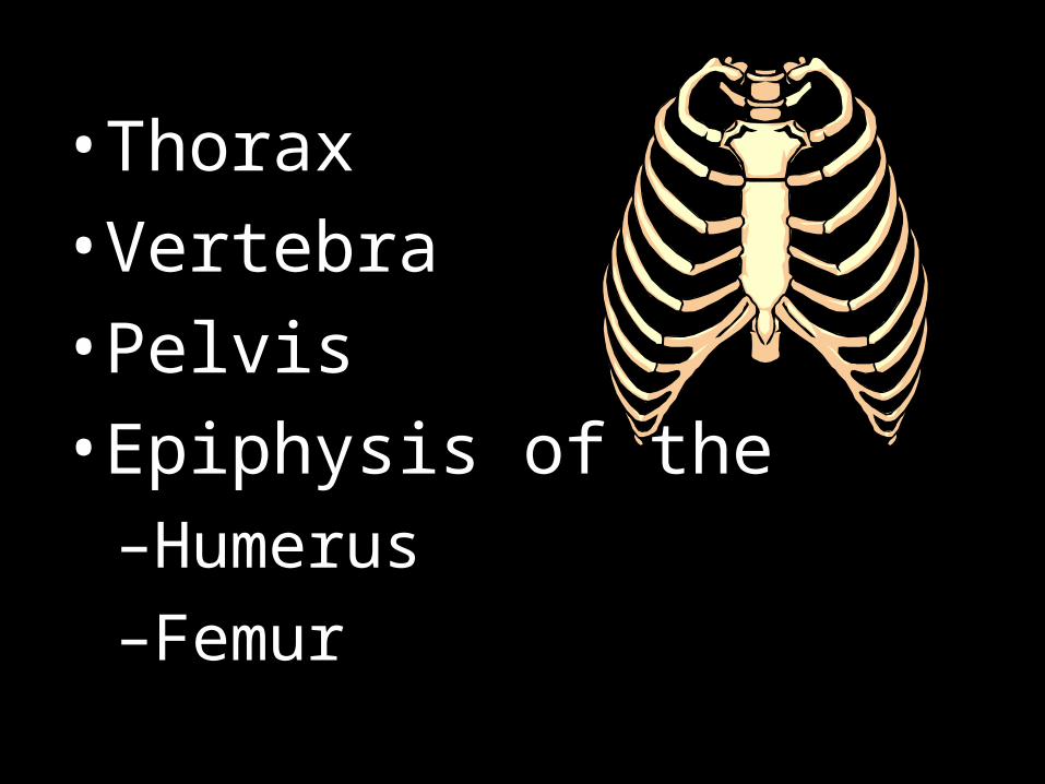
• Thorax
• Vertebra
• Pelvis
• Epiphysis of the–Humerus
–Femur
Bone as a Storage Unit
• Bone is used to store inorganic salts. (No Carbon)
• Primarily stores Ca+
• [Ca+] control–If [Ca+] in blood is too low

osteoclasts are stimulated to breakdown bone tissue to release Ca+.
–If [Ca+] in blood is too high, osteoblasts are stimulated to form bone tissue, trapping the Ca+.
Organization
• 2 major portions–Axial: head, neck, and trunk
–Appendicular: limbs and anchoring parts
Axial
1. Skull: cranium and facial bones
2. Hyoid bone: between jaw and neck, helps tongue function
3. Vertebral column: back bone, series of bony vertebrae and cartilaginous intervertebral discs.
4. Thoracic cage: used to protect the organs of the thorax and upper abdomen.
Appendicular
1. Pectoral girdle: clavicle and scapula, the attachment site of the arm.
2. Upper limb: humerus, radius, ulna, carpals, metacarpals, phalanges.
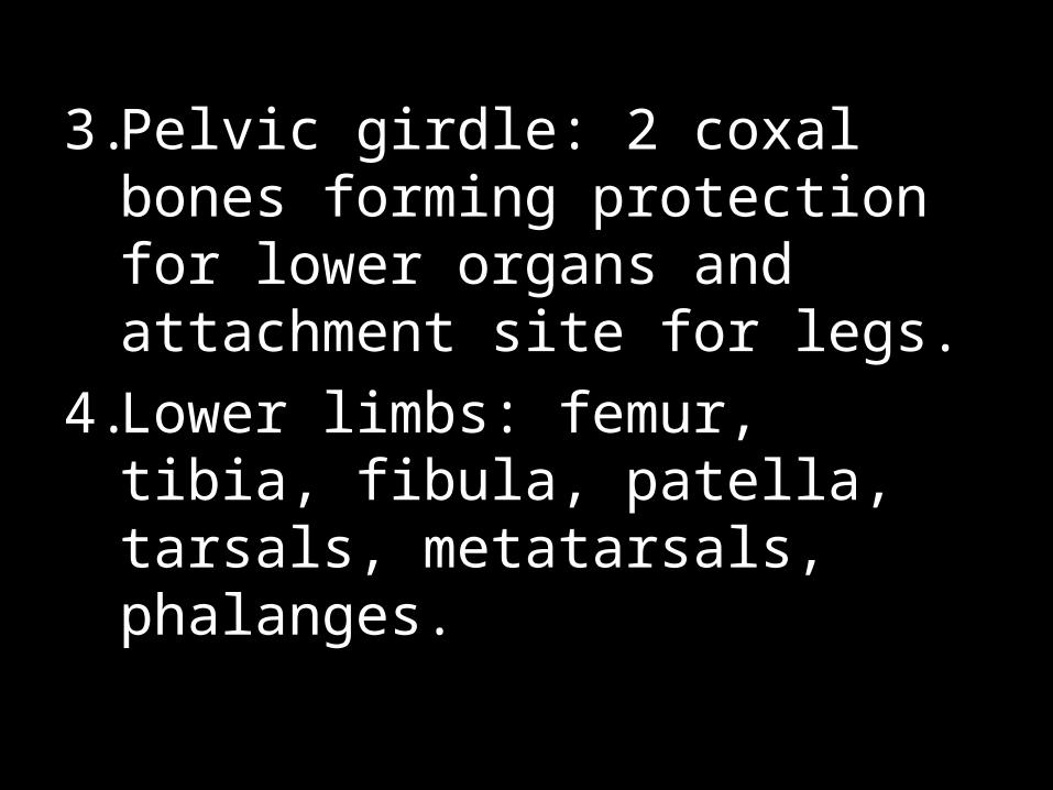
3. Pelvic girdle: 2 coxal bones forming protection for lower organs and attachment site for legs.
4. Lower limbs: femur, tibia, fibula, patella, tarsals, metatarsals, phalanges.


![Cartilage - facultymembers.sbu.ac.irfacultymembers.sbu.ac.ir/rajabi/ppt toPDF/Cartilage [Compatibility Mode].pdfFibrocartilage • Fibrous Cartilage • is a form of connective tissue](https://static.documents.pub/doc/80x56/6012989a4318862a0e5813ae/cartilage-topdfcartilage-compatibility-modepdf-fibrocartilage-a-fibrous.jpg)