80
م ي ح ر ل ا ن م ح ر ل ه ا ل ل م ا س ب م ي ح ر ل ا ن م ح ر ل ه ا ل ل م ا س ب
| Date post: | 30-Dec-2015 |
| Category: |
Documents |
| Upload: | russell-davis |
| View: | 220 times |
| Download: | 0 times |

بسم الله الرحمن الرحيمبسم الله الرحمن الرحيم
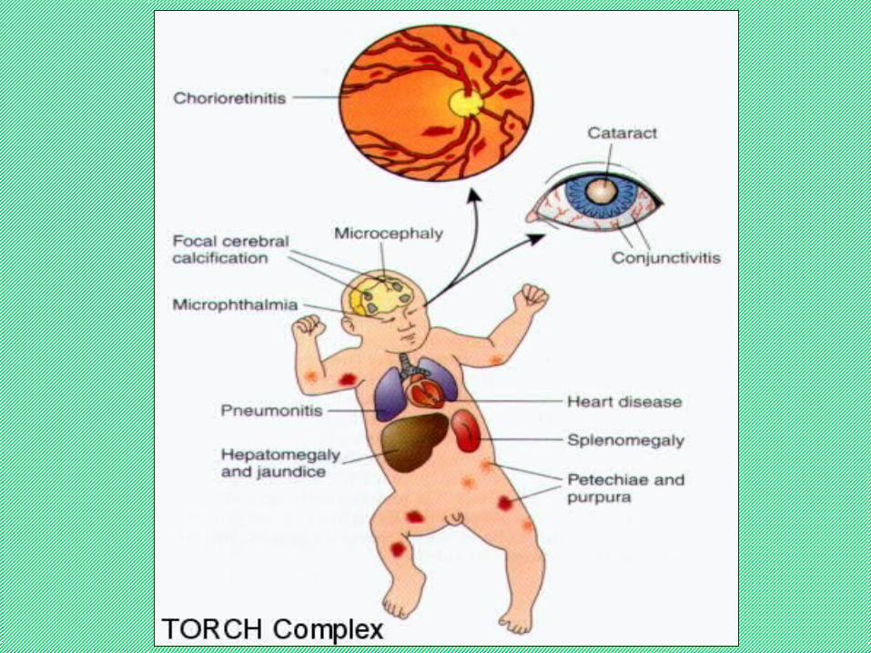
TORCHs Complex And CRS
PROFESSOR KARIMI
PIRC
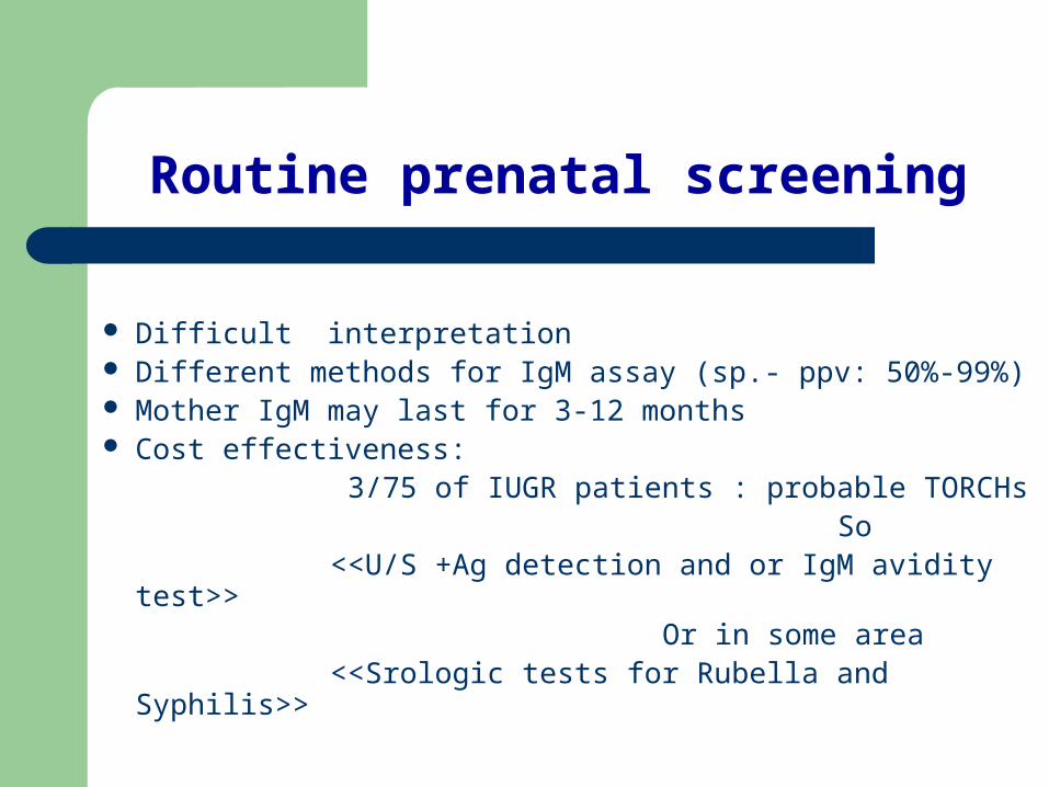
Routine prenatal screening
Difficult interpretation Different methods for IgM assay (sp.- ppv: 50%-99%) Mother IgM may last for 3-12 months Cost effectiveness: 3/75 of IUGR patients : probable TORCHs So <<U/S +Ag detection and or IgM avidity test>> Or in some area <<Srologic tests for Rubella and Syphilis>>
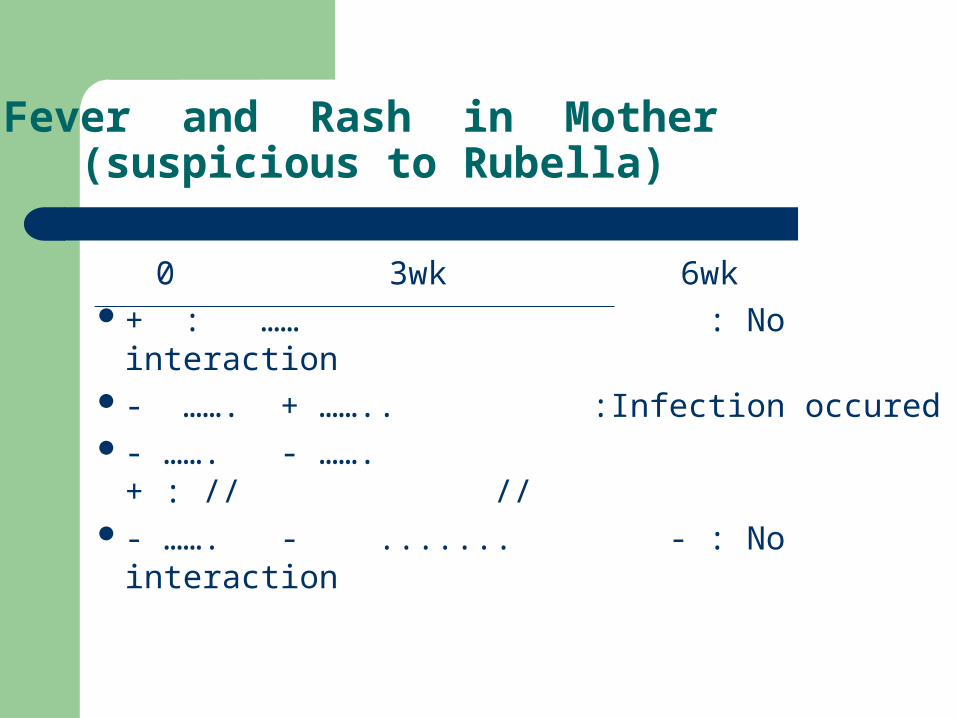
Fever and Rash in Mother (suspicious to Rubella)
0 3wk 6wk+ : …… : No interaction- ……. + …….. :Infection occured- ……. - ……. + : // //- ……. - ....... - : No interaction

Acquired Toxoplasmosis and CMV
90% asymptomatic Most common symptoms : LAP+ Fever + Fatigue Occasionally: Inf. Mono. Like sx Hepatitis , Encephalitis , Pneumonitis , Myocarditis Aseptic Meningitis and Mass lesion
Acquired CMV is very similar to EBV
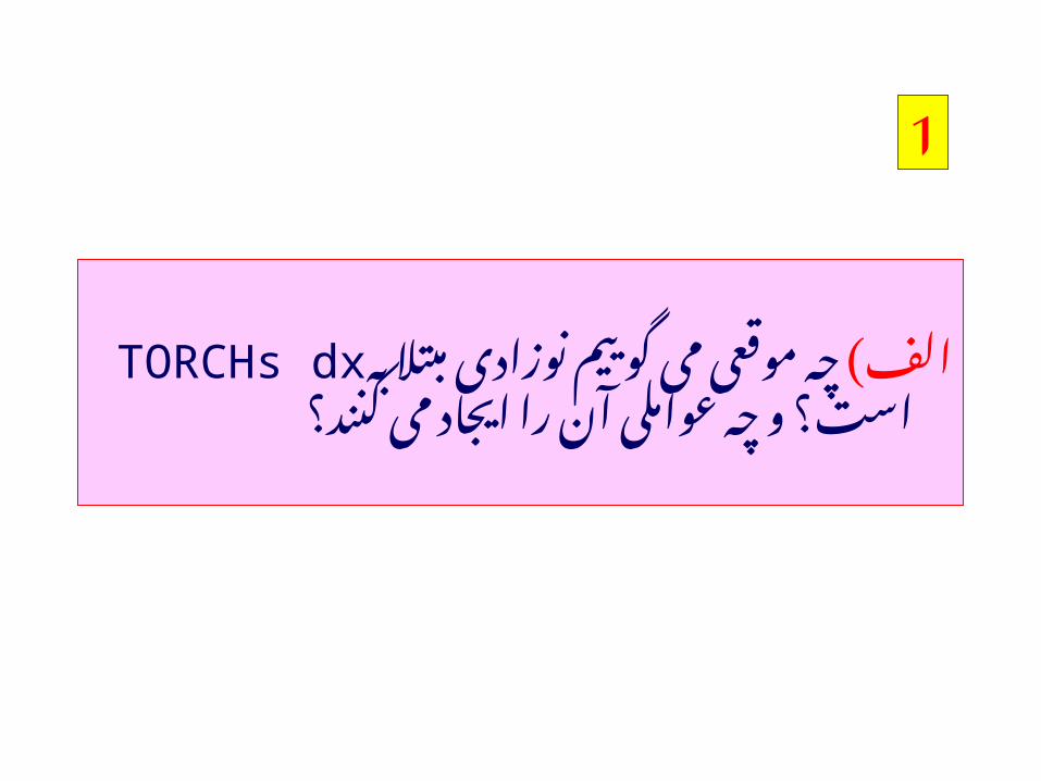
1
مبتال الف) نوزادی گوییم می موقعی چهرا TORCHs dxبه آن عواملی چه و است؟
کنند؟ می ایجاد
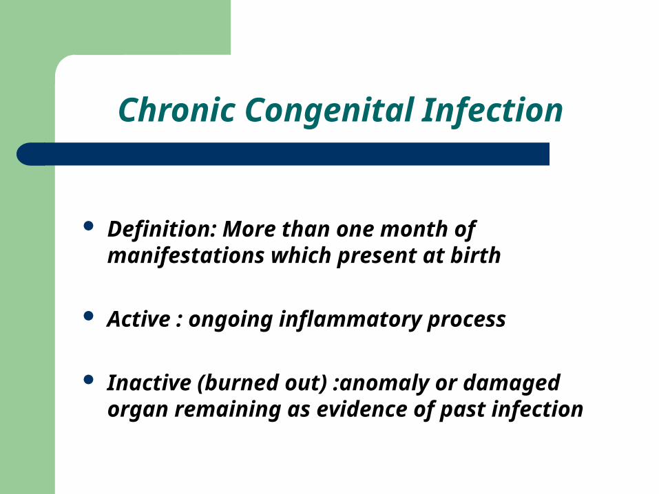
Chronic Congenital Infection
Definition: More than one month of manifestations which present at birth
Active : ongoing inflammatory process
Inactive (burned out) :anomaly or damaged organ remaining as evidence of past infection
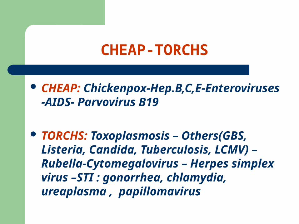
CHEAP-TORCHS
CHEAP: Chickenpox-Hep.B,C,E-Enteroviruses -AIDS- Parvovirus B19
TORCHS: Toxoplasmosis – Others(GBS, Listeria, Candida, Tuberculosis, LCMV) –Rubella-Cytomegalovirus – Herpes simplex virus –STI : gonorrhea, chlamydia, ureaplasma , papillomavirus
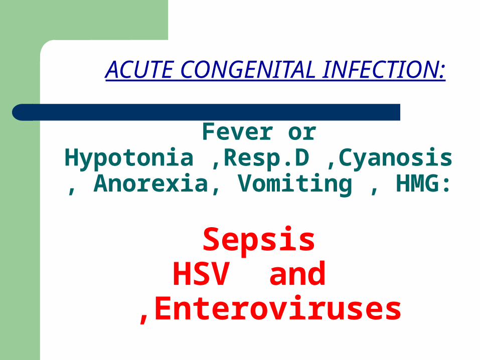
Fever or Hypotonia ,Resp.D ,Cyanosis,
Anorexia, Vomiting , HMG:
Sepsis
HSV and Enteroviruses ,
ACUTE CONGENITAL INFECTION:
1
تورچ ب) سندم برای را نوزاد مواردی چه دربررسی
کنید؟ می
1
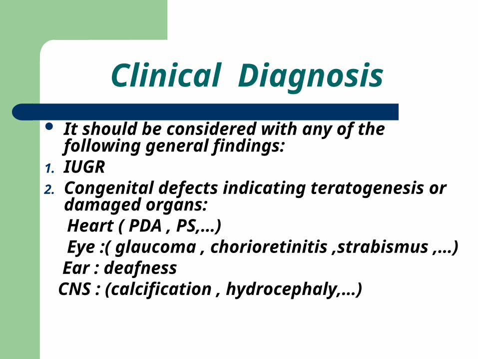
Clinical Diagnosis It should be considered with any of the
following general findings:1. IUGR2. Congenital defects indicating teratogenesis or
damaged organs: Heart ( PDA , PS,…) Eye :( glaucoma , chorioretinitis ,strabismus ,
…) Ear : deafness CNS : (calcification , hydrocephaly,…)
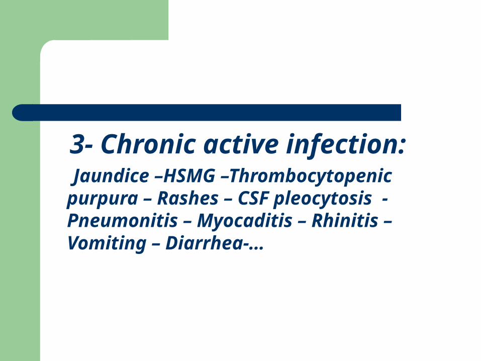
3- Chronic active infection:
Jaundice –HSMG –Thrombocytopenic purpura – Rashes – CSF pleocytosis - Pneumonitis – Myocaditis – Rhinitis – Vomiting – Diarrhea-…
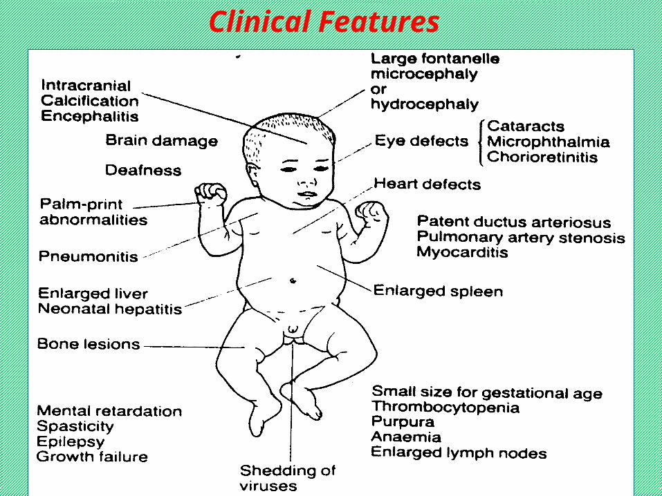
Clinical Features
چه ج ) تورچ سندرم برای نوزاد بررسی باکنید؟ می دنبال را اهدافی
1
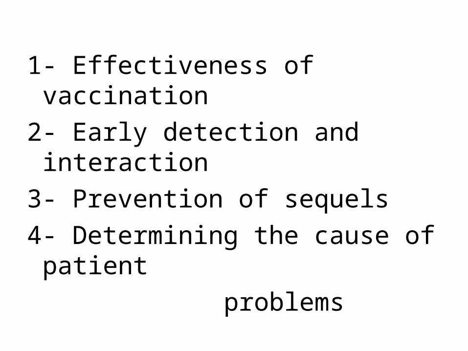
1- Effectiveness of vaccination
2- Early detection and interaction
3- Prevention of sequels
4- Determining the cause of patient
problems
آزمایشات الف) تورچ سندرم بررسی در ( سرولوژی ( از غیر اختصاصی و عمومی
کدامند؟
2
Nonspecific LAB W/U
U/S for IC abnormalities such as calcification Long bone X-Ray , CXR
ECG CSF analysis
CBC including platelet count Total IgM , which is neither S o SP .
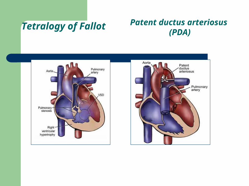
Tetralogy of Fallot Patent ductus arteriosus (PDA)
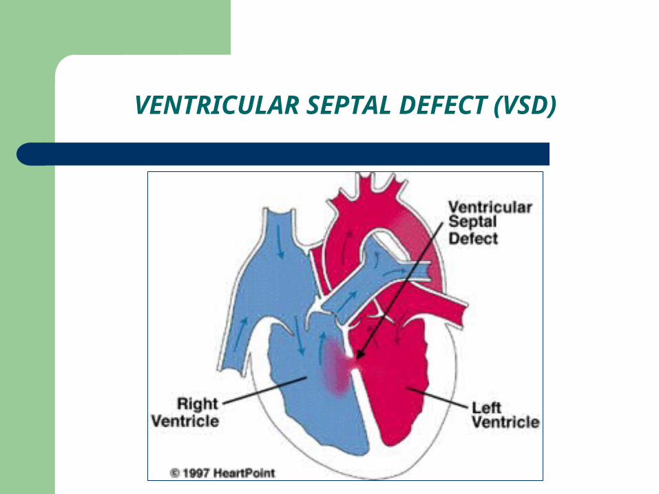
VENTRICULAR SEPTAL DEFECT (VSD)
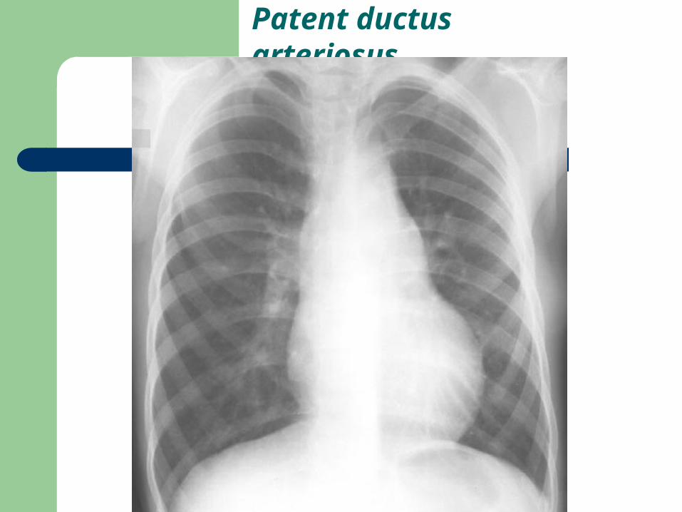
Patent ductus arteriosus
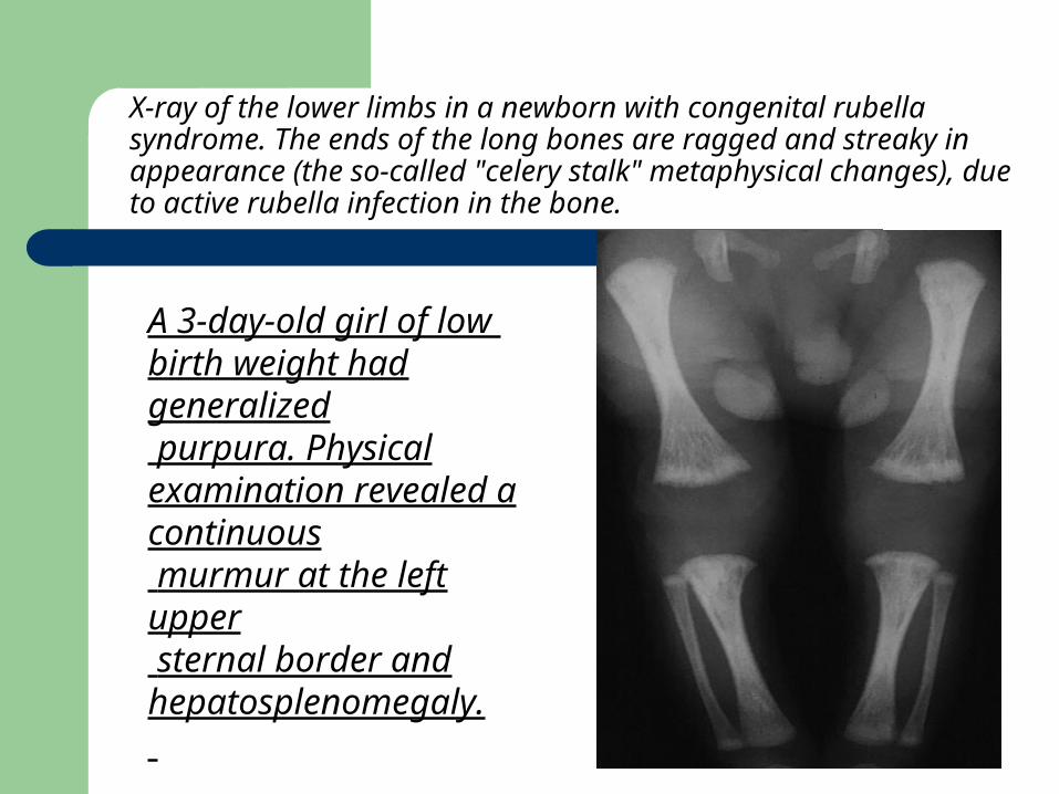
A 3-day-old girl of low birth weight had generalized purpura. Physical examination revealed a continuous murmur at the left upper sternal border and hepatosplenomegaly.
X-ray of the lower limbs in a newborn with congenital rubella syndrome. The ends of the long bones are ragged and streaky in appearance (the so-called "celery stalk" metaphysical changes), due to active rubella infection in the bone.
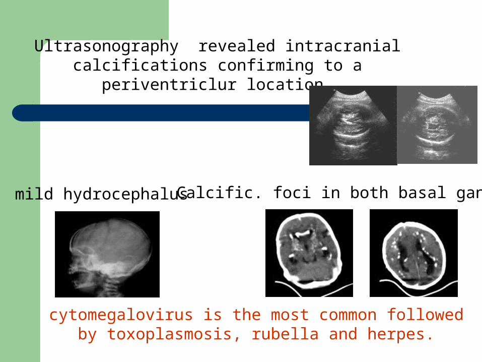
Ultrasonography revealed intracranial calcifications confirming to a periventriclur location
mild hydrocephalus Calcific. foci in both basal ganglia
cytomegalovirus is the most common followed by toxoplasmosis, rubella and herpes.
تمام ب) برای را شده نامبرده آزمایشات آیایا میفرمائید درخواست تورچ به مشکوک نوزادان
نظر در را معیارهاییگیرید؟ می
درخواست( برای خاصی راهنمای صورتیکه درفرمایید ارائه دارید نظر در ).آزمایشات
2
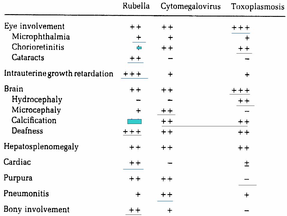
شباهتهای الف) و ها و CMV،Rubella تفاوتToxopl. بطور وآزمایشگاهی بالینی نظر از را
. فرمایید منعکس جدول دو در جداگانه
3
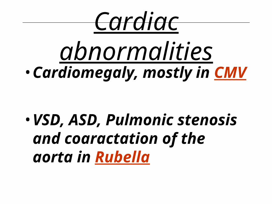
Cardiac abnormalities• Cardiomegaly, mostly in CMV
• VSD, ASD, Pulmonic stenosis and coaractation of the aorta in Rubella
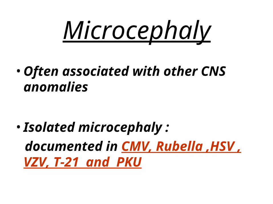
Microcephaly
• Often associated with other CNS anomalies
• Isolated microcephaly :
documented in CMV, Rubella ,HSV , VZV, T-21 and PKU
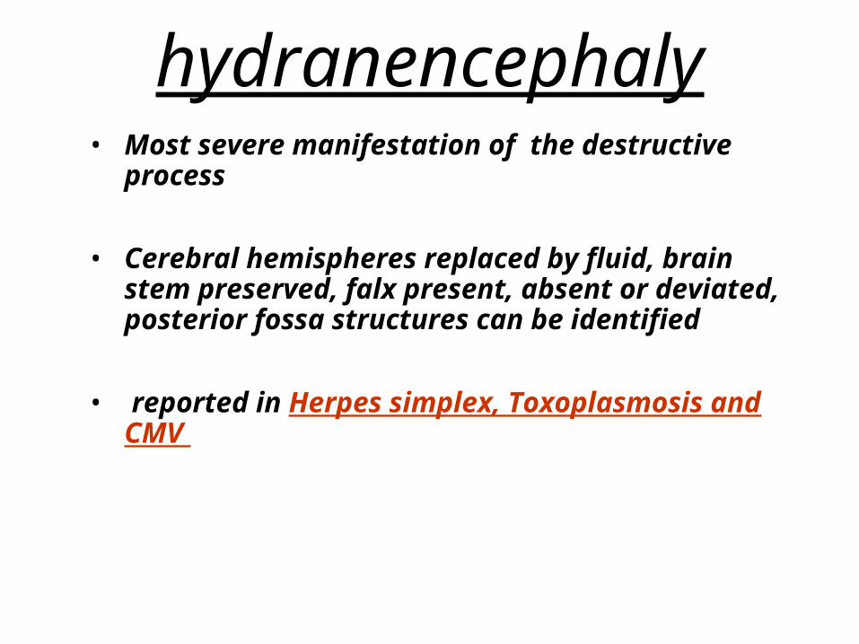
hydranencephaly• Most severe manifestation of the destructive
process
• Cerebral hemispheres replaced by fluid, brain stem preserved, falx present, absent or deviated, posterior fossa structures can be identified
• reported in Herpes simplex, Toxoplasmosis and CMV
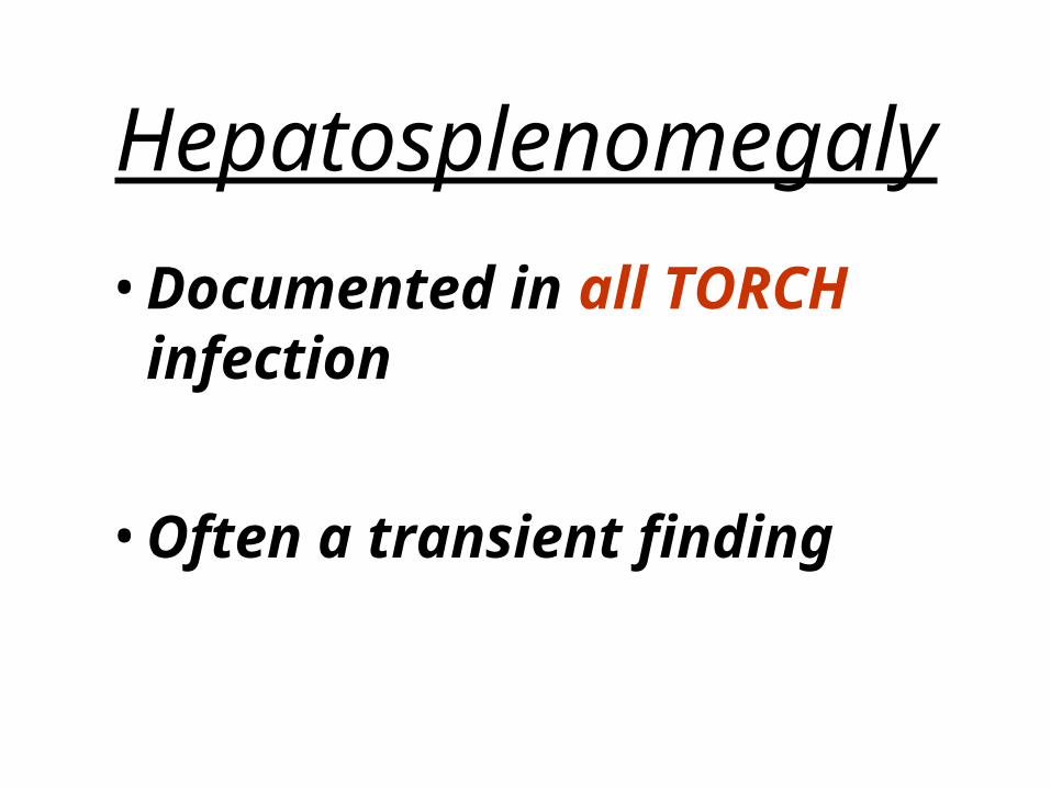
Hepatosplenomegaly
• Documented in all TORCH infection
• Often a transient finding
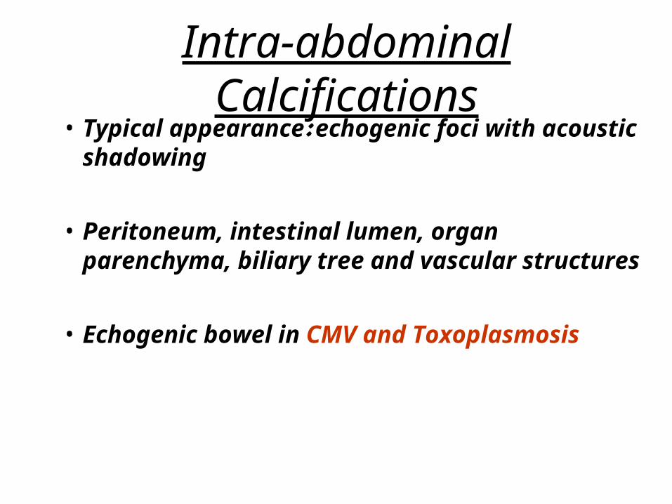
Intra-abdominal Calcifications• Typical appearance: echogenic foci with
acoustic shadowing
• Peritoneum, intestinal lumen, organ parenchyma, biliary tree and vascular structures
• Echogenic bowel in CMV and Toxoplasmosis
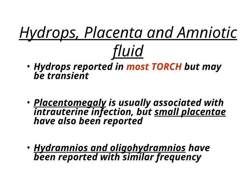
Hydrops, Placenta and Amniotic fluid
• Hydrops reported in most TORCH but may be transient
• Placentomegaly is usually associated with intrauterine infection, but small placentae have also been reported
• Hydramnios and oligohydramnios have been reported with similar frequency
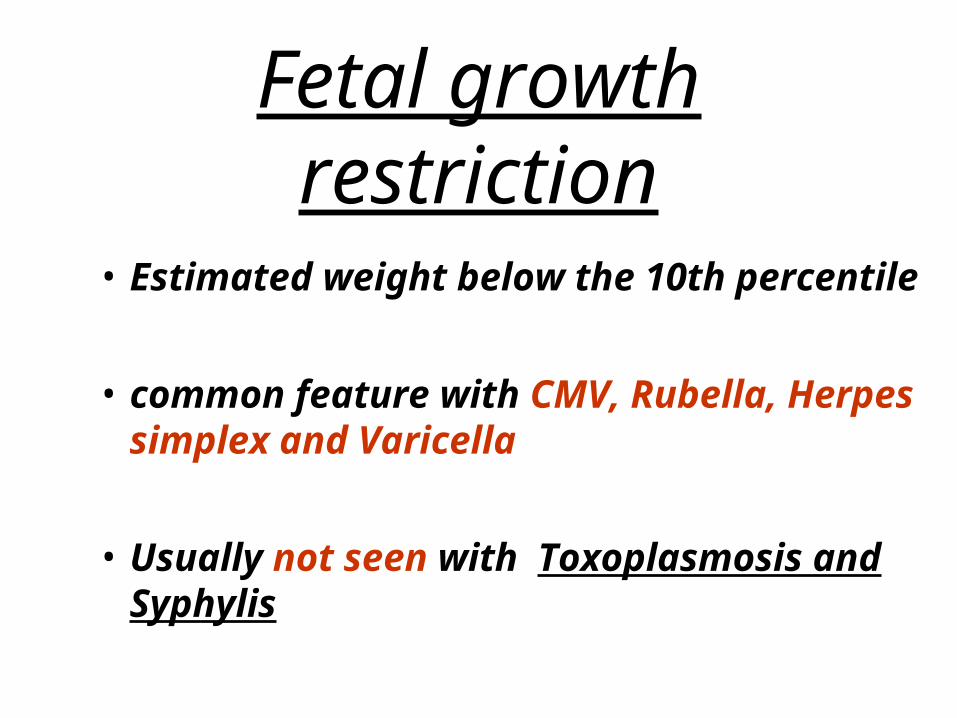
Fetal growth restriction
• Estimated weight below the 10th percentile
• common feature with CMV, Rubella, Herpes simplex and Varicella
• Usually not seen with Toxoplasmosis and Syphylis
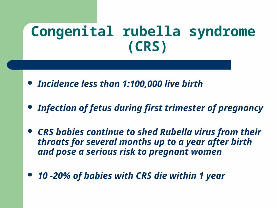
Congenital rubella syndrome (CRS)
Incidence less than 1:100,000 live birth
Infection of fetus during first trimester of pregnancy
CRS babies continue to shed Rubella virus from their throats for several months up to a year after birth and pose a serious risk to pregnant women
10 -20% of babies with CRS die within 1 year
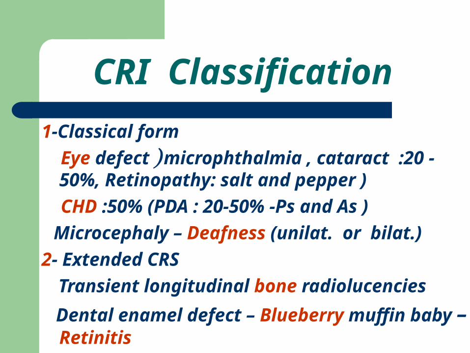
CRI Classification
1-Classical form
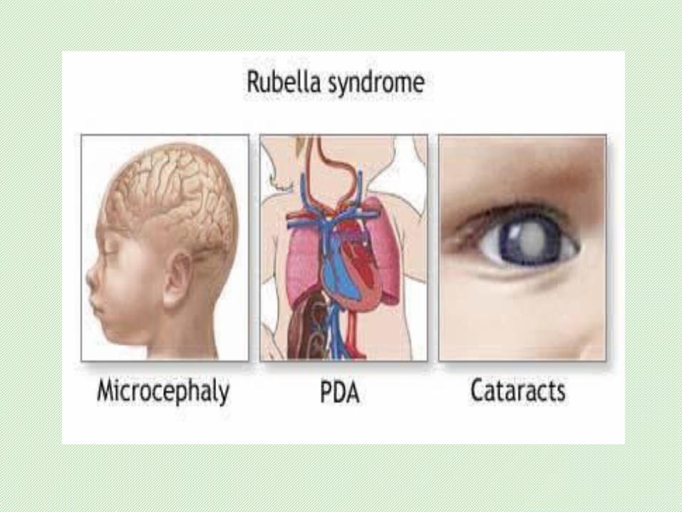
Eye defect ) microphthalmia , cataract :20 -50%, Retinopathy: salt and pepper )
CHD :50% (PDA : 20-50% -Ps and As )
Microcephaly – Deafness (unilat. or bilat.)
2- Extended CRS
Transient longitudinal bone radiolucencies
Dental enamel defect – Blueberry muffin baby – Retinitis
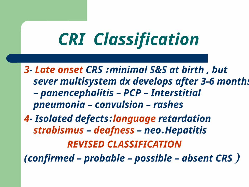
3- Late onset CRS : minimal S&S at birth , but sever multisystem dx develops after 3-6 months – panencephalitis – PCP – Interstitial pneumonia – convulsion – rashes
4- Isolated defects: language retardation strabismus – deafness – neo. Hepatitis
REVISED CLASSIFICATION
(confirmed – probable – possible – absent CRS )
CRI Classification
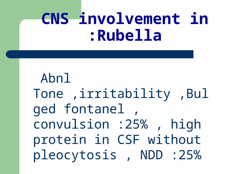
CNS involvement in Rubella:
Abnl Tone ,irritability ,Bulged fontanel , convulsion :25% , high protein in CSF without pleocytosis , NDD :25%
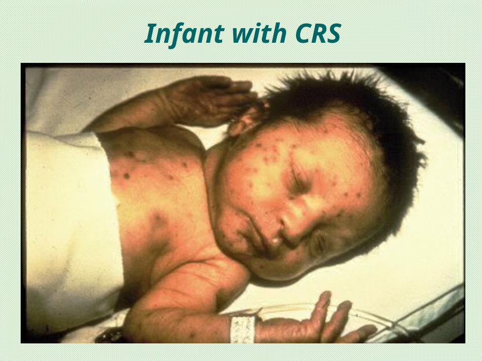
Infant with CRS
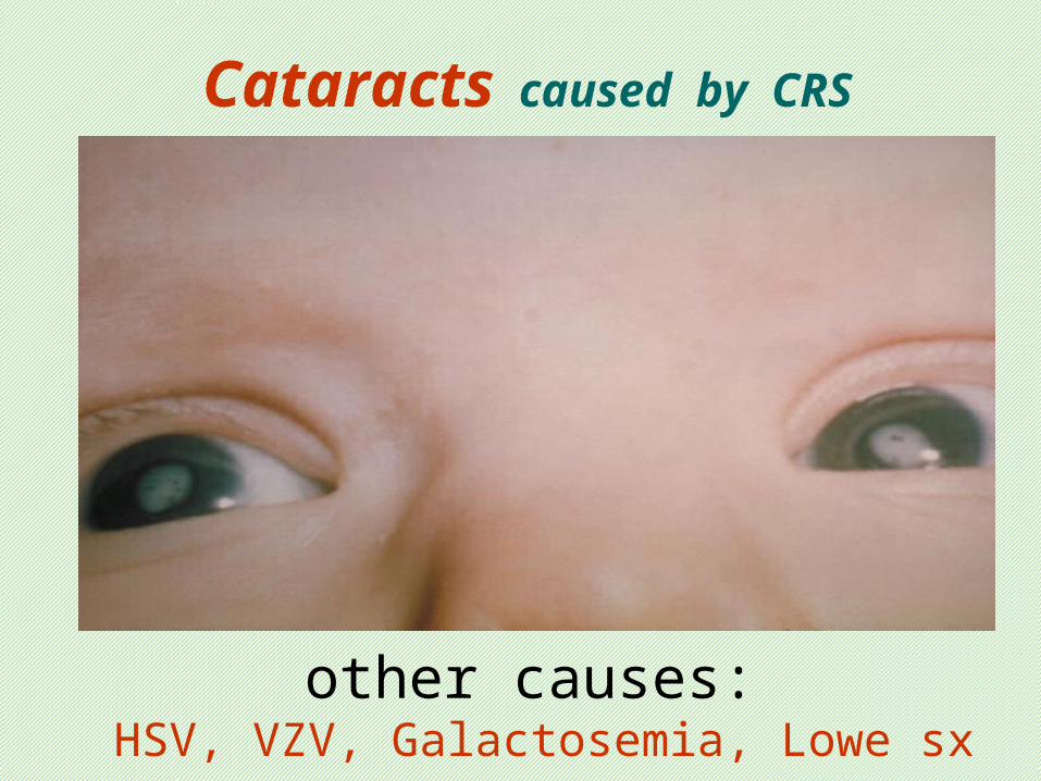
Cataracts caused by CRS
other causes: HSV, VZV, Galactosemia, Lowe sx
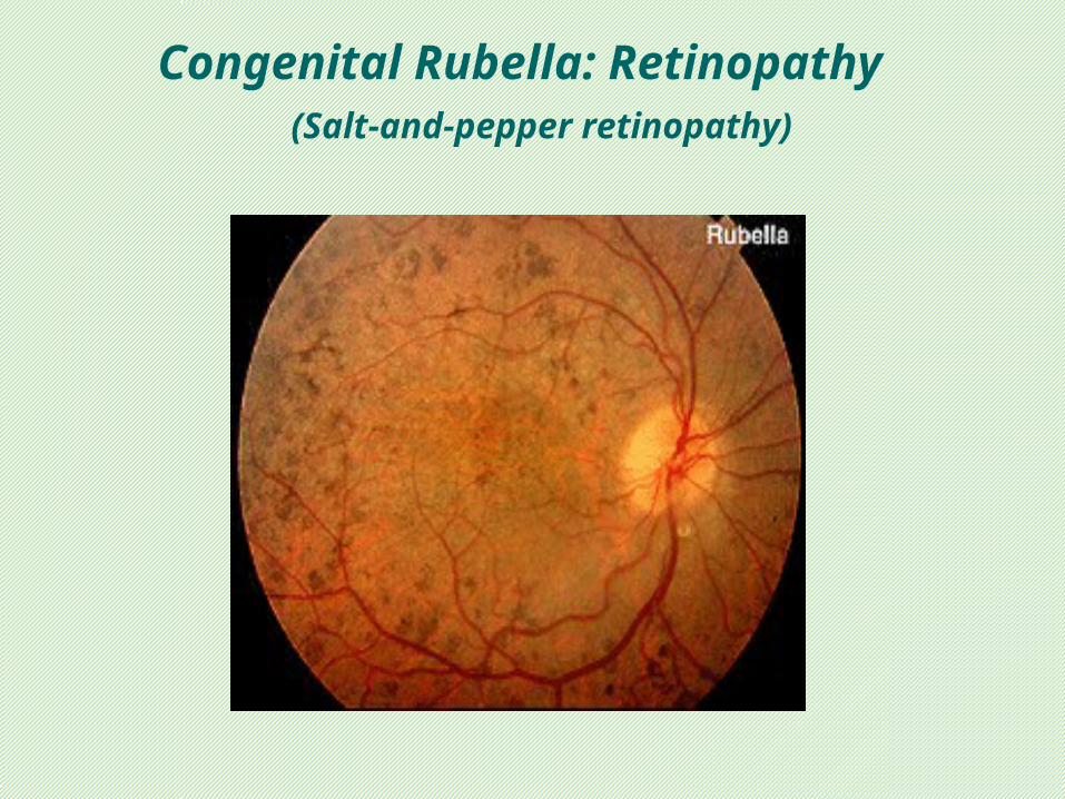
Congenital Rubella: Retinopathy (Salt-and-pepper retinopathy)
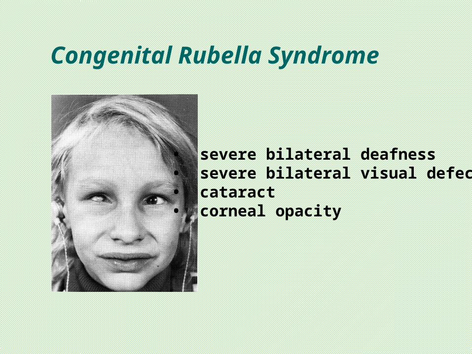
Congenital Rubella Syndrome
• severe bilateral deafness• severe bilateral visual defects• cataract• corneal opacity
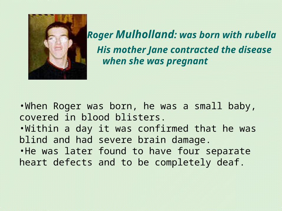
His mother Jane contracted the disease when she was pregnant
Roger Mulholland: was born with rubella
•When Roger was born, he was a small baby, covered in blood blisters. •Within a day it was confirmed that he was blind and had severe brain damage. •He was later found to have four separate heart defects and to be completely deaf.
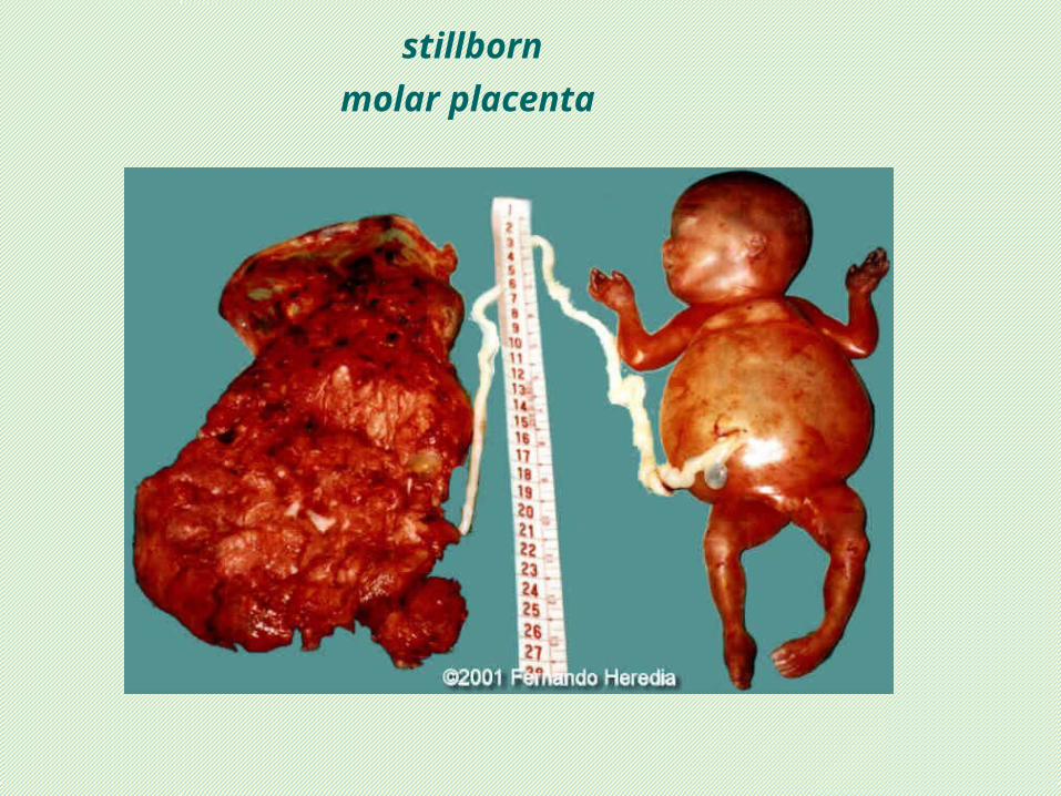
stillborn
molar placenta
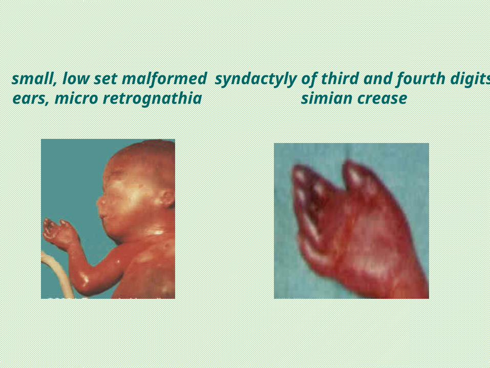
syndactyly of third and fourth digits simian crease
small, low set malformed ears, micro retrognathia
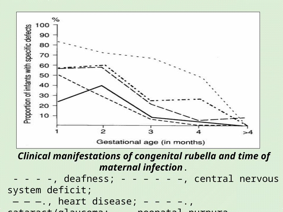
Clinical manifestations of congenital rubella and time of maternal infection.
- - - -, deafness; - - – - - –, central nervous system deficit; — — —., heart disease; – – – –., cataract/glaucoma; ——, neonatal purpura
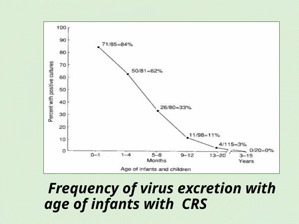
Frequency of virus excretion with age of infants with CRS
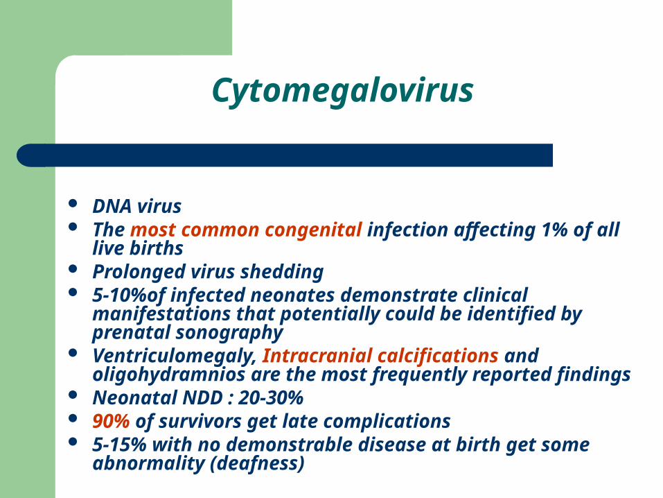
Cytomegalovirus
DNA virus The most common congenital infection affecting 1% of all
live births Prolonged virus shedding 5-10%of infected neonates demonstrate clinical
manifestations that potentially could be identified by prenatal sonography
Ventriculomegaly, Intracranial calcifications and oligohydramnios are the most frequently reported findings
Neonatal NDD : 20-30% 90% of survivors get late complications 5-15% with no demonstrable disease at birth get some
abnormality (deafness)
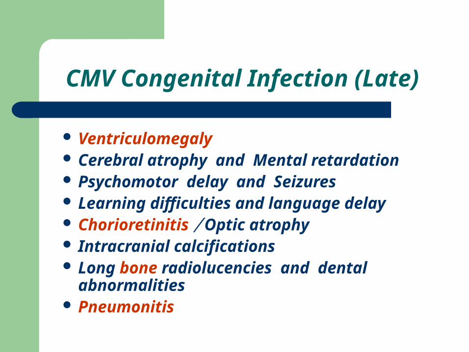
CMV Congenital Infection (Late)
Ventriculomegaly Cerebral atrophy and Mental retardation Psychomotor delay and Seizures Learning difficulties and language delay Chorioretinitis / Optic atrophy Intracranial calcifications Long bone radiolucencies and dental
abnormalities Pneumonitis
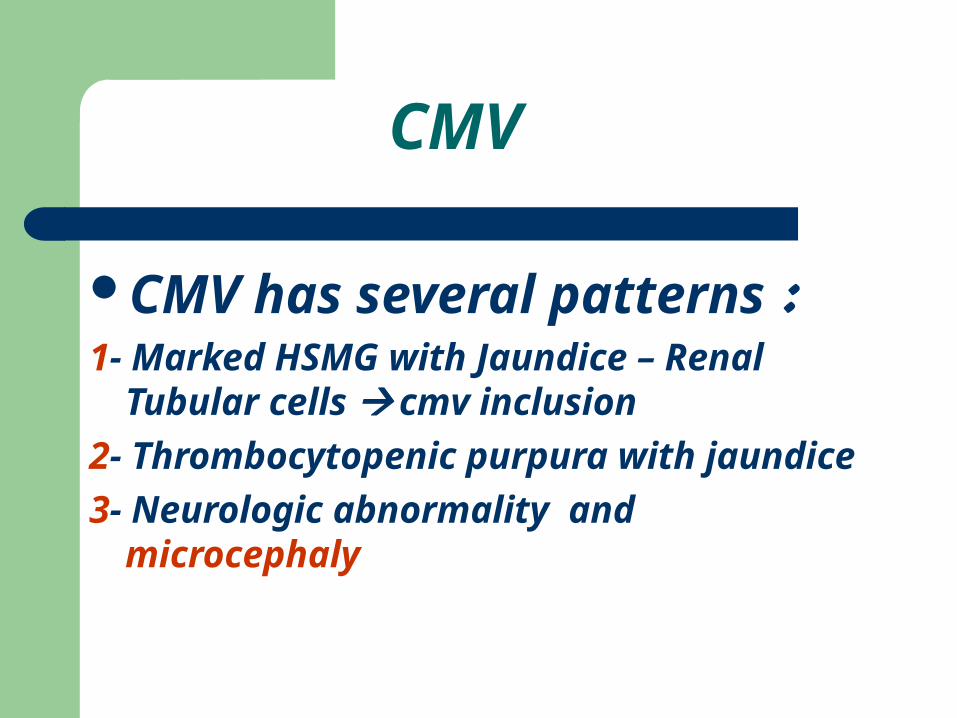
CMV
CMV has several patterns : 1- Marked HSMG with Jaundice – Renal
Tubular cells cmv inclusion
2- Thrombocytopenic purpura with jaundice
3- Neurologic abnormality and microcephaly
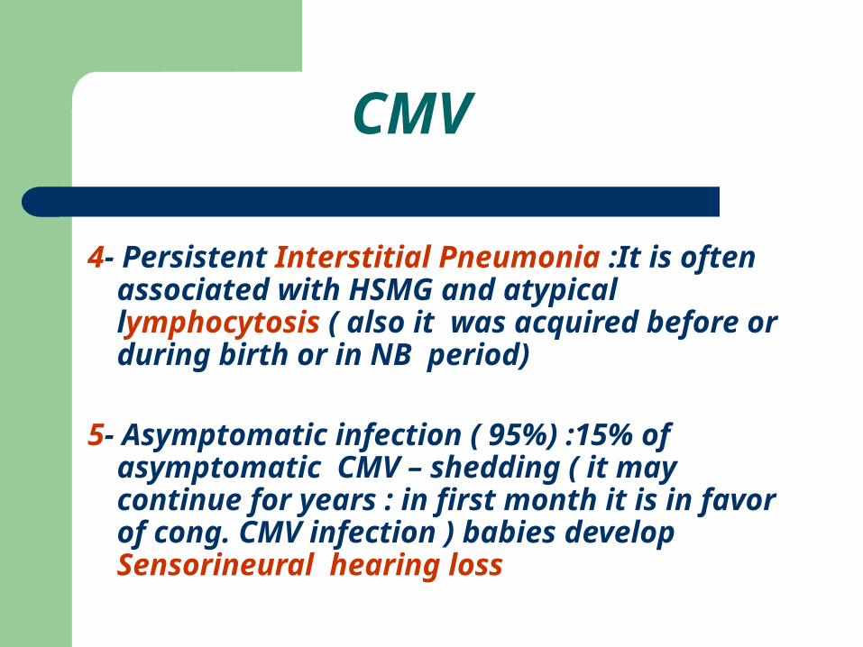
4- Persistent Interstitial Pneumonia :It is often associated with HSMG and atypical lymphocytosis ( also it was acquired before or during birth or in NB period)
5- Asymptomatic infection ( 95%) :15% of asymptomatic CMV – shedding ( it may continue for years : in first month it is in favor of cong. CMV infection ) babies develop Sensorineural hearing loss
CMV
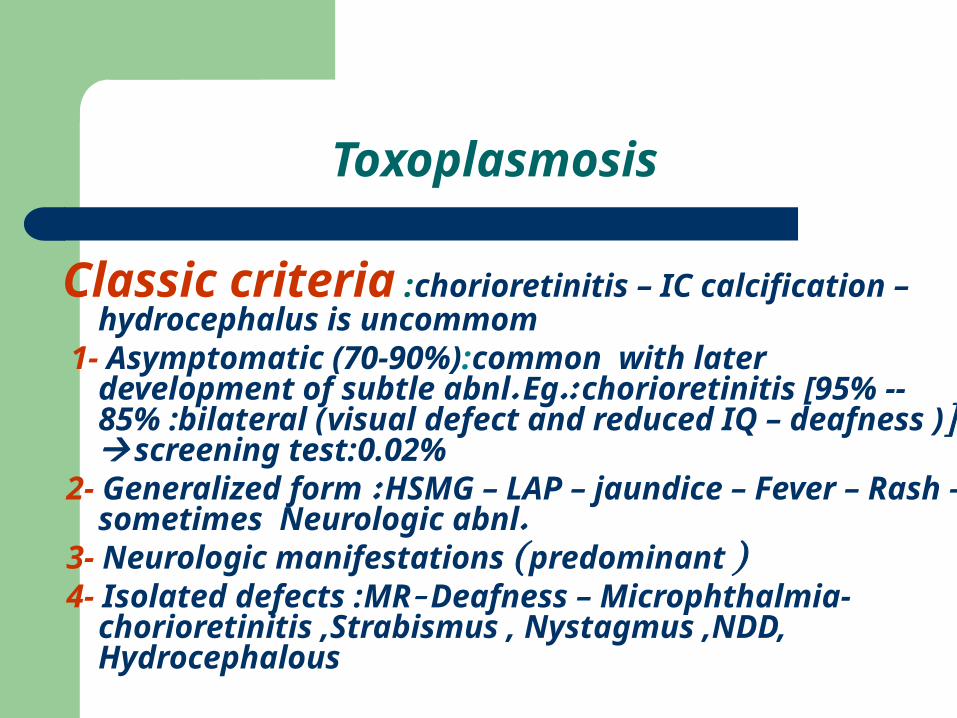
Classic criteria :chorioretinitis – IC calcification – hydrocephalus is uncommom
1- Asymptomatic (70-90%):common with later development of subtle abnl. Eg.: chorioretinitis [95% -- 85% :bilateral (visual defect and reduced IQ – deafness )] screening test:0.02%
2- Generalized form : HSMG – LAP – jaundice – Fever – Rash – sometimes Neurologic abnl.
3- Neurologic manifestations ( predominant ) 4- Isolated defects :MR- Deafness – Microphthalmia-
chorioretinitis ,Strabismus , Nystagmus ,NDD, Hydrocephalous
Toxoplasmosis
کدام ( ب هر برای کننده کمک سرولوژی تست دوعامل سه Toxoplasmosisو CMV،Rubellaاز
کنید؟ تفسیر را ببریدوآنها نام را
.
3
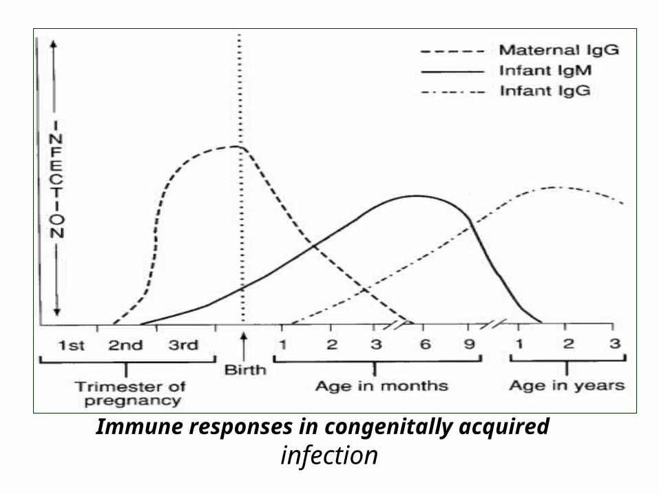
Immune responses in congenitally acquired infection
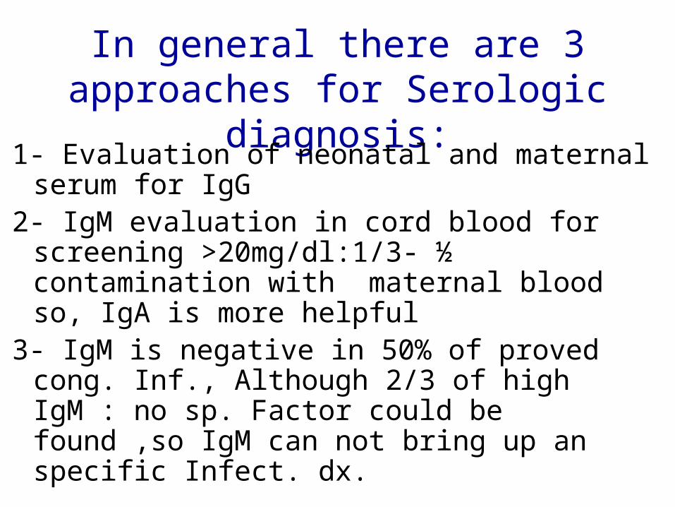
In general there are 3 approaches for Serologic diagnosis:
1- Evaluation of neonatal and maternal serum for IgG
2- IgM evaluation in cord blood for screening >20mg/dl:1/3- ½ contamination with maternal blood so, IgA is more helpful
3- IgM is negative in 50% of proved cong. Inf., Although 2/3 of high IgM : no sp. Factor could be found ,so IgM can not bring up an specific Infect. dx.
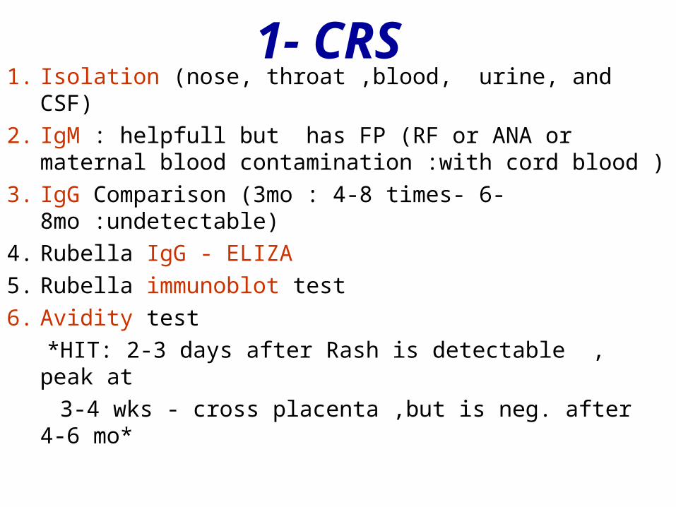
1- CRS1. Isolation (nose, throat ,blood, urine, and CSF)
2. IgM : helpfull but has FP (RF or ANA or maternal blood contamination :with cord blood )
3. IgG Comparison (3mo : 4-8 times- 6-8mo :undetectable)
4. Rubella IgG - ELIZA
5. Rubella immunoblot test
6. Avidity test
*HIT: 2-3 days after Rash is detectable , peak at
3-4 wks - cross placenta ,but is neg. after 4-6 mo*
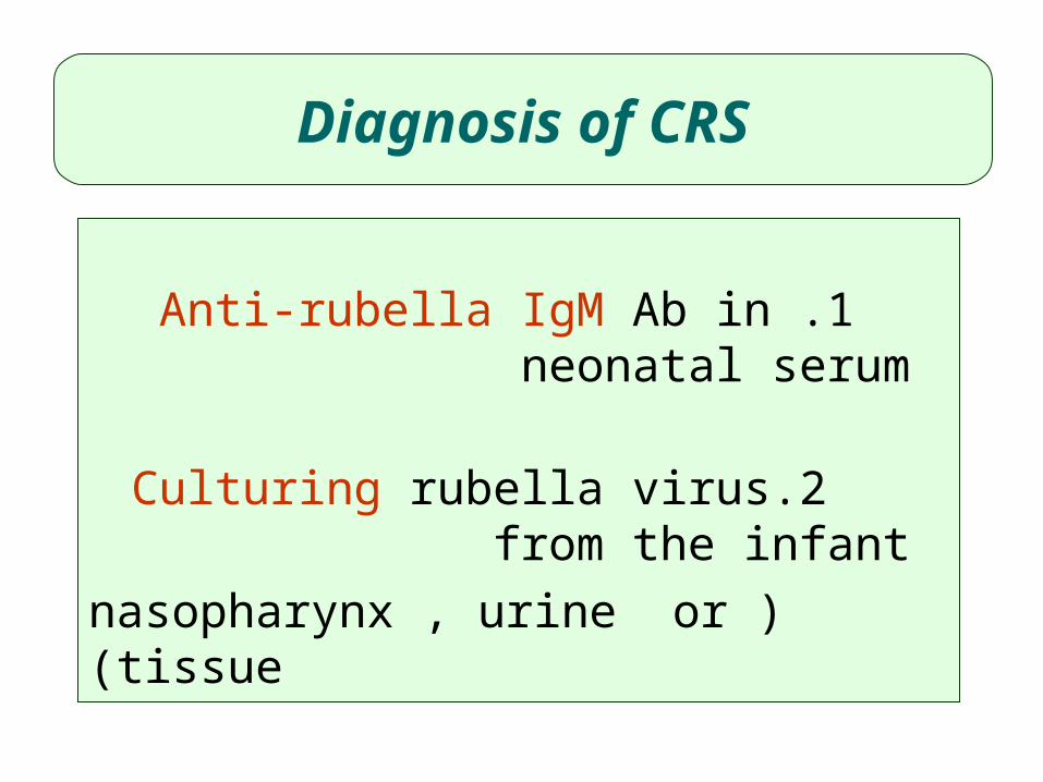
1 .Anti-rubella IgM Ab in neonatal serum
2.Culturing rubella virus from the infant ( nasopharynx , urine or tissue)
Diagnosis of CRS
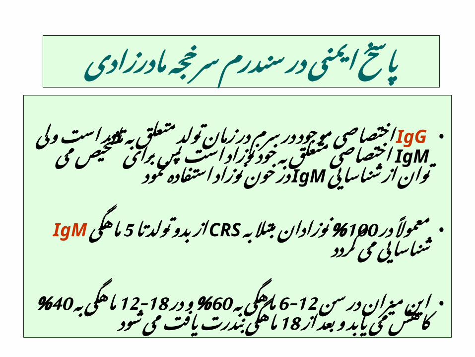
سرخجه سندرم در ایمنی پاسخمادرزادی
• IgG متعلق تولد زمان در سرم در موجود اختصاصیولی است مادر خود IgMبه به متعلق اختصاصی
از توان می تشخیص برای پس است نوزادنمود IgMشناسایی استفاده نوزاد خون در
در • به% 100معموًال, مبتال تا CRSنوزادان تولد بدو ازگردد IgMماهگی 5 می شناسایی
سن • در میزان به 6-12این در% 60ماهگی 12-18وبه از% 40ماهگی بعد و یابد می ماهگی 18کاهش
شود می یافت بندرت
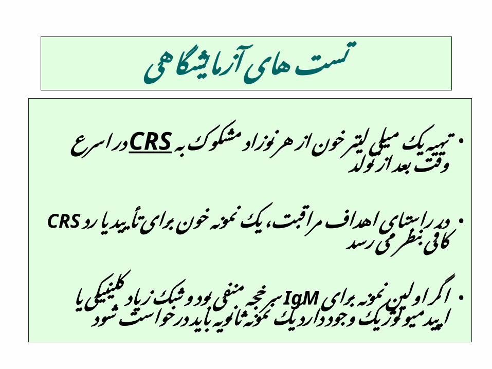
آزمایشگاهی های تست
مشکوک • نوزاد هر از خون لیتر میلی یک تهیهتولد CRS به از بعد وقت اسرع در
خون • نمونه یک مراقبت، اهداف راستای دررد یا تأیید رسد CRSبرای می بنظر کافی
برای • نمونه اولین و IgMاگر بود منفی سرخجهدارد وجود اپیدمیولوژیک یا کلینیکی زیاد شک
شود درخواست باید ثانویه نمونه یک
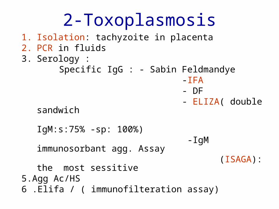
2-Toxoplasmosis1. Isolation: tachyzoite in placenta2. PCR in fluids 3. Serology : Specific IgG : - Sabin Feldmandye -IFA - DF - ELIZA( double sandwich IgM:s:75% -sp: 100%) -IgM immunosorbant agg. Assay (ISAGA): the most sessitive 5.Agg Ac/HS6 .Elifa / ( immunofilteration assay)
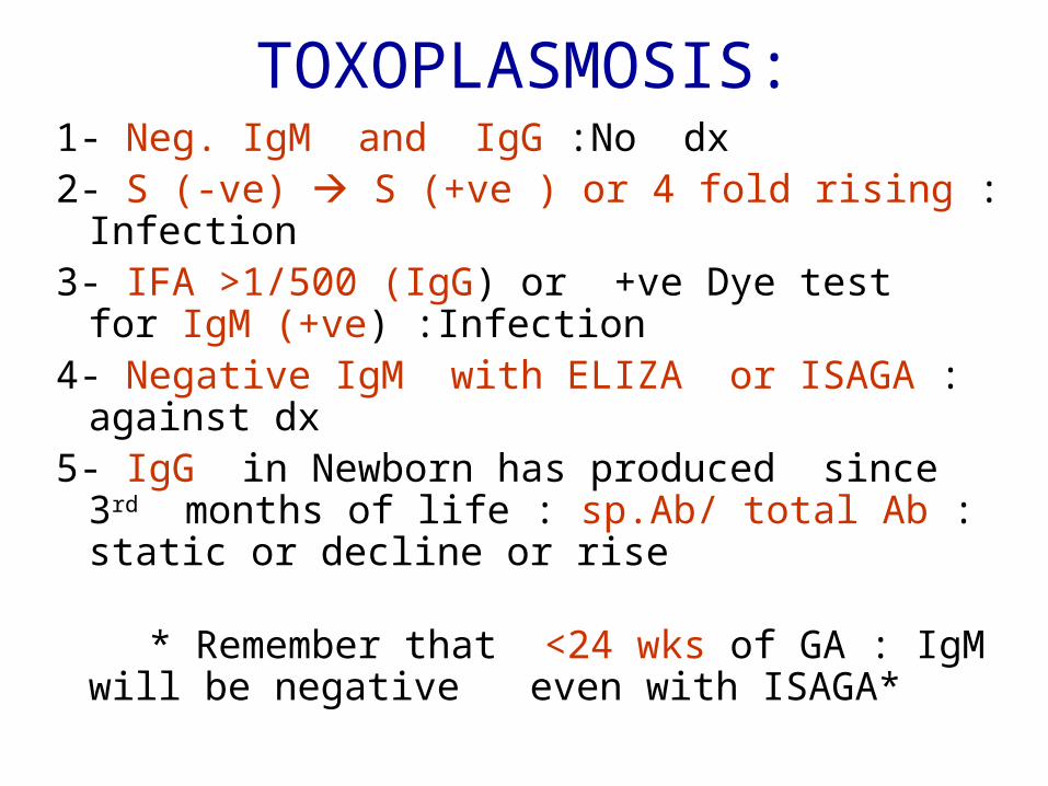
TOXOPLASMOSIS:1- Neg. IgM and IgG :No dx2- S (-ve) S (+ve ) or 4 fold rising : Infection3- IFA >1/500 (IgG) or +ve Dye test for IgM
(+ve) :Infection4- Negative IgM with ELIZA or ISAGA :
against dx 5- IgG in Newborn has produced since 3rd
months of life : sp.Ab/ total Ab : static or decline or rise
* Remember that <24 wks of GA : IgM will be negative even with ISAGA*
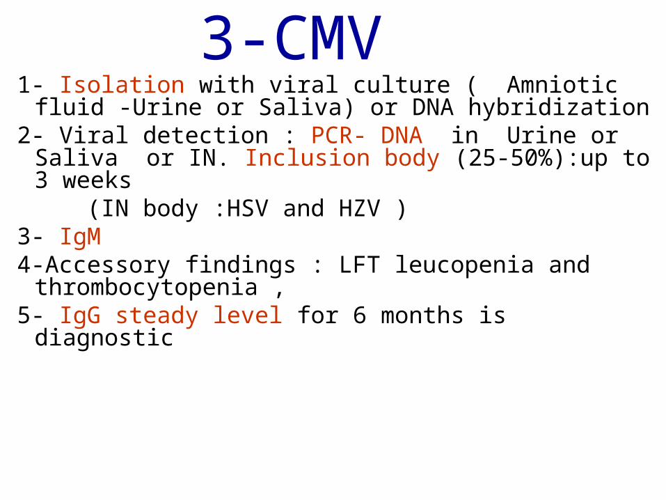
3-CMV1- Isolation with viral culture ( Amniotic fluid -
Urine or Saliva) or DNA hybridization 2- Viral detection : PCR- DNA in Urine or Saliva
or IN. Inclusion body (25-50%):up to 3 weeks (IN body :HSV and HZV )3- IgM4-Accessory findings : LFT leucopenia and
thrombocytopenia ,5- IgG steady level for 6 months is diagnostic
را درمانی چه مثبت موارد از کدام هر برایمی پیگیری را بیمار چگونه و کنید می شروع
نمایید؟
4
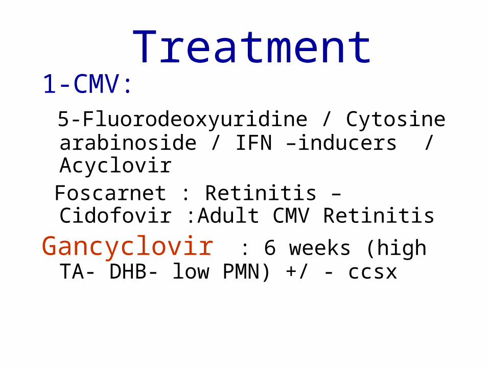
Treatment1-CMV: 5-Fluorodeoxyuridine / Cytosine
arabinoside / IFN –inducers / Acyclovir Foscarnet : Retinitis – Cidofovir :Adult CMV
Retinitis
Gancyclovir : 6 weeks (high TA- DHB- low PMN) +/ - ccsx
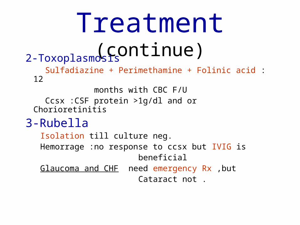
Treatment (continue)2-Toxoplasmosis Sulfadiazine + Perimethamine + Folinic acid : 12 months with CBC F/U Ccsx :CSF protein >1g/dl and or Chorioretinitis
3-Rubella Isolation till culture neg. Hemorrage :no response to ccsx but IVIG is beneficial Glaucoma and CHF need emergency Rx ,but Cataract not .
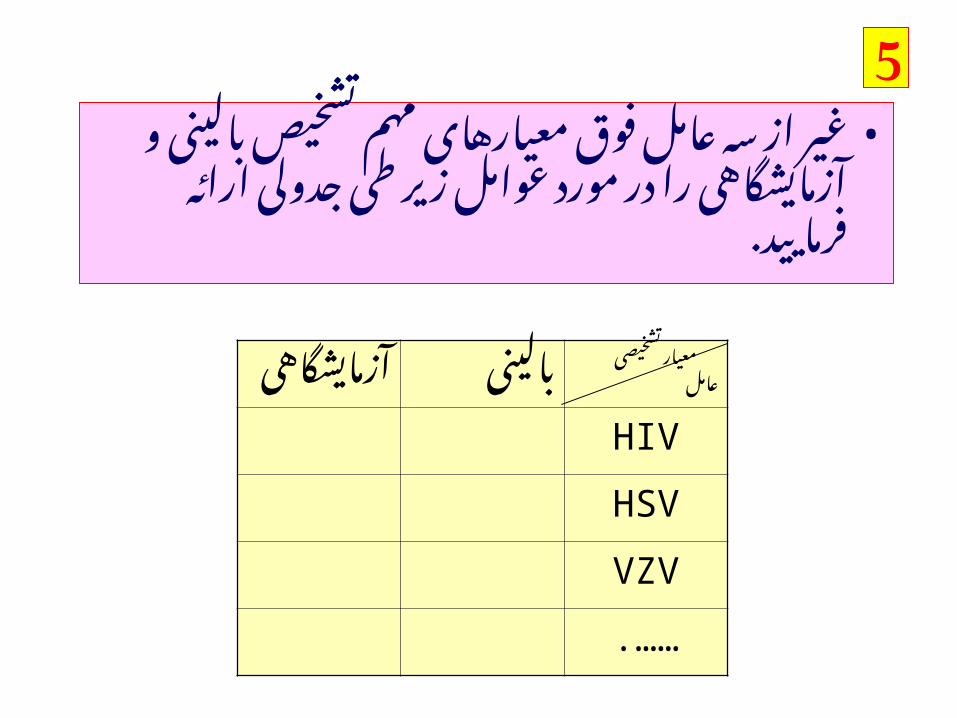
آزمایشگاهی
بالینی معیار تشخیصی
عامل
HIV
HSV
VZV
.……
5مهم • معیارهای فوق عامل سه از غیر
مورد در را آزمایشگاهی و بالینی تشخیص. فرمایید ارائه جدولی طی زیر عوامل
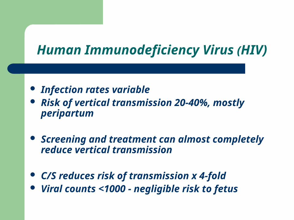
Human Immunodeficiency Virus (HIV)
Infection rates variable Risk of vertical transmission 20-40%, mostly
peripartum
Screening and treatment can almost completely reduce vertical transmission
C/S reduces risk of transmission x 4-fold Viral counts <1000 - negligible risk to fetus
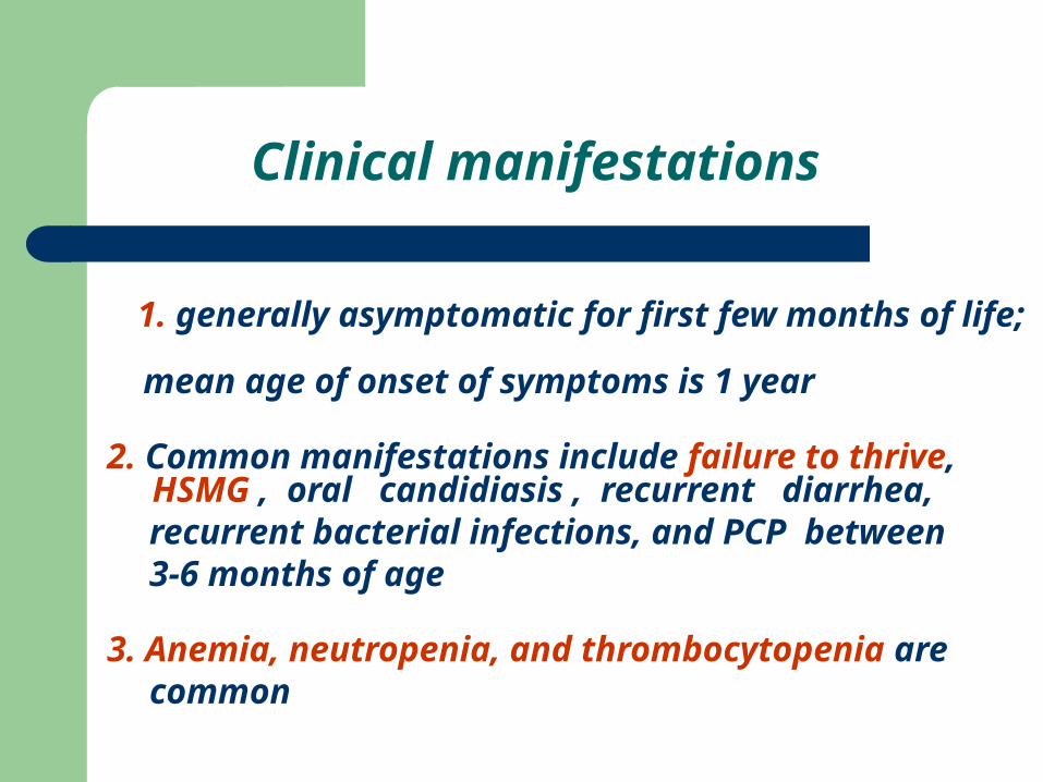
Clinical manifestations
1. generally asymptomatic for first few months of life; mean age of onset of symptoms is 1 year
2. Common manifestations include failure to thrive, HSMG , oral candidiasis , recurrent diarrhea,
recurrent bacterial infections, and PCP between 3-6 months of age
3. Anemia, neutropenia, and thrombocytopenia are common
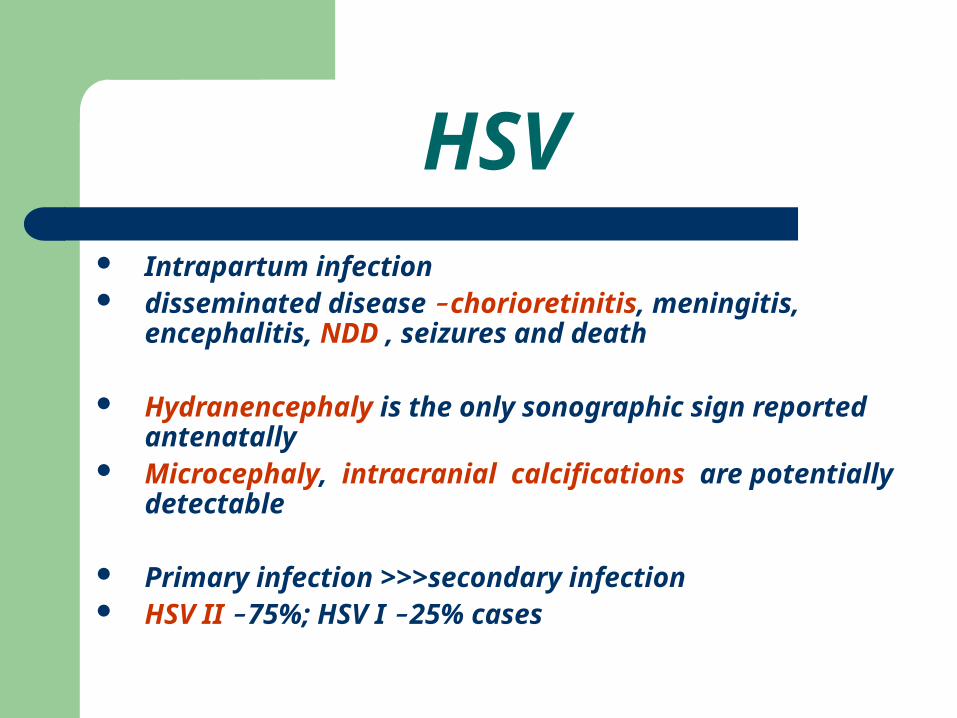
HSV Intrapartum infection disseminated disease - chorioretinitis, meningitis,
encephalitis, NDD , seizures and death
Hydranencephaly is the only sonographic sign reported antenatally
Microcephaly, intracranial calcifications are potentially detectable
Primary infection >>>secondary infection HSV II - 75%; HSV I - 25% cases
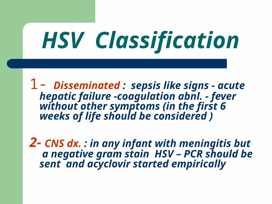
HSV Classification
1- Disseminated : sepsis like signs - acute hepatic failure -coagulation abnl. - fever without other symptoms (in the first 6 weeks of life should be considered )
2- CNS dx. : in any infant with meningitis but a negative gram stain HSV – PCR should be sent and acyclovir started empirically
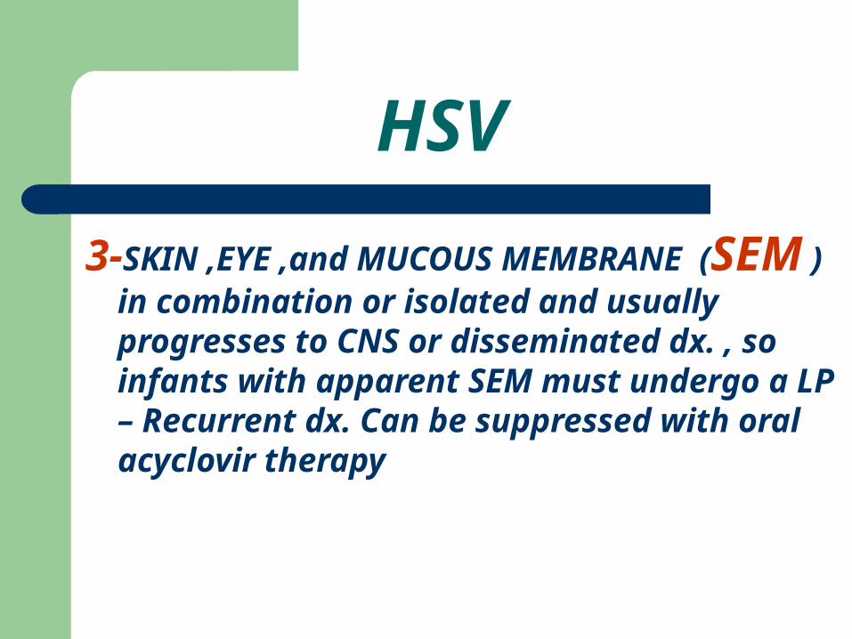
3-SKIN ,EYE ,and MUCOUS MEMBRANE
(SEM ) in combination or isolated and usually progresses to CNS or disseminated dx. , so infants with apparent SEM must undergo a LP – Recurrent dx. Can be suppressed with oral acyclovir therapy
HSV
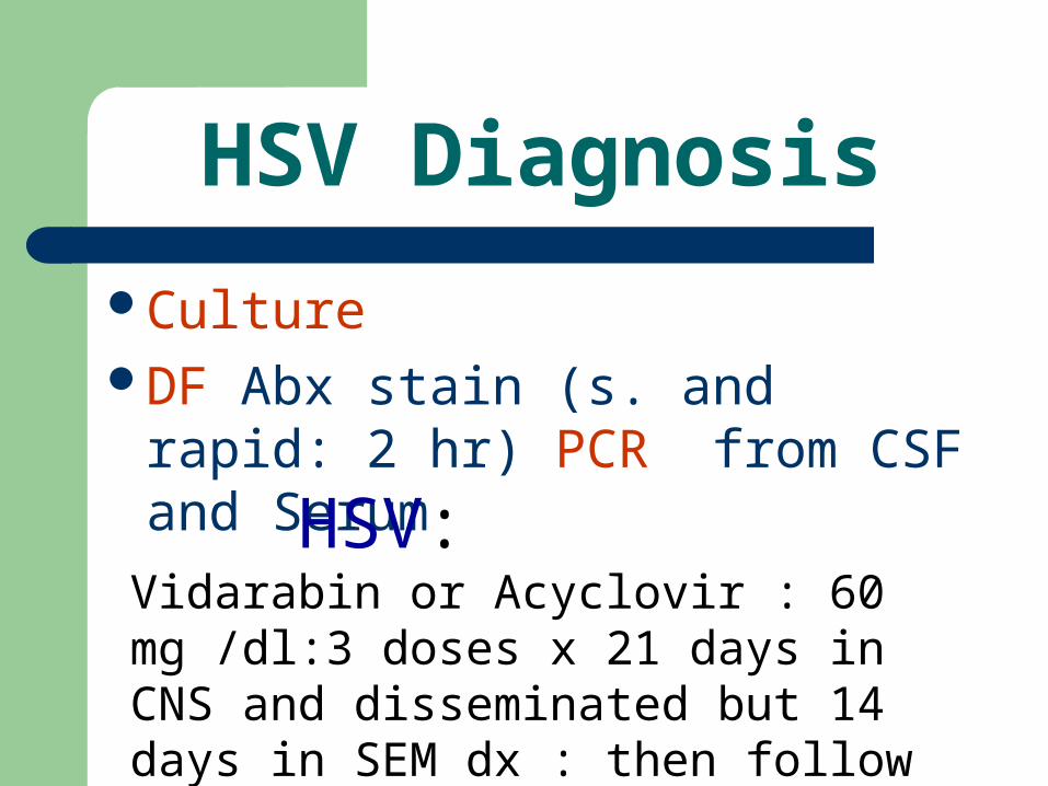
HSV Diagnosis
CultureDF Abx stain (s. and rapid: 2 hr)
PCR from CSF and Serum
HSV: Vidarabin or Acyclovir : 60 mg /dl:3 doses x 21 days in CNS and disseminated but 14 days in SEM dx : then follow by PCR
Parvovirus
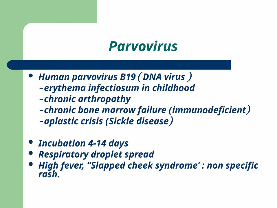
Human parvovirus B19 ( DNA virus )- erythema infectiosum in childhood- chronic arthropathy- chronic bone marrow failure (immunodeficient)- aplastic crisis (Sickle disease)
Incubation 4-14 days Respiratory droplet spread High fever, “Slapped cheek syndrome’ : non specific
rash.
Parvovirus and fetus
Hydrops (anaemia, myocarditis) Adults : 60% sero-positive 1/3 fetuses affected in acute infection Fetal loss rare with appropriate treatment Assess serology – IgG , IgM, paired
serology Serial ultrasound, intrauterine transfusion
Varicella and pregnancy
Mild immunocompromise of pregnancy increases risk
10% develop pulmonary complications - main cause of mortality
Fetal effectsPreterm deliveryVaricella syndromeNeonatal varicella
Varicella Syndrome
Cutaneous scarring Limb hypoplasia Missing/ hypoplastic digits Limb paralysis/muscle atrophy Psychomotor retardation and Convulsions Microcephaly and Cerebral atrophy Chorioretinitis/ choriod scarring/optic disc
hypoplasia , Cataracts Horner’s Syndrome Early childhood Zoster
SYPHILIS
several patterns: 1- Asymptomatic infection : the most
common type
2- Symptomatic dx :usually manifested by a rash : vesicular or bullous or may be EM (demarcated on diaper , palms and soles)
3- Chronic Rhinitis ( “the snuffles”) :often blood – tinged with fissures of the lips
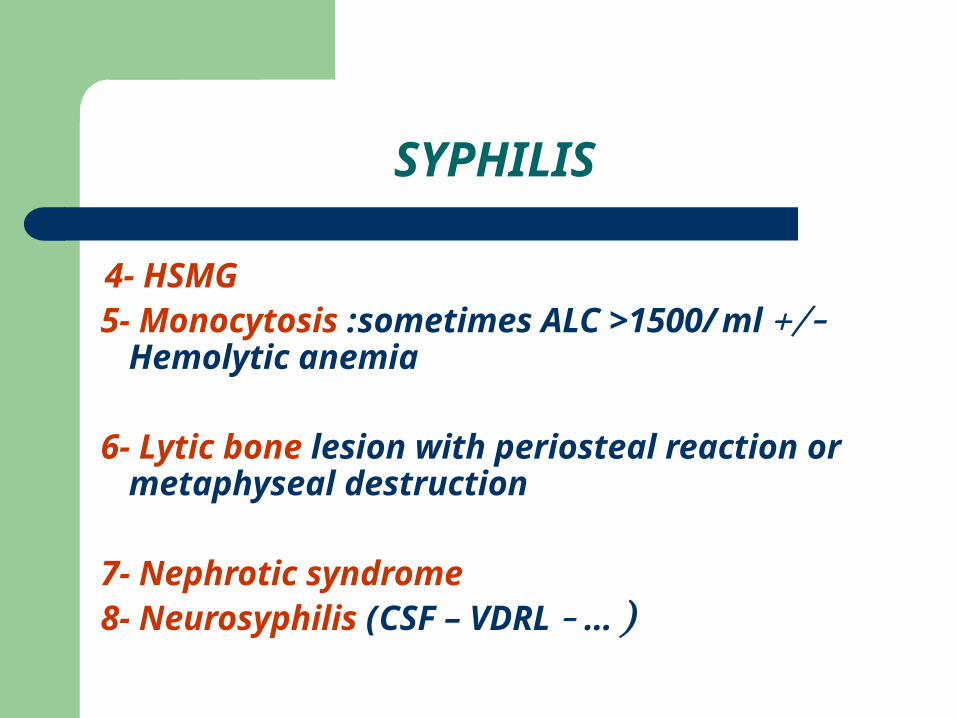
4- HSMG 5- Monocytosis :sometimes ALC >1500/ml +/-
Hemolytic anemia
6- Lytic bone lesion with periosteal reaction or metaphyseal destruction
7- Nephrotic syndrome 8- Neurosyphilis (CSF – VDRL - … )
SYPHILIS

THANKS A LOT