+ All Categories
Home > Documents > spiral.imperial.ac.uk · Web viewEpidemiological interactions between urogenital and intestinal...

spiral.imperial.ac.uk · Web viewEpidemiological interactions between urogenital and intestinal...

Date post: 10-Jun-2018
Category:
Upload: letu
View: 222 times
Download: 0 times
Share this document with a friend
54
Epidemiological interactions between urogenital and intestinal human schistosomiasis in the context of praziquantel treatment across three West African countries Sarah C. L. Knowles 1, 2 , Bonnie L. Webster 1, 3 , Amadou Garba 4 , Moussa Sacko 5 , Oumar T. Diaw 6 , Alan Fenwick 1 , David Rollinson 3 , Joanne P. Webster 1,7 1 Department of Infectious Disease Epidemiology, Imperial College London, St. Mary’s Campus, Norfolk Place, London, W2 1PG 2 Department of Life Sciences, Imperial College London, Silwood Park Campus, Ascot, Berkshire, UK 3 Natural History Museum, Parasites and Vectors Division, Department of Life Sciences, Cromwell Road London. 4 Réseau International Schistosomose, Environnement, Aménagement et Lutte (RISEAL), 333 Avenue des Zarmakoye, B.P. 13724, Niamey, Niger 5 Institut National de Recherche en Santé Publique (INRSP), Ministère de la Santé, B.P. 1771, Bamako, Mali. 6 Institut Sénégalais de Recherches Agricoles (ISRA), route des Hydrocarbures, Bel Air, 3120 Dakar, Sénégal 7 Department of Pathology and Pathogen Biology, Centre for Emerging, Endemic and Exotic Diseases (CEEED), Royal Veterinary College, University of London, AL9 7TA, UK 1
Transcript

Epidemiological interactions between urogenital and intestinal human schistosomiasis in the context of praziquantel treatment across three West African countries

Sarah C. L. Knowles1, 2, Bonnie L. Webster1, 3, Amadou Garba4, Moussa Sacko5, Oumar T. Diaw6, Alan

Fenwick1, David Rollinson3, Joanne P. Webster1,7

1 Department of Infectious Disease Epidemiology, Imperial College London, St. Mary’s Campus, Norfolk

Place, London, W2 1PG

2 Department of Life Sciences, Imperial College London, Silwood Park Campus, Ascot, Berkshire, UK

3 Natural History Museum, Parasites and Vectors Division, Department of Life Sciences, Cromwell Road

London.

4 Réseau International Schistosomose, Environnement, Aménagement et Lutte (RISEAL), 333 Avenue des

Zarmakoye, B.P. 13724, Niamey, Niger

5 Institut National de Recherche en Santé Publique (INRSP), Ministère de la Santé, B.P. 1771, Bamako,

Mali.

6 Institut Sénégalais de Recherches Agricoles (ISRA), route des Hydrocarbures, Bel Air, 3120 Dakar,

Sénégal

7 Department of Pathology and Pathogen Biology, Centre for Emerging, Endemic and Exotic Diseases

(CEEED), Royal Veterinary College, University of London, AL9 7TA, UK

1

Abstract

Background: In many parts of sub-Saharan Africa, urogenital and intestinal schistosomiasis co-

occur, and mixed species infections containing both Schistosoma haematobium and S. mansoni

can be common. During co-infection, interactions between these two species are possible, yet the

extent to which such interactions influence disease dynamics or the outcome of control efforts

remains poorly understood.

Methodology/Principal Findings: Here we analyse epidemiological data from three West African

countries co-endemic for urogenital and intestinal schistosomiasis (Senegal, Niger and Mali) to

test whether the impact of praziquantel (PZQ) treatment, subsequent levels of re-infection or long-

term infection dynamics are altered by co-infection. In all countries, positive associations between

the two species prevailed at baseline: infection by one species tended to predict infection intensity

for the other, with the strength of association varying across sites. Encouragingly, we found little

evidence that co-infection influenced PZQ efficacy: species-specific egg reduction rates (ERR)

and cure rates (CR) did not differ significantly with co-infection, and variation in treatment success

was largely geographical. In Senegal, despite positive associations at baseline, children with S.

mansoni co-infection at the time of treatment were less intensely re-infected by S. haematobium

than those with single infections, suggesting competition between the species may occur post-

treatment. Furthermore, the proportion of schistosome infections attributable to S. mansoni

increased over time in all three countries examined.

Conclusions/Significance: These findings suggest that while co-infection between urinary and

intestinal schistosomes may not directly affect PZQ treatment efficacy, competitive interspecific

interactions may influence epidemiological patterns of re-infection post-treatment. While re-

infection patterns differed most strongly according to geographic location,, interspecific

interactions also seem to play a role, and could cause the community composition in mixed

species settings to shift as disease control efforts intensify, a situation with implications for future

disease management in this multi-species system.

2

1

2

3

4

5

6

7

8

9

10

11

12

13

14

15

16

17

18

19

20

21

22

23

24

25

26

27

Author Summary

In many parts of Africa both urinary and intestinal schistosomiasis are endemic, and mixed

species infections can be common. However, little is known about potential within-host

interactions between the causative parasites, S. haematobium and S. mansoni, and how these

might influence treatment success and post-treatment patterns of re-infection. Here, we bring

together datasets from three West African countries to examine the epidemiological evidence for

interactions between these two schistosome species relevant to the impact of treatment

programmes using praziquantel (PZQ). Encouragingly, PZQ efficacy (in a double 40mg/kg dose

format) was not significantly altered by co-infection, though since co-infections tended to be

heavier, complete clearance was less likely than for single species infections. Despite positive

associations in infection intensity for these two species at baseline, Senegalese children that were

successfully treated for S. haematobium showed less intense re-infection if they were co-infected

with S. mansoni at the point of treatment. Furthermore, in all three settings, the proportion of

infections attributable to S. mansoni increased over successive rounds of PZQ treatment. These

data suggest asymmetric competition may occur between S. haematobium and S. mansoni in the

context of drug treatment, which may alter schistosome species composition as PZQ-based

control programmes proceed.

Introduction

Globally, at least 230 million people are estimated to have schistosomiasis [1]. In sub-Saharan

Africa where the disease burden is highest, Schistosoma haematobium and S. mansoni, causing

urogenital and intestinal schistosomiasis respectively, frequently overlap in their geographic

distribution [2-5] as do their respective snail hosts Bulinus and Biompharia spp. In such areas,

mixed species infections can be common [6-10], and may be even more widespread than

currently recognised if diagnostic methods with greater sensitivity than standard microscopy are

applied [11].

3

28

29

30

31

32

33

34

35

36

37

38

39

40

41

42

43

44

45

46

47

48

49

50

51

52

53

Co-infection with both S. haematobium and S. mansoni generates the potential for within-host

parasite interactions, whereby the presence of one species may alter the course of infection or

disease caused by the other. Such interactions could arise through competition for nutrients or

mates, or immune-mediated mechanisms, including cross-reactive immune responses. Immune-

mediated interactions may also arise through or be affected by drug treatment, if species differ in

drug susceptibility, or if the drug in question alters host immunity in a way that favours one species

over another [12, 13]. Although rarely investigated, such interspecific interactions may have

important implications for schistosomiasis epidemiology, associated morbidity, and the

effectiveness of control measures including PZQ treatment, the cornerstone of current

schistosomiasis control programmes [14].

Evidence for biologically relevant interactions between co-infecting S. haematobium and S.

mansoni comes from studies in animal models as well as humans. These two species can engage

in mate competition, since during co-infection, infertile interspecific mating pairs form resulting in

the release of ectopic eggs: S. haematobium eggs in faeces or S. mansoni eggs in urine [7, 15-

17]. Moreover, mixed species infections produce different morbidity profiles in humans, altering

the relative levels of bladder and liver morbidity [6, 8, 15, 18, 19]. Immune-mediated competition

between S. haematobium and S. mansoni has also been reported in animal models [20-22] and

immunological studies suggest widespread cross-reactivity among antigenic epitopes from

different schistosome species [23].

However, exactly how such interactions might play out in epidemiological settings, and in the

context of mass PZQ treatment, remains underexplored. In parts of Africa the species composition

of schistosomiasis infections has shifted notably over time. At sites in Senegal, Niger, Cameroon

and Egypt, S. mansoni has been introduced through changes in irrigation (e.g. dam construction),

and has been seen to increase in prevalence and subsequently ‘take over’ from S. haematobium

24-29]. While changes in the distribution and relative abundance of Biomphalaria and Bulinus

snails following water resource development have undoubtedly played a key role in such shifts

[25, 28, 30, 31], whether and how within-host interactions between S. haematobium and S.

4

54

55

56

57

58

59

60

61

62

63

64

65

66

67

68

69

70

71

72

73

74

75

76

77

78

79

80

mansoni might influence schistosomiasis epidemiology remains to be fully investigated. PZQ

treatment could also alter the relative abundance of these two species, if drug efficacy varies

between species or during co-infection, or if treatment alters interspecific interactions during re-

infection [14]. With increasing momentum behind scaling up schistosomiasis control programmes

across much of Africa [32], there is a clear need to understand how each species responds to

treatment, whether these responses depend on the parasite community context, and the

implications for epidemiology and morbidity.

Here, we use three epidemiological datasets from co-endemic areas of West Africa (Senegal,

Niger and Mali) to investigate potential interactions between S. haematobium and S. mansoni in

the context of PZQ treatment. In particular, we examine whether PZQ efficacy is altered by co-

infection, the impact of co-infection on individual re-infection post-treatment, and how schistosome

species composition, as well as prevalence and mean intensity of infection changes over the

course of successive treatment rounds at the population level.

Methods

Study sites and datasets

Two of the datasets analysed here come from co-endemic villages in Niger [4] and Senegal [5]

collected as part of the CONTRAST project, while the third comes from sentinel sites monitored

as part of Mali’s schistosomiasis control programme monitoring and evaluation activities [6].

These datasets are described in more detail below, their characteristics are summarised in

Table 1, and their locations are shown in Fig 1.

Table 1: Characteristics of study sites in Senegal, Niger and Mali used in this study.

5

81

82

83

84

85

86

87

88

89

90

91

92

93

94

95

96

97

98

99

100

101

102

103

104

Country Study type Site/s N children

Baseline uninfected

(%)

Baseline single S. haematobium

(%)

Baseline single S. mansoni

(%)

Baseline co-infected

(%)

Senegal PZQ efficacyTemeye 89 0 21.3 19.1 59.6

Nder 107 0 0 2.8 97.2

Niger PZQ efficacyDiambala 180 0 22.2 24.4 53.3

Namarigoungou 223 0 13.5 40.4 46.2

Mali

Monitoring & evaluation of national treatment programme

29 co-endemic schools in three regions (Bamako, Koulikoro and Ségou), 20 of which followed annually for 3 years.

2477 28.5 45 5.5 21

Senegal

Data were collected in 2007-8 from two villages (Nder and Temeye) in the Senegal River Basin

(Fig 1). In previous work [5], village-level variation in PZQ efficacy and re-infection dynamics was

documented over a one-year period at these sites. Here, we extend analysis of these data to

consider the influence of individual co-infection status on PZQ efficacy, parasite clearance and re-

infection dynamics. Full details on these sites and study design are given in [5], but a brief

description of the study design follows. At baseline, children aged 5 to 15 were recruited in each

village, given a unique identification number, and asked to provide a single urine and stool sample

on three consecutive mornings. S. haematobium egg counts were made from filtrations of 10 ml of

urine using the standard urine filtration method, and duplicate Kato-Katz thick smears were

examined from each stool sample in order to calculate the number of S. mansoni eggs per gram of

stool. Only children infected with either S. haematobium, S. mansoni, or both species were

recruited into the follow-up study if consenting, and all infected children were then treated with two

40mg/kg doses of PZQ, spaced 3 weeks apart. Follow-up surveys were conducted at 6 weeks

after baseline/the first PZQ dose (to monitor PZQ efficacy), 6 months from baseline and 12

months from baseline. All children were screened for both S. haematobium and S. mansoni at

each time point, using the same diagnostics used at baseline. No treatment was given at the 6

6

105

106

107

108

109

110

111

112

113

114

115

116

117

118

119

120

121

122

week follow-up, but all children were given two 40mg/kg PZQ doses 3 weeks apart at both the 6

and 12 month follow ups. For S. haematobium, the number of eggs per 10ml urine was calculated

for each sample, and infection intensity taken as the mean of these values across all available

samples from a 3-day sampling period. For S. mansoni, infection intensity was taken as the mean

number of eggs per gram of stool across all samples from the 3-day sampling period.

Niger

Data were collected in 2007-8 from two S. haematobium/S. mansoni co-endemic villages situated

in the western part of the country along the Niger River - Diambala and Namarigoungou. The

study design was very similar to the Senegal study described above, and is described fully in [4].

Briefly, children aged 6 to 15 infected with either S. haematobium or S. mansoni were recruited

into the study at baseline, given unique identifiers, and follow-up surveys were conducted at 6

weeks, 6 months and 12 months from baseline. As in Senegal, two 40mg/kg doses of PZQ were

given to all infected children after recruitment, and to all children in the study at the 12-month

sampling point, irrespective of infection status. However, unlike in Senegal, no treatment was

given 6 months from baseline. At each survey time-point, all children were screened for both S.

haematobium and S. mansoni using the same diagnostic techniques used in the Senegal study,

with the exception that one Kato-Katz slide was read per stool sample rather than two.

Mali

The Malian data analysed here formed part of the Monitoring and Evaluation component of the

Mali National Schistosomiasis Control Program, supported by the Schistosomiasis Control

Initiative (SCI). In 2004, a set of 33 schools (sentinel sites) were randomly selected from all

schools in three regions known a priori to be highly endemic for schistosomiasis: Bamako, Ségou,

and Koulikoro. Only the 29 schools that were co-endemic for S. haematobium and S. mansoni at

baseline are included in the analyses presented here, as our focus is on individual level

determinants of infection traits, rather than site to site variation in co-endemicity. Baseline data

were collected in 2004. At each school, 50-110 children (approximately equal numbers of boys

7

123

124

125

126

127

128

129

130

131

132

133

134

135

136

137

138

139

140

141

142

143

144

145

146

147

148

and girls) aged 7 to 14 years old were recruited, irrespective of infection status. Participants were

asked to provide a single stool sample, and two urine samples on two consecutive days. Urine

filtration and Kato-Katz examinations were carried out as described in [33], and infection intensity

was calculated as the arithmetic mean number of S. haematobium eggs per 10ml urine, or mean

number of S. mansoni eggs per gram of stool. Two subsequent follow-up surveys were conducted

on this cohort in 2005 and 2006, immediately prior to annual PZQ administration by the national

control programme.

Statistical analyses

All statistical analyses were performed in R v3.1.1. Descriptive statistics (prevalence, mean

infection intensity and confidence intervals) were calculated using the survey package [34],

accounting for clustering of the data by school or village where necessary. The significance of

model terms was assessed using likelihood ratio tests, which compared full models to models

excluding the term of interest.

Baseline associations between S. haematobium and S. mansoni

First, we tested whether the likelihood of infection by each schistosome species depended on co-

infection with the other, using baseline data from Mali where children were recruited irrespective of

infection status. Binomial generalized linear mixed models (GLMMs) with a logit link were

performed using the glmer function in package lme4, with either S. haematobium or S. mansoni

infection status as the response, and school fitted as a random intercept term. All children at the

29 co-endemic Malian schools surveyed at baseline were included. Co-infection status was coded

according to WHO infection intensity categories: for S. haematobium, uninfected, light (<50

eggs/10ml urine) or heavy (≥50eggs/10ml urine) infection, and for S. mansoni, uninfected, light (1-

99 epg), moderate (100-399 epg), or heavy (≥400 epg) infection. Linear and quadratic terms for

age (mean-centred) were included as covariates, as well as gender and geographic region (as a

3-level factor). An interaction term between region and co-infection status was also examined to

8

149

150

151

152

153

154

155

156

157

158

159

160

161

162

163

164

165

166

167

168

169

170

171

172

173

174

test for geographic heterogeneity in schistosome species associations. For all countries we also

assessed whether baseline infection intensity was associated with co-infection, by modelling log-

transformed egg counts for a given species as a function of co-infection status, age, gender and

location (as a 2-level factor for village in both Senegal and Niger, and a 3-level factor for region in

Mali). These infection intensity analyses included infected children only.

PZQ efficacy and parasitological cure

Data on the immediate effect of PZQ (6 weeks after the first of two 3-week spaced treatments)

were available for four villages in Senegal and Niger. To examine whether co-infection might

influence PZQ efficacy, for each country we calculated the egg reduction rate (ERR) for each

schistosome species, in single and co-infected individuals respectively. ERR was calculated using

the formula recommended in WHO guidelines [35], as the difference between the (arithmetic)

mean pre- and post-intervention egg counts divided by the mean pre-intervention egg count,

multiplied by 100. The calculation included only individuals infected with the focal species at

baseline, with known baseline co-infection status, and for which an egg count at 6 weeks post-

treatment was available. Although a wide range of formulas has been used in the past to calculate

ERR, this formula was used as it has been shown to be the most robust metric for evaluating drug

efficacy [36, 37]. Permutation tests were used to examine whether ERR differed significantly

(p<0.05) according to co-infection status. We also calculated species-specific cure rates (CR) –

the proportion of infected individuals that cleared infection six weeks after the first PZQ dose, by

country and co-infection status. With the exception of S. haematobium in Niger (for which

parasitological cure was almost universal) we modelled factors affecting individual parasite

clearance 6 weeks after PZQ using binomial GLMs. Alongside co-infection status as the key

predictor of interest, a number of covariates were controlled for in these models, including initial

infection intensity of the focal species (as logged epg or eggs/10ml urine) to control for differences

in efficacy arising from varying infection intensity and village.

9

175

176

177

178

179

180

181

182

183

184

185

186

187

188

189

190

191

192

193

194

195

196

197

198

199

200

Longitudinal dynamics and re-infection

Using the Mali dataset, we examined whether individuals’ probability of infection changed across

annual rounds of PZQ treatment, according to their initial co-infection status. These analyses

involved a subset of 20 schools from the original 29 co-endemic sentinel sites, as these had

follow-up data for both S. haematobium and S. mansoni from all three years. We constructed a

binomial GLMM, with S. haematobium or S. mansoni infection status as the response, and school

and child ID included as random intercept terms. Fixed effects for baseline co-infection status (as

a 3 or 4-level factor, depending on the species), year (as a 3-level factor: baseline, first and

second follow-up) and their interaction term were used to examine whether temporal changes in

infection for each species differed according to initial co-infection status. Region, gender and

mean-centred age at baseline (linear and quadratic term) were fitted as covariates. Only children

monitored at all three time-points (baseline, first and second follow-up years) were included, such

that we analysed changes in infection probability over time in the same set of individuals. To

examine whether drop-out from the Malian cohort might be biased according to children’s infection

status at baseline, we used a binomial mixed model to test which baseline variables predicted

whether children had full follow-up data. School was fitted as a random intercept term, alongside

S. haematobium and S. mansoni infection categories, with region, gender and mean-centred age

at baseline (linear and quadratic term) as covariates.

In Senegal and Niger it was also possible to separate effects of co-infection on parasite clearance

from those on re-infection (the latter potentially including recommencement of egg-shedding by

uncleared worms), since individuals were monitored six weeks after PZQ treatment. For each

schistosome species, we used a binomial GLM with a logit link to test whether, among individuals

infected at baseline but clear of infection 6 weeks later (after PZQ treatment), the probability of

being re-infected 6 months from baseline depended on baseline co-infection status (single vs. co-

infected). Covariates included were village, baseline intensity category for the focal species (2-

level factor for S. haematobium: light vs. heavy; 3-level factor for S. mansoni: light, moderate or

heavy), gender and mean-centred age (as a linear and quadratic term). An interaction term

10

201

202

203

204

205

206

207

208

209

210

211

212

213

214

215

216

217

218

219

220

221

222

223

224

225

226

227

between village and co-infection status was also included to test for geographic heterogeneity in

the influence of co-infection. Among the same individuals, we also used negative binomial GLMs

(using the glm.nb function in the MASS package) to examine whether the intensity of re-infection

at six months (eggs/gram stool for S. mansoni, or eggs/10ml urine for S. haematobium) was

dependent on co-infection status at baseline, including the same covariates used in the binomial

re-infection models.

Ethics statement

Full details on ethical approval granted are provided in the original publications from which these

data were gathered. In brief, ethical approval was obtained from the St Mary’s Hospital Local

Ethics Research Committee, R&D office (part of the Imperial College, London Research Ethics

Committee (ICREC; EC NO: 03.36. R&D No: 03/SB/033E) in combination with the ongoing

CONTRAST and SCI activities. Within Niger, Senegal and Mali, all aspects of sample collections

were carried out in the framework of the disease control activities implemented and approved by

the Ministry of Health (MOH) and adopted by regional and local administrative and health

authorities. In Senegal and Niger, the communities of the selected villages were informed about

the objectives, the methodology of the study and the advantages. A meeting was organized with

the population and verbal community consent was obtained for each selected village. Written and

verbal consent was also obtained from school directors and teachers, as well as the children’s

parents, prior to the recruitment of the children. Verbal assent was given by every child and their

acceptance documented. Participation was voluntary and children could withdraw or be withdrawn

from the study at any time without obligation. Results of the different diagnostic procedures

performed on children were briefly explained to them. In Senegal and Niger, all children diagnosed

as infected with schistosomiasis were immediately treated with 40mg/kg. In Mali, all children at

participating schools were treated shortly after each survey, as part of the national schistosomiasis

control programme activities.

11

228

229

230

231

232

233

234

235

236

237

238

239

240

241

242

243

244

245

246

247

248

249

250

251

252

253

254

Results

Baseline associations between S. haematobium and S. mansoni

At baseline, associations between the two schistosome species were generally positive, although

varied in strength across countries and regions. In Mali, infection probability for both schistosome

species was positively predicted by the level of infection with the other species, after controlling for

age and gender effects (Table 2). This relationship varied in strength across geographical regions

(S. haematobium: S. mansoni*region interaction χ26=22.28, p=0.001; S. mansoni: S.

haematobium*region interaction: χ24=12.63, p=0.013, Fig 2). Among infected individuals, infection

intensity increased with the intensity of co-infection for both schistosome species, although again

the strength of this relationship varied across countries and regions (Table 3; Fig 3). Across the 29

schools in Mali, these positive relationships for both S. haematobium and S. mansoni infection

intensity were usually observed within each school, with few exceptions (Fig A in S1 Text).

Table 2: Baseline predictors of (A) S. haematobium and (B) S. mansoni infection probability across 29 co-endemic schools in Mali (n= 2477 children). Parameter estimates (on the logit scale) are from binomial mixed models. χ2 and p values are from likelihood ratio tests comparing models with and without the term in question. Age was mean-centred in both analyses. ‘ref’ indicates the reference level of each factor.

Variable df Parameter estimate (SE) χ2 p

(A) S. haematobium

(Intercept) 0.016 (0.360)Age 1 0.105 (0.024) 18.760 <0.0001Age2 1 -0.010 (0.012) 0.766 0.3816Sex 1 Male (ref) 0 2.363 0.1243

Female -0.162 (0.105)

S. mansoni infection 3 Uninfected (ref) 0 51.066 <0.0001

Light 0.914 (0.207)

Moderate 1.411 (0.276)

Heavy 1.940 (0.306)

Region 2 Bamako (ref) 0 19.015 <0.0001

Koulikoro -0.894 (0.759)

Ségou 2.345 (0.545)

(B) S. mansoni

12

255

256

257

258

259

260

261

262

263

264

265

266

267

268

269270271272273

(Intercept) -3.391 (0.562)Age 1 0.161 (0.035) 21.817 <0.0001Age2 1 0.006 (0.017) 0.123 0.7253Sex 1 Male (ref) 0 2.808 0.0938

Female -0.255 (0.149)S. haematobium infection 2 Uninfected (ref) 0 56.630 <0.0001

Light 1.054 (0.202)Heavy 1.861 (0.253)

Region 2 Bamako (ref(A) 0 13.499 0.0012Koulikoro 4.441 (1.118)Ségou 0.127 (0.807)

13

274

Mali (n=1398) Senegal (n=171) Niger (n=269)Variable df Parameter estimate (SE) χ2 p df Parameter estimate (SE) χ2 p df Parameter estimate (SE) χ2 p(A) S. haematobium intensity(Intercept) 3.032 (0.224) -0.179

(0.600) 1.244 (0.131)

Age 1 -0.032 (0.019)

3.314 0.0687 1 0.014 (0.047) 0.172 0.6785 0.032 (0.028) 1.607 0.2049Age2 1 -0.013

(0.009)2.071 0.1502 1 0.005 (0.015) 0.129 0.7192 1 0.002 (0.013) 0.020 0.8890

Sex 1 Male (ref) 0 12.232 0.0005 1 Male (ref) 0 0.830 0.3623 1 Male (ref) 0 2.745 0.0976Female -0.283

(0.081)Female 0.257 (0.289) Female -0.169

(0.103)S. mansoni infection 3 Uninfected (ref) 0 15.967 0.0012 3 Uninfected (ref)

0 35.296 <0.0001 3 Uninfected (ref) 0 16.727 0.0008Light 0.399 (0.149) Light 1.684 (0.500) Light -0.059

(0.142)Moderate 0.591 (0.198) Moderate 3.040 (0.588) Moderate 0.276 (0.131)Heavy 0.813 (0.202) Heavy 3.513 (0.657) Heavy 0.668 (0.200)

Region / Village 2 Bamako (ref) 0 11.167 0.0038 1 Nder (ref) 0 8.633 0.0033 1 Diambala (ref) 0 0.070 0.7908Kouikoro -0.632

(0.474)Temeye 1.039 (0.357) Namarigoungou 0.028 (0.108)

Ségou 0.830 (0.318)Mali (n=654) Senegal (n=172) Niger (n=333)

df Parameter estimate (SE) χ2 p df Parameter estimate (SE) χ2 p df Parameter estimate (SE) χ2 p(B) S. mansoni(Intercept) 3.463 (0.322) 0.376

(11.233) 4.506 (0.131)

Age 1 0.123 (0.024) 26.700 <0.0001 1 0.054 (0.032) 2.178 0.1400 1 0.119 (0.027) 21.450 <0.0001Age2 1 0.002 (0.012) 0.017 0.8952 1 -0.011

(0.010)1.227 0.2680 1 0.003 (0.013) 0.045 0.8323

Sex 1 Male (ref) 0 11.280 <0.0001 1 Male (ref) 0 1.957 0.1618 1 Male (ref) 0 0.687 0.4073Female -0.361

(0.108)Female 0.281 (0.205) Female -0.085

(0.104)S. haematobium infection

2 Uninfected (ref) 0 38.585 <0.0001 3 Uninfected (ref)

0 14.790 0.0006 3 Uninfected (ref) 0 1.664 0.4351Light 0.623 (0.143) Light 0.189 (0.341) Light 0.099 (0.106)Heavy 1.106 (0.175) Heavy 1.025 (0.362) Heavy -0.749

(0.948)Region / Village 2 Bamako (ref) 0 12.864 0.0016 1 Nder (ref) 0 36.943 <0.0001 1 Diambala (ref) 0 24.310 <0.0001Kouikoro 1.848 (0.470) Temeye -1.464

(0.233)Namarigoungou 0.527 (0.106)

Ségou 0.667 (0.390)

Table 3: Baseline predictors of infection intensity for (A) S. haematobium and (B) S. mansoni across three co-endemic countries in West Africa. Results are from Gaussian GLMs (Senegal and Niger), or GLMMs with school as a random intercept term (Mali), using log transformed egg counts among infected individuals only (eggs/10ml urine or eggs/gram stool) as the response. χ2 and p values show results of likelihood ratio tests. Parameters from full models are shown, and age was mean-centred in analyses. ‘ref’ indicates the reference level of each factor.

14

275276277278

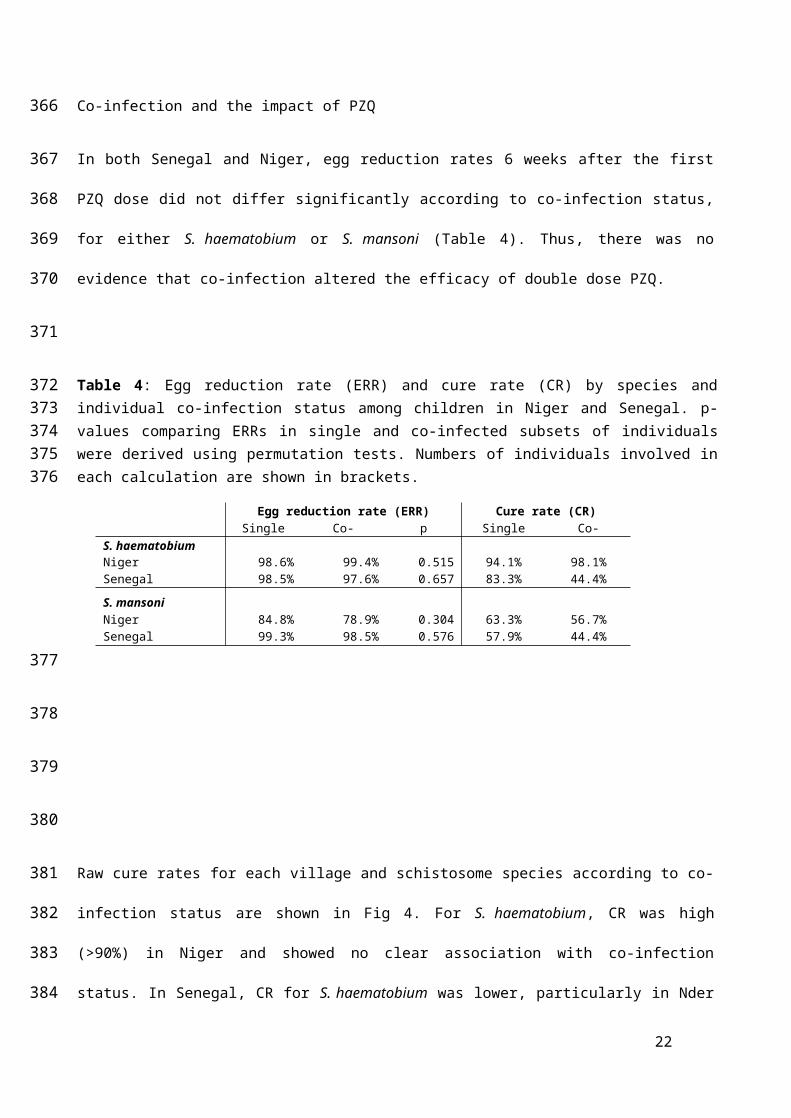
Co-infection and the impact of PZQ

In both Senegal and Niger, egg reduction rates 6 weeks after the first PZQ dose did not differ

significantly according to co-infection status, for either S. haematobium or S. mansoni (Table 4).

Thus, there was no evidence that co-infection altered the efficacy of double dose PZQ.

Table 4: Egg reduction rate (ERR) and cure rate (CR) by species and individual co-infection status among children in Niger and Senegal. p-values comparing ERRs in single and co-infected subsets of individuals were derived using permutation tests. Numbers of individuals involved in each calculation are shown in brackets.

Egg reduction rate (ERR) Cure rate (CR)Single Co-infected p

(permutatioSingle Co-infected

S. haematobiumNiger 98.6% (34) 99.4% (104) 0.515 94.1% (34) 98.1% (104)Senegal 98.5% (18) 97.6% (146) 0.657 83.3% (18) 44.4% (146)

S. mansoniNiger 84.8% (79) 78.9% (104) 0.304 63.3% (79) 56.7% (104)Senegal 99.3% (19) 98.5% (142) 0.576 57.9% (19) 44.4% (142)

Raw cure rates for each village and schistosome species according to co-infection status are

shown in Fig 4. For S. haematobium, CR was high (>90%) in Niger and showed no clear

association with co-infection status. In Senegal, CR for S. haematobium was lower, particularly in

Nder (where all individuals were co-infected with S. mansoni) as well as in co-infected individuals

in Temeye (Fig 4A, Table 5). In a model including data from all four villages in Senegal and Niger,

co-infection did not significantly explain variation in S. haematobium clearance, when controlling

for significant effects of village and initial S. haematobium infection intensity (Table 5A). In

Temeye, co-infected children had a lower CR than those with single infections (Fig 4A). However,

models showed that this effect was equally well explained by higher S. haematobium infection

15

279

280

281

282

283

284285286287

288

289

290

291

292

293

294

295

296

297

298

299

300

intensity as by co-infection. Considering data on parasitological cure from Temeye only, both co-

infection status and initial intensity had equal explanatory power in univariate models (likelihood

ratio tests for co-infection: χ21=4.16, p=0.041; log(eggs per 10ml): χ2

1=4.15, p=0.042). However,

neither term added significant explanatory power when the other was already present in the model

(likelihood ratio test for co-infection: χ2= 1.74, p=0.187; log(eggs per 10ml): χ2= 1.74, p=0.188),

suggesting that these two variables strongly confound one another in Temeye.

For S. mansoni, CR was lower in Niger than in Senegal, though in neither country did CR depend

strongly on co-infection status (Fig 4B). In a model including data from all four villages, co-

infection did not significantly explain S. mansoni clearance probability, when controlling for other

effects, particularly strong variation between the two countries (Table 5B). Among those who did

not clear S. mansoni entirely, the number of remaining eggs was also not predicted by S.

haematobium co-infection (GLM on logged egg counts: co-infection term χ2= 2.47, p=0.116). In a

model where clearance of all schistosome eggs of both species six weeks after PZQ was the

response, there was marginal evidence that co-infected individuals were less likely to clear all

parasites than those with single species infections, when controlling for differences in clearance

rates between villages (Table 5C).

S. haematobium longitudinal infection dynamics

Patterns of S. haematobium infection post-treatment varied widely across sites. In Senegal,

rapid re-infection with S. haematobium was seen in Temeye, though not in Nder (Fig 5A, B),

whereas in both Nigerien villages S. haematobium re-infection was much slower (Fig 5C, D). In

Mali, S. haematobium infection probability was higher in S. mansoni infected children at

baseline, and declined less rapidly, though by the second annual follow-up, no differences in

prevalence were apparent according to baseline S. mansoni status (Fig 6A; Table A in S1

Text). At the first annual follow-up (F1), an individual’s S. haematobium infection probability

was predicted by their change in S. mansoni infection since baseline: while controlling for

baseline S. haematobium status (i.e. predisposition to the focal species), children persistently

16

301

302

303

304

305

306

307

308

309

310

311

312

313

314

315

316

317

318

319

320

321

322

323

324

325

326

infected with S. mansoni (at baseline and follow-up) were more likely to be infected by S.

haematobium than those that never had S. mansoni, while those that either lost or gained S.

mansoni since baseline had intermediate infection probabilities (S. mansoni change: χ23=12.06,

p=0.007, controlling for significant effects of age, gender and baseline S. haematobium

infection category; Fig B in S1 Text). Among the 2197 children recruited at the 20 schools

where both schistosome species were monitored for all three years, 475 (22%) were only

sampled at baseline, 466 (21%) were seen in two of the three years, and 1256 (57%) were

followed-up in all three years. The mean number of follow-ups was 1.36. Drop-out analysis

showed that the probability of children having full follow-up data (and therefore being included

in these longitudinal analyses) was not affected by baseline infection status for S. haematobium

or S. mansoni, but declined with age and differed across regions (Table B in S1 Text).

Table 5: Factors predicting the probability of parasitological cure for (A) S. haematobium, (B) S. mansoni and (C) any schistosome infection, six weeks after PZQ treatment in Senegalese and

Variable df Parameter estimate (SE) χ2 p(A) S. haematobium (n=302)(Intercept) 0.519 (0.075)log(S. haematobium eggs/10ml) 1 -0.043 (0.014) 9.213 0.0024S. mansoni infection 3 Uninfected (ref) 0 1.101 0.7767

Light -0.032 (0.064)Moderate -0.038 (0.071)Heavy 0.031 (0.093)

Village 3 Nder (ref) 0 84.901 <0.0001Namarigoungou 0.543 (0.065)Diambala 0.516 (0.062)Temeye 0.244 (0.063)

(B) S. mansoni (n=344)(Intercept) 0.950 (0.110)log(S. mansoni epg) 1 -0.016 (0.020) 0.675 0.4112S. haematobium infection 2 Uninfected (ref) 0 3.000 0.2232

Light -0.040 (0.056)Heavy -0.152 (0.089)

Village 3 Nder (ref) 0 31.351 <0.0001Namarigoungou 0.250 (0.069)Diambala -0.262 (0.068)Temeye 0.121 (0.074)

(C) Both species combined (n=399) (Intercept) -0.402 (0.314)Infection status 1 One species (ref) 0 3.066 0.0799

Two species -0.411 (0.235)Village 3 Nder (ref) 0 16.075 0.0011

Namarigoungou 0.964 (0.313)Diambala 0.962 (0.313)Temeye 1.159 (0.322)

17

327

328

329

330

331

332

333

334

335

336

337

338

339340

Nigerien villages where both species are endemic. Parameter estimates (on the logit scale) are from binomial mixed models. χ2 and p values are from likelihood ratio tests comparing full models with and without the term in question. ‘ref’ indicates the reference level of each factor.

S. haematobium re-infection isolated from lack of clearance

No S. haematobium re-infection was observed after 6 months in Nigerien co-endemic villages, so

variation in S. haematobium re-infection was only examined in Senegal. Among individuals with S.

haematobium at baseline that had cleared infection six weeks after PZQ treatment, six month re-

infection rates were 14% in Nder (where 100% children had S. mansoni at baseline), and 98% in

Temeye (96% in children co-infected with S. mansoni at baseline vs. 100% in children singly

infected). In a model including data from both villages, re-infection probability was strongly

predicted by village (higher in Temeye than Nder, χ21=133.71, p<0.001) and was also higher in

individuals with heavy compared to light S. haematobium infection at baseline (χ22=6.17,

p=0.0130). As expected given the limited variation in re-infection rates in Temeye (the only village

where co-infection showed some variation), no significant effect of baseline S. mansoni co-

infection on re-infection probability was detected while controlling for village and other effects (χ21=

1.27, p=0.259). However, across both Senegalese villages, the intensity of S. haematobium re-

infection at six months post-treatment (which varied more widely, range 0-372 eggs/10ml), was

significantly lower in those individuals co-infected by S. mansoni at baseline (Table 6A, Fig 7A).

The negative binomial model provided a good fit to re-infection intensity data, with observed

counts closely matching expected values (Fig C in S1 Text) and a low overdispersion parameter

(α=1.2). It was not possible to test for an interaction between co-infection status and village, as all

individuals in Nder were co-infected at baseline. However, when limiting the analysis to the village

of Temeye, the same negative effect of S. mansoni co-infection on S. haematobium re-infection

intensity was seen (χ2= 12.02, p=0.0005). To explore this effect further, we tested how the change

in an individual’s S. mansoni infection status between baseline, 6 weeks and 6 months predicted

re-infection intensity, using data from both villages. This showed that the major difference in S.

haematobium re-infection intensity was between those individuals that were S. mansoni negative

at baseline but gained infection by 6 months (pattern 0-0-1), compared to those that were cleared

18

341342343

344

345

346

347

348

349

350

351

352

353

354

355

356

357

358

359

360

361

362

363

364

365

366

367

368

of their original S. mansoni infection (1-0-0 and 1-0-1; Table C in S1 Text). We also examined

geographical variation in S. haematobium re-infection rates across all of the seven villages in

Senegal and Niger assessed in our previous studies [4, 5]. We noted that village-level S.

haematobium re-infection rate at six months tended to decrease with increasing baseline mean S.

mansoni infection intensity (Fig 7B).

Table 6: Re-infection intensity 6-months after baseline among individuals that cleared their infection 6 weeks after PZQ treatment, modelled by negative binomial GLMs. Baseline infection intensity for the focal species was retained in models even when not significant, so the effect of co-infection over and above effects of the focal species could be assessed. χ2 and p values are from likelihood ratio tests comparing full models with and without the term in question. Age was mean-centred in analyses. ‘ref’ indicates the reference level of each factor.

Variable df Parameter estimate (SE) χ2 p

S. haematobium eggs/10ml (n=70; Senegal only)(Intercept)Baseline S. haematobium infection 1 Light (ref) 0 1.891 0.1691

Heavy 0.800 (0.556)Sex 1 Male (ref) 0 0.013 0.9092

Female 0.051 (0.429)Age 1 -0.189 (0.100) 4.322 0.0376Age2 1 -0.041 (0.037) 1.15 0.2836Village 1 Nder (ref) 0 50.374 <0.0001

Temeye 4.396 (0.720)Baseline S. mansoni infection 1 Not infected (ref) 0 10.713 0.0011

Co-infected -1.657 (0.498)S. mansoni eggs/gram(n=225, Senegal and Niger)(Intercept) 6.598 (0.393)Baseline S. mansoni infection 2 Light (ref) 0 2.798 0.2469

Moderate 0.440 (0.249)Heavy 0.092 (0.363)

Sex 1 Male (ref) 0 0.296 0.5866Female 0.119 (0.220)

Age 1 -0.021 (0.042)Age2 1 -0.039 (0.014) 6.818 0.0090Village 3 Nder (ref) 0 78.815 <0.0001

Temeye -0.902 (0.322)Diambala -3.608 (0.353)Namarigoungou -2.255 (0.343)

Baseline S. haematobium infection 1 Not infected (ref) 0 4.683 0.0305Co-infected -0.602 (0.266)

S. mansoni longitudinal patterns

19

369

370

371

372

373

374375376377378379

380

381

382

383

Similar to S. haematobium, re-infection for S. mansoni was much quicker in Senegalese than

Nigerien villages (Fig 8). In Mali, S. mansoni prevalence declined over two annual PZQ

treatments, and this pattern differed only a minor amount according to baseline S. mansoni

infection status (Table B in S1 Text): S. mansoni infection probability was higher in S.

haematobium positive children at baseline and declined less rapidly, though no differences

according to baseline S. haematobium infection status were visible after two years (Fig 7B). After

one year, similar to the pattern seen for S. haematobium, S. mansoni infection probability was

weakly predicted by changes in S. haematobium infection: children persistently infected with S.

haematobium (at both baseline and first follow-up) were more likely to be infected with S. mansoni

than those that never had S. haematobium, with those that either lost or gained S. haematobium

intermediate (S. haematobium change: χ23= 10.22, p=0.017, controlling for effects of age, gender,

region and baseline S. mansoni infection category; Fig B in S1 Text).

S. mansoni re-infection isolated from lack of clearance

In the four Senegalese and Nigerien villages, among children that had S. mansoni at baseline but

cleared it 6 weeks post-PZQ, re-infection probability at six months was strongly predicted by

village (χ23= 65.73, p<0.0001), largely reflecting differences between the two countries (Fig 8). S.

haematobium co-infection status did not predict S. mansoni re-infection probability (χ21= 2.312,

p=0.128), while controlling for village, baseline S. mansoni infection intensity category (χ22= 0.799,

p=0.671), and age (Age2 χ21=4.883, p=0.027). These results reflect inconsistent patterns of re-

infection rate according to S. haematobium co-infection status across the four villages (single vs.

co-infected in Temeye, Nder, Diambala and Namarigoungou: 100% vs. 88%, 67% vs. 97%, 33%

vs. 38% and 92% vs. 59% respectively). In models of S. mansoni re-infection intensity, again the

strongest effect was village (Table 6B). However, a weak main effect of S. haematobium co-

infection on re-infection intensity was also detected while controlling for covariates, indicating

slightly lighter S. mansoni re-infection overall among individuals co-infected with S. haematobium

at baseline (Table 6B). When an interaction term between village and co-infection status was

20

384

385

386

387

388

389

390

391

392

393

394

395

396

397

398

399

400

401

402

403

404

405

406

407

408

409

410

included in the model, however, this was also weakly significant (χ23=7.943, p=0.047), indicating

that the effect of co-infection differed somewhat across sites. This interaction term reflected a

weak positive association between co-infection and S. mansoni re-infection intensity in Nder and

Diambala compared to a weak negative association in Temeye and Namarigoungou (Fig D in S1

Text), though in single village analyses, the co-infection term was always non-significant (all

p>0.08). Overall therefore, S. mansoni re-infection was not strongly predicted by initial S.

haematobium co-infection status, and the relatively weak negative effect of co-infection on egg

counts at six months was not consistent across sites. We also checked whether any potential

effect of S. haematobium co-infection on re-infection intensity might depend on an individual’s

initial S. mansoni infection intensity, but there was no evidence for such an interaction (χ22 =3.451,

p=0.178).

Discussion

Despite frequent co-endemicity of S. haematobium and S. mansoni across Africa, little is known

about the potential for within-host interactions between these two species, or whether such

interactions might affect the impact of PZQ treatment or host immunity. Here, we analyse

epidemiological data from school-age children in three West African countries (Senegal, Niger and

Mali), to explore whether these two species may interact with one another in the context of PZQ

treatment.

At baseline, associations between urogenital and intestinal schistosomiasis were generally

positive in all three countries: for both S. haematobium and S. mansoni, infection intensity

increased with the degree of co-infection, while controlling for other effects such as age, gender

and location, and in Mali children with one species were more likely to be infected by the other,

with some geographic variation in the strength of this association. Similar positive associations

prior to treatment have been previously reported from other co-endemic villages in Senegal [7],

and Cameroon [38]. Such positive associations may be driven by a number of processes,

21

411

412

413

414

415

416

417

418

419

420

421

422

423

424

425

426

427

428

429

430

431

432

433

434

435

436

including correlated exposure of children to infective Bulinus and Biomphalaria snails at the same

transmission sites, correlated immune susceptibility of individuals to infection by the two species,

and/or facilitative interspecific interactions within the host. Few studies have examined the

evidence for correlated exposure to S. haematobium and S. mansoni, and while some have in fact

suggested these species may have distinct local transmission sites within villages [39, 40], further

work is needed to examine the extent to which correlated exposure, or other mechanisms, may

drive the positive associations between schistosome species documented here and elsewhere.

In analysing data from two efficacy studies with similar methodology in Senegal and Niger, we

found that the efficacy (ERR) of two (three week spaced) PZQ doses against either S.

haematobium or S. mansoni was not significantly altered by the presence of co-infection. This is

encouraging, though the effect of co-infection on efficacy still requires testing for the standard,

single 40mg/kg dose of PZQ widely used in control programmes. Parasitological cure rates told a

somewhat different story, with clearance probability for S. haematobium reduced in mixed

infections compared to single infections in Senegal, but not Niger. Further analysis showed that

this effect of S. mansoni on S. haematobium clearance disappeared once initial infection intensity

was controlled for, suggesting intensity differences likely underpinned reduced clearance of S.

haematobium from mixed infections in Senegal. While it is well known that heavy infections are

less likely to be cleared by a standard dose of PZQ [41-43], a key question arising from our

findings is to what extent S. mansoni co-infection causes, or is merely correlated with, increased

infection intensity for S. haematobium. The answer will determine whether co-endemicity in an

area per se is cause to consider higher or more repeated doses of PZQ to get full infection

clearance. Our finding of reduced overall clearance for S. haematobium/S. mansoni co-infections

compared to single infections agrees with a recent meta-analysis of studies using the standard

40mg/kg PZQ dose [44] in suggesting mixed infections may be harder to clear. Again whether this

observation is due to mixed infections simply being heavier, or other effects remains unclear. Our

results did not suggest a consistent difference in PZQ efficacy against the two species: S.

mansoni showed lower ERR and CR than S. haematobium in Niger [4], though this was not seen

22

437

438

439

440

441

442

443

444

445

446

447

448

449

450

451

452

453

454

455

456

457

458

459

460

461

462

463

464

in Senegal [5]. Another study with very similar methodology reported higher ERR and CR for S.

mansoni than S. haematobium in co-endemic sites in Cameroon [45]. These mixed findings echo

two recent studies synthesizing cure rate data for urogenital and intestinal schistosomiasis, which

showed large geographic variability but only limited species differences in PZQ-related clearance

overall [43, 44]. We also show that even after controlling for differences in initial infection intensity,

co-infection status and other variables, large differences in cure rates were observed between

Senegal and Niger for both urogenital and intestinal schistosomiasis. Moreover, while S.

haematobium ERR was similar in the Senegalese and Nigerien villages, ERR for S. mansoni was

notably lower in Niger. These geographic differences may reflect intraspecific genetic differences

in schistosomes, as seen across other West African countries [46], or geographic differences in

the prevalence of cryptic hybrid schistosomes that may differ in susceptibility to PZQ [47], both

possibilities warranting further investigation.

Patterns of re-infection for both schistosome species varied markedly between countries, and to a

lesser extent among sites within each country. Variation in transmission site ecology, for example

changes in the abundance of Biomphalaria and Bulinus snails, as well as seasonality are likely to

play important roles in driving these differences [48]. However, when controlling for geographic

location, we revealed a somewhat counterintuitive effect of interspecific co-infection on the degree

of short-term re-infection (6 months after PZQ treatment). Since baseline associations between S.

haematobium and S. mansoni were generally positive, one might expect to see higher levels of re-

infection among individuals that were co-infected at baseline, that would re-establish these

positive associations post-treatment. However, we found the opposite pattern in data from

Senegal. Despite S. mansoni co-infected individuals having higher S. haematobium egg counts at

baseline, after effective treatment they had significantly lower S. haematobium egg counts six

months later than those with single infections at baseline. This result could indicate some kind of

asymmetric competitive interaction between S. mansoni and S. haematobium, whereby S.

mansoni infection at the point of treatment limits the extent to which S. haematobium can re-infect

or shed eggs after treatment. Although it was not possible to test for the same individual-level

23

465

466

467

468

469

470

471

472

473

474

475

476

477

478

479

480

481

482

483

484

485

486

487

488

489

490

491

492

effect in Niger (since no participants were re-infected by S. haematobium at six months), we did

observe at a population-level that the proportion of individuals re-infected with S. haematobium in

a given location was negatively related to baseline mean intensity of S. mansoni infection. This

pattern that is at least consistent with the effect seen in Senegal, though may be influenced by

other factors varying across sites, such as environmental factors related to snail ecology. While

there was some evidence for the reverse pattern - S. haematobium co-infection at baseline limiting

S. mansoni re-infection, the effect was much weaker and geographically variable. Interestingly, a

study with similar methodology in Cameroon (conducted under the same multi-country

CONTRAST project as the Senegal and Niger studies reported here) also showed a reduced

probability of S. haematobium re-infection among children co-infected by S. mansoni at the time of

PZQ treatment, though the statistical significance of this difference was not tested [45]. Further

studies are now needed to test the generality of these patterns across a range of other co-

endemic settings.

If these patterns do reflect asymmetric competition between S. mansoni and S. haematobium after

treatment, a range of possible mechanisms could be responsible. First, if baseline S. mansoni co-

infection is associated with faster or more intense S. mansoni re-infection, our findings could arise

though interspecific competition during re-infection. Such competition could occur in the currency

of mates, nutritional resources like blood (an important food source for adult schistosomes) [49], or

immune-mediated competition via cross-immunity to cercariae, immature or adult worms. Mate

competition between S. mansoni and S. haematobium has been previously documented [14-16],

and competitive asymmetry could arise if S. mansoni males divert S. haematobium females away

from their preferred ovipositing site more often than the reverse pattern [16]. Competition for

resources among schistosome species may also affect patterns detected here, though to our

knowledge resource-based competition among schistosomes has not yet been explored. It also

remains unclear to what extent S. mansoni and S. haematobium elicit cross-protective immune

responses in humans. The idea of heterologous immunity in schistosomes has a long history, and

there is clear evidence from animal models that challenge by one species can sometimes protect

24

493

494

495

496

497

498

499

500

501

502

503

504

505

506

507

508

509

510

511

512

513

514

515

516

517

518

519

520

against, or at least limit egg output from, a challenge infection by another species [50]. Challenge

experiments in hamsters have shown that S. mansoni stimulates stronger cross protection than S.

haematobium [20], and that S. mansoni competes with S. haematobium at several stages,

impairing worm development, sexual maturation and egg shedding [22]. Moreover, a recent study

conducted close to our study sites in Senegal [9] provided preliminary evidence for cross-immunity

between these two species in humans, as measured by serum cytokine production in response to

egg or adult worm antigen. Intriguingly, this study also showed that S. mansoni elicited somewhat

stronger heterologous responses than S. haematobium [9], consistent with our finding that S.

mansoni was associated with reduced S. haematobium re-infection, but not necessarily the other

way around. Thus cross-immunity between S. mansoni and S. haematobium may well occur, and

its importance in humans warrants further investigation. To try and distinguish between mate,

resource and immune-mediated competition in our analyses, we tested whether the effect of

baseline S. mansoni status on S. haematobium re-infection varied according to subsequent S.

mansoni clearance or re-infection. Mate competition would be expected to show the strongest

effect in individuals that did not fully clear S. mansoni upon treatment, and cross-reactive immune

responses against re-infecting S. mansoni would predict the strongest effect in children who were

re-infected with S. mansoni six months later, compared to those with no patent infection. In fact,

we found that effect of baseline S. mansoni infection was similar regardless of whether individuals

were re-infected by S. mansoni six months after treatment, and was weakest in those that did not

clear S. mansoni in the first place. This suggests that the legacy of S. mansoni infection, rather

than active infection, is enough to generate competition with re-infecting S. haematobium, which is

possible if PZQ treatment exposes S. mansoni antigen that elicit heterologous immunity. Indeed,

PZQ treatment is known to alter immunity to schistosomes [51], an effect that could extend to

heterologous immunity. While we only examined school-age children here, immunologically-driven

patterns may differ depending on the length of exposure to schistosomes. Therefore, studies

involving adults would also be useful to further explore the patterns reported here.

25

521

522

523

524

525

526

527

528

529

530

531

532

533

534

535

536

537

538

539

540

541

542

543

544

545

546

547

In apparent contrast to findings from Senegal, at co-endemic sentinel sites in Mali, the children

most likely to have S. haematobium at the first annual follow-up were those that maintained S.

mansoni infection over the same period. Children maintaining S. haematobium were also more

likely to have S. mansoni at first follow-up. Despite appearing contradictory to findings from the

Senegalese and Nigerien villages (where negative associations between co-infection and re-

infection were seen), such positive correlations in Mali may simply reflect a lack of treatment in

certain individuals: maintenance of infection with a given species could well indicate that these

children missed PZQ treatment in the national treatment campaign, and therefore would also be

more likely to harbor the other schistosome species at follow-up. While data from sentinel sites

(where treatment coverage is imperfect), are less powerful than deliberate treatment follow-up

studies for examining potential interactions between schistosome species, they can contribute to

our understanding of how schistosome species composition is likely to change as preventative

chemotherapy programmes advance. In this respect, it is noteworthy that in all three co-endemic

West African countries examined here, among children that were infected at baseline and had

complete follow-up, over time the proportion of schistosome infections attributable to S. mansoni

as compared to S. haematobium, increased (Fig 9). A whole range of processes may drive such

population-level trends, including altered exposure via changes in the relative abundance of

Biomphalaria and Bulinus snails, or the re-infection effect we observed in Senegal, whereby S.

mansoni infection appeared to limit S. haematobium re-infection after treatment. Whatever the

underlying causes, it will be useful to study whether an increase in the contribution of S. mansoni

to infections post-treatment is a general pattern beyond the three countries examined here.

In conclusion, our multi-country analysis of associations between S. haematobium and S. mansoni

throughout the process of PZQ treatment and re-infection provided no evidence for altered

efficacy of double-dose PZQ in co-infections compared to single infections, though co-infections

were sometimes heavier and less likely to clear completely after treatment. We also show that

while controlling for other effects, children co-infected with S. mansoni in Senegal showed reduced

S. haematobium re-infection compared to singly infected children, which we suggest may be

26

548

549

550

551

552

553

554

555

556

557

558

559

560

561

562

563

564

565

566

567

568

569

570

571

572

573

574

575

driven by treatment-induced antigen exposure and stimulation of cross-protective immune

responses. Furthermore, although the temporal dynamics of prevalence and intensity for these

two species were most strongly affected by location, suggesting local environmental factors are

the dominant drivers of re-infection patterns, in all three co-endemic countries examined the

proportion of infections attributable to S. mansoni relative to S. haematobium increased over time

post-treatment. Studies in other co-endemic areas are warranted to test the generality of these

findings, which have implications for our understanding of basic schistosome biology, as well as

relevance for schistosomiasis control as we move towards elimination in mixed species settings.

Acknowledgements

We would like to thank the Senegal, Niger and Mali field teams for their assistance with collecting

and processing samples and all study participants for their help and cooperation. We also thank

Bruno Levecke for advice on assessing drug efficacy and code for permutation tests.

References

1. Vos T, Flaxman AD, Naghavi M, Lozano R, Michaud C, Ezzati M, et al. Years lived with

disability (YLDs) for 1160 sequelae of 289 diseases and injuries 1990–2010: a systematic

analysis for the Global Burden of Disease Study 2010. Lancet 2012;380: 2163-2196

2. Gryseels B, Polman K, Clerinx J, Kestens L. Human schistosomiasis. Lancet 2006;368: 1106-

1118.

3. Garba A, Barkire N, Djibo A, Lamine MS, Sofo B, Gouvras AN, et al. Schistosomiasis in infants

and preschool-aged children: Infection in a single Schistosoma haematobium and a mixed S.

haematobium-S. mansoni foci of Niger. Acta Trop. 2010;115: 212-219.

27

576

577

578

579

580

581

582

583

584

585

586

587

588

589

590

591

592

593

594

595

596

597

598

599

600

4. Garba A, Lamine MS, Barkire N, Djibo A, Sofo B, Gouvras AN, et al. Efficacy and safety of two

closely spaced doses of praziquantel against Schistosoma haematobium and S. mansoni and

re-infection patterns in school-aged children in Niger. Acta Trop. 2013;128: 334-344.

5. Webster BL, Diaw OT, Seye MM, Faye DS, Stothard JR, Sousa-Figueiredo JC, et al.

Praziquantel treatment of school children from single and mixed infection foci of intestinal and

urogenital schistosomiasis along the Senegal River Basin: monitoring treatment success and

re-infection patterns. Acta Trop. 2013a;128: 292-302.

6. Koukounari A, Donnelly CA, Sacko M, Keita AD, Landoure A, Dembele R, et al. The impact of

single versus mixed schistosome species infections on liver, spleen and bladder morbidity

within Malian children pre- and post-praziquantel treatment. BMC Infect Dis. 2010;10: 227.

7. Meurs L, Mbow M, Vereecken K, Menten J, Mboup S, Polman K. Epidemiology of mixed

Schistosoma mansoni and Schistosoma haematobium infections in northern Senegal. Int J

Parasitol. 2012;42: 305-311.

8. Gouvras AN, Kariuki C, Koukounari A, Norton AJ, Lange CN, Ireri E, et al. The impact of single

versus mixed Schistosoma haematobium and S. mansoni infections on morbidity profiles

amongst school-children in Taveta, Kenya. Acta Trop. 2013;128: 309-317.

9. Meurs L, Mbow M, Boon N, Vereecken K, Amoah AS, Labuda LA, et al. Cytokine responses to

Schistosoma mansoni and Schistosoma haematobium in relation to infection in a co-endemic

focus in Northern Senegal. PLoS Negl Trop Dis. 2014;8: e3080.

10. Poole H, Terlouw DJ, Naunje A, Mzembe K, Stanton M, Betson M, et al. Schistosomiasis in

pre-school-age children and their mothers in Chikhwawa district, Malawi with notes on

characterization of schistosomes and snails. Parasit Vectors 2014;7: 153.

11. Lodh N, Naples JM, Bosompem KM, Quartey J, Shiff CJ. Detection of parasite-specific DNA in

urine sediment obtained by filtration differentiates between single and mixed infections of

Schistosomia mansoni and S. haematobium from endemic areas in Ghana. PLoS One 2014;9:

e91144.

12. Mutapi F, Hagan P, Woolhouse ME, Mduluza T, Ndhlovu PD. Chemotherapy-induced, age-

related changes in antischistosome antibody responses. Parasite Immunol. 2003;25: 87-97.

28

601

602

603

604

605

606

607

608

609

610

611

612

613

614

615

616

617

618

619

620

621

622

623

624

625

626

627

628

13. Rujeni N, Nausch N, Midzi N, Cowan GJ, Burchmore R, Cavanagh DR, et al. Immunological

consequences of antihelminthic treatment in preschool children exposed to urogenital

schistosome infection. J Trop Med. 2013;2013: 283619.

14. Webster JP, Gower CM, Norton AJ. Evolutionary concepts in predicting and evaluating the

impact of mass chemotherapy schistosomiasis control programmes on parasites and their

hosts. Evol Appl. 2008;1: 66-83.

15. Cunin P, Tchuem Tchuente LA, Poste B, Djibrillia K, Martin PMV. Interactions between

Schistosoma haematobium and Schistosoma mansoni in humans in north Cameroon. Trop

Med Int Health 2003;8: 1110-1117.

16. Webster BL, Southgate VR, Tchuem Tchuente LA. Mating interactions between Schistosoma

haematobium and S. mansoni. J Helminthol. 1999;73: 351–356.

17. Huyse T, Van den Broeck F, Hellemans B, Volckaert FAM, Polman K. Hybridisation between

the two major African schistosome species of humans. Int J Parasitol. 2013;43: 687-689.

18. Friis H, Ndhlovu P, Kaondera K, Franke D, Vennervald BJ, Christensen NO, et al.

Ultrasonographic assessment of Schistosoma mansoni and S. haematobium morbidity in

Zimbabwean schoolchildren. Am J Trop Med Hyg. 1996;55: 290-294.

19. Meurs L, Mbow M, Vereecken K, Menten J, Mboup S, Polman K. Bladder morbidity and

hepatic fibrosis in mixed Schistosoma haematobium and S. mansoni Infections: a population-

wide study in Northern Senegal. PLoS Negl Trop Dis. 2012;6: e1829.

20. Smith MA, Clegg JA, Webbe G. Cross-immunity to Schistosoma mansoni and S. haematobium

in the hamster. Parasitology 1976;73: 53-64.

21. Webbe GC, James C, Nelson GS, Ismail MM, Shaw JR. Cross resistance between

Schistosoma haematobium and S. mansoni in the baboon. Trans R Soc Trop Med Hyg

1978;73: 42-54.

22. Mansour NS, Soliman GN, El-Assal FM. Studies on experimental mixed infections of

Schistosoma mansoni and S. haematobium in hamsters. Z Parasitenkd. 1984;70: 345-357.

29

629

630

631

632

633

634

635

636

637

638

639

640

641

642

643

644

645

646

647

648

649

650

651

652

653

654

23. Losada S, Chacon N, Colmenares C, Bermudez H, Lorenzo A, Pointier JP, et al. Schistosoma:

cross-reactivity and antigenic community among different species. Exp Parasitol. 2005;111:

182-190.

24. Talla I, Kongs A, Verle P. Preliminary study of the prevalence of human schistosomiasis in

Richard-Toll (the Senegal river basin). Trans R Soc Trop Med Hyg. 1992;86: 182.

25. Abdel-Wahab MF, Yosery A, Narooz S, Esmat G, el Hak S, Nasif S et al. Is Schistosoma

mansoni replacing Schistosoma haematobium in the Fayoum? Am J Trop Med Hyg. 1993;49:

697-700.

26. De Clercq D, Vercruysse J, Picquet M, Shaw DJ, Diop M, Ly A, Gryseels B. The epidemiology

of a recent focus of mixed Schistosoma haematobium and Schistosoma mansoni infections

around the 'Lac de Guiers' in the Senegal River Basin, Senegal. Trop Med Int Health 1999;4:

544-550.

27. Ernould JC, Ba K, Sellin B. Increase of intestinal schistosomiasis after praziquantel treatment

in a Schistosoma haematobium and Schistosoma mansoni mixed focus. Acta Trop. 1999a;73:

143-152.

28. Southgate V, Tchuem Tchuente LA, Sene M, De Clercq D, Theron A, Jourdane J, et al.

Studies on the biology of schistosomiasis with emphasis on the Senegal river basin. Mem Inst

Oswaldo Cruz 2001;96 Suppl: 75-78.

29. Steinman P, Keiser J, Bos R, Tanner M, Utzinger J. Schistosomiasis and water resources

development: systematic review, meta-analysis, and estimates of people at risk. Lancet Infect

Dis. 2006;6: 411–25

30. Ernould JC, Ba K, Sellin B. The impact of the local water-development programme on the

abundance of the intermediate hosts of schistosomiasis in three villages of the Senegal River

delta. Ann Trop Med Parasitol. 1999b;93: 135-145.

31. Labbo R, Garba A, Louboutin-Croc JP, Ernould JC, Sellin B, Chippaux JP, et al. The spread of

Biomphalaria pfeifferi in the Niger River valley, Niger. Ann Trop Med Parasitol. 2003;97: 209-

212.

30

655

656

657

658

659

660

661

662

663

664

665

666

667

668

669

670

671

672

673

674

675

676

677

678

679

680

681

32. Webster JP, Molyneux DH, Hotez PJ, Fenwick A. The contribution of mass drug

administration to global health: past, present and future. Philos Trans R Soc Lond B Biol Sci.

2014;369: 20130434.

33. Koukounari A, Sacko M, Keita AD, Gabrielli AF, Landouré A, Dembele R, et al. Assessment of

ultrasound morbidity indicators of schistosomiasis in the context of large-scale programs

illustrated with experiences from Malian children. Am J Trop Med Hyg. 2006;75: 1042-1052.

34. Lumley T. survey: analysis of complex survey samples. 2012. R package version 3.28-2.

35. WHO. Assessing the efficacy of anthelminthic drugs against schistosomiasis and soil-

transmitted helminthiases. 2013. WHO, Geneva.

36. Vercruysse J, Behnke JM, Albonico M, Ame SM, Angebault C, Bethony JM et al. Assessment

of the anthelmintic efficacy of albendazole in school children in seven countries where soil-

transmitted helminths are endemic. PLoS Negl Trop Dis. 2011;5: e948.

37. Dobson RJ, Sangster NC, Besier RB, Woodgate RG. Geometric means provide a biased

efficacy result when conducting a faecal egg count reduction test (FECRT). Vet Parasitol.

2009;161: 162-167.

38.Robert CF, Bouvier S and Rougemont A. Epidemiology of schistosomiasis in the

riverine population of Lagdo Lake, northern Cameroon: mixed infections and ethnic

factors. Trop Med Parasitol. 1989;40: 153-158.

39. Woolhouse ME, Chandiwana SK. Spatial and temporal heterogeneity in the population

dynamics of Bulinus globosus and Biomphalaria pfeifferi and in the epidemiology of their

infection with schistosomes. Parasitology 1989;98: 21-34.

40. Meurs L, Mbow M, Boon N, van den Broeck F, Vereecken K, Dieye TN, et al. Micro-

geographical heterogeneity in Schistosoma mansoni and S. haematobium infection and

morbidity in a co-endemic community in northern Senegal. PLoS Negl Trop Dis. 2013;7:

e2608.

41. Danso-Appiah A, De Vlas SJ. Interpreting low praziquantel cure rates of Schistosoma mansoni

infections in Senegal. Trends Parasitol. 2002;18: 125-129.

31

682

683

684

685

686

687

688

689

690

691

692

693

694

695

696

697

698

699

700

701

702

703

704

705

706

707

708

42. Olliaro PL, Vaillant MT, Belizario VJ, Lwambo NJ, Ouldabdallahi M, Pieri OS, et al. A

multicentre randomized controlled trial of the efficacy and safety of single-dose praziquantel at

40 mg/kg vs. 60 mg/kg for treating intestinal schistosomiasis in the Philippines, Mauritania,

Tanzania and Brazil. PLoS Negl Trop Dis. 2011;5: e1165.

43. Stothard JR, Sousa-Figueiredo JC, Navaratnam AM. Advocacy, policies and practicalities of

preventive chemotherapy campaigns for African children with schistosomiasis. Expert Rev Anti

Infect Ther. 2013;11: 733-752.

44. Zwang J, Olliaro PL. Clinical efficacy and tolerability of Praziquantel for intestinal and urinary

schistosomiasis - a meta-analysis of comparative and non-comparative clinical trials. PloS

Negl Trop Dis. 2014;20: 3286.

45. Tchuem Tchuente LA, Momo SC, Stothard JR, Rollinson D. Efficacy of praziquantel and

reinfection patterns in single and mixed infection foci for intestinal and urogenital

schistosomiasis in Cameroon. Acta Trop. 2013;128: 275-283.

46. Gower CM, Gouvras AN, Lamberton PH, Deol A, Shrivastava J, Mutombo PN, et al.

Population genetic structure of Schistosoma mansoni and Schistosoma haematobium from

across six sub-Saharan African countries: implications for epidemiology, evolution and control.

Acta Trop. 2013;128: 261-274.

47. Webster BL, Diaw OT, Seye MM, Webster JP, Rollinson D. Introgressive hybridization of

Schistosoma haematobium group species in Senegal: species barrier break down between

ruminant and human schistosomes. PLoS Negl Trop Dis. 2013b;7(4): e2110.

48. Sturrock RF, Diaw O-T, Talla M, Niang J-P, Capron A. Seasonality in the transmission

of schistosomiasis and in populations of its snail intermediate hosts in and around a

sugar irrigation scheme at Richard Toll, Senegal Parasitology 2001;123: S77–S89.

49. Skelly PJ, Da'dara AA, Li XH, Castro-Borges W, Wilson RA. Schistosome feeding and

regurgitation. PLoS Pathog. 2014;10(8): e1004246.

50. Nelson GS, Amin MA, Saoud MFA, Teesdale C. Studies on heterologous immunity in

schistosomiasis Bull World Health Organ 1968;38: 9-17.

32

709

710

711

712

713

714

715

716

717

718

719

720

721

722

723

724

725

726

727

728

729

730

731

732

733

734

735

51. Mutapi F, Burchmore R, Mduluza T, Foucher A, Harcus Y, Nicoll G, et al. Praziquantel

treatment of individuals exposed to Schistosoma haematobium enhances serological

recognition of defined parasite antigens. J Inf Dis. 2005;192: 1108-1118.

Figure legends

Fig 1: Map of the study sites in Senegal, Niger and Mali.

Fig 2: Baseline differences in infection probability for (A) S. haematobium and (B) S. mansoni

among Malian children according to intensity of co-infection. Raw data are plotted with 95%

exact confidence intervals, which account for clustering of prevalence data by school. Patterns

are plotted separately for each of the three regions surveyed in Mali.

Fig 3: Infection intensity at baseline of (A) S. haematobium and (B) S. mansoni according to

co-infection status in three West African countries. Arithmetic mean log-transformed egg counts

are plotted for each species among infected children only, with 95% confidence intervals that

account for clustering of the data by school in Mali, and by village in Senegal and Niger.

Fig 4: Parasitological cure rates for (A) S. haematobium and (B) S. mansoni with respect to co-

infection status, in four co-endemic villages in Senegal (red/pink) and in Niger (blue). Raw data

are plotted with exact 95% confidence intervals.

33

736

737

738

739

740

741

742

743

744

745

746

747

748

749

750

751

752

753

754

755

756

757

758

Fig 5: Longitudinal dynamics of S. haematobium in Senegal (A,B) and Niger (C,D), according

to co-infection status with S. mansoni at baseline. Means from raw data are plotted with 95%

confidence intervals. Confidence intervals for prevalence are exact, and for intensity data were

calculated using a negative binomial GLM on egg counts including zero counts. Black and red

lines indicate baseline single infections and co-infections with S. mansoni respectively. Solid

and dashed lines represent the villages of Temeye and Nder in A and B, and Diambala and

Namarigoungou in C and D respectively. There is no line for single infections in Nder because

all infections in that village were co-infections.

Fig 6: Predicted changes in the probability of (A) S. haematobium and (B) S. mansoni

infection, over successive rounds of treatment in the Malian cohort according to co-infection

status at basseline. Values shown are (cluster-specific) predicted values and standard errors

from a binomial mixed model, for a child of average age in the dataset. BL=baseline, F1=first

annual follow-up1, F2=second annual follow-up.

Fig 7: Relationship between S. haematobium re-infection 6 months post-PZQ in Senegalese

and Nigerien villages, and S. mansoni co-infection at the point of treatment (baseline): (A)

Negative association between S. mansoni co-infection and the intensity of S. haematobium re-

infection from a negative binomial GLM on Senegalese data (n=70). Values are predicted

values from the minimal model in Table 6A, for an individual of average age, living in Temeye

with a light S. haematobium infection prior to treatment. Errors bars represent 1 standard error.

(B) Relationship between mean S. mansoni infection intensity and the proportion of individuals

re-infected with S. haematobium six months after PZQ treatment across seven villages in Niger

(red dots) and Senegal (black dots).

34

759

760

761

762

763

764

765

766

767

768

769

770

771

772

773

774

775

776

777

778

779

780

781

782

783

Fig 8: Longitudinal dynamics of S. mansoni in Senegal (A, B) and Niger (C, D), according to co-

infection status at baseline. Means from raw data are plotted with 95% confidence intervals.

Confidence intervals for prevalence are exact, and for intensity data were calculated using a

negative binomial GLM on egg counts including zero counts. Black and red lines indicate

baseline single infections and co-infections with S. haematobium respectively. Solid and

dashed lines represent the villages of Temeye and Nder in A and B, and Diambala and

Namarigoungou in C and D respectively. There is no line for single infections in Nder because

all infections in that village were co-infections.

Fig 9: Changes in the proportion of schistosome infections at each follow-up point that were

attributable to S. mansoni alone (pale grey), S. haematobium alone (mid grey) or both species

(co-infections, in black). Only children that were positive at baseline, and monitored at all

follow-up occasions are included.

Supporting Information Legends

S1 Text Figure A Baseline patterns of schistosome infection intensity

according to co-infection status in individual Malian schools

S1 Text Figure B Infection probability among Malian children in year F1

according to changes in co-infection status from baseline

S1 Text Figure C Observed and expected distribution of S. haematobium

egg counts under NB2 model

S1 Text Figure D Variable relationship between S. mansoni re-infection

intensity and co-infection across sites in Senegal and Niger

35

784

785

786

787

788

789

790

791

792

793

794

795

796

797

798

799

800

801

802

803

804

805

806

807

S1 Text Table A Model results on changes in infection probability over time

in Malian children, according to baseline co-infection status

S1 Text Table B Predictors of follow-up in Malian cohort

S1 Text Table C Model results on how S. haematobium re-infection intensity

in Senegal was predicted by changes in S. mansoni

infection from baseline

Checklist S1 STROBE Checklist

36

808

809

810

811

812

813

814


Recommended