Page 1
A catalogue of teleosauroids (Crocodylomorpha: Thalattosuchia) from the Toarcian and Bajocian (Jurassic) of southern Luxembourg
Michela M. Johnson1*, Mark T. Young1, Stephen L. Brusatte1, Ben Thuy2, and Robert Weis2
1 School of GeoSciences, University of Edinburgh, King's Buildings, James Hutton Road, Edinburgh EH9 3FE, United Kingdom; +44 (0)131 650 1000; [email protected] , [email protected] , [email protected]
2 Musée national d’histoire naturelle Luxembourg, 25 Rue Münster, 2160 Luxembourg; +352 46 22 33 1; [email protected] , [email protected]
*Corresponding Author: [email protected]
Word count: 9,758.
Page 2
A catalogue of teleosauroids (Crocodylomorpha: Thalattosuchia) from the
Toarcian and Bajocian (Jurassic) of southern Luxembourg
Teleosauroids were a clade of semi-marine crocodylomorphs that attained near-global distribution
during the Jurassic Period. They were particularly common during the Toarcian (Early Jurassic) and
are well documented throughout the UK and Germany. However, Toarcian teleosauroids discovered in
Luxembourg have been little studied and rarely discussed in the scientific literature. Here we present a
comprehensive catalogue of Luxembourg thalattosuchian specimens, including nine teleosauroids (all
from the Toarcian) and five Thalattosuchia indeterminate (four from the Toarcian and one from the
Bajocian), many of which are noted in the literature for the first time. We describe these specimens
and identify two distinct genera (Steneosaurus and Platysuchus) as present in the sample as well as
three, or possibly four, distinct species. This represents a high diversity of teleosauroid taxa (both
common and rare forms) from the Toarcian rarely seen elsewhere in the world.
Keywords: Crocodylomorpha – Teleosauroidea – Steneosaurus – Platysuchus
Page 3
Introduction
Teleosauroid crocodylomorphs – distant extinct relatives of modern crocodiles – were a near-
globally distributed clade that frequented shallow marine and brackish ecosystems throughout
the Jurassic (Buffetaut 1982; Hua 1999; Foffa et al. 2015; Johnson et al. 2015; Martin et al.
2016; Johnson et al. 2017). Often they have been regarded as marine analogues of extant
gavials, as most species had an elongate and tubular snout, high tooth count and dorsally
directed orbits, suggesting a feeding style of catching small, fast-moving prey (Andrews
1909, 1913; Buffetaut 1982; Hua 1999; Young et al. 2014).
Teleosauroids were a key component of the marine reptile fauna in the Toarcian (Early
Jurassic) of England and western Europe (Westphal 1961, 1962; Benton and Taylor 1984;
Walkden et al. 1987; Mueller-Töwe 2006). Hundreds of specimens have been recovered and
researched from deposits in Germany (Jaeger 1828; Westphal 1961, 1962) and Britain (Seeley
1880; Westphal 1961; Benton and Taylor 1984; Williams et al. 2015; Brusatte et al. 2016).
The most common Toarcian teleosauroids include Steneosaurus bollensis Jaeger, 1828 (which
is well represented by many specimens from Germany and the UK), Steneosaurus brevior
Blake 1876, and Steneosaurus gracilirostris Westphal, 1961 (both of which are documented
from the UK). Another taxon, albeit rarer, is present in the Toarcian of Germany, Platysuchus
multiscrobiculatus Berckhemer, 1929 (Westphal 1961).
Teleosauroid specimens have also been reported from Luxembourg, but only some of these
have been mentioned in the literature, and few have been described in any detail (Godefroit
1994). Here we present and describe several Toarcian (and one Bajocian) specimens from
southern Luxembourg, many for the first time. We identify nine teleosauroids and five
Thalattosuchia indet., among which are specimens that can be assigned to two distinct
teleosauroid genera (Steneosaurus and Platysuchus) and three, or possibly four, distinct
species.
Geology
Page 4
The Grand-Duchy of Luxembourg is situated between the countries of Germany, France, and
Belgium (Fig. 1). While it is relatively small in size (roughly 2586 km2 in area), it displays a
wide range of geological strata (Weis and Mariotti 2007; Schintgen and Förster 2013), with
predominately Paleozoic and Triassic outcrops in the North and East, and Jurassic outcrops in
the central and southern areas. These Jurassic deposits, characterised by the Paris Basin
margin type, are typically Lower to Middle Jurassic (Hettangian-Bajocian) in age (Lucius
1948; Bintz et al. 1973; Weis and Mariotti 2007). The deposits are widespread, and run
through several southern communes (Fig. 1). Three lithological units are Toarcian in age and
particularly fossiliferous (Godefroit 1994; Guérin-Franiatte et al. 2010). These outcrops are
normally composed of bituminous black shales with intercalated nodular limestone beds
(Song et al. 2014; Hermoso et al., 2014; Nel and Weis 2017). One of these units is present
throughout southern Luxembourg, and can be assigned to the Harpoceras serpentinum
ammonite Zone (Guérin-Franiatte et al. 2010). It is approximately 40 -45 m thick and is
contemporaneous with both the Posidonienschiefer Formation in Germany and the ‘Schistes
Carton’ in France (Hermoso et al. 2014; Ruebsam et al. 2014; Song et al. 2014). Recently
referred to as ‘schistes bitumineux’, this zone is lower Toarcian in age and contains a variety
of invertebrate and vertebrate fossils, including cephalopods and marine reptiles, and insects
(e.g. Godefroit 1994; Henrotay et al. 1998; Delsate 1999 ; Nel and Weis, 2017; Szwedo et al.
2017; Vincent et al., 2017).
Institutions: IVPP, Institute of Paleontology and Paleoanthropology, Beijing; MMG,
Staaliches Museum für Mineralogie und Geologie, Dresden; MNHN, Muséum national
d'Histoire naturelle, Paris; MNHNL, Musée national d’histoire naturelle, Luxembourg;
NHMUK, Natural History Museum, London, UK; OUMNH, Oxford University Museum of
Natural History; SMNS, Staaliches Museum für Naturkunde, Stuttgart; YORYM, Yorkshire
Museum, York, UK.
Anatomical: XII, cranial nerve 12; al, alveolus; am, ammonite impression; an, angular; ?ant o,
possible area of antorbital fenestra; ar, articular; bao, basioccipital; ?bas, possible
basisphenoid; bel, belemnite; cen, vertebral centrum; cer r, cervical rib; corc, coracoid; cg,
costal groove (dorsal rib); den, dentary; ectp, ectopterygoid; ex, exoccipital; fm, foramen
Page 5
magnum; fr, frontal; hum, humerus; if, incisive foramina; jug, jugal; k, keel of osteoderms;
lac, lacrimal; ms, mandibular symphysis; mx, maxilla; na, nasal; ns, neural spine; oc, occipital
condyle; or, orbit; os, osteoderm; pal, palatine; par, parietal; paroc, paroccipital process; pc,
palatal canals (grooves); ?ph, possible phalanx; po, postorbital; prf, prefrontal; pt, pterygoid;
q, quadrate; ?rad, possible radius; rap, retroarticular process; rec p, reception pits; rib h, rib
head; spl, splenial; sq, squamosal; ste, sternal end; sup occ, supraoccipital; supr fen;
supratemporal fenestra; san, surangular; t, tooth; tub, tuberculum; tp, transverse process; 1st
mx al, first maxillary alveolus; 1st pmx al, first premaxillary alveolus; 2nd pmx al, second
premaxillary alveolus; 3rd pmx al, third premaxillary alveolus; 5th pmx al, fifth premaxillary
alveolus.
Systematic Palaeontology
CROCODYLOMORPHA Hay, 1930 (sensu Nesbitt, 2011)
THALATTOSUCHIA Fraas, 1901 (sensu Young & Andrade, 2009)
TELEOSAUROIDEA Geoffroy Saint-Hilaire, 1831 (sensu Young & Andrade, 2009)
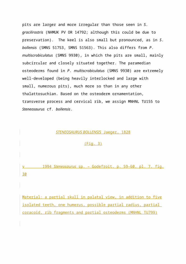
STENEOSAURUS cf. BOLLENSIS Jaeger, 1828
(Fig. 2)
Material: a nearly complete thorax, including one cervical rib and 17 dorsal ribs, 13 dorsal
vertebral neural spines and multiple osteoderms (MNHNL TU155).
Horizon and locality: Harpoceras serpentinum ammonite Zone (‘schistes bitumineux’),
Bascharage, Luxembourg; early Toarcian, Early Jurassic.
Page 6
Description: MNHNL TU155 is a well-preserved and nearly complete thoracic portion of a
postcranial skeleton preserved in dorsal view (Fig. 2). The neural spines of thirteen dorsal
vertebrae are preserved but the centra and the majority of the transverse processes are
obscured by the paramedian osteoderms. One transverse process (on the right side of the third
vertebra) is visible (Fig. 2). It is relatively mediolaterally short in length, anteroposteriorly
broad and appears to have a rounded distal end. The neural spines are dorsoventrally short and
anteroposteriorly elongated, with the edges slightly rounded. There is one partially complete
cervical rib (the tuberculum and capitulum are missing) and it is T-shaped with a
dorsomedially straight rim (Fig. 2). The dorsal ribs are dicephalous, narrow considerably
distally to the rib head and are dorsoventrally thin. The tuberculum is well pronounced and
rounded (Fig. 2). The costal groove (Fig. 2) is large and deep, and runs from the ventral edge
of the tuberculum to near the sternal end. The sternal end of the ribs are straight, thin in width,
and anteroposteriorly flat. The dorsal osteoderms (Fig. 2) are mediolaterally elongated and
roughly arranged in parallel rows (one row per side). A small yet pronounced keel is present.
The pits are large, roughly the same size as each other and irregularly shaped, and are situated
relatively close to one another.
Discussion: The thorax initially comes from a carbonate nodule. During the 1990s, it was
integrated into a “Posidonia shale” slab from Holzmaden, Germany, for esthetical reasons, by
the preparator of the museum at that time, M. John Heil. The presence of well-developed
paramedian osteoderms immediately identifies MNHNL TU155 as a teleosauroid and not a
metriorhynchoid (note that the basal metriorhynchoid Pelagosaurus typus Bronn, 1841
(Eudes-Deslongchamps 1864; Delfino & Dal Sasso 2006; Pierce & Benton 2006; Pierce et al.
2017), does have dorsal osteoderms but these are generally smaller, thinner and less extensive
when compared with teleosauroids, with the exception of Aeolodon priscus von Sömmerring,
1814). The pits in P. typus are also circular and closely packed together (MNHN.F RJN 463)).
There are thirteen preserved dorsal neural (vertebral) spines seen in MNHNL TU155 that are
shortened with rounded edges; however, neural spines rarely differ in Toarcian teleosauroids
(and Teleosauroidea in general), as they are similar to those seen in S. gracilirostris
(NHMUK PV OR 14792), P. multiscrobiculatus (SMNS 9930) and S. bollensis (SMNS
51753). The only preserved transverse process in MNHNL TU155 is similar to S. bollensis
Page 7
(SMNS 51753) in that is it mediolaterally shortened, dorsoventrally flat (although this could
be due to preservation) and anteroposteriorly broad. The transverse processes in P.
multiscrobiculatus (SMNS 9930) are even shorter with a dorsoventrally and anteroposteriorly
broad rounded end, which is not seen in MNHNL TU155 (the rounded end is much smaller).
In MNHNL TU155, the partial cervical rib is similar to S. bollensis (SMNS 51753) in that it
is (1) T-shaped, (2) anteroposteriorly elongated and (3) dorsomedially straight. In P.
multiscrobiculatus (SMNS 9930) and S. gracilirostris (NHMUK PV OR 14792), the cervical
ribs are not anteroposteriorly elongated to the extent seen in MNHNL TU155 (although those
in S. gracilirostris NHMUK PV OR 14792 are partially covered by matrix so their full shapes
are unclear). The dorsal ribs of MNHNL TU155 have a deep costal groove that begins slightly
ventral to the tuberculum, which is similar to both S. gracilirostris (NHMUK PV OR 14792)
and S. bollensis (SMNS 51563). However, the ornamentation of the osteoderms are is more
similar to that seen in S. bollensis (e.g. SMNS 51563). The pits are larger and more irregular
than those seen in S. gracilirostris (NHMUK PV OR 14792; although this could be due to
preservation). The keel is also small but pronounced, as in S. bollensis (SMNS 51753, SMNS
51563). This also differs from P. multiscrobiculatus (SMNS 9930), in which the pits are
small, mainly subcircular and closely situated together. The paramedian osteoderms found in
P. multiscrobiculatus (SMNS 9930) are extremely well-developed (being heavily interlocked
and large with small, numerous pits), much more so than in any other thalattosuchian. Based
on the osteoderm ornamentation, transverse process and cervical rib, we assign MNHNL
TU155 to Steneosaurus cf. bollensis.
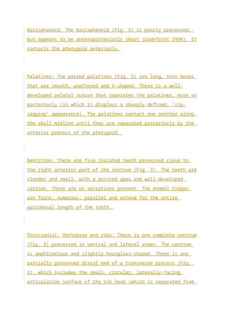
STENEOSAURUS BOLLENSIS Jaeger, 1828
(Fig. 3)
v 1994 Steneosaurus sp. – Godefroit, p. 59-60, pl. 7, fig. 30
Material: a partial skull in palatal view, in addition to five isolated teeth, one humerus,
possible partial radius, partial coracoid, rib fragments and partial osteoderms (MNHNL
TU799)
Page 8
Horizon and locality: Harpoceras serpentinum ammonite Zone (‘schistes bitumineux’),
Sanem, Luxembourg; early Toarcian, Early Jurassic.
Description: MNHNL TU799 is a partial skull exposed in palatal view, as well as additional
postcranial elements and isolated teeth (Fig. 3). The anterior and posterior portions of the
skull are not preserved, as well as the left lateral side. The palate is relatively smooth and
unaltered. There is a pair of well-developed palatal grooves running anteroposteriorly from
the anterior of the rostrum to the anterior palatines (Fig. 3).
Maxillae: The maxillae (Fig. 3) are only observed in ventral view. There are approximately 16
preserved on the right side, and a small damaged region posterior to the 16th preserved
alveolus may be an additional alveolus, but it is unclear (Fig. 3), and two are preserved on the
left side. The alveoli are relatively circular, being slightly mesiodistally longer than
mediolaterally wide. They are small and positioned close together with a relatively thin
interalveolar wall (smaller than the alveolar width). The tooth row is widely separated from
the lateral margin of the choanal opening (Jouve 2009). There is no ornamentation on the
ventral surfaces of the maxillae.
Jugal: The majority of the left jugal is preserved (Fig. 3) except for the posterior end. It forms
the lateral border of the orbit, as in other teleosauroids, and is mediolaterally thin.
Basioccipital: The basioccipital (Fig. 3) is poorly preserved and partially covered in matrix. It
forms the ventral part of the occiput.
Ectopterygoids: Only the anterior left ectopterygoid is present (Fig. 3) and it is a small bone
that contacts the maxilla anteriorly.
Page 9
Pterygoid: The pterygoid (Fig. 3) is a single, elongated, relatively thin bone. The anteromedial
pterygoid has a slight, anteroposteriorly elongated concavity. The anterior processes of the
pterygoid contact the posterior processes of the palatines at a mediolateral (horizontal) angle.
The pterygoid contributes to the medial and posterior borders of the sub-orbital fenestrae,
which are small, rounded posteriorly and teardrop-shaped with a lateral curvature (Fig. 3).
The pterygoid wings are not preserved.
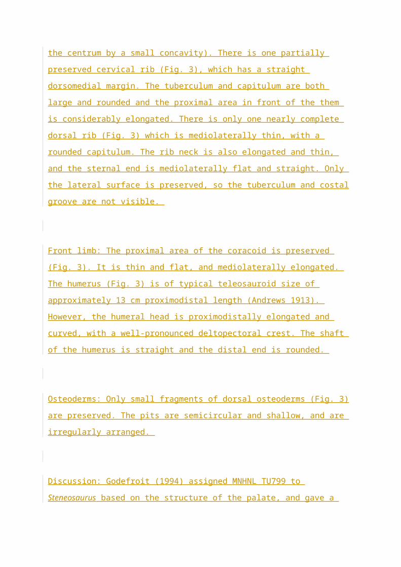
Basisphenoid: The basisphenoid (Fig. 3) is poorly preserved, but appears to be
anteroposteriorly short (Godefroit 1994). It contacts the pterygoid anteriorly.
Palatines: The paired palatines (Fig. 3) are long, thin bones that are smooth, unaltered and V-
shaped. There is a well-developed palatal suture that separates the palatines, more so
posteriorly (in which it displays a sharply defined, ‘zig-zagging’ appearance). The palatines
contact one another along the skull midline until they are separated posteriorly by the anterior
process of the pterygoid.
Dentition: There are five isolated teeth preserved close to the right anterior part of the rostrum
(Fig. 3). The teeth are slender and small, with a pointed apex and well-developed carinae.
There are no serrations present. The enamel ridges are faint, numerous, parallel and extend for
the entire apicobasal length of the tooth.
Postcranial: Vertebrae and ribs: There is one complete centrum (Fig. 3) preserved in ventral
and lateral views. The centrum is amphicoelous and slightly hourglass-shaped. There is one
partially preserved distal end of a transverse process (Fig. 3), which includes the small,
circular, laterally-facing articulation surface of the rib head (which is separated from the
centrum by a small concavity). There is one partially preserved cervical rib (Fig. 3), which
has a straight dorsomedial margin. The tuberculum and capitulum are both large and rounded
Page 10
and the proximal area in front of the them is considerably elongated. There is only one nearly
complete dorsal rib (Fig. 3) which is mediolaterally thin, with a rounded capitulum. The rib
neck is also elongated and thin, and the sternal end is mediolaterally flat and straight. Only the
lateral surface is preserved, so the tuberculum and costal groove are not visible.
Front limb: The proximal area of the coracoid is preserved (Fig. 3). It is thin and flat, and
mediolaterally elongated. The humerus (Fig. 3) is of typical teleosauroid size of
approximately 13 cm proximodistal length (Andrews 1913). However, the humeral head is
proximodistally elongated and curved, with a well-pronounced deltopectoral crest. The shaft
of the humerus is straight and the distal end is rounded.
Osteoderms: Only small fragments of dorsal osteoderms (Fig. 3) are preserved. The pits are
semicircular and shallow, and are irregularly arranged.
Discussion: Godefroit (1994) assigned MNHNL TU799 to Steneosaurus based on the
structure of the palate, and gave a brief description of the preserved cranial bones. However,
Rupert Wild (Stuttgart) labelled MNHNL TU799 as ‘Steneosaurus bollensis’ in the museum
catalogue in 2000, during an informal visit of the collections (no publication or publication
project followed). Nevertheless, we do agree that MNHNL TU799 belongs to S. bollensis,
based on the following observations:
(1) Small, circular alveoli, especially in the posterior maxillae, with small interalveolar
spacing (similar to unnumbered YORM S. bollensis). The alveoli are also small and
subcircular in S. gracilirostris specimens (e.g. NHMUK PV R 757), although the interalveolar
spacing is larger. The interalveolar spacing in S. brevior (NHMUK PV OR 14781) is also
larger (longer than the alveolus width).
(2) Small tear-shaped choanal openings being relatively the same size as the orbit which
are strongly posterolaterally curved (as seen in unnumbered YORM S. bollensis). The sub-
Page 11
orbital openings in S. gracilirostris (MNHNL TU515, NHMUK PV R 757) appear to lack this
curvature (although these specimens are poorly preserved in this area).
(3) Palatines are anterioposteriorly elongated, anteromedially constricted and V-shaped,
as opposed to S. gracilirostris (YORM 1994.3163.1, NHMUK PV R 757) in which the
palatines are shorter, lack anteromedial constriction and are more U-shaped (the palatines are
not visible in P. multiscrobiculatus SMNS 9930 and S. brevior NHMUK PV OR 14781).
(4) Well-developed, tightly interlocking palatal suture, with a characteristic ‘zig-zagging’
appearance in the posterior area (as seen in unnumbered YORM S. bollensis). In S.
gracilirostris (NHMUK PV R 5703, YORM 1994.3163.1), the suture is straight and not
tightly interlocking (the palatal suture is not visible in P. multiscrobiculatus SMNS 9930 and
S. brevior NHMUK PV OR 14781).
(5) A mediolaterally thin jugal with a noticeable lateral bulge (as seen in SMNS 53422,
SMNS 57153, OUMNH JZ176), which is absent in S. gracilirostris (NHMUK PV R 757,
MNHNL TU515) and P. multiscrobiculatus (SMNS 9930). This bulge is ventrolaterally
present in S. brevior (NHMUK PV OR 14781) and in some specimens of S. bollensis (e.g.
SMNS 51753) (note that this feature may be based on preservation).
(6) A relatively large and proximodistally elongated humeral head with a distinct
proximal curvature (as seen in S. bollensis SMNS 53422, SMNS 51753, SMNS 51957). The
humeral head in S. gracilirostris (NHMUK PV OR 14792) and P. multiscrobiculatus (SMNS
9930) is not elongated nor as curved.
STENEOSAURUS GRACILIROSTRIS Westphal, 1961
(Figs. 4-8)
v 1994 Steneosaurus gracilirostris Westphal – Godefroit, p. 50-54, pl. 5, fig. 27
Material: a nearly complete skull and dentary (MNHNL TU515).
Page 12
Horizon and locality: Harpoceras serpentinum ammonite Zone (‘schistes bitumineux’),
Dudelange-Bettembourg in southern Luxembourg; early Toarcian, Early Jurassic.
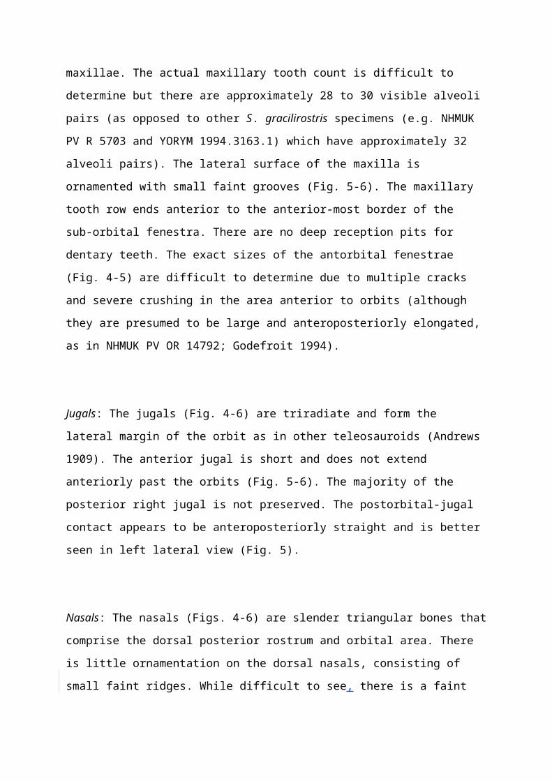
Description: MNHNL TU515 is a nearly complete skull and mandible (Figs. 4-8) (Godefroit
1994). The skull and mandible are cemented together with matrix, so the cranial palatal
surface and mandibular dorsal surface are not accessible. The cranium is approximately 569
mm in length; however, the premaxillae and anterior areas of the maxillae are not preserved.
The remaining cranial rostrum is severely dorsoventrally flattened and there is extreme
dorsoventral crushing just anterior to the orbits (Figs. 4-6), but the posterior cranium is well
preserved (Fig. 7). The orbits are large and comprise approximately 53% of the supratemporal
fenestrae length (Figs. 4-6). The foramen magnum is large and elliptical in shape (Fig. 7).
Maxillae: The maxillae (Figs. 4-6) form a substantial part of the rostrum and are paired
(although this is difficult to see in right lateral view due to deformation of the cranium (Fig.
6)). The nasals are separated from the premaxillae by the maxillae. The actual maxillary tooth
count is difficult to determine but there are approximately 28 to 30 visible alveoli pairs (as
opposed to other S. gracilirostris specimens (e.g. NHMUK PV R 5703 and YORYM
1994.3163.1) which have approximately 32 alveoli pairs). The lateral surface of the maxilla is
ornamented with small faint grooves (Fig. 5-6). The maxillary tooth row ends anterior to the
anterior-most border of the sub-orbital fenestra. There are no deep reception pits for dentary
teeth. The exact sizes of the antorbital fenestrae (Fig. 4-5) are difficult to determine due to
multiple cracks and severe crushing in the area anterior to orbits (although they are presumed
to be large and anteroposteriorly elongated, as in NHMUK PV OR 14792; Godefroit 1994).
Jugals: The jugals (Fig. 4-6) are triradiate and form the lateral margin of the orbit as in other
teleosauroids (Andrews 1909). The anterior jugal is short and does not extend anteriorly past
the orbits (Fig. 5-6). The majority of the posterior right jugal is not preserved. The postorbital-
jugal contact appears to be anteroposteriorly straight and is better seen in left lateral view
(Fig. 5).
Page 13
Nasals: The nasals (Figs. 4-6) are slender triangular bones that comprise the dorsal posterior
rostrum and orbital area. There is little ornamentation on the dorsal nasals, consisting of small
faint ridges. While difficult to see, there is a faint internasal suture (Fig. 4-5), suggesting that
the nasals are paired or partially fused (similar to NHMUK PV OR 14792 and YORYM
1994.3163.1). The posterior nasals are severely deformed, as mentioned above. The
anteroposterior length of the nasals is relatively short in comparison with the anteroposterior
length of the maxillae (roughly 47%, but due to deformation of the posterior nasals and
missing anterior rostrum this number is not reliable) (Fig. 4-6).
Prefrontals: The prefrontals (Figs. 4-5) are severely distorted due to crushing (slightly
anterior to the orbits, as mentioned above). Therefore, the majority of the anterior ends of the
prefrontals cannot be properly assessed. However, it is clear that the prefrontal forms the
anteromedial corner of the orbit (Fig. 4) and contacts the frontal medially, as in other
teleosauroids (Andrews 1909, 1913). The visible prefrontal-lacrimal contact is relatively
straight (Fig. 4-6).
Frontal: The frontal (Figs. 4-6) is large and has no evidence of a midline suture. The anterior
end of the frontal is distorted and slopes ventrally due to anterior crushing. The frontal
contributes to the posteromedial border of the orbits, forms the anterior medial borders of the
supratemporal fenestrae and forms a relatively straight vertical contact with the postorbital in
dorsal and lateral views (Figs. 4-5). The dorsal ornamentation of the frontal consists of
numerous small, but deep, circular-to-semicircular pits that radiate outwards from the midline
(Fig. 4).
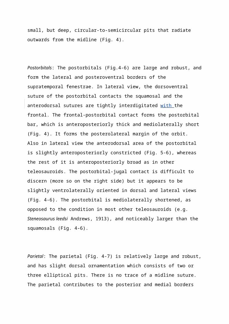
Postorbitals: The postorbitals (Fig.4-6) are large and robust, and form the lateral and
posteroventral borders of the supratemporal fenestrae. In lateral view, the dorsoventral suture
of the postorbital contacts the squamosal and the anterodorsal sutures are tightly interdigitated
with the frontal. The frontal-postorbital contact forms the postorbital bar, which is
anteroposteriorly thick and mediolaterally short (Fig. 4). It forms the posterolateral margin of
Page 14
the orbit. Also in lateral view the anterodorsal area of the postorbital is slightly
anteroposteriorly constricted (Fig. 5-6), whereas the rest of it is anteroposteriorly broad as in
other teleosauroids. The postorbital-jugal contact is difficult to discern (more so on the right
side) but it appears to be slightly ventrolaterally oriented in dorsal and lateral views (Fig. 4-6).
The postorbital is mediolaterally shortened, as opposed to the condition in most other
teleosauroids (e.g. Steneosaurus leedsi Andrews, 1913), and noticeably larger than the
squamosals (Fig. 4-6).
Parietal: The parietal (Fig. 4-7) is relatively large and robust, and has slight dorsal
ornamentation which consists of two or three elliptical pits. There is no trace of a midline
suture. The parietal contributes to the posterior and medial borders of the supratemporal
fenestrae and does not overhang the occiput in dorsal or occipital view. It is mediolaterally
thickened.
Squamosals: The squamosals (Figs. 4-6) are L-shaped; the anterior processes are
anteroposteriorly elongated (in dorsal view), and form the posterolateral border of the
supratemporal fenestrae. Its posterolateral surface is concave and it contacts the quadrate
posteroventrally in lateral view. The squamosal anteriorly contacts the postorbital, and
together they form the supratemporal arch.
Quadrates: The quadrates (Figs. 5, 7) are robust and strongly sutured to the squamosals and
quadratojugals. The anterodorsal region of the quadrate contacts the squamosal and
quadratojugal while the posteroventral margin articulates with the angular (=jaw joint) and
medially contacts the exoccipital. The posteroventral medial hemicondyle is slightly larger
than the lateral hemicondyle in size and mediolateral length. Both hemicondyles are elongated
mediolaterally, oval-shaped and have rounded posterior edges. On the occiput, the
hemicondyles posteriorly extend further than the exoccipitals (Fig. 7). The left quadrate is
well-preserved whereas the right quadrate is missing the hemicondyles.
Page 15
Quadratojugals: The quadratojugals are visible in lateral view, with the left being better
preserved than the right. The posterior region of the quadratojugal is expanded mediolaterally
to accommodate the quadrate and extends slightly further posteriorly than the posteroventral
corner of the quadrate.
Supraoccipital: The supraoccipital (Fig. 7) is positioned ventral to the parietal and is only
visible in occipital view. It forms the dorsomedial part of the occiput and contributes to the
dorsal edge of the foramen magnum (Fig. 7) (Brusatte et al. 2016). The ventral edge is
triangular and no nuchal crest is present. The supraoccipital is dorsoventrally tall and slightly
mediolaterally expanded (more so dorsally than ventrally). The supraoccipital is not broadly
exposed in dorsal view and is slightly concave.
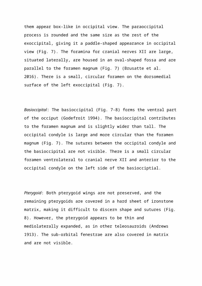
Exoccipital: the exoccipitals (Fig. 7) make up the majority of the occiput (Godefroit 1994),
are tilted dorsally, flared mediolaterally and are slightly concave on their occipital surfaces.
Both exoccipitals are strongly directed posteriorly (although this may be due to preservation).
The exoccipitals are dorsoventrally tall and mediolaterally short compared to other
teleosauroids (e.g. S. leedsi NHMUK PV R 3806) and contribute to the dorsal and lateral
borders of the foramen magnum. Laterally the exoccipitals descend rapidly, making them
appear box-like in occipital view. The paraoccipital process is rounded and the same size as
the rest of the exoccipital, giving it a paddle-shaped appearance in occipital view (Fig. 7). The
foramina for cranial nerves XII are large, situated laterally, are housed in an oval-shaped fossa
and are parallel to the foramen magnum (Fig. 7) (Brusatte et al. 2016). There is a small,
circular foramen on the dorsomedial surface of the left exoccipital (Fig. 7).
Basioccipital: The basioccipital (Fig. 7-8) forms the ventral part of the occiput (Godefroit
1994). The basioccipital contributes to the foramen magnum and is slightly wider than tall.
The occipital condyle is large and more circular than the foramen magnum (Fig. 7). The
sutures between the occipital condyle and the basioccipital are not visible. There is a small
circular foramen ventrolateral to cranial nerve XII and anterior to the occipital condyle on the
left side of the basiocciptial.
Page 16
Pterygoid: Both pterygoid wings are not preserved, and the remaining pterygoids are covered
in a hard sheet of ironstone matrix, making it difficult to discern shape and sutures (Fig. 8).
However, the pterygoid appears to be thin and mediolaterally expanded, as in other
teleosauroids (Andrews 1913). The sub-orbital fenestrae are also covered in matrix and are
not visible.
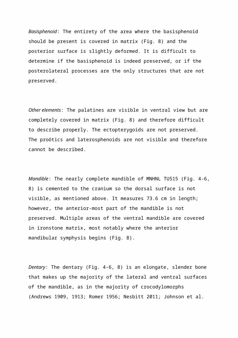
Basisphenoid: The entirety of the area where the basisphenoid should be present is covered in
matrix (Fig. 8) and the posterior surface is slightly deformed. It is difficult to determine if the
basisphenoid is indeed preserved, or if the posterolateral processes are the only structures that
are not preserved.
Other elements: The palatines are visible in ventral view but are completely covered in matrix
(Fig. 8) and therefore difficult to describe properly. The ectopterygoids are not preserved. The
proötics and laterosphenoids are not visible and therefore cannot be described.
Mandible: The nearly complete mandible of MNHNL TU515 (Fig. 4-6, 8) is cemented to the
cranium so the dorsal surface is not visible, as mentioned above. It measures 73.6 cm in
length; however, the anterior-most part of the mandible is not preserved. Multiple areas of the
ventral mandible are covered in ironstone matrix, most notably where the anterior mandibular
symphysis begins (Fig. 8).
Dentary: The dentary (Fig. 4-6, 8) is an elongate, slender bone that makes up the majority of
the lateral and ventral surfaces of the mandible, as in the majority of crocodylomorphs
(Andrews 1909, 1913; Romer 1956; Nesbitt 2011; Johnson et al. 2017). The anterior-most
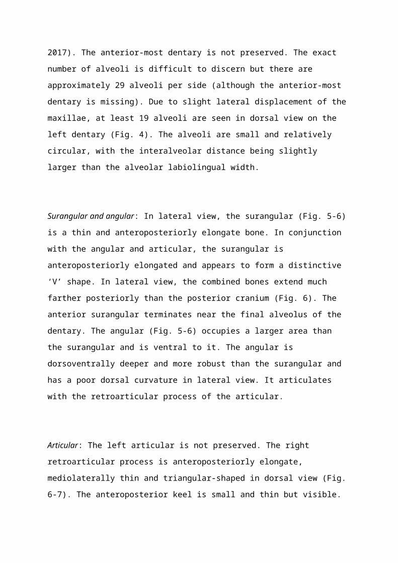
dentary is not preserved. The exact number of alveoli is difficult to discern but there are
approximately 29 alveoli per side (although the anterior-most dentary is missing). Due to
slight lateral displacement of the maxillae, at least 19 alveoli are seen in dorsal view on the
Page 17
left dentary (Fig. 4). The alveoli are small and relatively circular, with the interalveolar
distance being slightly larger than the alveolar labiolingual width.
Surangular and angular: In lateral view, the surangular (Fig. 5-6) is a thin and
anteroposteriorly elongate bone. In conjunction with the angular and articular, the surangular
is anteroposteriorly elongated and appears to form a distinctive ‘V’ shape. In lateral view, the
combined bones extend much farther posteriorly than the posterior cranium (Fig. 6). The
anterior surangular terminates near the final alveolus of the dentary. The angular (Fig. 5-6)
occupies a larger area than the surangular and is ventral to it. The angular is dorsoventrally
deeper and more robust than the surangular and has a poor dorsal curvature in lateral view. It
articulates with the retroarticular process of the articular.
Articular: The left articular is not preserved. The right retroarticular process is
anteroposteriorly elongate, mediolaterally thin and triangular-shaped in dorsal view (Fig. 6-7).
The anteroposterior keel is small and thin but visible. The posterior end of the retroarticular
process is slightly rounded.
Dentition: Both the maxillary and dentary teeth (Fig. 4-6) are small, slender and elongated
with a pointed apex. They are strongly posteriorly curved. The enamel ridges are slight and
faint, parallel to one another and do not reach the top of the apex. There are no serrations
present.
Discussion: MNHNL TU515 displays many characteristic features of teleosauroids including:
a relatively small frontal and anteroposteriorly elongated supratemporal fenestrae (Andrews
1913; Johnson et al. 2017). Godefroit (1994) referred MNHNL TU515 to Steneosaurus
gracilirostris based on: (1) the elongation and slender build of the skull; (2) the considerable
anteroposterior length of the antorbital fenestrae; (3) the lateral position of the orbits; and (4)
location of dorsal ornamentation (restricted to the frontal, postorbitals, parietal and posterior
area of the prefrontals). We agree with Godrefroit’s (1994) referral (although the actual size
Page 18
of the antorbital fenestrae is difficult to discern, due to the deformation of the skull; see
description), and here list how MNHNL TU515 is similar to the S. gracilirostris holotype
(NHMUK PV OR 14792) and paratype (NHMUK PV R 15500) based on the following
characters:
(1) A medium-sized skull (roughly 2 m in length) with an elongated narrow rostrum
comprising at least 70% of the total skull length (although the total length of the skull varies).
The rostra in S. bollensis (SMNS 51953), P. multiscrobiculatus (SMNS 9930), and S. brevior
(NHMUK PV OR 14781) contribute to less than 70% of the total skull length.
(2) Anteroposteriorly elongated maxillae and no elongation of the nasals, with a maxilla
that is over 55% of skull length. This is similar to S. bollensis (SMNS 51953) and P.
multiscrobiculatus (SMNS 9930), but differs in S. brevior (NHMUK PV OR 14781) in which
both the maxillae and nasals are not as elongated and the maxilla is less than 55% of the skull
length (note that S. brevior (NHMUK PV OR 14781) is a mesorostrine form).
(3) Laterally (and slightly dorsally) facing orbits. This character makes S. gracilirostris
unique amongst teleosauroids (this character is also shared with metriorhynchoids).
Steneosaurus bollensis (SMNS 51953), S. brevior (NHMUK PV OR 14781) and P.
multiscrobiculatus (SMNS 9930) all have orbits that are dorsally oriented.
(4) The tooth row and quadrate condyle are aligned on the horizontal plane, and are both
at a lower level than the occipital condyle. While this is similar to S. bollensis (SMNS 51753),
in P. multiscrobiculatus (SMNS 9930) both the tooth row and quadrate are unaligned (with
the quadrate being slightly ventral to the tooth row) and below the occipital condyle. In S.
brevior (NHMUK PV OR 14781) the tooth row and quadrate condyle appear to be unaligned.
(5) Nasals lack a midline concavity (although they are severely flattened). In S. brevior
(NHMUK PV OR 14781), S. bollensis (SMNS 51953) and P. multiscrobiculatus (SMNS
9930) this concavity is present (note that the holotype of S. gracilirostris (NHMUK PV OR
14792) also has this concavity).
(6) The antorbital fenestrae appear to be moderately large and anteroposteriorly elongated
(roughly 30 mm anteroposterior length), which is seen in the holotype (NHMUK PV OR
14792) (although MNHNL TU515 is severely deformed in these areas). Steneosaurus
Page 19
bollensis (SMNS 51953), S. brevior (NHMUK PV OR 14781) and P. multiscrobiculatus
(SMNS 9930) all have smaller, subcircular antorbital fenestrae.
(7) At least 29 maxillary alveoli, which is similar in S. bollensis (although the number of
alveoli can vary from 28 to over 32). The exact tooth count of S. brevior (NHMUK PV OR
14781) is difficult to discern but is it has fewer than 28 maxillary alveoli.
(8) Longitudinal, ellipsoid supratemporal fenestrae that show no anterolateral expansion.
While this is similar to S. bollensis (SMNS 51953), in S. brevior (NHMUK PV OR 14781)
and P. multiscrobiculatus (SMNS 9930) the anterior margin of the supratemporal fenestrae
are inclined anterolaterally.
(9) In dorsal view, the supratemporal fenestrae are subequal in size relative to the orbit. In
S. bollensis (SMNS 51953), P. multiscrobiculatus (SMNS 9930) and S. brevior (NHMUK PV
OR 14781) the supratemporal fenestrae are longer in length than the orbit.
(10) The anterior jugal is broad, with a roughly straight contact with the maxilla and does
not extend anteriorly past the level of the orbit. In S. bollensis (SMNS 20283, NHMUK PV R
756), S. brevior (NHMUK PV OR 14781) and P. multiscrobiculatus (SMNS 9930), the
anterior jugal tapers off dorsoventrally just anterior to the orbits in lateral view.
(11) The squamosal projects further posteriorly than the occipital condyle. This is similar
to S. brevior (NHMUK PV OR 14781); however, in S. bollensis (SMNS 51953) the
squamosal does not project further posteriorly than the occipital condyle.
(12) Angular is poorly curved (mostly horizontal) dorsally at its posterior end. This is
similar to S. brevior (NHMUK PV OR 14781); however, in S. bollensis (SMNS 51563) and
P. multiscrobiculatus (SMNS 9930) the angular is clearly yet gently curved.
(13) Mandible is poorly curved ventrally and the dorsal border is generally straight. In S.
brevior (NHMUK PV OR 14781) and P. multiscrobiculatus (SMNS 9930), the mandibular
dorsal border is gently dorsally arched.
PLATYSUCHUS MULTICROBICULATUS Berckhemer, 1929
(Fig. 109)
Page 20
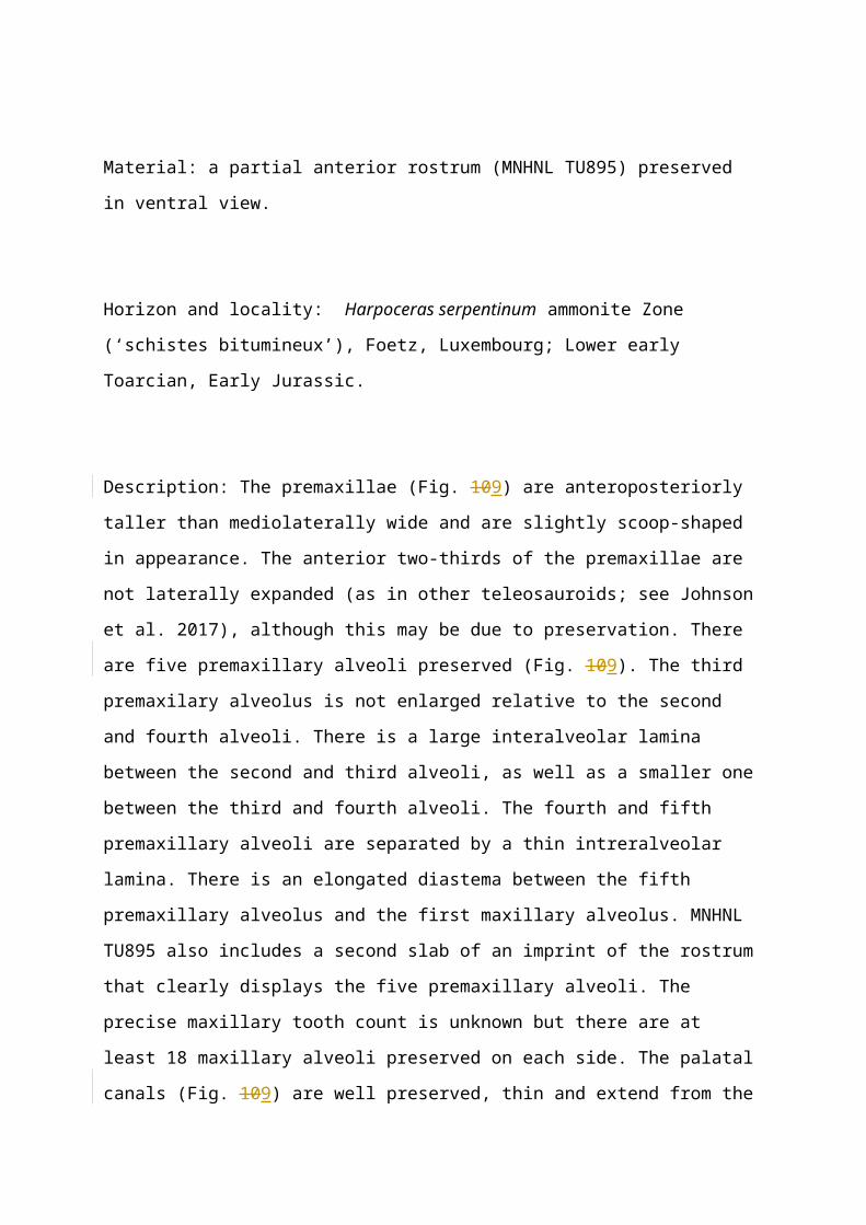
Material: a partial anterior rostrum (MNHNL TU895) preserved in ventral view.
Horizon and locality: Harpoceras serpentinum ammonite Zone (‘schistes bitumineux’),
Foetz, Luxembourg; Lower early Toarcian, Early Jurassic.
Description: The premaxillae (Fig. 109) are anteroposteriorly taller than mediolaterally wide
and are slightly scoop-shaped in appearance. The anterior two-thirds of the premaxillae are
not laterally expanded (as in other teleosauroids; see Johnson et al. 2017), although this may
be due to preservation. There are five premaxillary alveoli preserved (Fig. 109). The third
premaxilary alveolus is not enlarged relative to the second and fourth alveoli. There is a large
interalveolar lamina between the second and third alveoli, as well as a smaller one between
the third and fourth alveoli. The fourth and fifth premaxillary alveoli are separated by a thin
intreralveolar lamina. There is an elongated diastema between the fifth premaxillary alveolus
and the first maxillary alveolus. MNHNL TU895 also includes a second slab of an imprint of
the rostrum that clearly displays the five premaxillary alveoli. The precise maxillary tooth
count is unknown but there are at least 18 maxillary alveoli preserved on each side. The
palatal canals (Fig. 109) are well preserved, thin and extend from the fourth premaxillary
alveoli and continue posteriorly down the midline of the maxillae.
Discussion: The teleosauroid rostrum MNHNL TU895 was never formally classified and was
initially labelled in the museum catalogue as ‘crocodilian rostrum (by private collector and
discoverer of the specimen, M. Jo Simon, also a volunteer research associate of the MNHNL).
However, there is a critical character seen in MNHNL TU895: five distinct alveoli per
premaxilla (although it is noticeably harder to make out all alveoli on the right side). Other
Toarcian teleosauroids such as S. bollensis (e.g. SMNS 18699), S. brevior (NHMUK PV OR
14781) and S. gracilirostris (NHMUK PV R 5703) have four premaxillary alveoli per side.
The size of the alveoli also differ: in MNHNL TU895, the first two alveoli are similar in size,
whereas in S. bollensis (SMNS 18699) the first alveolus is slightly smaller than the second.
This character is diagnostic and, in Toarcian teleosauroids, is only present in P.
Page 21
multiscrobiculatus (SMNS 9930). Therefore, we attribute MNHNL TU895 to P.
multiscrobiculatus.
TELEOSAUROIDEA INDET.
(Fig. 10)
Material: a partially complete anterior rostrum in palatal view (MNHNL TU164).
Horizon and locality: Harpoceras serpentinum ammonite Zone (‘schistes bitumineux’),
Dudelange-Bettembourg in southern Luxembourg; early Toarcian, Early Jurassic.
Description: MNHNL TU164 (Fig. 10) is the anterior end of the rostrum and is approximately
187 mm in length. Once peculiar feature is that there are only three alveoli per premaxilla
(Fig. 10). All premaxillary alveoli are relatively the same size, with the second being slightly
larger than the first. The first premaxillary alveolus is procumbent. There is a large diastema
present between the last premaxillary and first maxillary alveoli. There are 14 and 15
preserved maxillary alveoli on the right and left sides, respectively, that are large and
semicircular, with a large interalveolar spacing between them (slightly larger than the alveolar
width). In ventral and lateral views (more so on the right side), there are faint but well-
developed reception pits for dentary teeth along the middle region of the ventral-lateral
margin of the maxillae (Fig. 10) The prenarial anterior premaxillary ridge, seen in
anteroventral view, is well-developed and, while relatively small, well pronounced. There are
three partial teeth associated with MNHNL TU164: one in situ in the right second
premaxillary alveolus, one in the left thirteenth maxillary alveolus (Fig. 10) and one
embedded in an opposing slab. The apices are not preserved in any of the teeth; however, the
apicobasal enamel ridges are small, well pronounced, numerous and run parallel to one
another towards the apex of the tooth (Fig. 10). There are no carinae preserved.
Page 22
Discussion: MNHNL TU164 was initially labelled as ‘Steneosaurus bollensis’ by Rupert
Wild (Stuttgart) in 2000 (this was an informal determination and there was no publication).
However, MNHNL TU164 has an unusual character: only three alveoli per premaxilla, as
opposed to P. multiscrobiculatus (MNHNL TU895, SMNS 9930), which has five, and S.
bollensis (SMNS 18699), S. gracilirostris (NHMUK PV R 5703) and S. brevior (NHMUK
PV OR 14781), which all have four. It is important to note that in Teleosauroidea, only the
genus Machimosaurus is known to have three alveoli per premaxilla (Young et al. 2014).
While the teleosauroid referred to as ‘Peipehsuchus’ teleorhinus Young, 1948, from China
(see Li 1993) is described as having three premaxillary alveoli, the specimen (IVPP 10098) in
actuality has four (the first premaxillary alveolus is much smaller than the other three). Three
premaxillary alveoli is a synapomorphy of Metriorhynchidae; however, the (1) shape of the
anterior maxilla in palatal view (straightened and sub-rectangular, as opposed to tapering and
sub-triangular in metriorhynchids), (2) overall shape of the premaxillae (spatulate) and (3)
spacing (large interalveolar distance) between premaxillary alveoli are more representative of
a teleosauroid than metriorhynchid. In addition, MNHNL TU164 has small yet noticeable
reception pits, which are absent in S. bollensis (SMNS 51753, SMNS 18699), P.
multiscrobiculatus (SMNS 9930) and S. gracilirostris (NHMUK PV OR 14792) (although
these are present in S. brevior (NHMUK PV OR 14781). The presence of three premaxillary
alveoli could potentially be a character diagnostic of a new species of teleosauroid, because
MNHNL TU164 is the only currently known Toarcian teleosauroid with this feature.
However, more specimens are necessary to demonstrate that this is a taxonomically diagnostic
feature and not individual or random variation. Therefore, at the current time, we assign
MNHNL TU164 to Teleosauroidea indet., but recognise that it may belong to a previously
unknown species.
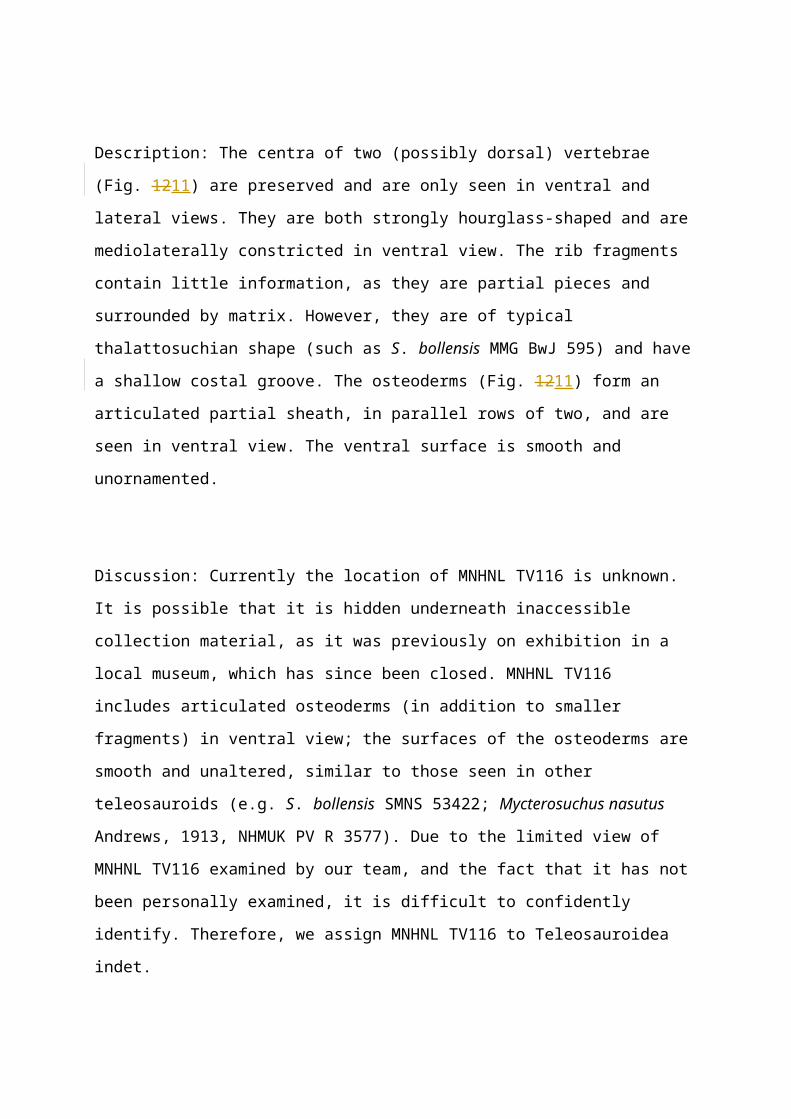
TELEOSAUROIDEA INDET.
(Fig. 1211)
Material: a large slab including: articulated osteoderms, rib fragments and two ?dorsal
vertebrae (MNHNL TV116).
Page 23
Horizon and locality: Harpoceras serpentinum ammonite Zone (‘schistes bitumineux’),
Dudelange-Bettembourg, Luxembourg; early Toarcian, Early Jurassic.
Description: The centra of two (possibly dorsal) vertebrae (Fig. 1211) are preserved and are
only seen in ventral and lateral views. They are both strongly hourglass-shaped and are
mediolaterally constricted in ventral view. The rib fragments contain little information, as
they are partial pieces and surrounded by matrix. However, they are of typical thalattosuchian
shape (such as S. bollensis MMG BwJ 595) and have a shallow costal groove. The
osteoderms (Fig. 1211) form an articulated partial sheath, in parallel rows of two, and are seen
in ventral view. The ventral surface is smooth and unornamented.
Discussion: Currently the location of MNHNL TV116 is unknown. It is possible that it is
hidden underneath inaccessible collection material, as it was previously on exhibition in a
local museum, which has since been closed. MNHNL TV116 includes articulated osteoderms
(in addition to smaller fragments) in ventral view; the surfaces of the osteoderms are smooth
and unaltered, similar to those seen in other teleosauroids (e.g. S. bollensis SMNS 53422;
Mycterosuchus nasutus Andrews, 1913, NHMUK PV R 3577). Due to the limited view of
MNHNL TV116 examined by our team, and the fact that it has not been personally examined,
it is difficult to confidently identify. Therefore, we assign MNHNL TV116 to Teleosauroidea
indet.
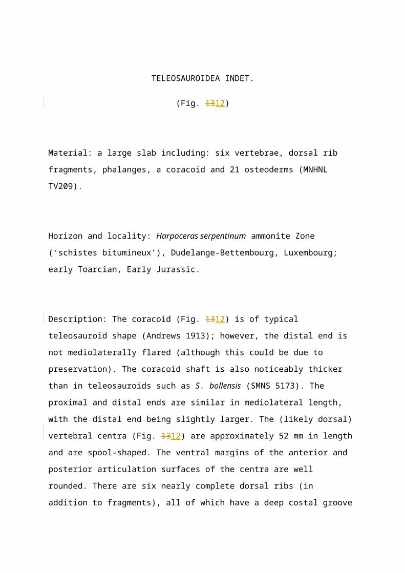
TELEOSAUROIDEA INDET.
(Fig. 1312)
Material: a large slab including: six vertebrae, dorsal rib fragments, phalanges, a coracoid and
21 osteoderms (MNHNL TV209).
Page 24
Horizon and locality: Harpoceras serpentinum ammonite Zone (‘schistes bitumineux’),
Dudelange-Bettembourg, Luxembourg; early Toarcian, Early Jurassic.
Description: The coracoid (Fig. 1312) is of typical teleosauroid shape (Andrews 1913);
however, the distal end is not mediolaterally flared (although this could be due to
preservation). The coracoid shaft is also noticeably thicker than in teleosauroids such as S.
bollensis (SMNS 5173). The proximal and distal ends are similar in mediolateral length, with
the distal end being slightly larger. The (likely dorsal) vertebral centra (Fig. 1312) are
approximately 52 mm in length and are spool-shaped. The ventral margins of the anterior and
posterior articulation surfaces of the centra are well rounded. There are six nearly complete
dorsal ribs (in addition to fragments), all of which have a deep costal groove (Fig. 1312). The
sternal rib end is flat, narrows substantially and is rounded. There are twenty-one osteoderms
preserved, and only seven are complete. The pits (Fig. 1312) are deep and semicircular to
elongate in shape. They are a variety of sizes, generally closely situated to one another,
separated by a small but thick lamina, and form a semi-circular spiralling pattern. The
majority of complete osteoderms possess a slight, elongated keel (Fig. 1213). One complete
osteoderm is preserved in ventral view, with a smooth and unornamented ventral surface (Fig.
1312). The phalanges are of typical thalattosuchian shape, similar to S. bollensis (SMNS
51753; however, they are much larger (over 50%) than those seen in S. bollensis). There are
possible stomach contents preserved on the ventral underside of the slab, located between the
dorsal ribs.
Discussion: The distal end of the preserved coracoid in MNHNL TV209 is less mediolaterally
flared in S. bollensis (SMNS 51753) and the shaft is noticeably thicker. It is similar to the
right coracoid preserved in P. multiscrobiculatus (SMNS 9930; although in this specimen the
coracoid is partially covered by the humerus). The dorsal ribs display a deep and
proximodistally wide costal groove, similar to both S. bollensis (SMNS 51563) and S.
gracilirostris (NHMUK PV OR 14792; although it is difficult to see in this specimen due to
preservation). The vertebral centa are of typical teleosauroid shape, being taller than wide and
mediolaterally constricted (hourglass-shaped), which is seen in S. bollensis (SMNS 51563), S.
Page 25
gracilirostris (NHMUK PV OR 14792) and P. multiscrobiculatus (SMNS 9930). The
ornamentation on the dorsal osteoderms consists of irregular large pits, with varying degrees
of closeness. This is similar to S. bollensis (SMNS 51953) and differs from S. gracilirostris
(NHMUK PV OR 14792), in which the pits are slightly smaller and more subcircular in
shape, and P. multiscrobiculatus (SMNS 9930), in which the ornamentation consists of small
pits situated close to one another. Due to this combination of features, we cannot be certain of
the species-level identity of MNHNL TV209 and conservatively assign it to Teleosauroidea
indet.
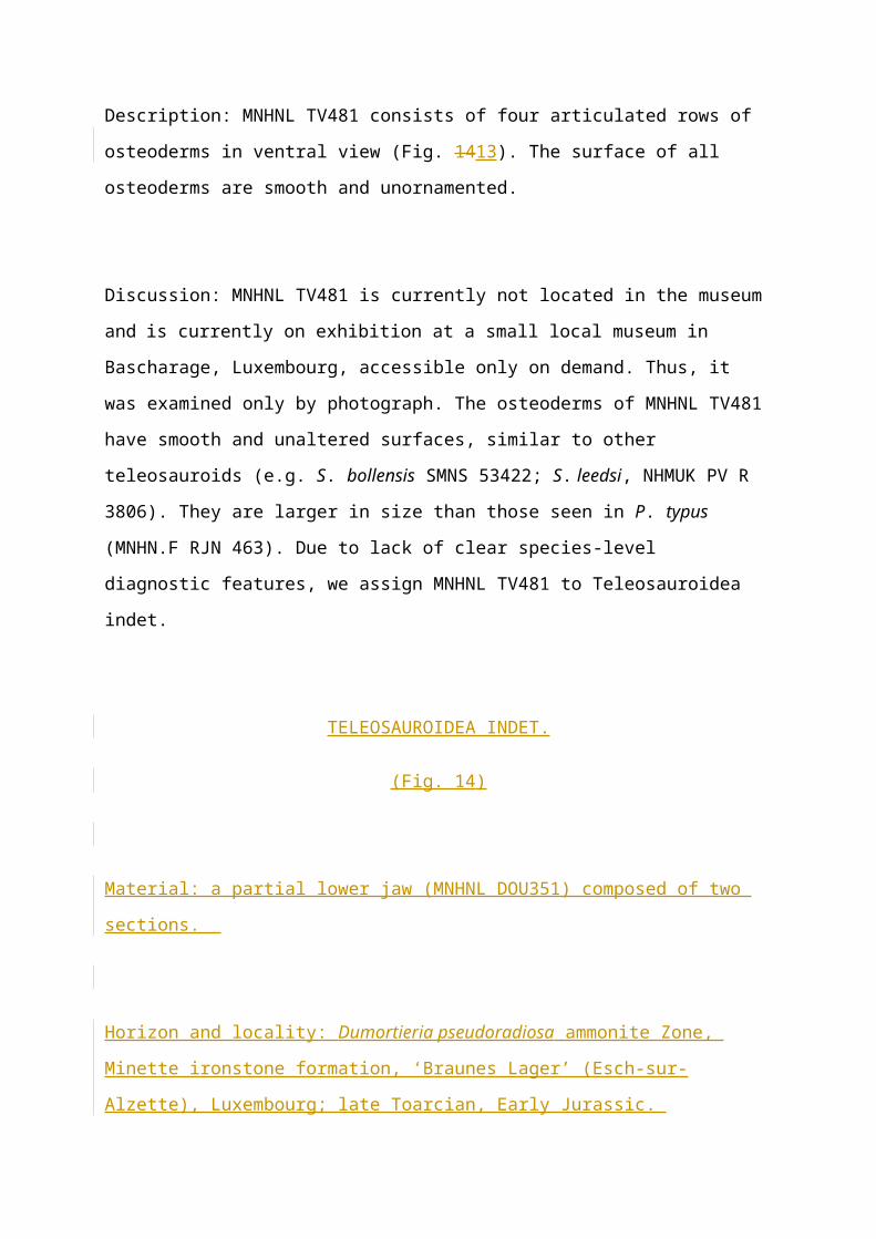
TELEOSAUROIDEA INDET.
(Fig. 1413)
Material: a large slab consisting of articulated ventral osteoderms (MNHNL TV481).
Horizon and locality: Harpoceras serpentinum ammonite Zone (‘schistes bitumineux’),
Dudelange-Bettembourg, Luxembourg; early Toarcian, Early Jurassic.
Description: MNHNL TV481 consists of four articulated rows of osteoderms in ventral view
(Fig. 1413). The surface of all osteoderms are smooth and unornamented.
Discussion: MNHNL TV481 is currently not located in the museum and is currently on
exhibition at a small local museum in Bascharage, Luxembourg, accessible only on demand.
Thus, it was examined only by photograph. The osteoderms of MNHNL TV481 have smooth
and unaltered surfaces, similar to other teleosauroids (e.g. S. bollensis SMNS 53422; S. leedsi,
NHMUK PV R 3806). They are larger in size than those seen in P. typus (MNHN.F RJN
463). Due to lack of clear species-level diagnostic features, we assign MNHNL TV481 to
Teleosauroidea indet.
Page 26
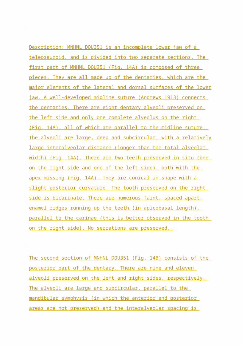
TELEOSAUROIDEA INDET.
(Fig. 14)
Material: a partial lower jaw (MNHNL DOU351) composed of two sections.
Horizon and locality: Dumortieria pseudoradiosa ammonite Zone, Minette ironstone
formation, ‘Braunes Lager’ (Esch-sur-Alzette), Luxembourg; late Toarcian, Early Jurassic.
Description: MNHNL DOU351 is an incomplete lower jaw of a teleosauroid, and is divided
into two separate sections. The first part of MNHNL DOU351 (Fig. 14A) is composed of
three pieces. They are all made up of the dentaries, which are the major elements of the lateral
and dorsal surfaces of the lower jaw. A well-developed midline suture (Andrews 1913)
connects the dentaries. There are eight dentary alveoli preserved on the left side and only one
complete alveolus on the right (Fig. 14A), all of which are parallel to the midline suture. The
alveoli are large, deep and subcircular, with a relatively large interalveolar distance (longer
than the total alveolar width) (Fig. 14A). There are two teeth preserved in situ (one on the
right side and one of the left side), both with the apex missing (Fig. 14A). They are conical in
shape with a slight posterior curvature. The tooth preserved on the right side is bicarinate.
There are numerous faint, spaced apart enamel ridges running up the teeth (in apicobasal
length), parallel to the carinae (this is better observed in the tooth on the right side). No
serrations are preserved.
The second section of MNHNL DOU351 (Fig. 14B) consists of the posterior part of the
dentary. There are nine and eleven alveoli preserved on the left and right sides, respectively.
The alveoli are large and subcircular, parallel to the mandibular symphysis (in which the
anterior and posterior areas are not preserved) and the interalveolar spacing is smaller than the
first part of MNHNL DOU351, being less than half but larger than a quarter of the alveolar
Page 27
width. There is no evidence of posterior curvature. There are faint reception pits seen in dorsal
view (Fig. 14B). The coronoid processes are not observed (although this could be due to
preservation).
Discussion: While the middle dentaries of MNHNL DOU351 can be interpreted as either
metriorhynchid or teleosauroid (as they are relatively similar; Andrews 1913), the shape and
interalveolar spacing of the alveoli are more representative of a teleosauroid than a
metriorhynchid. The interalveolar spacing is relatively large, which is similar to S.
gracilirostris (MNHNL TU515). The second section of MNHNL DOU351 has faint reception
pits in the anterior region of the lateral dentaries. These are present in S. brevior (NHMUK
PV OR 14781), whereas S. gracilirostris (NHMUK PV OR 14792, MNHNL TU515), S.
bollensis (SMNS 51953) and P. multiscrobiculatus (SMNS 9930) lack them. Therefore, we
assign MNHNL DOU351 to Teleosauroidea indet.
THALATTOSUCHIA INDET.
(Fig. 15)
Material: articulated caudal vertebrae, seven complete and one partial (MNHNL TU914).
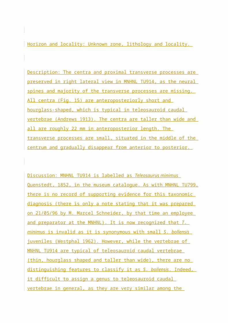
Horizon and locality: Unknown zone, lithology and locality.
Description: The centra and proximal transverse processes are preserved in right lateral view
in MNHNL TU914, as the neural spines and majority of the transverse processes are missing.
All centra (Fig. 15) are anteroposteriorly short and hourglass-shaped, which is typical in
teleosauroid caudal vertebrae (Andrews 1913). The centra are taller than wide and all are
roughly 22 mm in anteroposterior length. The transverse processes are small, situated in the
middle of the centrum and gradually disappear from anterior to posterior.
Page 28
Discussion: MNHNL TU914 is labelled as Teleosaurus minimus Quenstedt, 1852, in the
museum catalogue. As with MNHNL TU799, there is no record of supporting evidence for
this taxonomic diagnosis (there is only a note stating that it was prepared on 21/05/96 by M.
Marcel Schneider, by that time an employee and preparator at the MNHNL). It is now
recognized that T. minimus is invalid as it is synonymous with small S. bollensis juveniles
(Westphal 1962). However, while the vertebrae of MNHNL TU914 are typical of teleosauroid
caudal vertebrae (thin, hourglass shaped and taller than wide), there are no distinguishing
features to classify it as S. bollensis. Indeed, it difficult to assign a genus to teleosauroid
caudal vertebrae in general, as they are very similar among the teleosauroid taxa. The
posterior caudal vertebrae of teleosauroids are also similar in shape and size to those found in
metriorhynchids and Pelagosaurus typus. Therefore, we assign MNHNL TU914 to
Thalattosuchia indet.
THALATTOSUCHIA INDET.
(Fig. 1516)
Material: one partial caudal vertebra (MNHNL TV561).
Horizon and locality: Hildoceras bifrons ammonite Zone, Marnes à Bifrons, Sanem,
Luxembourg; early Toarcian, Early Jurassic.
Description: MNHN TV561 is of a centrum of one caudal vertebra (Fig. 1516). It is of typical
teleosauroid shape: mediolaterally constricted, taller than wide and strongly hourglass-shaped.
It measures roughly 20 mm in anteroposterior length. The neural spine is not preserved and
there is no evidence of a transverse process (the lateral surface of the centrum is smooth).
There are invertebrates associated with this vertebra: two belemnites and an ammonite
impression (Fig. 1516).
Page 29
Discussion: As mentioned above, MNHNL TV561 displays features typical of teleosauroid
caudal vertebrae: (1) mediolaterally thin; (2) hourglass shaped; and (3) taller than wide. These
are similar to other teleosauroids such as S. bollensis (SMNS 18699, SMNS 51753), S.
gracilirostris (NHMUK PV OR 14972) and P. multiscrobiculatus (SMNS 9930), as well as
the basal metriorhynchoid P. typus (NHMUK PV R 6213, MNHN.F RJN 463). MNHNL
TV561 is likely to be a posterior caudal vertebra, as it is quite small (only 20 mm
anteroposterior length) and lacks a transverse process. We assign MNHNL TV561 to
Thalattosuchia indet.
THALATTOSUCHIA INDET.
(Fig. 1617)
Material: two caudal vertebrae centra (MNHNL TV597) preserved on a rounded block of
matrix.
Horizon and locality: Harpoceras serpentinum ammonite Zone (‘schistes bitumineux’),
Sanem, Luxembourg; early Toarcian, Early Jurassic.
Description: Both centra (Fig. 1617) are amphicoelous and of typical teleosauroid shape.
They are mediolaterally thin, taller than wide, anteroposteriorly elongate and hourglass-
shaped. The neural spines and transverse processes are not preserved.
Discussion: Both centra preserved in MNHNL TV597 are representative of a typical
teleosauroid caudal vertebrae: mediolaterally constricted and thin, hourglass-shaped and taller
than wide. This is similar to S. gracilirostris (NHMUK PV OR 14792), S. bollensis (SMNS
18699, SMNS 51753) and P. multiscrobiculatus (SMNS 9930), as well as the basal
Page 30
metriorhynchoid P. typus (NHMUK PV R 6213, MNHN.F RJN 463). Thus, we assign
MNHNL TV597 to Thalattosuchia indet.
THALATTOSUCHIA INDET.
(Fig. 18)
Material: a single centrum (MNHNL DOU725) of a caudal vertebra.
Horizon and locality: Unknown zone, Minette ironstone formation, Esch-sur-Alzette,
Luxembourg; late Toarcian, Early Jurassic.
Description: The centrum of the vertebra is faintly amphicoelous and slightly mediolaterally
wider than dorsoventrally high (Fig 18). The neural canal is dorsoventrally tall and oval
shaped. In lateral view the centra centrum is anteroposteriorly short and slightly circular (Fig.
18). The prezygapophyses and postzygapophyses are not preserved, nor are the distal
transverse processes.
Discussion: MNHNL DOU725 is similar in shape to MNHNL TV602. The centrum of
MNHNL DOU725 is slightly mediolaterally longer (wider than tall) than those seen in a
typical cervical or dorsal vertebra of a teleosauroid (e.g. S. gracilirostris NHMUK PV OR
14792; S. bollensis SMNS 51753), although it is possible for cervical vertebral centra to be
wider than tall in teleosauroids, as mentioned above. The anteroposterior length of MNHNL
DOU725 is relatively short and while the centra of sacral vertebrae are typically marginally
wider than tall in teleosauroids (e.g. S. bollensis MMG BwJ 595, S. edwardsi NHMUK PV R
3701), the proximal part of the transverse processes in MNHNL DOU725 are more dorsally
inclined than in other teleosauroids. We therefore assign MNHNL DOU725 to Thalattosuchia
indet.
Page 31
THALATTOSUCHIA INDET.
(Fig. 19)
Material: a single partially preserved vertebra (MNHNL BM190).
Horizon and locality: Stephanoceras humphriesianum Zone, ‘Marnes sableuses d’Audun-le-
Tiche’, Rumelange, Luxembourg; early Bajocian, Middle Jurassic.
Description: The vertebral centrum (Fig. 19) is approximately 41 mm in length and is wider
than tall. The anterior and posterior central surfaces are rounded and slightly amphicoelous.
There is a flat, posteroventrally deflected surface on the ventral margin of the posterior
surface (Fig. 19). The transverse processes are small, anteroposteriorly thin and
dorsoventrally flat (Fig. 19). There is a slight elongated concavity anteroventral to each
transverse process.
Discussion: MNHNL BM190 is not of typical cervical, dorsal, sacral or caudal teleosauroid
shape (such as S. bollensis, SMNS 51753), as it is more rounded and expanded
mediolaterally. The transverse processes are also quite anteroposteriorly and dorsoventrally
thin, much more so than S. bollensis (SMNS 51753), S. gracilirostris (NHMUK PV OR
14792) and P. multiscrobiculatus (SMNS 9930). Therefore, we tentatively assign MNHNL
BM190 to Thalattosuchia indet.
Palaeodiversity
Page 32
The genus ‘Steneosaurus’ is considered paraphyletic (Mueller-Töwe 2006; Jouve 2009;
Young et al. 2014; Wilberg 2015a, b) and is currently under revision (M. Johnson, in prep).
However, for the time being we retain use of this genus-level name, and regardless of the
generic taxonomy, we can recognize two distinct species in Luxembourg: Steneosaurus
gracilirostris (MNHNL TV515) and Steneosaurus bollensis (MNHNL TU164 and MNHNL
TU799). There is also at least one representative of the species Platysuchus
multiscrobiculatus (MNHNL TU895) recognized by its apomorphic number of five
premaxillary teeth (Westphal 1961, 1962), as mentioned before. Otherwise, this taxon is only
known from the Toarcian of Germany, making MNHNL TU895 currently the only known
representative found outside of Germany. Finally, MNHNL TU164 is possibly a
representative of a new species, based on the unusual feature of three alveoli per premaxilla,
although we require more specimens with this unique character to be certain whether it is
diagnostic at the species-level. The presence of at least three, and possibly four, distinct
teleosauroid species in the same locality during the Toarcian is an unusual phenomenon,
rarely seen anywhere else (the Posidonienschiefer Formation in Germany would be another
example, although it is significantly dominated by the taxon S. bollensis). Thus, the Jurassic
marine crocodylomorph fauna of Luxembourg was particularly diverse, with many species
likely living together and filling different niches.
Conclusions
Luxembourg presents a wealth of fossil teleosauroids from the Early Jurassic, yet most have
not been previously studied. Here we describe and figure 14 specimens (nine teleosauroids
and five Thalattosuchia indeterminate) collected from southern Luxembourg, 13 of which are
Toarcian in age. We conclude that there are at least two distinct genera (Steneosaurus and
Platysuchus) and three distinct species (S. gracilirostris, S. bollensis, and P.
multiscrobiculatus) currently from Luxembourg, in addition to a possible fourth species
(MNHNL TU164). The presence of four possible distinct teleosauroid species in the same
location during the Toarcian is significant, as this is not observed elsewhere (with the
Posidonienschiefer Formation in Germany being an exception). In addition, Luxembourg
presents the only known occurrence of P. multiscrobiculatus, a very rare taxon, outside of
Page 33
Germany, as well as the only known appearance of a Toarcian teleosauroid with three
premaxillary alveoli. These observations indicate that the Luxembourg thalattosuchian fauna,
although comprised mostly of fragmentary specimens that have been little described in the
literature, is a globally important assemblage for understanding the diversity and evolution of
marine crocodylomorphs during the Age of Dinosaurs.
Acknowledgements
We thank X. Xing and L. Zhang (IVPP), M. Wilmsen (MMG), R. Allain (MNHN), L. Steel
(NHMUK), H. Ketchum (OUMNH), E. Maxwell and R. Schoch (SMNS), and S. King
(YORYM) for access to collections.
Additional Information and Declarations
This work was supported by the Natural Sciences and Engineering Council of Canada under
Grant PGSD3-487581-2016 to M. Johnson; and a Leverhulme Trust Research Project under
Grant RPG-2017-167 to S. Brusatte and M. Young. The funders had no role in study design,
data collection and analysis, decision to publish, or preparation of the manuscript.
Page 34
Figures
Figure 1. Map of Luxembourg. The red triangle indicates the capital, Luxembourg City, and
the green stars indicate communes (areas) where teleosauroid specimens have been found:
Bascharage, Sanem, Foetz (not a commune but rather a small town), Esch-sur-Alzette,
Rumelange and Dudelange. Note that all communes are situated in the south of Luxembourg,
and that all exact localities where specimens were found is unknown.
Figure 2. Photograph of Steneosaurus cf. bollensis (Jaeger, 1828), MNHNL TU155. Thoracic
postcranial skeleton in dorsal view. Refer to the main text for the abbreviations list. Scale bar:
50 mm.
Figure 3. Photograph (A) and line drawing (B) of Steneosaurus bollensis (Jaeger, 1828),
MNHNL TU799. Skull in palatal view along with assorted postcranial elements. Refer to the
main text for the abbreviations list. Scale bar: 20 mm.
Figure 4. Photograph (A) and line drawing (B) of Steneosaurus gracilirostris (Westphal,
1961), MNHNL TU515. Nearly complete skull in dorsal view. Refer to the main text for the
abbreviations list. Scale bar: 50 mm.
Figure 5. Photograph (A) and line drawing (B) of Steneosaurus gracilirostris (Westphal,
1961), MNHNL TU515. Nearly complete skull in left lateral view. Refer to the main text for
the abbreviations list. Scale bar: 50 mm.
Figure 6. Photograph (A) and line drawing (B) of Steneosaurus gracilirostris (Westphal,
1961), MNHNL TU515. Nearly complete skull in right lateral view. Note that shaded
Page 35
(striped) lines represent ironstone matrix. Refer to the main text for the abbreviations list.
Scale bar: 50 mm.
Figure 7. Photograph (A) and line drawing (B) of Steneosaurus gracilirostris (Westphal,
1961), MNHNL TU515. Nearly complete skull in occipital view. Note that shaded (striped)
lines represent ironstone matrix. Refer to the main text for the abbreviations list. Scale bar: 30
mm.
Figure 8. Photograph (A) and line drawing (B) of Steneosaurus gracilirostris (Westphal,
1961), MNHNL TU515. Nearly complete skull in ventral view. Note that shaded (striped)
lines represent ironstone matrix. Refer to the main text for the abbreviations list. Scale bar: 50
mm.
Figure 109. Photograph (A) and line drawing (B) of Platysuchus multiscrobiculatus
(Berckhemer, 1929), MNHNL TU799. Anterior rostrum in palatal view. Note the
characteristic five alveoli per premaxilla. Refer to the main text for the abbreviations list.
Scale bar: 10 mm.
Figure 10. Photograph (A) and line drawing (B) of Teleosauroidea indeterminate, MNHNL
TU164. Anterior rostrum in palatal view. Note the three alveoli per premaxilla. Refer to the
main text for the abbreviations list. Scale bar: 10 mm.
Figure 1211. Photograph of Teleosauroidea indeterminate, MNHNL TV116. Refer to the
main text for the abbreviations list. The photograph of MNHNL TV116 was provided by R.
Weis from the museum database, and no scale bar is present.
Page 36
Figure 1312. Photograph of Teleosauroidea indeterminate, MNHNL TV209. Note the
thickened coracoid. Refer to the main text for the abbreviations list. Scale bar: 50 mm.
Figure 1413. Photograph of Teleosauroidea indeterminate, MNHNL TV481. Refer to the
main text for the abbreviations list. The photograph of MNHNL TV481 was provided by R.
Weis from the museum database, and no scale bar is present.
Figure 14. Photograph of section 1 (A) and section 2 (B) of Teleosauroidea indeterminate,
MNHNL DOU351. Mandible in dorsal view. Refer to the main text for the abbreviations list.
Scale bar: (A) 10 cm and (B) 3 cm.
Figure 15. Photograph of Thalattosuchia indeterminate, MNHNL TU914 (previously referred
to as Teleosaurus minimus (Quenstedt, 1852)). Refer to the main text for the abbreviations
list. Scale bar: 10 mm.
Figure 1516. Photograph of Thalattosuchia indeterminate, MNHNL TV561. Note the two
belemnites and ammonite impression. Refer to the main text for the abbreviations list. Scale
bar: 10 mm.
Figure 1617. Photograph of Thalattosuchia indeterminate, MNHNL TV597. Refer to the main
text for the abbreviations list. Scale bar: 10 mm.
Figure 18. Photograph of Thalattosuchia indeterminate, MNHNL DOU725, in (A) anterior,
(B) left lateral, (C) posterior, (D) right lateral and (E) dorsal views. Refer to the main text for
the abbreviations list. Scale bar: 10 mm.
Page 37
Figure 19. Photograph of Thalattosuchia indeterminate, MNHNL BM190. Note the
mediolaterally and dorsoventrally thin transverse process. Refer to the main text for the
abbreviations list. Scale bar: 10 mm.
Page 38
References
Andrews CW. 1909. XXXVIII. - On some new Steneosaurs from the Oxford Clay of
Peterborough. Ann. Mag. Nat. Hist. 3: 299–308.
http://dx.doi.org/10.1080/00222930908692579.
Andrews CW. 1913. A descriptive catalogue of the marine reptiles of the Oxford Clay,
Part Two. London: British Museum (Natural History).
Benton MJ, Taylor MA. 1984. Marine reptiles from the Upper Lias (Lower Toarcian,
Lower Jurassic) of the Yorkshire Coast. Proc. Yorks. Geol. Soc. 44: 399-429.
Bintz J, Harry A. & Muller A. 1973. Luxembourg. In Waterlot G., Beugnies A. &
Bintz J. (eds.), Ardenne-Luxembourg. Guides géologiques régionaux: 135-192. Masson,
Paris.
Buffetaut E. 1982a. Radiation évolutive, paléoécologie et biogéographie des
crocodiliens mésosuchiens. Mém. Soc. Géol. Fr. 60: 1–88.
Brusatte SL, Muir A, Young MT, Walsh S, Steel L, Witmer LM. 2016. The braincase
and neurosensory anatomy of an Early Jurassic marine crocodylomorph: implications for
crocodylian sinus evolution and sensory transitions. Ann. Rec. 299:1551–1530.
Delfino M, Dal Sasso C. 2006. Marine reptiles (Thalattosuchia) from the Early
Jurassic of Lombardy (northern Italy). Geobios, 39: 346-354.
Page 39
Delsate D. 1999. L’ichthyofaune du Toarcien luxembourgeois. Cadre général et
catalogue statistique. Travaux scientifiques du Musée national d’histoire naturelle de
Luxembourg, 30: 1-101.
Eudes-Deslongchamps E. 1864. Description d’une espèce inedited de téléosaure des
environs de Caen, le Teleosaurus calvadosii. Bull. Soc. Linn. Norm.. V.10.
Foffa D, Young MT, Brusatte SL. 2015. Evidence of macrophagous teleosaurid
crocodylomorphs in the Corallian Group (Oxfordian, Late Jurassic) of the UK. PeerJ
3:e1497; DOI 10.7717/peerj.1497.
Godefroit, P. 1994. Les reptiles marins du Toarcien (Jurassique inferieur) Belgo-
Luxembourgeois. Mém. l'explic. géol. minibr. Belg. 39:1-98.
Guérin-Franiatte S, Maquil R, Münzberger P, 2010. Le Toarcien au Grand-Duché de
Luxembourg: Biostratigraphie dans la région de Belvaux. In: Weis, R. and GuérinFraniatte, S.
(Eds.), Le Jurassique inférieur et moyen au Luxembourg. Ferrantia, 62, Musée national
d’histoire naturelle Luxembourg, Luxembourg, 19–34.
Henrotay M, Marques D, Paicheler J-C, Gall J-C, Nel A. 1998. Le Toarcien inférieur
des régions de Bascharage et de Bettembourg (Grand-Duché du Luxembourg): évidences
paléontologiques et sédimentologiques d’environnements restreints proches de l’émersion.
Geodiver. 20: 263–284.
Page 40
Hermoso M, Delsate D, Baudin F, Le Callonnec L, Minoletti F, Renard M, Faber A.
2014. Record of Early Toarcian carbon cycle perturbations in a nearshore environment: the
Bascharage section (easternmost Paris Basin). Solid Earth 5: 793–804.
Hua S. 1999. Le crocodilien Machimosaurus mosae (Thalattosuchia, Teleosauridae)
du Kimmeridgien du Boulonnais (Pas de Calais, France). Palaeontogr. Abteilung Abt. A-
Palaozoologie 252:141-170.
Johnson MM, Young MT, Steel L, Lepage Y. 2015. Steneosaurus edwardsi
(Thalattosuchia, Teleosauridae), the largest known crocodylomorph of the Middle Jurassic.
Biol. J. Linnean Soc. 115: 911-918.
Johnson MM, Young MT, Steel L, Foffa D, Smith AS, Hua S, Havlik, P, Howlett EA,
Dyke G. 2017. Re-description of ‘Steneosaurus’ obtusidens Andrews, 1909, an unusual
macrophagous teleosaurid crocodylomorph from the Middle Jurassic of England. Zool. J.
Linnean Soc.1: 1–34. https://doi.org/10.1093/zoolinnean/zlx035.
Jouve S. 2009. The skull of Teleosaurus cadomensis (Crocodylomorpha;
Thalattosuchia), and phylogenetic analysis of Thalattosuchia. J. Vert. Paleon. 29: 88–102.
Li J. 1993. A new specimen of Peipehuschus teleorhinus from Ziliujing Formation of
Daxian, Sichuan. Vert. Palas. 31: 85-94.
Lucius M. (1948). Das Gutland. Erläuterungen zur geologischen Spezialkarte
Luxemburgs. Publications du Service Géologique du Luxembourg. Carte géol. Luxemb. 5: 1-
405.
Page 41
Martin JE, Deesri U, Liard R, Wattanapituksakul A, Suteethorn S, Lauprasert K.
Telouk P. 2016. Strontium isotopes and the long-term residency of thalattosuchians in the
freshwater environment. Paleobiol. 42: 143-156.
Mueller-Töwe IJ. 2006. Anatomy, phylogeny, and palaeoecology of the basal
thalattosuchians (Mesoeucrocodylia) from the Liassic of Central Europe. Unpublished PhD
thesis, Universität Mainz, Germany.
Nel A, Weis R. 2017. A new Early Jurassic damselfly from the Grand Duchy of
Luxembourg (Odonata: Campterophlebiidae). Alcheringa 41, 378–382.
Nesbitt SJ. 2011. The early evolution of Archosauria: relationships and the origin of
major clades. Bull. Am. Mus. of Nat. Hist. 352:1-292.
Pierce SE, Benton MJ. 2006. Pelagosaurus typus Bronn, 1841 (Mesoeucrocodylia:
Thalattosuchia) from the Upper Lias (Toarcian, Lower Jurassic) of Somerset, England. J.
Vert. Paleon. 26: 621-635.
Pierce SE, Williams M, Benson RB. 2017. Virtual reconstruction of the endocranial
anatomy of the early Jurassic marine crocodylomorph Pelagosaurus typus (Thalattosuchia).
PeerJ 5: DOI 10.7717/peerj.3225.
Romer AS. 1956. Osteology of the Reptiles. Univ. Chicago Press, Chicago. 772 pp.
Page 42
Ruebsam W, Münzbergerb P, Schwarka L. 2014. Chronology of the Early Toarcian
environmental crisis in the Lorraine Sub-Basin (NE Paris Basin). Earth Planet. Sci. Lett. 404:
273–282.
Schintgen T, Förster A. 2013. Geology and basin structure of the Trier-Luxembourg
Basin–implications for the existence of a buried Rotliegend graben. Z. Dt. Ges. Geowiss. 164,
615–637.
Seeley HG. 1880. Note on the cranial characters of a large teleosaur from the Whitby
Lias preserved in the Woodwaridan Museum of the University of Cambridge, indicating a
new species, Teleosaurus eucephalus. Q. J. Geol. Soc. 36: 627-634.
Song J, Littke R, Maquil R, Weniger P. 2014. Organic facies variability in the
Posidonia Black Shale from Luxembourg: implications for thermal maturation and
depositional environment. Palaeogeog. Palaeoclim., Palaeoecol. 410: 316–336.
Szwedo J, Weis R, Nel A. 2017: A bizarre sternorrhynchan wing from the Lower
Jurassic of Luxembourg (Hemiptera: Sternorrhyncha: Pincombeomorpha?). Hist. Biol. doi:
10.1080/08912963.2017.1395423.
Vincent P, Weis R, Kronz G, Delsate D. 2017. Microcleidus melusinae, a new
plesiosaurian (Reptilia, Plesiosauria) from the Toarcian of Luxembourg. Geol. Mag.
doi:10.1017/S0016756817000814.
Walkden GM, Fraser NC, Muir J. 1987. A new specimen of Steneosauraus
(Mesosuchia, Crocodilia) from the Toarcian of the Yorkshire coast. P. Yorks. Geol. Soc. 46:
279-287.
Page 43
Weis R, Mariotti N. 2007. A belemnite fauna from the Aalenian-Bajocian boundary
beds of the Grand Duchy of Luxembourg (NE Paris Basin). B. Soc. Paleontol. Ital. 46: 149-
174.
Westphal F. 1961. Zur Systematik der deutschen und englischen Lias-Krokodilier.
Neues Jahrb. Geol. Paläontol. 113:207–218.
Westphal F. 1962. Die krokodilier des Deutschen und Englischen oberen Lias.
Palaeontogr. Am. 116: 23–118.
Wilberg EW. 2015a. What’s in an outgroup? The impact of outgroup choice on the
phylogenetic position of Thalattosuchia (Crocodylomorpha) and the origin of
Crocodyliformes. Syst. Biol. 64: 621-637.
Wilberg EW. 2015b. A new metriorhynchoid (Crocodylomorpha, Thalattosuchia)
from the Middle Jurassic of Oregon and the evolutionary timing of marine adaptations in
thalattosuchian crocodylomorphs, J. Vert. Paleont. 35: DOI 10.1080/02724634.2014.902846.
Williams M, Benton MJ, Ross A. 2015. The Strawberry Bank lagerstätte reveals
insights into Early Jurassic life. J. Geol. Soc. 172: 683-692.
Young MT, Hua S, Steel L, Foffa D, Brusatte SL, Thüring S, Mateus O, Ruiz-
Omeñaca JI, Havlik P, Lepage Y, Andrade MB. 2014. Revision of the Late Jurassic
teleosaurid genus Machimosaurus (Crocodylomorpha, Thalattosuchia). Royal Soc. Open Sci.
R. 1: 140222. http://rsos.royalsocietypublishing.org/content/1/2/140222.