172
IMMUNOLOGIC ASPECTS OF PARASITIC INFECTIONS PAN AMERICAN HEALTH ORGANIZATION Pan American Sanitary Bureau, Regional Office of the WORLD HEALTH ORGANIZATION 1967 *-wrcD
IMMUNOLOGIC ASPECTS OFPARASITIC INFECTIONS
PAN AMERICAN HEALTH ORGANIZATION
Pan American Sanitary Bureau, Regional Office of the
WORLD HEALTH ORGANIZATION
1967
*-wrcD

IMMUNOLOGIC ASPECTS OFPARASITIC INFECTIONS
Proceedings of the Special Sessionheld during the Sixth Meeting
of thePAHO Advisory Committee on Medical Research
13 June 1967
Scientific Publication No. 150 September 1967
PAN AMERICAN HEALTH ORGANIZATIONPan American Sanitary Bureau, Regional Office of the
WORLD HEALTH ORGANIZATION525 Twenty-third Street, N.W.
Washington, D.C. 20037, U.S.A.
vzZD
NOTE
At each meeting of the Pan American Health Organization AdvisoryCommittee on Medical Research, a special one-day session is held on atopic chosen by the Committee as being of particular interest. At theSixth Meeting, which convened in June 1967 in Washington, D.C., thesession reviewed current information on the immunology of parasiticinfections and brought out facets of the subject about which more knowl-edge is needed. This volume records the papers presented and the ensuingdircussions.
ii

PAHO ADVISORY COMMITTEE ON MEDICAL RESEARCH
Dr. Hernán AlessandriEx Decano, Facultad de MedicinaUniversidad de Chile
, Santiago, Chile
Dr. Otto BierDiretor, Departamento de Microbiologia
e ImunologiaEscola Paulista de MedicinaSáo Paulo, Brasil
Dr. Roberto Caldeyro-BarciaJefe, Departamento de FisiopatologíaFacultad de Medicina
" Universidad de la RepúblicaMontevideo, Uruguay
Dr. Carlos ChagasChief, Brazilian Delegation to UNESCOParis, France
Dr. Philip P. CohenChairman, Department of Physiological
ChemistryThe University of WisconsinMadison, Wisconsin, U.S.A.
Dr. René DubosProfessor and MemberThe Rockefeller UniversityNew York, New York, U.S.A.
Dr. Herman E. HilleboeDirector, Division of Public Health PracticeSchool of Public Health and
Administrative MedicineColumbia UniversityNew York, New York, U.S.A.
Dr. Bernardo A. Houssayk Director, Instituto de Biología y
Medicina ExperimentalBuenos Aires, Argentina
Dr. Alberto HurtadoRectorUniversidad Peruana Cayetano HerediaLima, Perú
Dr. Walsh McDermottChairman, Department of Public HealthCornell University Medical CollegeNew York, New York, U.S.A.
Dr. James V. NeelChairman, Department of Human GeneticsUniversity of Michigan Medical SchoolAnn Arbor, Michigan, U.S.A.
Dr. Marcel RocheDirector, Instituto Venezolano de
Investigaciones CientíficasCaracas, Venezuela
Dr. James A. ShannonDirector, National Institutes of
HealthU.S. Public Health ServiceBethesda, Maryland, U.S.A.
Dr. John C. WaterlowDirector, Tropical Metabolism Research
UnitUniversity of the West IndiesKingston, Jamaica
Professor Abel WolmanEmeritus Professor of Sanitary
Engineering and Water ResourcesThe Johns Hopkins UniversityBaltimore, Maryland, U.S.A.
Dr. Salvador ZubiránDirector, Instituto Nacional de la
NutriciónMéxico, D.F., México
SECRETARIAT
Office of Research Coordination
Dr. Mauricio Martins da SilvaChief
Mr. Louis MunanResearch Scientist
PAN AMERICAN HEALTH ORGANIZATIONPan American Sanitary Bureau
Dr. Abraham Horwitz, Director
11i

Special Session on
IMMUNOLOGIC ASPECTS OF PARASITIC INFECTIONS
t
Moderator: Dr. Otto Bier
PARTICIPANTS
Dr. Carlos E. BiroInstituto Nacional de CardiologíaMéxico, D.F., México
Dr. Tibor BorsosNational Institutes of HealthU.S. Public Health ServiceBethesda, Maryland, U.S.A.
Dr. K. N. BrownNational Institute for Medical ResearchMill HillLondon, England
Dr. Irving FingerDepartment of BiologyHaverford CollegeHaverford, Pennsylvania, U.S.A.
Dr. Howard C. GoodmanWorld Health OrganizationGeneva, Switzerland
Dr. Irving G. KaganNational Communicable Disease CenterU.S. Public Health ServiceAtlanta, Georgia, U.S.A.
Dr. Franz C. von LichtenbergPeter Bent Brigham HospitalHarvard Medical SchoolBoston, Massachusetts, U.S.A.
Dr. Victor NussenzweigNew York University School of MedicineNew York, New York, U.S.A.
Dr. José Oliver-González *School of MedicineUniversity of Puerto RicoSan Juan, Puerto Rico
Dr. Jack S. RemingtonPalo Alto Medical Research FoundationPalo Alto, California, U.S.A.
Dr. S. R. SmithersNational Institute for Medical ResearchMill HillLondon, England
Dr. E. J. L. SoulsbyThe School of Veterinary MedicineUniversity of PennsylvaniaPhiladelphia, Pennsylvania, U.S.A.
Dr. William H. TaliaferroArgonne National LaboratoryUniversity of ChicagoArgonne, Illinois, U.S.A.
Dr. Paul P. WeinsteinNational Institutes of HealthU.S. Public Health ServiceBethesda, Maryland, U.S.A.
*Unable to attend.
iv
w
-
I

CONTENTS
Page
Opening Statement Otto Bier ........................................... 1
A Retrospective Look at the Immunologic Aspects of Parasitic Infections William H.Taliaferro .......... 3.............................................. 3
Nature and Variation of Parasite Antigens K. N. Brown ....................... 21
Characterization of Parasite Antigens Irving G. Kagan .................. 2....... 25
Discussion Irving Finger ............................................ 37
The Induction and Nature of Antibody Response to Parasites S. R. Smithers....... 43
Characterization of Antibodies to Parasites Jack S. Remington .................. 50
Discussion Howard C. Goodman .................................... 58
Victor Nussenzweig ..................................... 61
Lymphocyte, Macrophage, and Other Cell Reactions to Parasites E. J. L. Soulsby .... 66
Discussion Carlos E. Biro ........................................... 85
Immunologic Aspects of Parasitic Infections Paul P. Weinstein ................. 91
Discussion Tibor Borsos ............................................ 100
The Bilharzial Pseudotubercle: A Model of the Immunopathology of GranulomaFormation Franz C. von Lichtenberg ..................................... 107
Summary José Oliver-González .......................................... 128
References .......................................................... 130

OPENING STATEMENT
Otto Bier, Moderator
When the Advisory Committee on MedicalResearch decided last year that during thismeeting a special session should be dedicatedto the immunologic aspects of parasitic infec-tion, our Chairman, Professor René Dubos,rightly insisted upon a very important point.The session was not intended to cover thewhole field of parasitic immunity, but rathershould concentrate on a few selected topics inwhich discussion could eventually lead to newlines of investigatiori based, whenever possible,
on simplified experimental models.As one of the members of the committee that
suggested the theme, I was inevitably chargedwith the primary responsibility of organizingthe session and acting as moderator. Knowingpractically nothing about parasitology, andhaving no experience at all in the field ofimmunoparasitology, I was thus put in the
awkward position of having to fulfill a job forwhich I am really not prepared. Unlike Mon-sieur Jourdain, who could easily be convinced"qu'il faisait de la prose sans le savoir," I amnot at all convinced, in spite of having dedi-cated thirty years of my life to research onimmunology, that I shall be able to act as acompetent moderator in this session.
However, the decision had been made, and
we had to go ahead. Just after the meeting last
year, a prolonged discussion was held in Genevawith Drs. Niels K. Jerne, Howard C. Good-man, Zdenek Trnka, and Maurício Martins da
Silva. Our conclusion was rather pessimistic-we left with the fear that our session could re-sult in an inferior duplication of the excellentmeeting on immunology and parasitic diseasethat had been convened by WHO not long be-fore in Ibadan, Nigeria, with the participationof a most competent group of experts in basicand parasitic immunology.
In spite of this, a program was outlined andDr. Martins da Silva was given the task ofpreparing the first draft through further con-sultations with "immunologically competent"people in the United States. With the expertadvice of Drs. Louis Olivier, E. J. L. Soulsby,P. P. Weinstein, and Frans C. Goble, a veryfine draft was prepared within a relativelyshort time. Most important of all, Dr. Martinsda Silva succeeded in getting the collaborationof competent immunoparasitolgists and basicimmunologists not only for the presentation ofthe topics but also for their discussion.
In accordance with the requirements laiddown by our Chairman and other members ofthe committee, the session has been organized
in such a way as to emphasize the fact that its
objective is to encourage better work on
1

mechanisms or mechanics of immunity in para-sitic infection by bringing out aspects of thetopics about which more knowledge is neededand opening new avenues of approach. It wasalso agreed that, in view of the prime im-portance of schistosomiasis and Chagas' diseaseon the American continent, these parasitosesshould be used as illustrative examples when-ever possible.
The seven topics selected will be presentedin a logical sequence: parasite antigens andantibodies, cellular reactions, and effects ofthe immune response on both the parasite and
the host. Before we start with the topics re-lated to parasite antigens and their antibodies,we shall have an introduction by Dr. WilliamH. Taliaferro. Nobody could be better quali-fied than Dr. Taliaferro to give this introduc-tion. Besides being a pioneer in the field ofparasitic immunology-in which his contribu-tions are massive and frequently fundamental-he is also outstanding in the field of basicimmunology by virtue of his important workduring the past decade, in collaboration withhis wife, Dr. Jaroslow, and others, on the effectof X-radiation on antibody formation.
2
it

A RETROSPECTIVE LOOK AT THE IMMUNOLOGICASPECTS OF PARASITIC INFECTIONS'
William H. Taliaferro
My function today, as I understand it, is toemphasize the importance of some of the earlierbasic research on the immunology of parasiticinfections as a foundation for the very inter-esting papers that follow.
Each of us has his own ideas on basic as com-pared to applied research, even though we real-ize that there has always been a two-way streetbetween them and that they supplement eachother. In 1948 I defined a basic scientist as onewho approaches his research in terms of individ-ual interest to a greater extent than the appliedscientist and who is largely dependent for asuccessful outcome upon lucky guesses, inspira-tion, or-to use a fashionable word-seren-dipity (103). The basic scientist is interestedprimarily in how nature works, while the ap-
- plied scientist is interested primarily in benefit-ing mankind. At times, however, both aimsmay be accomplished.
Chance played a remarkable role in my re-search. When Dr. Robert Hegner invited meto join his Department of Protozoology at JohnsHopkins University in 1919, I had seen only oneparasitic protozoon-it happened to be Trypan-osoma lewisi-and I had had only a basic train-ing in general physiology. That background,combined with a knowledge of the genetic stud-ies of Dr. Herbert Jennings on variations infree-living protozoa, started me off on my workon T. lewisi. This sketchy beginning resultedin a study of the rat-T. lewisi relationship that
* Work supported by the Atomic Energy Commission.
has continued to intrigue me ever since (93, 96,98, 99, 109, 113, 118). Moreover, I feel par-
ticularly fortunate in having my co-workerPhilip D'Alesandro (22) investigating the spe-cific factors involved in the mechanism ofablastic action on T. lewisi.
In 1955 I congratulated the investigators intropical medicine for the healthy respect withwhich they regarded both basic and appliedbiological research, especially in view of the tre-mendous advances in applied science during andafter World War II (107). This attitude stim-ulated and benefited both fields and led to therapid application of many fundamental findingsto chemotherapy, control procedures, and alliedproblems. This statement is dramatically sub-stantiated by the intensive work of Jarrett andhis co-workers, at the Veterinary School of theUniversity of Glasgow, on bronchitis in cattlecaused by the lungworm, Dictyocaulus vivi-parus, which has led to the large-scale produc-tion of a vaccine consisting of heavily irradi-ated larvae (73). Similar work on other infec-tions has already been started, but successfulimmunizations have not yet been reported. Thisfield will undoubtedly be vigorously attacked,but the timing and dosage of irradiation and
the time of administering the challenging an-tigen will obviously have to be carefully stand-
ardized and evaluated, as has already been found
necessary in studying the hemolysin response inrabbits (see later discussion).
I also pointed out at that time that the defi-
3

nition of what is fundamental can only be rela-tive because what is fundamental for the clini-cian and public health worker may be appliedfor the biologist and what is fundamental forthe biologist may be applied for the chemist orphysicist. Thus, as biology develops, we movetoward the physical sciences. This situation isespecially evident today, when biology is beingcarried to the molecular level by the accelerateduse of tools from the fields of chemistry, phys-ics, and mathematics. Biologists are now work-ing with electron microscopes, with refinedchemical analyses and with complex methodsfor determining atomic and molecular struc-ture. At the other end of the spectrum, studentsare beginning to graduate in the field of biologi-cal engineering. With biology being approachedat these various levels, the tenuous line betweenbasic and applied work is being erased, but Ipredict that the "lone wolf," the young personwith an unorthodox mind and with apparentlyimprobable ideas, will uncover disproportion-ately outstanding results.
To turn now to the basic immunologicalwork on parasites, it seems remarkable in retro-spect how disinclined some investigators, espe-cially helminthologists, were in the first quarterof this century to recognize that acquired im-munity develops against the animal parasites(20, 21, 95, 100, 102). What makes it all themore surprising is that the classic studies ontrypanosomes by Ehrlich (29, 30) were report-ed in 1907 and that the equally valuable studieson immunity in malaria were begun in 1910 bythe Sergents (80). The main reason for thissituation was that most parasitologists were in-volved in systematics and life histories, whilethe medical research workers were concernedwith diagnosis, symptomatology, pathology,and therapeutics. Immunology with respect toanimal parasites was in its infancy. Knowledgewas scarce and hit-or-miss-usually fragmen-tary for a given host-parasite relationship andoften nonexistent.
Fortunately, the results of the study of somehost-protozoan relationships were sufficientlyclarified by 1926 to allow Hegner (39) to statethat host-parasite relationships in the trypano-
somiases and malarias were being aligned withimmunology of bacterial infections. By 1929,a series of papers demonstrating the productionof antibodies to animal parasites had been pub-lished and, in that year, my book The Immu-nology of Parasitic Infections (95) appeared.There I pointed out the uniqueness of parasitesin that their large size and accessibility allowedthem to be followed in vivo in relation to thehost's reactions and to be collected in largequantities for the preparation of antigens for invitro analysis.
During the succeeding 30 years, immunolog-ical phases of parasitic infections were attackedwith increasing interest (21, 90, 97, 100, 101,
102, 105, 108). More recent reviews, such asthose by Garnham and others (33, 40, 45, 57,70, 78), will undoubtedly be mentioned by sub-sequent speakers. In most of this work, acquiredantibody-mediated mechanisms were reported tobe superimposed upon innate, nonspecific, heter-ogeneous mechanisms that limit invasion orgrowth of the parasites after invasion. Manyof the innate mechanisms are inherited and inan over-all sense are more important than ac-quired immunity (33).
In the remainder of this paper I shall describesome results obtained by three different experi-mental approaches, which possess inherent ad-vantages. These are the cellular phases of im-munity, the separation of parasiticidal from re-production-inhibiting activities, and the role ofimmunity in a well-known antigen-antibodysystem.
The cellular phases of immunity
Various malarias have been invaluable instudying the cellular phases of immunity (101,102, 105), especially because malarial pigmentserves as a marker for a considerable time afiterthe parasite has been digested. From 1931 to1937, with my colleagues Paul Cannon (14,112), William Bloom (7, 111), and Hugh Mul-ligan (115), I studied the increase in maciro-phages in canaries and monkeys as they phago-cytosed plasmodia and overcame infections.Hematological studies were also carried outwith C. Kluver (114). One thing became
4
·1·

apparent from this work: our data did not sup-port the idea, current at that time, that addi-tional phagocytes needed for any but the mildestinfections arose exclusively by the division of
t pre-existing histogenous macrophages in theaffected area. We concluded that additionalphagocytes arose chiefly through the mitoticdivision of lymphocytes and monocytes inhematopoietic tissues and their migration viathe blood into strategic tissues and organs
where they subsequently developed heteroplasti-cally into macrophages. For purposes of thefollowing brief discussion, I shall use the term"lymphocyte" to include lymphocytes of allsizes (small, medium, and large) and restrictthe term "monocyte" to the typical bloodmonocyte, which is closely related to thelymphocyte.
To show some of the changes found, I haveselected two figures taken from later work
FIGURE 1. The parasitemia of blood-induced Plasmodium lophurae (unbroken line), in chickens initially infected,and macrophage activity of the host (dash lines), as gauged by macrophage content of malarial debris (active mac) andpigment clumps, in the red and white (SSS) pulp of the spleen and in the liver. From Taliaferro and Taliaferro (126)by permission of the authors and the University of Chicago Press.1 PP.ak of pc.rsitm¡a i N B LO OD
S-~/~
60.
sa
30
4
IN SPLEEN
\U Oji
o 2
r .o 42g 0
o.a. :0
10.
-- In SSSs.…_____ ____
IN LIVER1.
Pigment clumps/mac/jiald
Q
\?,
3
0.

(126) on chickens infected with Plasmodiumlophurae. In this severe but nonlethal infec-tion, macrophages increased (Figure 1); lym-phocytes, after being depleted, also increased(Figure 2). After the injection of a largenumber of plasmodia, the parasite count roseto a peak of 5.5 parasites per 10 red cells onday 4 and subsided to a subpatent level by day8. As gauged by the number of macrophagescontaining malarial material per microscopicfield, phagocytic activity was low on day 1 inboth the spleen and liver, but reached peaks justafter the parasitemia peak and subsided grad-ually thereafter. Figure 1 emphasizes themobilization of phagocytic macrophages thatsuppress the infection.
Additional macrophages, in our opinion, weresupplied by lymphocytes as gauged by changesin the lymphatic nodules of the spleen (Figure2). The nodules showed a mean number of 0.8per microscopic field before infection. Theyrapidly disappeared in 2 days, remained depletedthrough day 5, reappeared on day 6, reached alevel of about 2 per field on day 11, and attainedan approximate 3.5-fold increase over normalon day 22. The reappearing nodules frequentlycontained 10 to 20 dividing lymphocytes persection and were often abnormally large. Incontrast, reticular cells lining the sinusoids andmacrophages throughout the spleen and othertissues rarely divided. During the followingmonth and a half, the nodules gradually declinedin number and size until they reached thenormal level at three months. During thedepletion and subsequent increase of lympho-cytes, inflammatory mononuclear cells werenumerous in the spleen and in other strategictissues. These cells, also identified as mononu-clear exudate cells or polyblasts, varied in sizeand appearance over a wide range as the cyto-plasm swelled and the chromatin in the nucleibecame less compact. They were best seen inthinly cut, well-stained sections of tissues fixedimmediately upon the death of the infected host.
Thus, we concluded that the additionalmacrophages needed to suppress the malarial in-fection were supplied by the division andheteroplastic transformation of lymphocytes.
This idea is embodied in the term "lymphoid-macrophage system," which Mulligan and 1(115) proposed in 1937, in preference to theterm "reticulo-endothelial system," which wasadvanced by Aschoff (2) in 1927 to embraceall cells involved in defense but which did notinclude lymphocytes, monocytes, or interme-diate polyblasts. A fuller account of this sub-ject may be found in an earlier publication(106).
Our results fell in line with the classic workof Maximow beginning in 1902 (58, 60-62).He found that cells from the blood began tomigrate early and continued to migrate into aninflamed tissue. In the tissue, the mononuclearexudate cells rapidly developed through poly-blast stages into macrophages that were indis-tinguishable from the large tissue macrophagesat 36 to 48 hours. In 1928, Bloom (6) demon-strated the transformation of lymphocytesfrom rabbit thoracic duct into macrophages intissue culture.
Lymphocytes and, to a more limited extent:,monocytes are a part of the mesenchymal re-serves. That is, they are free, normally cir-culating connective tissue cells that retain tovarying degrees the power to undergo hetero-plastic development into more specialized celltypes (59, 106). These reserves are on occa-sion sources of red cells, granular leukocytes,phagocytes, and other cells of the connectiv-etissue involved in mechanical support and repaixrof injuries of certain types. An instance oftheir critical importance in the adult organisrnis illustrated in Figure 3, which shows theparasitemia and the number of lymphaticnodules during a superinfection by P. lophurae
of chickens about 1.5 months after initial in-fection. The parasite count rose to a peak of3 parasites per 10 red cells on day 3 and sub-sided to a subpateng level by day 5. Just beforesuperinfection, the number of nodules was high,with a mean of 2.4 per field, because of resi-dual activities connected with the initial in-fection; they decreased rapidly for 2 days,increased to a peak of about 2 per field on day11, and then declined slowly. As comparedto the initial infection (Figure 2), the higher
6
i,
_4
A

FIGURE 2. The parasitemia of blood-induced P. lophurae (dash lines), in chickens initially infected, and the numberi of lymphatic nodules (data points and unbroken line). As the parasitemia increased, the nodules decreased in number:
then, as the parasitemia declined and reached a subpatent level, the nodules markedly increased. Modified from Taliaferroand Taliaferro (126) by permission of the authors and the University of Chicago Press.
' i '4 ' b ' 2 Da ta4 Iniial to with ur*Doyi eJt·r Inltlel Inj·sction witn Ptophuro<
10 ' lO ' 22
level of nodules at the beginning of the super-infection, which represented augmented mesen-chymal reserves, was followed by a milderparasitemia and an earlier decline in the numberof nodules after the superinfection was sup-
'- pressed.To illustrate the rapid activity of blood
leukocytes, I have selected some unpublishedcamera lucida drawings from the skin of rabbitsbefore and immediately after the subcutaneousinjection of a few Trichinella larvae (Figure4).' These larvae serve as markers of the cellu-lar activities, as did the malarial pigment. Thefirst few hours after introduction of the larvaeare important because of leukocytic migrationand development.
Cells of the normal tissue consisted chieflyof faintly staining fibroblasts; macrophages and
* I am indebted to Mrs. E. Bohlman Patterson for thesedrawings and for those in Figures 5 and 6.
their close relative, adventitial cells; and en-dothelial cells lining the sinuses. Blood leuko-cytes were rare: in Figure 4A, only one is seen.Half an hour later (Figure 4B), this picturehad markedly changed in the vicinity of thelarvae. Numerous leukocytes were migratingfrom the venule into the tissue and around thelarvae. They consisted of heterophils (poly-morphonuclears), eosinophils, and lymphocytes,all of which were normal in appearance andunchanged in size. At six hours, the sitearound the larvae was filled with leukocytes.The small area in Figure 4C shows 80 hetero-phils, 5 eosinophils, and 24 mononuclear exudatecells. The latter are marked by arrows and arevariously labeled "lymphocytes," "monocytes,""monocytoid lymphocytes," and "medium poly-blasts." They ranged in size and appearancefrom the small lymphocyte in the upper left,through monocytes or monocytoid lympho-cytes, to the medium-sized polyblast shown a
7
i~~~~~~~~~~~~~~~~~~~~~~~~ip/4 o '
si g \ / ~~~~~~~~~~~~~~~~~inducsd<0*
o oo¡~~~~~~~~~ 0
li ui c1 0
o oI
/ 0u
"/ o I /'Ii /!/. . . . . . . . . . . . . . . . . . . . . .

FIGURE 3. The parasitemia of blood-induced P. lophurae (dash lines) in chickens during a superinfection and thenumber of lymphatic nodules (data points and unbroken line). The nodules decreased markedly in number as the para-sitemia increased: then they rose to a peak on day 11 and declined. These changes were superimposed on a high baseline because of the residual activation of the initial infection (see Fig. 7). Modified from Taliaferro and Taliaferro(126) by permission of the authors and the University of Chicago Press.
o
4
--
3o \
6 6 1,
A
a
6
a6
t;fA
N-\
_-4
ua>i't .
o4 -4r(
a4a)
-4
0C
1 Cr
O -
;21 Z;:
a.>
;,(O
susu
l
CO
i4
__________________X~~~~~~~~~~~~~~~C
iD i p4 ' 6 io 10 a2 14Days oft r superinf ction h POlP1phur.e
ló6 i 2 0
little below the lymphocyte. At this time,hypertrophy in the inflammatory mononuclearcells, as gauged by gradual swelling of thecytoplasm and lightening of the nuclear chro-matin, was evident but not pronounced. At 18hours, the small area in Figure 4D contains 35heterophils (many of which were degenerat-ing), 3 eosinophils, and 18 inflammatory mono-nuclear cells. The latter ranged in size from
the small migrating lymphocyte seen at thebottom right, through polyblasts, to the largehematogenous cells seen at the top left. Someof these larger cells were phagocytic and wereapproaching in appearance the large tissuemacrophage shown at the top right. Phagocy-tosed heterophil remnants are readily seen in theactive macrophages. Fibroblasts were inactivethroughout, and no dividing cell of any kind
FIGURE 4 (opposite). Camera lucida drawings from normal skin and from skin near Trichinella larvae %/, 6 or 18hours after the larvae were injected intracutaneously into rabbits. The tissues were fixed in Zenker-formol, embeddedin celloidin, and stained with hematoxylin-eosin azure II (10). The mononuclear exudate cells are identified by arrows.X 1500.
A. Normal derma and subcutaneous tissue containing inactive histogenous macrophages, adventitial cells, fibroblasts,and part of a small venule.
B. Small venule after %2 hour, from which leucocytes are migrating into the derma near a worm.C. Derma after 6 hours containing a worm surrounded by leukocytes. The heterophils and eosinophils are normal in
appearance and size; the mononuclear exudate cells show gradations in nuclear and cytoplasmic structures from lym-phocytes through monocytoid stages to medium sized polyblasts.
D. Derma after 18 hours near a larva showing many heterophils (some are degenerating), a few eosinophils, and theheteroplastic development of many mononuclear exudate cells from a typical lymphocyte through polyblasts of mediumsize to large actively phagocytic hematogenous macrophages which are almost the size of the phagocytically activetissue macrophage. The phagocytosed material consists largely of remnants of leukocytes. The fibroblasts are essentiallyunchanged.
8
;
o
1
-

A. Normal B. After 1/2 hour
B. s~~ ~ ~ 1t
;i
C. Affer 6 hours D. After 18 hours
* ·i-i·· ~4a !.
FIGURE 4
11
W_~0---~---

was seen in the inflamed area during all thisactivity. The migration of cells and theheteroplastic development of hematogenousmononuclear cells into macrophages continuedfor a long time thereafter.
The same rapid activity of blood leukocytesoccurred in the skin of guinea pigs .mmediatelyafter the intra- and subcutaneous injection ofa few killed staphylococci. Figure 5 showssome unpublished camera lucida drawings ofderma before and 1, 2, 6, 18, and 36 hours afterthe introduction of the bacteria. The onlymarked difference in defense against staphylo-cocci, as contrasted with defense againstTrichinella, was that heterophils played an earlyphagocytic role (Figure 5D). In other respects,the sequence was essentially similar. The leu-kocytes of the blood promptly migrated intothe tissue (Figures SB and 5C), and the lym-phocytes and monocytes developed heteroplas-tically through inflammatory mononuclearstages (Figures 5D and 5E) into phagocyticmacrophages. At 36 hours, heterophils and evenengorged macrophages were degenerating, whiletissue and hematogenous macrophages wereactively phagocytosing bacteria and debris(Figure 5F). Throughout, fibroblasts werestrikingly inactive, and no dividing cell of anykind was seen in the affected area. At 7 days,the cellular content of the inflamed area con-sisted of macrophages, a few of which werestill phagocytic, fibroblasts, cells intermediatebetween macrophages and fibroblasts, and a fewinflammatory mononuclear cells. The areacontained at least twice as many cells as before
the entrance of the bacteria.Cellular reactions were also studied by Pizzi
and me (117) during infections and superin-fections of C3 H mice with a reticulotropic strainof Trypanosoma cruzi that localizes and thrivesfor a time in macrophages and reticular, ad-ventitial, and Kupffer cells (Figure 6). Themice usually died in 9 to 11 days. In markedcontrast, mice immunized with avirulent try-panosomes, when challenged with the virulentstrain subcutaneously, were able to suppress theinfection to a low level in 2 days. Histopatho-logical material revealed that destructive pro-cesses predominated in the nonimmune mice,whereas marked myeloid, macrophage, andlymphoid proliferations protected the immunemice.
The question arises, How was the protectionbrought about? Antibodies were probablyimportant, as the successful immunization sug-gests (71), but, in addition, stretch prepara-tions of the subcutaneous loose connectivetissue at or near the site of the challenginginoculation revealed that the free inflammatory,newly developing macrophages appeared to dis-pose of the parasites more effectively than thehistogenous macrophages of the area, as is shownin Figure 6B. This finding needs further study-not only of the macrophage itself but alsoof the parasite. For example, it may be feas-ible to adapt some of the approaches alreadyused to study interactions between heterophilsand bacteria (89). Ihdeed, it would be ofgreat interest to ascertain whether the meta-bolic pathways in the macrophages of normal
FIGURE 5. Camera lucida drawings from normal skin and from skin at 1, 2, 6, 18, and 36 hours near or at thesite of the intracutaneous injection of killed Staphylococcus aureus into guinea pigs. The tissues were prepared as forFigure 4. The mononuclear exudate cells are identified by arrows. X 1500.
A. Normal derma containing inactive histogenous macrophages, fibroblasts, and part of a small venule.B. Small venule near the bacteria after 1 hour, containing a concentration of leukocytes some of which are migrating
into the derma.C. Derma near the bacteria after 2 hours, containing a fibroblast, a phagocytic histogenous macrophage, and hetero-
phils and mononuclear exudate cells which have migrated from adjacent venules. At the bottom is shown the phago-cytic activity of the histogenous macrophages in the area at this time.
D. Derma after 6 hours, showing (1) an area near the bacteria and (2) an area containing the bacteria, which arebeing phagocytosed by the heterophils. Both areas exhibit a concentration of hematogenous leukocytes and some of themononuclear exudate cells are larger than after 2 hours.
E. The derma after 18 hours, showing many medium-sized polyblasts, some of which are phagocytic. The fibroblastsare not phagocytic.
F. The derma after 36 hours, showing degenerate heterophils and phagocytes of both blood and tissue origin. Thephagocytosed material consists of remnants of staphylococci and of leukocytes.
10

B. After I hour
FIGURE 5
nrwr·, b"-

FIGURE 6. Camera lucida drawings from tissues of C3H mice initially infected from 4 to 11 days with a virulentstrain of Trypanosomna cruzi. The tissues were prepared as for Fig. 4 except for Fig. 6D which was stretched, dried inair, and stained with Giemsa. X 1500.
A, C, and D. Normal-appearing leishmanial stages in a Kupffer cell of the liver, an adventitial cell in the intestine,and a macrophage in the omentum, respectively.
B. Normal and abnormal (identified by arrows) parasites, which probably arose from the rupture of a cell similar tothat in Figure 6A, are being phagocytosed by inflammatory macrophages (polyblasts). From Taliaferro and Pizzi (117)by permission of the authors and the University of Chicago Press.
hosts differ from those in hosts during innateand acquired immunity. Chemotaxis, enzymes,and opsonins undoubtedly play a role. Moulder
and I (65) have already found that enlarged
spleens in chicken malaria involve an actual
increase in functional tissue and that new cells
12

4 have the same glucose metabolism as cells inthe uninfected spleen.
Cellular activities of the host against otherparasites have been documented-by Singer(82) with Plasmodium berghei in mice andby Barnett with Theileria parva in cattle (4),for example.
The foregoing studies, based on closelyspaced, early, serial sampling of fixed tissues,strongly suggest that defense in a wide assort-ment of hosts against a wide assortment ofparasites depends upon the mesenchymal poten-
r cies of the lymphocyte and monocyte. Thisconcept has been maintained for decades bysuch workers as Metchnikoff (63), Dominici
(24, 25), Maximow (58-62), Downey and
Weidenreich (26, 27), Bloom (6), Jordan(51), Kolouch (56) and Rebuck (74-76), as
well as by my associates and me ( 112, 115, 114,
106, 82, 108, 117, 126, 4). Many scientists,
however, have questioned this idea (74, 137).
They have even been skeptical about whether
the lymphocyte, especially the small lympho-
cyte, has any mesenchymal potencies. This
attitude is beginning to change (38, 137) as
specialists use new techniques and materials.
These include refined methods of tissue culture,
the "skin window" technique devised by
Rebuck, the intraperitoneal diffusion chamber,
fluorescent dyes, radioactive antigens, auto-
radiography, and electron microscopy. For ex-
ample, Howard et al. (41, 42), using genetic
markers to identify cells and the graft-versus-
host reaction as well as irradiation, have demon-
strated that thoracic duct lymphocytes in mice,
after settling in the liver, divide and acquire
the character of mononuclear phagocytes, in-
distinguishable from the macrophages of the
area. Lymphocytes have also been studied both
in vivo and in vitro with regard to plasma cell
and antibody formation (3, 16, 19, 67, 68, 85,
138, 139).
Thus, from a cell that 30 years ago was
widely thought to have no particular function,
the lymphocyte has become one that is being
closely scrutinized to assess its function in
defense (137).
The separation of parasiticidal andreproduction-inhibiting activities
Parasites lend themselves admirably to a
study of the cellular activities of the host be-
cause of attributes such as size or pigment thatact as markers for their presence in the host.These same attributes, however, hamper the
study of humoral activities because large sizeis accompanied by complexity. Further troubles
occur. The parasites reproduce, and they con-sist of a baffling array of antigens, some ofwhich are certainly common to the host.Finally, the antibodies that arise are just be-
ginning to be accurately measured (15, 52,
53). Nevertheless, antibodies have been knownto exist against parasites since the detailedstudies of Ritz (77) in 1914 on trypanosomes.In addition, aside from many studies on im-
munity to superinfection (20, 21, 95, 100,
102), Coggeshall and Kumm in 1937 (17)first established the 'fact that immune serumprotects monkeys against lethal infections ofPlasmodium knowlesi. They survived and theirparasitemia was almost completely suppressed
when they were injected initially with a suita-blc number of parasites and several compara-tively large daily doses of immune serum from
chronic drug-treated infections.Further study fortunately revealed that
parasiticidal mechanisms can be differentiated
from reproduction-inhibiting activities in syn-
chronously reproducing, blood-inhabiting spe-
cies of plasmodia because the reproduction of
the plasmodia and the number that die can be
independently ascertained. In the absence of
an adequate test for antibody, antibody-inducedacquired immunity was assumed to be super-imposed upon innate immunity when the
parasitemia in an infection rapidly decreased
after a peak, as is shown in Figure 1. Figure 7
illustrates the results of such a study for 6
malarial species (104, 120).
On the one hand, reproduction of the para-
sites (merozoites-produced) was progressivelyinhibited in four infections (Figures 7B, C, E,
and F) during innate immunity and was tem-
porarily inhibited in all six infections just after
13

FIGURE 7. Schematic diagrams of 6 species of plasmodia during blood-induced infections of monkeys or birdsshowing (1) the reproductive rate of the parasites (merozoites produced per asexual cycle) and (2) the number ofparasites that lived and died as gauged from parasitemia counts such as that shown in Fig. 1. For comparative purposes,the stages of the 6 infections are drawn at the same scale although peak parasitemia varied from 4 days (see Figure 1) to aweek or more because of differences in the number of parasites injected and the length of the asexual cycle. At the begin-ning of the infections, the merozoites produced per segmenter during each asexual cycle varied from 10 in P. brasilianunito 25 in P. gallinaceum, but only from 3 to 16 of these survived during the first asexual cycle. Particular attentionis directed to the following: (1) In all 6 species of parasites there was a marked but temporary inhibition of reproduc-tion and a marked increase in the number that died just after peak parasitemia. (2) More parasites died in thenonpathogenic species (A, B, D, E) than in the pathogenic species (C). The exception to this generalization (F) maybe partly explained by the high reproductive rate of P. gallinaceum. (3) No discernible change in reproduction couldbe detected during minor relapses (A, B, D, F) except perhaps during the terminal stage of infections of P. knowlesi(C). Modified from Taliaferro (104) by permission of the author and the Williams and Wilkins Press.
A 3 day cylt
"<arasilt'aoum
'i~'a
I ivQ
D 4 PRophuraeCHICKEN1 aay dy,~
IB Pcynomolg/RHESU5 MONKEB ; ~
s4: '··"
f_ . :-iI; 1:r -. I/ ?....
i:iiiii!9.
Rcathemerium
·*/ ,..,~ ·~ ·.-..
i.*
O'' y Sr.i/S :' L .:'<..«:. ..r;: . j. ; fci::? ;'..{'i: ~ :~:' .,:·" : ~L: ; :': ·: i:, :,
!L. .: ?.:
PknC IRHtSU.lo 1%,
\ . aa.
a· -1-
oi. .....a i¡ ., o,.a ,,:
.'. , ~ .\ f:", ?::: :. - . , * ..:
, '',' ':.; ;,, c? ?:::'i: :: :? i? * < *:?
i -" g\ ;!i';.'q~;:,~:~: >. ;t.;:':.*, - - -
peak parasitemia (arrows). This inhibition canbe partly ascribed to athreptic innate factors,especially that occurring before peak parasi-temia, in view of the work on malnutrition byHuff and his associates (43). On the otherhand, parasite death was undoubtedly morepronounced after than before peak parasitemiain all six infections. The clearest-cut differencewas encountered in infections of P. brasilianum(Figure 7A) and P. lophurae (Figure 7D).Whereas 64 to 70 per cent of the parasites diedinitially-that is, during innate immunity-96 to 98 per cent died just after peak parasi-temia and about 90 per cent died (only about
-`·`·'·""" :- :-:·�I-:::::·::··, ·i; (: 1�:i·::"'ik '?·i- i··:i·1·
jl:;
i··i· i·rJ r���,is i�l�r·i·r a···i·p�.P-L inl·i"·n
1 out of 10 or 11 merozoites survived) duringthe developed infections, with occasionally asomewhat lower percentage dying (slightlymore than one parasite surviving) during re-lapses. In three of the remaining infections(Figure 7B, E, and F), as compared to thedeath of 0 to 83 per cent during innate im-munity, 94 to 98 per cent died just after peakparasitemia and slightly fewer (92 to 95 percent) died during the developed infection.Infections of P. knowlesi (Figure 7C) in rhesusmonkeys differed from those of P. gallinaceum(Figure 7F) in chickens only in the unexpectedterminal survival of all the parasites produced.
14
-1
olesiS MONKEYY Y'c
4 Further inspection of Figure 7 indicates that thedeath of parasites was greater for such non-pathogenic species as P. lophurae than for the
< pathogenic species, P. knowlesi. The apparentexception to this generalization, the markeddeath of P. gallinaceum, which is lethal formany young chickens, is at least partially ex-plained by the large number of merozoites itproduced (25 at the beginning of the infec-tion, as compared to 10 to 16 in other species).
Parasiticidal and reproduction-inhibitingactivities of the host have also been analyzedwith respect to acquired immunity in bloodinfections of certain trypanosomes. This anal-ysis was carried out by obtaining parasite countsand indirectly measuring reproduction. Theactivities of the host with respect to innateimmunity-that is, the suitability of the non-immune host as a culture medium for thetrypanosomes-were not determined becausethe total number of trypanosomes producedcould not be ascertained for the asynchro-nously reproducing trypanosomes. The indirectmeasures for reproduction consisted in obtainingthe percentage of dividing forms or coefficientof variation constants for size, since a highcoefficient of variation of, for example, 20 percent indicated growth stages and a low one of3 per cent indicated no growth and no divi-sion. Such data during the course of variousinfections revealed the following: The mousedevelops little or no acquired immunity againstthe so-called pathogenic trypanosomes (Try-panosoma brucei, T. rhodesiense, T. equinum,T. equiperdum)-it was found that the para-sitemia increased logarithmically until themouse died, while reproduction was maintainedat a fairly constant high rate (94, 102, 119).Other hosts, like the guinea pig and the dog,develop lysins against the trypanosomes, butdo not inhibit their reproduction, as is evidencedby recurrent increases and decreases in parasitepopulations while high rates of parasite repro-duction prevail (72, 94, 108, 119). In con-trast, the rat not only develops lysins but formsthe reproduction-inhibiting antibody ablastinagainst T. lewisi. As a consequence, althoughT. Iewisi rapidly divides and increases in num-
bers at first, it is nonpathogenic because iteventually cannot reproduce and is killed (93,94, 96, 99, 102, 104, 108, 118, 119, 22). Themouse reacts somewhat similarly against T.duttoni (98, 102, 104, 116).
Parasiticidal factors have also been studiedin various leishmaniases, especially by Stauberand his associates (1, 84), but the reproductiverates of the leishmania have not been measured.
Modifications of the host-parasite relationshipoffer an inviting field of study. Nonimmuno-logical factors may greatly influence the courseof infections. Some of the simplest proceduresproduced surprising results. As early as 1928,L. G. Taliaferro (92) delayed the highly syn-chronous cycle of Plasmodium cathemerium byplacing parasitized blood in the icebox for 12hours. During this interval the parasites ap-parently stopped growing, but when they wereinjected into canaries they proceeded to seg-ment faster for a week until they were againsegmenting on time. After Boyd (8) foundthat the timing of the cycle was controlled bylight and dark, Stauber (83) used "dunce"caps to control the malarial cycle. He foundthat the cycle, especially the young tropho-zoite, was measurably affected by changing thetemperature and periods of rest of the host.Hibernating squirrels are a unique host forstudy, as has been shown more recently byJaroslow and his associates (11, 48). In addi-tion, the course of some malarias is intensifiedby the parasites' preference for normal ratherthen sickled red cells (33) or for immaturered cells rather than mature ones (81). Thislatter result was demonstrated by Singer (81)in an unexpected manner in X-ray experimentson the fatal infections of Plasmodium bergheiin mice. In mice whose hematopoietic systemwas injured by 550 R, the parasitemia reacheda peak in 5 days and then declined to a sub-patent level because of a lack of immature redcells, whereas in control unirradiated mice itmounted for 2 weeks or more. Goble andSinger (37) studied the effect of daily intra-venous injections of such substances as Thoro-trast, saccharated iron, or polyvinyl pyrrolidonein mice at the beginning of infections of
15
Plasmodium berghei or of Trypanosoma con-
golense. They found, on the one hand, thatThorotrast enhanced the malarial and trypano-some infections-that is, depressed innateimmunity-whereas some of the other ma-terials prolonged the trypanosome infections butonly suppressed the initial minor malarial criseswhile not delaying the final fatal outcome.These authors (37) thoroughly reviewed pre-vious work designed to modify the course ofvarious malarias and trypanosomiases, andGoble (36) reviewed the immunoreactions inantiparasitic chemotherapy.
Irradiation at critical times has also beenfound to suppress immunity against certain in-fections (133). For example, Jaroslow (46,47) infected mice with nonpathogenic Try-panosoma duttoni 14 days before to 22 daysafter 550 R. He found that all the mice diedwith overwhelming parasitemias and high re-productive rates when infected from 4 daysbefore to 15 days after 550 R, but showed littlechange in their infections when infected 14days before or 22 days after 550 R. An analysisof the data indicated that X-rays markedlysuppressed the formation of anti-duttoni anti-bodies, that the reproduction-inhibiting capa-city of the host was more sensitive to X-rayinjury than the trypanocidal activity, that bothactivities were resistant if antibody titers werehigh (infection 2 weeks before X-rays) andthat recovery from X-rays began in three weeks(infection 22 days after X-rays). These find-ings fall in line with irradiation studies on thehemolysin response as reported by my associatesand me (124, 125, 131, 132). Somewhat simi-lar results were reported previously for T. lewisiby Naiman (66) and for Plasmodium gallina-ceum and P. lophurae by us (135). From ourresults, we concluded that an X-ray-induceddecrease in immunity, as gauged by increasesin parasitemia, is only detectable when the sumtotal of innate and acquired immunity is at anintermediate level. Thus, a dose of 550 R tomice a week after infection with T. duttoni
caused a relapse (intermediate level of im-
munity), but not when given 2 weeks afterinfection (strong immunity).
Delayed hypersensitivity
The problem of delayed hypersensitivity issurrounded by perplexities and the absence ofquantitative measurements. The phenomenondevelops slowly as a lesion over a period of 24to 72 hours at the site of antigen deposition ina sensitized animal and in the absence of cir-culating antibody. Moreover, it can only bepassively transferred, by cells-not by serum-from peritoneal exudates of lymphoid tissues ofa sensitized animal (31, 34, 44, 54, 55). Asfar as I am aware, it has not been induced byprotozoa, but has been developed in the guineapig to Trichinella spiralis larval antigens (54,55).
The parasite
The humoral activities of the host dependupon what parts of the parasites act as effec-tive antigens. Early in this century, the anti-genic character of African pathogenic try-panosomes was studied with respect to relapsevariants (95). Ritz (77) found 22 immu-nological variants of T. brucei in 600 mice, andone of his mice, which was incompletely curedwith drugs 20 times, produced 17 immunolo-gically different relapse strains. The differencesencountered were based on the fact that amouse cured of an infection with a givenpathogenic trypanosome by drugs is refractoryfor about 20 days to a second infection of thesame strain. In 1963 Weitz (33) summarizedwork on the antigenicity of some Africantrypanosomes, and in the same year Brown(33) summarized work he and his collabora-tors have been engaged in on the characteriza-tion of the Trypanosoma brucei antigens byvarious chemical, physical, and immunologicalmethods. Zuckerman (33, 140, 141) has like-wise been undertaking a systematic study ofantigens in malaria.
Canning in 1929 (13) was the first workerto study the antigenic mosaic of helminths.Before there was any general interest in anal-yzing the mechanisms of immunity to theseparasites, he found antigenic similarities anddifferences in such isolated tissues as egg,sperm, muscle, intestine, and cuticle of ascaris.
16
w

He concluded that certain of these were bettersuited for use in immunological tests than thewhole worm, where conflicting elements wouldobscure the results. More concerted attackshave been carried out within the last ten years.For example, as has been reported by Kaganand his coworkers (52, 53), with sheep hydatidfluid globulins of Echinococcus granulosusseparated by immunoelectric methods and testedby gel diffusion techniques, 10 of 19 detectableantigens were of sheep-serum origin and couldbe removed by absorption; in similar tests with
r human hydatid fluid, 4 of 23 detectable anti-gens were of parasitic origin, 6 of host origin,and 13 of undetermined origin. In a compar-able study, Toxoplasma gondii showed 14 hostcomponents and 3 to 4 parasite components(19).
Work on the biochemistry of plasmodia, in-cluding metabolic pathways and nutritionalrequirements, has been reviewed from a stimu-lating point of view by Moulder (64), and thechemical composition and metabolism of pro-tozoa-chiefly the free-living protozoa-havebeen brought into focus in the review bySeaman and Reifel (79). The recent monographon the biochemistry of parasites by von Brand(9) describes the newer trends in dealing withbiochemical aspects of parasitology, includingintermediate carbohydrate and protein meta-bolism. Further study on the mechanismsunderlying antigenic variations needs the co-ordination of serological, biochemical, andgenetic approaches, as Beal and Wilkinson note(5). Such studies, in addition to their in-trinsic value, may bring to light commonantigens in the parasite and host that may inter-fere with host resistance. Host mimicry has beendiscussed by Damien (23).
The hemolysin response
The difficulties inherent in demonstratinghumoral phases of host-parasite relationshipscan be circumvented in other antigen-antibodyreactions. It was for this reason that we startedwork on the hemolysin response induced by anonreproducing, foreign, benign antigen, whichcan be accurately titrated by colorimetric
methods (12, 121, 127). I should like to de-scribe briefly a few salient results that we haveobtained with this response since we are nowin agreement that the host reacts in a some-what stereotyped manner against all foreignproteins.
The hemolysin response can be induced inrabbits by the intravenous injection of sheepred blood cells (sRBC) containing the Forss-man antigen. As is shown in Figure 8, the re-sponse is characterized by a latent period whenno hemolysin can be detected in the serum, arapid rise of hemolysin to peak titer, and a 'sub-sequent less rapid decline. This curve isremarkable in that the individual segments aremore or less linear and allow various parts ofparameters to be measured for times, rates, andpeak titer. Peak titer is important because itgives a relative measure of the amount ofhemolysin formed.
The most spectacular result we found wasthat the latent period can be divided into two
0 4 8 1i 16 20o 24 28 32Doys After Injection of SRBC
FIGURE 8. The mean hemolysin response in a groupof rabbits following one intravenous injection of 10Osheep red blood cells (sRBC) per kg rabbit, as ascertainedby hemolysin log titer (determined colorimetrically in 50per cent units). After a latent period, hemolysin rose rap-idly to peak titer and then declined. From data of Talia-ferro and Taliaferro.
17
sRBCinjected
Peaok
-E E
J eE se

parts. Moreover, the first part-induction ofantibody cells by antigen-occurs in an ex-tremely short time and determines the amountof hemolysin formed; the second and muchlonger part involves the elaboration of the anti-body-synthesizing mechanism, which thereafterworks rapidly at first and more slowly later.These results were obtained by using radiationas a dissecting tool. Parenthetically, it shouldbe emphasized that the following results inrabbits are based not only on determining thetiming and dosage of X-rays (124, 131, 132),assembling and testing adequate serum samplesover a sufficient length of time to show length-ening latent periods, and so forth, but on athorough knowledge of the response in unirra-diated controls with respect to their variability
A
-4
-3
-2
sRBC
when similarly treated (121-123, 129, 130),
the suitability of a given amount of antigen(122, 130), and the route used to introduceit (28). Such variables have to be reassessedwhen mice, rats, or other species are studied(133).
Figure 9 illustrates pertinent data obtainedfrom the irradiation experiments. The meancontrol response when only red cells were given(Figure 8) is repeated in each section of thisfigure for comparative purposes. Thus, after alatent period of 4 days, hemolysin rose rapidlyto a peak titer of 3.5 log units on day 8.4. Inmarked contrast, hemolysin in rabbits givensheep red cells 4 hours after 500 R did notappear in the serum for 8.9 days and onlyreached a titer of 2.5 log units on day 19.3
B
sRBC
3.5 on 8.4
o
1-
Ec
o.J
in
i
E
0
_i
C
4.1 on 8.2
3.5 on 8.4
-Control(No X)
oys ofterSpleen
2.5 on 19.3
-I
0 4 8 12 16 2'0 0 4Ooys After
8 12 16 20 0 4 8 12i.v. Injection of sRBC
FIGURE 9. The mean hemolysin response in 3 groups of rabbits following one intravenous injection of 100 sheep redcells (sRBC) and variously irradiated as compared to the mean hemolysin response in unirradiated rabbits (Control:no X). From data of Taliaferro and Taliaferro.
A. In irradiated (500 R) rabbits given sRBC 4 hours later, the latent period was lengthened and the peak delayedand decreased.
B. In irradiated (400 R) rabbits given sRBC + toxic doses of colchicine 1 day later, the latent period was lengthenedbut the peak titer was almost completely restored.
C. In spleen-irradiated (5000 R) rabbits given sRBC 2 days later, the latent period was normal in length and peaktiter was enhanced.
18
I _ I I _ · _· I I I I I I 1 I I. . . . .

4 (Figure 9A). Here, induction and the syn-
Yt thetic mechanism were both injured (125, 131,132). When, however, sheep red cells plus largetoxic doses of colchicine were given to rabbits1 day after 400 R, hemolysin rose to a peak(3.2 log units) practically as high as the con-trol value, but only after a long latent periodof 8.9 days (Figure 9B). Here induction wasrestored but the antibody-synthesizing mecha-nism remained injured (49, 50). Finally, whensheep red cells were injected into rabbits 2days after the spleen alone was irradiated with5,000 R, hemolysin rose to a remarkably highlevel after a latent period comparable to that inthe control. Here not only were both parts of
7> the latent period restored, but stimulation oc-curred (88, 127).
Jaroslow and I (49, 50) concluded fromthese and other irradiation experiments that-either directly or indirectly-the materialsor procedures that restore induction releasenucleic acid degradation products that are inshort supply in the host. These materials insome way facilitate induction of certain primi-tive mesenchymal cells of various lymphatictissues (16, 91, 129, 139).
The antigenicity of sheep red cells was alsostudied. Talmage and I (136) found that twoForssman hemolysins arise as a result of in-jecting heated sRBC and that these two to-
gether with two isophile hemolysins arise as aresult of injecting fresh sRBC. These anti-bodies differ in several respects. When anti-Forssman hemolytic serums were separatedelectrophoretically, 1 cm fractions in starchblocks 50 cm long showed two peaks of hemo-lysin in the globulin area. The fast-movingglobulin with a peak at 38 cm, which wasidentified as the IgM (y,) component, alwayspredominated; the slow-moving one with apeak at 44 cm, which was identified as the IgG
(y,) component, was proportionately small inamount (0.2 per cent) during initial immuni-zation but increased to 10 per cent or moreduring hyperimmunization. Moreover, thelarge IgM hemolysin, with a molecular weightof about 900,000, appeared early in immuniza-tion, was markedly avid, and decayed with ahalf life of 2.8 days, whereas the small one,with a molecular weight of 160,000, appearedlate in immunization, was only moderately avid,and decayed with a half life of 5.6 days. Thesedata are shown in Table 1. Thus, in the rabbit,the predominant IgM Forssman hemolysin ap-pears in detectable arnounts sooner than theIgG Forssman hemolysin. Moreover, it seemsprobable that both the IgM and IgG isophilehemolysins appear late in immunization. Thestructure and biological activities of otherimmunoglobulins have recently been inten-
TABLE 1. Characteristics of IgM (-i) and IgG (-2) Forssman hemolysins in rabbits injected with heated stromatafrom sheep red-blood cells
Separation Reference *
Characteristic IgM (-i) IgG (y2) a, e, g, h, j
Appearance and peak titer (after Early Late and especially in c, d, g, h, i, jimmunization) hyperimmune animalsMolecular weight 900,000 160,000 a, d, gRate of hemolysis Varies as the square of Varies as the fourth power f
of the concentration of the concentrationBlood/tissue equilibration 80/20 50/50 bHalf life 2.81 4 0.12 days 5.56 4- 0.17 days bAviditv High Moderately low f, i, jAction of 2-mercaptopurine Degraded Not degraded g
* Data from (a) Stelos (1956); (b) Taliaferro and Talmage (1956); (c) Talmage et al. (1956a); (d) Talmage et al.(1956b); (e) Stelos and Talmage (1957); (f) Weinrach et al. (1958) and Weinrach and Talmage (1958); (g) Stelos andTaliaferro (1959); (h) Stelos et al. (1961); (i) Taliaferro, Taliaferro and Pizzi (1959); (j) Taliaferro and Taliaferro(1961). These papers may be found in one or more of the following references: 86, 87, 110, 128, 133, 134, 136.
19

sively studied (18, 32, 35, 69).The foregoing characteristics of the hemo-
lysins against sheep red blood cells should be
considered in searching for antibodies against
parasites, especially when parasites inhabit redblood cells or contain antigens with a Forssman
specificity. Other facets of the hemolysin re-
sponse may with profit be considered a pattern
upon which to establish likenesses and differ-
ences in the various host-parasite relationships,and the effects of irradiation and the restorative
procedures may be a touchstone for assessing
future parasitological work.
Summary
These introductory remarks are concerned
with past accomplishments. Parasitologists
during the first quarter of this century were
mainly interested in systematics and life his-
tories, and some of them, especially helmin-
thologists, were disinclined to recognize that
acquired immunity develops against the animal
parasites. Soon thereafter, however, the im-munology of parasitic infections began to be
brought into focus; it gathered momentumduring the second quarter of the century chiefly
because changes in the blood populations ofplasmodia and trypanosomes could be relatedto host reactions. The cellular activities of thehost were explored, and the developmental
potencies of the lymphocyte began to be real-ized-although in this field as in others, many
were disinclined to attribute any function atall to this cell. During the middle of the cen-
tury immunological parasitology flourished. Of
the subjects investigated, differences betweenpathogenic and nonpathogenic infections were
clarified by separating parasiticidal from repro-
duction-inhibiting mechanisms and by modify-ing infections in various ways.
Basic data on the hemolysin response in
rabbits after the intravenous injection of sheepred blood cells are described in order to give a
general idea of the rise and fall in antibody
formation in a system with two distinctadvantages. This system is initiated by anonreproducing benign antigen, and serum
antibody can be accurately measured photo-
colorimetrically. The results from these data
are paralleled by certain results obtained in
parasitic infections.The third quarter of this century promises
steady advances on problems related to the
antigenic mosaic and biochemistry of the para-sites, and critical analyses of the cellular andhumoral activities of the invaded host. These
advances will undoubtedly rest on the use ofnew methods involving the electron micro-
scope, genetic and isotopic markers, auto-
radiography, and electron microscopy. As
biochemical phases of parasitism are developed,
we should be better equipped to understandthe basis of nonantibody and antibody im-munity.
Moderator: Dr. Taliaferro's presentationhas given us a remarkable example of the need
for basic knowledge and applied knowledge togo hand in hand. It was also very inspiring to
hear how, starting from the study of applied
immunology, Dr. Taliaferro could make such
a contribution to basic immunology.Let us now have the papers by Dr. K. Neil
Brown and Dr. Irving G. Kagan, and thendiscuss both these topics afterward.
20
i1
14
NATURE AND VARIATION OF PARASITE ANTIGENS
K. N. Brown
Introduction
The life history of protozoa and helminthsparasitic in vertebrates usually includes a vectorand, with some helminths, free-living stages aswell. These separate habitats, together with theneed for transmission and sometimes the locali-zation of the parasite in a number of distincttissues, are all factors that require some particu-lar specialization in parasite structure andphysiology.
In protozoa, this adaptation occurs at the uni-cellular level, but in helminths multicellular or-ganization is involved. Cellular differentiationproduces tissue-specific antigens in mammals(21), and, equally, each specialized form of theparasite has certain unique antigenic charac-ters. In addition, at least three species of proto-zoa can change their antigenicity repeatedlywithout visible alteration in cell structure.
This paper will discuss the capacity of para-site antigens to vary in one life cycle. In orderto underline the relevance of parasite specializa-tion and adaptability to the immunology ofparasitic infections, emphasis will be placed onantigenic differences rather than on similarities.Vector- and free-living forms will be comparedwith the vertebrate-infecting organism, andthen the various types of parasite developingwithin the vertebrate will be considered.
Vector- or free-living and vertebrate-dwelling forms
Brucei-group trypanosomes occur as "tsetsemid-gut trypanosomes," "crithidia," and "meta-cyclics" in the vector and in another "trypano-
some" form in the vertebrate. Besides being dif-ferent in shape, the "mid-gut" and "blood"trypanosomes are known to show differences intheir respiratory systems (27) and lipid content(Dixon, personal communication). Antigeni-cally they are very different. Immunization withthe mid-gut parasites gives no protection againstthe blood forms (Pittam, personal communica-tion), and only two of the many precipitinogenspresent in the blood trypanosomes occur in themid-gut parasite (39, 28). In fact, blood try-panosomes from two separate strains may havemore in common than mid-gut and blood formsof the same strain (28).
In Leishmania donovani, the insect-dwellingleptomonads and the vertebrate-dwelling leish-manoids show respiratory differences analogousto the Trypanosoma brucei mid-gut and bloodtrypanosomes (16). Leptomonads, however,convert directly to leishmanoids, and this trans-mission can be accomplished in vitro by increas-ing the temperature from 250 to 340 C (17).A similar leptomonad-to-leishmanoid conver-sion follows in vitro treatment with anti-lepto-
monad serum (2), and the two stages give dis-
tinct agglutination reactions (8, 3). T. cruzi
crithidia, normally found in the triatomid vec-tor, can also be modified in vitro by immuneserum to produce the vertebrate-infecting try-panosome and leishmanoid parasites, leishmanoidproduction requiring a higher concentration ofantiserum than the leptomonad-to-trypanosomechange; a single flagellate in immune serum canproduce colonies of trypanosomes and leishma-noids (1). Like the change from leptomonad to
21

leishmanoid in L. donovani, the crithidia-to-trypanosome conversion can be reproduced ex-perimentally in vitro by raising the tempera-ture-from 26° to 370C (35). In the normallife cycle these changes are probably physiologi-cally induced, but elimination of the T. cruzitrypanosome parasite to leave only leishmanoidsoccurs when acute Chagas' disease gives way to
.the chronic condition, presumably as the resultof host immune reactions.
In malaria, immunization with sporozoites ofPlasmodium gallinaceum isolated from mosqui-toes gives some protection against sporozoitechallenge but none against erythrocytic parasites(24). Fowls immunized with erythrocytic formsare somewhat protected against erythrocyticand sporozoite challenge, not an unexpected re-sult since the infection progresses from the spo-rozoite to the erythrocytic stage.
Antigenic differences between vector- orfree-living and adult helminths have also beendescribed. The reaginic antigen of Nippostron-gylus braziliensis, a "metabolic product," ispresent in adults and fourth-stage larvae butonly in trace amounts in the free-living thirdstage (22). In Schistosoma mansoni, protectiveimmunity develops in rhesus monkeys after in-travenous transplantation of adult worms, butmonkeys immunized with schistosome eggs arenot protected (33). Egg and cercarial antigenscan be differentiated from adult antigens bygel diffusion (31 ).
Antigenic changes within the vertebrate
Once the parasite is inside the vertebrate,morphological and physiological specializationcontinues and further antigenic changes follow.These developments can be divided into twotypes: (1) those inherent in the life cycle, likethe sporozoite, pre-erythrocytic, and erythro-cytic progression of malaria; and (2) those thatcan apparently occur as a result of the host's im-mune response, for example the T. cruzi try-panosome to leishmanoid transition referred toabove.
1. There is only limited evidence on changesinherent in the life cycle. Pre-erythrocytic anderythrocytic P. vivax may be antigenically dis-
tinct, since pre-erythrocytic parasites can de-velop even if subsequent blood invasion is pre-vented in the immune host (29). X-irradiatedSchistosoma cercariae can develop into stuntedschistosomula but still fail to immunize effec-tively as transplanted adult worms (32, 33).
2. In vivo modifications of parasite struc-ture, apparently associated with host immunereactions, have been reported in several species.The trypanosome-to-leishmanoid transition ofT. cruzi has already been cited, and host im-munity also alters the structure of Toxoplasma(37). In both these species the modified para-sites tend to remain together in "nests" or"pseudocysts," and T. cruzi cultivated in vitroin immune serum forms clumps and syncitia(1). Since the modified parasites survive, theyare, by inference, antigenically different fromthe form they replace, although their distribu-tion within the host's tissues may affect theirsusceptibility to immune reactions.
The immunological environment may alsomodify helminths, for worms in immune ani-mals may be alive but stunted. Host immunitypresumably inhibits parasite physiology and de-velopment, and antigenic substances that arenormally produced by the adult worm butwould be lethal to the parasite in an immunehost may fail to develop fully; possibly analo-gous stunting and cellular modification occursin embryonic bone cultivated in immune se-rum (10).
Therefore, the structure, the function, andprobably the antigenicity of parasites can bemodified by immunity. Additional antigenicchanges not involving visible morphological al-teration occur in at least three protozoan spe-cies. This phenomenon, antigenic or relapse var-iation in the classical brucei trypanosome sense,will be described in the next section.
Classic "brucei-type" antigenic variation
i "Brucei-type" variation is used here to meanrepeated antigenic changes similar to those de-scribed for T. brucei over fifty years ago (26).Similar variation was later shown in T. congo-lense (12) and more recently in simian malaria(6). The variants are morphologically indis-
22

tinguishable, but brucei variants are known toshow some differences in drug sensitivity (14)and in the electrophoretic mobility of their ma-jor variant-specific antigens, the 4S group ofproteins (39, 18). Variation occurs continuallyin chronic infections, and over twenty variantshave been described for one strain of T. brucei
(25) ; in some experimental hosts protectionmay be completely variant-specific (12). Aftercyclical development in the tsetse, variants tendto revert to a "parent" serotype that appears asthe first parasitemia following an infective bite(4, 13). Because of the continuous variation,random isolations are of limited value whenstrains are being compared, but the immediatepost-tsetse parasites have been used with somesuccess (13).
Variant trypanosome antigens can be detect-ed by agglutination, lysis, protection, and pre-cipitin tests. In P. knowlesi malaria, on theother hand, only an agglutination reaction withschizont-infected cells has been used. Erythro-cytes infected with immature trophozoites donot agglutinate, which suggests that the schi-zont develops antigen that the trophozoite lacks,or, alternatively, that the breakdown of red-cellstructure that accompanies parasite maturationallows parasite antigenic material to reach the
red-cell membrane surface (7). Schizonts freedfrom erythocytes by immune lysis with goatanti-monkey red-cell serum barely agglutinatein anti-malarial serum, so perhaps the aggluti-nogen consists of parasite and red cell compo-nents; a combined parasite and red-cell antigenhas been described in Anaplasma (30). Game-tocytes of P. knowlesi also may show antigenicvariation (15), and the effect of vector trans-mission on antigenicity could provide an in-teresting comparison with the asexual bruceitrypanosomes.
By repeated antigenic changes of the bruceitype, parasites can persist for many months inone host. Chronic infections are characteristicof many protozoan diseases, which indicates thatbrucei variation may be common in protozoa.Variation may not be confined to one formof the parasite-in T. cruzi, for example, itmay perhaps occur in both trypanosomes and
leishmanoids. Nevertheless, in spite of variation,parasites usually become increasingly scarce asimmunity develops, although they remain fullyvirulent for the nonimmune animal. Some ofthe factors involved in this generalized partialimmunity have been discussed elsewhere (5),but it may depend in part upon antigenic de-terminants shared by all variants. In antigenicstudies on protozoa, a clear distinction between"common" and possible "variable" antigens isnecessary. "Common" antigens may or may notbe shared by different morphological forms ofthe parasite.
With the more complex multicellular hel-minths, antigenic modification without morpho-logical change is more difficult to visualize, butthis possibility cannot be excluded. Recent ex-periments show that adult schistosomes trans-ferred from hamsters to rhesus monkeys die orfail to produce eggs although rhesus-to-rhesustransfers are fully fertile (33). These resultscould be interpreted as showing that the primi-tive cells differentiating in the developing schis-tosomula are influenced or selected by hostantibody in such a way that only molecular con-figurations nonantigenic in that particular hostdevelop. An immune response would occur ontransfer to another species of host. Possiblesimilarities have been suggested between hostand parasite antigens in another context (34,
9).
General considerations
Although the antigenic structure of mostprotozoa and helminths remains unknown, theirlife cycle may include several, and sometimesmany, antigenic forms. Antigenic change may
follow obvious structural reorganization stimu-
lated by the physiological and immunological
environment, but in at least three species-T.
brucei, T. congolense, and P. knowlesi-repeat-
ed antigenic variation occurs without obvious
alteration in morphology. The extent and sig-
nificance of this antigenic liability must be
recognized and fully characterized before we
can understand parasite immunology. Several
interconnecting lines of approach are possible
23

and desirable; they include examination of thefollowing:
1. In any given species, the degree of vari-ability occurring in a life cycle, particularlywithin the vertebrate.
2. The extent to which total antigenic struc-ture is involved in any changes that occur.
3. The comparative protective value of theseantigens.
4. The effect of vector transmission on theantigenic repertoire of the parasite.
5. The comparison of strains in relation totheir geographical distribution, using a fixedpoint of reference in the life cycle.
6. Antigen immunochemistry and its rela-tionship to parasite physiology, including thecomparative value of live and dead antigen, andthe possible role of "exo-antigens." The use oflive parasites of known antigenic character in-hibited by mitomycin or actinomycin (11)might prove very revealing.
7. The character of the immune responsestimulated by each stage of the parasite. The re-sponse will be related not only to antigenicstructure but also to the location of the para-site within the body. For example, cutaneousleishmaniasis produces a delayed skin reactionbut no demonstrable antibody, while visceralleishmaniasis produces antibody but no delayedresponse. These effects are presumably due tothe contrasting localized and systemic type ofinfection. An understanding of this aspect isessential for a full appreciation of the antigenicpotential of parasites.
The experimental approach to these problemsinvolves four fundamental points:
1. Parasites must be maintained under con-stant conditions, with strain histories recordedin full. Where possible, reference "stabilates"(20) must be established to which all parasiteisolations may be related. In protozoa this canbe done by keeping viable parasites in deep-freeze (23). Where strains are maintained invivo, the animal species used for keeping thestrain should, for preference, be different from
the experimental host. Parasite antigens maycome to mimic host antigens (34, 9), and theymay not be detected if strain and experimentalhosts are of the same species.
2. The development of suitable immunologi-cal tests. Classic serological techniques may notbe sufficient, and host cells, for example ma-crophages in toxoplasmosis (36), may be re-quired. Although in vitro tests may not alwayscorrespond directly to events in vivo, they haveone advantage in the initial stage óf an investi-gation: that they concentrate observation on aparticular selection of parasites, antibodies, andhost cells collected at a defined time. In vivoexperiments, preferably in inbred animals, arelikely to prove most valuable where cell-medi-ated immunity is suspected; examples includecoccidiosis (19) and cutaneous leishmaniasis.Host species vary in their response to parasiticinfections; therefore the experimental animalchosen should, if possible, suffer a similar typeof infection to that occurring in man or thepertinent domestic animal. In some instancesit may even be preferable or necessary to use analternative species of parasite as a model.
3. The isolation of parasite material free ofhost cells. This is often difficult, but a new andimproved technique has recently been devised(40). The possibility of host cell components'being included in parasite antigenicity must beconsidered.
4. Micromethods of analysis. Techniques in-volving live parasites or their constituents willoften depend on small amounts of material.Microscopic observation of parasites in experi-mental situations will be necessary, and in im-munochemical analyses some newer micrometh-ods (38) should prove valuable.
In any experimental approach, many morefactors will have to be considered than thoseoutlined here, and each parasite will set its owntechnical limitations. Nevertheless, the pointsmentioned, some of which are necessarily fairlyobvious, are likely to contribute to a full analy-sis of protozoa and helminth parasite antigens.
24
41
.4'
.,
CHARACTERIZATION OF PARASITE ANTIGENS
Irving G. Kagan
The biological and immunological activitiesof parasite antigens have been under investiga-
,>- tion since the turn of the century, andantigen-antibody interactions in helminthiases,particularly, have been discovered to be many
7, and complex (111). Hydatid fluid from cystsof Echinococcus granulosus was used as anantigen in the complement-fixation (CF) testin 1906 (39). Since then parasite serology hasgrown in variety of tests standardized and inkinds and types of antigens employed. Manyserologic tests have lacked specificity, but todaywe do have specific tests for a number of para-site infections (50). There is still the need forimprovement. Almost without exception, theserologic antigens employed have been mixtures
of many components. Research to isolate andcharacterize diagnostic parasite antigens hasbeen made. Some of these studies will be re-viewed.
The use of parasitic antigens has not beenlimited to serology. They have also been usedas vaccines to stimulate host resistance. Initi-ally, crude homogenates of parasite materialwere injected to stimulate immunity. Earlyinvestigators differentiated between the somaticantigens obtained from the body of the para-site and the secretory and excretory antigens ofthe living organism. The latter were believedto be the important ones in immunity. Withimproved techniques for antigenic analysis, thedifferences between these two types of antigensbecame less significant. Today we group para-sitic immunogens into "functional" and "non-functional" antigens. Soulsby (112) veryskillfully reviewed this subject. The functional
antigens are the ones that interest us, and whenwe have isolated and characterized them fullywe may be able to synthesize them or attacha synthetic immunogenic group to a biologicalcarrier for vaccination purposes.
Dineen's (30, 31) and Damian's (26) pro-vocative speculations on the host-parasite re-lationship suggest that the immune response ofthe host may exert a selective pressure on theparasites that have less antigenic disparity withthe host. The parasite can then be thought ofas a successful tissue graft that does not stimu-late a rejection response on the part of thehost. In a successful host-parasite relationshipmany antigenic determinants must be sharedbetween the parasite and the host. If this istrue, then "eclipsed" antigens and "molecularmimicry" between parasite and host have broadbiological significance. Differentiating betweenhost and parasite components becomes impor-tant in developing specific antigens for serologicand immunologic studies.
Nonspecific, cross-reacting parasiticantigens
Antigens with broad specificity in helmin-thology are the polysaccharides of numerousspecies that exhibit blood-group activity. Thebiological activity of these antigens was re-viewed by Oliver-González (87), who has con-tributed many of the observations in this area.A more recent review was made by Damian(26). Campbell (19) analyzed the polysac-charide of Ascaris suum and found hexoses andglucose but no hexuronic acid, pentoses, ketoses,or manoses. Kagan et al. (57) were also unable
25

to find pentose in polysaccharide extracts ofA. suum. Ascaris polysaccharide is reported
to have blood group antigens of the ABO sys-
tem (86, 113).Oliver-González and Kent (89) present
evidence that the A2 isoagglutinogen-like sub-
stance prepared from the cuticle of A. suum
is serologically related to Clostridium collage-
nase. They assayed the Ascaris material by
specific action and degree of inhibitory activity
against A2 isoagglutinins in human sera of bloodgroups O and B, in hemagglutination tests with
antisera against the blood-group factor and
cells coated with collagenase, and by gel diffu-
sion analysis. This is one example of cross-
reacting antigenic substances found in phy-logenetically distantly related organisms that
react antigenically in serologic tests. The
collagenase from the Clostridium and the
collagenase-like extract from the cuticle ofA. lumbricoides killed dogs with an anaphylac-
toid reaction, and both caused similar histo-
pathology, as was seen at autopsy.Insight into the antigenic nature of some
parasitic materials has been derived by inferenceand not by direct isolation and characterization.Another example of an antigen shared between
a helminth and a microorganism is the relation-ship between Trichinella spiralis and Salmonella
typhi (124, 125). Since the antigenic con-
figuration of Salmonella species is known,various Salmonella were reacted with an anti-trichinella serum in an agglutination test. The
major cross-reacting antigens involved in these
tests were the somatic 12 antigen of Salmonella
and, to a lesser extent, the somatic 9 antigen.The somatic 12 Salmonella antigen successfully
immunized mice and rats against experimentalinfection with larvae of T. spiralis. The so-
matic 12 antigen of S. typhi has been character-ized as having two molecules of carbohydrate,
one terminating in glucose and the second inrhamnose (71).
Another instance of croás-reactivity betweenAscaris and pneumococcus (Diplococcus pneu-
moniae) was described by Heidelberg et al.
(45). Glycogen of Ascaris is thought to beclosely related to mammalian glycogen com-
posed of 12-13 glucosyl chains linked a (1-6)with many a (1-4) branch parts and with anaverage molecular weight on the order of 9 x10⁶. Due to the 1-4, 1-6 linkage, Ascaris gly-cogen will cross-react with various pneumo-coccal antisera.
An antigenically active polyglucose wasisolated by Sawada et al. (98, 99) from Clonor-chis sinensis. The antigen was isolated after
delipidization with diethyl ether and extractionin distilled water. The concentrated material
was then passed through a Sephadex G-100column, a CM-cellulose column, and a DEAE-
Sephadex A-50 column and deproteinized by90 per cent phenol extraction. The purified car-
bohydrate antigen contained 90.6 per cent glu-
cose and perhaps 1 per cent pentose, plus negli-gible amounts of nucleic acid and phosphorus.On infrared spectographic analysis the polyglu-cose of C. sinensis gave a pattern almost iden-tical with a polygluclose isolated from Myco-bacterium tuberculosis.
Antigens from mycobacteria cross-react inLeishmania serologic tests (83). A recent re-
port (129) indicated that BCG could be sub-stituted for the Mycobacterium butyricumpreviously used in serologic tests for leish-maniasis.
Since Yamaguchi (130) reported the Forss-man antigen in the larvae of Gnathosomaspinigerum in 1912, other parasitic worms havebeen shown to contain it, including the larvae
of T. spiralis (78), the third-stage larvae ofOesophagostomum dentatum (110), Hymeno-lepis diminuta (43), and Schistosoma mansoni
(88, 28).
The presence of C reactive protein in at
least 13 species of helminths, including nema-tode, trematode, and cestode species, wasdemonstrated by Biguet et al. (12). C reactiveprotein is distributed quite widely in the animalkingdom.
The occurrence of cross-reacting antigens inparasites of different species may be due to anumber of causes. Most obvious is the cross-reactivity to be expected if the parasites arephylogenetically related. Another reason may
simply be the chance occurrence of similar
26
-B.
.
,w

antigens among unrelated organisms. However,? if the parasites have hosts in common and
therefore are ecologically related, cross-reac-tivity may have yet other bases. Two alterna-tive hypotheses for this phenomenon were re-cently advocated: Damian (26) suggested thatconvergent evolution of eclipsed antigens maybe responsible, and Schad (100) proposed thatthe development of nonreciprocal cross-im-munity may have a significant effect on thedistribution of a parasite. By possessing cross-reacting antigens, one parasite may exert alimiting effect on another's distribution through
* the agency of the host's immune response.Several examples of such parasitic relationshipsare reviewed in his paper.
Host antigens present in the parasite mayconstitute a final area of nonspecificity. Kaganet al. (58) demonstrated that serum of patientsill with a number of collagen diseases containedantibodies that cross-reacted nonspecificallywith host antigens found in echinococcushydatid fluid.
Chemical identification of helminthantigens
The chemical identification of parasite anti-gens has followed an empirical course. In mostinstances, techniques that have proved usefulin the isolation of microbial antigens havebeen employed.
yt The antigenic components active in the CFtest for schistosomiasis have been investigatedby several groups. Rieber et al. (93) separatedadult worms into lipid, carbohydrate, and pro-tein fractions. As expected, two of the fivelipid fractions fixed complement with syphi-litic serum but were inactive with schistosomeantibody. The carbohydrate fraction was non-reactive. The acid-insoluble protein fraction(which can be precipitated in 30 per centsaturated ammonium sulfate) contained theantigenic component. This antigen was elec-trophoretically homogeneous. Sleeman (104)extracted schistosome adult worms withsodium desoxycholate, a reagent also used bySchneider et al. (102), followed by fractiona-tion with ethanol and precipitation with cal-
cium. On chemical analysis this antigen wasfound to contain protein and lipid in a ratio of2.5:1. The purified antigen was free of carbohy-drate and after acid hydrolysis was negative forpurines and pyrimidines. Since Cohn's methodfor isolating fraction III-O was employed,Sleeman suggested the antigen may be a beta-lipoprotein or a "lipo-poor euglobulin."
An antigenic polysaccharide material wasextracted from cercariae and eggs of S. man-soni by Smithers and Williamson (107, 127).Extensive analysis indicated that the antigenwas a "glucan polysaccharide of glycogen-likeproperties." A similar antigen was preparedfor the intradermal test by Pellegrino et al.(92) from cercariae of S. mansoni. Theseworkers concluded from their studies thatchemical components other than carbohydrateswere active in the schistosome skin test. Kaganand Goodchild (55) evaluated the polysac-charide content of a series of antigens thatwere adjusted to similar nitrogen content andgave similar reactivity in the skin (the whealareas in 25 infected individuals were not sig-nificantly different). The carbohydrate con-tent did not correlate with the intradermalactivity. Gazzinelli et al. (38) fractionatedcercarial extract in a DEAE-Sephadex A-S0column and found the most active fraction inthe intradermal tests to be free of polysac-charide.
A lipoprotein was isolated from Fasciolahepatica by precipitation with dextran sulfate;final purification was by differential ultracen-trifugation in a high-density salt medium.Immunochemical electrophoretic analysis indi-cated a pure fraction. The antigen was immu-nogenic and similar in chemical composition toalpha lipoprotein of human serum. The activelipoprotein constituted 2 per cent of the worms'dry weight; it had a sedimentation constant of4.9S and a molecular weight of 193,000 (65-67).
Maekawa and Kushibe (73, 74) isolated andcharacterized an antigen from a heated extractof F. hepatica by means of precipitation byammonium sulfate and phenol treatment fol-lowed by extraction with potassium acetate
27

and ethanol. One of the antigenic componentswas further analyzed and found to be com-posed of ribonucleic acid (95 per cent) andsmall amounts of peptides. This antigen was apotent intradermal reagent in cattle (75) andwas earlier crystallized by these authors (76).A serologic antigen devoid of protein- andlipid-containing polysaccharide material wasprepared by Babadzhanov and Tukhmanyants
(5).Protein complexes of helminths have been
under active study. Kent (59) reviewed hisearly work on the isolation of proteins fromMoniezia expansa, Hymenolepis diminuta, andRaillietina cesticillus. In his studies on A. suzum(60, 61) five protein fractions were isolatedby DEAE cellulose chromatography. The frac-tions were all glycoprotein complexes contain-ing glucose and ribose with different aminoacids. The two fractions with the highestcarbohydrate content were the most antigenic.Working with larvae of T. spiralis, Kent (62)isolated *four antigenic glycoprotein fractionsby column chromatography.
The antigens of T. spiralis have been studiedextensively. Witebsky et al. (128) prepareda CF antigen by heating an extract of larvaein a boiling-water bath. Melcher (79) pre-pared acid-soluble and -insoluble fractionsfrom an extract of delipidizing lyophilizedlarvae. Labzoffsky et al. (70) isolated eightfractions from larvae with a pyrimidine extrac-tion. Chemical analysis revealed glycoproteinand carbohydrate characteristics. The antigensreacted differently to circulating antibody inthe serum of rabbits at different stages of theinfection. Sleeman and Muschel (106) frac-tionated the larval antigen into ethanol-solubleand -insoluble components. Of interest is thefact that Witebsky used his boiled antigen attwo dilutions (1:2 and 1:20) for maximumsensitivity in the CF test. These dilutionscorresponded to Sleeman and Muschel's frac-tions with regard to serologic reactivity. Theethanol-soluble antigen absorbed S. typhosaagglutinins present in the sera of trichinellapatients. Chemical analysis for these antigens(105) revealed that the ethanol-soluble antigen
was a glycoprotein (75 per cent protein and15 per cent carbohydrate), with the carbohy-drate portion composed of only glucose units.In the light of Weiner and Neely's (125)studies, one would expect to find some rham-nose as well. Attempts to split off the proteinor the carbohydrate resulted in denaturing theantigen. The ethanol-insoluble antigen was anucleoprotein, with the nucleic-acid portioncontaining DNA (60 per cent) and protein(14 per cent). The protein was the antigenicsubstance in the complex.
Tanner and Gregory (121) analyzed extractsof larvae of T. spiralis by immunoelectrophore-sis (IE). Tanner (119) found that while mostof the trichina antigens were proteins that couldbe precipitated with 5 per cent trichloraceticacid, the major antigen could not be so precipi-tated and contained some polysaccharides. Thiscomponent had an isoelectric point similar tohuman gamma globulin and was heat-labile.Enzyme susceptibility studies (120) identifiedit as a mucoprotein. The specific enzyme em-ployed to degrade this antigen was mucopro-tenase lysozyme.
The antigens of Echinococcuzs species (hyda-tid fluid, scolices, and membranes) have beenpopular materials for antigenic analysis. Wechose hydatid fluid of E. granulosus early in ourantigenic analysis work because it was a biologi-cal fluid with a strong antigenicity and bore astriking resemblance to paper electrophoreticpatterns obtained with serum of the host (42).To date we have identified 19 antigenic com-ponents in sheep hydatid fluid (24). At leasttwo polysaccharides have been described (2,64), as have end products of carbohydrates andprotein metabolism (1).
Polysaccharide antigens have been isolatedfrom laminated membrane and probably ger-minal membrane by a number of workers.Agosin et al. (2) separated the polysaccharideantigens in two components by electrophoresisand found a mobility similar to that of glyco-gen. The second contained glucosamine andgalactose. Kilejian et al. (64) isolated a muco-polysaccharide. Working in our laboratory,she was able to coat latex particles with this
28
u-
*1
>i
-

antigen and found it to be reactive with serafrom immunized animals but not with serafrom infection. Magath (77) reported that anechinococcus antigen reactive in the CF testmoved like a gamma globulin by immunoelec-trophoresis. Paulete-Venrell et al. (90) reported
r that their antigen moved in an immunoelec-trophoretic field like beta and gamma globulins.Harari et al. (44) identified an immunologi-cally active component in hydatid fluid as aglobulinoid protein. Glycolipid and glycopro-tein have been identified by Pautrizel and
y- Sarrean (91) in hydatid fluid antigens. Theantigens of Echinococcus were recently re-viewed by Kagan and Agosin (51).
Gel-diffusion and IE analyses ofhelminth antigens
The characterization of parasitic antigens by.the various gel-diffusion methods has eluci-dated their complexity and has provided auseful assay for their purification. The tech-niques are relatively simple and do not requireelaborate equipment. They have certain limita-tions: the number of lines observed in an agargel precipitin test represents minimum numbersof antigenic components that are at equivalence.It is therefore important to evaluate severaldilutions of antigen, or more rarely of anti-serum, for the maximum development ofantigenic complexes. The introduction ofradiolabeled parasite antigens has extended theusefulness of this technique in parasitologicstudies (34).
The strength of the gel-diffusion test isusually limited by the antibody content of theantisera employed. Antisera prepared in rabbitsagainst a number of helminth worms in ourlaboratory were made by injecting rabbits with2 mg of lyophilized antigen suspended in 0.5ml of saline with an equal amount of completeFreund's adjuvant. A rabbit received six in-jections over a three-week period, or a total of12 mg of antigen. We thought we were in-jecting large doses of antigen, but Biguet andCapron use 20 mg of antigen per inoculation(14). The antisera they employ after sixmonths or a year of immunization contain many
more antibodies to major and trace componentsin the antigens they assayed. It is for thisreason that Biguet et al. (19) reported so manycross-reactions between cestodes, helminths,and nematode species. The differentiation ofclosely related species is also difficult with suchcomposite antisera (13).
At least four antigens common to adult S.mansoni and the laboratory mouse host weredemonstrated by Damian (28). In addition,a Forssman hemolysin was demonstrated inrabbit anti-schistosome sera. Analyses of thevarious stages of the schistosome life cyclewere made by Capron et al. (20). Theseworkers were able to demonstrate 21 antigensin extracts from adult worms, 11 shared byadult and egg, 14 with cercariae, and 12 withexcretions and secretious products. There werefour bands common to the parasite and thehamster host and five common to the parasiteand the snail host (Australorbis glabratus).By labeling extracts of S. mansoni with I 131,
Dusanic and Lewert (34) were able to differen-tiate five or six antigen-antibody complexesby cellulose acetate electrophoresis, as con-trasted to two to five lines demonstrable inagar gel precipitin tests with the same sera.
Capron et al. (22) reviewed their work ongel-diffusion analysis of S. haematobium, S.japonicum, and S. mansoni that had been com-pleted since 1962. They were able to find 19of 21 immunoelectrophoretic 'fractions of S.mansoni common to S. haematobium and tenantigens common to S. japonicum. Analysisof a large number of sera from infected in-dividuals indicated at least nine precipitinbands in serum from patients with schisto-somiasis mansoni, six in schistosomiasis haema-tobium and seven in schistosomiasis japonicum:In experimental schistosomiasis mansoni theseworkers found 18 antiadult, ten anticercarial,and at least ten anti-egg precipitins. Similarimmunodiffusion studies of schistosome antigenwere made by Damian (27) and Sadun et al.(94). Dodin et al. (32) found six to eightprecipitin bands by the Ouchterlony and IEtechnique in sera of patients under treatment.Of great interest was the fact that they could
29
visualize circulating antigen on the seventhday of treatment in the serum of these patients.This antigen migrated toward the anodic sideof the reaction. Kronman (68) analyzed acercarial extract of S. mansoni and was ableto resolve it into three components by DEAEcellulose chromatography. Peak 1 moved 35mm anodically and reacted with human anti-sera; peak 2, 22 mm; and peak 3, 14 mm. Thelatter two components were not active in diag-nostic tests.
Caetano da Silva and Guimaraes Ferri (17)found one to four precipitin bands in theserum of 78 per cent of patients with hepa-tosplenic schistosomiasis, as against one bandin only 38 per cent of patients with hepatoin-testinal schistosomiasis. In a second paper (18)these authors published data on a reverse im-munoelectrophoretic technique. Serum wasfractionated in an electrical field and developedwith antigen of S. mansoni. Precipitin bands inthe IgM and IgG position were visualized.
Kent (63) analyzed adult and cercarial ex-tracts in terms of protein, carbohydrate, andlipid. He was able to show that a considerableportion of the lyophilized antigen is water-soluble. Ten protein systems in adult and eightin cercariae were detected by immunoelectro-phoresis. One cross-reacting antigen with T.spiralis was demonstrated. Biguet et al. (10)were able to demonstrate eight proteins, fiveglycoproteins, and one lipoprotein in adult ex-tracts of S. mansoni. Stahl et al. (116) wereable to demonstrate antibodies to egg antigen-antibody complexes.
In our work (53) we were able to demon-strate by agar gel analysis seven specific adultworm, three cercarial, and five egg antigens.In all, 25 different antigenic bands were demon-strated by Ouchterlony gel-diffusion analysis.Analyses were made of antigens prepared byvarious methods such as delipidization withanhydrous ether (Chaffee antigen), petroleumether (Melcher antigen), and crude extract.In these extracts, five of seven adult antigenswere shared. Immunoelectrophoretic studieswith antisera prepared in rabbits showed thecomplexity of our schistosome extracts. An
extract of adult S. mansoni containing 0.87mg N/ml was developed after electrophoresiswith a serum prepared against the crude anti-gen (Figure 1 ). * An extract of adult worms pre-pared by the Melcher (79) technique was devel-oped with the same serum (Figure 2). At least16 components in the crude extract and 11 com-ponents in the Melcher extract were identified. Adelipidized cercarial extract (prepared by ex-traction with anhydrous ether) developedagainst the same antiserum revealed at least18 components (Figure 3). The same antigendeveloped with an anti-Chaffee adult serumshows a slightly different configuration (Fig-ure 4). Absorption studies indicated that allbut perhaps one band are shared by the cer-cariae and the adult.
An immunoelectrophoretic analysis of F.hepatica antigen by Biguet et al. (11) revealed7 protein fractions, 2 glycoproteins, and 6lipoproteins. Of 15 fractions visualized withrabbit antisera, 5 were specific. Szaflarski et al.(117) attempted to. characterize an antigenicmucoprotein prepared with sulphosalicylic acidusing papain and rivanol without success.Capron et al. (21) identified C reactive proteinin extracts of F. hepatica as well in a number ofother helminth parasites (12).
Tanner and Gregory (121) showed in theirgel-diffusion studies on extracts of larvae T.spiralis that of the 11 bands they identifiedindividual rabbits developed antibodies to onlysome. They also compared the crude extractof larvae and a Melcher-type larval antigen.In most instances they found that the differ-ences between a crude buffered saline extractand antigens prepared by alkaline and acidextraction after delipidization (Melcher-type)were quantitative, not qualitative. Dymowskaet al. (35) fractionated larvae of T. spiralis ona starch block and assayed 26 protein fractions.Among these fractions, 9 to 14 proved to beserologically active. They contained acid phos-phatase and hyaluronidase. The antigenic struc-ture of T. spiralis was analyzed in detail by
* In each figure the numbers designated to a line of pre-cipitation are based on order of appearance and not anti-genic relationship.
30
t-
(1·

(-11t 1
An immunoelectrophoretic analysis of adult and cercarial antigens of Scistosoma mansoni. FIGURE 1. The antigen isa crude extract of adult worms of S. mansoni developed with a homologous antiserum prepared in rabbits. FIGURE 2.
A delipidized (Chaffee) extract of adult S. mansoni developed with an antiserum against the crude worm extract.FIGURE 5..A delipidized extract (Chaffee) developed with the crude adult worm antiserum. FIGURE 4. A delipidizedcercarial extract (Chaffee) developed with a homologous rabbit antiserum.
31
or�

Biguet et al. (14). With antisera producedby immunization in rabbits, 19 antigenic com-ponents were identified; with antisera frominfected rabbits, 10 bands. The appearanceof antibodies in the serum during the courseof infection was also studied. The total num-ber of 19 antigenic components is reachedafter five weeks of immunization in rabbits.Recent studies on the specificity of T. spiralisantigens were made by Lupasco et al. (72),Moore (80), and Dusanic (33).
In our own immunoelectrophoretic studieswith a larval antigen of T. spiralis prepared bythe Melcher technique (79) containing 2.34mg N/ml, we identified 12 bands in serumfrom infected rabbits, 5 bands in human diag-nostic serum, 11 bands with an antiserumprepared against a metabolic antigen, and 16bands with an immunized-rabbit antiserum.The reaction of this antigen developed afterelectrophoresis with a human diagnostic serum(1401) and with a rabbit immunized antiserum(crude D) shows the antibody complexity ofthese sera and a lack of identity in the bands,which were visualized since they did not joinafter three days of incubation (Figure 5). InFigure 6 the antigenic development of thisantigen with the sera of two infected rabbitsis depicted. Note the difference in the patternsdeveloped on the cathodic portion of the re-action. Figure 7 shows antigen developed withan antiserum prepared against a crude larvalantigen; after three days of incubation, com-mon antigenic components joined and coalesced.In Figure 8 an infected rabbit serum (N26)and a rabbit antiserum (crude D) were usedto develop the reaction to detect common com-ponents in these bleedings; only two or threeantigen-antibody bands were shared. In Figure9 a human diagnostic serum (1401) and an in-fected rabbit serum (N26) were used to de-velop the antigenic pattern; only bands 4 and7 are common. In Figure 10 a similarity inpattern may be noted between the infected rab-bit serum above (N20) and an immune rabbitserum (LXS) prepared against metabolic secre-
tions of larvae (LXS antigen). In Figure 11 theLXS antiserum is compared to the crude larvae
antiserum with very little evidence for anti-genic sharing of components. In Figure 12 theLXS antiserum is compared to a human diag-nostic serum and one component appears to -beshared. The antigenic complexity of a delipi-dized extract of larvae of T. spiralis as revealedby this type of analysis is very great. In all thereactions, only a few components are clearlyshared-the remainder may be different.
Agar gel analysis of Ascaris tissues and ex-tracts were reported by Kagan (48), Kaganet al. (56), Soulsby (109), and Huntley andMoreland (46). Tormo and Chordi (123)prepared polysaccharide and protein extracts ofA. suum for analysis by immunoelectrophoresis.A total of 20 antigenic components were visual-ized by their antisera. Of this group only sevenantigenic components were found in sera of in-fected animals and natural infections in man.
Our studies on analysis of E. granulosushydatid fluid and extracts of cysts of E. multi-locularis have been reviewed in several publica-tions (49, 53). We found only 9 of 19components in hydatid fluid to be of parasiticorigin. We were able to isolate gamma globulinand albumin antigen that gave lines of identitywith serum gamma globulin and serum albuminof the host (54). With the technique of right-angle agar gel analysis, diffusion coefficients ofour diagnostic antigens were measured (4).When antigen and antibody at equivalence areallowed to diffuse from troughs cut at rightangles in an agar plate, a narrow line of precip,itate is formed. The tangent of the angle madeby this line with the antigen trough is equalto the square root of the ratio of the diffusioncoefficients of antigen and antibody. Whenrabbit or human antibody is used the diffusioncoefficient of the test antigen can be calculated.Values from 3.2 to 7.2 x 10- 7 cm2 /sec. wereobtained for seven hydatid fluid componentstested with a rabbit antiserum. Three of fourparasitic components in an analysis of humansera had diffusion coefficients of 1.6, 1.7, and2.0 x 10` 7 cm2 /sec. The diffusion coefficientdata suggest molecular weights close to onemillion for these diagnostic antigens (3).
Our recent studies on chromatographic
32
d,
+
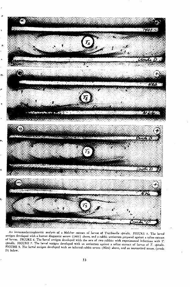
An immunoelectrophoretic analysis of a Melcher extract of larvae of Trichinella spiralis. FIGURE 5. The larvalantigen developed with a human diagnostic serum (1401) above, and a rabbit antiserum prepared against a saline extractof larvae. FIGURE 6. The larval antigen developed with the sera of two rabbits with experimental infections with T.spiralis. FIGURE 7. The larval antigen developed with an antiserum against a saline extract of larvae of T. spiralis.FIGURE 8. The larval antigen developed with an infected rabbit serum (N26) above, and an immunized serum (crude.D) below.
33
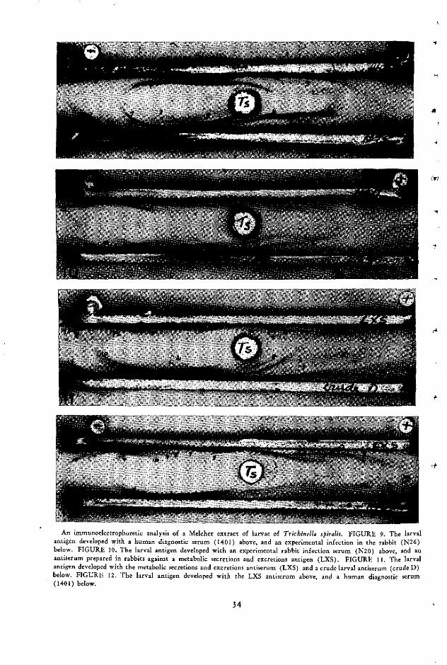
-
tt
An immunoelectfophoretic analysis of a Melcher extract of larvae of Trichinella spiralis. FIGURE 9. The larvalantigen developed with a human diagnostic serum (1401) above, and an experimental infection in the rabbit (N26)below. FIGURE 10. The larval antigen developed with an experimental rabbit infection serum (N20) above, and anantiserum prepared in rabbits against a metabolic secretions and excretions antigen (LXS). FIGURE 11. The larvalantigen developed with the metabolic secretions and excretions antiserum (LXS) and a crude larval antiserum (crude D)below. FIGURE 12. The larval antigen developed with the LXS antiserum above, and a human diagnostic serum(1401) below.
34

separation of diagnostic antigens emphasize the
importance of gel-diffusion assay. Although
column chromatograph of hydatid fluid of E.granulosus and E. multilocularis (18, 82) ap-
peared to separate host from parasite com-
ponents, agar gel analysis indicated that
complete separation of the two groups did not
take place, since molecular screening techni-
c ques cannot separate many at and a2 globulin-
like antigens of host origin that migrate with
similar parasite antigens.
t' Protozoan antigens
Antigenically reactive polysaccharides have
been isolated from Trypanosoma cruzi (41).
Fife and Kent (36) separated protein and poly-
saccharide components from T. cruzi and
'evaluated their sensitivity and specificity in
the CF test. The fractionated antigens were
more specific than the crude extract but less
sensitive. The protein component was the best
and most economical antigen to use. Von Brand
(15) reviewed the information on the chemi-
cal composition of T. cruzi. Exoantigens or
secretory antigens produced by T. cruzi have
i been studied and a glycoprotein has been de-
scribed (122).The chemical composition of an African try-
panosome was studied by Williamson and Brown
(126) and Brown and Williamson (16).
The Leishmania organisms must share a com-
+ mon antigen with mycobacteria, since the lat-
ter have been used by a number of workers in
South America as a diagnostic antigen in the
CF test for leishmaniasis. This antigen, how-
ever, could not be isolated or characterized in
gel-diffusion studies (52).A number of protozoan species have been
studied by agar gel and immunoelectrophoretic
analysis. Krupp (69) recently evaluated 11
amebic antigens by IE, and similarities between
some strains of Entamoeba histolytica with high
and low pathogenicity was observed. Goldman
and Siddique (40) analyzed two substrains of
E. histolytica and showed some antigenic dis-
parity.The studies of Schneider and Hertog (101) on
16 strains of Leishmania indicated that two im-
munologic groups of human leishmaniasis were
present in Panama with wide geographic dis-
tribution. García (37) showed that L. tropica
has three heat-labile and one heat-stabile com-
ponent.Nussenzweig et al. (85) separated a number
of T. cruzi strains into three antigenic group-
ings by means of agglutination and precipitin
tests in agar. Both type- and group-specific sub-
stances in groups A and B were reacted. In a
further analysis (84) 23 strains were studied
and it was shown that most human strains are
Type A but some were of Type B.
Antigenic analyses of plasmodia made by im-
munoelectrophoresis and agar gel by Spira and
Zuckerman (114) revealed seven components
in extracts of P. vinckei. Zuckerman (133)
compared P. vinckei and P. berghei, and found
several common antigenic components. Using
polyacrylamide gels, Sodeman and Meuwissen
(108) found at least 21 bands in P. berghei ex-
tracts. From 3 to 12 participating antigens have
also been described in plasmodia extracts (7, 8,
29, 103, 25). Chavin (23) found 10 to 15
bands in extracts of P. berghei in polyacryla-
mide gel, 4 to 7 lines on IE, and 8 to 10 lines
by double diffusion in tubes. An interesting
aspect of Chavin's work was the presence of all
the bands in IE on the anodic side of the elec-
trical field. Mouse-hemoglobin protein com-
prised a significant portion of the extract. The
parasite components had electrophoretic mobil-
ity in the beta-to-albumin range and could not
be separated from the host components. Similar
difficulties are reported in our hydatid-fluid
fractionations in separating host and parasite
components by ion-exchange chromatography
(81, 82). Spira and Zuckerman (115) have
extended the analysis of plasmodia species by
disc electrophoresis for seven plasmodia species.
Differences were apparent between all the spe-
cies, and their chemical complexity is evident
from the large number of components devel-
oped in their preparations.
Comment
This review is far from complete, and many
excellent publications on the analysis of para-
35

sitic components in the immunology and serol-ogy of paragonimiasis (132, 47, 96, 97, 131)
and filariasis (118, 95) and other parasites ofveterinary (6) and medical importance havebeen omitted. Studies on the fractionation andcharacterization of parasitic materials are at acrucial stage of development. It is importantto characterize the antigenic complexity of ourdiagnostic and immunogenic materials. Researchmust be focused, however, on characterizationof the specific immunological components. Toaccomplish this end we need a stronger biochem-ical approach. We have to staff our laboratorieswith scientists capable of working with the en-zymes and chemical components that interactin our immunologic reactions. We need person-nel who can use the complex preparative chemi-cal techniques, such as preparative column elec-trophoresis, gas chromatography, and othertools emerging from immunologic research inrelated fields.
Parasitic materials are excellent sources forboth applied and basic immunologic studies. The
successful parasite has solved the host's "graft- 'rejection" response. The self-not-self problemin immunology might be as fruitfully studiedwith a host-parasite system as with the graft-rejection system.
Finally, we need specific antigens for parasiticvaccines and diagnostic tests. Some hosts de-velop strong functional immunity against theirparasites. Our feeble attempts to stimulate thisimmunity by vaccination have been far fromsuccessful in most parasitic infections. Once the
immunogenic substances have been character-ized and synthesized, practical vaccines for e'parasitic infections will be available. Parasiticimmunodiagnosis will be greatly enhanced whenthe laboratory can prepare specific and active 'diagnostic reagents. The infected hosts developa large number of antibodies. Analysis of theseantibodies and the development of specific anti-gens for their detection are the challenges ofthe future.
Moderator: The discussion of these topicswill be introduced by Dr. Finger.
36

DISCUSSION
Irving Finger
It may seem a little unusual for a geneticistwho studies a completely nonparasitic organ-ism to be speaking at such a session, but I hopethe relevance of our studies to those describedby the two previous speakers will become
apparent.We have studied primarily the genetics and
regulation of antigen synthesis in Parameciumaurelia. It is obvious that the major advantagepossessed by free-living protozoa for studieson these topics lies in the ability to make con-trolled matings and therefore to analyze therelative importance of genetic and nongeneticdeterminants in the control of a particulartrait. Similarly, cytoplasmic as contrasted withnuclear contributions to the determination ofa character can be distinguished. The otherprimary benefit to be derived from studies offree-living forms is, of course, that the en-vironment can be more carefully controlledand it may be possible to analyze the variouscontributors in that environment that deter-mine antigenic composition.
I should like to describe briefly some of thecharacteristics of the surface antigens of para-mecia and the genetics and regulation of theirsynthesis, particularly as these bear on thetopics discussed by the previous speakers. Everycell can express at least a dozen surface anti-gens. These antigens are generally recognizedby a cell's immobilization in a specific anti-serum prepared against particular serotypes.Even though a cell has the ability to make amultiplicity of antigens, generally only oneantigen is expressed at a time-which one beingdetermined by a complex interaction of nu-
cleus, cytoplasm, and environment.The antigens themselves are proteins with a
molecular weight of about 310,000. They areprobably not made on the surface but migrateto the surface and ultimately to the cilia. Themost disturbing thing about these antigens, asfar as the experimenter is concerned, is thattheir function is completely unknown. A cellapparently has no particular selective advan-tage in possessing one antigen rather than an-other. Apart from the phenomenon of mutualexclusion-the expression of but one antigenat a time-the striking feature of these anti-gens is that one antigenic type can easily betransformed into another. This transformationis readily reversed and is not due to mutation.
From this brief summary, I should like toturn to more specific features that are relevantto the antigens of parasites. When antigenictypes transform to each other, they oftentransform to unrelated antigenic types. Buteven if two antigens are serologically quitedistinct, they appear still to possess the same"function" and to be remarkably similar intheir chemical features. It is obvious that thistransformation process, and the fact that theantigens may be immunologically distinct, willhave a great bearing on assigning a particularfunction to a class of antigens, since one ofthe criteria for correlating function with amolecule is whether this function disappearswhen the molecule disappears. Here, however,the function of a class of molecules persistseven when the original specific molecule nolonger exists.
Secondly, the antigens that can be detected
37

on the surface are not necessarily the onlyciliary antigens that the cell possesses. By thisI mean that there may be primary antigensthat are recognized by the immobilization testand can definitely be stated to be external, andthere may be secondary immobilization anti-gens that are not recognized by immobilizationtests but can be detected when the cell isbroken open and all the antigens are extracted.
Therefore, in a sense, the phenomenon ofmutual exclusion that I mentioned earlier is notnecessarily an all-or-none phenomenon; whatmay be excluded on the surface may not beexcluded internally. These secondary antigens,obviously, may have potential significance byeliciting the formation of antibodies, especiallyif there is lysis of cells within the organism.
A third point of relevance, particularly tothe work described by Dr. Brown, is that theenvironmental influences that determine theparticular types a paramecium will possess atany time are manifold and include such thingsas antiserum, temperature, cultural conditions,enzymes, temperature shock, inert proteins, andstage in the life cycle.
Another feature of the antigens of para-mecium that is of possible significance tostudies with parasitic protozoa is the recentfinding in our laboratory that the variousserotypes release into their medium materialthat can induce transformation when added toother serotypes. The material released does notact in a nonspecific fashion. Different mediahave propensities for inducing different kindsof transformations. Our studies thus far sug-gest that the substances put out into the mediaare not related to the antigens and act byspecifically repressing the activity of certaingenes.
I should like now to discuss briefly the effectof serum on cells. Not only does immune seruminduce transformation, but normal serumfrom an unimmunized animal (in our case, therabbit) may have a very profound effect onthe kind of antigen expressed by a cell. Thefactor in serum that has this effect is presentin the 0-40 per cent ammonium sulfate cut ofthe serum, the same fraction that contains the
immunoglobulins. The orginal cell is not visiblyaffected by such serum, but the serotypes of itsdescendants are altered.
With regard to immune serum, there areseveral features of interest here. It might beexpected that, when antigen is found on thesurface, immobilizing antibody would inducetransformation by binding this surface antigen.We have found, however, that this is not neces-sarily the case. Rather, it seems more likely thatthe antibodies against surface antigens onlyinfluence the transformation of antigenic typeif that antigen is being actively synthesized.It is probable that antibodies do not actthrough tying up the surface antigens butprobably penetrate the surface of the cell andact at the synthetic sites, or conceivably thesites where the secondary antigens are located.
We have also been able to show that anti-sera that are prepared against media in whichparamecia have been living and that possess noimmobilizing titer can markedly influence thepattern of antigen synthesis. Apparently whatis found in these sera is antibodies directedagainst the substances released by the parameciathat in turn repress gene activity.
So we see that the effect of a serum can bequite complex. It may contain nonimmunesubstances that can induce antigenic transfor-mation and perhaps allow the descendants ofthe original cell to escape the effect of specificantibodies. Antiserum can affect the synthesisof antigens that are not on the surface; and,finally, antibodies against substances other thanthe antigens themselves, substances that con-trol the synthesis of the antigens, may have amarked effect on transformation.
Another thing that is pertinent to some ofthe topics Dr. Brown discussed is that we havefound no antibodies except those against theciliary antigens lethal to the paramecia. Thereappears to be no general antigenic substancethat when combined with antibody causes thedeath of the cell.
Let me conclude with a few remarks aboutthe gel-diffusion techniques that have beenextensively used by Dr. Kagan and manyothers in the study of the antigenic mosaics
38
-L
Vr

that make up cells. There are several advan-tages to analysis by means of gel-diffusion tech-
niques compared with the more classical pre-cipitin tests, hemagglutination, complementfixation, and so on. For example, a purified anti-gen is not needed for unambiguous results;multiple mixtures may be studied and roughlyquantitated. The major difficulty with the gel-diffusion techniques-including those usingtubes, such as Preer's and Oudin's, and thoseusing flat surfaces, such as Ouchterlony's andimmunoelectrophoresis-is that' there is analarming tendency for artifacts to appear. Thepossibility of these artifacts must be very care-fully controlled for. However, once these aretaken into account, the Preer technique, whichis the most sensitive of those I have mentioned,may be especially useful in following the isola-
' tion of antigenic components and for diag-nostic purposes, as Dr. Kagan has shown. Infact, the Preer technique can be used (by
following a modification of the Oudin profilemethod) to make qualitative comparisons ofvarious antigens with a several-fold increase insensitivity.
A problem with gel-diffusion techniques isthat they are much too easy to use. Anyonecan set up gel-diffusion tests from scratch in
an afternoon and have data the following day.It is the interpretation that takes a great deal
of care and experience and is not always easy.
* Reactions of identity in Ouchterlonys mustalways be considered tentative, depending on
the serum. Reactions of partial identity are only
convincing when accompanied by absorptionexperiments.
Lastly, I want to emphasize, along with Dr.
Kagan, that the determination of the number
of antigens in a particular preparation mustalways take into account the possibility ofband splitting and the question of whether oneis in antibody- or antigen-excess regions of theprecipitin curve.
Moderator: I want to thank you particu-larly, Dr. Finger, for introducing the conceptsderived from the studies on the genetics of
paramecia. This knowledge is so fundamental
for immunoparasitologists that I think we
should discuss it thoroughly. As I understandit, the production of antigens by the para-mecium depends, if I recall my genetic studies,on the genes from the macronucleus as well as
on the cytoplasm and also on environmentalfactors.
Finger: Yes. The environment determinesthe particular cytoplasmic state, which deter-mines which gene in the macronucleus willwork.
Moderator: I am not very clear about themechanism of mutual exclusion; I believe it
is a rather complex matter.
Finger: No one is clear about that mechan-ism. In fact, that is the subject of our research.
Moderator: One of the things I want toask is how you would interpret the stabilizingeffect of the vector mentioned by Dr. Brown.You remember that he called attention to the
fact that the vector had a stabilizing effect on
the antigenic variants of trypanosomes pro-duced in the course of infection, so that the
parental type was obtained when it got backinto the vertebrate.
Finger: Offhand I cannot think of a hypo-thesis, because of the apparent nonspecificity ofthe phenomenon. We are carrying out workbased on a hypothesis that invokes the per-sistence of an antigen due to the feedback of
the antigen itself on its synthesis. But I do notknow what the factor might be in Dr. Brown'svector for maintaining a particular parentaltype.
Brown: There was the point that when theparasite passes through the vector, it changesstructurally and the electrophoretic pattern ofits cytoplasmic proteins also changes. In par-ticular, the 4S group, which is the major groupof variable antigens, appears to be much lessevident in the insect form. It is apparentlyrepressed there. But why, when it gets back
into the vertebrate, the parasite should develop
as the parent antigenic type, I do not know.This reversion is not always complete. Often
traces of the original variants come throughalong with the parental type.
Finger: Do you know whether this couldbe due to selection in a mixed population?
39

Brown: It could be, yes. I have perhapsoversimplified slightly: when I say there is asuccession of populations, I mean that there isa succession of population mixtures, particu-larly late in the infection. I have in fact iso-lated a parent form from such a mixture quitelate in the infection, so it is possible that thisis the one that is selected and that comesthrough the vector.
Finger: With reference to the parameciumsituation, there is no selection among theparamecium serotypes. It is a conversion of apopulation of one type to a new population.
I wonder if you would expand on what 1found to be of particular interest-the conver-sion of the crithidial to the trypanosome to theleishmania type by increasing the concentra-tion of an anticrithidial serum.
Brown: Unfortunately, there is not muchmore to say about that. This work was donea few years ago by Professor Adler. He pre-pared the rabbit anticrithidial serum and in-cubated his crithidia in it and in that way
was able to show that a single crithidium pro-duced a colony of either trypanosome or leish-manoid forms, depending on the serum con-centration. This work has not been continued.
Finger: The crithidium then shares antigenswith trypanosomes, and these antibodies areperhaps responsible for the conversion?
Brown: They do share some antigens, andone would think that the difference may per-haps be quantitative rather than qualitative.
Finger: That is right.
Moderator: I think this illustrates the needfor immunologists and geneticists to get to-gether and study certain fundamental prob-lems.
I was very much interested in what Dr.Brown said about the hemagglutination reac-tion's depending on the red cell membrane. Doyou think it is some antigen diffusing from theparasite that sensitizes the red cells or modifiesa protein of the red cell and produces this?And, just to ask all the question at the sametime, if complement is added, do the red cellslyse or not? Is the phenomenon related to pas-sive hemagglutination and passive hemolysis?
Brown: In reply to the first question, it isinteresting that the red cell infected with theimmature parasite, the trophozoite, does notagglutinate. It is only when the parasite ma-tures and forms the schizont that it will do so.At this stage, the surrounding red cell itself isbreaking down. The membrane is visible in EMsections, but the contents are pretty muchdestroyed, and I imagine-although there is noevidence for this-that the parasite antigen canthen get into the red cell membrane. If the redcell membrane is removed, these naked para-sites will not agglutinate in immune serum. Thismay be because the removal of the red cellmembrane removes a lot of the antigen, orperhaps the complete antigen involves a para-site component and a red cell component. Wehave in fact shown that if the red cell is lysedwith anti-red-cell serum, a certain amount ofparasite material is released and can be collectedin the lysate formed.
As for your second question, if complementis added to schizont defective cells in immuneserum, they will not lyse. The reason for this,we think, is probably that the red cell, beingso empty, is more or less lysed anyway. Evenif the complement punches holes in the redcell membrane, there will be no osmotic effectto burst the cell.
Goodman: Along the same line as Dr.Bier's questions on your very interesting ob-servation about the hemagglutination of thered cell having something to do with the redcell membrane, and perhaps with associatedantigen from the malaria parasite, is thisagglutination only of the red cells with theschizonts in them? Does the agglutination in-volve red cells that are not parasitized? Haveyou been able to find any factor you can puton a normal cell, let us say, that would causeit to be agglutinated? Those are my specificquestions.
On a more general level, there have beensome philosophical interpretations of the de-struction of red cells in malaria in which auto-immune responses have been implicated in
theory. It sounds as if this would be ratherunlikely in the light of your observations.
40
.s ,

Brown: With regard to the first point, inour system normal red cells are not aggluti-nated at all; they are completely distinct. And,as I said earlier, the cells containing the im-mature parasite do not agglutinate either. Sothe reaction of the schizont-infected cell is
' quite distinct.
Goodman: When you do the agglutina-tion test, are 100 per cent of your red cellsparasitized so that they all agglutinate, or doyou just see agglutination of those that have themature schizonts? Is, for example, how youdo the test affected?
Brown: We have done both. Normally androutinely for the test, we collect the schizont-
infected cells, but we have also tested mixtures;in fact, we have also transfused immune seruminto monkeys containing infected and nonin-fected cells and shown that when the schizont-infected cells are put onto a warm stage theywill then agglutinate and the normal cells willstay quite separate.
Your question about autoimmune responsesraises, I think, a very debatable point. I thinkthe lysis of normal red cells could be explainedin quite a number of ways. One thing we arelooking into at Mill Hill is the possibility thatcysfaccinic(?) acid, which is present in theparasite and is quite a strong lytic agent, maybe responsible. I think it can be explained inbiochemical terms just as easily as in immu-nological ones.
Cohen: My comment is directed to Dr.Finger, but it bears on the earlier presenta-tions. One can not help being impressed withthe apparent multiplicity of proteins duringthe various stages of differentiation of theseintermediate forms. I feel inclined to wonder,as a biochemist, whether the entities are toocrude for determining whether they are in factgenetically different structures. We havelearned, of course, that it is the subunits of theproteins that are genetically controlled, and itwould seem to me that perhaps the importantthing is to devote some effort to characterizinga few good proteins and finding out the natureof their polymeric and other tendencies to
aggregate. Reference was made to their relative
ability to form glyco and other proteins. I donot know whether these have covalent bondsor whether they are merely adducts of somekind, but it would seem to me that before thegenetic implications in going from one stage toanother can really be analyzed, a subunit struc-ture for identification ought to be settled on.We certainly know that many proteins in theirsubunit form will have one kind of immunespecificity but in the aggregate form may beblocked because of the specific groups that areinducing the antibody formation. While thismay only complicate the story, it does seemthat we should be rather cautious at this stagein taking the apparent complexity too literally.There may be a simplicity if we can get to thesubunit stage.
Finger: With reference to paramecium, Ican answer in this way: Unfortunately, thesituation is as complex as I presented it. Theproteins have been purified and characterizedchemically and physically. They are made up ofnine subunits, apparently arranged in sets ofthree trimers, and each distinct antigenic typeis controlled by a different locus. The geneticwork that has been done shows that these lociare unlinked. We know that the various formsof antigenic types I have described, althoughthey have related amino acid compositions andsedimentation coefficients, also have differences.Fingerprints have been made. It may be a littleunfair to talk about the paramecium antigens,because we have been fortunate. Dr. Preer andDr. Steers have purified them sufficiently foractual analysis. In this case, therefore, I do notbelieve the question of one's being a polymerof another is applicable. In fact, we have evenstudied complementation and hybrid forms ofthe antigens. I do think that, as Dr. Kaganindicated, in the parasitic forms there is greatercomplexity; I am delighted to be working withparamecium.
Borsos: Transformation of cells may be theresult of the action of an antibody on thecell surface. However, there are other sub-stances-such as phytohemagglutinins-thatcan induce lymphocytic transformation, that
can induce biochemical changes inside the cell
41

and stimulate synthesis of DNA, proteins, andcell divisions. Has anything like phytohemag-glutinin been tried on parasites of this sort toinduce changes in the antigens?
Another point I should like to make is thatantibodies, as far as we can tell, do not kill.They may occupy sites and the result of theoccupation may be a steric hindrance of somesort, but there seems to be no biochemicalactivity involved in antibody interaction withantigens. It is therefore not surprising thatantibodies will not kill a parasite. Perhapsoccasionally a miracidium may be blocked inits movement. But other substances, such ascomplement, are needed to make an antibodycytocidal.
Furthermore, not all classes of antibodiescan bind complement, and there can be a greatvariety of responses as a result of antibody-antigen interaction.
Finger: I cannot answer the parasitologicalpart of your question. As I have mentioned, ofcourse, in paramecium many reagents otherthan antibodies induce transformation-enzymes, temperature, shock, actinomycin D,puromycin, and so on.
With regard to the killing of a cell by anti-body, we still do not know the mechanism ofdeath in paramecia that are immobilized byantibodies. By the way, this occurs with de-complemented serum. It can occur very rapidly,within less than a minute, and we have madesome preliminary pictures with the EM to seeprecisely what may be happening. So far wehave no clue at all to why the antibody in
combination with the antigen should result indeath. In more dilute concentrations of anti-body there is no death, and there can be trans-formation of type.
Borsos: I mentioned the phytohemagglu-tinins because they react at the surface of cells.The other substances are probably not actingon the surface. Actinomycin is not; heat is not.Any one of these changes induced is not dueto surface action. However, antibodies do acton the surface.
Finger: As I have tried to stress, we thinkthat the antibodies that are having their effecton transformation may really not be acting atthe surface.
Bruce-Chwatt: I should like to put to Dr.Taliaferro a question that has puzzled para-sitologists since 1948. What explanation canhe give for the fact that the pre-erythrocyticor extra-erythrocytic schizont in the liver ofmammalian parasites does not produce anycellular response? Dr. Brown mentioned that inPE forms the development is so fast that theremay have been no time for an immunocellularresponse, but this is not so when it comes tosecondary tissue stages, which produce nocellular response in the liver.
Moderater: Dr. Taliaferro, would you careto comment?
Taliaferro: I am afraid I can't.Moderatór: We shall continue now with
the papers by Dr. Smithers and Dr. Remington.Again, the discussion will follow both presenta-
tions.
42
4-

THE INDUCTION AND NATURE OF ANTIBODYRESPONSE TO PARASITES
,, S. R. Smithers
The literature on antibodies in parasitic in-fections is extensive, and nothing like a com-plete review of this subject is possible in thetime available. I am therefore going to dealwith the subject in broad outline, drawing ex-amples from various infections that illustrateimportant concepts. Throughout, I shall con-centrate on the significance of antibodies inresistance to infection.
Changes in the immunoglobulins inparasitic diseases
Raised immunoglobulin levels in parasiticdiseases are common. Unfortunately, most ofthe studies on this subject were made before therecognition of the immunoglobulins as four dis-tinct types-IgG, IgM, IgA, and IgD-and inmost infections the relative increase in each im-munoglobulin type is not known.
In experimental and in human schistosomiasisthere are large increases (as much as twofold)in the y and P globulin levels, as well as in-creases in the a2 globulins (20, 21, 29, 61, 68,79, 89). These increases occur at the time ofegg deposition by the parasite and are probably
due to the increase in antigenic stimulation thatresults from the excretions and secretions of the
fully mature worms and the larval stages withinthe eggs. However, some alteration in the glob-ulin values may be due to other causes. The a2
globulin increase, for example, may be the re-sult of tissue damage caused by the depositionof eggs in the liver and intestine (4), and the
increase in the level of macroglobulin in chronicschistosomiasis that has been reported (6, 21)may be due to splenomegaly (44). In rhesusmonkeys exposed to Schistosoma mansoni, theincrease in immunoglobulin at the time of eggdeposition occurs in the absence of an increasein macroglobulin. Figure 1 shows the averagemacroglobulin values (determined by ultra-centrifugation) from three monkeys estimatedon the day of exposure to 1,000 S. mansoni cer-cariae, four weeks later, and eight weeks later.Although the total immunoglobulin value hadrisen at eight weeks, the macroglobulin valuehad remained constant.
In trypanosomal and malarial infections, theIgG, IgA, and IgM levels are all raised (47, 77,88). Especially high IgM levels are reachedearly in the infection, and it has been suggested
FIGURE 1. Average gamma globulin values and macro-globulin values of three rhesus monkeys after exposure to1000 cercariae of S. mansoni.
80
60 -
E
20 -
0.25gm.% 0-24gm.% 0.24gm.% macroglob.
gamma glob
_~
0 2 4 6 8 10 12weeks o! i-f-! on
43
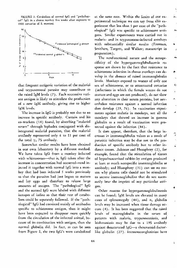
FIGURE 2. Catabolism of normal IgG and "pathologi-cal" IgG in a rhesus monkey five weeks after exposure to1000 cercariae of S. mansoni.
loo
150
-r;
o i
o
1o
-5
; 2
ILO 5
Ooys10
that frequent antigenic variation of the malarialand trypanosomal parasite may contribute tothe raised IgM levels (7). Each successive vari-ant antigen is likely to stimulate the productionof a new IgM antibody, giving rise to higherIgM levels.
The increase in IgG is probably not due to anincrease in specific antibody. Curtain and hisco-workers (14) found, by absorbing "malarialserum" through Sephadex conjugated with dis-integrated malarial parasites, that the malarialantibody represented only 6 to 11 per cent ofthe total ¥2 7S antibody.
Somewhat similar results have been obtainedin our own laboratory by a different method.We have taken IgG from a monkey infectedwith schistosomes-that is, IgG taken after theincrease in concentration had occurred-and in-jected it together with normal IgG into a mon-key that had been infected 5 weeks previouslyso that the parasites had just begun to matureand lay eggs and therefore to release largeamounts of antigen. The "pathological" IgGand the normal IgG were labeled with differentisotopes of iodine so that their rate of catabo-lism could be separately followed. If the "path-ological" IgG had consisted mainly of antibodiesspecific to schistosome antigen, then it mighthave been expected to disappear more quicklyfrom the circulation of the infected animal, be-cause of its combination with antigen, than thenormal globulin did. In fact, as can be seenfrom Figure 2, the two IgG's were catabolized
at the same rate. Within the limits of our ex-perimental technique we can say from this ex-periment that less than 5 per cent of the "path-ological" IgG was specific to schistosome anti-gens. Similar experiments were carried out inmalaria- and in trypanosome-infected monkeyswith substantially similar results (Freeman,Smithers, Targett, and Walker; manuscript inpreparation).
The nonfunctional nature and the nonspe-cificity of the hypergammaglobulinemia re-sponse are shown by the fact that resistance toschistosome infection in rhesus monkeys can de-velop in the absence of raised immunoglobulinlevels. Monkeys exposed to worms of only onesex of schistosome, or to attentuated cercariae(infections in which the female worms do notmature and eggs are not produced), do not showany alteration in their serum proteins, but nev-ertheless resistance against a normal infectiondoes develop (29, 76). In vaccination experi-ments against malaria in monkeys, not all themonkeys that showed an increase in gammaglobulin as a result of vaccination were pro-tected against the infection (86).
It does appear, therefore, that the large in-creases in immunoglobulin values as a result ofparasitic infection may be due not to the pro-duction of specific antibody but to other in-direct causes. Askonas and Humphrey (2), forexample, found that the stimulation of tissuesof hyperimmunized rabbits by antigen producedat least as much nonspecific immunoglobulin asantibody; and Humphrey (31) can see no rea-son why plasma cells should not be stimulatedto secrete immunoglobulins that do not neces-sarily bear the imprint of any particular anti-gen.
Other reasons for hypergammaglobulinemiacan be found; IgM levels are elevated in manycases of splenomegaly (44), and a2 globulinlevels may be increased when tissue damage oc-curs (4). It has been suggested that the raisedlevels of macroglobulin in the serum ofpatients with malaria, trypanosomiasis, andleishmaniasis may be due to a 19S antibodyagainst denaturated IgG-a rheumatoid-factor-like globulin (27). Immunoconglutinins have
44
-4
t
1-

been demonstrated in Trypanosoma brucei in-fections of rabbits and cats (32) and may wellcontribute to raised immunoglobulin levels.
The etiology of hypergammaglobulinemia inparasitic infections is obviously complex, andyet it is fundamental to our knowledge of para-site immunity and pathology and of antibodyproduction. With the recent expansion of ourknowledge of the immunoglobulins and withtechniques becoming available for their easyassay, we should be able to make considerableadvances in our understanding of the signifi-cance of raised immunoglobulins in the pathol-ogy and immunity of parasitic diseases.
Specific antibody response to parasites
Protozoa
The liberation of large amounts of variedantigens from parasitic infections induces acomplex antibody response. Almost every typeof antibody known in classical immunologyhas been described. Thus, precipitins, lysins,agglutinins, opsonins, complement-fixing anti-bodies are all known in parasitological infec-tions, and there are some antibodies peculiar toparasitology, such as ablastin in T. lewisi infec-tions. Undoubtedly many antigens will induce
an antibody that will agglutinate, precipitatefix complement, lyse, and so forth, dependingon the appropriate conditions. Many of thesein vitro reactions may therefore be manifesta-tions of the same antibody under different con-ditions; on the other hand, one antigen mayinduce more than one antibody. It is easy tosee, therefore, why the antibody response toparasites is complex and still poorly understood.
The induction of parasite antibody dependson the natural history of the infection. This iswell illustrated in leishmaniasis, a disease causedby an intracellular protozoan. There are twoforms of the disease: a cutaneous form result-ing in a local infection of the skin, and a serioussystemic infection, visceral leishmaniasis, in-volving the spleen and other reticulo-endothe-lial organs. In cutaneous leishmaniasis circulat-ing antibody has not yet been demonstrated,although delayed skin reactions are obtained
early in the infection. In visceral leishmaniasis,on the other hand, circulating antibodies arereadily demonstrated even if there is little orno immunity to this disease (1). In malaria itis difficult to detect antibody before the para-sites can be demonstrated in the red cells, butduring the erythrocytic cycle antibody is easilydetected (87). This does not imply that theother stages lack antigenicity, but rather thatantigen is present in too low a concentration toinduce a response (62). It is well known thatwhen antigen is presented intravascularly, thespleen is of major importance in antibody pro-duction, and, as would be expected, splenec-tomy in malaria and African trypanosome in-fections results in an increase in the parasit-emia. On the other hand, splenectomy haslittle effect in chronic Chagas' disease where theparasites are found within tissue cells (23).
At present it is difficult to correlate any oneantibody with a protective role. In malaria ithas been shown that neither the complement-fixation titer nor the fluorescent-antibody titeris necessarily correlated with protection (22,86). It is known, however, from the success-ful passive transfer experiments of Coggeshalland Kumm (9) and Cohen and McGregor(10), that humoral agents must be playing animportant role in protection. In the latter workthe protective agent was in the IgG fractionof the immunoglobulin. Even so, large amountsof transferred IgG are necessary in order todemonstrate its protective effect. The reasonsfor the difficulty in demonstrating passive trans-fer are not clear. It has been suggested thatthe antibodies formed in the spleen, liver, andbone marrow occur in sufficient quantities to beactive in those sites but are insufficient afterdilution in the serum to be passively trans-ferred (84). Apart from this possibility, pas-sive transfer is complicated by the fact that agiven species of Plasmodium may consist of anumber of races or strains that may differ intheir antigenicity. Cross-immunity may existbetween strains of the same species, although toa lesser extent than against the homologousstrain (83).
In trypanosomiasis, most of the detailed stud-
45

ies have been on the nonpathogenic rodentforms, against which three types of protectiveantibody have been described. One is ablastin,a 7S antibody that inhibits division of the try-panosomes without being immediately lethal; itcan be absorbed from serum only with difficulty.The other two antibodies are trypanocidal; oneis a 7S and the other a 19S immunoglobulin(15). In the African trypanosomes of humanand veterinary importance, the antibody re-sponse is complicated by the antigenic variantstrains that appear as a counter to antibody re-sponse (24). However, serum from animalswith T. gambiense is able to confer passive im-munity, but recent work has suggested that theagglutinating antibody present in such serumis not the antibody responsible for protection(71).
The antibody response in Chagas' disease isstill poorly understood. In the acute form ofthe disease trypanosomes circulate in the bloodstream, whereas in the chronic stages they arefound only within cells. Lysins (16), precipi-tins (72), and fluorescent-antibody titers (93)have been demonstrated in sera from infectedanimals, but it is not known which, if any,of these antibodies has a protective function.Resistance has been conferred by transferringserum from infected hosts in mice and inrats (13, 41, 72). It is believed, however, thatthe transferred serum affects only the parasitescirculating in the blood; the intracellular stageis probably unaffected by the immune serum.Pizzi believes that phagocytosis is an importantdefensive mechanism in the acute stage, andit is likely that the process is assisted by theaction of an opsonin-like antibody; lysins ap-parently do not develop during the acute stage(60). Recently it was found that in a rhesusmonkey infected with T. cruzi, the IgM levelrose to a maximum on the tenth day of in-fection and then fell over a period of twoweeks (unlike African trypanosome infections,in which the IgM level remains high through-out the disease) (93). This period roughlycorresponded to the acute stage of the infec-tion. The rhesus monkey or other primatesmay prove useful experimental animals for
studying Chagas' disease.It is clear that many antibodies are induced
by protozoal infections, but most of theseantibodies have no protective function. Inmost infections a humoral protective agent hasbeen detected by passive transfer of serum, butthe type of antibody responsible and the pos-sible and likely interplay between cellular andhumoral factors remain largely unknown.Little is known about the immune response tochronic Chagas' disease, in which the parasitesare located in tissue cells, particularly heartmuscle. It is conceivable that the immunereaction at thís stage will enhance rather thanreduce the pathology of the disease (60).According t'o Mackaness (45), organisms thatcan survive and multiply within host macro-phages caused delayed-type hypersensitivity to-ward the corresponding microbial antigens.Leishmania, and to a lesser extent T. cruzi (17),are found within host macrophages, and it maybe that investigations into cell-bound immu-nity would yield significant results in theseinfections.
Helminths
Much of what has been said about protozoalinfections applies to helminthiasis. A widevariety of antigens is liberated from wormparasites, and a complex antibody response con-sisting of precipitating, agglutinating, com-plement-fixing, immobilizing, skin-sensitizing,nonprecipitating, and blocking antibodies isproduced. In helminthiasis, unlike protozoal in-fections, lysins have not been detected. Thelysis of a relatively large organism with astrong impermeable cuticle, such as a nema-tode, is probably outside the bounds of anti-body capability. Schistosomes, however, haveno cuticle as such but a living cytoplasmicmembrane (42) that is probably concernedin the uptake of nutriment from the host (73).It is possible that antibody and complementcould damage the schistosome surface, althoughno lysis of larval or adult schistosomes byspecific antibody has been observed.
In schistosome infections various antibodiesare produced (58, 81). Peak antibody titers
46
*.4
x,

generally appear when the parasite matures andeggs are deposited in the tissues (50, 67, 75);it is at this stage that there is a sudden increase
'< in the liberation of antigen. If, however, sensi-tive techniques such as complement fixation orfluorescent-labeled antibody are used, otherantibodies can be demonstrated earlier in infec-tion (30, 36, 46).
Antibody reactions have been describedaround living schistosomes in vitro; the cer-carienhullen reaction or CHR antibody is mani-fested as an envelope-like structure aroundcercariae when placed in specific antiserum(91), and circumoval precipitins or the COPreaction is seen when schistosome eggs are in-cubated in schistosome antiserum (57, 63).There is one report of precipitates formingaround the oral sucker of three-week-oldschistosomula when cultured in immune ba-boon serum (64). Quite recently it has beenshown that the Hoeppli phenomenon, which issometimes seen around schistosome eggs in hosttissue, is an antigen-antibody complex (43).
Apart from the usual display of antibodiesnormally present in helminth infections, pre-
cipitates have been clearly demonstrated aroundthe oral and excretory orifices when nematodes
are incubated in immune serum (18, 48, 56,
59, 65, 69, 74). Jackson, using fluorescent-labeled antibody, demonstrated that these pre-cipitates are the result of antigen-antibodycombination in infections of Nippostrongylusbraziliensis and Trichinella spiralis (34, 35).Taliaferro and Sarles have also shown precipi-tates in and around N. braziliensis adults andlarvae in infected rats (85).
What significance have anti-worm antibodiesin protecting the host against the parasite? Atthe present time no demonstrable antibody inhelminth infections can be directly correlatedwith acquired resistance. In schistosome infec-tions the CHR, COP, complement-fixing anti-body, fluorescent-antibody titers, skin tests, andvarious precipitins show no relation to protec-
tion (33, 76, 92). Animals usually produce a
marked antibody response after the injection of
dead parasite material, but protection againstreinfection cannot be induced in this way.
Moreover, resistance can be induced in the ab-sence of eggs and fully mature worms (that is,in the absence of the stages of the life cyclethat induce the greatest antibody response) aseasily as in a normal infection (76, 78).
Ogilvie has shown that the precipitates thatdevelop around N. braziliensis larvae when in-cubated in immune rat serum are no guide toimmunity. These precipitates are not detectedafter one and sometimes two normal infections,although the rats are resistant to challenge atthis time. On the other hand, they do developin the serum from rats infected with extractsof adult worms in adjuvant, yet rats treated inthis way are not resistant to challenge (52).
Thus, as in protozoal infections, a host ofantibodies is produced but their role in protec-tion is not clear. In protozoal diseases at least,a protective role by humoral factors can bedemonstrated, with some difficulty, by thetransfer of immune serum to a normal animal.In schistosomiasis, although many attemptshave been made to transfer protection withserum from resistant animals, most have failed(80); and, as Kagan has pointed out, even in
the few successful passive-transfer experiments(where only slight protection was conferred)absorption experiments have not been per-formed to determine that specific antibodies
were responsible for the effect (40).In several nematode infections passive trans-
fer of protection with serum has been demon-strated (8, 12, 25, 37, 65, 70, 82, 90). But
protection obtained in this way is seldom com-parable to that produced by an active infec-tion. Even when enormous quantities of serumare given, the infection is at best only dampeddown, the result being a slight reduction inworm take or egg output. Painstaking workby my colleagues has highlighted some of thedifficulties of demonstrating passive transfer;during the past three years, 18 pools of immuneserum from rats immunized against N. brazi-liensis by similar infection schedules have beentested for their protective properties. Only 11
of the serum pools have given passive protec-tion, and the efficacy of each positive poolshowed considerable variation (Ogilvie and
47

Jones, personal communication). Similar ex-
periences have been reported by Crandall
working with Ascaris in mice (11) and by
Miller working with hookworm in dogs (49).
These results lend support to Stirewalt's con-
clusion that lack of passive transfer in schis-
tosomiasis indicates not so much an absence orineffectiveness of the antibodies as a lack of
understanding on our part of how they func-
tion and how to handle them (80). In this
connection, one is reminded of the recent re-
ports of cytophilic antibodies, a term applied to
globular components of antiserum that be-
come attached to certain cells in such a waythat the cells are subsequently capable of
specifically absorbing antigen (51). Transfer
of guinea pig cytophilic antibody is rapidlycleared from the circulation of recipient guineapigs; only a small fraction of the injected
cytophilic antibody is detectable after 24hours, whereas the more conventional anti-
bodies are still present in relatively high titersat this time (38). There is also evidence that
conventional antibodies may even suppressphenomena in which cell-bound antibodies play
a part, and Nelson and Boyden suggest that
passive transfer with serum might be possible
if the serum contained an unusually large
amount of cytophilic antibody and an un-
usually small amount of conventional antibody
(51). No cytophilic antibodies have as yetbeen described in parasitic infections, but asearch for them could prove profitable.
Reagins. An interesting feature of the anti-
body response to helminth infections has beenthe subject of recent research. It has longbeen known that in helminth infections of
man, an immediate-type skin reaction is elicitedby intradermal injection of homologous anti-
gen. Skin tests have been widely used indiagnostic and survey work, but it was perhapsnot generally appreciated by parasitologists thatsuch immediate reactions are mediated by aspecial class of antibodies called reagins.
Recently reagin-like antibodies have beendemonstrated in experimental infections of
helminths; in rats infected with Nippostrongy-lus (53, 54); in rats and monkeys infected with
Schistosoma (19, 28, 55, 66); in rabbits in-fected with Schistosoma (5) and with several
species of Ascaroids (26); in rats infected with
the filarial parasite Litomnosoides carinii (Worms,
personal communication); and in sheep in-
fected with Trichostrongylus (Ogilvie, per-
sonal communication). Reagin-like antibodies
of rats, rabbits and monkeys are similar inproperties to the reagins of man (5, 19).
Reagins are heat-labile, nonprecipitatingantibodies that possess the property of pro-
longed tissue fixation to homologous skin or to
the skin of closely related species. They appear
to be slightly larger in molecular size than 7S
gamma globulins and have fast electrophoretic
mobility (19, 39).The induction and nature of reagins in
experimental helminth infections has been in-
vestigated mainly in rats infected with Nippo-
strongylus and in monkeys infected with
Schistosoma. It is difficult to induce reagin
formation by the inoculation of dead parasitematerial, although reagins are readily induced
by a viable infection. In rats infected with
Nippostrongylus, reagins appear in the serum
about the time the animals acquire resistance
and eliminate the majority of their worms.
Further infections stimulate an anamnestic
rise in reagin production, but if reagins are
stimulated by vaccination with worm extracts
a second vaccination will not induce an anam-nestic response (54). In rhesus monkeys in-fected with S. mnansoni, reagins appear any time
after the sixth week of the first infection, buttheir appearance at this stage is transitory, for
they disappear from the serum within the next
two to four weeks. Subsequent challenge with
cercariae, however, causes their reappearancewithin two weeks; thereafter they may persist
for long periods (19).The evidence so far is against the involve-
ment of reagins in any protective function in
monkeys infected with Schistosoma (19, 28).In rats infected with Nippostrongylus, theevidence at the moment suggests that reagins
are not protective in the usual sense-that is, ininitiating direct damage to the worm-but maybe involved in the sudden termination of prim-
48
ary worm infections by an anaphylactic mech-anism (3, 39).
The stimulation of reaginic antibodies byhelminth infections poses some fascinatingproblems. Why are reagins produced in re-sponse to helminths as opposed to other para-sites? Why are reagins induced only by theliving infection process and not by the injec-tion of helminth antigen? What is the linkbetween helminthic infections and allergicdiseases such as asthma and hay fever? Havereagins a significant role in the host-parasiterelationship, or is their appearance just anothermisdirected function of the immunologicalmechanism? Answers to these questions wouldbe of interest to allergists as well as to para-sitologists.
Comment
I have pointed out in this brief review thatit has seldom been possible to correlate anti-body response with protective immunity in
parasitic infections. In no host-parasite system
(except T. lewisi in the rat) has protective
antibody been characterized in terms of itsbiological function.
It follows that parasitologists are still faced
with two basic problems in spite of the large
amount of work done in this field: first, forthe great majority of parasitic infections it isnot yet possible to induce resistance by a safeand effective vaccine; second, although ac-quired immunity is common in parasitic infec-tions, we know almost nothing of how it acts.
The attack on these problems is conductedalong two main lines, which derive from classi-cal immunology; these are active immunizationby antigens and passive immunization by anti-serum. Both approaches are aimed ultimatelyat the indentification of the functional anti-
gens. Thus living parasites actively immunizetheir hosts, but living parasites are complexsystems and must be simplified by fractiona-
tion and isolation of parasite antigens, with theprotective power of the antigens determined ateach step. Ultimately, pure antigens will beobtained that not only will vaccinate the hostagainst the parasite but also may be charac-terized in terms of parasite physiology. Asregards passive immunity, where this is pos-sible, the protective serum must be fraction-ated in terms of the specific immunoglobulins.Purified functional antibody can then be ab-sorbed with parasite antigens and in this waythe functional antigens may also be identified.
These approaches are obvious to immunolo-gists, who may well wonder why parasitologistshave not already solved their problems. Wecan only plead that in practice the difficultiesare enormous. To begin with, it is usually im-possible to induce a high-grade protective im-munity with nonliving parasite material. Inthe protozoa, this may be due to antigenicvariation of the parasite; in helminths, it isprobably due to the small amount of functional
antigen present in dead worms. In any case, itis a hard task to attempt to isolate functionalantigens when the starting material will notitself stimulate protective immunity. Again,in many cases it is very difficult to induce
passive protection with serum; in some host-
parasite systems it seems impossible, and in
others the protection is erratic and requires
very large amounts of serum. Thus the isola-tion of functional antibody is severely ham-
pered.It is small wonder, then, that many para-
sitologists, including myself, tend to skatearound these two basic problems. We must,however, be continually aware of these prob-lems and our own inadequacy in solving them.We must face up to the fact that it is littleuse analyzing the antibody response withoutattempting to relate it to the biology of theinfection, and it is little use analyzing parasiticantigens without attempting to relate them tothe stimulation of active immunity.
49

CHARACTERIZATION OF ANTIBODIES TO PARASITES°
Jack S. Remington
In marked contrast to the voluminous litera-ture on the immunochemical and physicochem-ical characterization of antibodies formed in re-sponse to infection with a variety of bacteriaand viruses, and to numerous pure antigens,relatively little is known about the character-
istics of antibodies formed in response to infec-
tion with protozoa and helminths. Multiple
serologic tests are available for the diagnosis o'f
parasitic diseases, but we lack knowledge of thenature of the antibodies that participate in these
reactions. Such characterization would not
necessarily be of academic interest only butmight shed additional light on the mechanisms
of parasitism and host response.When I saw the proposed title of my talk, I
realized that of necessity the information dis-
cussed would be largely prospective in naturerather than both prospective and retrospective,
as is possible with the subjects covered by theother discussants. In many ways this is an ad-
vantage-in an area as embryonic as this it isoften useful to direct one's attention to what
might be accomplished in the future.The characterization of antibodies is a vast
area of research and includes the definition ofprimary, secondary, tertiary, and quaternarystructure; molecular and genetic variations; the
determination of protein-bound carbohydrates,
synthesis, distribution and turnover, serologicreactivity and specificity, and so forth. Here I
shall concern myself solely with methods for
* Supported by Grant No. AI-04717 from the NationalInstitutes of Health and by a grant from the John A.Hartford Foundation, Inc.
immunochemical and/or physicochemical char-
acterization and thus with recognition of the
three major immunoglobulin classes of antibody-IgG, IgA, and IgM. Suitable references are
given for each method.First I should like to mention the data avail-
able on the characterization of antibodies in
three parasitic infections. In 1965 Abele et al.
(1) published their findings on alterations in
serum proteins and 19S antibody production
during the course of induced malarial infectionsin man. These authors noted a consistent in-
crease in IgM in immunoelectrophoretic patternsof serum obtained from volunteers infected with
Plasmodium vivax and Plasmodium cynomolgi.
Sera were separated by gel filtration on columns
of Sephadex G-200, and the fractions weretested for antibody activity by means of the
fluorescent-antibody test using a fluorescein-
labeled rabbit anti-human immunoglobulin an-
tiserum. In addition, reductive cleavage with
mercaptoethanol was used to help differentiateIgG and IgM antibodies. In several of the volun-
teers antibody activity was confined to the IgMfractions of serum during the first few days of
antibody production, but thereafter was found
in both IgM and IgG. Approximately 40 days
after infection antibody activity was present
only in IgG. Of significance for our discussionhere is the use by the authors of both blood-
induced and sporozoite-induced infections and
the variability in response in the different volun-
teers. For example, in one case of sporozoite-
induced infection antibody activity was dem-
onstrable only in IgG and never in IgM frac-
50

tions. Characterization of the antibodies inthese cases has revealed previously unrecognizedparameters requiring further exploration, suchas the relative efficacy of these different classesof antibodies in protection against infections(2), their reactivity and specificity in differentserologic tests, the antigenic structures stimu-lating their production, and the relation ofinoculum size to class of immunoglobulinresponse.
Recently we have demonstrated the presenceof IgM toxoplasma antibodies in humans withacute acquired and congenital toxoplasmosis(3). A complete review of our data has re-cently been submitted for publication andwill not be described here (4). Characteriza-tion of toxoplasma antibodies was performedto determine whether a demonstration of IgMtoxoplasma antibodies would prove diagnosti-cally significant. Since IgG toxoplasma anti-bodies freely traverse the placenta, their pres-ence in the newborn may only reflect maternalinfection (5). IgM antibodies do not normallytraverse the placenta but may be formed inutero in response to infection (6). Their dem-onstration in a newborn, in the absence of aplacental leak, has proved diagnostic of con-genital toxoplasmosis.
Initially, the demonstration of IgM toxo-plasma antibodies was accomplished by the useof sucrose density gradient ultracentrifugationand reductive cleavage. Recently we devel-oped a modification of the indirect fluorescent-antibody technique, using a fluorescein-taggedantiserum specific for IgM. This technique canbe employed in any laboratory that has a fluo-rescent microscope and can be completed with-in about two hours. It may prove useful instudies of characterization of antibodies to otherparasites and in the diagnosis of other infections-especially in the newborn. A description ap-
'pears on pages 55 and 56.
We are now employing the technique in a pro-spective study of 5,000 newborns to determinethe incidence of congenital toxoplasmosis in theSan Francisco Bay Area. Because of the wideclinical spectrum of congenital toxoplasmosisin the newborn (7) and the presence of mater-nally transmitted antibodies, such a study could
not be performed previously.Crandall, Cebra and Crandall have recently
described the relative proportion of IgG-, IgA-,and IgM-containing cells in rabbit tissues dur-ing experimental trichinosis (8). Direct count-ing of cells containing two different immuno-globulin classes in a single tissue section was ac-complished by staining with pairs of immuno-fluorescent reagents, specifically reactive withy-, It-, or a-immunoglobulin heavy chains andlabeled with contrasting fluorochromes.
By the use of two different pairs of reagentson adjacent sections, the relative numbers ofcells containing IgG, IgM and IgA were calcu-lated. The most striking change in the relativeproportions of immunoglobulin-containing cellsin the intestinal mucosa of infected animals wasan increase in cells containing IgM in early in-fections. Later in the infection, cells contain-ing IgG made up a larger proportion of the totalIgM plus IgG population. This change in rela-tion to time after infection correlates with thesequence of appearance of the immunoglobulinclasses of humoral antibody after antigenicstimulation. The number of immunoglobulin-containing cells in the diaphragm suggested thatskeletal muscle is an important site for immu-noglobulin production in Trichinella infections.The authors detected humoral IgG and IgMantibodies to Trichinella but not IgA. Althoughthis study was oriented mainly toward thestudy of cellular production of the variousimmunoglobulin classes of antibody, it canreadily be seen that such an experimental ap-proach to characterizing antibodies and anti-body formation might be used in the study ofhost response to a variety of parasites.
There are large numbers of techniques nowavailable for characterizing antibodies. No at-tempt will be made here to review or evenmention all of them. Information about manyof the techniques may be found in references
(5), (9), and (10).The characterization of antibodies in the
three parasitic infections mentioned aboveillustrates what can be done in this area in thefield of parasitology. Each of the techniquesused in those studies had been previously em-ployed to characterize antibodies formed
51

*r .. *:
¿~~~· ·
FIGURE IA. Diagrammatic presentation of the sedimentation of particles in a swinging-bucket centrifuge tube.The distribution of particles initially is shown in tube A. Tubes B to E show the sedimentation of particles during cen-trifugation and illustrate the sources of cross contamination. The sedimentation rates of particle boundaries are indi-cated by the angled lines. The bars at the right indicate the distribution of solvent and particles in the last tube.FIGURE IB. Rate-zonal centrifugation in a conventional swinging-bucket certrifuge tube. Initially a thin sample zoneis layered over a liquid density gradient (A) with the tube at rest. After centrifugation (B) particles having differentsedimentation rates are separated into zones at different density levels in the gradient. (Reproduced by permission from"Zonal Ultracentrifugation" by Norman G. Anderson, Fractions, No. 1, 1965, published by Spinco Division of Beck-man Instruments, Inc.)
against many unrelated organisms and antigens.One of them, density gradient ultracentrifuga-tion, is shown diagramatically in Figures 1Aand lB. For separating the various molecularspecies of immunoglobulins, gradients of sucroseare usually employed. In Figure 2 are shown
9 '541 k ' T 53 W sk r16(- : 1 T , (---OS)
. *s
· i . . .
syx f 13 1 1.6 1 (IC -oye +eÁt t;ter
FIGURE 2. Separation of 19S and 7S dye test antibodiesby sucrose density gradient ultracentrifugation in a caseof acute toxoplasmosis. Ordinate = sucrose gradient frac-tions. 1 through 6 = IgM. 7 through 19 = IgG.
the results obtained by this method in a serumof a patient with acute acquired toxoplasmosis.Only IgM or 19S antibodies were demonstrableby this method at 5 weeks of illness, and onlyIgG or 7S antibodies were demonstrable at 9and 53 weeks. This technique requires theuse of a preparative ultracentrifuge, which isexpensive and is not readily available in mostlaboratories now involved in research in para-
sitology. Another method for separating theimmunoglobulins is gel filtration on columnsof Sephadex (11, 12). An example of resultsobtained using this method is shown in Figure3. Using this technique we not only were ableto show the absence of cross-reactivity betweenheterophile and dye test antibodies but alsolearned something of the molecular size of thesetwo antibodies in the serum samples em-ployed (13). Although IgA antibodies were notlooked for in this experiment, they usually residein eluates collected between the first andsecond peaks.
To actually define an antibody as IgG, IgA,or IgM one must usually resort to studies em-ploying antisera specific for the heavy chains ofthese three immunoglobulins. (There is a fourthclass, IgD, but antibody activity has not yetbeen reported in it.) This area of immunologyhas already been reviewed very adequately andwill not be discussed here (5, 9, 10, 14). Thetechniques are simple, and usually immunodif-fusion in agar is employed. The antisera areavailable from a number of supply houses inthe United States and Europe. Great care mustbe exercised to verify the stated specificity ofany purchased antiserum. For this purposecontrols (specific immunoglobulins) may bepurchased or obtained by chromatographic pro-
.
f
52
4
1 1 )11(11 (I ) I

(n(D
~ a
W
U.)U.)
wO 5) o
0 w0
V> O
lo
oz -
o
0 o
5 0
53
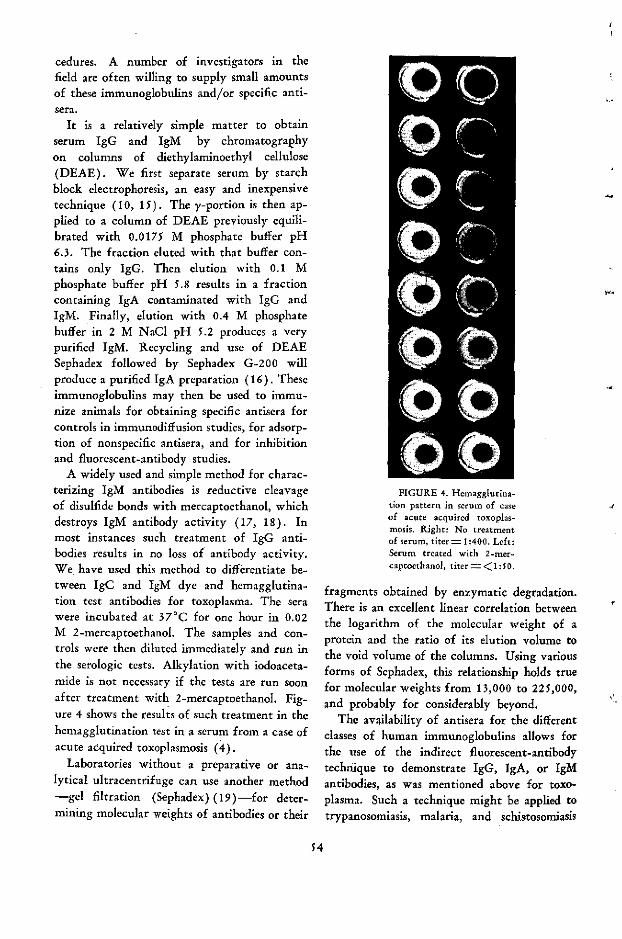
cedures. A number of investigators in thefield are often willing to supply small amountsof these immunoglobulins and/or specific anti-sera.
It is a relatively simple matter to obtainserum IgG and IgM by chromatographyon columns of diethylaminoethyl cellulose(DEAE). We first separate serum by starchblock electrophoresis, an easy and inexpensivetechnique (10, 15). The y-portion is then ap-plied to a column of DEAE previously equili-brated with 0.0175 M phosphate buffer pH6.3. The fraction eluted with that buffer con-tains only IgG. Then elution with 0.1 Mphosphate buffer pH 5.8 results in a fractioncontaining IgA contaminated with IgG andIgM. Finally, elution with 0.4 M phosphatebuffer in 2 M NaCI pH 5.2 produces a verypurified IgM. Recycling and use of DEAESephadex followed by Sephadex G-200 willproduce a purified IgA preparation (16). Theseimmunoglobulins may then be used to immu-nize animals for obtaining specific antisera forcontrols in immunodiffusion studies, for adsorp-tion of nonspecific antisera, and for inhibitionand fluorescent-antibody studies.
A widely used and simple method for charac-terizing IgM antibodies is reductive cleavageof disulfide bonds with mercaptoethanol, whichdestroys IgM antibody activity (17, 18). Inmost instances such treatment of IgG anti-bodies results in no loss of antibody activity.We have used this method to differentiate be-tween IgC and IgM dye and hemagglutina-tion test antibodies for toxoplasma. The serawere incubated at 370C for one hour in 0.02M 2-mercaptoethanol. The samples and con-trols were then diluted immediately and run inthe serologic tests. Alkylation with iodoaceta-mide is not necessary if the tests are run soonafter treatment with 2-mercaptoethanol. Fig-ure 4 shows the results of such treatment in thehemagglutination test in a serum from a case ofacute acquired toxoplasmosis (4).
Laboratories without a preparative or ana-lytical ultracentrifuge can use another method-gel filtration (Sephadex) (19)-for deter-mining molecular weights of antibodies or their
FIGURE 4. Hemagglutina-tion pattern in serum of caseof acute acquired toxoplas-mosis. Right: No treatmentof serum, titer= 1:400. Left:Serum treated with 2-mer-captoethanol, titer = <1:50.
fragments obtained by enzymatic degradation.There is an excellent linear correlation betweenthe logarithm of the molecular weight of aprotein and the ratio of its elution volume tothe void volume of the columns. Using variousforms of Sephadex, this relationship holds truefor molecular weights from 13,000 to 225,000,and probably for considerably beyond.
The availability of antisera for the differentclasses of human immunoglobulins allows forthe use of the indirect fluorescent-antibodytechnique to demonstrate IgG, IgA, or IgMantibodies, as was mentioned above for toxo-plasma. Such a technique might be applied totrypanosomiasis, malaria, and schistosomiasis
54
1

and to many other parasitic diseases of man aswell. In animal studies the problem is moreinvolved, since specific antisera for the variousclasses of animal immunoglobulins are notreadily available. Here it would be necessaryto resort to preparing the purified immuno-globulins and their respective antisera.
These techniques lend themselves to thestudy not only of serum antibody and cellularproduction of immunoglobulins but also ofantibodies of body secretions and excretions(15, 20, 21, 22). Relatively little is known ofthe characteristics of coproantibody response tointestinal parasites. About 90 per cent of theimmunoglobulin-producing cells of the laminapropria of the duodenum and jejunum produceIgA (23). What'is the role of this immunoglob-ulin in parasitic infections of the intestine?What is the sequence of the formation of anti-body in the intestine to various parasites? Sincelittle is known of the importance and functionof IgA as exocrine antibody, studies employingamoebae and certain helminths may help clarifythis subject.
Although it is not within the scope of thismanuscript to cover extensively the techniquesthat might be used in such studies, sufficientreferences are given to allow one to becomeacquainted with them. It is unfortunate thatthese newer techniques, many of them rela-tively simple to perform, have not been usedby more laboratories in the study of parasitesand of parasitism. For years the immunologyof parasitic diseases has been associated withhistopathology and the study of antigens fordiagnosis and immunization. In recent yearstremendous advances have been made in thetechnology of imunochemistry-and thereby inour knowledge of immunity. In this countrylaboratories such as those of Drs. Elvio Sadunand Irving Kagan have served as an impetusto other workers in parasitology not previouslyoriented to immunology. With laboratorymethods now at hand for the characterizationof antibodies to parasites, it is hoped that suchstudies will be performed at least in part by,or in laboratories of, those with the greatestknowledge of the field of parasitology and
appreciation of its problems-parasitologists.
Description of indirect fluorescent-anti-body test used for rapid demonstration ofIgM-toxoplasma antibodies
The technique employed is essentially thatdescribed by Walton, Benchoff, and Brooks(24). The RH strain of toxoplasma is ob-tained from the peritoneal fluids of three-dayinfected mice. The fluid is added to a smallamount of phosphate-buffered NaCI pH 7.2(PBS) containing 1 per cent formaldehydeand heparin. The suspensión is mixed thor-oughly with a 10-ml syringe and a No. 22needle to disrupt cells containing organisms.The volume is then brought to 40 ml by addingadditional PBS containing the formaldehydeand is centrifuged at 1,500 rpm for 10 minutes.The supernatant is discarded, and the sedimentis resuspended and washed twice in the PBS-formaldehyde solution. After the second washthe sediment is resuspended in an amount ofPBS-formaldehyde solution sufficient to give100-200 organisms per high-power field whendelivered to the 1 cm circled areas on the slide(24). If many leukocytes are present in theperitoneal fluid, the organisms may be separatedby filtration through a Baird-Tatlock filter asdescribed by Fulton and Spooner (25). How-ever, by careful timing of the period betweenthe inoculation of animals and the obtainingof peritoneal fluid, exudate may be obtainedthat contains relatively few leukocytes. Slidesfor future use may be frozen at -200 C forat least four months (the longest periodtested).
PBS is used for diluting samples of serumand as a wash solution for the slides after eachstep in the procedure. The fluorescein-con-jugated goat anti-human IgM antiserum waspurchased from Hoechst Pharmaceuticals, Inc.,Cincinnati, Ohio. Through the excellent co-operation of Mr. H. Jelinek of the DiagnosticReagents Division, a number of such sera weretested for the presence of toxoplasma anti-bodies prior to conjugation with the fluoro-chrome. Lot F132 was found to be negative

(<1:2) in the dye test and was employed inall the studies we have performed to date.No precipitin arc resulted when this antiserumwas reacted by double diffusion in agar gelagainst IgG. Reactions of this same antiserumwith normal human serum resulted in a singleprecipitin arc that gave a reaction of identitywith that formed by the reaction of the anti-serum with IgM. Dilutions of the conjugatedantiserum were made with a variation of thediluent recommended by Goldman (26), usingPBS containing 1.2 per cent bovine serumalbumin. The appropriate dilution of the anti-serum was 1:100 to 1:150. The slides wereexamined with a Zeiss Fluorescence Photomicro-scope using an Achromatic-aplanatic condenserwith a BG 12 exciter filter and barrier filters53 and 44. 12.5X oculars were used with the40X objective and 1.25 optivar giving a mag-nification of 625X. The photographs weretaken with high-speed Ektachrome daylightcolor film using the dark field condenser andBG 12 exciter filter and 65/50/41 barrierfilters. The exposures were for 30 minutes withthe 40X objective and 1.6 optivar giving amagnification of 800X.
The initial dilution of the serum sampleswas 1:8 or 1:10, and thereafter serial twofolddilutions were made. The titer of a given serumis reported as that dilution in which at least 50per cent of the organisms in multiple fieldsappeared to have completely outlined cell walls.Two other types of fluorescence that were notedwere the staining of one tip of the organism orfragmentary staining of the cell wall. Such re-actions were not considered positive. Threecontrols were run each time the test was per-formed: (a) a serum negative (<1:8) in thedye test, (b) serial dilutions of a serum knownto contain IgM dye test antibodies, and (c) a1:8 dilution of a serum known to contain onlyIgC dye test antibodies (titer=1:256). Inthe IgM-fluorescent-antibody test controls (a)and (c) there was either no fluorescence of theorganisms or a staining of some tips. Control(b) resulted in brightly fluorescent rims at atiter of 1:8 through 1:128, and at a titer of
1:256 there were rare faint rims or no fluores-cence. As a control in some experiments afluorescein-conjugated goat anti-human IgGantiserum (Lot 7V-818) obtainéd from HylandLaboratories, Los Angeles, California, was usedin a dilution of 1:100.
To test the specificity of the fluorescein-tagged anti-IgM antiserum, 34 sera with IgM-toxoplasma antibodies were treated with 2-mercaptoethanol (2-ME) and therea'fter testedin the IgM-fluorescent-antibody test. The dyetest titers in these sera ranged from 1:1024 to1:65000. Treatment with 2-ME completelydestroyed the ability of sera with IgM-fluores-cent-antibody test titers to cause fluorescence.In contrast, this treatment did not affect theirability to cause brilliant fluorescence when afluorescein-tagged anti-IgG antiserum was em-ployed. Pooled IgG isolated from sera of humancases containing both IgG and IgM-toxoplasmaantibodies was tested in the IgM-fluorescent-antibody test. The titer of the pooled IgG in thedye test was 1:32000. This IgG gave no fluores-cence in the IgM-fluorescent-antibody test.
Moderator: Before. introducing the dis-cussant, 1 should like to call attention to onepoint that has not been mentioned by thespeakers: the possible role of antienzymes inparasitic immunity. I think this possibility wasraised fifteen years ago by Chandler when hesuggested that the stunting of worms in theimmune host was the effect of the antibody onthe metabolism of the parasite. The idea wasfurther developed by a group of workers at theUniversity of Chicago, who showed that whilemany serological reactions observed withSchistosoma mansoni-such as complementfixation, circumoval precipitation, and mira-cidial and cercarial agglutination-are defi-nitely associated with IgG antibodies, the in-hibition of collagenase activity in cercariaedepends on a heat-stable factor migrating asan alpha globulin. The comparative suscepti-bility of different species ran parallel to thelevel of the inhibitor, which would affect thepenetration and development of cercariae.
It is quite possible, however, that antibodiesto enzymes play an important role in the
56
4
·;

penetration of larvae or affect their life cycleinside the immune host. It is known, for ex-ample, that the sera of infected rats canneutralize lipase of Nippostrongylus larvae andthat the sera of infected dogs neutralize theproteolytic activity of esophageal extracts ofadults and larvae of Ancylostoma caninum.
Leucin aminopepsidase is produced under themolting stimuli in both Haemonchus contortusand Trichostrongylus colubriformus and seemsto be responsible for the attack of the larvalsheath leading to its liberation.
There are people working on enzymes ofSchistosoma mansoni, and at least two groupsare working with the techniques developed byUriel in France using electrophoresis or im-munoelectrophoresis on agarose gel. I refer tothe work recently published in France by a
group of investigators in Lille, who report thepresence in the adult Schistosoma mansoni offour dehydrogenases (mallic, lactic, alanine, andglucose-6-phosphate dehydrogenases), alkalinephosphatase, chymotrypsin, and carbonic anhy-drase. These same enzymes were found inFasciola hepatica.
In our laboratory we have been interestedparticularly in cercariae, and our experimentsshow positive results for leucin aminopeptidase,carboxypeptidase A, chymotrypsin, and alka-line protease.
As a complement to what has been said bythe speakers on the antibodies to parasites, Ithought, something should be added about theantienzymes.
Now I call on Dr. Goodman to start the dis-cussion and Dr. Nussenzweig to follow.
S7
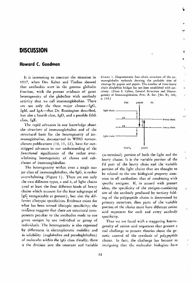
DISCUSSION
Howard C. Goodman
It is interesting to contrast the situation in1937, when Drs. Kabat and Tiselius showedthat antibodies were in the gamma globulinfraction, with the present evidence of greatheterogeneity of the globulins with antibodyactivity that we call immunoglobulins. Thereare not only the three major classes-IgG,IgM, and IgA-that Dr. Remington described,but also a fourth class, IgD, and a possible fifthclass, IgE.
The rapid advances in our knowledge aboutthe structure of immunoglobulins and of thestructural basis for the heterogeneity of im-munoglobulins, documented in WHO nomen-clature publications (10, 11, 12), have far out-
stripped advances in our understanding of thefulnctional significance of the rather over-whelming heterogeneity of classes and sub-classes of immunoglobulins.
The heterogeneity within even a single ma-jor class of immunoglobulins, the IgG, is ratheroverwhelming (Figure 1). There are not onlythe two different types, K and A, of light chains(and at least the four different kinds of heavychains which account for the four subgroups ofIgG recognizable at present), but also the dif-ferent allotypic specificities. Evidence exists forwhat has been termed idiotypic specificity; theevicence suggests that there are structural com-ponents peculiar to the antibodies made to onegiven antigen by one individual or group ofindividuals. The heterogeneity is also expressedby differences in electrophoretic mobility andin solubility (euglobulins and pseudoglobulins)of molecules within the IgG class. Finally, thereis the division into the constant and variable
FIGURE 1. Diagrammatic four-chain structure of the im-munoglobulin molecule showing the probable sites ofcleavage by papain and pepsin. The number of inter-heavychain disulphide bridges has not been established with cer-tainty. (From S. Cohen, General Structure and Hetero-geneity of Immunoglobulins, Proc. R. Soc. [Ser. B], 166;p. 114.)
Fab papain
light chnin .
Fd S
Fd S,
light chnin 5
Fo
-hory chain
F(Qb'), plgp}il
(n-terminal) portion of both the light and theheavy chains. It is the variable portion of theFd part of the heavy chain and the variableportion of the light chains that are thought tobe related to the one biological property com-mon to all antibodies: that of combining withspecific antigens. If, in accord with presentideas, the specificity of the antigen-combiningsite of the antibody produced by tertiary fold-ing of the polypeptide chains is determined byprimary structure, then parts of the variableportion of the chains must have different aminoacid sequences for each and every antibodyspecificity.
Thus we are faced with a staggering hetero-geneity of amino acid sequences that present areal challenge to present theories about the ge-netic control of the synthesis of polypeptidechains. In fact, the challenge has become sointriguing that the molecular biologists have
58
I { S~~nnnw nna n

decided to devote this year's Cold Spring Har-bor Symposium in June to the consideration ofthe genetic basis for the synthesis of immuno-globulin molecules.
Returning to the classes of immunoglobulinsin human sera, it seems established that the gen-eral basis for differences in the four majorclasses of immunoglobulins depends upon thefour different kinds of heavy chains character-istic for each class, the y, a, p and 8. There arestructural variations within each class of heavychains, accounting for the currently recognizedsubclasses of IgG, IgA, and IgM (which sub-classes obviously do not mark the end of ourability to subdivide major classes of immuno-globulins). The existence of such subclasses ofimmunoglobulins may provide an explanationfor some of the conflicting results on the bio-logical activities of antibodies that were gen-erally considered to be within one class, IgG,which can really be regarded as including awhole family of immunoglobulins.
One of the major challenges in immunologyis to correlate the new knowledge about differ-ences in amino acid sequence resulting in thedifferent polypeptide chains characteristic ofdifferent kinds of immunoglobulins with thedifferent biological functions that presumablyare also characteristic of the different kinds ofimmunoglobulins. We are at the beginning ofwhat might be termed "molecular immunol-ogy," of being able to understand the molecu-lar basis for what hitherto have necessarilyoften been descriptive phenomena of immunol-ogy, such as complement fixation, tissue sensiti-zation and anaphylaxis, cytophilic and opsonicproperties of immunoglobulins, and the way inwhich immunoglobulins cross the placenta orappear in external secretions.
My comments will be limited to focusingattention on Dr. Smithers' references toanaphylactic antibodies and Dr. Remington'sto antibodies in external secretions.
The presence of antibodies in stools (copro-antibodies) was first demonstrated in 1922, butthe renewal of interest in these antibodies and
their possible protective role is due to the re-
cent demonstration that IgA is the predominant
immunoglobulin in most external se*cretions, in-cluding the gastrointestinal secretions. It islikely that this IgA is a dimer of the serummonomer plus a protein "piece." The proteinpiece is found in epithelial cells and may be in-volved in transporting the IgA into the lumenof the gut. The probability exists that theseIgA molecules are produced locally as a resultof antigenic stimulation of the gut-associatedlymphoid tissue by ingested antigens. Diseasesin which various stages of protozoan and meta-zoan parasites reside in the intestinal tract pro-vide opportunities for study of the importanceof this system of local production of antibodiesto parasite antigens in immunity against theseinfections. These diseases also provide modelsfor fundamental studies of the immunologicalcapacities and reactivities of the gut-associatedlymphoid system.
Fixation of immunoglobulins to cells:Skin sensitization, anaphylaxis
We have only incomplete knowledge aboutwhich classes or subclasses of immunoglobulinsfix to different types of tissue cells. Ideally, weshould know the specific structural configura-tion of the heavy chain of each class of immu-noglobulins that is responsible for its sensitiza-tion of (presumably by fixation to) differenttissue cells.
The capacity to sensitize tissue cells is aproperty of antibodies essential for inductionof the anaphylactic reactions that follow com-bination with antigen.
1. Reaginic skin-sensitizing antibodies (ho-mologous anaphylactic antibody, homocyto-tropic antibody) are those antibodies that sen-sitize tissue cells (presumably mast cells) oftheir own or closely related species to producesystemic, local, and, under certain experimentalconditions, in vitro anaphylactic reactions. Inthe human, these antibodies are detected bypassively transferring serum into the skin ofother humans (Prausnitz-Kustner reaction) orof certain primates, and allowing 24 hours ormore to pass before injecting antigen to testfor increase in vascular permeability. It hasrecently been shown that these antibodies pro-
59
duce in vitro sensitization of monkey ileum ina modified Schultz-Dale technique (1). Thenecessity for employing Prausnitz-Kustner re-actions as an assay for this type of antibody hasheretofore been one of the main obstacles to itscharacterization and purification. It is knownto occur in the electrophoretically fast immuno-globulins (y-l mobility), t' be present invery small amounts and to sediment in the ul-tracentrifuge faster than the bulk of the IgGmolecules. Reaginic skin-sensitizing antibodysticks in the skin for weeks, although in vitroit is heat-labile and losses in activity occur withexposure to mercaptoethanol. Although thoughtfor some time to reside in the IgA globulins,skin-sensitizing activity has now been reportedin what may be a new class (IgE) of immuno-globulins (6).
Antibodies with similar reaginic skin sensitiz-ing activity have been reported in the rat (4,7), dog (14), and rabbit (13, 16). These anti-bodies are also found in trace amounts in theserum, persist in the skin for weeks after pas-sive transfer within the species, are heat-labile,and are destroyed by mercaptoethanol treat-ment. The rat antibodies are reported to sedi-ment faster than the bulk of the 7S antibodies.In rabbit they are reported to be 7S (13) orperhaps sormewhat larger than 7S (16).
2. A second group of antibodies, also termedanaphylactic antibodies, with similar, but notidentical; biological properties, and with differ-ent physicochemical properties, has been de-scribed in the guinea pig (3, 15) and in themouse (2, 12). These antibodies also reside inan electrophoretically fast class of immuno-globulins, but this class, provisionally termedy-l, is present in large amounts in the serumand has a sedimentation constant of 7S. Inthese two species, these y-l anaphylactic anti-bodies also mediate and transfer anaphylacticreactions within the species or in closely relatedspecies, but the skin-sensitizing activity is notdestroyed as readily by heat or mercaptoethanoltreatment and passively transferred antibodiespersist in the skin for days rather than weeks(5, for review).
While it may be dangerous to make close
comparisons between different animal species inwhich different immunoglobulin classes couldwell serve similar functions, it does seem tomake sense from a plhylogenetic point of viewto look for both types of reaginic antibodies inboth groups of species. It is interesting thatMota (8) has reported evidence of the exist-ence of both types of anaphylactic (homocyto-tropic) antibody in the mouse, and I suspectthat this will also be found to hold for theguinea pig. In the human, the rabbit, and thedog, on the other hand, where no discrete y-1has been reported, it is possible that the analo-gous immunoglobulih may turn out to be oneof the four subclasses of IgG (12).
In any case, Johannson and Bennich (per-sonal commiunication) have recently discovereda myeloma protein that does not react withmonospecific antisera against IgG, IgM, IgA, orIgD and that has been found to have antigenicdeterminants in common with what has beentermed IgE by K. Ishizaka (personal communi-cation). If this inyeloma protein has homo-cytotropic properties, the way may be open forchemical studies to relate a structural compo-nent of the heavy chain of the new immuno-globulini to the property of tissue (mast-cell?)sensitization. In addition, we need more stud-ies of the kind Dr. Smithers described to learnwhether the antibodies that produce anaphy-laxis by virtue of their tissue-sensitizing prop-erties may also play a role in immunity in theparasitic diseases. Certainly experimental infec-tions with parasites appear to be an excellentmeasure for eliciting the production of theseanaphylactic antibodies.
There is need for a better understanding ofthe functional significance of the heterogeneity
of immunoglobulins in terms meaningful forbetter control of public health problems. This
need and the potentially short step betweenadvances in basic knowledge about the struc-ture and function of immunoglobulins and the
application of this knowledge to problems ofpublic health are reflected in the development
of the WHO immunology research program
and the WHO Reference Laboratory for Im-
60
1
1

munoglobulins at the Institute of Biochemistryin Lausanne.
Victor Nussenzweig
In the ten minutes allowed me, I should like
to discuss only two aspects of the precedingpapers which are perhaps more controversialand might have important theoretical implica-tions.
The first is the possibility that in many para-
sitic diseases in which there are high levels ofimmunoglobulins in the serum, these are non-specific. The immunoglobulins would not beantibodies against the parasite or against itsmetabolic products or antibodies against cross-reacting antigens elicited through an anamnes-tic response. It is very difficult to understandthe mechanism of such a nonspecific stimula-tion of y-globulin production. Two possibili-ties come immediately to iny mind. First, thatthe antigen specifically stimulates the immuno-competent cells. The plasma cells synthesizeantibodies but simultaneously produce somenonspecific immunoglobulins. According to
this hypothesis, one plasma cell would have toproduce more than one kind of immunoglobu-lin, and this is not supported by most of the
experimental evidence. Second, that duringthese infections there is a nonspecific stimula-tion of immunocompetent cells, and they pro-liferate and synthetize antibodies directed
against products that have nothing in commonwith the antigens of the parasite.
Although I think these are interesting possi-
bilities, there are no firm experimental data tosupport them, and at least in some cases otherexplanations can be found for this increasedproduction of immunoglobulins. The situations
in which very high "nonspecific" levels of se-rum immunoglobulin are found result, in gen-eral, from immunization with complex anti-gens. When well-defined substances are used
as antigens, a substantial proportion of the se-rum immunoglobulins is very frequently found
to be antibodies against the immunizing anti-gens. When some haptenic systems are usedand the antibody production is studied at thecellular level, it can be demonstrated that dur-ing the first weeks after immunization as much
as 50 per cent or more of the cells engaged inimmunoglobulin synthesis are actually produc-ing antihapten antibody (3).
It is also well known that many substancesand bacterial products are potent immunogensin extremely small doses. If a minor parasitecomponent or metabolic product can induce theformation of high levels of antibodies, the ab-sorption of these antibodies with a mixture ofdifferent antigens would be very difficult if theimportant one constituted a minute fraction.
I should also like to bring to your attentionsome known facts on serological reactions invisceral leishmaniasis. This is one example of aparasitic disease in which extremely high levelsof immunoglobulins are found in the serum.Many years ago I tried to obtain from culturedLeishmania donovani antigenic fractions thatwould react strongly and specifically with theimmunoglobulins present in the patient's serum,but without success; this has been, I think, theexperience of many investigators. However,when I used extracts of tubercle bacillus as
antigen (as others had done before) I obtainedvery high complement-fixation titers with thepatients' sera. Just to give you an idea of theresults, in the complement fixation test I used.05 ml of the patients' serum and 6 C'H 50; inmany cases the serum could be diluted 3,000times and the results would still be positive (1).If this test is capable of detecting 1 ~tg/ml ofantibody protein, some sera would contain about3 mg/ml of antibodies against some componentsof the tubercle bacillus. The conclusion wouldbe that a sizable portion of the immunoglobu-lins in the patients' sera have a well-definedspecificity. It is fair, I think, to postulate the
presence of cross-reacting antigens in L. dono-vani and tubercle bacillus. These antigens would
perhaps be difficult to extract from the flagel-
lates or be in some way altered during the ex-traction, while they could be easily obtained
from tubercle bacillus.
61

The second general point I want to discussis related to the problem of selection of im-munoglobulin chains during the immune re-sponse. This is indeed an intriguing problem.A great number of different polypeptide chainsare found among immunoglobulins, but a cer-tain selection seems to take place during theimmune response, and, as a consequence, anti-bodies may contain a restricted number oftypes of polypeptide chains. Why, for instance,are reagins produced in larger amounts in hel-minth infestations than in other infections?Why are certain antibodies preferentially lo-cated in one or other of the immunoglobulinclasses? It is known, for example, that anti-penicillin hemagglutinins or cold agglutininsare mostly IgM, while antidextran antibodiesare IgG. In collaboration with Dr. Benacerraf,we have recently done some experiments thatshow that antihapten antibodies, containingcertain classes of chains in preference to others,are synthesized by immunized animals becausethese chains are better fitted for the formationof the antibody-combining site (2). We havefound that during the immune response ofguinea pig to dinitrophenol (DNP) conjugates,a selection of types of L chains in the anti-DNP antibodies can be demonstrated. At thebeginning of the immunization, when the bind-ing affinities of the anti-DNP antibodies fore-DNP-L-lysine are low, they contain both xand X types of L chain. However, when thebinding affinities of the anti-DNP antibodiesincrease, only the x type of L chain can befound. We have also found an increasingamount of L molecules (containing A chains)in fractions of anti-DNP antibodies with de-creasing affinities for the hapten. The implica-tion would be, then, that x chains are betterfitted to produce anti-DNP antibodies of highbinding affinity. This may be a general mecha-nism of selection of immunoglobulin chainsduring the immune response, and it would im-ply, for example, that the increased productionof reaginic antibodies in some parasitic diseases isdue simply to the fact that the H chain presentin this kind of immunoglobulin is more ade-
quate than other kinds of H chains to form
"good" antibodies against some important para-sitic antigen. It is possible, as was suggested byDr. Bier, that some antienzyme antibodies playan important role in the mechanism of resist-ance in parasitic diseases and these antibodiesmight contain restricted types of H and Lchains. Of course, a consequence of this rea-soning might be that the reaginic antibodiesthat participate in the immune defenses mech-anisms in some helminth infections are effectivenot because of their biological properties butbecause they specifically combine with someimportant antigen. This hypothesis can ofcourse be tested experimentally, in view of thepossibility of separating, by appropriate enzy-matic treatment, the Fc portion of the anti-body, which mediates its biological property,from the portion containing the combiningsites.
I should like to finish with a word of cau-tion about the assumption that IgM antibodiesalways precede the formation of IgG anti-bodies. I think that in many cases this issuehas been confused by the differences in sensi-tivity of assays for those immunoglobulins. Forexample, Robbins, Kenney, and Sutter (4) haveshown that rabbit IgM antibody to Salmonellatyphimurium is 22 times more active than IgGin inducing agglutination, 120 times more po-tent in sensitizing bacteria for complement fix-ation, and 500 to 1000 times more efficient asan opsonin. Recent investigations using moreadequate methods to detect antigen-antibodyreactions show that in many cases IgM andIgG appear simultaneously after immunization.
Moderator: I should like to have the com-ments of Dr. Smithers and Dr. Remington onwhat has been said by the two discussants.
Smithers: This question of reaginic forma-tion by helminth infection has interested usfor a long time, and we have been very muchpuzzled as to why reagins are induced by'hel-minth infections and only living helminth in-fections. The fact that they are not induced bydead worm homogenates but only by the livinginfection does suggest in fact that it is an anti-
gen produced by the living worm-an excre-
tion or secretion from the worm. I was greatly
62
-
4

interested in the suggestion of the last speakerthat reagins may be induced by the stimulation
T from a particular type of antigen. In fact, wehave found that allergens-that is, the anti-gens responsible for inducing reagins-can beproduced best from a helminth by in vitro cul-ture. We can produce a high level of allergenfree in the culture fluid by maintaining schisto-somes for four hours in Hank's solution, andthe actual schistosome worms themselves showno depreciation in their allergen content, whichsuggests that they are actively producing aller-gen. The same applies even more to Ni po-strongylus. If Nippostrongylus is incubated inbuffered saline for three hours, two to threetimes as much allergen will be produced as ifthe adult worms were crushed.
And so it certainly looks as if the allergenwere some sort of metabolic product. Indeed,some investigations have shown that these al-lergens are fairly small proteins, with a molec-ular weight of about 10 to 15 thousand, andthey could well be, as the Moderator has sug-gested, an enzyme produced by the parasite.
The other small comment I should like tomake is on Dr. Goodman's chart. The rhesusmonkey could now be added to the human,dog, rat, and rabbit group, because it has beenshown that rhesus monkey reagin is similar tothose others. Dr. Sadun, 1 know, has foundreagins in a chimpanzee infected with S. man-soni, and I am sure those would also be foundto be the same.
Remington: I was very happy to hear Dr.Goodman's remarks about external body secre-tions. I think it is important to remember thatthe gastrointestinal tract is in the externalworld, not really inside our bodies, and thateven in lower forms of animals, invertebrates,immunoglobulin similar to those of IgA arefound; perhaps in prehistoric days this was oneof the main modes of defense of such animals.We know very little about it in the human, andI think the parasitologists can help us learnmore. I hope that some of you here, and yourcolleagues, will be interested in working in thisarea.
One comment about Dr. Nussenzweig's
work: I was very much interested to hear aboutthe antibody to the tubercle bacillus. As I amsure he is aware, if one wants to get a good dealof antibody against many antigens one finds apatient with cirrhosis of the liver who hashypergammaglobulinemia; his serum will con-tain large amounts of antibody. I wonderwhether the mere finding of high titers totubercle bacillus without adsorption of theimmunoglobulins really shows that any of thishyperglobulinemia had anything to do withthe tubercle bacillus, or whether this is just asimple manifestation of extra antibody beingmade in a hyperglobulinemic animal. Did youadsorb the gamma globulin portion with thetubercle bacillus to show a reduction in thetotal gamma globulin in those animals, Dr.Nussenzweig, and was this antibody present inthe controls?
Nussenzweig: The reaction of kala-azarsera with tubercle bacillus has been krown fora long time, and its sensitivity and specificityhave been well studied. It has all the character-istics of a true antibody-antigen reaction. It isquite specific in the sense that these antigensdo not react with sera from other types of leish-maniasis or with sera from patients with otherdiseases (except, of course, leprosy or tubercu-losis) in which high levels of gamma globulinare found.
Moderator: I should like to comment onwhat Dr. Nussenzweig has just said. I shouldsay that what you are really detecting in kala-azar is one of the antibodies whose productioncan actually be demonstrated, but there maybe other antibodies produced at the same timethat we haven't the right reagent to demon-strate.
On the other hand, the parallelism betweenthe total amount of antibody and the high titerof complement fixation does not mean verymuch, because the amount of antibody neces-sary to give such high titers in the comple-ment-fixation tests may not represent a con-siderable fraction of the total amount of gammaglobulin. What I would like to know is the
homogeneity or heterogeneity of the globulin
produced in kala-azar.
63

Nussenzweig: I think I was misunder-stood. I was not implying that all the gammaglobulins found in the serum of patients withkala-azar is anti-tubercle-bacillus antibody.What is clear, 1 think, is that part of what iscalled "nonspecific" gamma globulin has awell-defined specificity.
Moderator: I understood you quite well,but I raised the point because you talked aboutthe parallelism between the increase in gammaglobulin and increase in complement fixation.
Remington: I do not want to press thepoint too hard, Dr. Nussenzweig, but I do notknow what the data are to suggest that even ahigh proportion of the gamma globulins areformed against this tubercle bacillus extract.
Have you adsorbed the gamma globulins toshow that a significant percentage are directedagainst that antigenic component?
Nussenzweig: No, I have not.Moderator: My remark about the activity
was just to answer your question, because evenif the serum is adsorbed the fact that therewould not be a great reduction in gamma glob-ulin would not be very enlightening; you mayjust adsorb a very small amount of gammaglobulin that is responsible for the activity.The only assumption we can make is that prob-ably many other antibodies are being formed,but it is just a hypothesis.
Borsos: In this connection I should like tocite some experiments. Dr. Richard Asofskyat NIH has used purified endotoxin as the im-munizing antigen, and he measured the produc-tion of antibody in germ-free mice by bacterio-cidal reactions that are exceedingly sensitive, asDr. Bier knows so well from his complement-fixation experiments. He is capable of detecting102 antibody molecules per ml, which makesthis reaction a very sensitive test for antibodyproduction. On the injection of the endotoxin,antibody is formed; there is an accompanyingincrease in gamma globulin content. However,as much as 80 per cent of the immunoglobulinproduced contains no antibody activity againstthe endoroxin. These experiments demonstratethat there is indeed production of immuno-
globulin that has no antibody activity to the
particular antigen used. It might be arguedthat the principle of the original antigenic sinoperates here, and that to some related antigensan anamnestic response was produced. This ishypothetical; there is no evidence for it. So Ithink we have to accept the fact that non-specific gamma globulin can be produced in re-sponse to antigenic stimulation.
Kagan: I should like to interject a note ofcaution about some of these immunoglobulinexperiments. It is very important to evaluatethe assay methods for measuring immunoglob-ulins. Hemagglutination tests, for example,will detect IgM antibody much more readilythan IgG, and if a hemagglutinating system isused to evaluate antibody levels, the result willbe a biased evaluation of the amounts of IgMversus IgG antibody. In fact, Stavitsky hasrecently suggested that the hypothesis thatIgM is produced first, followed by IgG, is nottrue; he believes they are produced at the sametime, but detecting these antibodies dependsupon the assay used for evaluation.
My other comment concerns Dr. Reming-ton's work. Using specific immunoglobulinantisera in the fluorescent-antibody tests, hecan detect acquired congenital toxoplasmosisby measuring IgM antibody production in thefetus. He can also detect IgG antibody in pas-sive transfer of globulin from the mother tothe fetus. This has a very practical use in de-termining whether what the infant has is anacquired infection or antibody obtained bypassive transfer. We need studies like this forother parasitic infections. In our laboratory weare busy characterizng the various classes ofimmunoglobulins. They should be studied asthey evolve during infection and their levelsshould be detected quantitatively. Recentstudies of this nature have been published byTobey and his group for malaria, and we needother such studies for other parasitic infections.
The third area that we are particularly inter-ested in is the role of the various classes ofimmunoglobulin in our diagnostic tests. Wehave to evaluate the efficiency of various diag-nostic serologic procedures in detecting thesevarious types of antibodies. It may turn out
64
J
Jl

that one test has a higher sensitivity than an- aspects of immunoparasitology connected witht other for detecting a certain class of immuno- antigens and antibodies. Our next period will, globulin, and therefore serves as a better param- be devoted to cellular reactions and the effects
eter of infection. of the immune response on the parasites. First,Ir Moderator: So far we have discussed a few Dr. Soulsby.
65

Lymphocyte, Macrophage, and Other Cell Reactions to Parasites °
E. J. L. Soulsby
Introduction
Almost without exception, a marked cellu-
lar response is a characteristic feature of para-
sitic infections. Even a cursory examination
of a supposedly simple reaction will reveal its
complexity, and when a more major reaction is
examined an evaluation of it would seem to
be an extremely hazardous enterprise. For-
tunately, the newer knowledge of the various
cell types will now permit a closer study of the
functional aspects of the cell response, and it
is this aspect which will be emphasized. A
familiarity with the morphology and origin ofthe cells will be assumed.
The chief concern of this review will be the
cell types that are closely associated with the
immune response of the host, those that re-
spond as a result of it and other forms that
make up the acute inflammatory response. No
attention will be paid to degenerative changes
that occur in parenchymatous cells of organs
or proliferative changes in, for example, fibro-
blasts, but the newer knowledge about the
change or loss of function of epithelial cells in
parasitic infection warrants brief considera-
tion.
Epithelial cell reactions to parasitism
Hyperplasia of epithelial cells is a common
response to parasitism. It is seen in the bile duct
or pancreatic duct when these are parasitized
by helminths such as Fasciola, Clonorchis,
Ascaris, Hymenolepis, and Stilesia or by pro-
: The experimental work reported in this review was
carried out with the support of USPHS Grant Al 06262.
tozoa (e.g., Eimeria stiedae). Similar responses
occur in the urinary bladder (polyp formation)
in schistosomiasis, in the bronchioles and
bronchi in lungworm infection, and in the
gastrointestinal mucosae in nematode, trema-
tode, and sporozoan infections. Such changes
are frequently accompanied by a loss of 'func-
tion of various specialized cells or their ac-
quisition of new functions. For example, in
Ostertagia infection in the abomasum of sheep
and cattle, the specialized parietal cells that
produce hydrochloric acid and the peptic cells
that produce pepsinogen may lose their func-
tion and be replaced by hyperplastic undifferen-
tiated cuboidal cells ( 51). In the same
infection, mucoid metaplasia may occur, and
in fact this reaction-seen in the bile duct, in
the stomach mucosa, in the bronchi, and else-
where-is a not uncommon response to para-
sitic helminths.
Recent investigations have correlated such
changes with the molecular biology of the para-
sitism. 'In the Ostertagia situation, the loss of
cells with specialized secretory function leads
to a rise in the pH of the abomasum and a
failure to activate pepsinogen, a leakage of pep-
sinogen into the blood, and an increased leakage
of plasma macromolecules into the lumen. The
last two of these effects may be due to imper-
fectly formed cell junctions as a result of the
hyperplasia (51).
The functional changes in the rat intestinal
epithelium, which becomes hyperplastic in
Nippostrongylns braziliensis infection, have
been studied by Symons and Fairbairn (101).
Under normal conditions, it is said, there is a
66
~4
r

progressive differentiation of the function ofepithelial cells as they migrate distally to beshed at the tips of the villi (83). N. braziliensisinfection appears to accelerate this migration,leading to an immaturity of the cells with aconcomitant loss, or reduction, of the levels ofmaltase, alkaline phosphatase, and leucine aminopeptidase on the cell surface and in the mi-crovilli of the epithelial cells. Comparablehyperplastic changes have been reported inNecator americanus infection (94), and aresaid to contribute to impaired absorption ofvitamin A, xylose, and fat.
Though there is much to indicate that suchchanges are essentially nonspecific, being seenin nontropical sprue and niacin deficiency, they
h. are associated with cell hyperplasia in the laminapropria, and this in parasitic infections includesinfiltration of polymorphonuclear leukocytes,eosinophils, macrophages, plasma cells, and lym-phocytes, the latter in various stages of trans-formation.
Local accumulation of lymphoreticularcells
Lymphocytes, plasma cells, and macrophagesare traditionally associated with immunologicalfunctions, and in many instances the local ac-cumulation of them may progress to a definite,and at times macroscopic, focus of lymphoreti-cular elements that has the general appearanceof a lymph node. Such structures frequentlydevelop around a parasite or its larval stage thathas been trapped in the tissues. They are seen
t later in the course of an infection and fre-quently at a time when immunity (sensustricto) develops. It is difficult to avoid theconclusion that these lesions are related to themechanism of immunity, and in fact there isoften much to indicate that immunity againsta parasite is mediated at a local level. Thisstatement would not, of course, imply that themajor antibody-producing organs such as thespleen and lymph nodes do not contribute to thepicture.
The local nature of the immune response atan organ level is well illustrated by bovinetrichomoniasis, in which protective immunity
appears to be mediated solely in the uterus andvagina. A similar situation possibly occurs alsoin Trichomonas vaginalis in man. This impliesthat antibody-producing cells are located in theuterine and vaginal walls, and in fact accumu-lations of plasma cells have been found there(88). Furthermore, it seems that the vaginacan produce antibody independently of theuterus and of the general antibody-producingorgans (87).
Another example of a locally mediated im-mune response, with local cell accumulations,occurs with Eimeria tenella of the cecum of thechicken. In this case, however, immunity cre-ated in one cecum is somehow transferred to asurgically isolated collateral cecum (45). Themechanism is not yet fully understood, thoughthe transfer does not appear to be mediated byserum antibodies (46). Pierce and Long (88)suggest that the immunity that develops at asecond, previously unstimulated site (the sec-ond cecum) may be analogous to the second-set homograft rejection reaction (65).
Accumulations of lymphoreticular cells inhelminth infections are seen perhaps to theirbest advantage in such entities as the Impho-reticular broncho-occlusive lesions in lungwormdisease of cattle that occur around larvaetrapped in the tissues or eggs and larvae thathave been aspirated into bronchioles and alveoli(53) and also in schistosomiasis with the for-mation of pseudotubercles around immatureschistosomes and eggs in the liver.
Lymphoid hyperplasia in Leishmaniainfections
The immunological role of lymphoid cells inOld World cutaneous leishmaniasis seems fairlyclear and has been documented by Adler (1, 2).After infection with Leishmania tropica, thereis a local proliferation of macrophages in whichthe leishmaniae multiply. This continues untilthe area is infiltrated with lymphocytes andplasma cells; when this happens the macro-phages cease to proliferate, the population de-clines, and the number of parasites also de-creases until they can be demonstrated byculture techniques only. Eventually the orga-
67

nisms disappear completely and the cutaneouslesion resolves. The sequence of events varieswith the individual and in the absence of spe-cific therapy may take three to eighteen months.Spontaneous cure is followed by lasting immu-nity to the causative strain, which may persistfor as long as 20 years. Immunity is establishedonly after the lesion has progressed through theseries of cellular reactions that result in spon-taneous cure (1); if the cutaneous sore is re-moved surgically before spontaneous cure, theindividual remains susceptible to reinfection.
A similar situation occurs with certain formsof New World cutaneous leishmaniasis. TheUta of Peru is usually followed by immunity;however, with mucocutaneous leishmaniasis,though the initial cutaneous lesion usually healsspontaneously, metastatic lesions occur in theskin, the mucosa of the mouth, and the carti-lages of the nose, mouth, and nasopharynx. Asubsequent lymphocytic and plasma cell infil-tration may reduce or eliminate the parasites,but in the mucocutaneous form of the diseasechronic metastatic lesions continue to occur. Acommon feature of these forms of cutaneousleishmaniasis is the lymphoid infiltration, ac-companied by a positive Montenegro skin re-action of the delayed type . The reaction devel-ops early in the course of infection and persistslong after spontaneous cure. It can be inducedby leptomonads of L. tropica or other species ofLeishmania, including those of cold-bloodedanimals, and also antigens from TrypanosomacruLzi and antigens of T. equiperdum (32).
A type of leishmaniasis in which there is littleor no lymphoid cell invasion and no positiveMontenegro reaction is Leishmaniasis tegumen-taria diffusa (19). In this, extensive areas ofskin are involved and masses of infected macro-phages are found in the dermis, with no second-ary invasion of lymphocytes or plasma cells. Ithas been suggested that the condition, whichhas been recorded in a small number of personsin Bolivia, Venezuela, and Brazil, may be oneof immunological unresponsiveness on the partof the infected person (1). However, no workhas been done to determine whether such per-sons are genetically deficient or whether the
unresponsiveness is one induced by the parasite.The immunological response to cutaneous
leishmaniasis.would suggest that it is mediatedby delayed-hypersensitivity mechanisms. Cir-culating antibodies are not readily demonstratedin the infection, and there is no evidence thatthe immunity has an antibody basis.
The situation with visceral leishmaniasis isquite different from that of the cutaneous form.The cellular reactions are similar, character-ized by a massive proliferation of histiocytesand secondary infiltration of lymphoid elementsgeneralized throughout the spleen, liver, bonemarrow, and lymphatic glands. However, inmost untreated human infections the disease isfatal, though spontaneous cure may occur-upto 25 per cent of such cases have been reportedin India (71) and in Portugal (32). Thera-peutic cure of kala-azar is followed by resist-ance to reinfection, and in about 10 per centof these a local skin lesion (post-kala-azardermal leishmanioid) may develop. Though thislesion contains numerous parasites, they do notbecome generalized but rather appear to repre-sent a residuum of infection for the mainte-nance of immunity or premunition.
Whereas delayed skin reactions are commonin dermal leishmaniasis, active cases of Indiankala-azar do not show a Montenegro reaction(93) and an absence of the reaction has beennoted in Brazilian cases of kala-azar (3). Asimilar situation appears to obtain in the Medi-terranean form of the disease. However, somecases with post-kala-azar dermal leishmanioiddo give a positive Montenegro reaction (2).The situation appears different in the East Afri-can form of kala-azar: positive Montenegroreactions have been observed in treated infec-tions and in 95 of 119 normal volunteers inoc-ulated with a gerbil strain of Leishmania thatlocalizes in the dermis (2).
The functional value of the lymphoid hyper-plasia in untreated visceral leishmaniasis is diffi-cult to evaluate. Basically, it fails to containthe infection, but there are indications that thevigor of the lymphoid response may have somevalue in prolonging life in man and dog. Onthe other hand, spermophils are very susceptible
68

to infection and show excessive macrophage
4 proliferation with less lymphoid cell infiltrationthan man or dog. In the spermophil the spleenmay be converted to "a nearly solid histiocy-toma" (2).
Specific antibodies for Leishmania have beendetected in visceral leishmaniasis by comple-ment-fixation techniques, either by the use ofleishmania antigens or by extracts of mycobac-teria, the former being more satisfactory forthis (19). There is, however, little correlationbetween the elevated levels of gamma globulinand the development of immunity or betweenthe complement-fixation tests and immunity.
Lymphoid hyperplasia in Theileriainfections
The Theileria genus occurs in ruminants, and> though a detailed consideration of the cell re-
actions in this infection is not germane to thediscussion, a brief consideration of the immuneresponse serves to illustrate an infection inwhich immunity, when it does occur, is solidfor many years. The important species inTheileria parva, which causes East Coast fever,a disease that is usually fatal and is character-ized by lymphoid hyperplasia followed by ex-haustion of the lymphoid tissues and leukopenia(48). Immunity cannot be reduced by splenec-tomy, and its level is not influenced by the de-gree of clinical response to the first infection.Antibodies have not been regularly detected ininfected or immune animals (9).
In this infection, however, the lymphoid cellsappear to play a dual role: besides their pre-sumed importance in the immune response, theyalso serve as host cells for the parasites. Recentwork has shown that the two replicative formsof the parasite, the "macroschizonts" and "mi-croschizonts," behave differently in lymphoidcells (48). The former has been cultivated inbovine lymphocytes in association with babyhamster kidney cells, and in these the organismpropagates in the multiplyng lymphoid cells but
does not destroy the host cell (47). TheTheileria organism appears to divide at the same
rate as the host cell, the parasitic forms being
closely associated with the mitotic apparatusand distributed to daughter cells in late mito-sis. This process has yet to be conclusivelydemonstrated in vivo, but so far there is no evi-dence for new infection of cells by particlesliberated from distintegrated lymphoid cells.
It has yet to be determined whether the para-sitized lymphocyte or its clonal descendantscan eventually become immunologically com-petent or whether a separate line of cells is in:volved in this process.
Lymphocyte populations andimmunoglobulin types in parasiticinfections
It is only recently that efforts have beenmade to determine the relative proportions ofimmunoglobulin-containing cells found at thelocal site of an immunological event in a para-sitic infection. Recent studies (26) of rabbittissues during experimental trichinosis haveused pairs of immunofluorescent reagents spe-cifically reactive with the y-, jp-, and a-heavychains and labeled with contrasting fluoro-chromes (17).
A preponderance of IgA-containing cells wasfound in intestinal sections, but this was highboth in normal and in infected rabbits and cellscontaining the a chain made up, 80 to 90 percent of the immunoglobulin-containing cells innormal animals. A comparable finding has beenreported for the human intestine (24). A rela-tive increase in IgM-containing cells was ob-served early in T. spiralis infection, followedby an increase in IgG-containing cells late inthe infection and after hyperimmunization. Thedistribution of the immunoglobulin-containingcells in the spleen and popliteal lymph nodesdiffered from that in the intestinal mucosa, withIgA cells constituting only 2 to 10 per cent ofthe fluorescing cells. Soon after infection theproportion of IgG cells to IgM cells was rela-tively high, but late in the infection and inhyperimmunized animals the proportion wasusually reversed.
In the diaphragm, after larval encystment,each type of immunoglobulin-containing cellwas observed; the distribution was similar to
69

that in the spleen, and IgM cells were the mostabundant.
The role of IgA cells in immunoglobulinproduction in the intestinal mucosa and in im-munity to parasites in general has yet to beclarified. Crandall et al. (26) failed to observefixation of IgA immunoglobulin to T. spiralislarvae when sections of diaphragm containinglarvae were exposed to various immunoglobu-lins. Specific staining was obtained only withanti-y chain reagent.
Interest in IgA antibody has increased re-cently because of the demonstration of ana-phylactic antibody in rats infected with N.braziliensis, in monkeys infected with Schisto-soma mnansoni, and in other parasitic infections(78). Though not all the anaphylactic anti-body detected in these infections may belongto the IgA type, the occurrence of IgA-con-taining cells at the site of an immunologicalevent, especially when anaphylactic mechanismsare postulated as mediators of the immune re-sponse, may indicate that these cell types areimportant in the response. The situation in N.braziliensis infection may, however, requiresome reconsideration in the light of recent workwith neonatally thymectomized rats, in whichstrong resistance developed in the absence ofhigh levels of anaphylactic antibody (113).
Effect of immunosuppressive agents onimmunity to parasites
The manipulation of the immune response toparasites by immunosuppressive drugs, irradia-tion, thymectomy, and bursectomy has beenlittle studied to date. It should, however, offeran invaluable tool in the analysis of immunityto parasites.
The adrenal steroids have been used in a vari-ety of studies on immunity to parasites, and ithas been demonstrated, for example, that theelimination of adult worms of Trichinella spi-ralis in mice, probably an immune event, can bemarkedly inhibited by cortisone (20, 58). Insheep infected with gastrointestinal nematodes,excessive doses of adrenal steroid (prenidsolin)failed to have any effect on the immune status(Soulsby, unpublished), but chlorambucil
markedly affected immunity and allowed a pop-ulation of inhibited larvae to attain patencywithin a few days (100). On the other hand,prenidsolin has been used successfully to inhibitthe immune elimination of N. braziliensis fromthe gut of rats (77) and cortisone has beenused to overcome "innate" resistance to suchhelminth parasites as Litomosoides carinii (15)and Nematospiroides dubuis (28).
Interpreting the effects of adrenal steroids onimmunity to parasites is difficult. These com-pounds have a wide range of effects on almostevery aspect of the immune response (see 40 forreview), and consequently it would be unwiseto infer a common basis for the immunity froma common end effect of the drugs.
Total body irradiation has been used to studyimmunity to T. spiralis infection, and exposedmice failed to show a significant elimination oftheir adult worms as compared with controlanimals (115). As might be expected, the ir-radiation produced a severe leukopenia, but cir-culating antibody levels were not markedly al-tered over the experimental period.
Studies on the effect of bursectomy and thy-mectomy on immunity to the chicken coccidianEimeria tenella were carried out by Pierce andLong (88). Chickens deprived of bursal tissueby in ovo treatment with testosterone were suc-cessfully immunized against E. tenella eventhough they failed to produce serum antibodiesand showed markedly reduced or undetectablelevels of immune globulins. In addition, pyro-ninophilic cells in the ceca or spleen and sec-ondary foci in the spleen and cecal lymphoidtissue were also very much reduced in numbers.The inhibition of bursal development is recog-nized as a major factor in reducing the abilityof fowls to synthesize immunoglobulins; never-theless, such fowls are still able to reject skingrafts (108). The indication is therefore thatimmunity to E. tenella is mediated more by cel-lular elements than by humoral antibody. Un-equivocal evidence that immunity to E. tenellain the chick was dependent on cells derivedfrom the thymus was not obtained, but this wasprobably due to the difficulty of ensuring thatall thymic tissue had been removed. In any case,
70
-,
`,

thymic tissue is present in chicken embryos
after 14 days of incubation, and there is thepossibility that lymphocytes from the thymus' had already been distributed in the body by thetime of hatching (85).
The effect of thymectomy on immunity inthe rat to N. braziliensis has been reported, and
. in this work neonatal thymectomy failed toalter the acquisition of immunity to the para-site (113). It is of interest also that thymec-tomy caused a marked reduction in the level ofanaphylactic (PCA) antibody.
Much of the foregoing evidence repeatedlyinvites the idea that in many cases immunityto parasites is mediated more by "cellular im-munity" than by classical humoral antibody.There is, indeed, increasing justification for thisbelief in respect to some infections, but itshould not be forgotten that other entities(such as malaria, trypanosomiasis, and larvalcestode infections) do appear to depend on hu-moral factors for the protective immune re-sponse. In fact, it should be no surprise to findthat a whole range of immune responses occursto parasitic infection and that the protectivedevices employed vary from parasite to parasite.
Relationship of delayed hypersensitivity(cell-mediated immunity) to immunityto parasites
In many parasitic infections there has longbeen an inability, or controversy about the abil-ity, to passively transfer immunity with serum.In cases where this has been achieved, compara-tively large volumes of serum have been re-quired and frequently only a moderate degreeof passive immunity has been achieved. Localpassive transfer of antibody into the skin, withsubsequent challenge of the sensitized site withcercariae, has been used to demonstrate serumtransfer of immunity in schistosomiasis (79).On the other hand, protective immunity hasbeen transferred by lymphoid cells in at leasttwo nematode infections in which serum trans-fer failed to convey immunity: with lymphnode cells from guinea pigs infected with Tri-chostrongylus colubriformis (34, 107) andwith peritoneal cavity cells with Trichinella
spiralis (5 8). A recent report has indicatedthat serum or lymphoid cells, or the two to-gether, could transfer immunity to Ancylo-stoma caninum in dogs (66). Larsh (58) hasgone as far as to conclude that the mechanismscausing the expulsion of adult T. spiralis in miceare mediated by a specific delayed hypersensi-tivity reaction. Hypersensitivity of the de-layed type to larval antigens of T. spiralis hasbeen demonstrated following the injection ofantigens with Freund's complete adjuvant intothe foot pad of guinea pigs (56).
Delayed hypersensitivity is well known inleishmaniasis and is the basis of the Montenegroskin reaction for the diagnosis of mucocuta-neous leishmaniasis. It is of interest to note thatthe leishmaniae are intracellular parasites ofmacrophages, and in a recent review of delayedhypersensitivity and microbial infection, Macka-ness (62) has stated that "without any knownexception, organisms which can survive andmultiply within host macrophages cause de-layed-type hypersensitivity to . . . the microbialantigens." A feature of microbial infections inwhich delayed hypersensitivity plays a pro-nounced part is that immunization with killedvaccines (except for Mycobacterium) usuallydoes not lead either to a delayed hypersensitiveresponse or to marked protection against thechallenge infection (62). Living vaccines, onthe other hand, produce both. The similaritybetween this situation and that seen with anumber of parasitic infections is striking, andit would be all too tempting to ascribe many ofthe difficulties in understanding immunity toparasites, and to helminths in particular, to de-layed hypersensitivity phenomena. There areno adequate data at present, however, to sup-port any such claim, though there, are indica-tions in several directions of a closer relation-ship between the delayed type of hypersensitiv-ity and parasite immunity than has hithertobeen suspected.
A major objection to such an idea might bethat skin reactions of the delayed type have notbeen regularly observed in parasitic infection.However, a delayed skin response is seen inLeishmania, Toxoplasma (39), T. cruzi (63),
71

and the early stages of infection with a numberof helminths. It is possible also that the delayedskin reaction has not been searched for, espe-cially when an infection already has a markedimmediate-type response. A further point isthat a skin response may be only one of severalmanifestations of delayed hypersensitivity andits absence may imply nothing about the reac-tions occurring at a cellular level.
Mackaness (62j has suggested that continu-ing antigenic stimulation is necessary for theinduction of acquired cellular resistance. Sucha situation could certainly obtain in parasitic,especially helminth, infections where materialsmay persist in the tissues for several months.
In any consideration of specific cell-mediatedimmunity, the lymphocyte plays a prominentpart. Cells that derive from small lymphocytes,pyroninophils, or "immunoblasts" (29) arevery much in evidence in the lymph nodesdraining a skin homograft, at the rejection siteitsel'f, and in lymph nodes draining a site towhich a contact sensitizing agent has been pri-marily applied. If searched for, such cells arealso common in the local sites of an immuneevent in a variety of parasitic infections.
Hitherto, a major problem in the study ofdelayed hypersensitivity has been the absence ofan in vitro correlate of the condition. The situ-ation is rendered more difficult in the field ofparasitology because of the lack of suitably de-fined antigens. Recently, however, several invitro and experimental in vivo systems havebeen suggested as in vitro correlates, includingthe inhibition of migration of macrophagesfrom capillary tubes, the transformation ofsmall lymphocytes to active blast forms bysoluble antigen (or homologous or heterologouslymphocytes), and the disappearances of macro-phages from the peritoneal cavity on the injec-tion of antigen. Of less certain significance isthe antibody that is cytophilic for macrophages.
A reaction that might, after further study,serve as an in vitro correlate of immunity inhelminth infections is one in which pyronino-philic lymphoid cells become strongly adherentto the antibody-sensitized surface of helminthlarvae.
Interaction between pyroninophils andparasites
Original studies on this system were con-cerned with the in vivo interaction of Ascarissuum larvae with cell populations in the peri-toneal cavity of immune rabbits (97). It wasfound that within one to four hours third-stagelarvae became covered with a mass of cells,which, when stained, were seen to be a mixtureof cells with a distinct pyroninophilic cytoplasmand eosinophils. The reaction could be followedin vitro with a peritoneal cell exudate inducedby a bland oil. The adherence of cells to larvaewas rapid and firm. An essential requirement
FIGURE 1. Cells from peripheral blood culture adherentto third-stage Ascaris larvae (x 350).
for the reaction was that the larvae must havebeen previously sensitized with antibody; thetreatment of larvae with normal serum failedto induce the reaction. The most reactive cellpopulations were those containing a high pro-portion of lymphoid cells; exudates consistingprincipally of macrophages produced poor re-actions or none at all. Since the major reactingcell appeared to be of the lymphoid origin, thereactivity of cells from various lymphoid or-gans was examined.
72
1
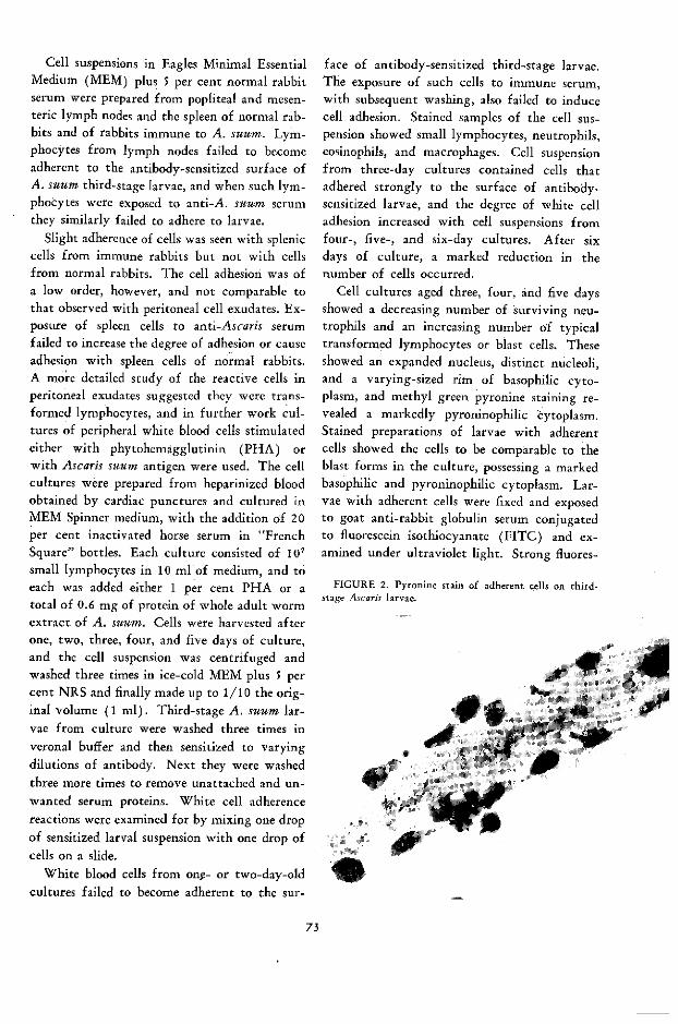
Cell suspensions in Eagles Minimal EssentialMedium (MEM) plus 5 per cent normal rabbitserum were prepared from popliteal and mesen-teric lymph nodes and the spleen of normal rab-bits and of rabbits immune to A. suum. Lym-phocytes from lymph nodes failed to becomeadherent to the antibody-sensitized surface ofA. suum third-stage larvae, and when such lym-phocytes were exposed to anti-A. suum serumthey similarly failed to adhere to larvae.
Slight adherence of cells was seen with spleniccells from immune rabbits but not with cellsfrom normal rabbits. The cell adhesion was ofa low order, however, and not comparable tothat observed with peritoneal cell exudates. Ex-posure of spleen cells to anti-Ascaris serumfailed to increase the degree of adhesion or causeadhesion with spleen cells of normal rabbits.A more detailed study of the reactive cells inperitoneal exudates suggested they were trans-formed lymphocytes, and in further work cul-tures of peripheral white blood cells stimulatedeither with phytohemagglutinin (PHA) orwith Ascaris suum antigen were used. The cellcultures were prepared from heparinized bloodobtained by cardiac punctures and cultured inMEM Spinner medium, with the addition o'f 20per cent inactivated horse serum in "FrenchSquare" bottles. Each culture consisted of 107small lymphocytes in 10 ml of medium, and toeach was added either 1 per cent PHA or atotal of 0.6 mg of protein of whole adult wormextract of A. suum. Cells were harvested afterone, two, three, four, and five days of culture,and the cell suspension was centrifuged andwashed three times in ice-cold MEM plus 5 percent NRS and finally made up to 1/10 the orig-inal volume (1 ml). Third-stage A. suum lar-vae from culture were washed three times inveronal buffer and then sensitized to varyingdilutions of antibody. Next they were washedthree more times to remove unattached and un-wanted serum proteins. White cell adherencereactions were examined for by mixing one dropof sensitized larval suspension with one drop ofcells on a slide.
White blood cells from onç- or two-day-oldcultures failed to become adherent to the sur-
face of antibody-sensitized third-stage larvae.The exposure of such cells to immune serum,with subsequent washing, also failed to inducecell adhesion. Stained samples of the cell sus-pension showed small lymphocytes, neutrophils,eosinophils, and macrophages. Cell suspensionfrom three-day cultures contained cells thatadhered strongly to the surface of antibody-sensitized larvae, and the degree of white celladhesion increased with cell suspensions fromfour-, five-, and six-day cultures. After sixdays of culture, a marked reduction in thenumber of cells occurred.
Cell cultures aged three, four, and five daysshowed a decreasing number of surviving neu-trophils and an increasing number of typicaltransformed lymphocytes or blast cells. Theseshowed an expanded nucleus, distinct nucleoli,and a varying-sized rim of basophilic cyto-plasm, and methyl green pyronine staining re-vealed a markedly pyroninophilic cytoplasm.Stained preparations of larvae with adherentcells showed the cells to be comparable to theblast forms in the culture, possessing a markedbasophilic and pyroninophilic cytoplasm. Lar-vae with adherent cells were fixed and exposedto goat anti-rabbit globulin serum conjugatedto fluorescein isothiocyanate (FITC) and ex-amined under ultraviolet light. Strong fluores-
FIGURE 2. Pyronine stain of adherent cells on third-stage Ascaris larvae.
1 e·
73
i

FIGURE 3. Cells on Ascaris larvae stained with goatanti-rabbit globulin serum conjugated with FITC.
cence occurred in the cytoplasm of the attachedcells. Similar preparations of larvae and cellswere exposed to A. suum antigen and after ade-quate washing were then exposed to a rabbitanti-A. suum globulin conjugated to FITC. Inthis case strong fluorescence occurred both inthe cells and on the surface of larvae, givingpresumptive evidence that antibody to A. suumoccurred both on the surface and in the cyto-plasm of the attached cells.
The reaction showed evidence of specificityin that the most marked reactions occurred withcell cultures obtained from rabbits that werestrongly immune to A. suum. These had beenimmunized by repeated subcutaneous injectionof infected eggs (96) or by vaccination withcultures of third-stage larvae obtained fromculture (97). Cultures of cells from rabbitsimmune to unrelated antigens and stimulatedwith the appropriate homologous antigen failed,with one exception, to give strong lymphoblastadhesion (see Table 1). The significance of thereactions with cells from rabbits immune to eggalbumin is unclear.
At low dilutions of immune serum, sensitizedlarvae attracted cells with equal effect fromcultures stimulated with either antigen or phy-tohemagglutinin; however, with increasing di-lutions of antiserum the cells from PHA-stimu-
lated cultures were less reactive, and serum
could be diluted to a point at which PHA-stimulated cells were nonreactive whereas anti-gen-stimulated cells still gave good leukocyteadhesion. Such results might be explained onthe ground that the mitogenic effect of PHAstimulated the transformation of lymphocytes"committed" to a wide range of antigens, in-
cluding Ascaris, but the number of cells in sus-
pension specifically committed to Ascaris would
be much smaller than in suspensions of lympho-cytes stimulated by the Ascaris antigen.
The system was antibody-dependent, and the
most satisfactory sensitization was achievedwith serum from rabbits immune to A. suum.Thus, rabbits that had been immunized repeat-edly with A. suum eggs (96) or with antigens
prepared from in vitro cultured larvae (97)
provided reactive sera, and the increasing abil-ity of serum to sensitize larvae to attract cellscould be titrated during the immunizationschedule. Normal rabbit serum failed to sensi-
tize larvae, and minimal sensitization was ob-tained with sera from rabbits immunized with
various tissues of adult Ascaris (whole worm,
cuticle, testes, and so on). No reaction was ob-tained when third-stage larvae were sensitizedwith immune serum against human blood groupA substance, sheep red blood cells, fowl red
blood cells, Necator americanus, or Turbatrix
aceti (Table 2). In all these cases, sensitizationof the cuticle by antibody could be demon-
strated either by mixed antiglobulin agglutina-tion (22), mixed agglutination, or immune ad-herence techniques (98).
The complement requirements for the reac-
tion have yet to be fully clarified. Some sera
retain their sensitizing ability after inactivation
at 56° C for 20 or 30 minutes, whereas others
fail to do so. The reactivity can be partiallyrestored by fresh guinea pig sera and more or
less completely restored with normal rabbit
serum. The sera that are not inactivated by
healing are, invariably, those that show the
greatest sensitizing ability and are still activewhen inactivated at 56 ° C for 60 minutes,
when treated with zymosan or ammonia, or
when absorbed with sheep red-blood-cell stroma
74
A4
'a
wlo
a
A
4
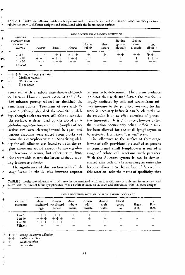
TABLE 1. Leukocyte adhesion with antibody-sensitized A. suum larvae and cultures of blood lymphocytes fromrabbits immune to different antigens and stimulated with the homologous antigen
LYMPHOCYTES FROM RABBITS IMMUNE TO
ANTIBODY
DILUTION USED Bovine BovineTO SENSITIZE Normal Sheep gamma serum Egg
LARVAE Ascaris Ascaris Ascaris rabbit serum globulin albumin albumin
1 in 5 +-+ + + + + + + + + + --+ + + -1 in 10 -+ + - + ---- - - - + + +-+1 in 20 ++ +A+-+ ++ - - - - + +Diluent
.+ + + Strong leukocyte reaction+ + Medium reaction, + Weak reaction- No reaction
sensitized with a rabbit anti-sheep-red-blood-cell serum. However, inactivation at 560 C for120 minutes greatly reduced or abolished thesensitizing ability. Treatment of sera with 2-mercaptoethanol abolished the sensitizing abil-
V ity, though such sera were still able to sensitize
the surface, as determined by the mixed anti-globulin agglutination reaction. Samples of re-active sera were electrophoresed in agar, andvarious fractions were eluted from blocks cutfrom the electrophoretic run. Sensitizing abil-
* ity for cell adhesion was found to lie in the re-$ gion where one would expect the macroglobu-
lin fraction of serum, but other serum frac-: tions were able to sensitize larvae without caus-
ing leukocyte adhesion.The significance of this reaction with third-
stage larvae in the in vivo immune response
remains to be determined. The present evidenceindicates that with such larvae the reaction islargely mediated by cells and serum from ani-mals immune to the parasite; however, furtherwork is necessary before it can be claimed thatthe reaction is an in vitro correlate of protec-tive immunity. It is of interest, however, thatthe reaction occurs only when sufficient timehas been allowed for the small lymphocytes tobe activated from their "resting" state.
The adherence to the surface of third-stagelarvae of cells provisionally classified at presentas transformed small lymphocytes is one of arange of white cell reactions with parasites.With the A. suum system it can be demon-strated that cells of the granulocytic series alsobecome adherent to the surface of larvae, butthis reaction lacks the marks of specificity that
TABLE 2. Leukocyte adhesion with A. suum larvae sensitized with various dilutions of different immune sera andtested with cultures of blood lymphocytes from a rabbit immune to A. suum and stimulated with A. suum antigen
LARVAE SENSITIZED WITH SERUM FROM RABBITS IMMUNE TO
ANTIBODY Ascaris Ascaris Ascaris AscariJ Ascaris BloodDILUTION vaccinated vaccinated whole adult adult group Sheep Fowl
eggs larvae worm cuticle testes A1 RBC RBC
lin5 +++ ++ + + + - -
1 in 10 +-- - + --- - +lin20 ++-+ -+-- .+Diluent
A-+ - - strong leukocyte adhesion- + medium reaction
9 + weak reaction-- no reaction
75
characterize the response with lymphoid cells.The reactions with granulocytes, together with
other reports on the interreaction between para-sites and white cells, will be discussed in thesection dealing with neutrophils and eosinophils.
Lymphocyte transformation by parasiteantigens
The transformation of small lymphocytesinto larger blast forms able to synthesize DNA
and undergo mitosis can be induced by mito-gens such as phytohemagglutinin (75), by var-
ious antigens to which the donor is sensitive(such as tuberculin) (86), and by mixed leu-kocyte cultures (7). As far as can be deter-mined, there has been little or no work usingthis technique for parasitic infections, but therewould seem to be every justification for studyin this direction in view of the lymphoid hyper-plasia frequently seen in parasitic infection.
Preliminary experiments at the author's lab-oratory have indicated that lymphocyte trans-
formation can be induced in rat lymphocytesderived from animals immunized with A. suumand stimulated with an extract of whole adultworm. Table 3 presents some of these prelimi-nary results. Similar reactions have been ob-tained with peripheral blood lymphocytes of aman sensitive to Ascaris as the result of labora-
tory contact over many years (Table 4).
The significance of these preliminary find-ings is not yet clear. They may indicate that adelayed hypersensitivity component is a part ofthe immune response to Ascaris infection, andthis would be in line with certain aspects of thehistological picture of the immunity to Ascaris.However, it is certainly not the sole form ofhypersensitivity that occurs, since an immedi-ate type of hypersensitivity is also much inevidence (100). Nevertheless, it is possible thatboth could exist in the same animal, perhapsmediated by different antigens. The systemdoes, however, provide a useful technique forthe study of lymphocytes from different sourcesusing small quantities of antigen, and the re-cent demonstration by Oppenheim et al. (82)that delayed hypersensitivity in the guinea pigis expressed by increased lymphocyte transfor-
mation should encourage work with Ascaris inthe guinea pig.
Macrophages
Reticulo-endothelial hyperplasia has alreadybeen noted in leishmaniasis, the macrophageserving as a host cell for the parasite. Infectedmacrophages are very active and are able topenetrate into many tissues and appear to ex-change material with neighboring cells bymeans of pseudopodia through which materialpasses from one cell to another (1). The gen-eral failure of the immune response in visceralleishmaniasis has been suggested as due to anequivalent of blockade of the RE system. How-ever, the fact that the macrophages are activelyphagocytic-indeed, they show increased indis-criminate activity-has been presented as evi-
dence against immunologic paralysis (2), butthis may not be a valid consideration, since ithas been shown that the macrophages of rabbitsrendered tolerant in neonatal life to BGG cantake up antigen and stimulate DNA synthesisin spleen cells of an immunized animal (44).The absence of an obvious defect of recognitionis comparable to the results of in vivo studieswith bacterial antigens showing that tolerantanimals were able to recognize antigen in thesame way as immune animals (74).
The phagocytic function of the macrophageis of particular interest in the hemoprotozoa,and it is now clear that erythrophagocytosis isa major factor in the pathology of malaria(115). Extensive erythtrophagocytosis of both
parasitized and normal erythrocytes is seen in
the internal organs in Plasmodium falciparum
infection and has also been observed in P. ber-
ghei in rats (23) and in P. lophurae in duck-lings (64) and chickens (95).
Phagocytosis of abnormal numbers of normal
erythrocytes in malaria has been attributed to
various immunological entities, both antigen
and antibody, including an opsonizing antibody
that is said to develop via an autoimmunity
mechanism. However, George et al. (42) con-
cluded in recent studies that red cell destruc-
tion in P. berghei infection was caused by hy-
76
14
~w
4
4
4-
4-,
iV

TABLE 3. Transformation by Ascaris antigen of human lymphocytes from an Ascaris-sensitive person
SIZE OF CELLS (MICRONS) TOTAL % TOTAL %
ANTIGEN DAYS OP OP CELLS 71. OF CELLS 9 g
ADDBD CULTURE <751 7 to <91. 91. to <11. >11 OR MORE OR OVER
PHA 3 37 30.25 20.5 12.25 63 32.75(4-8.1) (=-5.2) (+4.7) (-5.4) (48.3) (48.2)
PHA 5 9.25 24.25 41.5 25 90.75 66.5(4-3.49) (46.38) (45.41) (4-10.17) (43.6) (-6.9)
Ascaris 3 58.75 29.5 10.25 1.5 41.25 11.75-4 0.06 mg/ml (-18.6) (4-6.1) (47.69) (40.87) (421.5)* (4-8.39)*
Ascaris 5 68.5 24.25 6.75 0.5 31.5 7.250.06 mg/ml (421.7) (-4.6) (41.5) (-0.5) (43.3)* (42.0)
Ascaris 5 83 13.0 3.75 0.25 17.00 4.0w, 0.006 mg/ml (4-3.81) (41.87) (+1.92) (40.13) (43.75) (42)
b Ascaris 3 80.5 17.75 1.75 0 19.5 1.750.006 mg/ml (-25.49) (43.24) (4-1.3) (-4) (-1.29)
Control 3 87.75 9.75 2.5 0 12.25 2.5(±1.47) (40.8) (41.12) (41.4) (-1.12)
>r Control 5 81.75 14 4.25 0 18.25 4.25(426.04) (42.92) (42.58) (-3.3) (44.08)
TABLE 4. Transformation by Ascaris antigen of rat lymphocytes from rats immune to Ascaris suum
SIZE OF CELLS (MICRONS)
ANTIGEN DAYS OF --- TOTAL % OF
ADDED CULTURE <71. 7,u to <9,U 91. to <11h >11 CELLS >7,p
)4 PHA 3 55.75 (42.45) 35 (45.2) 8.75 (±5.85) 0.5 (-.5) 44.25 (49.29)
PHA 5 68.25 (4-2) 15.75 (43.3) 7.75 (43.87) 8.25 (4-3.15) 31.75 (+2)
Á Ascaris0.06 mg/ml 3 91.5 (+1.4) 6.5 (42) 2 (41.23) 0 8.5 (4-2.65)
Ascaris, 0.06 mg/ml 5 88 (41) 3.25 (4-1.7) 6.0 (41.35) 2.75 (41.35) 12 (41)*
Ascaris0.006 mg/ml 3 96.25 (=10.9) 2.0 (-1.2) 1.0 (41.2) .25 (±-14) 3.25 (4-35)
Ascaris0.006 mg/ml 5 93.25 (4-1.68) 3.75 (±I1.65) 1.75 (42) 1.25 (4.26 6.75 (41.78)
Control 3 97.25 (±=1) 2.75 (-1) 0 0 2.75 (-1)
Control 5 96.5 (41.4) 2.75 (±-.7) 0.5 (-.9) 0.25 (4.14) 3.5 (41.44)
Values represent percentage of cells in each group.400 cells measured in duplicate cultures for each set.Figures in parenthesis = standard deviation.* Significant difference (p= <0.01) between test and controls.
77
persplenism and not by an opsonin. It was sug-gested that circulating parasites, even in lownumbers, stimulate the phagocytic capacity ofthe RE systerif, which leads to an increased rate
of sequestration of blood cells in the spleen, in-cluding normal ones, which ultimately producesa cumulative effect of greater splenic functionand greater cell destruction. These authorswere unable to detect IgG on red cells and pre-sumed the absence of IgM because of the ab-sence of agglutination and hemolysis. In a con-sideration of the possible mechanisms of theimmunopathology of malaria, Dixon (35) hassuggested a mechanism based on the formationof antigen-antibody complexes, unrelated tothe erythrocytes, that nonspecifically adsorb tothe surface of red cells with the subsequent fix-ation of complement. Such cells are then liableto lysis, which would explain intravascularhemolysis and erythrophagocytosis.
Erythrophagocytosis has been claimed as acause of anemia in several other intracellularparasitoses-for example, Babesia in rodents(92) and Toxoplasma in humans (54).
Phagocytic activity is also seen in helminthinfections, especially where tissue destructionoccurs. In parasitosis of the lungs, for example,hemosiderin is frequently seen in macrophages,but apart from this there is little evidence of adirect action of macrophages on parasitic stagesof parasites in vivo, though this is presumed inview of the frequency of macrophages in areaswhere parasites are or have migrated. In thiscase, it is likely that they are performing apurely phagocytic function without referenceto, for example, delayed hypersensitivity mech-anisms.
The adherence of peritoneal macrophages tothe surface of second-stage Ascaris larvae whenthese are placed in the peritoneal cavity ofmice immune to Ascaris has been reported (26),and this finding contrasts with the results ofthe author (97), who failed to observe a reac-tion with second-stage larvae of Ascaris in theperitoneal cavity of rabbits immune to Ascaris.
It is possible, however, that different animalhosts respond differently.
Cytophilic antibody
Cytophilic antibody (sensu stricto) was orig- .,inally described by Boyden (12) as a globulin ,-component of serum that would become at-tached to certain cells so that the cells would *
then specifically absorb antigen. The originaldescription was applied to a class of rabbit anti-bodies that would attach to spleen cells of rab- >
bit or guinea pig. Another type of cytophilicantibody, apparently distinct from the spleencell cytophilic antibody and capableof becom- 4
ing attached to macrophages but not to mostother cells, has now been described (13). Therole of cytophilic antibodies in delayed hyper-sensitivity has recently been assessed by Nelsonand Boyden (72), who conclude that thus farthere is no clear-cut evidence of a definite role 4
for cytophilic antibody in delayed hypersensi-tivity.
Preliminary studies on the possible occur- 4rence of cytophilic antibody in Ascaris infec-tion have been made in the author's laboratory.None has been found in the infection, but diffi-culty has been encountered in obtaining a satis-factory bis-diazotized benzidine linkage ofAscaris antigen to red cells, and radio-labeledantigen has not been investigated.
The possibility of an antibody cytophilic forlymphocytes has been suggested (72) as a result Cof work by Koprowski and Fernandes (57),who showed that normal lymphocytes, whentreated with serum of rats immune to guineapig spinal cord tissue, acquired an affinity forcultures of cells of the brain of puppies.
It would seem worthwhile, in view of thestrong association of immunity to certain para- 5sites with cells, especially lymphocytes andmacrophages, to examine for the various cyto-philic antibodies, especially where the spleen <
plays an important role in erythrophagocytosis.
Neutrophils
Neutrophils are the characteristic cells foundearly in parasitic infections, especially wherethere is inflammation and tissue destruction,and local foci of neutrophils are frequently seenaround dying or dead parasites in various tis-
78
..
sues of the body. At times neutrophilia may bemassive and may lead to an increase in thepathological changes associated with the infec-tion (30). In experimental infection of themouse with Fasciola hepatica, the later stage ofmigration of t' .s parasite in the liver is associ-ated with a iassive outpouring of leukocytes
(0, into the tra ,s left by the migrating liver fluke.The ear y neutrophil response is usually re-
placed b' one of lymphoid elements and macro-
# phages and possibly this represents the onset ofa spe.ific reaction to the parasite by sensitizedcells. There is much to commend this idea,
+W since second infections frequently produce aninitial lesion beginning at the lymphoid stagerather than at the neutrophil stage.
The role of the neutrophils, like that of theother cells, in parasitic infections is unclear.They are part of the early response to a parasite
4 and appear before well-formed immunologicr eactions are in operation. They are also seen incertain immune reactions, particularly the Ar-thus reaction, but in both cases they are a re-sponse to inflammation and vascular injurywith subsequent sticking of cells to the blood-vessel endothelium and the migration of thecells to the local tissue.
An account of the substances that promoteW leukocyte migration has been given by Hurley
(50), who suggested that a factor (or morethan one) of serum was activated by damaged
9 tissue rather than by direct liberation from in-jured cells.
The role of antibody in the chemotaxis ofpolymorphonuclear leukocytes (PML's) wasdemonstrated by Boyden (11), who showedthat the incubation of antigen-antibody pre-cipitates in rabbit serum caused the serum tobecome markedly chemotactic to rabbit PML's.Studies with PML's and Ascaris antigens andantibodies have demonstrated a similar finding
(25).
Adhesion of neutrophils to the surfaceof parasites
In vitro studies of the adhesion of neutro-phils to the surface of second-stage larvae of
A A. suum have been in progress at the author's
laboratory. When sensitized with antibody,second-stage larvae readily become covered withlarge numbers of neutrophils. Stained prepara-tions of antibody-sensitized larvae exposed tobuffy coat preparations of rabbit blood showthe cells to be mainly PML's with a few adher-ent eosinophils. No pyroninophilia is seen withmethyl green pyronine, and the cells do notshow specific fluorescence when exposed to agoat anti-rabbit globulin conjugated withFITC. Larvae sensitized with rabbit serum willattract PML's from both immune rabbits andguinea pigs and also polymorphs from normalanimals. The reaction is antibody- and comple-ment-dependent and is produced with a varietyof immune sera; these include those that willcause the adhesion of pyroninophils from pe-ripheral blood culture to third-stage larvae andantisera to various fractions of the'adult As-caris worm and to unrelated parasites such asTurbatrix aceti and Necator americanus. Even
normal sera of bovine, rat, and guinea pig willproduce the reaction. While the several typesof immune serum might be expected to contrib-ute to the reaction, the ability of normal serumto induce it was somewhat unexpected. How-ever, other studies have shown that second-stage larvae of Ascaris, unlike third-stage lar-vae, will nonspecifically become coated with aserum component, and this may have the abilityto fix complement and cause the adhesion ofpolymorphs.
Second-stage larvae of A. suum show a defi-nite sequence of cell adhesion with immuneserum, especially when examined with antigen-stimulated peripheral blood cultures after one,two, three, four, and five days of culture. Theadherent cells from one-day cultures consistonly of neutrophils and eosinophils; as the cul-tivation time increases, the adherent cells con-sist of a mixture of neutrophils with increasingnumbers of pyroninophils, and finally the ma-jority of adherent cells are pyroninophils withonly a few adherent polymorphs. The adher-ence of pyroninophils to second-stage larvae, aswith third-stage, is mediated by immune serum.Consequently, the adherence of PML's to sec-ond-stage larvae is to a great extent nonspecific,
79
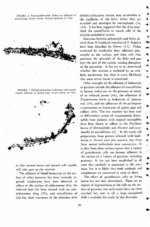
FIGURE 4. Polymorphonuclear leukocytes adherent tosecond-stage Ascaris larvae. Stained pyronine (x 750).
FIGURE 5. Polymorphonuclear leukocytes and pyroni-nophils adherent to second-stage Ascaris larvae (x 750).
,44
IK
in that normal serum and normal cells equally
will take part in the reaction.The adhesion of blood leukocytes to the sur-
face of other parasites has been variously re-
ported. Leukocytes have been observed to
adhere to the surface of schistosomes after the
infected host has been treated with an anti-
schistosome drug (71), and microfilariae of
Loa loa, after treatment of the infection with
diethyl-carbarazine citrate, may accumulate in
the capillaries of the liver, where they are
attacked and enveloped by macrophages (16,114). It has been suggested that the drug sensi-tized the microfilariae to attack cells of the
reticulo-endothelial system.Reactions between polymorphs and flukes de-
rived from X-irradiated cercariae of F. hepatica
have been described by Dawes (31). Flukes
weakened by irradiation show adherent poly-
morphs on the surface, and these cells then
penetrate the epicuticle of the fluke and pass
into the core of the cuticle, causing disruption
of the epicuticle. It has yet to be determined
whether this reaction is mediated by an anti-
body mechanism, but there is every likelihood
that some serum factor is concerned.
Other examples of the adhesion of leukocytes
to parasites include the adhesion of microfilariae
to human leukocytes in the presence of serum
of an infected person (84), the adherence of
Trypanosoma lewisi to leukocytes of immune
rats (59), and the adherence of the pathogenic
trypanosomes to leukocytes of guinea pigs and
rabbits (60). This last reaction has been used
to differentiate strains of trypanosomes. Eosin-
ophils from patients with tropical eosinophilia
have been shown to adhere to the filariform
larvae of Strongyloides and Necator and occa-
sionally to microfilariae (8). In this work, cell
preparations from persons infected with hook-
worm or Ascaris were also reactive, but those
from normal individuals were nonreactive. It
is clear from these various reports that a variety
of granulocytic cells can become adherent to
the surface of a variety of parasites, including
protozoa. It has not been established in all
cases that antibody is concerned in the reac-
tions, but it is likely that both antibody and
complement are concerned in most of them.
The effect of granulocytic cells on living
larvae has not been determined. There are no
reports of degranulation of the cells on the sur-
face of parasites, but such events have not been
searched for, and, in all, a large unexplored
field is available for study in this direction.
80
4*
él
4
al4.
.. IAYdl

Eosinophils
The eosinophil is probably the cell that ismost traditionally associated with parasitic in-fection, and yet, despite its characteristic ap-pearance, there are no definitive statements
O available on its function or the factors thatmediate its accumulation-sometimes in a spec-tacular manner-in the tissues. Usually, theeosinophil is not part of the early acute inflam-matory response but occurs at a later stage ofinflammation when round cells and plasma cells
q become evident. To some extent, its appearanceis an indication of the age of an inflammatorylesion, and the frequent association of eosino-
' philia with allergic disorders, and especiallyparasitic infections, has led to the presumptionthat this cell type is associated with immunityprocesses.
*,, Among the many theories that have been de-vised to account for the accumulation of eosin-
" ophils either in the blood or locally, three havereceived more attention than others. These arevariations on a theme to do with histamine andcan be stated (a) histamine attracts eosino-phils, (b) eosinophils contain an antihistamine,or (c) eosinophils contain histamine (5). Amore recent study of eosinophils and eosino-
, philia (61) has helped greatly to clarify thefactors that induce eosinophilia. Evidence has
; been provided that it is due essentially to anti-gen-antibody complexes: antigen alone, anti-body alone, or the by-products of an antigen-antibody reaction have little eosinophilotactic
· activity. If this work is confirmed, it wouldprovide an explanation for the marked accumu-
, lation of eosinophils in parasitic infection, sincein many cases the stimulating antigen or anti-gens (the parasite) can persist for some time inthe body. Eosinophils have also been shown to
* have a phagocytic function, being capable of
ingesting antigen-antibody precipitates (4, 61,
90), this is also associated with degranulation
of the cells, and it may be a mechanism wherebythe antigen-antibody aggregate is inactivated
or degraded. A variety of enzymes have beenidentified from the granules of eosinophils, in-
· cluding cathepsin, P-glucuronidase, nucleases,
phosphatases, and peroxidases (myeloperoxidase,verdoperoxidase), but their exact function isstill in doubt. Some evidence of degranulationof eosinophils on the surface of Ascaris larvaehas been noted by the author, but this effecthas not been studied in detail or examined forits possible effects on the cuticle of larvae.
The idea that antigen-antibody aggregatesprovide the eosinophilotactic response is anattractive one; however, there are several re-ports that indicate the occurrence of eosinophiliain circumstances where the immune responseplays a small part in the mechanism. Arean(6) has shown that the appearance of eosino-phils at the site of injected Ascaris eggs was toorapid to be explained by an antigen-antibodyreaction, and marked eosinophilia has beenobserved in a child with visceral larva migransbut who was agammaglobinemic and showedalmost a complete absence of immune globulinsin the serum (47). There are many reports,published over the last few decades, indicatingthat antigens from several parasites (includingAscaris) will induce eosinophilia after an initialinjection (41, 106). Early work in this areawas almost certainly with animals (e.g., dogs)that may have been naturally sensitized toascarids, but even the more recent experimentsshould be intepreted with care, since animalsare commonly parasitized with a low burden of"normal" parasites and these may be sufficientto sensitize an animal to a cross-reacting anti-gen.
In a study of eosinophilia to Toxocara canisinfection in guinea pigs, Olsen and Schulz (81 )showed that the onset and extent of eosinophiliawere somewhat dose-dependent; guinea pigs re-ceiving the largest dose of eggs (5,000) showedeosinophilia on the second day, while those re-ceiving only 50 eggs developed it on the tenthday. Maximum eosinophilia was seen approxi-mately two weeks after infection in all cases.Schultz-Dale tests with antigens from Toxo-cara eggs showed a dose dependency betweenthe number of eggs given and the time whenreactions could be elicited. Maximal eosinophilicresponses at about two weeks after infectionhave also been observed in other nematode in-
81

fections, such as Trichinella spiralis (89) andDictyocaulzus viviparus in cattle (109).
An interesting aspect of the work by Olsenand Schulz was that though the eosinophiliapersisted in infected animals for 28 days, theToxocara larvae did not persist for more than14 days, as judged by digestion techniques.
Support for the idea that eosinophils are an-tagonists of histamine is obtained from studieson the mast-cell, eosinophil, and histaminelevels in rats infected with N. braziliensis(110). A marked eosinophilia that occurredfrom 12 days after infection onward Was asso-ciated with a marked decrease in the numberof tissue mast cells; this was interpreted to indi-cate that the disruption of mast cells, with therelease of histamine, served to attract eosino-phils whose function was to remove the excesshistamine (110). However, an alternative ex-planation might be that mast-cell degranulationwas induced by a mast-cell-sensitizing antibodyand that eosinophils were attracted by the re-sulting antigen-antigen aggregates.
There is little doubt that the role of theeosinophil in parasitic infection requires muchmore detailed study.
Mast cells
Earlier studies of the cell response to initialand repeated infections of N. braziliensis inrats demonstrated that connective tissue mastcells (connective tissue basophils) fluctuated innumbers during such infection (102). Thishas been confirmed by Wells (110), whoshowed that mast-cell numbers in the rat in-testine fell markedly about the fifteenth day ofinfection and later rose to levels much higherthan those prior to infection. It is unlikely tobe fortuitous that at the same time these eventsoccur there is also a loss of adult worms (self-cure) of an initial N. braziliensis infection. Theconcurrent increased accumulation of eosino-phils has been mentioned previously.
Degranulation of mast cells can be broughtabout by a number of agencies, includingtrauma, bacterial toxins, heat, cold, and ion-izing radiation (104). An additional mecha-nism is degranulation by an antigen-antibody
interaction, and it is now well established thatthe mast cell plays an important part in theanaphylactic syndrome (70, 105). A detailedconsideration of the various aspects of this isgiven by Keller (55). Essentially, mast-celldegranulation may occur on contact with anti-gen after active sensitization and, accordingto the animal species, after passive immuniza-tion with serum. Mota has postulated a "mast-cell-sensitizing" antibody that is nonprecipi-tating, appears early after immunization, andmay be present in the blood for only a shorttime (67, 68). The most satisfactory methodof demonstrating it is homologous passive cu-taneous anaphylaxis, and in the rat its produc-tion is much accentuated by the use of Haemo-philus pertussis vaccine. Consequently, itclosely resembles the "reagin" type of antibody(69).
The occurrence of an antibody similar toreagin has been reported to be closely associatedwith immunity to N. braziliensis in the rat (76,78), the reagin being detected in some ratsimmediately after the acquisition of resistanceand in all rats one week later. High levels ofthe reagin were stimulated only by infectionwith living, adult worms and not by vaccina-tion with worm extracts. Rats immune to N.braziliensis undergo severe anaphylaxis on intra-venous injection of antigen, and since the gutof the rat appears to be the major shock organ(111), it has been suggested that a local ana-phylactic reaction in the gut may be responsiblefor the termination of an adult worm infection(103).
It has been shown recently that anaphylacticshock induced by an unrelated antigen-anti-body system will enhance passive immunityconferred with antiserum. This produces a sig-nificant expulsion of worms compared to ratswhich were only passively sensitized (10). Inthis circumstance, it appears that the anaphy-lactic lesion has increased the passage of im-munoglobulin to the lumen of the bowel.
It would be tempting to ascribe this sequenceof events to a degranulation of tissue mast cellsin the intestinal wall brought about by an anti-gen-antibody interaction, possibly with the
82
4
,*i
'9
1,u-0
$
u
e

» characteristics of a mast cell sensitizing anti-" body. Conclusive evidence for this, however,
must await further study." There is ample evidence to indicate that hel-
minth antigens do have a profound effect onmast cells. A principle of Ascaris will degranu-late rat peritoneal mast cells (104) and studieswith mice infected with T. spiralis have shownthat local (subcutaneous) degranulation ofmast cells is dependent on prior sensitizationwith the parasites (14). In the latter studiesmaximal reactions were obtained one monthafter infection, using either metabolic or so-
* matic antigens of T. spiralis. Sensitizationcould be passively transferred to normal mice,though in this case mast-cell injury was less
_ than that observed in actively sensitized mice.Mast-cell degranulation in skin pouches of miceimmunized against Strongyloides ratti has been
t described (43); in this case "excretory andsecretory" antigens of infective larvae provedmore effective than somatic antigens.
Various other reports have indicated an in-crease in the number of mast cells in the skin
3 of mice infected with S. mansoni, Hymenolepisnana, and Syphacia and in the skin of patientswith schistosomiasis and filariasis (37, 38). Anincrease in the number of bone marrow mast
a cells (basophils) and an increased release ofthese and eosinophils into the blood of guineapigs after the injection of Ascaris body fluidhas been reported (18). Comparable workusing a highly purified polysaccharide of As-caris muscle showed that intraperitoneal injec-
w tions caused infiltrations of primitive hema-topoetic elements of the erythrocytic and leuko-cytic series into the liver (80).
Globule leukocytes
* These cells have received occasional attention_ over the last several years, having been noticed
especially in parasitic infection of the gastro-* intestinal tract of ruminants. Their relation-
ship to parasitism and in particular to theimmunological process has hitherto been some-what unclear, since they have been found inthe abomasal and intestinal mucosae of bothinfected and normal animals. Recently, how-
ever, a clear relationship between gastrointesti-nal parasitism and the globule leukocyte hasbeen reported, the cell being common in themucosae of parasitized animals but infrequentin worm-free animals (112). An increase inglobule leukocyte numbers has been observedin the intestinal mucosa of rats infected withN. braziliensis; this was marked on the twelfthday and was coincidental with the self-curemechanism (112).
The nature and function of the globuleleukocyte have yet to be fully clarified. Somehave regarded it as comparable to the "Russellbody" cell of the plasma cell series, since bylight microscopy it appears very much thesame as that cell (36). The Russell body cellhas been shown to contain immunoglobulin,and in the work by Crandall et al. (27) suchcells showed intense cytoplasmic fluorescencewith anti-/u heavy chain reagents. A re-exami-nation of the identity of mast cells and globuleleukocytes by Jarrett et al. (52) showed thatin rat, sheep, and bovine three related cell typesoccurred that could be differentiated on theirstaining reactions with toluidine blue andfluorescence with acridine orange. One form ofmast cell was commonly found in the laminapropria of the intestine, gastric wall, and per-ibiliary area in F. hepatica infected livers. Theultrastructure of mast cells and globule leuko-cytes was reported to be similar, but differentfrom that of the "Russell body" plasma cell; ofspecial interest was the fact that mitotic activ-ity could be detected in mast cells and globuleleukocytes. The globule leukocyte was sug-gested as an end cell of a range of mast-celltypes, derived from the type of mast cell foundin the lamina propria of the digestive tract.
If globule leukocytes are in fact a form ofmast cell, then a function 'for them could beenvisaged in gastrointestinal parasitism. Theycould, as for example in N. braziliensis infec-tion, accumulate in the mucosa and underappropriate stimulation release biologically ac-tive amines, which in turn could alter thepermeability of the gut mucosa for largermacromolecules and possibly also for immuno-logically competent cells.
83
Summary
A wide range of cellular reactions are asso-ciated with parasitic infections. Some of theseare probably nonspecific responses to tissue in-jury and inflammation, whereas others appearto be mediated by an immunological response.Since protozoan and, especially, metazoan para-sites are not only complex antigenic entities butmay also cause tissue destruction, it is to beexpected that the cell response to them will be
complex. Perhaps this complexity has been anunattractive prospect to workers in the past,but with an abundance of information nowavailable on the morphology, ultrastructure,physiology, and functions of the wide range ofcell types that can be found in parasitic infec-tions, there is much less reason for the field tobe neglected.
Moderator: The discussion of this topicwill be initiated by Dr. Biro.
4.-
%A
84
4
b4*
4
DISCUSSION
Carlos E. Biro
My comment must of necessity be general in+ nature, and I should like to keep it short.
To begin with, when the cells involved inthe immune response are mentioned, the firstconfusion that strikes me personally concernsthe cells actually involved in the immune re-sponse itself-in other words, in the prepara-tion or production of antibody-and those thatare called in after the antibody response hastaken place, as effecters of damage. I thinkthat this is a very important distinction tomake in studying immunity to parasites andthe cells involved in this kind of phenomenon.
9 It is necessary to study on the one hand theinitiation of the immune response, and on theother the phenomena that occur afterward.
To put it very simply, the first kind of cellinvolved in an immune response is probably amacrophage, a phagocytic cell. Most antigensare particulate or large matter, and these willbe the first ones involved. By some mechanismthat need not be discussed here, they seem to
!\' be in touch with or communicate with cells ofanother origin, lymphoid cells, and these lymph-oid cells or their relatives differentiate to give
r plasma cells.
Along this line of differentiation, a series ofevents takes place. The first seems to be the
·E- production of IgM. The second seems to be theestablishment of immunological memory, andpossibly the last would be the establishment ofIgG production. Again, I do not think it isimportant at the moment to discuss whichstarts first.
I have deliberately left IgA production anddelayed hypersensitivity in a separate, not-too-
clear position, because I do not think their posi-tion is clear. The only thing that, at least frommy point of view, is interesting is the associa-tion between these two. There seems to be areasonable and growing body of evidence link-ing the presence of IgA to delayed hypersensi-tivity. Anti-IgA antisera are able to block somein vitro models that are probably representa-tions of delayed hypersensitivity. This, I think,would be important in studying parasite im-munity.
Now, once all these cells have been involvedin the production of antibody or an immuneresponse, in another totally different stage-again, I do this for the sake of simplicity-there will be antigen-antibody complexes. 1include in this term the interaction betweenwhatever produces the delayed hypersensitivityand the corresponding antigen. As a result, wecan have histamine release, changes in vascularpermeability, polymorphonuclear cells attractedto the site of the lesion, and eosinophilia.
I start at this end because, not being a para-sitologist interested in immunology but an im-munologist interested in parasitology, I have to.I now ask myself, Where within this scheme doI fit immunity to parasites? I find that this isnot as big a problem as I would have thought.I know that my colleagues in different special-ties who deal with immunological problemswould like to have a special immune responsefor each of them. The transplant people wantone, the parasitologists want one, and why not?Yet I think that economy in a living being leadsto the idea of the immune response as probably,if not one event, a series of linked events. In
8S5

this case the only question that need be askedis what the characteristics of the antigen are,so that we can know something of the charac-teristics of the antibody produced or the im-mune response produced to it. We find herethe first division I must make.
I have been a little uneasy today hearing youtalk simultaneously about monocellular, uni-cellular, intracellular parasites, and about verybig things like helminths. I think the prob-lems are totally different. We are not going toadvance by lumping them; we have to separatethem. In the case of the helminth, we have tothink of a parasite that is outside the organism;as Dr. Remington said, the gut is really in theouter world. Here, at least, we have to con-sider a parasite that is big and lives part of itslife outside the being it inhabits. The otherthing we have to think about is a parasite witha very resistant surface. In this context, theproduction of antibody, a protein capable ofweak interactions would seem to be very littleprotection against the parasite. One would ex-pect the intervention of cells very particularlyat this level. At the other extreme are theprotozoa, which are necessarily intracellularparasites, and here again we have a problem.Under normal circumstances, immunoglobulinsdo not, or appear not to, penetrate in normalform into the cytoplasm of healthy living cells;therefore, once the parasite is admitted into anintracellular space, the only point at whichthere will be interaction between antibody andthe parasite will be in its passage between onecell and another, or after the rupture of onecell and before the parasite is admitted to thenext.
These two different approaches-one to thebigger parasite, on which a humoral factor willhave little or no effect, and the other to theintracellular parasite, in which the moment ofthe antibody's action must be just the onewhen the parasite is outside the cell-are whatI want to call to your attention.
On the negative side, one might add thatthe immune response was not made for eitherof these types of problems. I know this isteleology, and I apologize for putting it this
way. But, to repeat, what I mean is this: Inthe case of the big parasites, humoral factorswill have a very slim chance of helping toeliminate infection. In the case of the intra-cellular parasites, too, perhaps research shouldbe directed toward something different fromthe immune response. There are other exam-ples, such as viruses, in which nature appar-ently needed some other kind of solution be-cause antibody production was not goodenough.
If this sounds negative, I am sorry; it is notmeant to. It is meant only to separate outproblems so that we can tackle them one byone, instead of lumping them together in thishard-to-handle mixture which is the field ofparasitology.
Moderator: I think Dr. Soulsby presentedvery interesting evidence that the attachmentof pyroninophilic cells around the surface oflarvae may play a role in the actual destruc-tion of the parasite, and this is to my mind avery important point. We know that in de-layed hypersensitivity tissues suffer a lesion onaccount of lymphoid sensitized cells, and whyshould not the tissues of the parasite suffer thesame thing? We know that lymphocytes frompeople who have delayed hypersensitivity willproduce lesions of target cells in vitro, andwhy not on the parasites?
I should also like to provoke a comment byDr. Soulsby on the eosinophils. In the not verydistant past, people have been very interested inthe role played by the eosinophils in the variousstages of the immunological sequence. Weknow that the antigen-antibody interactionattracts eosinophils. The eosinotactic factorhas not been isolated as the chemotactic factorthat produces the attraction of neutrophils andresults from the interaction of antigen, anti-body, and complement. But there is probablysuch a thing as an eosinotactic factor producedby antigen-antibody interaction, and we alsoknow that eosinophilia is very characteristic ofmany parasitoses, particularly of helminthiasis.
I remember a paper written in 1963 in whichDr. Soulsby considered and speculated aboutSpier's point of view on the participation of
86
14
*
'
l'4
-4,
-
*·
k:,;

eosinophils. To my mind, the sequence ofevents suggested by Spier called for too many
;, hypotheses: First, phagocytosis of the antigen
by neutrophils. Second, phagocytosis of the- necrotic neutrophils by macrophages. Third,
lymphocyte macrophage cells (hemocytoblasts)undergoing DNA synthesis and dividing to
- form medium and small lymphocytes. Fourth,some of the mononuclear cells involved in theinflammatory reaction being injured by anti-
' gen, becoming swollen, and forming numerousvesicles. Fifth, attraction of eosinophils bythe injured cells penetrating their plasma mem-
1 branes and causing disruption of the cytoplasm.
, Sixth, macrophages engulfing cell fragmentswith adherent antigen as well as eosinophils as-
r sociated with these fragments. Finally, micro-phages losing their phagocytic activity and
undergoing transformation into antibody-pro-' ducing cells.
I believe Dr. Soulsby will agree that this con-cept is based on a chain of unnecessary and un-proved steps. It recalls the old dictum known
as Occam's Razor: Essentia non sunt multipli-canda praeter necessitatem (One should not
multiply the hypothesis beyond necessity). Ther only thing we really know is that the antigen-
antibody complex, as well as certain macro-n molecules, including Ascaris keratin, are
strongly eosinotactic, and we also know thatAscaris extracts are potent histamine liberators,
·" and there are indications that eosinophils mayplay a role in the removal and neutralization ofhistamine. These are a good many assorted
¶ things that might be put together, perhaps,but I do not know how.
Like the attraction of lymphoid cells in de-1 layed hypersensitivity, the attraction of eosino-
, phils under the conditions of Dr. Soulsby'sexperiments a few years ago still requires ex-
planation, I think.
Soulsby: I agree entirely that Spier's se-quence of requirements for his theory of anti-body initiation and production is so compli-cated and requires so many unproved steps that
L it is more or less unacceptable. I think thatnowadays almost everyone would agree.
If there was anything that would have made
me refuse to cooperate in this conference, itwas any consideration of the eosinophil. It isextremely difficult to find definite informationabout it. As you well know, there are so manytheories about it-that it is a histamine carrier,that it is an antihistamine carrier. The recentpublication of Litt's in the Annals of the New
York Academy of Sciences seems to offer afairly good explanation of eosinophilia in para-sitic infections. He suggests that eosinophiliais very common in helminth infections becausethe antigen persists for some time in the tissues.Consequently, a residuum of antigen-antibodyaggregates might be expected to persist there.
We have been able, using the in vitro tech-nique mentioned earlier with larvae, to demon-strate the adherence of eosinophils to larvae.This is similar to Bang's demonstration of a
comparable reaction with eosinophils and mic-rofilariae in cases of tropical eosinophilia inIndia. The reaction depends on sensitization ofthe larvae by antibody and is also complement-dependent. This would fit with Litt's work.
I want to return for just a minute to thepyroninophil-adherence reaction. I want to em-phasize that this system we are working withis a very artificial one. I would be the last tosuggest that this is a definite in vivo mechan-ism. A point I raised in my presentation wasthat perhaps we should think not only in
terms of the parasite, but also in terms of thelocal environment of the parasite. In examining
a lesion, it may be seen that these cells are verynumerous in the local environment. When theparasite is transferred to an artificial system,there is no longer any local environment, butin this situation cells are observed adheringdirectly to the parasite.
Moderator: I appreciate the caution ofyour statement.
Nussenzweig: I have two questions forDr. Soulsby. What is the role of the comple-ment in these reactions, and their relation to
the immune-adherence phenomenon? Are thelarge cells already present in the lymph node ofthe animals or are they transformed on the sur-face of the larvae?
Soulsby: We produce these cells in culture
87
and then use them. It is only when cells haveundergone transformation that they are activein the reaction. Cells will transform if they arecultured with living larvae, presumably as aresult of the release of antigen into themedium. But we have never observed an ad-herence of unstimulated lymphocytes to larvae,and consequently it does not appear that theyare stimulated to transform on the surface oflarvae.
Goodman: I have a comment and then aquestion. The comment is the same one I madeabout immunoglobulins: that we do not reallyknow the role of delayed hypersensitivity or ofcirculating antibodies in many of the classicalinfections by viruses and bacteria. Therefore,we should not expect to understand clearly therole of delayed hypersensitivity in parasitic dis-eases without a good deal of work. The gaps inour knowledge about the function of differentkinds of circulating antibodies persist althoughwe can purify them and even determine theirstructure. In the case of the delayed hypersensi-tivity system, on the other hand, we have veryfew in vitro techniques. It is good to hear thatDr. Soulsby is making a start on applyingthem to the parasitic diseases.
My question concerns the reaction he isworking with; I believe it involves thelymphocyte, which has changed, been "trans-formed," become a blast cell. It has been veryinteresting to follow the development of invitro systems devised to study delayed hyper-sensitivity; to note that Dr. Perlman in Stock-holm and Dr. Brunner in Lausanne have beenable to take sensitized lymphocytes (whichhave not previously been transformed) directlyfrom the spleen (Brunner) or peripheral bloodcells (Perlman) and mix them with the targetcells (to which the animal has delayed hyper-sensitivity). The damage to the target cell canbe determined by culturing them or by measur-ing the release of labeled chromium from thecell. Have your parasites been damaged by thetransformed lymphocytes? Have you been ableto apply the Perlman or the Brunner techniqueand expose any stage of the parasite to lympho-cytes (either from peripheral blood or from
lymph nodes or spleen) and show that theseparasites then do not grow normally or cannotbe transferred and grown normally when putinto another animal? It would seem that para-sites should offer an excellent system for thistype of in vitro investigation of delayed hyper-sensitivity.
Soulsby: You have summarized our researchfor the next two years. It would be most in-teresting to be able to demonstrate a lesionon the parasite with these cells, but at presentwe have not detected any. But it may be thatsuch a lesion takes longer to form than oursystem will demonstrate. The study of an in-dividual preparation is terminated after abouthalf an hour. We are, however, initiating workon the continuous in vitro culture of cells withlarvae, and at present we have a little evidencethat a parasite population survives less readilyin a population of immune cells than of normalcells.
Biro: I still insist on my separation. Hista-mine release, polymorphonuclear attraction,circulating antibody, and immediate hypersen-sitivity all belong on one side, whereas delayedhypersensitivity-or, at least, so there is an in-creasing body of evidence to show-probablybelongs on the other. I like to think of delayedhypersensitivity as the local manifestation of asecondary immune response. This is very im-portant in relation to Dr. Soulsby's model, be-cause he is dealing with the adherence of cells,and I think that the stickiness of the surfaceof a cell is what decides whether a cell will orwill not circulate or recirculate. Not muchimmune response goes on in circulating cells.It is usually in cells that are stuck. What makesthem stick? This is an important question.
In the adherence of blastoid or transformedcells to larvae, two different models, the mean-ing of which would be entirely different, haveto be separated. One of them is as Dr. Soulsbyhas been carrying it out: the cells have alreadyacquired this stickiness, which I tend to feelmight be very nonspecific. A question I mightask is what happens when one takes anotherkind of barrier for immunoglobulin, anotherbig particulate kind of antigen, and covers
88
(45
\
gn E

> it with its antibody. Does this also stick tothe cells and not change the specificity of theantibody or of the cells but change the
r specificity of the carrier?ipx The other model is the one Dr. Goodman
suggested, in which transformation wouldj take place locally, on the cell. I think that
this acquisition of stickiness shows the begin-ning of an immune response. It might be
; worth while to remember that these blastcells or blastoid cells do not appear in thelymph that comes from the site of antigeninto a lymph node. They appear in the nodeand in the efferent lymph-the lymph goingaway from the node. It seems to take some
t kind of adhesion or mutual interaction be-tween cells to get the phenomenon started.This is very difficult to correlate with in vitromodels.
R. S. Nussenzweig::* I should like, if I may,to raise one more problem concerning the cel-lular aspects of host response to parasitic in-fections, in addition to those that have al-ready been discussed: the stimulation of thereticuloendothelial (RE) system that is knownto occur in many cellular and blood parasiticinfections, the importance of which has been
'* recognized since the classical work of Talia-ferro. But I have the feeling that since thenthis problem has not had enough experimental
- attention and that many related problems re-main to be clarified.
I want to present some data we obtained
(Y in malaria infection, using rodent malariaparasites in mice. Immunizing animals withsporozoites, the insect form of the malariaparasite, incorporated in Freund's completeadjuvant, we observed the development of acertain degree of protective immunity, shown
r by a two- to threefold increase in the survivaltime of the immunized animals. But similarresults were obtained in a control group in-
4 jected repeatedly with Freund's adjuvantalone, without parasite antigens.
We thought of RE stimulation as enhancingthe natural resistance to the malaria infection,
* Assistant Professor, Department of Preventive Medi-cine, New York University Medical School.
and the next step was to try out other-andif possible more potent-stimulants of the REsystem to verify whether these effects couldbe reproduced. For this we used Coryne-bacterium parvum, which had been shown byDr. Biozzi and his colleagues to have a verypotent accelerating effect on carbon particleclearance. We treated animals (A/J mice)with killed C. parvum and later challengedthem, and also untreated controls, with viablesporozoites of Plasmodium berghei. A quiteextensive protection was observed: only 30 to50 per cent of the pretreated animals becameinfected, compared to 100 per cent of thecontrol animals. Furthermore, some of the pre-treated animals that became patent survivedthe P. berghei infection; this has never beenobserved with this parasite under normal con-ditions. In conclusion, there was a marked in-crease in host resistance produced by this non-specific stimulation of the RE system. I thinkit would be worth while to see whether some-thing similar could not be observed in othersystems with different malaria parasites, andeven in other parasitoses.
Moderator: This is a very interesting ob-servation. As far as I know, Dr. Biozzi neverhad a conspicuous degree of protection againstbacteria with this stimulation. Against Sal-monella typhimurium in the mouse, I believehe never got any protection.
R. S. Nussenzweig: I am not aware of pub-lished results on the effect of administeringC. parvum on Salmonella typhimurium orany other bacterial infection. As for theeffect of C. parvum in other systems, Halpernand his colleagues obtained a certain inhibi-tion of tumor development and increased sur-vival rates in several experimental tumors inmice pretreated with C. parvum. Biozzi andhis colleagues also reported on a reduction ofmortality rates due to graft-versus-host reac-
tions in mice pretreated with C. parvum.
Dubos: Since the time is so short, I shalllimit my remarks to Dr. Biro's category of
biochemical events, taking place as a result
of antigen-antibody reaction and also of aller-
gic reaction, that reveal changes in the activi-
89

ties of the cells due to the immunologicalprocess. He denoted the biochemical changeby the word "histamine." He used histaminebecause this happens to be a substance thatimmunologists are professionally interested in,but I am sure that he meant it to symbolizemany other kinds of biochemical changes thattake place. It is a fact, as you well know,that during the past five or six years an enor-mous amount of information has been accu-mulating on the changes that occur in thephysiological and biochemical activities ofvarious types of cells in various immunologicalsituations.
Let me just mention two or three that Ibelieve are directly relevant to the questionthat has been worrying you today; that is,how the immune reaction exerts its anti-in-fectious effect on the pathogen, whatever thenature of the pathogen.
There is no doubt that after any one ofthe classical immunologic reactions there isa change in the metabolic activity of the cellconcerned. This has been illustrated by thephenomenon so well kriown since Metchnikoff,and so well documented since, that a changein the glycolytic activity of the cell takesplace, so that suddenly there is the classicalburst of acidity that results from an increas-ing glycolytic activity associated withphagocytic processes, for example.
Moreover, and also extending early observa-tions of Metchnikoff's, there has been spec-tacular demonstration that not only as a re-sult of phagocytosis but also as a result ofmere contact between the antigen and theimmunologically competent cell, explosivechanges occur in the different types of gran-ules of the cells-to such an extent that thephenomenon has been described as degranula-tion. This might more properly be referred toas the destruction or explosion of the lysosome,with a sudden release into the cell and thevicinity of the cell of all the components ofthe lysosome system, which in the final analysis
means all the enzymes that are stored in thelysosome complex.
There is also a great deal of evidence thatalmost any kind of stimulation of an activecell by the antigen to which it has been sensi-tized results in an accumulation within thatcell of many types of biochemical equipment,as can so well be demonstrated by growingmacrophages in tissue cultures and showingthat the richness in different metabolic' po-tentialities can be changed at will.
It seems to me that you should have all thisin mind. You will find that the question youhave been asking all day-how does the im-mune mechanism really affect the susceptiblecell?-is probably much simpler than weknew a very few years ago.
By the way, let me say that your questionsare not at all peculiar to parasites; they applyto all bacterial species. They apply exactlyto immunity against tuberculosis. I amusedmyself by taking several sentences from Dr.Smithers' presentation and inserting "tuberclebacilli," and I found that his text was abso-lutely applicable, with the same queries, thesame uncertainties, but also I believe the samepossibilities of explanation.
So let me just suggest that in your thinkingabout this problem you bear in mind that theactivities of the cells involved in the immuneprocess are profoundly affected by any formof immunologic reaction, whether it be theclassical antigen-antibody or the delayed aller-gic type of. reaction. I believe you will findthen that most of the phenomena of protectionthat appear so mysterious are due in reality notto a direct effect of the antibody on the parasite(whether virus, bacterium, or helminth) butto the secondary manifestations of the allergicprocess-the release of a whole variety ofchemically metabolically active substances allproduced in Dr. Biro's second category.
Moderator: I think we had better proceednow with Dr. Weinstein's presentation. Ihope we may take up this matter again later.
90
i(4
u
1

IMMUNOLOGIC ASPECTS OF PARASITIC INFECTIONS
Paul P. Weinstein
The effects of the immune response on para-sites are manifested in a wide variety of ways,
-~t and many studies have been devoted to a de-scription of diverse aspects of the response,such as behavioral and morphologic changes,
structural damages, changes in infectivity andin subsequent growth and differentiation, andmetabolic alterations. In few instances, how-ever, has there been a systematic attempt tostudy in detail and interrelate the variety ofconsequences to a parasite that may follow animmunologic event.
For the sake of discussion, the effect of theimmune response on parasites will be arbitrarilydivided into several levels of reaction: gross,physiological, and biochemical. It is quiteevident, however, that these categories areartificial and may operate simultaneously ina complex interplay. Therefore, the descriptionof the consequences of an immunologic reac-
(' tion upon a parasite will, like the proverbial
blind men and the elephant, depend upon theanalytical tools that are brought to bear andupon the perceptiveness of the investigator.
No attempt will be made here to present adetailed review for each of the subjects dis-cussed. Instead, selected examples will be used
,wt¿- in an attempt to assess the major reactions ex-hibited by protozoa and helminths. Many of
4 the observations that will be referred to havebeen made in vitro only, and their relevanceto the in vivo condition is frequently not clearly
1) understood. It is similarly apparent that inmost instances insufficient information is avail-able for determining whether a given reaction
plays a role in the ultimate destruction of aparasite. It is unfortunately still true that theunderlying mechanisms of protective immunityin the great majority of parasitic infections area mystery.
Gross aspects of reaction
Immobilization reactions
Antibody acting on various species of pro-tozoa and helminths may result in retardationin movement and immobilization.
Entamoebae. This type of reaction com-monly exhibited by amoebae was first describedby Cole and Kent (8) for Entamoeba histoly-tica exposed to the serum of rabbits that hadbeen immunized to the organism. When placedin antiserum, motile trophozoites soon ceasedto form pseudopodia and rounded up. Maximalimmobilization was obtained in twenty tothirty minutes, after which the amoebae re-gained their activity. Serum from infectedhumans was also shown to possess the immo-bilizing factor (3). Previous investigators hadconsidered that surface antigens were involved,and Biagi et al. (2) provided data to strengthenthis hypothesis. Using fluorescent-antibodyprocedures, they observed that as immobiliza-tion occurred, the fluorescence was localized
principally on the surface of the organism.After 45 minutes, it was regularly distributedin the ectoplasm and endoplasm. At 60 min-utes, activity in the cytoplasm was noted withthe appearance of non-fluorescent areas. At75 minutes, all the fluorescent material was
91

present in a large vacuole in the endoplasm,and at 105 minutes it was barely perceptible;at this time remobilization of the trophozoitewas observed. Loss of fluorescence was notseen in trophozoites that were not remobilized.
Rabbit antiserum to E. histolytica alsomarkedly inhibited the ability of the amoebae toingest red blood cells in vitro, apparently be-cause of the immobilizing effect of the anti-serum (63).
Similar immobilization reactions have beenshown to occur with E. invadens, E. coli, E.ranarum, and E. moshkovskii (89). The firsttwo species with E. histolytica fall into oneantigenic group, distinct from the two latterspecies. Biagi et al. (2) believe that the anti-body responsible for the immobilization reac-tion is a gamma globulin. Antiserum that isnot inactivated will produce lysis of theamoebae; the inactivated antiserum produces,in addition to immobilization, a certain degreeof agglutination (89).
According to Zaman (89), a strain of E.invadens when grown in the presence of specificimmobilizing antiserum appeared to change itsantigenic structure; growth in the presence ofantiserum was very poor. However, whentransferred back to medium with normal serum,it reverted to the original antigenic type.
Trypanosoma cruzi. Immobilization andstructural changes of T. cruzi have been notedin vitro by Adler (1). Specific serum added tonormal cultures results in rapid agglutination,
chain formation, and immobilization of thelarge majority, though not all, of the flagel-lates. Flagellates exposed to concentrationsinhibiting growth lose their capacity to mul-tiply several days before flagellar activityceases completely. Specific serum results inapparent fusion of some flagellates owing tothe destruction of the cell membranes of
agglutinated individuals. The trypanosome
form appears to be less susceptible to theimmobilization effect of specific serum thanthe crithidial. Lysis of the crithidial form of
T. cruzi, and in some cases of the trypanosome
stage, by normal sera from various animals fre-
quently occurs. Warren and Borsos (88) haveshown by immunologic methods that fowlsera contain at least two factors against thecrithidial form; a heat-stable factor, which isin all probability a strongly agglutinating andsensitizing antibody, and a heat-labile factor,which is probably complement. It was con-cluded that the agglutinating factor (antibody)is due to response to a cross-reacting antigenpossibly present in a common contaminant offowls.
Balantidium coli. When added to dilutedfresh serum from immunized rabbits, the ciliateswere rapidly immobilized and lysed. However,inactivated antiserum produced immobiliza-tion almost immediately, without lysis.Although the organisms remained stationaryduring the reaction, local ciliary movementcontinued with gradually diminishing activity;after 6 to 7 hours disintegration occurred. In-activated normal rabbit serum had no effecton the activity of Balantidium (90).
Miricidial immobilization. Immune serumcapable of rapidly immobilizing miricidia ofSchistosoma mansoni has been reported for in-fections with S. mansoni (61, 29), S. japoni-cum (61), and S. douthitti (29). Thereaction occurs in sera inactivated at 560 Cfor 30 minutes and is due to the clumpingand immobilization of the surface cilia. In thehigher dilutions of immune serum, some ofthe cilia on a given miracidium were clumpedwhile others on the same organism continuedto beat normally. The miracidia remainedalive, as determined by flame cell motion, whichapparently was unimpaired during the periodof observation (62). No immobilization oc-curred in sera from uninfected control animals.The evidence was that the response was due to
an antigen-antibody reaction: activity wascompletely abolished by absorbing serum with
schistosome eggs, adults, or cercariae, and lyo-philized preparations of these worm stages
injected into animals gave rise to immobilizing
antibody. Miracidial-immobilization factorswere also demonstrated in all the globulinfractions of a schistosome patient's serum (34).
92
,¥1
1
>'s).
-

Using fluorescent-antibody reaction, Sadun etal. (57) reported that the ciliated epitheliumand cilia of the miracidium stained brightly inimmune serum, whereas the remainder of theorganism was nonreactive.
Electron microscopy has revealed alterationsin the surface of miracidia of S. japonicumexposed to serum from infected rabbits (28).The epithelial cells were greatly swollen, andtheir outer surface was covered with a proteincomplex that appeared as a coat of amorphousmaterial. The most striking change was theswelling of the cilia, which caused their axial
<i· filaments to bend and coil. No alteration of
subepithelial structures was observed. Themiracidia incubated in normal serum showedno change in surface or internal structure.
For Fasciola hepatica infection in sheep, thefindings are somewhat different (66). Heat-inactivated sera from either infected or normal
animals have no miracidial-immobilizing ac-tivity. However, immobilizing and cytotoxicactivity is quite marked in both types of serain the presence of complement, with some slightindication that specific augmentation of thereaction may occur during the early stage ofinfection; it is at such a period that antibodiesto F. hepatica infection are highest.
Membrane and Precipitate Formation
Trematodes. The cercarienhüllen reaktion(CHR), also referred to as the cercarial-en-velope reaction and pericercarial-membranephenomenon, was first described by Vogel andMinning (85, 86) and has subsequently beenstudied by several other investigators. Vogeland Minning noted that cercariae of S. japoni-cum and S. mansoni incubated in serum fromschistosome-infected animals soon became in-vested with a transparent membrane. Detailed
1L: observations made by Stirewalt and Evans (71)using phase microscopy revealed that the cuticleof cercariae of S. mansoni in serum from un-infected mice appears as two very dark greento black parallel lines separated by a clearyellow-orange to orange-green space about 2 to3 microns wide. The cercariae remained activelymotile and showed no cuticular 'changes for
several hours. In serum from infected mice,however, changes occurred rapidly. Within afew minutes, large amounts of a sticky ma-terial were secreted orally. Soon thereafter thewidth of the cuticle increased, as though thecuticular material was swelling or fluid wascollecting within it. Associated with this wasa shift in its color from orange to a deep green.These changes spread over the entire cuticleduring the course of the first hour and werefollowed by the apparent detachment of theswollen area, which resulted in the ensheath-ment of the cercariae in a transparent mem-brane. The membrane was pliable at first butbecame cross-ridged in consequence df thecercarial movement and soon "set" and hard-ened. Finally, it loosened and ballooned away,encasing the cercaria as though it was in amold. Of particular interest is the finding thatschistosomules that were rapidly recoveredfollowing penetration of the host did not formthe CHR reaction (70). This suggests that asurface change occurred in the parasites withtheir penetration of skin.
The CHR serum factor is relatively thermo-stable, though strongly reacting serum whenheated at 560 C for 30 minutes showed amarked reduction in its capacity to induce en-velope formation. The addition of unheatednormal guinea-pig serum not only restored theCHR factor, but enhanced it. The rapid form-ation of the pericercarial envelop apparentlyprotected the organism against any "cidal"activity of the serum (71). Serum fractiona-tion studies led to the suggestion that the
gamma globulin in infected mice contains both
the pericercarial envelope-forming and cercari-
cidal factors. Inhibition of the cercaricidal
factor appeared to depend upon the presence of
an alpha globulin component perhaps aided by
dilution with albumin; such inhibition allowedthe expression of the CHR factor present in the
gamma globulin (20). Further investigationwith serum from humans infected with S.
mansoni demonstrated that both the CHR and
cercarial agglutinating activity were present in
an electrophoretically homogeneous, fast-mov-
93

ing portion of the gamma-l-globulin (T-globulin). Ultracentrifugal analysis showedthe fraction to be polydisperse, having twominor comparatively fast-sedimenting com-ponents (S20=11.9 and 7.7, respectively) anda major, slowly sedimenting component (S2 0 =
5.1). CHR and cercarial agglutinating activitywere isolated in the major component (19).
Nematodes. Precipitate formation at one ormore of the various apertures of nematodes(excretory, oral, anal, reproductive) and withinthe lumen of the gut has been described formany species of nematodes incubated in serumfrom infected animals. Sarles and Taliaferro(59) and Taliaferro and Sarles (79) first de-scribed this reaction as occurring in vivo inthe tissues of rats showing a marked immuneresponse to Nippostrongylus braziliensis. A de-tailed description of the reaction developing invitro against various stages of the living para-site was presented by Sarles (58), who demon-strated that it would occur in heat-inactivatedserum. The addition of complement had nofurther effect and did not elicit lytic or lethalreactions. Since then the formation of similarprecipitates on various other species has beendemonstrated: Trichinella spiralis (47), Ascari-dia galli (55), Heterakis spumosa (64), andOesophagostomum radiatum (52).
Although there is a possibility that precipi-tate formation is correlated in some instanceswith decreased activity of the worm (58), theresults have been inconclusive or negative forthe most part. In a study with Neoaplectanaglaseri (27), the organisms grew to the adultstage in culture in the presence of specific anti-serum from immunized rabbits. No change in
activity or injurious effect was noted, despite
the development of precipitates at all the var-
ious openings through which excretions and
secretions passed. In a comparable study in vitro
with Oesophagostomum radiatum, Douvres(52) found no evidence that the antibodies
responsible for precipitate formation adversely
influenced the development of the larvae. In
this latter study it was also reported that a
"coating" of the body surface of larvae oc-
curred, and that both the precipitate and thecoating phenomena took place only in intestinaltissue extracts from infected or immune ani-mals, not in sera from these hosts.
Evidence that the precipitates forming onnematodes are antigen-antibody complexes waspresented by Jackson (25, 26), who demon-strated that fluorescent antibody was specificallyincorporated into the precipitates developed atthe orifices of N. braziliensis and Trichinellaspiralis. Similar fluorescent antibody findingshave been reported for Ascaris lumbricoides(75) and A. suum (75, 9).
Physiological aspects of reaction
The immune response to helminths is fre-quently manifested by a repression of growthand differentiation that results in various de-grees of retarded development and, with adultworms, in inhibition of egg production. Michel(39), in his analysis of these phenomena ex-hibited by various species of nematodes para-sitizing domestic animals, has suggested thatinhibition of development should be distin-guished from stunting or interference withgrowth. True inhibition of developmentarrests the growth of the worm at a precisepoint in its life cycle (either third, fourth, orfifth stage), which varies among species. Forexample, the great majority of Ostertagiaostertagi become inhibited early in the fourthstage and the resulting worm population is veryuniform in size. The worms may survive inthe tissues of the host 'for a relatively longperiod. They may ultimately resume their de-velopment and become undersized adults, thesenow being considered "stunted" organisms(39). If this differentiation between inhibition
of development and stunting proves to be
generally correct, it would imply an effect of
the immune response, directly or indirectly, on
the growth-regulatory mechanisms of nema-todes. That such inhibition of larval develop-ment is based on immune reaction has been
deduced from several lines of evidence. Inhibi-
tion is readily reversed upon the transplantation
of larvae fsom the immune to a nonimmune
94
'1
1
*e'
4%

' host, as has been shown for N. braziliensis(59, 7), Oesophagostomum radiatum (52),and HaemQnchus placei (51); the transplantedorganisms rapidly develop to mature adults.
.t Soulsby and Owen (68), using an alkylatingagent, chlorambucil, were able to depress theimmunity of sheep containing a large popula-tion of inhibited nematode larvae. Theprogressive fall in hemagglutination and com-plement-fixation titers and white blood cell
, counts was inversely related to the steep rise infecal egg counts. Similarly, as is discussed bySoulsby (67), a reactivation of dormant larvae
(¢ due to a decrease in the level of immunity_, during the winter months may contribute sub-
stantially to the so-called "spring rise" in para-- sitism seen in domestic animals. It should be
pointed out, however, that serologic responseand the existence of protective immunity fre-
- quently show little correlation in parasiticinfections.
Reduction in the infectivity of larvae afterrelatively brief treatment in vitro with immuneserum before being introduced into a host mayrepresent another facet of larval inhibition.
, Observations on various nematode species havebeen reported: Ancylostoma caninurm (48),T. spiralis (37), N. braziliensis (81). This
"* effect was considered to be due to antibody,since the sera reducing infectivity also causedprecipitate formation at larval orifices and in
, the case of T. spiralis were demonstrated tocontain complement-fixing antibody and im-munizing capacity on passive transfer. This
T effect in the case of T. spiralis was associatedwith the euglobulin fraction of the serum(38).
The immune response to nematode infec-, tion is also frequently characterized by a de-
crease in egg production, as for example withN. braziliensis (59, 42) and Ostertagia oster-tagi (39). Inhibition of ovulation, although itsuperficially appears to result from the stuntingof the worms, seems on analysis to be a sepa-rate phenomenon. This conclusion is based onthe fact that within any worm population inan immune host there was no correlation be-
,.
954
tween the size of a female and the number ofeggs it contained (39). Evidence that inhibi-tion of egg-laying is a reversible phenomenonwas presented by Chandler (7) and Mulliganet al. (42). When adult N. braziliensis femalesshowing inhibited egg production in immunerats were transplanted to nonimmune animals,their egg-laying capacity was rapidly restored.
It appears highly probable that the variousphenomena just described are due primarily toan immune response. However, it is not at allcertain that antibody acting directly on theorganism constitutes the complete explanation.For example, at a particular period in an in-testinal nematode infection, the organism maybe expelled from the gut, which will result ina fairly abrupt diminution of the infection.This reaction, first observed by Stoll (72) inhaemonchosis in sheep in response to challengewith infective larvae, has its counterpart inN. braziliensis in the rat, in which the originalinfection rapidly induces a sufficiently intenseimmune state to result in expulsion of theworms. Stewart (69) demonstrated that ahypersensitivity reaction of the immediate type,resulting in edematous and histological changesin the gut mucous membranes, occurred inassociation with "self-cure," and discussed thepossibility that such environmental alterationsin worm habitat might be involved in themechanism of the reaction. In studies on themechanism of the self-cure reaction in N.braziliensis, it was similarly postulated that theimportance of antibody lies not in its directaction on the parasites, but in its inducing astate of hypersensitivity in the gut, in whichfixed antibody and worm antigen would giverise to a local anaphylactic reaction. Such areaction could conceivably render the para-site's environment "unsuitable" (42, 84). Thesame response, however, could be induced bythe passive transfer of serum, and it was recog-nized that a direct action on antibody on theworms in the intestine need not necessarily beexcluded by this interpretation. Increasedcapillary permeability associated with localanaphylaxis could well result in a leakage of
plasma proteins containing antibody, and infact it was found that immune serum didsignificantly inhibit worm oxygen uptake afterovernight incubation (42).
The same concept, but based on a differentimmunological mechanism, has been invokedfor T. spiralis infection. Larsh (33) has con-cluded that the mechanism causing the ex-pulsion of the adult worms from the host bycreating an unsuitable biochemical environmentis triggered by a specific delayed, rather thanimmediate, hypersensitivity reaction in the gut.Gordon (22) has objected to this interpretationon the ground that the protection-giving celltypes used were not lymphocytes and that otherpertinent controls were not included. AlthoughKim (31) has reported the development ofdelayed hypersensitivity to T. spiralis antigen,he failed to accomplish passive transfer of im-munity by splenic cells. Wagland and Dineen(87), have presented more convincing evidencefor the cellular transfer of immunity to Tri-chostrongylus colubriformis in an isogenicstrain of guinea pig, using mesenteric lymphnodes as their cell source. But since they didnot employ normal cell controls, the resultsare difficult to evaluate. Findings suggestiveof transfer of immunity to A. caninum in pupsby serum and lymphoid cells have also beenreported recently (40). It is obvious from thesevarious findings that much work remains to bedone in this interesting area of research.
If the idea of anaphylactic shock inducingthe expulsion of nematodes from the gut is avalid one, then it seems from the results of
Stewart (69) and Ogilvie (45) with N.
braziliensis that the reaction must be "worm"-but not necessarily species-specific. A severeanaphylactic reaction induced by an unrelatedantigen-antibody reaction (egg white) had nodetectable effect on the worm population in theshocked rats. Furthermore, when homologousshock was induced in rats by the injection ofworm antigen 28 days after the initial infec-tion, at which time most of the worms had
already been expelled, the small residual popula-tion was found to be unaffected. Ogilvie (45)
points out that this result does not necessarilymean that a local worm-specific anaphylacticreaction fails to remove the major part of theworm population; worms that survive may doso because of their ability to endure such areaction.
Ogilvie (44, 45) has presented evidence thatantibodies resembling human reagins are closelyassociated with both the primary and thesecondary immune response of rats to N.braziliensis infection, and that stimulation ofreagin production is related to infection withliving worms. She has suggested that theseantibodies may be involved in protective im-munity, but no mechanism has as yet beenproposed. The effects of reagins in protectingagainst S. mansoni infection have been equivo-cal (46), and the results against S. japonicumhave been negative (24).
Although for many years the matter of pro-tective immunity occurring in schistosomiasiswas in considerable doubt, the evidence that hasgradually accumulated from epidemiologicaland laboratory studies, particularly during thepast two decades, indicates that it does developat least to some degree in various host species(30, for review). The immunologic mechanisminvolved is still obscure. The stunting ofworms and diminished egg excretion have beenreported to occur in the case of S. mansoni inthe rhesus monkey (43) and in the rat (65).The latter authors, however, have rightlypointed out that the stunting could be due toa failure of these abnormal hosts to supply anessential nutrient or provide some other physio-logical need of the worm. However, in thecase of S. japonicum the stunting of worms isperhaps more likely to be due to an immuneresponse (56).
Biochemical aspects of reaction
One of the most interesting areas of im-munological research concerns the effects of
antibody on cell metabolism and physico-chemi-
cal structure, yet relatively little work has beendone in this field with parasites. The first such
study was performed with Trypanosoma lewisi,
96
'4
4-
si
-4
-
7-1'
4(
a parasite of the rat. The parasites reproduceactively in the blood of the host during thefirst few days of an in'fection until a crisisoccurs in which trypanocidal antibody destroys
t most of the organisms. Reproductive activityalso diminishes during this period, so thatshortly before or after the crisis there is acomplete cessation of division. The survivorsremain in the blood for several weeks or monthsas nonreproducing adults. Taliaferro (76)demonstrated that this latter phenomenon isbrought about by the development in the rat
r of a passively transferable serum component< that specifically inhibits the mitosis and growth
of the parasites without detectably affecting' their vitality or infectivity. This serum com-
ponent was named ablastin (77); unlike thetrypanocidal antibodies, it does not sensitizetrypanosomes in vitro and cannot be removed
s from immune serum by absorption with livingparasites (77). In a study of the biochemicalchanges that occur during the course of theinfection, Moulder (41) demonstrated thattrypanosomes inhibited by ablastin have ahigher oxygen uptake and higher respiratoryquotient, but a lower glucose utilization, thanthe dividing forms. On the basis of effects withmetabolic inhibitors such as azide and dinitro-
" phenol, he suggested that ablastin may inhibitthe oxidative assimilation of glucose in repro-ducing trypanosomes in such a manner as tostop cell division and growth. In further studiesusing isotopically labeled amino acids and
r adenine, Taliaferro and Pizzi (78) demon-.yr strated that inhibition of the division and
growth of T. lewisi in the presence of ablastinwas accompanied by an essentially complete
y'j cessation of nucleic acid synthesis, and inhibi-tion of protein synthesis by more than 50 per
cent. Ablastin also markedly inhibited the+CL uptake of certain purines, pyrimidines, and
nucleosides (50). Using in vitro studies,
D'Alesandro (11) found that ablastic serum
inactivated at 560 C for 20 minutes did not
o) lose its inhibitory activity, which indicates that
ablastin is not complement-dependent. Para-
sites grown at room temperatures were not
affected by the antibody, which suggests thatthere are basic antigenic differences betweenbloodstream forms at 370 C and culture formsat room temperature. Ultracentrifugal studies(10), revealed that both ablastin and the earlytrypanocidal antibody are associated with aglobulin component of a small molecular weightwith a sedimentation constant of 6S. D'Alesan-dro (11) has discussed possible modes of actionof ablastin but was unable to reach any specificconclusion. Lactic dehydrogenase, involved inthe formation of lactic acid from glucose, wasfound to have three times the activity in theearly-reproducing parasites as in the nonrepro-ducing, ablastin-inhibited ones (12). Variousdata suggested that ablastin does not directlyaffect the enzyme level within the trypano-somes.
Changes in the physiological integrity of thecell membrane of T. lewisi have been reportedto occur from the action of rat antiserum (16).Organisms exposed to antiserum were rapidlypenetrated by tetracycline, whereas the drugwas completely excluded in normal serum. Inaddition, leaching of material absorbing at 260ml, from the trypanosomes was more pro-nounced in antiserum than in normal serum.
Warburg studies on T. vivax of cattledemonstrated that serum containing specificantibody inhibited the parasite's respiratoryrate and that the degree of inhibition of oxygenconsumption appeared to be related to the titerof antibody present (13). Although heatingthe serum diminished the activity of the anti-body in this system, it did not destroy it (14).Fulton and Spooner (21) similarly found thathuman sera containing dye test antibodies re-
duced the respiration of Toxoplasma gondii as
compared to normal serum.Recently Strannegard, Lund, and Lycke
(74) have reported on the effect of Toxo-
plasma antibodies and normal serum factors on
the respiration of Toxoplasma by means of an
ampulla diver technique. The respiration-in-
hibiting effect of antibody was found to re-
quire the presence of properdin and a high
concentration of fresh human serum. The
97

development of morphologic alterations in theparasites was not directly correlated to inhibitedrespiration. Morphologic alternations and in-hibition of cell penetration by Toxoplasma canbe induced by antibody plus properdin, in theabsence of demonstrable complement com-ponents (73).
The effect of dye test antibody on T. gondii
and the mechanism of action of the dye test(54) have been the subject of considerablecontroversy, but it is quite apparent fromelectron-micrograph and cytochemical studiesthat morphological and structural changes areinduced. Early investigations with regular andphase microscopy described swelling followedby partial lysis and cytoplasmic expulsion (35,49). Electron micrograph studies on sectionedorganisms (4) have revealed the destruction oforganelles such as conoid, toxonemes, and ul-timately the nucleus; these effects were inter-preted as due to the penetration of antibodyinto the cell. On the basis of electron-micro-scopical (5) and cytochemical studies (32) ithas been suggested that the ribonucleic acidnormally present in T. gondii disappears orbecomes undemonstrable owing to depolymeri-zation, as a result of the action of dye testantibodies.
Toxoplasma exposed to ferritin-conjugatedantibody and examined by electron microscopywere found to have a high concentration offerritin particles on their surface (36). How-ever, organisms contained within intact vacuolesof a host cell remained unlabeled by this pro-cedure. Antibody was arrested at the limitingmembrane of the vacuole, which accounts forthe protection of Toxoplasma against antibodyaction.
In an attempt to explain stunting and inhibi-tion of egg-laying in N. braziliensis, Chandler(6) proposed that the protective antibodiesformed by the host acted as antienzymes againstthe enzymes produced by the worm for in-vasion and for its nutrition. Thorson (80, 83)ultimately demonstrated that secretions andexcretions liberated by the worm in vitro wereantigenic and contained lipolytic activity.Immune serum prepared against these materials
almost completely inhibited the lipolytic ac-tivity of the metabolite preparations. Similarly,serum from a dog immune to Ancylostomacaninum inhibited the proteolytic activitypresent in esophageal extracts obtained from theworm (82). The extract was used to immunizepuppies, and stunted worms were produced onchallenge infection (83). Dusanic (17) ob-tained evidence to indicate that lactic dehy-drogenase is secreted by the worm duringinfection and stimulates the production ofspecific antibodies in the rabbit. In this re-gard, it is of interest that Henion, Mansour,and Bueding (23) found the lactic dehydro-genases of S. mansoni, S. japonicum, and S.haematobium to be inhibited by an antiserumagainst the S. mansoni enzyme. The same im-mune serum had no effect on another glycolyticenzyme from S. mansoni, phosphohexose iso-merase. In a somewhat different approach,immune serum to N. braziliensis was 'found toinhibit the oxygen consumption of infectivethird-stage larvae, but only slight activity wasapparent against parasitic third-stage organ-isms, which may indicate some stage specificity(60).
Remarks
In the course of this discussion I have at-tempted to depict the spectrum of effectsproduced by immune responses on animal para-sites. Of fundamental importance is recogni-tion of the fact that in those instances in whichprotective immunity develops, the underlyingmechanisms for the most part remain obscure.Particular in vitro effects elicited by a specificantigen-antibody reaction, such as formationof precipitates or immobilization, may be oftemporary consequence only, causing littlepermanent injury to the organism. However,the potential importance of these reactions invivo should not be too readily dismissed. AsJackson (27) has pointed out, "worms cannotbe swept out of a closed glass vessel containingantibody as they can from the gut of animmune host."
It is obvious, however, that physiologicaland biochemical alterations induced by the
98
k,
'4
luf
Íw-
-4
, .
'4
1
'1 .
4(

immune response may play a highly significantrole not only in the growth and differentiation
~-., of parasites, but in their ultimate rejection bythe host. In view of the very small numberof studies existing in this area, the field affords
4 many intriguing research opportunities.Many of the studies that have been concerned
"' with the effects of antibody action on parasitesi-+ have arisen from an interest in exploiting a
particular phenomenon as a diagnostic test forinfection. Far fewer have been devoted to adetailed examination of the basic mechanismsof these reactions. Yet it is quite clear that
' such investigations are urgently needed if we.-í are to gain a full understanding of protective
immunity.New advances in fundamental immunology,
cell and cancer immunobiology, and develop-mental embryology obviously need applicationto parasitological problems. For example, the
more complete understanding recently obtainedof the membrane defects of red blood cellscaused by complement lysis (53) may have animportant bearing on the problem of lytic de-struction of trypanosomes and other parasitesknown to be killed by such antibody action.There is also much in common betweeninhibition of development and stunting ofhelminths, and embryological developmentalstudies of higher organisms in which repressionof organ growth may occur from the action ofspecific antiserum ( 18). Unicellular and multi-cellular parasites offer valuable tools with whichto investigate many parameters of antibodyeffects on cells, and it is apparent that trulysystematic exploitation of them has been rela-tively rare.
Moderator: May I ask Dr. Borsos to initiatethe discussion.
r
99
:?

"4
DISCUSSION
Tibor Borsos
The study of immunity of parasites byparasitologists is a sort of three-way system.We have the parasite on the one hand, thehost on the other hand, and the parasitologiston a third hand. Today we are discussingwhat the parasitologist should do when he isconfronted with a parasite and a host in thepresence of an immunologist. I hope that thesomewhat pessimistic remarks made by theimmunologists are not inhibitory on theparasitologists.
First I should like to discuss some of theproblems of antigen separation and identifica-tion by immunological techniques.
It seems to me that the most importantantigens in terms of the host's defense againstthe parasite or any other kind of invadingorganism ought to be the surface antigen ofthe invading organism. These are, after all,the very first antigens that come in contactwith the host. Yet surface antigens are veryhard to deal with, because they are difficult toextract in a form that still corresponds to thestructural identity as it was in the intact sur-face of the parasite.
If one exposes a parasite to ethanol andall sorts of reagents, organic and inorganic,one gets something that one dries down andcalls an antigen and puts into an animal toget an antibody. Can one expect that antigenstreated in this way still correspond to theantigenic structure which was present in theintact host? Even though we may get a re-action between the antibody and the host inthe long run, there is little chance that theintegrity of the molecule as it was in sitiu-
that is, in a three-dimensional geometrical areaof other antigens-still exists.
I think that this point should be rememberedin view of the different classes of immunoglob-ulins that may be generated against a givenantigen. This is especially important because,as Dr. Kagan mentioned, antibodies of thedifferent classes of immunoglobulins have dif-ferent efficiencies in terms of agglutination,complement fixation, and other reactions. Wehave been studying the complexity of cell sur-face antigen-antibody interaction, using themodel referred to by Dr. Taliaferro and alsoother models, including human red cells andtheir iso antibodies. We have been able to showthat, in addition to immunoglobulin class,the geometry of the antigen is also importantin the action of antibodies on cell surfaceantigen.
Let me elaborate on this point a little. IgMmolecules are highly efficient both in agglutina-tion and in complement fixation. IgM mole-cules have at least five reactive sites. By thisfact alone, the avidity of this molecule differsfrom that of an IgG molecule, which has onlytwo reactive sites. In addition, one IgM mole-cule has ten heavy chains, the chains that areinvolved in complement fixation. One IgGmolecule has only two heavy chains. TheIgM molecule can be looked upon as an aggre-gation, and I use the word very carefully here-I do not want to imply that it is aggregatedIgG-whose effectiveness lies in its being "pre-aggregated." We know that one IgG moleculecannot fix complement; at least two must bepresent at the same antigenic site. We do not
100
+
.ço,
-'3
rr-

know how close the antigen sites have to be,- but certainly the two IgG molecules must land
close enough to one another to form an effec-tive unit, a unit that can fix complement.
$ Possibly this is the unit that is also able toinduce some of the other reactions indicatedby Dr. Biro.
?p Now, if the generation of a unit of IgG-ar doublet, as we call it-is a random process,
the number and geometry of antigenic sites, will determine whether a doublet can be
formed. If there are very few antigenic sites,c and they are all close together, the generationtF of doublets will be very efficient. If there are
a very large number of antigenic sites dis-tributed widely over the cell's surfaces, thenthe chance of doublet formation is virtuallynil, unless thousands of molecules of the IgG
b molecule are supplied. In contrast, a singleIgM is capable of initiating complementfixation under either of these conditions.
So depending upon how we handle theseantigens, we may get shifts in the efficienciesof antibodies. If the A substance of the humanred cell is studied-it is a very convenientantigen to study because a soluble form of itcan be obtained and it can be had on a cell
i~ surface-the inefficiency of complement fixa-,, tion at cell surfaces by the anti-A substance
IgG molecule can be shown. In contrast,complement fixation by soluble A substanceand anti-A-substance IgG antibody is veryefficient. The difference in efficiency lies in our
r' ability to manipulate the fluid phase concen-tration of both the A substance and the IgGat our convenience. Under ordinary condi-tions we cannot manipulate the concentrationof A substance on a cell surface at our con-venience; we can only manipulate the cell
'. concentration. The very same problem facesparasitologists. They cannot manipulate antigenconcentration on the surface of the parasite,
b/ but they can manipulate the concentration ofthe extracted antigen. Thus the extraction ofantigens may alter not only their secondary
or tertiary structures but also their geometric
positions in space.
A further difficulty arises from the fact that
to the same antigen can be produced antibodiesbelonging to more than one class of immuno-globulin. These immunoglobulins, as was saidearlier, may manifest different activities. De-pending on the ratios of the different anti-bodies (immunoglobulins) in a given antiserum,one antibody may interfere with the action ofanother; this interference may be the result ofsteric hindrance of neighboring sites, competi-tion for the same site, differences in aridity, andso on; thus, different results may be obtained,depending on experimental conditions. Theseconditions include, among others, the dilutionof antiserum, the source of antiserum, and thetime of removal of antiserum after antigenicstimulation of an animal. All these factors maycontribute to a great deal of difficulty in inter-preting results.
I want to direct my final remarks to whatDr. Dubos was saying. I think that Dr. Biro'sdistinction is well made.
I do not want to resurrect Metchnikoff, Ehr-lich, and the theories of humoral versus cellulardefense mechanisms, but there are extracellularsubstances such as antibody and complementthat by themselves are capable of killing certaininvading organisms; the role of the macro-phage in this instance seems to be the elimina-tion of debris left behind. The first demonstra-tion of the effectiveness of huimoral factors inkilling bacteria in vivo was by Pfeiffer in 1894.
I also want to point out that often the lastrefuge of a parasite is the inside of a cell. Anumber of viruses and other parasites survivefor many years inside cells in the presence ofantibodies. Some of these parasites, indeed, sur-vive inside cells where there are lysosomes. Idon't want to say that lysosomes and macro-phages are not important-that is not my point.The point I am raising is that we have more
than one kind of defense mechanism to worry
about and that one should not be ignored infavor of the other.
Moderator: I believe Dr. von Lichtenberghas a comment to make.
Lichtenberg: With your permission, I shouldlike to show some slides that have a bearing on
the remarks of Dr. Smithers and Dr. Weinstein
101
on the surface characteristics of the differentlife phases of Schistosoma mansoni. These areimportant, I thought, because they illustratesome of the antigens that the host antibodies-about which there has been so much discussionhere-are reacting to.
My first figure shows an electron micrographof a cercaria of Schistosoma mansoni at thepoint of attachment of the body to the tail,represented by the gray zone in the middle. Asyou see, the outer surface of the cercaria iscovered by finely fibrillary material that isnoncytoplasmic and constitutes a glycoproteinthat has been called the "cercarial envelope."This has been observed by Dr. Stirewalt and byothers. In addition, Dr. Stirewalt showed thatthe reaction with host antibody took place withjust this material, and you can visualize herehow such a reaction would appear in the low-power micrograph of the cercarienhullen reac-tion that was shown this morning by Dr.Smithers.
The integument itself-the living cytoplasmof the cercaria-is organized very much likethat of the succeeding life form, which will beshown in the second figure. This represents theschistosomulum at its lung stage. This stage isseen to lack a mucopolysaccharidal envelope-which, by the way, is not a product of theparasite integument but comes from an un-known source in the cercariae. Here in theschistosomulum we can see a very active cyto-plasmic symplasium with a unit membrane onits outside that-at least in this static picture-appears to be engaging in a lot of movement,invagination, and resicle formation similar topirocytosis. The third figure is an electronmicrograph of an adult organism seen by "thicksection," showing details of the spines and thedorsal integumental surface. We have incu-bated this worm in a medium containing horse-radish peroxidase, a protein macromolecule thatwas used by Karnovski and others to study thetransport of macromolecules across endothelial
surfaces. Here it can be seen that the integu-ment of the schistosome is in fact taking up
the macromolecular material labeled by the
benzidine stain, and that this material is also
appearing in the somatic cells of the organism.Therefore, it would seem that an antibody
directed against the surface material of thecercaria would not be effective against schisto-somula or adult worms. Further, one wouldthink, as Dr. Smithers has said, that perhapsthe living surface of the adult schistosomewould be able to treat host proteins, includingany antigen-antibody complexes present, asfood, and ingest and metabolize them.
Remington: Dr. Weinstein mentioned thatmany of the studies on delayed hypersensitivityin nematode infections reported in the past fewyears leave much to be desired. It appears thatthese workers have equated cellular resistanceor cellular immunity to delayed hypersensitivityand the role of the lymphocyte or immuno-competent cell. In some experimental models,"cellular resistance" develops simultaneouslywith the appearance of delayed hypersensitivityto certain antigens of the organism. In vitrostudies such as those reported by John Davidand his colleagues have demonstrated that themigration of macrophages is inhibted in thepresence of the antigens to which the host ishypersensitive. This inhibition of macrophagemigration appears to be dictated or mediatedby lymphocytes of the immunized animal. Butthe relationship of these findings to actual im-munity is unclear. Mackaness has demonstrated"immunity" of peritoneal macrophages in vitroand in vivo in animals immunized with livelisteria or salmonella. In such animals there isno good correlation of circulating antibodylevels with immunity, and immunization withdead bacteria does not confer immunity. Mostimportant is the fact that animals immune tolisteria are also immune to challenge withvirulent strains of salmonella. As Dr. Duboshas said, nonspecific stimulation of the produc-tion of lysosomal particles within macrophagescan be produced even with glycogen. By in-creasing the numbers of lysosomal particles inthese cells, the macrophages appear to have an
enhanced "nonspecific" resistance to bacterialinvasion.
There is one other point I should like to men-tion in regard to the effect of the immune re-
102
Y'
',

FIGURE 1. Cercaria of S. mansoni at point of attachment of body to tail (gray zone in middle).
103
FIGURE 2. Schistosomulum at its lung stage.
104

¥
aka
F
6·
*:: Ai: -: 8 á
4.
FIGURE 3. Adult organism of S. mansoni seen by "thick section."
Uf
* 105
de

sponse on parasites. Dr. Biro mentioned in a interferon resulting from infection with toxo-roundabout way the difficulties encountered in plasma or that produced by viral infection cancellular and humoral immunity to virus infec- prevent or even suppress toxoplasma infection. ,tions. I suspect that he was indirectly referring However, the interferon resulting from infec-to agents such as interferon. We have recently tion with toxoplasma can definitely inhibit viral 'demonstrated the stimulation of interferon pro- infection, as has been demonstrated in our ex-duction by toxoplasma. This discovery raises periments with mengo virus in mice.the question of the role of interferon in infec- Moderator: We come now to the last topiction with other intracellular parasites. As yet, on our program, presented by Dr. von Lichten- i,,we have been unable to demonstrate that the berg.
4
.,6:
106

THE BIIHARZIAL PSEUDOTUBERCLE: A MODEL OFTHE IMMUNOPATHOLOGY OF GRANULOMA FORMATION'
Franz C. von Lichtenberg
t, Introduction
Host reactions in parasitic infections areruled by the same immunological principles thatregulate all other infectious diseases (6).
Therefore, if any of their features appearunique, they must be derived from the specific
1 biological properties of parasites and their prod-
ucts. By the same token, analysis of thesevariegated responses, so different from classicalmodels, will help to enlarge our perspective of
the total range of host defense mechanisms andtheir interactions in disease.
) Schistosome flukes are among the largest* tissue-dwelling agents and secrete or excrete a
variety of enzyme-containing and antigenicf products (4, 5, 17, 24, 51, 55, 59). Their
mammalian phase culminates with intense andsustained reproductive activity inside host veins.
ry Yet in their natural hosts they rarely produce
critical illness and characteristically achieve astable and long-lasting host-parasite balance
't7 that may remain entirely subclinical or shift
, toward eventual host disability or death. InSchistosoma mansoni infection, the following
p key events have been identified through experi-., mental study:
About 28 days after primary exposure the
flukes that have survived penetration and mi-*J gration begin reaching sexual maturity; oviposi-
tion starts and through about the tenth week
* This work was supported in part by a grant from theInstitute of Allergy and Infectious Diseases, National In-stitutes of Health (ROI-AI-02631) and by a contract
* with the U.S. Army Medical Research and DevelopmentCommand, Armed Forces Epidemiological Board (DA-49-193-MD-2253).
of infection the number of eggs deposited inhost tissue rapidly increases. Egg destruction,a slower process, lags behind at first, but some-time around the fourth month of infection itattains a rate equivalent to deposition, so thategg turnover in host tissue stabilizes (7). Even
before oviposition, antigen is released by thematuring worms; thus immunofluorescent anti-body can be detected by the third week (21)and, in massive murine infection, circulatingantigen is discovered by immunoelectrophoresison the 26th day (4). Likewise, immediatelyafter the deposition of each new schistosomeegg, immunofluorescent stainable antigen isdiffused for a limited period of time, probably
aggregating a substantial antigen influx in earlyinfection (27). However, during this acutephase, which may be marked by disseminatedlymphoreticular activation (21, 37) and bysystemic illness (13), multiple antibodies are
formed (25, 51 ) and their titer ascends steeply,as does the degree of cellular sensitization (21).As a result, the disposal of schistosome antigenbecomes accelerated and increasingly efficient,as will be shown in greater detail below. Bythe time egg turnover becomes constant,symptomatic remission has ushered in the
chronic phase of the infection (13). By thistime, too, flukes residing in the mesenteric
radicles have completed their differentiation and
acquired the capacity to assimilate and trans-
port various metabolites and macromolecules
across their integument (54). Whether for
this or for other reasons, their living surface
becomes inured to surrounding host cells and
107

antibodies (which readily attack any damagedworms). Similarly, miracidia protected by in-tact egg shells and by the release of theirsecretion maintain an unimpaired 21- to 32-dayviability period (38, 60) even while surroundedby host cell granulomas. Finally the host be-comes resistant to reinfection, although thisvaries in degree by species and experiment de-sign (48). In the natural hosts of schistosomes,immunity is feeble compared to that of speciescapable of self-cure (57), but it is likely that,even in man, the worms do not go on increas-ing indefinitely and a relatively stable ceilingor-under favorable conditions-a decline ofthe infection is eventually reached (41).
Thus, in schistosomiasis, host-parasite bal-ance is the result of mutually counteravailingdefense mechanisms of host and parasite, ratherthan of low parasite virulence or suppressedhost reactivity. During the chronic phase ofthe infection, the balance is rarely disturbed
except by such events as massive "toxic" super-infection, ectopic lesions in vital areas, or inter-current pathology. In the long run, gradualand repetitive formation and resolution ofpseudotubercles leads to structural distortion oforgans and to impaired flow in sensitive vascularterritories. This late and sometimes life-threatening pathology is poorly understood, butthere is evidence that it is related to high eggburdens over long periods of time (9, 50).
The immunopathology of schistosomiasispresents two major problems: the factors thatdetermine acquired resistance and those thatplay a role in defense against established infec-tion. While the former have been studied inmore detail (28), the latter are of at least equalsignificance. Part of our research has there-fore dealt with the disposal of schistosome eggantigen by the mammalian host. These studiesand the pertinent hypotheses will be summarizedbelow.
i
FIGURE 1. Mouse lung, 32 days after intravenous injection of Schistosoma mansoni eggs and of di-vinyl-benzene-copolymer beads (Hematoxylin-Eosin, x 240). The schistosome pseudotubercle is at its peak development, whereas thereaction to the plastic bead is reduced to a thin fibrous sheath.
108
y
4 '
t
Studies on the schistosome pseudotubercle
Production of experimental pseudotubercles
When viable S. mansoni eggs obtained frommouse livers (11) are injected intravenouslyinto unsensitized mice, they disperse in the lung,forming discrete pseudotubercles that can besequentially measured and compared withreactions to control particles. Purified eggsuspensions are heterogeneous as to age andpreservation of individual ova and have lostsome antigenicity, but experimental granu-lomas, while averaging about 15 0 t less in meandiameter, are otherwise similar to their naturalcounterparts in location and cell composition.After an initial lag of all response, primary ex-perimental pseudotubercles increase to theirpeak size within 16 to 32 days, then slowlyinvolute and heal, probably earlier than thesixth month after onset (26). Ascaris suis
FIGURE 2. Mouse liver, 8 weeks after cercarial infec-tion with S. mansoni; cryostat section stained with fluores-cein-conjugated immune Mastomys globulin by the directCoons technique (x 370). The paired miracidial cephalic
"O' glands and the antigen deposits on the inner and outer egg-shell surface are intensely positive. Antigen is seen in adja-cent granuloma cells, fading toward the periphery. Somegranulocytes show nonspecific fluorescence.
granulomas are similar in course, but somewhatfaster in onset and healing than schistosomegranulomas, and both differ markedly from re-actions to insoluble polyvinyl spheres, whichtend to terminate early by the formation of athin, fibrous sheath around each plastic bead(26), as may be seen in Figure 1.
Stainable schistosome egg antigen inpseudotubercles
By means of the Coons immunofluorescenttechnique with its proper controls (27), it canbe shown that in the unsensitized host an amor-phous, specifically stainable product is diffusedfrom eggs for at least 24 hours following in-jection, after which the material is taken upby phagocytes congregated around the egg. Bythe fourth day stainable antigen, contrastingvividly with the orange-yellow autofluores-cence of the egg shell, is deposited on both itsinner and its outer surfaces and in cytoplasmicparticles within granuloma cells. Thus evidenceof antigen diffusion is now replaced by a visualimage of "antigen sequestration" (Figure 2).From the fourth through the eighth day, stain-able antigen is rapidly depleted, but fine,powdery particles in the miracidia and hostcells remain demonstrable for 60 to 70 days-past the onset of involution of the pseudo-tubercle. This sequence, originally referred toas "rapid" and "slow" antigen disappearance,is probably the composite result of ending anti-gen generation in the presence of continuingcatabolism (27). The homologous, immuno-fluorescent circulating antibody does not be-come detectable until two weeks after egginjection (21); thus, as in the case of solubleprotein antigen (58), catabolism precedes de-tectable antibody formation.
When naturally infected mouse-liver tissueis stained with the immunofluorescent tech-nique, most granuloma centers fluoresce selec-tively as if lit by a magic lantern, and thevarious sequential phases seen after egg injectionall appear concurrently. In the best prepara-tions, the miracidial cephalic glands and cortexare stained intensely, together with the glassyantigen deposits along the egg shells (Figure
109
y,
r.'
1k 1

2). When specimens are demounted, washed,
and restained, all these deposits are positive with
the periodic acid-Schiff stain (27).
Reactions to separate egg components
Pure miracidia (31) when injected intra-
venously, cause a mild leukotactic response(Figure 3) and disappear without trace within
48 hours. Diffusion of stainable antigen occurs
during the first hours after injection, as with
whole eggs, but antigen sequestration and
granuloma formation do not follow. Pure eggshells, whether obtained by maceration or by
sonication-centrifugation (31), cause an in-
flammatory cell reaction lasting somewhat over
two weeks. Some of these egg shells retain
traces of Coons-stainable material, and a feweosinophils and epithelioid cells participate in
the early cell response; later, as in the case of theplastic spherules, the shells remain ensheathed
by a few stereotactic giant cells or histiocytes(Figure 4). The reactions to miracidia or eggshells do not qualify as true pseudotubercles, and
even their aggregate size and duration are less
than the corresponding reaction to intact eggs,whether viable or heat-killed (Figure 5). Live
and dead intact ova cause the formation of
pseudotubercles similar in cell composition and
reaction profile, but the reaction to autoclaved
whole eggs is less in size and duration than thatto viable eggs (Figure 5) (31). When com-
pared by the immunofluorescent technique,viable eggs are found to generate amorphousstainable, antigen during at least the first four
days of "their residence in host tissue, whileheat-killed eggs show a gradual depletion of
this material, together with a bluish-white
autofluorescence suggestive of protein denatura-
tion. Nevertheless, antigen diffusion and its
uptake by host cells can be observed in both
(27).
Hypothesis of antigen sequestration
According to this evidence, both miracidial
antigen and relatively inert shell material (29)
FIGURE 3. Mouse lung, 24 hours after injection of pure miracidia; periodic acid-Schiff stain (x 420). The stronglypositive miracidium, impacted in a capillary, is disintegrating and has attracted numerous leukocytes to its vicinity.There is no granuloma formation.
110
i-é
t,,

FIGURE 4. Mouse lung, 64 days after injection of purified egg shells obtained by sonication-centrifugation (Hema-toxylin-Eosin, x 420). The egg appears as a basophilic spiral surrounded by a few histiocytes. This represents theresidual stage of the reaction.
are required for granuloma formation, but themiracidium must be contained in the intact egg
shell so that antigen is gradually and continu-
ously released rather than quickly dissipated.The larger size of pseudotubercles caused by
Y) viable as against heat-killed eggs is explained by
their more generous endowment with immuno-fluorescent diffusible antigen and suggests that
this material is a miracidial secretion product.Direct evidence of- miracidial. secretion and of
submicroscopic pores in schistosome egg shells
will be supplied below to show that diffusibleantigen can indeed be gradually released byschistosome eggs much as drugs are released
from so-called "spansules."A close analogy is evident when the pseudo-
tubercle is compared with the "adjuvant effect"-the enhancement of local cell reaction and
antibody formation resulting when diffusible
antigen is injected in the form of oil- or wax-
coated particles. In both cases, soluble antigenat first diffuses freely, but as soon as host phago-
cytes become nonspecifically attracted to theparticles these cells take up newly emerging an-
tigen on contact provided it is macromolecularor attached to a phagocytable carrier. Contin-ued antigen release then results in antigen se-
questration and in primary granuloma forma-
tion. This sequence can be triggered in theabsence of host sensitization, depending only on
the manner in which diffusible antigen becomesavailable to host cells in situ. Eventually a gra-dient of antigen concentration develops from
the center to the periphery of the primarygranuloma, systemic antigen diffusion is re-duced, and, as circulating antibody makes itsappearance, the host becomes immunologicallyresponsive.
Effect of sensitization on granulogenesis
When mice are sensitized intraperitoneallywith S. mansoni eggs and are then challengedintravenously after suitable intervals, a modi-fied, secondary granulomatous response occurs,
111

which has been analyzed in some detail by our sensitizatigroup, including the tragically deceased Dr. quantitatiRamón Gómez Mazzei (18) of Asunción, Para- any cellguay, and by K. S. Warren and collaborators municatic(63j. Some of these studies are still in progress group areat this writing. induction
Accelerated and enhanced granuloma forma- of the se,tion. This effect was first reported as a twofold will be di1or greater enhancement of granuloma size on Accelerthe fourth day after challenge in 24-hour- and accelerate2-week-old mice sensitized and challenged with earlier anAscaris suis eggs (39). Later, Gómez Mazzei explored,showed in experimental S. mansoni pseudotu- well doctbercles that both the total cell and the eosino- stainablephil response were markedly enhanced by the tinguishecsecond day, and he was able to demonstrate the against tlspecificity of this effect (18). Warren con- (27); infirmed this further (63) and has shown that containini
GRANULOMA SIZES AFTER INJECTION OF:
2401
210`
180I
MICRA
90.
60'
on is detectable after 24 hours bying the proportion of eggs showing Hresponse whatsoever (personal com-in). With this method, he and hiscurrently exploring the factors in thesuppression, and passive transmission
condary granulomatous response thatscussed below.rated antigen disappearance. Whetherd secondary cell response results intigen sequestration has not yet beenbut accelerated antigen destruction isimented. In sensitized hosts, Coons-antigen is found to be virtually ex- 9'd by the 32nd day after challenge, asie 70th day in unsensitized subjectsfact, the proportion of granulomas
g stainable antigen is already signifi- <r
LIVE SCHISTOSOMg EGGS: ---
DEAD SCHISTOSOME EGGS: .........-
SONICATED EGG SHELLS:
KOH- DIGESTED EGG SHELLS ................. 4.
,4
30
..... I
4 16 12864DAYS
*VERTICAL BARS SHOW STANDARD ERRORS
FIGURE S. Graphic representation of cellular reaction diameters around viable and heat-killed S. mansoni eggs andaround two types of purified egg-shell preparations (see text), at successive intervals after intravenous injection intounsensitized mice.
112
I
1
"11\i---1--1... .......

,k cantly reduced by the eighth day after chal-0,,lenge (44). Using this experimental endpoint,
Peterson showed that the antigen in pseudotu-bercles is sensitizing in vivo as long as it can bevisualized there by immunofluorescence andirrespective of its coexistence with circulatingantibody in the same host (44). While an
Ñ~ anamnestic antibody response to repeated eggchallenge has not yet been studied, it has beendemonstrated in analogous experimental situa-tions (21 ).
Reduced total duration of pseudotubercles.v% Gómez Mazzei noted that, although initially, larger, secondary granulomas actually became
smaller than those of unsensitized controls by 70days after challenge, and that fewer egg shellswere detectable at that time. Since this experi-ment had not been repeated, he mentioned it
* without descriptive detail (18). Warren's re-sults also showed a steeper fall in the size of
secondary granulomas between the 1 6th and32nd days after challenge than occurs in pri-mary granulomas (63).
Increase of concomitant alterations. In allthese experiments, including the very earliest,systemic alterations were found to accompanygranuloma formation. In secondary response,pulmonary alveolitis (26), lymphoid cell man-tling of blood vessels (39), and intimal pro-liferation in pulmonary arterioles were found tobe increased (26, 39). When schistosome eggswere injected prior to cercarial infection, sple-nomegaly was increased over that found in thecontrols (33). When viable schistosome eggswere given by repeated intravenous injectionup to 14 times, a pulmonary arteritis was pro-duced in mice that resembled human bilharzialpulmonary arteritis in that hiatuses of the elas-tica layer and multiple channel formation inarterial lumina (Figure 6) were present (2);
~W e -
FIGURE 6. Mouse lung after 4 successive monthly injections of viable S. mansoni eggs (Hematoxylin-Eosin, x 220).The arteriole shows marked intimal proliferation with multiple cleft-like lumina, appears thickened and surrounded by adense lymphoid cell infiltrate. An involuting pseudotubercle containing an egg shell is seen in the vessel wall, right ofcenter. '
113
,"?P;·$1 '
·( ·
vi . J-1~,~

however, typical angiomatoids and right ven-tricular hypertrophy did not appear. The designof these unpublished experiments precludedstatistical analysis, but the granuloma size didnot seem to increase beyond the time of thethird or fourth successive challenge-that is,the second to fourth month of observation.
Pseudotubercles in natural infection
Compared to any of the preceding models, theacute phase of cercaria-induced schistosome in-fection is a condition of maximal host reactiv-ity: the granuloma reaches its largest attain-able size during the second to third month ofinfection (7), often giving rise to presinusoidalportal hypertension and to the early murinehepatosplenic syndrome first described by War-ren and deWitt (64). Stainable antigen is cal-culated to persist for less than 34 days (27),close to the calculated maximal life span ofmiracidia; in view of the better preservation ofeggs left in situ compared to those in purifiedsuspensions, this may be at least as effective ahost performance as is found in the artificiallysensitized model. Circulating antibody experi-ences a steep, anamnestic type of rise with afivefold or greater increment over the titers at-taired by a single purified-egg injection (21).CJfcomitant alterations, including scatteredlymphoid cell infiltrates, are likewise maximalat this stage; "lymphoreticular activation" (21,37, 45) results in hepatosplenomegaly withsplenic follicular enlargement and, frequently,hyperglobulinemia (37, 45). As has been shownby Raslavicius, these manifestations are trans-mitted to the uninfected parabionts of schisto-some-infected mice in the absence of cross-pas-sage of schistosome eggs (45). If the portalvein is ligated to induce collateral formationand egg passage into the mouse lung, a floridpulmonary arteritis replaces the sporadic lesionsusually found; this arteritis is more intense thanits experimental analogue induced by repeatedintravenous egg challenge (62), but neither ofthese models fully reproduces human bilharzialcor pulmonale.
Two additional features appear in naturalschistosome infection that have not yet been
observed in any other experimental model men-tioned so far-central necrosis of granulomasand in vivo circumoval eosinophilic precipitate,also called the Hoeppli phenomenon.
Central necrosis of pseudotubercles. Thislesion is most frequent in acute sublethal orlethal infection of mice and in other heavilyexposed small laboratory mammals and primates(8, 32, 49). Characteristically, the necrosis iscircumoval, well limited, and not as extensiveas in mycobacterial infection. The necroticzone may be eosinophilic or may contain baso-philic nuclear fragments or dust. The floridtype of pseudotubercle, with numerous centrallyaggregated neutrophils, is probably a variant orprecursor of this lesion. As Cheever has shown,maximal granuloma size in the liver decreasesafter the transition from the acute to thechronic stage of infection, although meangranuloma size does not clearly diminish (7),but with the waning of large, florid pseudo-tubercles, central necrosis also becomes rare inchronic infection.
The Hoeppli phenomenon. Stellate eosino-philic precipitates similar to those of other para-sitic and fungal infections (Figure 7) have beendescribed in schistosomiasis since before 1932(20), although they have not always been rec-ognized as a lesion distinct from central necrosis.Since 1954 (42), in vitro circumoval precipi-tate has been identified as an antigen-antibodycomplex, reactive with heterologous antiglobu-lin (23, 42, 47). Unlike the in vitro complex,the Hoeppli phenomenon requires an especiallyintense degree of infection and is correlatedwith large or rapidly accumulating egg loads inhost tissues. It appears during acute schistoso-miasis, most frequently between the 9th and15th week of infection and usually in organsheavily infested with ova. No more than 10 percent of all granulomas are affected, and nonecontaining eggs with immature miracidia showprecipitate (34). Although the immunologicalsetting of the Hoeppli phenomenon resemblesthat of central necrosis, these two features donot often coincide within the same single granu-loma. Detailed immunofluorescent studies of S.mansoni-infected livers of Mastomys coucha
114
4,
1

Ñ, FIGURE 7. Colonic submucosa of baboon, 7 months after exposure to 1000 cercariae (Hematoxylin-Eosin, x 400).Cluster of Hoeppli phenomena in a composite granuloma with central necrosis (an unusual occurrence). Egg on left
y shows a mature miracidium and fully developed, spectacular Hoeppli corona. Egg on right has a degenerate miracidiumand blotchy, aging Hoeppli phenomenon.
were undertaken in adjacent serial sections, toshow that both antigen and fixed host globulinare present in the Hoeppli precipitate; antigen
'+'predominates in the center and the presumedantibody globulin in the peripheral zone of thecomplex (Figure 8). Both the Hoeppli phenom-enon and the in vitro circumoval precipitateare formed in the outer surface of the egg shell
' and do not appear to affect the vitality of mira-o cidia. Once formed, the Hoeppli precipitate
!, matures and undergoes degradation parallel tothe involution of the entire pseudotubercle(34).
4 Pathogenesis of the secondary granulomatous
7 response
We have shown that in the sensitized host theevents that take place in primary granulomaformation are accelerated and their total dura-
tion is reduced, which results in more eflicientand quicker disposal of schistosome egg antigen;this gain is achieved at the expense of enhancedcellular response both in the pseudotuberclesand systemically, and is accompanied by anti-body formation. While "infectious allergy" isusually defined by the conversion of a previouslynegative skin test, the changes described aboveare probably more significant.
Just as intact eggs are required in primarypseudotubercle formation (31), the secondaryresponse also requires sensitization with wholeschistosome eggs, either viable or subjected tofreezing-thawing; sonicated or mechanicallydisrupted eggs were found to be ineffective byWarren et al. (personal communication). Onthe other hand, homogenates of primary pseudo-tubercles remain sensitizing for several weeksafter their onset, as has been shown by Peterson
115

(44). This evidence strongly supports the hy-pothesis of antigen sequestration and the pro-posed analogies between schistosome eggs andantigen-adjuvant mixtures that have been dis-cussed earlier.
Procedures that suppress the homograft re-sponse also inhibit primary pseudotubercle for-mation but have little effect on secondary re-sponse. Thus primary A. suis granuloma for-mation is delayed during the first eight dayspost partum in mice (30), but sensitization isnot abolished despite their apparent immuno-logical immaturity (39). Warren et al. havesuccessfully inhibited primary S. mansoni pseu-dotubercle formation by neonatal thymectomy(15), antilymphocytic serum (63), and a vari-ety of immunosuppressive drugs (14).
How sensitization is mediated remains un-solved. Warren et al. were able to transmit the
FIGURE 8. Mastoinys liver infected for 11 weeks withS. rnansoni; serial cryostat sections stained with rabbit-antimastomys globulin conjugate only (x 370). An apple-green halo surrounds the autofluorescent spikes of theHoeppli phenomenon and blurs their outline. This demon-strates the predominant peripheral location of fixed hostglobulin in the precipitates. Adjacent sections, stained forantigen, showed the central distribution of the latter.
secondary granulomatous response passively bymeans of sensitized spleen cells, but not by im- »'
mune serum (63). This experiment clearly provesthe systemic nature of sensitization and suggeststhat it is mediated by delayed hypersensitivity,as is indicated also by the relatively slow on- dset of granuloma formation and the frequencyof concomitant perivenular lymphoid cell in- Cafiltrates (61). On the other hand, even a singleinjection of schistosome eggs induces circulatingantibody (21), and the early eosinophilotaxisobserved in secondary granuloma formationsuggests the formation of antigen-antibodycomplexes (18, 35). In acutely infected and 'highly sensitized hosts, clearcut antigen-anti- 4body precipitates occur both in vitro and invivo (34). I would therefore propose that bothdelayed hypersensitivity and circulating anti-body have closely interrelated roles in mediat-ing secondary pseudotubercle formation, and x~that antibody formation increases in importanceproportionally to the degree of host sensitiza-tion. Until methods are found to decode thisinterplay, cellular and humoral factors in gran-ulogenesis are perhaps best regarded as insepa-rable. This situation can be allegorically repre- 4sented by the celebrated Koan riddle attributed,.to the Zen master Mokurai in a challenge tohis pupil Toyo. "Show me the sound of twohands clapping," demands the Master, and Toyoclaps his hands. "Good, now show me the soundof one hand clapping." tr(
Perhaps a more tractable enigma is the rela-tionship of in vivo precipitation and centralnecrosis in pseudotubercles, both of which tendto occur in highly sensitized hosts. Assumingthat circulating antibody reactive with schisto-some egg antigen is ordinarily not sufficient t0oovercome antigen excess in the granuloma cen-.ter, this relationship might express itself in thecustomary form of phagocytic antigen seques-tration. Should antibody titer rise to a levelsufficient to create a zone of antigen-antibodyequivalence adjacent to the mature ovum, in4
vivo precipitation would occur. In this con-,text, precipitation can be considered a form ofhighly effective antigen sequestration, a con-cept applicable to other infections produced by
116

' bulky organisms or colonies of organisms and-r'~ accompanied by marked host sensitization (19,36). In the light of this concept, central ne-
crosis might have a critical humoral compo-nent, perhaps the formation of soluble antigen-antibody complexes similar to those responsiblefor other types of immunological cell damage
*. such as arteritis (10, 65), but undoubtedly cel-
lular sensitivity also plays a role (46). Whilein many infections central necrosis persists dur-ing the entire course of their activity, in schis-tosomiasis it is largely confined to the acutephase that precedes the stabilization of egg
,J turnover in host tissues. These clues, and othersmentioned earlier, deserve to be followed up by
further experimental studies.The familiar language of immunology has
served us well in this discussion, by identifyinguseful precedents and analogies for most of thephenomena studied. However, if the pseudotu-
bercle is to be fully understood, it must be ana-
lyzed on the biochemical and enzymatic levelas well.
Antigen sources in schistosome eggs
Although the basic structure of miracidiahas long been known (16), their physiology
and ultrastructure are still poorly understood(22). Likewise, the chemistry and ultrastruc-
ture of schistosome egg shell is just beginningto come under scrutiny. Recently Smith hasshown that egg shells of S. mansoni, as seen inpurified suspensions, consist of an inner elec-tron-dense layer, a wide middle zone containingsubmicroscopic pores, and a thin outer layercovered by "microspikes" (Figure 9). Each of
these spikes shows a dense core, a light middlelayer, and an outer lining formed by an array
of globular subunits (unpublished) (Figure10). Similar structures were seen by Seiti (52)
and by Stenger et al. (56) in eggs surroundedby granulomas.
FIGURE 9. Electron micrograph of schistosome egg shell obtained from a purified suspension; glutaraldehyde-osmium
fixed (x about 43,000). Shows the trilaminate egg shell structure described in text, with prominent pores in middle
layer and with closely spaced superficial microspikes. The fine structure of the latter is better seen at higher
magnifications.
117
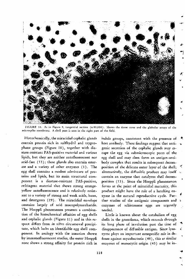
a'P- " i
4* * *,t -' .s 1 9
i # ' 'W '
,,Vx lf 55 2z i
1<
FIGURE 10. As in Figure 9, tangential section (x 80,000). Shows the dense cores and the globular arrays of themicrospike membrane. A shell pore is seen in the right part of the field.
Histochemically, the miracidial cephalic glandscontain protein rich in sulfhydril and trypto-phane groups (Figure 10), together with dia-stase-resistant PAS-positive material and variouslipids, but they are neither autofluorescent noracid-fast (53); these glands also contain ester-ase and a variety of other enzymes (1). Theegg shell contains a modest admixture of pro-teins and lipids, but its main structural com-ponent is a diastase-resistant PAS-positive,refringent material that shows strong orange-yellow autofluorescence and is relatively resist-ant to a variety of strong and weak acids, bases,and detergents (29). The miracidial envelopecontains largely of acid mucopolysaccharide.The Hoeppli phenomenon presents a combina-tion of the histochemical affinities of egg shellsand cephalic glands (Figure 11) and in this re-spect differs from in vitro circumoval precipi-tate, which lacks an identifiable egg shell com-ponent. In analogy with the zonation shownby immunofluorescent studies, the outer Hoepplizone shows a strong affinity for protein rich in
indole groups, consistent with the presence ofhost antibody. These findings suggest that anti-genic secretion of the cephalic glands may es-cape the egg via submicroscopic pores of theegg shell and may .then form an antigen-anti-body complex that results in subsequent decom-position of the delicate outer layer of the shell;alternatively, the diffusible product may itselfcontain an enzyme that catalyzes shell decom-position (53). Since the Hoeppli phenomenonforms at the point of miracidial maturity, thisproduct might have the role of a hatching en-zyme in the natural reproductive cycle. Fur-ther studies of the antigenic components andenzymes of schistosome eggs are urgentlyneeded.
Little is known about the catabolism of eggshells in the granuloma, which extends throughits long phase of involution past the time ofdisappearance of diffusible antigen. Since lyso-zyme plays an important nonspecific role in de-fense against mycobacteria (40), this or similarenzymes of monocytic origin (43) may be in-
118
'
4 %~~;w 4 *r rj& e*0 .16 w 1*
1~1 -4b
j i�� 11

FIGURE 11. Egg of S. mansoni in mastomys liver,stained by the dimethylamino-benzaldehyde nitrite(Adams, 1960) method for indole groups (x 420). Boththe cephalic glands and the early Hoeppli phenomenonseen along the left egg-shell border are strongly positive.
volved in schistosome egg-shell catabolism.Identification of the distinctive egg-shell mate-rial, likely to contain highly polymerized glu-cosamine or glycoprotein (55), could be a firststep into an interesting borderland of biochemi-cal immunology, since there is evidence thatgranuloma resolution can be accelerated by sen-sitization although egg shell material is not yetknown to be responsive to any physiologicallyactive mammalian enzymes.
, Immunological significance of pseudotubercles
From the aggregate evidence presented here,pseudotubercles appear to function as auxiliarysubunits of the lymphoreticular "establishment"in handling particulate pathogens from whichdiffusable antigen is gradually released, thus in-
7 ducing antigen sequestration in situ. Thesemacrophagic cell factories act as immunologicalreceptors and effectors and potentiate the abil-ity of the sensitized host to catabolize antigenand to break down residual inert matter during
granuloma involution. In this manner, granu-lomas are uniquely equipped to defend the hostagainst a variety of antigens produced by mi-croorganisms and haptenic chemical depositstoo large or too toxic to be handled by singlecell units.
Askonas and Humphrey have suggested thatantibody may be locally generated in adjuvant-antigen granulomas (3); since pseudotuberclesshow a gradient of antigen concentration thatdecreases toward their periphery, antibodymight be a function of lympho-plasmocyticcells that surround the phagocytic core of ma-ture granulomas. This attractive hypothesiswould partly account for the enhanced anti-body-generating potency of antigen-adjuvantmixtures and for the high antibody levels foundin schistosomiasis. However, in order to avoidmisunderstanding (12) it should be re-empha-sized here that antigen sequestration cannot to-tally prevent diffusible antigen from reachingand activating the entire lymphoreticular sys-tem; therefore, peusdotubercles act by supple-menting, rather than supplanting, the classicalimmunological responses to antigenic stimula-tion.
Summary
The immunopathology of schistosome pseu-dotubercles has been reviewed, with particularemphasis on antigen sequestration and on ac-celerated antigen destruction in sensitized hosts.The sequence of events in primary and second-ary pseudotubercle formation has been analyzedand correlated with currently available data onthe nature of schistosome egg antigens; themiracidial cephalic glands and the egg shell havebeen identified as major antigen sources. Theinterrelated cellular and humoral factors in hostsensitization have been explored, with particu-lar attention to precipitate formation and tocentral necrosis in pseudotubercles, and theHoeppli phenomenon has been identified as anin vivo antigen-antibody complex. The modi- "fications of granulomatous inflammation and itsconcomitant pathology in the course of naturalschistosomi infection have been summarized;
119

.nally, a brief evaluation of the immunologicrole of granulomas has been presented.':
Moderator: I have nothing to add to theexcellent report presented by Dr. von Lichten-berg except to endorse his view that in chronicnatural infection the host-parasite balance isthe result of antagonistic forces of both thehost and the parasite and not of unresponsive-ness on the part of either.
It has been speculated even today during oursession that well-adapted parasites are recog-nized by the host as "self," whereas pathogenicparasites are definitely regarded as "non-self"and therefore elicit an immune response. Itshould be borne in mind, however, that para-sites undergo antigenic changes within the hostas a result of antibody production, and thatsequestered antigens may therefore be releasedin the course of infection. The liberation offunctional antigens may lead to protection ofthe host, to immunity, but if the antigens re-leased are nonfunctional the coating of theparasite by the corresponding antibody mayvery well protect the parasite by a mechanismsimilar to the enhancement phenomenon intransplantation immunology. Thus the elimi-nation of worms could be interpreted as theresult of the predominance of the stimulus pro-vided by functional antigens over the enhance-ment effect induced by antibodies to nonfunc-tional antigens. Contrariwise, if enhancementovercomes immunity, tolerance will be estab-lished and the infective larvae will remain in adormant state.
This last mechanism may be operative inhelminthic infections terminated by self-cure,which have been observed in both natural andexperimental infections. As was pointed outby Dr. Weinstein, sheep in Great Britain sub-
* The tragic death, in 1965, of my collaborator andfriend Ramón Gómez Mazzei has been a permanent lossto science and to humanity. Many others have given aidand encouragement to this work, which now extends overmore than nine years, and as far as possible their sharehas been acknowledged in each personal article reviewedas a source. Whatever new understanding may haveemerged from these studies, let it be a tribute to thememory of Ramón and a source of satisfaction to all whohave so generously helped me in this labor.
ject to continuous infection with larvae ofHaemonchus contortus show an increased out-put of eggs every spring, and this spring rise isfollowed by self-cure and by a period of protec-tion over the rest of the year. It seems wellestablished that in this particular case self-cureis associated with the molting of dormant larvaeof the fourth or fifth stages and the concomi-tant liberation of potent functional antigenswith the excreted fluids.
I hope that Dr. Soulsby will comment lateron the possibility that the trigger mechanism ofself-cure in the Haemonchus infection may es-sentially be a switch from tolerance to immu-nity through the decoating of dormant larvaeand the formation of antibody to previouslymasked functional antigens.
In the case of schistosomiasis, self-cure mayalso be achieved, for instance, in laboratorymonkeys or in poor hosts such as white rats,guinea pigs and rabbits. In mice and in men,however, the infection usually leads to achronic state, in which the parasite is seques-tered and slowly destroyed in a pseudotubercle.The situation is complicated by the migrationof the parasite to ectopic areas, particularly inthe liver, causing widespread damage. It haseven been claimed that chemotherapy of schis-tosomiasis in heavily infected mice aggravatesthe disease as a result of the arrest of deadschistosomes and the development of obstruct-ing lesions, which leads to a distortion of theintrahepatic vascular system and to the so-called axial fibrosis as it is seen in human liverpreceding the coarse fibrosis (Symmer's clay-pipestem cirrhosis) characteristic of the laterstages of the disease.
There are indications, however, that thispotential danger of chemotherapy has beenexaggerated. Moreover, although lesions mayappear surrounding dead parasites, schistosomeor adult worms, the basic histopathologicaldamage in Schistosoma mansoni seems to be agranulomatous obliterating vasculitis produced
by embolized eggs. It is also believed that liv-
ing eggs in the tissues may be necessary for the
development of immunity in the mouse. If themiracidium is an important source of functional
120
-4
,4
1

antigen, the granulomatous reaction around theegg could be compared to immunization withantigen and Freund's adjuvant, and this couldlead either to the formation of antibodies or tothe development of delayed hypersensitivity.
Suggestive evidence has been presented in fa-vor of an immunological barrier to the penetra-tion of cercariae of S. mansoni in rats, whenapplied to skin sites previously infiltrated withreagin-like homologous PCA antibodies. How-ever, this barrier seems to be effective only inthe rat, which is endowed with a conspicuous
t degree of natural resistance. No protective ef-fect was obtained, so far as I know, under simi-lar experimental conditions with reagin-likeantibodies from superinfected monkeys whencercariae of both mansoni and japonicum wereapplied to skin treated sites.
These are only a few reflections I should liketo submit for your consideration in introduc-ing the discussion of the topic presented byDr. von Lichtenberg.
Dubos: When I spoke a little earlier I ap-parently gave some of the participants theimpression that I was downgrading the impor-tance of specific reactions in immunity andallergic processes. This was very far from mythought. In fact, it is because I wanted tomake it clear that I considered the specificphase of the immunological process fundamen-tal to all phenomena of immunity that I intro-duced my remarks with a statement that I wasgoing to speak only of the second part of Dr.Biro's scheme.
So let me just restate my personal views onthe mechanisms through which specific immu-nity processes are converted into reactions thatare effective in controlling infection. There isno doubt whatever in my mind that the firstand fundamental aspect of these processes isthe classical induction of immunological re-sponse, with all its specificity and occurring indifferent forms with which we are familiar, in-cluding the production of the classical anti-bodies and the production of the complex setof reactions that result in delayed hypersensi-tivity.
But it is also my conviction that after this
mechanism has been triggered into activity,there follows a set of secondary reactions thatare of immense and probably decisive impor-tance in determining the manifestations of thespecific immunological process.
Since this aspect of the nonspecific conse-quences of specific immunological processeshas not been well studied, I can at best give afew suggestions on how it might affect the fateof the infection. So please consider the two orthree (or perhaps four, if they come to mymind fast enough) mechanisms that I am goingto suggest not as exhausting the possibilitiesbut merely as illustrations.
To begin with, it has been demonstratedtime and time again that antigen-antibody re-action does result in the activation of severalenzymes, in particular of proteolytic enzymes.All of us are familiar with the classical demon-stration that cathepsins can attack certain para-sites, certain worms especially, and it mightwell be imagined that the activation of a pro-teolytic system through an antigen-antibodyreaction might create active enzyme mecha-nisms that can attack the parasite.
It has also been demonstrated countless timesthat an inflammatory reaction is accompaniedby a greater acidity of the site of the inflam-matory reaction through an exaggeration of theglycolytic processes. As you well know, meas-urements of local pH have shown that the pHat the site of the inflammatory reaction canfall to 4.5 to 5. There is very little doubt thatthis change of pH can affect certain parasitesdirectly, and perhaps indirectly, by providingan environment in which certain enzymes canact.
It has also been shown more recently thatlysosomes and all sorts of granules will becaused to disrupt and discharge their contentsas a result of contact between a certain sensi-tized cell and the antibody capable of reactingwith that cell. Here again the potentialities ofrelease of biologically active substances areenormous. You are probably familiar with therecent demonstration that probably all the lyso-zyme is stored in lysosomes and is released onlywhen the lysosome is destroyed. You may also
121
be familiar with the release of those small basic
peptides of various kinds that have been shown
to be stored in the lysosomes and to be released
and to adsorb nonspecifically on the surface of
many types of parasites.I believe I could spend the next fifteen min-
utes listing from memory some o: the biologi-
cally active substances that are either released
or rendered active by different forms of im-
munological reactions, but I wish only to con-
clude by summarizing in one phrase the over-
all point of view that I have tried to communi-
cate to you: All immune processes involve at
the beginning some highly specific reactions.
They are those of classical immunological spec-
ificity. But once this specific triggering of the
reaction has occurred, then there is a nonspe-
cific release of biologically active mechanisms
that certainly operate in all pathological proc-
esses.
Smithers: I should like to take up a coupleof the points made by the Moderator. First,
the suggestion that the egg and the granuloma
formation may be responsible for inducing the
protective response in schistosomiasis. After the
last couple of years' work in our laboratory, I
am absolutely convinced that the egg has noth-
ing at all to do with the development of pro-
tection against a challenge infection. The evi-dence for this comes from some worm transfer
experiments we have been doing-transferringadult worms from hosts into normal rhesus
monkeys.
Moderator: Excuse me, Dr. Smithers. Idid not mean to suggest that. I compared the
miracidium-releasing antigen in the living eggto an immunization with Freund's adjuvant,
but I did not mean that this was a protectingantibody. It could lead to delayed hyper-sensitivity or to antibody formation, but I did
not mean to say that it was protective.
Dr. Smithers: I am sorry; I must have mis-understood you. But to finish (because I believe
it is important that this point should be made
clear), protection is induced by transferring
male worms alone, or female worms alone, or
even worms that have been cut in half and are
still alive although they produce no eggs; but
protection cannot be induced by introducing
into the hepatic portal system of a normal mon-
key half a million living viable eggs. This has
no effect upon protection at all. Although, as
Dr. von Lichtenberg has shown, the egg is re-
sponsible for reducing the granuloma in sensi-
tized animals, it has no effect on the protection
against a challenge.The other very interesting point was the fact
that worms may become coated by antibody,
and in this way they may protect themselves
against the host's immune response. In our ex-
periments we have transferred worms from
hamsters and mice and monkeys into normal
monkeys. We find that the results are different,
depending on the donor animals used. If worms
are transferred from monkeys to monkeys, then
the transfer take is very good, and after a week
or so eggs begin to appear in the recipient, and
so on. If worms are transferred from hamsters
to monkeys, then the results are extremely poor.
Very few eggs are produced, and the worms are
quickly killed off. If worms are transferred
from mice into monkeys, you get an in-between
stage. The initial response-that is, the egg
production-is poor, but after five or six weeks
the egg production increases until a level is
reached that is comparable to that of monkey-
to-monkey transfers. We believe this shows
that the worms have become adapted in some
way to their definitive host; thus, worms from
hamsters are adapted to hamsters and cannot
make the necessary readaptation when they are
transferred to the monkey host. It could well
be that the worm has become coated with ham-
ster antibody; when we transfer this worm to
the monkey, the monkey does not recognize it
as a schistosome worm but recognizes it as ahamster, and consequently kills it off. However,
this is pure speculation.
Cohen: It does not seem appropriate for abiochemist to discourage people from recogniz-
ing the importance of the biology of a cell, be-
cause I have participated, as so many of you
have, in grinding things up and looking for
this or that. But I should like to mention an
observation-one I am not sure has been pub-
lished yet-that bears in some rather important
(1
y,
122 -r

respects on some of the discussions today. Thisobservation, reported by Dr. Jerry Gross of theMassachusetts General Hospital, has to do withthe disappearance of the tail of the tadpole un-der certain conditions. A rather rapid and ex-tensive dissolution of collagen, which exists inrather large quantities, is involved. As bio-chemists are inclined to do, Dr. Gross made avery serious effort to look for the enzyme col-lagenase in the system thus triggered. It seemsvery clear that in this system there is neither aproenzyme nor an inhibitor that normally re-
,Ty presses the enzyme. But apparently there is alayer of cells in the tail (and I should interjecthere that if the tail is cut off and put in aPetrie dish under sterile conditions with thy-roxine, the same process takes place as in thewhole animal) that is separated from the colla-gen fibers, and when certain changes occur andthese cells come into contact with certain seg-ments of the fibers, and only under these condi-tions, a specific collagenase is induced in thecells, and then the dissolution takes place.
This leads me to suggest that the absenceof an enzyme may be very important. This isnot a lysosome process; I do not quite shareyour enthusiasm about the lysosomes. I thinkit is another kind of process. But it seems to meto point to the fact that a cell, whether a para-sitic form or a host cell, may have potentialsbecause of the presence, if not the actual adher-ence, of specific proteins or other cells that leadto the induction of activities not found in theabsence of those particular circumstances.
I believe Dr. Gross showed that the effectwas puromycin-sensitive, and thus truly an in-duction. In any case, what this means is that,biochemically, the biology of the cell is a chal-lenge. Simply grinding up structures, as one ofthe earlier speakers said, shows nothing; onedoes not know for sure what it is one is extract-ing, nor, afterward, what one has extracted.Other dimensions may have a bearing. I think
this is an excellent example of a specific effectinduced to serve a particular role-in this case,to dissolve the collagen-and the enzyme is notpresent or not discernible under any circum-stances until a certain biological event occurs.
Lichtenberg: I want to ask Dr. Soulsby,since his findings have had such a vivid re-sponse here, for some additional comment aboutthe significance of what happens to the third-stage larvae specifically in ascariasis; whetherhe believes that something like a parasitical ef-fect could occur simply by obstructing thetransit of an organism that is in the process ofmigration and maintaining it at a stage that isnot the one corresponding to its subsequentmetabolic requirements. Is this part of his inter-pretation of the cell adherence phenomenon, andis it something that might happen in other hel-minthic diseases in general?
Soulsby: I should like to think that thesecells are so responsive that on a second infectionthey would behave as you suggest. I am notsure, though, that this is the case. In examiningan infection serially, it always seems that thecellular aspect of the response is a little behindthe progress of the parasitic infection. On sec-ond infection, it appears to catch up. I am notsure what delays the infection so that the cellscan catch up; it may be antibody. The sequencemay well be that antibody slows down an in-fection and then the cells have a chance tooperate.
Remington: I hesitate to raise another con-troversy here, and this is not necessarily for therecord. As I have sat here today I have won-dered about something that I think may also bein the minds of many of you. Are we sittinghere in a relative vacuum? Are we, each of us,traveling down different paths in our own re-search toward similar goals, and are all thesepaths going to end in different places? Cannotthere somehow be more of a cohesiveness-arecognized "core" for each problem and somesort of cooperative effort to answer the "core"questions? As a physician interested in clinicalinfectious diseases, I am interested in host re-sponse and immunity to infectious agents, andin the prevention of infections and the morbid-ity and mortality caused by them. The virolo-gists and bacteriologists now have a large arma-mentarium of vaccines and effective chemo-therapeutic agents with which to prevent, treat,
and/or study their infectious diseases. And
123

how stands the parasitologist in comparison?More infection and disease than can be claimedby either of the other groups, with relativelylittle in the way of advances to compare withtheirs. Is this because there is a lack of knowl-edge of how to approach parasitic diseases, alack of knowledge of the parasite and its host'sresponse, or is the lack also one of no commonpath or goal, or are these simply ill-defined? Isit the diffuseness of efforts that is causing thisscientific lag?
We have seen today that there seems noshortage in talent-veterinary medicine, pathol-ogy, immunology, parasitology. But there seemsto be a very real gap in the sharing of knowl-edge-not in willingness to share but perhapsin facilitation. I wonder what is going to bedone about this. How many more years are wegoing to remain in this relative void withoutsignificant advances toward answering age-oldproblems? Are we to be satisfied with advancesthat carry us only micrometers toward answer-ing a core question, or shall we seek ways thatmay help us leap by giant steps toward answer-ing questions that plague those concerned withthe health of the world? It may be that theanswer is greater cooperation among us all.
Moderator: I think that we are makingthat sort of effort here today. How far we willsucceed, I do not know. But we are making aneffort to put basic immunology and immuno-parasitology together.
Bruce-Chwatt: As a malariologist of longstanding, I feel much more optimistic than youseem to be. I feel that a very great deal hasbeen done with regard to the immunologicalapproach-not only toward an understandingof the process of infection and response to in-fection, but also toward coming to grips withthe problem of the still hypothetical malariavaccine. I feel that we understand today bet-ter than ever before the interrelation betweenthe humoral and the cellular aspects of immu-nity in malaria. We are still very far indeedfrom the end; for a long time we are going tohave to rely on chemotherapy of malaria.Nevertheless, I feel that we are much closer to
understanding and solving the problem ofmalaria.
Let me put one question here to my friendand colleague Dr. Brown, who has describedso well his work on the antigenic change inmalaria parasite in the course of infection. Doeshe believe that this undoubtedly importantphenomenon he has discovered will have an im-pact-negative or positive, probably the latter-on the development of the still hypotheticaland faraway malaria vaccine?
Brown: No, I do not believe it will affectthe ultimate development of the vaccine. Ithink that, unfortunately, antigenic variationcan be used to explain anything-especially anynegative result. If you do not happen to pro-tect by immunization one time, you say youhave used the wrong variant, or something ofthat sort. But in fact, to speak in particular ofFreund's original experiments, he kept hisstrains under quinine and they were obviouslyrelapsing continuously. Almost certainly thevariants he challenged with were different fromthe ones that he immunized with. I think thereis good reason to suppose that with suitableadjuvant it should be possible to immunizequite effectively with the blood forms.
Bruce-Chwatt: I forgot to say before thata great deal of what we know today about theimmunology of malaria is due to the tremen-dous efforts of Dr. Sadun and his group, and Ishould like here to pay tribute to his work.
Cohen: I hope that Dr. Remington will notthink I am directing this remark at him, butI think what we have seen today is an exampleof the fact that you cannot parlay two halftruths into a whole truth. Until we have wholetruths to deal with, we are going to be a longway from dealing as directly with the situationas he believes we must. I am not against get-ting together, but we need more information,and information substantial enough to meansomething, before we can make much of it.
Remington: In essence, I certainly agreewith Dr. Cohen. However, we are well awareof the government-funded chemotherapy pro-grams for malaria in the last world war and for
124
1
cf·
-.1
.i
-

cancer at present. Most of us in basic researchare loath to perform directed research or evento take contracts that dictate a complete pro-gram differing from our major interests. Thereis evidence in favor of laissez-faire for scien-tists and their research. But there is also evi-dence in favor of directed programs formulatedby experts in the field who consider that cer-tain experimental designs will lead to morerapid answers to questions vital to world health.I want to proceed in my work without inter-ference, and I hope that government grantingagencies will be tolerant of my approach andviews. Yet I also consider it very importantthat someone, somewhere, be able to ask the"core" questions on each of the maladies ofmankind and that there be institutions that willhelp guide research more directly to the answers,by a less devious route than is being taken inmany laboratories today. To put it in thevernacular, "someone must keep his eye on theball." The "ball" that is rolling here today isthe parasitic infestation of millions of people.Public health preventive measures, includingvaccines or specific drug therapy, must be
- found for each of these infectious diseases. Myanswer to the malariologists is that I sympa-thize with the splendid statement made byMartin Young several years ago in a lead articlein the Journal of Tropical Medicine and Hy-giene: "Medical progress must allow for theeradication of the disease before liquidating theinvestigator." In this country the malariologistwas "destroyed" before malaria itself was eradi-cated. Now, confronted with the problem ofmalaria in Vietnam, we have a resurgence ofmalaria research-because someone, somewhere,
t' has his "eye on the ball" and is making funds
available for research. I hope that we can do
the same in other fields of parasitology.Waterlow: As a Committee member who
knows virtually nothing about immunology, Ihave the impression at the end of today that,left to themselves, the body's defenses againstthese parasites are not very good and need sup-plementing-just as I think that with manyforms of bacterial disease most of us would take
antibiotics or sulfonimides and not rely entirely
on the antibody response. The impression I havereceived is that possibly the point to whichmost attention needs to be paid, from a practi-cal point of view, is the biochemistry of theparasites and the point at which they can behit hard with drugs without hitting the host.
Moderator: Gentlemen, I am afraid that wemust bring this to an end. Dr. Kagan has beenkind enough to take on the task of briefly sum-marizing today's discussions, since Dr. Oliver-González was unable to attend.
Kagan: Mr. Moderator, I feel like thematador who comes into the arena after thebull is dead. It has seemed to me, in thinkingabout what to say in summing up this veryinteresting day, that most of my ideas havealready been expressed within the past halfhour. I should like to point out, as a publichealth parasitologist, that the comments madeby Dr. Remington were uppermost in my mind.We have to remember that we are talking aboutdiseases that are of tremendous importance inthe world, and our ultimate aim is to providetools and measures for their control and possi-ble eradication.
We are never going to arrive at a stage atwhich we can control and perhaps eradicateparasitic diseases unless we have the wholetruths that Dr. Cohen alluded to. It is onlyby wedding basic research to applied researchthat we can reach that stage.
I was also impressed by Dr. Dubos' remarks.It occurred to me that fifty years ago peoplesaid in print that parasitic diseases did not in-voke an immune response. Dr. Taliaferro wasone of the early investigators who pointed outrepeatedly in his writings the similarity betweenthe immune response of microorganisms andthat of parasitic organisms. Today we havebeen reminded again that all the mechanismsthat hold true in immunologic problems re-lated to viruses and bacteria are also true ofparasitic organisms, and that some of the ad-vances made in those fields can be exploited inworking out the problems in parasitology.
We are relative beginners in the immunology
of parasitic infections in terms of working out
mechanisms. We are recovering from the flush
125

of some initial successes in this area to realizethat we cannot look for a primary mechanismin the immunologic response of parasites, thatthey are exceedingly complex, and although wemay not be able to lump immunologic mecha-nisms, we can lump concepts. The more westudy the response of the host to his parasite,the more complex it becomes. Dr. Smitherstalks about the schistosome egg not being theimmunologic stimulus in the monkey. Thismay be true for the monkey, but perhaps notfor another host. The recent work of Lichten-berg and Sadun in a detailed study of the im-munologic response to schistosomes in a varietyof hosts indicates the complexity of response onboth the cellular and the humoral level.
Another point I want to make is that beforewe arrive at "whole truths" we first have to"survey the territory" we are studying. Wehave a big field with many organisms, manyspecies. Characterizing and describing the im-munologic responses and developing assay meth-ods for their study have occupied parasitolo-gists for the past twenty to twenty-five years.It is only after we have such assay systems thatwe can begin to look for the immunologicmechanisms involved. When we have themethods we need, we can get down to somebasic immunology.
Before I make some specific comments aboutareas that remain to be studied, I should liketo revert to Dr. Cohen's remark that we oughtto characterize some of these antigens. Wehave not done so because it is difficult work,and until we have identified specific antigensand characterized their biological and immu-nological role, this aspect of research will lag.After this has been done, I believe, we can de-vote the energy necessary to characterize theantigens, identify their subunits, and perhapseven attempt to synthesize them and attachthem to artificial groups to make vaccines. Butwe are a long way from that, because we havenot done our homework in all these areas.
The hallmark of this meeting, I think, hasbeen complexity-complexity of antigenicstructure in malaria and other microorganismsoutlined by Dr. Brown, complexity in patho-
genic structure as outlined in my presentation.Dr. Goodman brought up the question of thecomplexity of immunoglobulin and alluded tothe complexity that goes beyond the IgM, IgA,and IgG level. We now have IgD and IgE.But even just in the IgG class we have at leastfour subgroups that have light chains. Weknow that the light chains have at least tensubgroups. We know we have kappa andlambda light chains. We thus have a wholehost of complex immunoglobulins and we haveno idea of their biological function. Not onlythat, we have no way of studying them ade-quately as they relate to parasitic diseases andinfections.
A tremendous challenge confronts us, there-fore-on one hand to elucidate the antigenicstructure on one side, and on the other to re-late these to the development of specific anti-bodies in the various subclasses by other im-munologists and by parasitic immunologists.
In conclusion, let me point out that we arenot going to find our answers in studies of thehumoral aspects .of parasitology, and we arenot going to find them in studies on the cellu-lar aspects and on delayed hypersensitivity. Weshall find the answer in a combination of thetwo and in the interaction of antigen-antibodycomplexes on these cellular reactions.
I believe that parasitic organisms use everyimmunologic trick that has been developed bya living organism. We know some of them.There are immunologic processes that we arejust beginning to become aware of. As weunderstand how they relate to other micro-organisms, we shall find that these processescan be applied to the study of parasitism ofhelminths and protozoa. In this lies the chal-lenge and the need for people with diversebackgrounds to apply their knowledge to thestudy of parasitic infection. Only by weddingbasic immunologic information to practicalgoals will we ever reach a point at which wecan diagnose parasitic infections with confi-dence, prepare vaccines for their eliminationand for host protection, and develop controlprograms in the world.
Moderator: I close this meeting with the
126
-
-f

feeling that we have indeed accomplished our of the Advisory Committee, I wish to expressobjective, which was to encourage new ap- our gratitude to each of the speakers and also toproaches to the problem of parasitic immunity Dr. Martins da Silva and to Dr. Louis Olivier,by utilizing concepts and methods of modern who are the persons chiefly responsible for thebasic immunology. On behalf of my colleagues organization and plan of this session.
127

Appendix
IMMUNOLOGIC ASPECTS OF PARASITIC INFECTIONS: SUMMARY'
Jose Oliver-González
Past accomplishments on immunologic as-pects of parasitic infections were very well re-viewed by a pioneer worker and authority inthis field, Dr. William H. Taliaferro. Amongthe most significant of these accomplishmentshave been the recognition of host reactionagainst parasites mediated through specificantibodies and cellular by-reactions, and of theimportant part played by lymphocytes-migrating to foci where parasites are present
and changing into macrophages-in immunereactions. The biochemistry of the parasitesand antigenic constituents have been alsoemphasized, mostly in connection with serologi-cal diagnosis. The electron microscope, isotopicmarkers, and autoradiography could be usedto great advantage. Electron microscope studieson the fine structure of parasitic tissue arerevealing important features of the secretionof antigens.
Brown and other investigators mentionedfundamental points in connection with thenature and variation of parasitic antigens. Theantigenic structure of parasites, particularlyprotozoa, changes during infection; with suchversatility, they become less vulnerable to hostimmune reactions. These changes, however,need to be correlated with changes in enzymecharacteristics and followed by tissue culturestudies.
The antigens responsible for immunizing thehost do not originate only in the parasite but
* Dr. Oliver González, who was unable to attend theSpecial Session, prepared this paper after reviewing thesummaries of the papers presented.
also consist of host-parasite complexes. Anti-bodies that develop against these host-parasitecomplexes may lead to phenomena related toautoimmune diseases.
A great deal of progress has been made inthe immunodiagnosis of parasitic infections,although more specific antigens are needed toincrease the efficiency of the diagnostic tests.Very few antigenic substances of establishedpurity have been isolated. As Kagan empha-sized, immunochemical analysis of the antigenicmosaic will lead to effective methods for theassay of purity.
High immunoglobulin levels are encounteredin parasitic infections. Hypergammaglobuline-mia occurs in schistosomiasis, malaria, try-panosomiasis, and Chagas' disease, at differenttimes releated to course of infection. An in-crease in immunoglobulin is not necessarily dueto an increase in specific antiparasite antibody.
Reagins have been demonstrated duringhelminth infections of man and other hosts.These are induced by the living organism and
not by dead worm materials. Their role inprotective immunity is now being uncovered.
Parasitologists are still faced with the prob-lems of understanding the role of antibodies inprotection against infection and of preparing
effective vaccines. Very little is known aboutthe characteristics of antibodies formed in re-sponse to parasitic infections; this knowledgeis necessary for additional light on the mecha-nisms of parasitism and host response. Newtechniques have been developed for character-izing these antibodies. By means of exclusion
128
ni
t:
-

chromatography on columns of cross-linkeddextran gels, the molecular weights of unknownproteins may be determined through correla-tion with the elution of proteins with knownmolecular weights. Demonstration of IgM and
IgG antibody during different stages of infec-tion has proved to be of diagnostic significancein certain parasitic infections.
The role of the lymphocyte and macrophagein immune reactions is again emphasized. Im-munity has been transferred by lymph nodes.The role played by lymphocytes in delayedhypersensitivity has been found to be of im-
portance in resistance. The role of themacrophage in immunity is not clear, since in
some infections parasites multiply within the
macrophage whereas in others intense phagocy-
tosis of the parasite is a manifestation of
acquired immunity.Weinstein summarized very effectively the
observed effects of immune response on para-sites. These effects, at a gross level, includesuch phenomena as retardation of movement,immobilization, and obliteration of orifices
and lumena. The effects at physiological andbiochemical levels are a decrease in infectivity,stunting, changes in uptake of dyes, and altera-
tions in metabolic processes.Lichtenberg used infection with Schistosoma
mansoni to illustrate host reactions against
parasites. The antigen proceeding from the
t schistosome egg is withheld or "sequestered"within the pseudotubercule. With sensitizationof the host, cell reaction and antigen destruc-
tion are greatly enhanced. The antigen-antibodyprecipitate has been shown in vivo around eggs
in animals highly sensitized (Hoeppli's phe-
nomenon).
CommentsAlthough the need for specific antigens is
very obvious, recent investigations indicate
that parasitic species, particularly nematodes,
contain common antigens that may be re-sponsible for a particular reaction or pathologi-
cal phenomenon within the host. A glycopro-tein has been isolated from the coelomic fluid
of adult Ascaris suum that has been detectedin the larvae of Trichinella spiralis, in third-
and fourth-stage larvae of Ascaris lumbricoides
and Toxocara canis, and in adult Dirofilariaimmitis (1). Sera from children with the
syndrome of visceral larva migrans reacted
positively against this antigen in direct and in-
direct hemagglutination tests. This suggeststhat the antigen responsible for the syndrome
is present in a large number of parasites.The presence of specific antigens in develop-
mental stages of parasites is still debatable. Some
investigtaors claim (2) that stage specificitymay be only apparent, since immunizing ex-
tracts from different stages may stimulatedemonstrably distinct antibodies by virtue ofthe differences in their relative proportions of
common antigens. Stage specificity may alsobe a function of the time at which serum hasbeen obtained. The presence or absence ofstage-specific antigens should be studied in re-lation to enzyme systems possessed by the vari-
ous forms, since enzymes probably representthese specific antigens.
Immunization against infection with anti-gen-antibody complexes, rather than with anti-
gen alone, has been overlooked. Antigen-anti-body precipitates have been shown to occur invivo; therefore, this complex must play a rolein host-parasite immune relationship.
Investigations on immunity to parasitic in-fections are burgeoning as a result of newtechniques, the advance of knowledge aboutantibody proteins, and newer methods for iso-lation of antigens.
129
REFERENCES+
WILLIAM H. TALIAFERRO: A Retrospective Look at the Immunologic Aspectsof Parasitic Infections
1. ADLER, S. Leishmania. Advanc. Parasit. 2:35-96, 1964. r2. AscHoFF, L. Das reticulo-endotheliale System. Ergeb. inn. Med. Kinderheilk.
26:1-118, 1924.3. ATTARDI, G., M. COHN, K. HORIBATA, and E. S. LENNOX. Antibody formation
by rabbit lymph node cells. J. Immunol. 92:335-390, 1964.4. BARNETT, S. F. Connective tissue reaction in acute fatal East Coast fever
(Theileria parva) of cattle. J. Inf. Dis. 107:253-282, 1960.5. BEALE, G. H., and J. F. WILKINSON. Antigenic variation in unicellular organ-
isms. Ann. Rev. Microbiol. 15:263-296, 1961.6. BLOOM, W. Mammalian lymph in tissue culture. From lymphocyte to fibro-
blast. Arch. exp. Zellforsch. 5:269-307, 1928.7. BLOOM, W., and W. H. TALIAFERRO. Regeneration of the malarial spleen in
the canary after infarction and after burning. 1. Inf. Dis. 63:54-70, 1938.8. BOYD, G. H. Experimental modification of the reproductive activity of Plas-
modium cathemerium. J, Exp. Zool. 54:111-126, 1929. "9. BRAND, T. v. Biochemistry of Parasites. New York, Academic Press, 1966.
10. BUCHSBAUM, R. and C. G. LOOSLI. Methods of Tissue Culture In Vitro andOutlines of Histological Methods. Chicago, University of Chicago Press,1936.
11. CAHILL, J. E., R. M. LEWERT, and B. N. JAROSLOW. Effect of hibernation oncourse of infection and immune response in Citelus tridecemlineatus infectedwith Nippostrogylus brasiliensis. 1. Parasit. 53:110-115, 1967. f
12. CAMPBELL, D. H., J. S. GARVEY, N. E. CREMER, and D. H. SUSSDORF. Methodsin Immunology. New York, W. A. Benjamin, 1963.
13. CANNING, G. A. Precipitin reactions with various tissues of Ascaris lumbri-coides and related helminths. Amer. J. Hyg. 9:207-226, 1929.
14. CANNON, P. R., and W. H. TALIAFERRO. Acquired immunity in avian malaria.
J. Prev. Med. 5:37-64, 1931.15. CHORDI, A., K. W. WALLS, and 1. G. KAGAN. Analysis of Toxoplasma gondii
antigens by agar diffusion methods. J. Immunol. 93:1034-1044, 1964.16. COCHRANE, C. G., and F. J. DIXON. Antibody production by transferred cells.
Advanc. Immunol. 2:205-239, 1962.17. COGGESHALL, L. T., and H. W. KUMM. Demonstration of passive immunity
to experimental monkey malaria. J. Exp. Med. 66:177-190, 1937.18. COHEN, S., and R. R. PORTER. Structure and biological activity of immuno-
globulins. Advanc. Immunol. 4:287-349, 1964.19. COONs, A. H. Some reactions of lymphoid tissues to stimulation by antigens.
Harvey Lectures, Ser. 53 (1957-1958):113-129, 1959.20. CULBERTSON, J. T. Recent contributions to the immunology of helminthic
infections. Arch. Pathol, 25:85-117 and 256-280, 1938.
130

21. CULBERTSON, J. T. Immunity against Animal Parasites. New York, ColumbiaUniversity Press, 1941.
22. D'ALESANDRO, P. A. Immunological and biochemical studies of ablastin, thereproduction-inhibiting antibody to Trypanosoma lewisi. Ann. N. Y. Acad.Sci. 129:834-852, 1966.
23. DAMIEN, R. T. Molecular mimicry: antigen sharing by parasite and host andits consequences. Amer. Naturalist 98:129-149, 1964.
24. DOMINICI, H. Sur le plan de structure du systéme hématopoiétique des mam-miferes. Arch. de méd. exp. et d'Anat. pathol, 13:473, 1901.
25. DOMINICI, H. Études sur le tissu conjonctif et les organes hématopoiétiquedes mammiferes. Arch d'Anat. microscop. 17:3-76, 1920-21.
26. DOWNEY, H. Reactions of blood and tissue cells to acid coloidal dyes underexperimental conditions. Anat. Rec. 12:429-454, 1917.
27. DOWNEY, H., and F. WEIDENREICH. Ueber die Bildung der Lymphozyten inLymphdrusen und Milz. Arch. f. mikr. Anat. 80:306-395, 1912.
28. DRAPER, L. R., and D. H. SUSSDORF. The serum hemolysin response in intactand splenectomized rabbits following immunization by various routes. J.Inf. Dis. 100:147-161, 1957.
29. EHRLICH, P. Chemotherapeutische Trypanosomen-Studien. Berl. klin. Wschr.44:233-236, 280-283, 310-314, 341-344, 1907.
30. EHRLICH, P. The Harden Lectures, 1907, London, 1908.31. FELDMAN, J. D. Ultrastructure of immunologic processes. Advanc. Immunol.
4:175-248, 1964.32. FUDENBERG, H. H. The immune globulins. Ann. Rev. Microbiol. 19:301-338,
1965.33. GARNHAM, P. C. C., A. E. Pierce, and I. Roitt. Immunity to Protozoa. Oxford,
Blackwell, 1963.34. GELL, P. G. H., and B. BENACERRAF. Delayed hypersensitivity to simple pro-
tein antigens. Advanc. Immunol. 1:319-343, 1961.35. GITLIN, D. Current aspects of the structure, function, and genetics of the im-
munoglobulins. Ann. Rev. Med. 17:1-22, 1966.36. GOBLE, F. C. Immunoreactions in antiparasitic chemotherapy. Advanc. Chemo-
therapy 1:355-395, 1964.37. GOBLE, F. C., and I. SINGER. The reticuloendothelial system in experimental
malaria and trypanosomiasis. Ann. N. Y. Acad. Sci. 88:149-171, 1960.38. GOWANS, J. L., and D. D. MCGREGOR. The immunological activities of lympho-
cytes. Progr. Allergy. 9:1-78, 1965.39. HEGNER, R. W. The biology of host-parasite relationships among protozoa
living in man. Quart. Rev. Biol. 1:393-418, 1926.40. HORTON-SMITH, C., and P. L. LONG. Coccidia and coccidiosis in the domestic
fowl and turkey. Advanc. Parasit. 1:67-107, 1963.41. HOWARD, J. G., G. H. CHRISTIE, J. L. BOAK, and E. EVANS-ANFOM. Evidence
for the conversion of lymphocytes into liver macrophages during graft-versus-host reaction. In La Greffe des Cellules Hématopoiétiques Allogén-iques. Paris, Centre Nat. de la Recherche Sc., 1965.
42. HOWARD, J. G., J. L. BOAK, and G. H. CHRISTIE. Further studies on the trans-formation of thoracic duct cells into liver macrophages. Ann. N. Y. Acad.Sci. 129:327-339, 1966.
43. HUFF, C. G., D. F. MARCHBANK, and T. SHIROISHI. Changes in infectiousnessof malarial gametocytes. II. Analysis of the possible causative factors. Exp.Parasit. 7:399-417, 1958.
44. HUMPHREY, J. H. Cell-mediated immunity. Brit. Med. Bull. 23:93-97, 1967.
131

45. JACOBs, L. Toxoplasma and toxoplasmosis. Ann. Rev. Microbiol. 17:428-450,1963.
46. JAROSLAW, B. N. The effect of X irradiation on immunity of the mouse toTrypanosoma duttoni. J. Inf. Dis. 96:242-249, 1955.
47. JAROSLOW, B. N. The effects of X or neutron irradiation, India ink blockade,or splenectomy on innate immunity against Trypanosoma duttoni in mice.J. Inf. Dis. 104:119-129, 1959. "
48. JAROSLOW, B. N., and D. E. SMITH. Effects of hibernation on the latent period
in normal and X-irradiated ground squirrels. J. Immunol. 93:649-655, 1964.49. JAROSLOW, B. N., and W. H. TALIAFERRO. The restoration of hemolysin-form- +
ing capacity in X-irradiated rabbits by tissue and yeast preparations. J.Inf. Dis. 98:75-81, 1956.
50. JAROSLOW, B. N., and W. H. TALIAFERRO. The effect of colchicine on the he-
molysin response in unirradiated and irradiated rabbits. J. Inf. Dis.116:139-150, 1966.
51. JORDAN, H. E. Comparative hematology. In H. Downey, Handbook of Hema-tology 2:703-863, see esp. 712. New York, Paul B. Hoeber, 1938.
52. KAGAN, I. G., and M. AGOSIN. Echinococcus antigens. Bull. Wld. Hlth. Org.1967 (in press).
53. KAGAN, I. G., and L. NORMAN. Analysis of helminth antigens by agar gel
methods. Ann. N. Y. Acad. Sci. 113:130-153, 1963.54. KIM, C. W. Delayed hypersensitivity to larval antigens of Trichinella spiralis.
J. Inf. Dis. 116:208-214, 1966.55. KIM, C. W. Delayed hypersensitivity to Trichinella spiralis antigens in ir-
radiated guinea pigs. J. Parasit. 52:722-726, 1966.56. KOLOUCH, F., JR. The lymphocyte in acute inflammation. Amer. J. Path.
15:413-428, 1939.57. LARSH, J. E., Jr. Experimental trichiniasis. Advanc. Parasit. 1:213-286, 1963.58. MAXIMOW, A. A. Experimentelle Untersuchungen iber entzundliche Neu-
bildung von Bindegewebe. Beitr. path. Anat. Suppl. 5, 1902.59. MAXIMOW, A. A. Relation of blood cells to connective tissues and endothelium.
Physiol. Rev. 4:533-563, 1924.60. MAXIMOW, A. A. Bindegewebe und blutbildende Gewebe. In W. v. Mollen-
dorff, Handb. d. mikrosk. Anat. d. Menschen 2:232-583, Berlin, JuliusSpringer, 1927. T
61. MAXIMow, A. A. Morphology of the mesenchymal reactions. Arch. Path. Lab.Med. 4:557-606, 1927.
62. MAXIMOW, A. A. Cultures of blood leucocytes. From lymphocyte and mono-cyte to connective tissues. Arch. exp. Zellforsch. 5:169-268, 1928.
63. METCHNIKOFF, E. Ueber die phagocytáre Rolle de Tuberkelriesenzellen. Vir-chows Arch. f. path. Anat. 113:63-94, 1888. r
64. MOULDER, J. W. The Biochemistry of Intracellular Parasitism. Chicago, Uni-versity of Chicago Press, 1962.
65. MOULDER, J. W. and W. H. TALIAFERRO. Reactions of the connective tissue inchickens to Plasmodium gallinaceum and Plasmodium lophurae. II. Glucosemetabolism during initial infections. J. Inf. Dis. 97:137-142, 1955.
66. NAIMAN, D. N. Effect of X irradiation on rats upon their resistance to Try-panosoma lewisis. J. Parasit. 30:209-228, 1944.
67. NOSSAL, G. J. V. Cellular genetics of immune responses. Advanc. Immunol.2:163-204, 1962.
68. NOSSAL, G. J. V., and O. MXKELX. Elaboration of antibodies by single cells.Ann. Rev. Microbiol. 16:53-74, 1962.
132 r

69. OVARY, Z. The structure of various immunoglobulins and their biologic activi-ties. Ann. N. Y. Acad. Sci. 129:776-786, 1966.
70. PIERCE, A. E., P. L. LONG, and C. HORTON-SMITH. Immunity to Eimeria te-
nella in young fowls (Gallus domesticas). Immunol. 5:129-152, 1962.71. PIzzI, T., Imunología de la enfermedad de Chagas. Santiago, Chile, Stanley,
1957.72. PIzzI, T., and W. H. TALIAFERRO. A comparative study of protein and nucleic
acid synthesis in different species of trypanosomes. J. Inf. Dis. 107:100-107,1960.
73. POYNTER, D. Parasitic bronchitis. Advanc. Parasit. 1:179-212, 1963.74. REBUCK, J. W. The functions of the white blood cells. Amer. J. Clin. Path.
17:614-630, 1947.75. REBUCK, J. W., and J. H. CROWLEY. A method of studying leucocytic func-
tions in vivo. Ann. N. Y. Acad. Sci. 59:757-805, 1955.
76. REBUCK, J. W., R. W. MONTO, E. A. MONAGHAN, and J. M. RIDDLe. 'Poten-
tialities of the lymphocyte, with an additional reference to its dysfunction
in Hodgkin's disease. Ann. N. Y. Acad. Sci. 73:8-38, 1958.
77. RITZ, H. Ueber Rezidive bei experimenteller Trypanosomiasis. Deutsch. med.
Wschr. 40:1355-1358, 191478. SADUN, E. H. Research in malaria. Military Med. 131(Suppl. 9):1-1272, 1966.
79. SEAMAN, G. R. and R. M. REIFEL. Chemical composition and metabolism of
protozoa. Ann. Rev. Microbiol. 17:451-472, 1963.80. SERGENT, Ed., and Et. SERGENT. Sur le paludisme des oiseaux di au Plas-
modium relictum (vel Proteosoma). Ann. Inst. Pasteur (Paris) 32:382-388,1918.
81. SINGER, I. The effect of X irradiation in infections with Plasmodium bergheiin the white mouse. J. Inf. Dis. 92:97-104, 1953.
82. SINGER, I. The cellular reactions to infections with Plasmodium berghei in the
white mouse. J. Inf. Dis. 94:241-261, 1954.
83. STAUBER, L. A. Factors influencing the asexual periodicity of avian malarias.
J. Parasit. 25:95-116, 1939.
84. STAUBER, L. A. Immunity to leishmania. Ann. N. Y. Acad. Sci. 113:409-417,
1963.85. STAVITSKY, A. B. In vitro studies of the antibody response. Advanc. Immunol.
1:211-261, 1961.
86. STELOS, P., L. G. TALIAFERRO, and P. D'ALESANDRO. Comparative study of
rabbit hemolysins to various antigens. J. Inf. Dis. 108:113-119, 1961.
87. STELOS, P., and W. H. TALIAFERRO. Comparative study of rabbit hemolysins
to various antigens. 2. Hemolysins to the Forssman antigen of guinea pigkidney, human type A red cells and sheep red cells. J. Inf. Dis. 104:105-118,1959.
88. SUSSDORF, D. H., and L. R. DRAPER. The primary hemolysin response in rab-
bits following shielding from X rays or X irradiation of the spleen, ap-
pendix, liver or hind legs. J. Inf. Dis. 99:129-142, 1956.
89. SUTER, E., and H. RAMSEIER. Cellular reactions in infection. Advanc. lmmunol.4:117-173, 1964.
90. Symposium on Resistance and Immunity in Parasitic Infections. The Rice Inst.Pamphlet 45:1-207, 1958.
91. SZENBERG, A., and K. SHORTMAN. Fractionation of fowl blood lymphocytes
by their buoyant density: localization of cells active in graft-versus-host re-actions. Ann. N. Y. Acad. Sci. 129:310-326, 1966.
133

92. TALIAFERRO, L. G. Return to normal of the asexual cycle in bird malaria after
retardation by low temperatures in vitro. J. Prev. Med. 2:525-540, 1928.93. TALIAFERRO, W. H. A reaction product in infections with Trypanosoma lewisi
which inhibits the reproduction of the trypanosomes. J. Exp. Med. 39:171-190, 1924.
94. TALIAFERRO, W. H. Host resistance and types of infections in trypanosomiasis
and malaria. Quart. Rev. Biol. 1:246-269, 1926.95. TALIAFERRO, W. H. The Immunology of Parasitic Infections. New York,
Century, 1929.96. TALIAFERRO, W. H. Trypanocidal and reproduction-inhibiting antibodies to
Trypanosoma lewisi in rats and rabbits. Amer. J. Hyg. 16:32-84, 1932.97. TALIAFERRO, W. H. Some cellular bases for immune reactions in parasitic
infections. J. Parasit. 20:149-161, 1934.98. TALIAFERRO, W. H. Ablastic and trypanocidal antibodies against Trypanosoma
duttoni. J. Immunol. 35:303-328, 1938.99. TALIAFERRO, W. H. The effects of splenectomy and blockade on the passive
transfer of antibodies against Trypanosoma lewisi. J. Inf. Dis. 62:98-111,1938.
100. TALIAFERRO, W. H. The mechanism of acquired immunity in infections with
parasitic worms. Physiol. Rev. 20:469-492, 1940.101. TALIAFERRO, W. H. The cellular basis for immunity in malaria. In Amer.
Assn. Adv. Sc. Publication 15:239-249 and 371-398, 1941.
102. TALIAFERRO, W. H. The immunology of the parasitic protozoa. In G. N.
Calkins and F. M. Summers, Protozoa in Biological Research. New York,Columbia University Press, 1941.
103. TALIAFERRO, W. H. Science in the universities. Science 108:145-148, 1948.
104. TALIAFERRO, W. H. The inhibition of reproduction of parasites by immune fac-tors. Bact. Rev. 12:1-17, 1948.
105. TALIAFERRO, W. H. Immunity to the malaria infections. In M. F. Boyd, ed.,
Malariology, II. Philadelphia, W. B. Saunders, 1949.106. TALIAFERRO, W. H. The cellular basis of immunity. Ann. Rev. Microbiol.
3:159-164, 1949.107. TALIAFERRO, W. H. President's message. Trop. Med. and Hyg. News 4:3-5,
1955.108. TALIAFERRO, W. H. Specificity in the relationship between host and animal
parasites. In E. G. Butler, Biological Specificity and Growth. Princeton, NewJersey, Princeton University Press, 1955.
109. TALIAFERRO, W. H. Functions of the spleen in immunity. Amer. J. Trop. Med.Hyg. 5:391-410, 1956.
110. TALIAFERRO, W. H. General introduction: synthesis and degradation of anti-
body. J. Cell. Comp. Physiol. 50 (Suppl. 1):1-26, 1957.
111. TALIAFERRO, W. H., and W. BLOOM. Inflammatory reactions in the skin of
normal and immune canaries and monkeys after the local injection of ma-
larial blood. J. Inf. Dis. 77:109-138, 1945.
112. TALIAFERRO, W. H., and P. R. CANNON. The cellular reactions during primaryinfections and superinfections of Plasmodium brasilianum in Panamanian
monkeys. J. Inf. Dis. 59:72-125, 1936.
113. TALIAFERRO, W. H., P. R. CANNON, and S. GOODLOE. The resistance of rats
to infection with Trypanosoma lewisi as affected by splenectomy. Amer. J.Hyg. 14:1-37, 1931.
114. TALIAFERRO, W. H., and C. KLivEBR, Tbe bematology of malaria (Plasmodiumbrasilianum) in Panamanian monkeys. 1. Numerical changes in leucocytes.
134 r
2. Morphology of leucocytes and origin of monocytes and macrophages. J.Inf. Dis. 67:121-176, 1940.
115. TALIAFERRO, W. H., and H. W. MULLIGAN. The histopathology of malariawith special reference to the function and origin of the macrophages in de-fence. Indian Med. Res. Memoir. 29:1-113. Calcutta, Thacker, Spink, 1937.
116. TALIAFERRO, W. H., and Y. PAVLINOVA. The course of infection of Trypano-soma duttoni in normal and in splenectomized and blockaded mice. J.Parasit. 22:29-41, 1936.
117. TALIAFERRO, W. H., and T. PIzzI. Connective tissue reactions in normal andimmunized mice to a reticulotropic strain of Trypanosoma cruzi. J. Inf.Dis. 96:199-226, 1955.
118. TALIAFERRO, W. H., and T. PIzzi. The inhibition of nucleic acid and proteinsynthesis in Trypanosoma lewisi by the antibody ablastin. Proc. Nat. Acad.Sci. 46:733-745, 1960.
119. TALIAFERRO, W. H., and L. G. TALIAFERRO. The resistance of different hoststo experimental trypanosome infections, with especial reference to a newmethod of measuring this resistance. Amer. 1. Hyg. 2:264-319, 1922.
120. TALIAFERRO, W. H., and L. G. TALIAFERRO. Reproduction-inhibiting and para-siticidal effects on Plasmodium gallinaceum and Plasmodium lophurae dur-ing initial infection and homologous superinfection in chickens. J. Inf. Dis.86:275-294, 1950.
121. TALIAFERRO, W. H., and L. G. TALIAFERRO. The dynamics of hemolysinformation in intact and splenectomized rabbits. 1J. Inf. Dis. 87:37-62, 1950.
122. TALIAFERRO, W. H., and L. G. TALIAFERRO. The role of the spleen in hemol-ysin production in rabbits receiving multiple antigen injections. J. Inf. Dis.
88:143-168, 1951.123. TALIAFERRO, W. H., and L. G. TALIAFERRO. The role of the spleen and the
dynamics of hemolysin production in homologous anamnesis. 1J. Inf. Dis.90:205-232, 1952.
124. TALIAFERRO, W. H., and L. G. TALIAFERRO. Effect of X rays on hemolysinformation following various immunization and irradiation procedures. J.Inf. Dis. 95:117-133, 1954.
125. TALIAFERRO, W. H., and L. G. TALIAFERRO. Further studies on the radiosensi-tive stages in hemolysin formation. J. Inf. Dis. 95:134-141, 1954.
126. TALIAFERRO, W. H., and L. G. TALIAFERRO. Reactions of the connective tissuein chickens to Plasmodium gallinaceum and Plasmodium lophurae. 1. His-topathology during initial infections and superinfections. J. Inf. Dis. 97:99-136, 1955.
127. TALIAFERRO, W. H., and L. G. TALIAFERRO. X-ray effects on hemolysin for-mation in rabbits with the spleen shielded or irradiated. J. Inf. Dis. 99:109-128, 1956.
128. TALIAFERRO, W. H., and L. G. TALIAFERRO. Amino acid incorporation intoprecipitin at different stages in the secondary response to bovine serumalbumin. J. Inf. Dis. 101:252-274, 1957.
129. TALIAFERRO, W. H., and L. G. TALIAFERRO. Immunologic unresponsivenessduring the initial and anamnestic Forssman hemolysin response. I. Re-peated injections of heated sheep red cell stromata into rabbits before and
after splenectomy. II. Spleen, bone marrow, lymph node and other tissuetransfers. J. Inf. Dis. 110:165-180, 1962.
130. TALIAFERRO, W. H., and L. G. TALIAFERRO. The effect of antigen dosage on
the Forssman hemolysin response in rabbits. J. Inf. Dis. 113:155-169, 1963.
135
131. TALIAFERRO, W. H., and L. G. TALIAFERRO. The relation of radiation desage 7to enhancement, depression and recovery of the initial Forssman hemolysinresponse in rabbits. J. Inf. Dis. 114:285-303, 1964.
132. TALIAFERRO, W. H., L. G. TALIAFERRO, and E. F. JANSSEN. The localization
of X-ray injury to the initial phases of antibody response. 1. Inf. Dis.91:105-124, 1952.
133. TALIAFERRO, W. H., L. G. TALIAFERRO, and B. N. JAROSLOW. Radiation and ,Immune Mechanisms. New York, Academic Press, 1964.
134. TALIAFERRO, W. H., L. G. TALIAFERRO, and A. PIzzI. Avidity and intercellu-lar transfer of hemolysin. J. Inf. Dis. 105: 197-221, 1959.
135. TALIAFERRO, W. H., L. G. TALIAFERRO, and E. L. SIMMONS. Increased para-sitemia in chicken malaria (Plasmodium gallinaceum and Plasmodiumlophurae) following X-irradiation. J. Inf. Dis. 77:158-176, 1945.
136. TALIAFERRO, W. H., and D. W. TALMAGE. Antibodies in the rabbit with dif- Wferent rates of metabolic decay. J. Inf. Dis. 99:21-33, 1956.
137. TROWELL, O. A. Lymphocytes. In E. N. Willmer, Cells and Tissues in Cul-ture, 2. London, Academic Press, 1965.
138. WOLSTENHOLME, G. E. W., and M. O'CONNOR, eds. Cellular Aspects of
Immunity. Ciba Found. Symp. 1960.139. YOUNG, R. L., H. WARD, D. HARTSHORN, and M. BLOCK. The relationship
between antibody formation and the appearance of plasma cells in newbornhamsters. J. Inf. Dis. 112:67-76, 1963.
140. ZUCKERMAN, A. The antigenic analysis of plasmodia. Amer. J. Trop. Med.13 (Suppl.):209-213, 1964.
141. ZUCKERMAN, A. Autoimmunization and other types of indirect damage tohost cells as factors in certain protozoan diseases. Exper. Parasit. 15:138-183, 1964.
K. N. BROWN: Nature and Variation of Parasite Antigens
1. ADLER, S. The action of specific serum on a strain of Trypanosoma cruzi. Ann.Trop. Med. Parasit. 52:282-301, 1958.
2. ADLER, S. Immune phenomena in Leishmaniasis. In P. C. C. Garnham, A. E.Pierce, and 1. Roitt, eds., Immunity to Protozoa. Oxford, Blackwell,1963:235-245.
3. ADLER, S., and J. ADLER. The agglutinogenic properties of various stages ofLeishmania. Bull. Res, Council Israel 4:396-7, 1955.
4. BROOM, J. C., and H. C. BROWN. Studies in trypanosomiasis. IV. Notes on theserological characters of Trypanosoma brucei after cyclical development inGlossina morsitans. Trans. R. Soc. Trop. Med. Hyg. 34:53-64, 1940.
5. BROWN, I. N., K. N. BROWN, and L. A. HILLS. Immunity to malaria: theantibody response to antigenic variation by Plasmodium knowlesi. Immunol. ,(in press).
6. BROWN. K. N., and 1. N. BROWN. Immunity to malaria: antigenic variation inchronic infections of Plasmodium knowlesi. Nature (Lond.) 208, 1286-1288,1965.
7. BROWN, K. N., and D. J. HOCKLEY. Immunity to protozoa: antigenic variationin trypanosomiasis and malaria. Parasitology 56:6P, 1966.
8. D'ALESANDRO, P. A. A serological comparison of the leishmaniform and lepto-monad stages of Leishmania donovani. M.S. thesis, Rutgers University, NewBrunswick, N.J., 1954.
9. DINEEN, J. K. Antigenic relationships between host and parasite. Nature(Lond.) 197:471-472, 1963.
r136

10. FELL, H. B., R. R. A. COOMBS, and J. T. DINGLE. The breakdown of embryonic(chick) cartilage and bone cultivated in the presence of complement suffi-cient antiserum. Int. Arch. Allergy 30:146-176, 1966.
11. FERNANDES, J. F., M. HALSMAN, and O. CASTELLANI. Effect of Mitomycin C,Actinomycin D, and pyrimidine analogs on the growth rate, protein andnucleic acid synthesis, and on the viability of Trypanosaoma cruzi. Exp.Parasit. 18:203-210, 1966.
12. FULTON, J. D., and E. M. LOURIE. The immunity of mice cured of trypano-
some infections. Ann. Trop. Med. Parasit. 40:1-9, 1946.13. GRAY, A. R. The antigenic relationship of strains of Trypanosoma brucei iso-
lated in Nigeria. J. Gen. Microbiol., 44:263-271, 1966.14. GRAY, A. R. Antigenic variation in clones of Trypanosoma brucei. II. The drug-
sensitivities of variants of a clone and the antigenic relationships of trypano-somes before and after drug treatment. Ann. Trop. Med. Parasit. 60:265-275, 1966.
15. HAWKING, F., M. J. WORMS, K. GAMMAGE, and P. A. GODDARD. The biologi-cal purpose of the blood-cycle of the malaria parasite Plasmodium cynomolgi.Lancet, August 20, 422-424, 1966.
16. KRASSNER, S. M. Cytochromes, lactic dehydrogenase and transformation inLeishmania. 1. Protozool. 13:286-290, 1966.
17. LEMMA, A., and E. L. SCHILLER. Extracellular cultivation of the leishmanoidbodies of species belonging to the protozoan genus Leishmania. Exp. Parasit.15:503-13, 1964.
18. LE PAGE, R. W. F. Studies on the variable antigens of Trypanosoma brucei.Trans. R. Soc. Trop. Med. Hyg. 61:139-140, 1967.
19. LONG, P. L., and A. E. PIERCE. Role of cellular factors in the mediation of im-munity to avian coccidions (Eimeria tenella). Nature (Lond.) 200:426-427,1963.
20. LUMSDEN, W. H. R., and G. T. C. HARDY. Nomenclature of living parasitematerial. Nature (Lond.) 250:1032, 1965.
21. MILGROM, F., Z. M. TUGGAC, and E. WITEBSKY. Organ specific antigens ofliver, testicle and pituitary. J. Immunol. 94:157-163, 1965.
22. OGILVIE, B. M. Reagin-like antibodies in rats infected with the nematode para-site Nippostrongylus brasiliensis. Immunol. 12:113-131, 1967.
23. POLGE, C., and M. A. SOLTYS. Preservation of trypanosomes in the frozen state.Trans. R. Soc. Trop. Med, Hyg. 51:519-526, 1957.
24. RICHARDS, W. H. G. Active immunization of chicks against Plasmodium galli-naceum by inactivated homologous sporozoites and erythrocytic parasites.Nature (Lond.) 212:1492-1494, 1966.
25. RITZ, H. Ueber Rezidire bei experimenteller Trypanosomiasis. II. Arch. F.Schriff u Trop. Hyg. 20:397-420, 1916.
26. RODET, A., and G. VALLET. Nagana experimental. Sur les variations du nom-bre des trypanosomes dans le sang du chien. Trypanolyse intravasculaire etpouvoir trypanolytique de serum. Compt. rend. Acad. Sci. 143:327-328,1906.
27. RYLEY, J. F. Studies on the metabolism of the protozoa. 9. Comparativemetabolism of blood stream and culture forms of Trypanosoma rhodesiense.Biochem. J. 85:211-223, 1962.
28. SEED, J. R. The characterization of antigens isolated from Trypanosoma rho-desiense. J. Protozool. 10:380-389, 1963.
29. SHORTT, H. E., and P. C. C. GARNHAM. The pre-erythrocytic development of
137

Plasmodium-cynomolgi and Plasmodium vivax. Trans. R. Soc. Trop. Med.Hyg. 41:785-795, 1948.
30. SIMPSON, C. F., J. M. KLING, and F. C. NEAL. The nature of bands in para-sitized bovine erythrocytes. J. Cell. Biol. 27:225-235, 1965.
31. SMITHERS, S. R. Gel-diffusion studies on Schistosoma mansoni. Trans. R. Soc.Trop. Med. Hyg. 54:8, 1960.
32. SMITHERS, S. R. Acquired resistance to Bilharziasis. In. Wolstenholme andO'Connor, eds., Ciba Foundation Symposium on Bilharziasis. London, J. &A. Churchill, 1962, 239-258.
33. SMITHERS, S. R. and R. J. TERRY. Resistance to experimental infections ofSchistosoma mansoni in rhesus monkeys induced by the transfer of adultworms. Trans. R. Soc. Trop. Med. Hyg. 1967 (in press).
34. SPRENT, J. F. A. Parasitism, immunity and evolution. In The Evolution ofLiving Organisms. Victoria, Australia, ,Melbourne University Press, 1959.
35. TREJOS, A., G. A. GODOY, C. GREENBLATT, and R. CEDILLUS. Effects of tem-perature on morphologic variation of Schizotrypanum cruzi in tissue culture.Exp. Parasit. 13:211-218, 1963.
36. VISCHER, W. A., and E. SUTER. Intracellular multiplication of Toxoplasmagondii in adult mammalian macrophages cultivated in vitro. Proc. Soc. Exp.Biol. N.Y. 86:413-419, 1954.
37. WERNER, H. Uber die formvariablitat von Toxoplasma gondii unter dem ein-fluss von antikorpen. Zbl. Bakt. (Orig). 189:497-510, 1963.
38. WILLIAMSON, J., and A. C. ALLISON. Thin-layer Sephadex chromatographyand immunodiffusion. Nature (Lond.) 1967 (in press).
39. WILLIAMSON, J., and K. N. BROWN. The chemical composition of trypano-somes. III. Antigenic constitutents of Brucei trypanosomes. Exp. Parasit.15:44-68, 1964.
40. WILLIAMSON, J., and B. COVER. Separation of blood-cell-free trypanosomesand malaria parasites on a sucrose gradient. Trans. R. Soc. Trop. Med.Hyg. 60:425-427, 1966.
IRVING G. KAGAN: Characterization of Parasite Antigens1. AGOSIN, M. Biroquímica de Echinococcus granulosus. Biológica (Santiago,
Chile) 27:3-32, 1959.2. AGOSIN, M., T. VON BRAND, G. F. RIVERS, and P. McMAHON. Studies on the
metabolism of Echinococcus granulosus. 1. General chemical compositionand respiratory reactions. Exp. Parasit. 6:37-51, 1957.
3. ALLISON, A. C., and J. H. HUMPHREY. Estimation of the size of antigens bygel diffusion methods. Nature (Lond.) 183:1590-1592, 1959.
4. ALLISON, A. C., and J. H. HUMPHREY. A theoretical and experimental analy-sis of double diffusion precipitin reactions in gels, and its application tocharacterization of antigen. Immunol. 3:95-106, 1960.
5. BABADZHANOV, S. N., and A. A. TUKHMANYANTS. Preparation and testingof Fasciola hepatica antigen. Uzbek. Biol. Zh. 5:27-33, 1958.
6. BAISDEN, L. A., and F. G. TROMBA. DEAE-Cellulose chromatography of kid-ney worm antigens. J. Parasit. 49:375-379, 1963.
7. BANKI, G., and A. BuccI. Research on an antigenic structure of Plasmodiumberghei. Parassitologia 6:251-257, 1964.
8. BANKI, G., and A. Bucci. Antigenic structure of Plasmodium cynomolgi andits relationships with the antigenic structure of Plasmodium berghei. Paras-sitologia 6:269-274, 1964.
138

9. BIGUET, J., A. CAPRON, P. TRAN VAN KY, and R. D'HAUSSY. Immunochimie-étude immunoélectrophorétique comparée des antigenes de divers helminthes.Acad. Sci. 254:3600-3602, 1962.
10. BIQUET, J., A. CAPRON, and P. TRAN VAN KY. Les antigenes de Schistosomamansoni. I. £tude électrophotétique et immunoélectrophorétique. Charac-térisation des antigénes spécifiques. Ann. Inst. Pasteur (Paris) 103:763-777,1962.
11. BIGUET, J., A. CAPRON, and P. TRAN VAN KY. Les antigénes de Fasciola hepa-tica. etude électrophorétique et immunoélectrophorétique. Identification desfractions et comparaison avec les antigénes correspondants a sept autreshelminthes. Ann. de Parasit. 39:221-231, 1962.
12. BIGUET, J., A. CAPRON, P. TRAN VAN KY, and F. RosÉ. Présence de sub-stances de Type C dans les antigénes vermineux et de protéine anti-C aucours des helminthiases humaines ou expérimentales. 1. Pltude immuno-logique preliminaire et répercussions pratiques. Rev. d'lmmunol. (Paris)29:233-240, 1965.
13. BIGUET, J., F. RosÉ, A. CAPRON, and P. TRAN VAN KY. Contribution del'analyse immunoélectrophorétique a la connaissance des antigénes vermi-neux. Incidences pratiques sur leur standardisation, leur purification et lediagnostic des helminthiasis par immuno-électrophorése. Rev. d'lmmunol.(Paris) 29:5-29, 1965.
14. BIGUET, J., P. TRAN VAN KY, Y. MOSCHETTO, and D. GNAMEY-KOFFY.Contribution a l'etude de la structure antigenique des larves de Trichinellaspiralis et des precipitines experimentales.du lapin. Wiad. Parazyt. 11:299-315, 1965.
15. BRAND, T. VON. Old and new observations on the chemical composition ofTrypanosoma cruzi. Rev. Inst. Med. Trop. Sao Paulo 4(2):53-60, 1962.
16. BROWN, K. N., and J. WILLIAMSON. The chemical composition of trypano-somes. IV. Location of antigens in subcellular.fractions of Trypanosomarhodesiense. Exp. Parasit. 15:69-86, 1964.
17. CAETANO DA SILVA, L., and R. GUIMARÁES FERRI. Immunodiffusion studiesin human schistosomiasis mansoni. I. Hepatointestinal and hepatosplenicforms. Rev. Inst. Med. Trop. Sáo Paulo 7(1):1-6, 1965.
18. CAETANO DA SILVA, L., and R. GUIMARÁES FERRI. Immunodiffusion studies inhuman schistosomiasis mansoni. II. Localization of antibodies by immuno-electrophoresis. Rev. Inst. Med. Trop. Sáo Paulo 7(1):7-10, 1965.
19. CAMPBELL, D. H. An antigenic polysaccharide fraction of Ascaris lumbri.coides (from hog). J. Inf. Dis. 59:266-280, 1936.
20. CAPRON, A., J. BIGUET, F. ROSÉ, and A. VERNES. Les antigénes de Schisto-soma mansoni. II. ttude immunoélectrophorétique comparée de diversstades larvaires et des adultes de deux sexes. Aspects immunologiques desrelations hote-parasite de la cercaire et de l'adulte de S. mansoni. Ann.Inst. Pasteur (Paris) 109:798-810, 1965.
21. CAPRON, A., G. RosÉ, G. LUFFAU, J. BIGUET, and F. RosÉ. Apport de ladistomatose expérimentale a la connaissance de la distomatose humaine aFasciola hepatica. Aspects immunologiques. Revue d'lmmunol. (Paris)29:25-42, 1965.
22. CAPRON, A., A. VERNES, J. BIGUET, F. RosÉ, A. CLAY, and L. ADENIS. Lesprécipitines sériques dans les Bilharzioses humaines et expérimentales aSchistosoma mansoni, S. haematobium and S. japonicum. Ann. de Parasit.(Paris). 41:123-187, 1966.
23. CHAVIN, S. I. Studies on the antigenic constituents of Plasmodium berghei.
139

I. Immunologic analysis of the parasite constituents. II. Fractionation ofthe parasite constituents. Milit. Med. 131:1124-1136, 1966.
24. CHORDI, A., and I. G. KAGAN. Identification and characterization of antigencomponents of sheep hydatid fluid by immunoelectrophoresis. J. Parasit.51:63-71, 1965.
25. CORRADETTI, A., F. VEROLINI, A. ILARDI, and A. BuccI. Immunoelectro-
phoretic analysis of water-soluble antigens extracted from parasitic bodies tof Plasmodium berghei separated from the blood. Bull. Wld. Hlth. Org.35:802-805, 1966.
26. DAMIAN, R. T. Molecular mimicry: Antigen sharing by parasite and host andits consequences. Amer. Naturalist 98:129-149, 1964.
27. DAMIAN, R. T. An immunodiffusion analysis of some antigens of Schistosomamansoni adults. Exp. Parasit. 18:255-265, 1966.
28. DAMIAN, R. T. Common antigens between adult Schistosoma mansoni andthe laboratory mouse. J. Parasit. 53:60-64, 1967.
29. DIGGS, C. L. Immunodiffusion studies of Plasmodium berghei: Interactions ofan extract of the erythrocyte forms with rabbit antisera. Exp. Parasit.19:237-248, 1966.
30. DINEEN, J¿ K. Immunological aspects of parasitism. Nature (Lond.) 197:268-269, 1963.
31. DINEEN, J. K. Antigenic relationship between host and parasite. Nature(Lond.) 197:471-472, 1963.
32. DODIN, A., RATOVONDRAHETY, J. P. MOREAU, and J. RICHAUD. Itude im-munologique de bilharziens traités par le CIBA 32644-Ba. Ann. 'Inst.Pasteur 109:35-44, 1965.
33. DUSANIC, D. G. Serologic and enzymatic investigations of Trichinella spiralis.I. Precipitin reactions and lactic dehydrogenase. Exp. Parasit. 19:310-319,1966.
34. DUSANIC, D. G., and R. M. LEWERT. Electrophoretic studies of the antigen-antibody complexes of Trichinella spiralis and Schistosoma mansoni. J.Inf. Dis. 116:270-284, 1966.
35. DYMOWSKA, Z., A. ZAKRZEWSKA, and J. ALEKSANDROWICZ. Antigens ofTrichinella spiralis. 1. Methods of preparation of antigenic fractions. ActaParas. Polonica 13:183-190, 1965.
36. FIFE, E. H., and J. F. KENT. Protein and carbohydrate complement fixing an-tigens of Trypanosoma cruzi. Amer. J. Trop. Med. Hyg. 9:512-517, 1960.
37. GARCíA, B. S. Antigenic components of Leishmania tropica. J. Philipp. Med.Assn. 41:647-652, 1965.
38. GAZZINELLI, G., F. J. RAMALHO-PINTO, J. PELLEGRINOj and J. M. P.MEMORIA. The intradermal test in the diagnosis of schistosomiasis man-soni. IX. Skin response to a purified fraction isolated from cercarial ex-tracts. J. Parasit. 51:753-756, 1965.
39. GHEDINI, G. Ricerche sul siero di sangue di individuo affecto da ciste deechinococco e sul liquido in essa contenuto. Gazz. Osped. Milano 27:1616-1617, 1906.
40. GOLDMAN, M., and W. A. SIDDIQUI. Antigenic comparison of two substrainsof Entamoeba histolytica by gel diffusion and immunoelectrophoresis. Exp.Parasit. 17:326-331, 1965.
41. GON^ALVES, J. M., and T. YAMAHA. Immune polysaccharide of Trypanosomacruzi. Congr. Inter. Doenfa Chagas, Rio de Janeiro 159-160, 1959.
42. GOODCHILD, C. G., and I. G. KAGAN. Comparison of proteins in hydatid fluidand serum by means of electrophoresis. J. Parasit. 47:175-180, 1961.
140

43. HACIG, A., P. -SOLOMON, and R. WEINBACH. Recherches serologiques surl'hymenolepidose l'étude d'un antigene d' Hymenolepis diminuta dans les
réactions antigene-anticorps "in vivo" et "in vitro". Arch. Roum. Path.
Exp. Microbiol. 18:611-625, 1959.44. HARIRI, M. N., C. W. SCHWABE, and M. KOUSSA. Host-parasite relationships
in echinococcosis. XI. The antigen of the indirect hemagglutination test forhydatid disease. Amer. J. Trop. Med. Hyg. 14:592-604, 1965.
45. HEIDELBERGER, M., A. C. AISENBERG, and W. Z. HASSID. Glycogen, an im-
munologically specific polysaccharide. J. Exp. Med. 99:343-353, 1954.
46. HUNTLEY, C. C., and A. MORELAND. Gel diffusion studies with Toxocara
and Ascaris extracts. Amer. J. Trop. Med. Hyg. 12:204-208, 1963.47. ISHII, Y., and S. MORISAWA. Intradermal test for paragonimiasis. Specificity
of skin test with purified peptides. Fukuoka Acta Medica 52:594-602, 1961.48. KAGAN, I. G. Serum agar double diffusion studies with Ascaris antigens.
J. Infect. Dis. 101:11-19, 1957.49. KAGAN, I. G. Seminar on immunity to parasitic helminths. VI. Hydatid disease.
Exp. Parasit. 13:57-71, 1963.50. KAGAN, I. G. Evaluation of routine serologic testing for parasitic diseases.
Amer. J. Public Health 55:1820-1829, 1965.51. KAGAN, I. G., and M. AGOSIN. Echinococcus antigens. Bull. Wld. Hlth. Org.
1967 (in press).52. KAGAN, I. G., and H. BIJAN. Immunologic and biologic studies with Leish-
mania species (unpublished).53. KAGAN, I. G., and L. NORMAN. Analysis of helminth antigens (Echinococcus
granulosus and Schistosoma mansoni). Ann. N. Y. Acad. Sci. 113:130-153,1963.
54. KAGAN, I. G., and L. NORMAN. The isolation and characterization of two hostantigens in hydatid fluid of Echinococcus granulosus. Amer. J. Trop. Med.Hyg. 12:346-347, 1963.
55. KAGAN, I. G., and C. G. GOODCHILD. Polysaccharide content of schistosome
skin test antigens with comparisons of reactivity of nitrogenous and carbo-
hydrate components. Amer. J. Trop. Med. Hyg. 12:179-183, 1963.56. KAGAN, I. G., E. L. JESKA, and C. J. GENTZKOW. Serum agar double diffusion
studies with Ascaris antigens. II. Assay of whole worms and tissue antigen
complexes. J. Immunol. 80:400-406, 1958.57. KAGAN, I. G., L. NORMAN, and D. S. ALLAIN. Studies on the serology of
Ascaris antigens (Abst.). Fedn. Proc. 18:576, 1959.58. KAGAN, I. G., L. NORMAN, D. S. ALLAIN, and C. G. GOODCHILD. Studies on
echinococcus: Nonspecific serologic reactions of hydatid fluid antigen withserum of patients ill with diseases other than echinococcosis. J. Immunol.84:635-640, 1960.
59. KENT, N. H. Biochemical aspects of specificity in cestodes. Proc. First Inter-
national Symposium on Parasitic Specificity, Neuchtael, Switzerland 293-307, 1958.
60. KENT, N. H. Isolation of specific antigens from Ascaris lumbricoides (var.suum). Exp. Parasit. 10:313-323, 1960.
61. KENT, N. H. Seminar on immunity to parasitic helminths. V. Antigens.Exp. Parasit. 13:45-56, 1963.
62. KENT, N. H. III. Fractionation isolation and definition of antigens fromparasitic helminths. Amer. J. Hyg. Monographic Series. 22:30-46, 1963.
63. KENT, N. H. Comparative immunochemistry of larval and adult forms ofSchistosoma mansoni. Ann. N. Y. Acad. Sci. 113:100-113, 1963.
141

64. KILEJIAN, A., K. SAUER, and C. W. SCHWABE. Host-parasite relationships inechinococcosis. VIII. Infrared spectra and chemical composition of thehydatid cyst. Exp. Parasit. 12:377-392, 1962.
65. KORACH, S. Isolation and properties of a soluble lipoprotein from Fasciolahepatica. Biochtm. et Biophys. Acta 125:335-351, 1966.
66. KORACH, S., and J. BÉNEX. A lipoprotein antigen in Fasciola hepatica.I. Isolation, physical and chemical data. Exp. Parasit. 19:193-198, 1966.
67. KORACH, S., and J. BÉNEX. A lipoprotein antigen in Fasciola hepatica. II.Immunological and immunochemical properties. Exp. Parasit. 19:199-205,1966.
68. KRONMAN, B. S. Immunochemistry of Schistosoma mansoni cercariae.J. Immunol. 95:13-18, 1965.
69. KRUPP, I. M. Immunoelectrophoretic analysis of several strains of Entamoebahistolytica. Amer. J. Trop. Med. Hyg. 15:849-85'4, 1966.
70. LABZOFFSKY, N. A., E. KUITUNEN, L. P. MORRISEY, and J. J. HAMVAS.Studies on the antigenic structure of Trichinella spiralis larvae. Can. J.Microbiol. 5:396-403, 1959.
71. LUDERITZ, O., A. M. STAUB, and O. WESTPHAL. Immunochemistry of O andR antigens of salmonella and related enterobacteriaceae. Bact. Rev.30:192-255, 1966.
72. LUPASCO, G., A. HACIG, P. SOLOMON, and L. IANCO. Recherches sur laconstitution et la spécificité des antigénes de Trichinella spiralis. Arch.Roum. Path. Exp. Microbiol. 23:877-882, 1964.
73. MAEKAWA, K., and M. KUSHIBE. Sur la composition chimique de l'antigénepour la dermo-réaction allergique vis-a-vis de Fasciola hepatica. C. R. Soc.Biol. (Paris) 4:832-834, 1956.
74. MAEKAWA, K., and M. KUSHIBE. Studies on allergen of Fasciola hepatica.Part III. Separation of allergenic substances (C 5 and P 4). Agr. Biol.Chem. 25:542-549, 1961.
75. MAEKAWA, K., and M. KUSHIBE. Studies on allergen of Fasciola hepatica.Part IV. Composition of allergen P 4. Agr. Biol. Chem. 25:550-552, 1961.
76. MAEKAWA, K., K. KITAZAWA, and M. KUSHIBE. Purification et cristallisationde l'antigéne pour la dermo-réaction allergique vis-a-vis de Fasciolahepatica. C. R. Soc. Biol. (Paris) 148:763-765, 1954.
77. MAGATH, T. B. The antigen of echinococcus. Amer. J. Clin. Path. 31:1-81,1959.
78. MAuss, E. A. Occurrence of Forssman heterogenetic antigen in the nematodeTrichinella spiralis. J. Immunol. 42:71-77, 1941.
79. MELCHER, L. R. An antigenic analysis of Trichinella spiralis. J. Inf. Dis.73:31-39, 1943.
80. MOORE, L. L. A. Studies in mice on the immunogenicity of cuticular antigensfrom larvae of Trichinella spiralis. J. Elisha Mitchell Scientific Society81:137-143, 1965.
81. NORMAN, L., and I. G. KAGAN. Preparation and evaluation of antigens foruse in the serologic diagnosis of human hydatid disease. I. Identificationand partial purification of the reactive elements in Echinococcus granulosusantigen prepared from sheep hydatid fluid. J. Immunol. 96:814-821, 1966.
82. NORMAN, L., I. G. KAGAN, and D. S. ALLAIN. Preparation and evaluation'of antigens for use in the serologic diagnosis of human hydatid disease.II. Isolation and characterization from extracts of ¿ysts of Echinococcusmultilocularis of serologically reactive elements found in hydatid fluid ofEchinococcus granulosus. J. Imrmunol. 96:822-828, 1966.
142
y

83. NUSSENZWEIG, V. Serodiagnosis of human and canine visceral leishmaniasis.Proc. 6th Int. Cong. Trop. Med. Mal. 3:779-790, 1958.
84. NUSSENZWEIG, V., and F. C. GOBLE. Further studies on the antigenic constitu-tion of strains of Trypanosoma (Schizotrypanum) cruzi. Exp. Parasit.18:224-230, 1966.
85. NUSSENZWEIG, V., L. M. DEANE, and J. KLOETZEL. Differences in antigenicconstitution of strains of Trypanosoma cruzi. Exp. Parasit. 14:221-232, 1963.
86. OLIVER-GONZÁLEZ, J. Functional antigens in helminths. J. Inf. Dis. 78:232-237, 1946.
87. OLIVER-GONZÁLEZ, J. Immunological properties of polysaccharides fromanimal parasites. Ann. Rev. Microbiol. 8:353-361, 1954.
'88. OLIVER-GONZÁLEZ, J., and M. V. TORREGROSA. A substance in animal para-sites related to the human isoagglutinogens. J. Inf. Dis. 74:173-177, 1944.
89. OLIVER-GONZÁLEZ, J., and N. H. KENT. Serological relationships between
collagenase and the A2 -isoagglutinogen-like substance of animal parasites.Proc. Soc. Exp. Biol. Med. 106:710-714, 1961.
90. PAULETE-VENRELL, J., W. T. CATICHA, SAAGLIA DE PAULETE, and N. MAT-TERA. Bioquímica de las diversas fracciones del líquido hidático. Arch.Int. Hyd. 21:190-198, 1964.
91. PAUTRIZEL, R., and C. SARREAN. Fractionnement de l'antigéne hydatique etintradermoréaction de Casoni. C. R. Soc. Biol. (Paris) 141:1061-1062,1947.
92. PELLEGRINO, J., E. PAULINI, J. M. P. MEMORIA, and D. G. MACEDO. Areacáo intradérmica na esquistossomose com uma franco polissacarideaisolada de cercárias de Schistosoma mansoni. Rev. Brasil. Malar. 8:527-534,1956.
93. RIEBER, S., R. I. ANDERSON, and M. G. RADKE. Serologic diagnosis ofSchistosoma mansoni infections. III. Isolation and purification of antigenfrom adult S. mansoni for the complement fixation text. Amer. J. Trop.Med. Hyg. 10:351-355, 1961.
94. SADUN, E. H., M. J. SCHOENBECHLER, and M. BENTZ. Multiple antibody re-sponse in Schistosoma mansoni infections: Antigenic constituents in eggs,cercariae, and adults (excretions and secretions) determined by floccula-tion reactions, cross absorption and double diffusion studies. Amer. J. Trop.Med. Hyg. 14:977-995, 1965.
95. SAWADA, T., and K. TAKEI. Immunological studies on filariasis. III. Isola-tion and purification of antigen for intradermal skin test. Japan J. Exp.Med. 35:125-132, 1965.
96. SAWADA, T., K. TAKEI, and K. YONEYAMA. Studies on the immunodiagnosisof paragonimiasis. II. Intradermal tests with fractionated antigens. J. Inf.Dis. 114:315-320, 1964.
97. SAWADA, T., K. TAKEI, and K. YONEYAMA. Studies on the immunodiagnosisof paragonimiasis. I. The precipitin reaction with crude and fractionatedantigens. J. Inf. Dis. 114:311-314, 1964.
98. SAWADA, T., K. TAKEI, J. E. WILLIAMS, and J. W. MOOSE. Isolation andpurification of antigen from adult Chlonorchis sinensis for complement fixa-tion and precipitin tests. Exp. Parasit. 17:340-349, 1965.
99. SAWADA, T., Y. NAGATA, K. TAKEI, and S. SATO: Studies on the substanceresponsible for the skin tests on clonorchiasis. Japan J. Exp. Med.34(6) :315-322, 1964.
100. SCHAD, G. A. Immunity, competition, and natural regulation of helminth pop-ulations. Am. Nat. 100:359-364, 1966.
143

101. SCHNEIDER, C. R., and M. HERTIG. Immunodiffusion reactions of Panamanian7Leishmania. Exp. Parasit. 18:25-34, 1966.
102. SCHNEIDER, M. D., M. G. RADKE, and M. T. COLEMAN. Immunologicallyreactive substance from Schistosoma mansoni. Exp. Parasit. 5:391-397,1956.
103. SHERMAN, I. W. Antigens of Plasmodium lophurae. J. Protozool, 11:409-417,1964.
104. SLEEMAN, H. K. Isolation and study of a specific complement fixing antigenfrom adult Schistosoma mansoni. Amer. J. Trop. Med. Hyg. 9:11-17, 1960.
105. SLEEMAN, H. K. Studies on complement fixing antigens isolated fromTrichinella spiralis larvae. II. Chemical analysis. Amer. J. Trop. Med. Hyg.10:834-838, 1961.
106. SLEEMAN, H. K., and L. H. MUSCHEL. Studies on complement fixing antigensisolated from Trichinella spiralis. , I. Isolation, purification and evaluationas diagnostic agents. Am. 1. Trop. Med. Hyg. 10:821-833, 1961.
107. SMITHERS, S. R., and J. WILLIAMSON. Antigenic polysaccharide material incercariae and eggs of Schistosoma mansoni. Trans. R. Soc. Trop. Med. Hyg.55:308-309, 1961.
108. SODEMAN, W. A., and J. H. E. T. MEUWISSEN. Disk electrophoresis ofPlasmodium berghei. J. Parasit. 52:23-25, 1966.
109. SOULSBY, E. J. L. Antigenic analysis of Ascaris tissues by the double diffusionprecipitin test. Trans. R. Soc. Trop. Med. Hyg. 51:9-10, 1957.
110. SOULSBY, E. J. L. Studies on the heterophile antibodies associated with hel-minth infections. III. Heterophile antibody in Oesophagostomum dentatum.1. Comp. Path. Therap. 68:380-387, 1958.
111. SOULSBY, E. J. L. Antigen-antibody reactions in helminth infections. Advanc.Immunol. 2:265-308, 1962.
112. SOULSBY, E. J. L. The nature and origin of the functional antigens in helminthinfections. Ann. N.Y. Acad. Sci. 113:492-509, 1963.
113. SOULSBY, E. J. L., and R. R. A. COOMBS. Studies on blood group substancesassociated with Ascaris lumbricoides. Parasit. 49:505-510, 1959.
114. SPIRA, D., and A. ZUCKERMAN. Antigenic structure of Plasmodium vinckei.Science 137:536-537, 1962.
115. SPIRA, D., and A. ZUCKERMAN. Recent advances in the antigenic analysisof plasmodia. Milit. Med. 131:1117-1123, 1966.
116. STAHL, W., J. OLIVER-GONZÁLEZ, and A. RIVERA DE SALA. Antibody response
to immunization witl Schistosoma mansoni egg antigen-antibody complex.Exp. Parasit. 13:204-210, 1963.
117. SZAFLARSKI, J., Z. DUDZIAK, Z. KAPP, and J. SZURMAN. Beitrag zur antigen-struktur von Fasciola hepatica. Int. Vet. Congr. (17th), Hanover I:787-789,1963.
118. TADA, I., and K. KAWASHIMA. Studies on the skin reaction in human filariasiswith a purified antigen from Dirofilaria immitis. Japan. J. Parasit. 13:427-434; 1964.
119. TANNER, C. E. Immunochemical study of the antigens of Trichinella spiralislarvae. II. Some physicochemical properties of these antigens. Exp.Parasit. 14:337-345, 1963a.
120. TANNER, C. E. Immunochemical study of the antigens of Trichinella spiralislarvae. III. Enzymatic degradation of the major precipitating antigen.Exp. Parasit. 14:346-357, 1963b.
121. TANNER, C. E., and J. GREGORY. Immunochemical study of the antigens of
144
Trichinella spiralis. 1. Identification and enumeration of antigens. Can. J.Microbiol. 7:473-481, 1961.
122. TARRANT, C. J., E. H. FIFE, and R. I. ANDERSON. Serological characteristicsand general chemical nature of the in vitro exoantigens of T. cruzi.J. Parasit. 51:277-285, 1965.
123. TORMO, J., and A. CHORDI. Immunoelectrophoretic analysis of Ascaris suumantigens. Nature (Lond.) 205:983-985, 1965.
124. WEINER, L. M., and S. PRicE. A study of antigenic relationships betweenTrichinella spiralis and Salmonella typhi. J. Immunol. 77:111-114, 1956.
125. WEINER, L. M., and J. NEELY. The nature of the antigenic relationship be-tween Trichinella spiralis and Salmonella typhi. J. Immunol. 92:908-911,1964.
126. WILLIAMSON, J., and K. N. BROWN. The chemical composition of try-panosomes. III. Antigenic constituents of Brucei trypanosomes. Exp.Parasit. 15:44-68, 1964.
127. WILLIAMSON, J., S. R. SMITHERS, and B. COVER. Analysis of the glycogen-
like antigens of Schistosoma mansoni eggs. Trans. R. Soc. Trop. Med. Hyg.59:368-369, 1965.
128. WITEBSKY, E., P. WELS, and A. HEIDE. Serodiagnosis of trichinosis by meansof complement fixation. N.Y. State J. Med. 42:431-435, 1942.
129. WITREMUNDO, TORREALBA, J., and J. CHAVES-TORREALBA. Empleo de
antígeno de B.C.G. en la reacción de fijación del complemento para eldiagnóstico de la leishmaniasis visceral. Rev. Inst. Med. Trop. Sáo Paulo6:252-253, 1964.
130. YAMAGUCHI, T. [Immunological studies on human gnathostomiasis. IV. Onthe antigenicity] (in Japanese, English summary). J. Kurume Med. Assoc.15:223-228, 1952.
131. YOGORE, M. G., R. M. LBWERT, and E., D. MADRASO. Immunodiffusion studieson paragonimiasis: Am. J. Trop. Med. Hyg. 14:586-591, 1965.
132. YOKOGAWA, M., and T. OSHIMA. Intradermal test for paragonimiasis. VI.Analysis of the antigenicity of the V.B.S. antigen with ammonium sulfateand cold methanol fractionation. Jap. J. Parasit. 8:44-49, 1959.
133. ZUCKERMAN, A. The antigenic analysis of plasmodia. Am. J. Trop. Med.Hyg. 13(Supp.):209-214, 1964.
S. R. SMITHERS: The Induction and Nature of Antibody Response to Parasites
1. ADLER, S. Immune phenomena in leishmaniasis. In Garnham, P. C. C., A. E.Pierce, and I. Roitt, eds., Immunity to Protozoa. Oxford, Blackwell, 1963.
2. ASKONAS, B. A., and J. H. HUMPHREY. Formation of antibody by isolated per-fused lungs of immunised rabbits. Biochem. J. 70:212-222, 1958.
3. BARTH, E. E. E., W. F. H. JARRETT, and G. M. URQHART. Studies on the mecha-nism of the self-cure reaction in rats infected with Nippostrongylus brasili-ensis. Immunol. 10:459-464, 1966.
4. BELFRAGE, S. Plasma protein pattern in the course of acute infectious disease.Acta Med. Scand. Supple. 395:169, 1963.
5. BLOCH, K. J. The anaphylactic antibodies of mammals including man. Progr.Allergy 10:84-150, 1967.
6. BOUR, H. M. MOMAN, and G. SCHAISON. Bilharziose urinaire a Schistosomahaematobium accompagnée d'une splenomegalie importante guerie par leseul traitement (A propos d'un cas). La Presse Medicale 70:2140-2142,1962.
145
7. BROWN, K. N., and I. N. BROWN. Immunity to malaria: antigenic variationin chronic infections of Plasmodium knowlesi. Nature (Lond.) 208:1286-1288, 1965.
8. CHANDLER, A. C. Studies on the nature of immunity to intestinal helminths. V.
Experiments on the role of the skin in parenteral immunity, and further ex-
periments on passive immunization in Nippostrongylus infection in rats.Am. J. Hyg., 26:292-308, 1937.
9. COGGESHALL, L. T., and H. W. KUMM. Demonstration of passive immunity in
experimental monkey malaria. 1. Exp. Med. 66:177-190, 1937.
10. COHEN, S., and I. A. McGREGOR. Gamma globulin and acquired immunity tomalaria. In Garnham, P. C. C., A. E. Pierce, and I. Roitt, eds., Immunity to
Protozoa: Oxford, Blackwell, 1963.
11. CRANDALL, C. C. Studies on the transfer of immunity to Ascaris suum in mice
by serum, parabiotic union and cells. J. Parasit. 51:405-408, 1965.
12. CULBERTSON, J. T. Passive transfer of immunity to Trichinella spiralis in the
rat. J. Parasit. 28:203-206, 1942.
13. CULBERTSON, J. T., and M. H. KOLODNY. Acquired immunity in rats against
Trypanosoma cruz¡. J. Parasit. 24:83-90, 1938.14. CURTAIN, C. C., C. KIDSON, D. L. CHAMPNESS, and J. G. GORMAN. Malaria
antibody content of gamma2 7 S globulin in tropical populations. Nature
(Lond.) 203:1366-67, 1964.
15. D'ALESANDRO, P. A. Electrophoretic and ultracentrifugal studies of antibodies
to Trypanosoma lewisi. J. Inf. Dis. 105:76-95, 1959.16. DENISON, N. Immunologic studies on experimental Trypanosoma cruzi infec-
tions: I. Lysins in blood of infected rats. Proc. Soc. Exp. Biol. Med. 52:26-27,
1943.17. DIAS, E. Le Trypanosoma cruzi et ses rapports avec le systéme reticulo-endo-
thelial. Compt. rend., Soc. Biol. 110:206-210, 1932.
18. DOUVRES, F. W. The in vitro cultivation of Oesophagostomum radiatum, the
nodular worm of cattle. II. The use of this technique to study immune re-sponses of host tissue extracts against the developing nematode. J. Parasit.
48:852-864, 1962.19. EDWARDS, A. J., V. E. JONES, S. R. SMITHERS, and R. J. TERRY. The occurrence
and properties of reagins in rhesus monkeys infected with Schistosoma
mansoni. Ann. Trop. Med. Parasit. (in press).20. EVANS, A. S., and M. A. STIREWALT. Serologic reactions in Schistosoma man-
soni infections. IV. Comparative ionographic study of sera of hamsters,
mice, and albino rats. Exp. Parasit. 7:165-177, 1958.
21. FIORILLO, A. M. Electroforese das proteínas do sangue na esquistossomose
mansónica forma hepatesplénica. Rev. Inst. Med. Trop. Sáo Paulo. 8:1-8,1966.
22. FREUND, J., K. J. THOMSON, H. E. SOMMER, A. W. WALTER, and T. M. PISANI.
Immunization of monkeys against malaria by means of killed parasites with
adjuvants. Am. J. Trop. Med. Hyg. 28:1-22, 1948.
23. GOBLE, F. C., and I. SINGER. The reticuloendothelial system in experimental
malaria and trypanosomiasis. Ann. N.Y. Acad. Sci. 88:149-171, 1960.
24. GRAY, A. R. The influence of antibody on serological variation in Trypanosoma
brucei. Ann. Trop. Med. Parasit. 56:4-13, 1962.25. HENDRICKS, J. R. Passive transfer of resistance to Trichinella spiralis in mice.
J. Parasit. 39(Sect. 2):40-41, 1953.
26. HOGARTH-SCOTT, R. S. Rabbit reagin-like antibodies. Int. Arch. Allergy
(in press).
146

27. HOUBA, V., and A. C. ALLISON. M-antiglobulins (rheumatoid-factor-like globu-lins) and other gamma globulins in relation to tropical parasitic infections.Lancet 1:848-852, 1966.
28. Hsi, H. F., and S. Y. LI Hsü. Reagin-like antibody in rhesus monkeysimmune to Schistosoma japonicum. Zeit. Tropenmed. Parasit. 17:166-176,1966.
29. Hsu, S. Y. LI, and H. F. Hsu. Serum protein changes in rhesus monkeys duringimmunization and following challenge by Schistosoma japonicum. Zeit.Tropenmed. Parasit. 15:43-56, 1964.
30. Hsu, S. Y. LI, and H. F. Hsi. Complement fixation reaction in rhesus monkeysinfected with Schistosoma japonicum. Zeit. Tropenmed. Parasit. 17:264-278,1966.
31. HUMPHREY, J. H. What are gamma globulins? Lectures on the Scientific Basisof Medicine VIII:87-106, 1958-59.
32. INGRAM, D. G., and M. A. SOLTYS. Immunity in trypanosomiasis. IV. Immuno-conglutinin in animals infected with Trypanosoma brucei. Parasit. 50:231-239, 1960.
33. JACHOWSKI, L. A., R. I. ANDERSON, and E. H. SADUN. Serologic reactions toSchistosoma mansoni. I. Quantitative studies on experimentally infectedmonkeys (Macaca mulatta). Am. J. Hyg. 77:137-145, 1963.
34. JACKSON, G. J. Fluorescent antibody studies of Trichinella spiralis infections.J. Inf. Dis. 105:97-117, 1959.
35. JACKSON, G. J. Fluorescent antibody studies of Nippostrongylus muris infec-tions. J. Inf. Dis. 106:20-36, 1960.
36. JAIMES, S., and F. VON LICHTENBERG. Host response to eggs of Schistosomamansoni. IV. Fluorescent antibody titers in mice infected with normal cer-cariae, gamma-irradiated cercariae and with purified eggs. Am. 1. Trop.Med. Hyg. 14:727-735, 1965.
37. JARRETT, W. F. H., F. W. JENNINGS, W. I. M. MCINTYRE, W. MULLIGAN andG. URQUHART. Immunological studies on Dictyocaulus viviparas infection.Passive immunization. Vet. Rec. 67:291-296, 1955.
38. JONAS, W. E., B. W. GURNER, D. S. NELSON, and R. R. A. CooMBs. Passivesensitization of tissue cells. I. Passive sensitization of macrophages by guinea-pig cytophilic antibody. Int. Arch. Allergy 28:86-104, 1965.
39. JONES, V. E., and B. M. OGILVIE. Reaginic antibodies and immunity to Nippo-strongylus braziliensis in the rat. II. Some properties of the antibodies andantigens. Immunol. 12:583-597, 1967.
40. KAGAN, I. G. Mechanisms of immunity in trematode infection. In E. J. L.Soulsby, ed., Biology of Parasites. New York, Academic Press, 1966.
41. KAGAN, I. G. and L. NORMAN. Passive transfer and pathogenicity. Experimentswith an American and a Chilean strain of Trypanosoma cruzi. J. Parasit.48(Sec. 2):31, 1962.
42. LEE, D. L. The structure and composition of the helminth cuticle. In B. Dawes,ed., Advances in Parasitology, 4. Academic Press. London and New York,1966.
43. LICHTENBERG, F. VON, J. H. SMITH, and A. W. CHEEVER. The Hoeppli phe-nomenon in schistosomiasis. Comparative pathology and immunopathology.Am. J. Trop. Med. Hyg. 15:886-895, 1966.
44. LUMSDEN, W. H. R. Estimation of serum IgM in the diagnosis of humantrypanosomiasis. Trans. R. Soc. Trop. Med. Hyg. 60:125, 1966.
45. MACKANESS, G. B. The relationship of delayed hypersensitivity to acquiredcellular resistance. Br. Med. Bull. 23:52-54, 1967.
46. MAGALHAES, A. F., I. M. KRUPP, and E. A. MALEK. Localization of antigen and
147

presence of antibody in tissues of mice infected with Schistosoma mansonias indicated by fluorescent antibody techniques. Am. J. Trop. Med. Hyg.14:84-99, 1965.
47. MATTERN, P., R. MASSEYEFF, R. MICHEL, and P. PERETTI. Etude immuno-
chimique de la 82 macroglobuline des serums de malades atteints de try-panosomiase Africaine a T. gambiense. Ann. Inst. Pasteur (Paris) 101:382-388, 1961.
48. MAUSS, E. A. The in vitro effect of immune serum upon Trichinella spiralislarvae. Am. J. Hyg. 32:80-83, 1940.
49. MILLER, T. A. Transfer of immunity to Ancylostoma caninum infection in pupsby serum and lymphoid cells. Immunol. 12:231-241, 1967.
50. NAIMARK, D. H., J. OLIVER-GONZÁLEZ, E. F. CHAFFEE, and R. I. ANDERSON.Studies on Schistosomiasis mansoni in primates. (1) Initial occurrence ofserologic antibodies correlated with egg recovery. J. Parasit. 43(Suppl.) :26,1957.
51. NELSON, D. S., and S. V. BOYDEN. Macrophage cytophilic antibodies and de-layed hypersensitivity. Br. Med. Bull. 23:15-20, 1967.
52. OGILVIE, B. M. Nippostrongylus brasiliensis (Travassos, 1914; Travassos andDarriba, 1929). A study of the life cycle and the immunological responseof the host. Ph.D. thesis, University of Cambridge, 1963.
53. OGILVIE, B. M. Reagin-like antibodies in animals immune to helminth parasites.Nature (Lond.) 204:91-92, 1964.
54. OGILVIE, B. M. Reagin-like antibodies in rats infected with the nematode para-site Nippostrongylus brasiliensis. Immunol. 12:113-131, 1967.
55. OGILVIE, B. M., S. R. SMITHERS, and R. J. TERRY. Reagin-like antibodies in ex-
perimental infections of Schistosoma mansoni and the passive transfer ofresistance. Nature (Lond.) 209:1221-1223, 1966.
56. OLIVER-GONZÁLEZ, J. The in vitro action of immune serum on the larvae andadults of Trichinella spiralis. J. Inf. Dis. 67:292-300, 1940.
57. OLIVER-GONZÁLEZ, J. Anti-egg precipitins in the serum of humans infectedwith Schistosoma mansoni. J. Inf. Dis. 95:86-91, 1954.
58. OLIVER-GONZÁLEZ, J., P. M. BAUMAN, and A. S. BENENSON. Immunologicalaspects of infections with Schistosoma mansoni. Am. J. Trop. Med. Hyg.4:443-452, 1955.
59. OTTO, G. F. The reaction between hookworm, Ancylostoma caninum larvaeand immune serum. J. Parasit. 25(Suppl.):29, 1939.
60. PIzzI, T. The immunology of Trypanosoma cruzi infections. Proc. XVI Int.Congr. Zoology 4:196-197, 1963.
61. RAMIREZ, E. A., A. RIVERA DE SALA, D. SERRANO, and M. CANCIO. Electro-
phoretic serum protein studies in chronic human Manson's schistosomiasis.Am. J. Trop. Med. Hyg. 10:530-536, 1961.
62. RICHARDS, W. H. G. Active immunization of chicks against Plasmodium galli-naceum by inactivated homologous sporozoites and erythrocytic parasites.Nature (Lond.) 212:1492-1494, 1966.
63. RIVERA DE SALA, A., R. MENÉNDEZ CORADA, and R. RODRIGUEZ MOLINA.
Detection of circumoval precipitins by the fluorescent antibody technique.Proc. Soc. Exper. Biol. Med. 111:212-215, 1962.
64. ROBINSON, D. L. H. The behaviour of young and adult Schistosoma mansoniin immune serum. Trans. R. Soc. Trop. Med. Hyg. 50:105-106, 1956.
65. SADUN, E. H. The antibody basis of immunity in chickens to Ascaridia galli.Am. J. Hyg. 49:101-116, 1949.
66. SADUN, E. H., F. VON LICHTENBERG, R. L. HICKMAN, J. I. BRUCE, J. II. SMITH,
148

and M. J. SCHOENBECHLER. Schistosomiasis mansoni in the chimpanzee:Parasitologic, clinical, serologic, pathologic and radiologic observations. Am.J. Trop. Med. Hyg. 15:496-506, 1966.
67. SADUN, E. H., M. J. SCHOENBECHLER, and M. BENTZ. Multiple antibody re-sponse in Schistosoma mansoni infections: antigenic constituents in eggs,cercariae and adults (excretions and secretions) determined by flocculationreactions, cross absorption and double diffusion studies. Am. J. Trop. Med.Hyg. 14:977-995, 1965.
68. SADUN, E. H., and B. C. WALTON. Studies on the host parasite relationships toSchistosoma japonicum. II. Quantitative changes in the concentration ofserum proteins in human and rabbits. Am. J. Trop. Med. Hyg. 7:500-504,1958.
69. SARLES, M. P. The in vitro action of immune serum on the nematode Nippo-strongylus muris. J. Inf. Dis. 62:337-348, 1938.
70. SARLES, M. P., and W. H. TALIAFERRO. The local points of defense and thepassive transfer of acquired immunity to Nippostrongylus muris in rats. J.Inf. Dis. 59: 207-220, 1936.
71. SEED, J. R. and A. A. GAM. The properties of antigens from Trypanosomagambiense. J. Parasit. 52:395-398, 1936.
72. SENECA, H., and P. PEER. Immunochemistry of Trypanosoma cruzi and im-munochemotherapy of experimental Chagas' disease. In J. C. Sylvester, ed.,Antimicrobial Agents and Chemotherapy. 1963, pp. 560-565.
73. SENFT, A. W., D. E. PHILPOTT, and A. H. PELOFSKY. Electron microscopeobservations of the integument, flame cells and gut of Schistosoma mansoni.J. Parasit. 47:217-229, 1961.
74. SILVERMAN, P. H., and J. E. PATTERSON. Histotrophic (parasitic) stages ofHaemonchus contortus. Nature (Lond.) 185:54-55, 1960.
75. SMITHERS, S. R. Gel-diffusion studies on Schistosoma mansoni. Trans. R. Soc.Trop. Med. Hyg. 54:8, 1960.
76. SMITHERS, S. R. Stimulation of acquired resistance to Schistosoma mansoni inmonkeys: role of eggs and worms. Exp. Parasit, 12:263-273, 1962.
77. SMITHERS, S. R., and R. J. TERRY. Changes in the serum proteins and leucocyte
counts of rhesus monkeys in the early stages of infection with Trypanosomagambiense. Trans. R. Soc. Trop. Med. Hyg. 53:336-345, 1959.
78. SMITHERS, S. R., and R. J. TERRY. Resistance to experimental infections ofSchistosoma mansoni in rhesus monkeys induced by the transfer of adultworms. Trans. R. Soc. Trop. Med. Hyg. 61, 1967 (in press).
79. SMITHERS, S. R., and P. J. WALKER. Serum protein changes in monkeys in-
fected with Schistosoma mansoni, with special reference to the metabolismof albumin. Exp. Parasit. 11:39-49, 1961.
80. STIREWALT, M. A. Seminar on immunity to parasitic helminths. IV. Schisto-some infections. Exp. Parasit. 13:18-44, 1963.
81. STIREWALT, M. A. and A. S. EVANS. Serologic reactions in Schistosoma mansoniinfections. I. Cercaricidal, precipitation, agglutination, and CHR phenomena.Exp. Parasit. 4:123-142, 1955.
82. TAFFS, L. F. The passive transfer of immunity in guinea pigs infected with thenematode Ascaris suum Goeze, 1782. J. Helm. 35:215-318, 1961.
83. TALIAFERRO, W. H. Immunity to malaria infections. In M. F. Boyd, ed.,Malariology. II. Philadelphia, W. B. Saunders, 1949.
84. TALIAFERRO, W. H., and P. R. CANNON. The cellular reactions during primaryinfections and superinfections of Plasmodium brasilianum in Panamanianmonkeys. ]. Inf. Dis. 59:72-125, 1936.
149
85. TALIAFERRO, W. H., and M. P. SARLES. The cellular reactions in the skin,lungs and intestine of normal and immune rats after infection with Nippo-strongylus muris. J. Inf. Dis. 64:157-192, 1939.
86. TARGETT, G. A. T., and A. VOLLER. Studies on antibody levels during vaccina-tion of rhesus monkeys against Plasmodium knowlesi. B. M. J. 2:1104-1106,1965.
87. TOBIE, J. E., D. C. ABELE, G. J. HILL, P. G. CONTACOS, and C. B. EVANS.Fluorescent antibody studies on the immune response in sporozoite-inducedand blood-induced vivax malaria and the relationship of antibody productionto parasitaemia. Am. J. Trop. Med. Hyg. 15:676-683, 1966.
88. TOBIE, J. E., D. C. ABELE, S. M. WOLFF, P. G. CONTACOS, and C. B. EVANS.Serum immunoglobulin levels in human malaria and their relationship toantibody production. J. Immunol. 97:498-505, 1966.
89. TOMODA, I. Paper electrophoretic studies on the serum protein in domesticanimals. III. Serum protein changes in schistosomiasis. Jap. J. Vet Sci.25:67-91, 1963.
90. THORSON, R. E. Absorption of protective antibodies from serum of rats im-mune to the nematode Nippostrongylus mutris. J. Parasit. 40:300-303, 1954.
91. VOGEL, H., and W. MINNING. Hullenbildung bei Bilharzia-cercarien im serumBilharzia-infizieter Tiere und Menschen. Centralbl.f.Bakteriol. 153:91-105,1949.
92. VOGEL, H., and W. MINNING. Uber die erworbene Resistenz von Macacusrhesus gegenuber Schistosoma japonicum. Zeit. Tropenmed. Parasit. 4:418-505, 1953.
93. VOLLER, A., P. D. MARSDEN, and D. S. RIDLEY. Infection with a Peru strainof Trypanosoma cruzi in a rhesus monkey. Trans. R. Soc. Trop. Med. Hyg.61 (in press), 1967.
JACK S. REMINGTON: Characterization of Antibodies to Parasites1. ABELE, D. C., J. E. TOBIE, G. J. HILL, P. G. CONTACOS, and C. B. EVANS.
Alterations in serum proteins and 19S antibody production during the courseof induced malarial infections in man. Amer. J. Trop. Med. Hyg. 14:191-197,1965.
2. COHEN, S., I. A. McGREGOR, and S. CARRINGTON. Gamma-globulin and ac-quired immunity to human malaria. Nature 192:733-737, 1961.
3. REMINGTON, J. S., and M. J. MILLER. 19S and 7S anti-toxoplasma antibodiesin the diagnosis of acute congenital and acquired toxoplasmosis. Proc. Soc.Exp. Biol. Med. 121:357-363, 1966.
4. REMINGTON, J. S., M. J. MILLER, and I. BROWNLEE. IgM antibodies in acuteacquired congenital toxoplasmosis: Diagnostic significance and a method fortheir rapid demonstration. (Submitted for publication)
5. SCHULTZE, H. E., and J. S. HEREMANS. Molecular Biology of Human Proteins,Nature and Metabolism of Extracellular Proteins, Vol. 1. New York, Amer-ican Elsevier, 1966.
6. VAN FURTH, R., H. R. SCHUIT, and W. HYMANS. The immunological develop-ment of the human fetus. J. Exp. Med. 122:1173-1188, 1965.
7. MILLER, M. J., E. SEAMAN, and J. S. REMINGTON. The clinical spectrum ofcongenital toxoplasmosis: Problems in recognition. J. Pediat. 70:714-723,1967.
8. CRANDALL, R. B., J. J. CEBRA, and C. A. CRANDALL. The relative proportionsof IgG-, IgA-, and IgM-containing cells in rabbit tissues during experi-mental trichinosis. Immunol. 12:147-158, 1967.
150

9. KABAT, E. A., and M. M. MAYER. Experimental Immunochemistry. Springfield,Illinois, Charles C Thomas, 1961.
10. CAMPBELL, D. H., J. S. GARVEY, N. E. CREMER, and D. H. SUSSDORF. Methodsin Immunology. New York, W. A. Benjamin, 1964.
11. FLODIN, P. Dextran Gels and Their Applications in Gel Filtration. Uppsala,Sweden, Pharmacia, 1962.
12. Description of methods and extensive bibliography are available from Phar-macia Fine Chemicals, Inc., Uppsala,. Sweden, or 800 Centennial Avenue,Piscataway, New Jersey.
13. REMINGTON, J. S., and E. MERLER. Absence of cross reactivity betweenheterophile and dye test antibodies. Proc. Soc, Exp. Biol. Med. 115:115-118,1964.
14. FRANKLIN, E. C. The immune globulins--their structure and function andsome techniques for their isolation. Progr. Allergy 8:58-148, 1964.
15. MERLER, E., J. S. REMINGTON, M. FINLAND, and D. GITLIN. Characterizationof antibodies in normal human urine. 1J. Clin. Invest. 42:1340-1352, 1963.
16. FAHEY, J. L., E. C. FRANKLIN, R. S. NEZLIN, and T. WEBB. Antibody purifica-tion on insoluble adsorbents and purification and characterization of im-munoglobulin A. Bull. Wld. Hlth. Org. 35:779-781, 1966.
17. ALTEMEIER, W. A., J. B. ROBBINS, and R. T. SMITH. Quantitative studies ofthe immunoglobulin sequence in the response of the rabbit to a somaticantigen. J. Exp. Med. 124:443-460, 1966.
18. BAUER, D. C., M. J. MATHIES, and A. B. STAVITSKY. Sequences of synthesisof y-1 macroglobulin and y-2 globulin antibodies during primary and sec-ondary responses to proteins, salmonella antigens, and phage. J. Exp. Med.117:889-907, 1963.
19. LEACH, A. A., and P. C. O'SHEA. The determination of protein molecularweights of up to 225,000 by gel-filtration on a single column of SephadexG-200 at 250 and 40 ° . J. Chromatogr. 17:245-251, 1965.
20. PAGE, C. O., and J. S. REMINGTON. Immunologic studies in normal humansweat. J. Lab. Clin. Med. 69:634-650, 1967.
21. REMINGTON, J. S., K. L. VOSTI, A. LIETZE, and A. L. ZIMMERMAN. Serumproteins and antibody activity in human nasal secretions. 1. Clin. Inves.43:1613-1623, 1964.
22. TOMASI, T. B., E. M. TAN, A. SOLOMON, and R. A. PRENDERGAST. Characteris-tics of an immune system common to certain external secretions. ]. Exp.Med. 121:101-124, 1965.
23. HEREMANS, J. F., P. A. CRABBE, and P. L. MASSON. Biological significance ofexocrine gamma-A-immunoglobulin. Acta Med. Scand. 179:(Suppl.)445:84-88, 1966.
24. WALTON, B. C., B. M. BENCHOFF, and W. H. BROOKS. Comparison of theindirect fluorescent antibody test and methylene blue dye test for detectionof antibodies to Toxoplasma gondii. Amer. 1. Trop. Med, Hyg. 15:149-152,1966.
25. FULTON, J. D., an& D. F. SPOONER. Preliminary observations on the metabo-lism of Toxoplasma gondii. Trans. R. Soc. Trop. Med. Hyg. 51:123-124,1957.
26. GOLDMAN, M. Staining Toxoplasma gondii with fluorescein-labelled antibody.II. A new serologic test for antibodies to toxoplasma based upon inhibitionof specific staining. ]. Exp. Med. 105:557-573, 1957.
151

HOWARD C. GOODMAN: Discussion
1. ARBESMAN, C. E., P. GERARD, and N. R. ROSE. Demonstration of humanreagin in the monkey. II. In vitro sensitization of monkey ileum withsera of untreated atopic patients. J. Allergy 35:520, 1964.
2. BARTH, W. F., and J. L. FAHEY. Heterologous and homologous skin-sensitizingactivity of mouse 7Syl and 7Sy2 globulins. Nature (Lond.) 206:730, 1965.
3. BENACERRAF, B., Z. OVARY, K. J. BLOCH, and E. C. FRANKLIN. Properties ofguinea-pig 7S antibodies. 1. Electrophoretic separation of two types ofguinea-pig 7S antibodies. J. Exper. Med. 117:937, 1963.
4. BINAGHI, R. A., B. BENACERRAF, K. BLOCH, and F. KOURILSKY. Properties ofrat anaphylactic antibody. J. Immunol. 92:927, 1964.
5. BLOCH, K. The anaphylactic antibodies of mammals, including man. Progressin Allergy 10, 1966.
6. ISHIZAKA, K., T. ISHIZAKA, and M. HORNBECK. Physico-chemical propertiesof human reaginic antibody. IV. Presence of a unique immunoglobulin asa carrier of reaginic activity. J. Immunol. 97:75, 1966.
7. MOTA, I. The'mechanism of anaphylaxis. I. Production and biological proper-ties of mast cell sensitizing antibody. Immunol. 7:681, 1964.
8. MOTA, I. Biological characterization of mouse "early" antibodies. Immunol.12:343, 1967.
9. Nomenclature for human immunoglobulins. Bull. Wld. Hlth. Org. 30:447-450, 1964.
10. Notation for genetic factors of human immunoglobulins. Bull. Wld. Hlth.Org. 33:721-724, 1965.
11. Notation for human immunoglobulin subclasses. Bull. Wld. Hlth. Org.35:953, 1966.
12. NUSSENZWEIG, R. S., C. MERRYMAN, and B. BENACERRAF. Electrophoreticseparation and properties of mouse antihapten antibodies involved in passivecutaneous anaphylaxis and passive hemolysis. J. Exp. Med. 120:315, 1964.
13. ONOUE, K., N. TANIGAKI, Y. YAGI, and D. PRESSMAN. IgM and IgG anti-hapten antibody: Hemolytic hemagglutinating and precipitating activity.Proc. Soc. Exp. Biol. Med. 120:340, 1965.
14. PATTERSON, R., J. PRUZANZKY, and W. CHANG. Spontaneous canine sensitivityto ragweed. Characterization of the serum factor transferring skin, bronchialand anaphylactic sensitivity. J. Immunol. 90:135, 1963.
15. WHITE, R. G., G. C. JENKINS, and P. C. WILKINSON. The production of skinsensitizing antibody in the guinea-pig. Int. Arch. Allergy, 22:156, 1963.
16. ZVAIFLER, N. J., and E. L. BECKER. Rabbit anaphylactic antibody. J. Exp. Med.123:935, 1966.
VICTOR NUSSENZWEIG: Discussion
1. NUSSENZWEIG, V. Diagnostico sorologico da leishmaniose visceral humana ecanina. Proc. 6th Internat. Congr. Trop. Med. Mal. (Lisbon) 3:779, 1958.
2. NUSSENZWEIG, V., and B. BENACERRAF. Antihapten antibody specificity and Lchain type. J. Exp. Med. 1967 (in press).
3. NUSSENZWEIG, V., I. GREEN, P. VASSALLI, and B. BENACERRAF. Changes in theproportion of guinea pg -y, and y2 antibodies during immunization and thecellular localization of these immunoglobulins. Immunol. 1967 (in press).
4. ROBBINS, J. B., K. KENNY, and E. SUTTER. The isolation and biological activi-ties of rabbit yM and YG-anti-Salmonella typhimurium antibodies. 1. Exp.Med. 122:385, 1965.
152

E. J. L. SOULSBY: Lymphocyte, Macrophage, and Other Cell Reactions to Parasites1. ADLER, S. Immune phenomena in leishmaniasis. In P. C. C. Garnham, A. E.
Pierce, and.I. Roitt, eds., Immunity to Protozoa. Oxford, Blackwell, 1963.2. ADLER, S. Leishmania. In Ben Dawes, ed., Advances in Parasitology, Vol. 2,
Academic Press, New York, 1964.3. ALENCAR, J. E. Aspectos clinicos do calazar americano. 6th Int. Congr. Trop.
Med. Malaria, Lisbon 3:718, 1958.4. ARCHER, G. T., and J. G. HIRSCH. Motion picture studies of horse eosinophils
during phagocytosis. J. Exp. Med. 118:287-294, 1963.5. ARCHER, R. K. The Eosinophil Leukocytes. Oxford, Blackwell, 1963.6. AREAN, V. M. Ascaridic granuloma, an experimental study. Arch. Path.
66:417-438, 1958.7. BAIN, B., M. R. VAS, and L. LOWENSTEIN. The development of large imma-
ture mononuclear cells in mixed leukocyte cultures. Blood 23:108-128,1964.
8. BANG, F. B., T. K. SAHA, and A. K. BANDYOPADHAYA. The reactions of
eosinophils with helminthic larvae in tropical eosinophilia. Bull. Calcut.Sch. Trop. Med. 10:152-153, 1962.
9. BARNETT, S. F. The biological races of the bovine theileria and their host-parasite relationship. In P. C. C. Garnham, A. E. Pierce, and I. Roitt, eds.,Immunity to Protozoa. Oxford, Blackwell, 1963.
10. BARTH, E. E. E., W. F. H. JARRETT, and G. M. URQUHART. Studies on themechanism of the self-cure reaction in rats infected with Nippostrongylusbraziliensis. Immunol. 10:459-464, 1966.
11. BOYDEN, S. V. The chemotactic effect of mixtures of antibody and antigen onpolymorphonuclear leukocytes. J. Exp. Med. 115:453-466, 1962.
12. BOYDEN, S. V. Cytophilic antibody. In B. Amos and D. Koprowski, eds., CellBound Antibodies. Philadelphia, Wistar Institute Press, 1963.
13. BOYDEN, S. V. Cytophilic antibody in guinea pigs with delayed type hyper-sensitivity. Immunol. 7:474-483, 1964.
14. BRIGGS, N. T. Hypersensitivity in murine trichinosis: some responses oftrichinella-infected mice to antigen and 5-hydroxy tryptophan. Ann. N. Y.Acad. Sci. 113:456-466, 1963.
15. BRIGGS, N. T. The effects of cortisone treatment on natural resistance andacquired responses of the white rat to infection with Litomosoides carinii.J. Parasit. 49:225-230, 1963.
16. BRUMPT, L. C. Le mode d'action de la diethylcarbamazine sur les filaires.C. R. Soc. Biol. 146:209-211, 1952.
17. CEBRA, J. J., J. E. COLBERG, and S. DRAY. Rabbit-lymphoid cells differentiatedwith respect to a- y- and ,i-heavy polypeptide chains and to allotypicmarkets Aa-1 and Aa-2. 1. Exp. Med. 123:547-558, 1966.
18. CHAN, B. S. T. Quantitative changes in the basophil cells of guinea pig bonemarrow following the administration of Ascaris body fluid. Immunol.8:566-577, 1965.
19. CHUNG, A. L., and N. C. CHANG. A kala-azar complement fixation test: itsdiagnostic and prognostic value. Chinese Med. J. 69:3-18, 1951.
20. COKER, C. M. Cellular factors in acquired immunity to Trichinella spiralis asindicated by cortisone treatment of mice. J. Inf. Dis. 98:187-197, 1956.
21. CONVITT, J. Leishmaniasis tegumentaria difusa. Nueva entidad clínico pato-lógica y parasitaria. Rev. Sanidad Ass. Soc. 23:1-28, 1958.
22. CooMBS, R. R., D. D. POUT, and E. J. L. SOULSBY. Globulin, possibly of
153
antibody nature, combining with the cuticle of live Turbatrix aceti. Exp.Parasit. 16:311-317, 1965.
23. Cox, A. W., N. F. SCHROEDER, and M. RISTIC. Erythrophagocytosis associatedwith anemia in rats infected with Plasmodium berghei. J. Parasit. 51(2) :35-36, 1965.
24. CRABBÉ, P. A., A. D. CARBONARA, and J. E. HEREMANS. The normal humanintestinal mucosa as a major source of plasma cells containing A immuno-globulin. Lab. Invest. 14:235-248, 1965.
25. CRANDALL, R. B. Chemotactic response of polymorphonuclear leukocytes toTrichinella spiralis and Ascaris suum extracts. J. Parasit. 51:397-404, 1965.
26. CRANDALL, C. A., and V. M. AREAN. Electron microscope observations on thecuticle and submicroscopic binding of antibody in Ascaris suum larvae. J.Parasit. 53:105-109, 1967.
27. CRANDALL, R. B., J. J. CEBRA, and C. A. CRANDALL. The relative proportionsof IgG-, IgA- and IgM-containing cells in rabbit tissues during experimentaltrichinosis. Immunol. 12:147-158, 1967.
28. CRoss, J. R. The natural resistance of the white rat to Nematospiroides dubuisand the effect of cortisone on this resistance. J. Parasit. 46:175-185, 1960.
29. DAMESHEK, N. Immunoblasts and "immunocytes": an attempt at a functionalnomenclature. Blood 21:243-245, 1963.
30. DAWES, B. Some observations of Fasciola hepatica L. during feeding opera-tions in the hepatic parenchyma of the mouse, with notes on the nature ofthe liver damage in this host. Parasit. 53:135-143, 1963.
31. DAWEs, B. Fasciola hepatica L., a tissue feeder. Nature (Lond.) 198:1011-1012, 1963.
32. DE AZEVEDO, J. F. Sur le diagnostic du kala-azar. Ann. Parasit. Hum. Comp.35:687-703, 1960.
33. DEPIEDS, R., H. COLLOMITE, J. MATHURIN, and J. RANQUE. L'intraderme-reaction a Trypanosoma equiperdum dans le bouton d'Orient. Bull. Soc.Path. Exot. 51:501-504, 1959.
34. DINEEN, J. K., and B. M. WAGLAND. The cellular transfer of immunity toTrichostrongylus colubriformis in an isogenic strain of guinea pig. II. Therelative susceptibility of the larval and adult stages of the parasite to im-munological attack. Immunol. 11:47-57, 1966.
35. DIXON, F. J. Comments on immunopathology. Milit. Med. 131(Suppl.):1233-1234, 1966.
36. DOBSON, C. Studies on the immunity of sheep to Eosophagostomum colum-bianum. The nature and fate of the globule leukocyte. Austral. J. Agric.Res. 17:955-966, 1966.
37. FERNEX, M., and P. FERNEX. Increased number of mast cells and helminthicdiseases. Experimental mastocytosis in mice. Acta. Trop. (Basel) 19:248-251, 1962.
38. FERNEX, M., and R. SARASIN. Topographical distribution of mast cells inhuman skin. Pathogenesis of tropical elephantiasis. Acta. Trop. (Basel)19:258-260, 1962.
39. FRENKEL, J. K. Dermal hypersensitivity to toxoplasma antigens (toxoplas-mins). Proc. Soc. Exp. Biol. (New York) 68:634-639, 1948.
40. GABRIELSON, A. E., and R. A. GOOD. Chemical suppression of adoptiveimmunity. In F. J. Dixon and J. H. Humphrey, eds., Advances in Immu-nology, 6. New York, Academic Press, 1967.
41. GAZZINELLI, G., M. M. GuIA, A. G. A. NEVES, J. PUDLES, W. T. BERALDO,and W. DIAs DA SILVA. Purification of the toxic fractions from Ascaris
154

lumbricoides and their effect on the guinea pig. Nature (Lond.) 190:813,1961.
42. GEORGE, J. N., E. F. STOKES, D. J. WICKEN, and M. E. CONRAD. Studies onthe mechanism of hemolysis in experimental malaria. Milit. Med.131(Suppl.):1217-1224, 1966.
43. GOLDGRABER, M. B., and R. M. LEWERT. Immunological injury of mast cells
and connective tissue in mice infected with Strongyloides ratti. 1. Parasit.51:169-174, 1965.
44. HARRIS, G. Macrophages from tolerant rabbits as mediators of a specific im-munological response in vitro. Immunol. 12:159-163, 1967.
45. HORTON-SMITH, C., J. BEATTIE, and P. L. LONG. Resistance to Eimeria tenellaand its transference from one caecum to the other in individual fowls.Immunol. 4:111-121, 1961.
46. HORTON-SMITH, C., P. L. LONG, A. E. PIERCE, and M. E. RosE. Immunityto coccidia in domestic animals. In P. C. C. Garnham, A. E. Pierce, and I.Roitt, eds., Immunity to Protozoa. Oxford, Blackwell, 1963.
47. HULLIGER, L., J. K. H. WILDE, and C. G. D. BROWN. Mode of multiplication
of Theileria in cultures of bovine lymphocytic cells. Nature (Lond.)203:728-730, 1964.
48. HULLIGER, L., C. G. D. BROWN, and J. K. H. WILDE. Transition of develop-mental stages of Theileria parva in vitro at high temperature. Nature(Lond.) 211:328-329, 1966.
49. HUNTLEY, C. C., and M. C. COSTAR. Eosinophilia and agammaglobulinemia.Pediatrics 36:425-428, 1965.
50. HURLEY, J. V. Substances promoting leukocyte emigration. Ann. N. Y. Acad.Sci. 116:918-935, 1964.
51. JARRETT, W. F. H. Pathogenic and expulsive mechanisms in gastro-intestinalnematodes. In A. E. R. Taylor, ed., The Pathology of Parasitic Diseases.Oxford, Blackwell, 1966.
52. JARRETT, W. F. H., H. R. P. MILLER, and M. MURRAY. The relationship be-tween mast cells and globule leukocytes in parasitic infections. Vet. Rec.80:505-506, 1967.
53. JARRETT, W. F. H., and N. C. C. SHARPE. Vaccination against parasitic dis-ease: reactions in vaccinated and immune hosts in Dictyocaulus viviparusinfection. J. Parasit. 49:177-189, 1963.
54. KALDERSON, A. W., Y. KIKKAWA, and J. BERNSTEIN. Chronic toxoplasmosisassociated with severe hemolytic anemia. Arch. Int. Med. 114:95-102, 1964.
55. KELLER, R. Tissue mast cells in immune reactions. In P. Kallos, H. C. Good-man, and T. Inderbitzin, eds., Monographs in Allergy, 2. New York,American Elsevier, 1966.
56. KIM, C. W. Delayed hypersensitivity to larval antigens of Trichinella spiralis.J. Infect. Dis. 116:208-214, 1966.
57. KOPROWSKI, H., and M. V. FERNANDES. Auto-sensitization reaction in vitro:contactual agglutination of sensitized lymph nodes in brain tissue cultureaccompanied by destruction of glial elements. J. Exptl. Med. 116:467-476,1962.
58. LARSH, J. E., JR. The present understanding on the mechanism of immunityto Trichinella spiralis. Amer. J. Trop. Med. Hyg. 16:123-132, 1967.
59. LAVERAN, A., and F. MESNIL. Recherches morphologiques et experimentalessur le trypanosome des rats (Tr. lewisi, Kent). Ann. Inst. Pasteur 15:673-714, 1901.
Is5

60. LAVERAN, A., and A. THIROUX. Identification des trypanosomes pathogenes.Comp. Rend. 152:487-490, 1911.
61, LTT, M. Eosinophils and antigen-antibody reactions. Ann. N. Y. Acad. Sci.116:964-985, 1964.
62. MACKANESS, G. B. The relationship of delayed hypersensitivity to acquiredcellular resistance. Brit. Med. Bull. 23:52-54, 1966.
63. MAYER, M. and C. F. PIFANO. O diagnóstico da molestia de Chagas por intra-dermo-reaccao com cultura de Schizotrypanum cruzi. Brasil Med. 55:317-319, 1941.
64. Mc GHEE, R. B. Erythrophagocytosis in ducklings injected with malariousplasma. In Prog. in Protozool. 2nd Intl. Congr. Protozool., London, 1965.
65. MEDWAR, P. B. Reactions in homologous tissue antigens in relation to hyper-sensitivity. In H. S. Lawrence, ed., Celllular and Humoral Aspects of Hyper-sensitive States. New York, Hoeber-Harper, 1959.
66. MILLER, T. A. Transfer of immunity to Ancylostoma caninum infection inpups by serum and lymphoid cells. Immunol. 12:231-241, 1967.
67. MOTA, E. Mechanism of action of antigen-antibody complexes: their effect onmast cells. Nature (Lond.) 191:572-573, 1961.
68. MOTA, 1. Biological characterization of "mast cell sensitizing" antibodies. LifeSci. 1:465-474, 1963.
69. MOTA, 1. The mechanism of anaphylaxis. 1. Production and biological prop-erties of "mast cell sensitizing" antibody. Immunol. 7:681-699, 1964.
70. MOTA, K., and W. DIAZ DA SILVA. Antigen-induced damage to isolated sensi-tized mast cells. Nature (Lond.) 186:245-246, 1960.
71. NAPIER, L. E. The Principles and Practice of Tropical Medicine. New York,Macmillan, 1946.
72. NELSON, D. S., and S. V. BOYDEN. Macrophage cytophilic antibodies and
delayed hypersensitivity. Brit. Med. Bull. 23:15-20, 1966.73. NEWSOME, J. Immune opsonins in Schistosoma infestation. Nature (Lond.)
195:1175-1179, 1962.74. NOSSAL, G. J. V., and G. L. ADA. Recognition of foreignness in immune and
tolerant animals. Nature (Lond.) 201:580-587, 1964.75. NOWELL, P. C. Phytohemagglutinin: an initiator of mitosis in cultures of nor-
mal human leukocytes. Cancer Res. 20:462, 1960.76. OGILVIE, B. M. Reagin-like antibodies in animals immune to helminth para-
sites. Nature (Lond.) 204:91-92, 1964.77. OGILVIE, B. M. Use of cortisone derivatives to inhibit resistance to Nippo-
strongylus braziliensis and to study the fate of parasites in resistant hosts.Parasit. 55:723-730, 1965.
78. OGILVIE, B. M. Reagin-like antibodies in rats infected with the nematode para-site, Nippostrongylus braziliensis. Immunol. 12:131, 1967.
79. OGILVIE, B. M., S. R. SMITHERS, and R. J. TERRY. Reagin-like antibodies inexperimental infections of Schistosoma mansoni and the passive transferof resistance. Nature (Lond.) 209:1221-1223, 1966.
80. OLIVER-GONZÁLEZ, J. Histopathological and unilogical observations afterinoculation of substances isolated from the muscle and cuticle of Ascarislumbricoides. J. Inf. Dis. 107:94-99, 1960.
81. OLSEN, J., and C. W. SCHULZ. Nematode-induced hypersensitivity reactionsin guinea pigs: onset of eosinophilia and positive Schultz-Dale reactionsfollowing graded infections with Toxocara canis. Ann. N. Y. Acad. Sci.113:440-455, 1963.
82. OPPENHEIM, J. J., R. A. WOLSTENCROFT, and P. G. H. GELL. Delayed hyper-
156

sensitivity in the guinea pig to a protein-hapten conjugate and its relation-ship to in vitro transformation of lymph node, spleen, thymus and periph-eral blood lymphocytes. Immunol. 12:89-102, 1967.
83. PADYKULA, H. A. Recent functional interpretations of intestinal morphology.Fedn. Proc. 21:873-879, 1962.
84. PANDIT, C. G., S. R. PANDIT, and P. V. SEETHARAMA IYER. Adhesion phe-nomenon in filariasis: preliminary note. Indian J. Med. Res. 16:946-953,1929.
85. PAPERMASTER, B. W., and R. A. GOOD. Relative contributions of the thymusand the bursa of fabricius to the maturation of the lympho-reticular spleenand immunological potential in the chicken. Nature (Lond.) 196:838-840,1962.
86. PEARMAIN, C., R. R. LYCETTE, and P. H. FITZGERALD. Tuberculin-inducedmitosis in peripheral blood leukocytes. Lancet 1:637-639, 1963.
87. PIERCE, A. E. Specific antibodies at mucous surfaces. Vet. Revs. Annot. 5:17-36, 1959.
88. PIERCE, A. W., and P. L. LONG. Studies on acquired immunity to coccidiosisin bursaless and thymectomized fowls. Immunol. 9:427-439, 1965.
89. ROBERTSON, M. Antibody response in cattle to infection with Trichomonasfoetus. In P. C. C. Garnham, A. E. Pierce, and I. Roitt, eds., Immunity toProtozoa. Oxford, Blackwell, 1963.
90. SABESIN, S. M. A function of the eosinophil phagocytosis of antigen-antibodycomplexes. Proc. Soc. Exptl. Biol. Med. 112:667-670, 1963.
91. SCARDINO, V., and H. ZAIMAN. The absence of eosinophilia in rats infectedwith irradiated Trichinella spiralis. Amer. J. Clin. Path. 37:470-474, 1962.
92. SCHROEDER, W. F., H. W. Cox, and M. RISTIC. Mechanisms of anemia result-ing from Babesia rodhaini and Plasmodium berghei infection in rats. InProgr. in Protozool. 2nd Intl. Congr. Protozool., London, 1965.
93. SEN GUPTA, P. C., and A. M. MUKHERJEE. Intradermal test with Leishmaniadonovani antigens in post-kala-azar dermal leishmaniasis. Ann. Biochem.Exp. Med. 63; 1962.
94. SHEEHY, T. W., W. H. MERONEY, R. S. Cox, and J. E. SOLER. Hookwormdisease and malabsorption. Gastroenterology 42:148-156, 1962.
95. SLOAN, B. L., and R. S. McGHEE. Autoimmunity in chickens infected withP. lophurae. J. Parasit. 51(Sect. 2):36, 1965.
96. SOULSBY, E. J. L. Immunization against Ascaris lumbricoides in the guineapig. Nature (Lond.) 179:783-784, 1957.
97. SOULSBY, E. J. L. The nature and origin of the functional antigens in helminthinfections. Ann. N. Y. Acad. Sci. 113:492-509, 1963.
98. SOULSBY, E. J. L. The demonstration of antibodies to helminths. In D. M.Weir, ed., Experimental Immunology, Oxford, Blackwell, 1967 (in press).
99. SOULSBY, E. J. L., and L. N. OWEN. Lowering of immunity in sheep follow-ing injections of chlorambucil. Nature (Lond.) 205:719-720, 1965.
100. SPRENT, J. F. A. On the toxic and allergic manifestations caused by the tissuesand fluids of Ascaris. III. Hypersensitivity through infection in the guineapig. J. Inf. Dis. 88:168-177, 1951.
101. SYMONS, L. E. A., and D. FAIRBAIRN. Biochemical pathology of the ratjejunum parasitized by the nematode Nippostrongylus braziliensis. Exp.Parasit. 13:284-304, 1963.
102. TALIAFERRO, W. H., and M. P. SARLES. The cellular reactions in the skin,lungs and intestine of normal and immune rats after infection with Nippo-strongylus muris. J. Inf. Dis. 64:157-192, 1939.
157

103. URQUHART, G. M., W. MILLIGAN, R. M. EADIE, and F. W. JENNINGS. Im-munological studies on Nippostrongylus braziliensis infection in the rat:the role of local anaphylaxis. Exp. Parasit. 17:210-215, 1965.
104. UVNXS, B. Release processes in mast cells and their activation by injury. Ann.N. Y. Acad. Sci. 116:880-890, 1964.
105. UVNAS, B., and I. L. THON. Isolation of "biological intact" mast cells. Exp.Cell. Res. 18:512-520, 1959.
106. VAUGHN, J. Experimental eosinophilia: local tissue reaction to Ascaris ex-tracts. J. Allergy 32:501-513, 1961.
107. WAGLAND, B. M., and J. K. DINEEN. The cellular transfer of immunity to
Trichostrongylus colubriformis in an isogenic strain of guinea pig. Austr.1. Exp. Biol. Med. Sci. 43:429-438, 1965.
108. WARNER, N. L., and A. SZENBERG. The immunological function of the bursaof fabricius in the chicken. Ann. Rev. Microbiol. 18:253-264, 1964.
109. WEBER, T. G., and R. RUBIN. The eosinophil response to infection with thecattle lungworm, Dictyocaulus viviparus. 1. Inf. Dis. 102:214-218, 1958.
110. WELLS, P. D. Mast cell, eosinophil and histamine levels in Nippostrongylusbraziliensis infected rats. Exp. Parasit. 12:82-101, 1962.
111. WEST, G. B. Some factors involved in anaphylactic shock. Intern. Arch.Allergy Appl. Immunol. 15:231-236, 1959.
112. WHUR, P. Mast cell globule leukocyte response to Nippostrongylus braziliensisinfection and to induced anaphylaxis. Int. Arch. Allergy 30:351-359, 1966.
113. WILSON, R. J. M., V. E. JONES, and S. LESKOWITZ. Thymectomy and ana-phylactic antibody in rats infected with Nippostrongylus braziliensis. Na-ture (Lond.) 213:398-399, 1967.
114. WOODRUFF, A. W. Destruction of microfilariae of Loa loa in liver in loiasistreated with banocide (hetrazan). Trans. R. Soc. Trop. Med, Hyg. 44:479-480, 1951.
115. YARINSKI, A. The influence of x-irradiation on the immunity of mice toinfection with Trichinella spiralis. J. Elisha Mitchell Sci. Soc. 78:29-43,1962.
116. ZUCKERMAN, A. Recent studies on factors involved in malarial anemia.Milit. Med. 131(Suppl.):1201-1216, 1960.
PAUL P. WEINSTEIN: Immunologic Aspects of Parasitic Infections
1. ADLER, S. Action of specific serum on a strain of Trypanosoma cruzi. Ann.Trop. Med. Parasit. 52:282-301, 1958.
2. BIAGI, F. F., F. H. BELTRÁN, and P. S. ORTEGA. Remobilization of Entamoebahistolytica after exposure to immobilizing antibodies. Exp. Parasit. 18:87-91,1966.
3. BIAGI, F. F., and L. BUENTELLO. Immobilization reaction for the diagnosis ofamoebiasis. Exp. Parasitol. 11:188-190, 1961.
4. BRAUNSTEINER, H., F. PAKESCH, and O. THALHAMMER. Elektronenmikrosko-
pische Untersuchungen uber die Morphologie des Toxoplasma gondii unddas Wesen des Farbtestes nach Sabin and Feldman. Wien. Z. inn. Med.38:16-27, 1957.
5. BRINGMANN, G., and J. HOLZ. Licht- und elektronen-mikroscopische Unter-suchungen zum Sero-Farbtest auf Toxoplasmose nach Sabin und Feldman.Z. Hyg. Infectkr. 138:151-154, 1953.
6. CHANDLER, A. C. Susceptibility and resistance to helminth infections. J. Parasit.18:135-152, 1932.
158

7. CHANDLER, A. C. Studies on the nature of immunity to intestinal helminths.
III. Renewal of growth and egg production in Nippostrongylus after trans-
fer from immune to non-immune rats. Am. J. Hyg. 23:46-54, 1936.8. COLE, B. A., and J. F. KENT. Immobilization of Entamoeba histolytica in vitro
by antiserum produced in the rabbit. Proc. Soc. Exp. Biol. Med. 83:811-814,
1953.9. CRANDALL, C. A., R. ECHEVARRIA, and V. M. AREAN. Localization of antibody
binding sites in the larvae of Ascaris lumbricoides var. suum by means of
fluorescent technics. Exp. Parasit. 14:296-303, 1963.10. D'ALESANDRO, P. A. Electrophoretic and ultracentrifugal studies of antibodies
to Trypanosoma lewisi. J. Inf. Dis. 105:76-95, 1959.
11. D'ALESANDRO, P. A. In vitro studies of ablastin, the reproduction-inhibiting
antibody to Trypanosoma lewisi. J. Protozool. 9:351-358, 1962.12. D'ALESANDRO, P. A., and I. W. SHERMAN. Changes in lactic dehydrogenase
levels of Trypanosoma lewisi associated with appearance of ablastic immu-
nity. Exp. Parasit. 15:430-438, 1964.13. DESOWITZ, R. S. Effect of antibody on the respiratory rate of Trypanosoma
vivax. Nature (Lond.) 177: 132-133, 1956.
14. DESOWITZ, R. S. Studies on immunity and host-parasite relationships. I. The
immunological response of resistant and susceptible breeds of cattle to try-
panosomal challenge. Ann. Trop. Med. Parasit. 53: 293-313, 1959.15. DOUVRES, F. W. The in vitro cultivation of Oesophagostomum radiatum, the
nodular worm of cattle. II. The use of this technique to study immune
responses of host tissue extracts against the developing nematode. 1. Parasit.
48: 852-864, 1962.16. DU BUY, H. G., C. L. GREENBLATT, J. E. HAYES, JR., and D. R. LINCICOME.
Regulation of cell membrane permeability in Trypanosoma lewisi. Exp.
Parasit. 18:231-243, 1966.17. DUSANIc, D. G. 1966. Serologic and enzymatic investigations of Trichinella
spiralis. I. Precipitin reactions and lactic dehydrogenase. Exp. Parasit.
19:310-319, 1966.18. EDDs, M. V., JR. Immunology and Development. Chicago, University of Chi-
cago Press, 1958.19. EVANS, A. S., and M. A. STIREWALT. Serologic reactions in Schistosoma man-
soni infections. V. Localization of CHR and cercarial agglutinating factorsin electrochromatographically fractionated human sera. Exp. Parasit. 8:1-9,
1959.20. EVANS, A. S., M. A. STIREWALT, and M. MAcKENzIE. Serologic reactions in
Schistosoma mansoni infections. II. Cercarial behavior in electrophoretically
separated fractions of sera of infected and uninfected mice. Exp. Parasit.
4:419-426, 1955.21. FULTON, J. D., and D. F. SPOONER. Metabolic studies on Toxoplasma gondii.
Exp. Parasit. 9:293-301, 1960.22. GORDON, B. L., II. Functional immunity to nematode disease: A review. Ann.
Allergy 25:137-144, 1967.
23. HENION, W. F., T. E. MANSOUR, and E. BUEDING. The immunological spec-ificty of lactic dehydrogenase of Schistosoma mansoni. Exp. Parasit. 4:40-
44, 1955.
24. Hsu, H. F., and S. Y. L. Hsu. Reagin-like antibody in rhesus monkeys immuneto Schistosoma japonicum. Zeitr. Tropenmed. u. Parasit. 17:166-176, 1966.
25. JACKSON, G. J. Fluorescent antibody studies of Trichinella spiralis infections.J. Inf. Dis. 105:97-117, 1959.
159

26. JACKSON, G. J. Fluorescent antibody studies of Nippostrongylus muris infec-tions. J. Inf. Dis. 106:20-36, 1960.
27, JACKSON, G. J. The parasite nematode, Neoaplectana glaseri, in axenic culture.1. Effects of antibodies and anthelminthics. Exp. Parasit. 11:241-247, 1961.
28. JAMUAR, M. P., and R. M. LEWERT. Effect of immune serum on the miracidialsurface of Schistosoma japonicum. J. Parasit. 53:220-221, 1967.
29. KAGAN, I. G. Studies on the serology of schistosomiasis. 1. The in vitro activityof cercariae and miracidia in serum of experimental, natural, and immunizedhosts. Exp. Parasit. 4:361-376, 1955.
30. KAGAN, I. G. Mechanisms of immunity in trematode infection. In E. J. L. Soulsby,ed., Biology of Parasites. New York, Academic Press, 1966.
31. KIM, C. W. Delayed hypersensitivity to Trichinella spiralis antigens in irradi-ated guinea pigs. J. Parasit. 52:722-726, 1966.
32. KULASIRI, C., and B. DASGUPTA. A cytochemical investigation of the Sabin-Feldman phenomenon in Toxoplasma gondii and an explanation of itsmechanism on this basis. Parasit. 49:586-593, 1959.
33. LARSH, J. E., JR. The present understanding of the mechanism of immunityto Trichinella spiralis. Am. J, Trop. Med. Hyg. 16:123-132, 1967.
34. LEE, C. L., and R. M. LEWERT. The distribution of various reactants in humananti-Schistosoma mansoni serums fractionated by starch electrophoresis. J.Inf. Dis. 106:69-76, 1960.
35. LELONG, M., and G. DESMONTS. Sur la nature du phenoméne de Sabin andFeldman. C. R. Soc. Biol. (Paris) 146:207-209, 1952.
36. MATSUBAYASHI, H., and S. AKAO. Immuno-electron microscopic studies onToxoplasma gondii. Am. J. Trop. Med. Hyg. 15:486-491, 1966.
37. MAUss, E. A. The in vitro effect of immune serum upon Trichinella spiralislarvae. Am. J. Hyg. 32:80-83, 1940.
38. MAuss, E. A. The serum fraction with which anti-trichinella (Trichinellaspiralis) antibody is associated. Am. J., Hyg. 34:73-80, 1941.
39. MICHEL, J. F. The phenomena of host resistance and the course of infection ofOstertagia ostertagi in calves. Parasit. 53:63-84, 1963.
40. MILLER, T. A. Transfer of immunity to Ancylostoma caninum infection in pupsby serum and lymphoid cells. Immunol. 12:231-241, 1967.
41. MOULDER, J. W. Changes in the glucose metabolism of Trypanosoma lewisiduring the course of infection in the rat. J. Inf, Dis. 83:42-49, 1948.
42. MULLIGAN, W., G. M. URQUHART, F. W. JENNINGS, and J. T. M. NEILSON.Immunological studies on Nippostrongylus brasiliensis infection in the rat:The "self cure" phenomenon. Exp. Parasit. 16:341-347, 1965.
43. NAIMARK, D. H., A. S. BENENSON, J. OLIVER-GoNZÁLEZ, D. B. MCMULLEN,and L. S. RITCHIE. Studies of schistosomiasis in primates: Observations onacquired resistance (progress report). Am. J. Trop. Med. Hyg. 9:430-435,1960.
44. OGILVIE, B. M. Reagin-like antibodies in animals immune to helminth parasites.Nature (Lond.) 204:91-92, 1964.
45. OGILVIE, B. M. Reagin-like antibodies in rats infected with the nematode para-site Nippostrongylus brasiliensis. Immunol. 12:113-131, 1967.
46. OGILVIE, B. M., S. R. SMITHERS, and R. J. TERRY. Reagin-like antibodies inexperimental infections of Schistosoma mansoni and the passive transfer ofresistance. Nature (Lond.) 209:1221-1223, 1966.
47. OLIVER-GONZÁLEZ, J. The in vitro action of immune serum on the larvae andadults of Trichinella spiralis. J. Inf. Dis. 67:292-300, 1940.
160

48. OTTO, G. F. A serum antibody in dogs actively immunized against the hook-worm, Ancylostoma caninum. Am. J. Hyg. 31:23-37, 1940.
49. PANGALOS, G., M. PAVLATOS, and P. MERCIER. Sur le dye-test de Sabin-Feld,man. (Modification de la technique. Essai d'explication. Valeur diagnos-tique.) Arch. Inst. Pasteur, Hellénique 1:2, 1955.
50. PIzzI, T., and W. H. TALIAFERRO. A comparative study of protein and nucleicacid synthesis in different species of trypanosomes. J. Inf. Dis. 107:100-107,1960.
51. ROBERTS, F. H. S. Reaction of calves to infestation with the stomach wormHaemonchus placei. Austral. J. Agric. Res. 8:740-767, 1957.
52. ROBERTS, F. H. S., R. F. RIEK, and R. K. KEITH. Studies on the resistance incalves to experimental infection with the nodular worm, Oesophagostomumradiatum (Rudolphi, 1803). Austral. J. Agric. Res. 14:704-715, 1963.
53. RoSSE, W. F., R. DOURMASHKIN, and J. H. HUMPHREY. Immune lysis ofnormal human and paroxysmal nocturnal hemoglobinuria (PNH) red bloodcells. III. The membrane defects caused by complement lysis. 1. Exp. Med.123:969-984, 1966.
54. SABIN, A. B., and H. FELDMAN. Dyes as microchemical indicators of a newimmunity phenomenon affecting a protozoan parasite (Toxoplasma).Science 108:660-663, 1948.
55. SADUN, E. H. The antibody basis of immunity in chickens to the nematode,Ascaridia galli. Am. J. Hyg. 49:101-116, 1949.
56. SADUN, E. H. Immunization in schistosomiasis by previous exposure to homol-ogous and heterologous cercariae by inoculation of preparations from schis-tosomes and by exposure to irradiated cercariae. Ann. N. Y. Acad. Sci.113:418-439, 1963.
57. SADUN, E. H., R. I. ANDERSON, and J. S. WILLIAMS. The nature of fluorescentantibody reactions in infections and artificial immunizations with Schisto-soma mansoni. Bull. Wld. Hlth. Org. 27:151-159, 1962.
58. SARLES, M. P. The in vitro action of immune rat serum on the nematode,Nippostrongylus muris. J. Inf. Dis. 62:337-348, 1938.
59. SARLES, M. P., and W. H. TALIAFERRO. The local points of defense and thepassive transfer of acquired immunity to Nippostrongylus muris in rats.J. Inf. Dis. 59:207-220, 1936.
60. SCHWABE, C. W. Effects of normal and immune rat sera upon the respirationof free-living and parasitic Nippostrongylus muris larvae. Am. J. Hyg.65:338-343, 1957.
61. SENTERFIT, L. B. Immobilization of Schistosoma mansoni miracidia by immuneserum. Proc. Soc. Exp. Biol. Med. 84:5-7, 1953.
62. SENTERFIT, L. B. Immobilization of the miracidia of Schistosoma mansoni byimmune sera. I. The nature of the reaction as studied in hamster sera. Am.J. Hyg. 68:140-147, 1958.
63. SHAFFER, J. G., and J. ANSFIELD. The effect of rabbit antisera on the abilityof Entamoeba histolytica to phagocytose red blood cells. Am. J. Trop. Med.Hyg. 5:53-61, 1956.
64. SMITH, P. E. Life history and host-parasite relations of Heterakis spumosa, anematode parasite in the colon of the rat. Am. J. Hyg. 57:194-221, 1953.
65. SMITHERS, S. R., and R. J. TERRY. Acquired resistance to experimental infec-tions of Schistosoma mansoni in the albino rat. Parasit. 55:711-717, 1965.
66. SOULSBY, E. J. L. An antagonistic action of sheep serum on the miracidia ofFasciola hepatica. J. Helminthol. 31:161-170, 1957.
161

67. SOULSBY, E. J. L. Textbook of Veterinary Clinical Parasitology. Vol. I, Hel-minths, Chapter 13, 1965.
68. SOULSBY, E. J. L., and L. N. OWEN. Lowering of immunity in sheep followinginjections of chlorambucil. Nature (Lond.) 205:719-720, 1965.
69. STEWART, D. F. Studies on resistance of sheep to infestation with Haemonchuscontortus and Trichostrongylus spp. and on the immunological reactions ofsheep exposed to infestation. Austral. J. Agric. Res. 4:100-117, 1953.
70. STIREWALT, M. A. Cercaria vs. Schistosomule (Schistosoma mansoni): Ab-sence of the pericercarial envelope in vivo and the early physiological andhistological metamorphosis of the parasite. Exp. Parasit. 13:395-406, 1963.
71. STIREWALT, M. A., and A. S. EVANS. Serologic reactions in Schistosoma man-soni infections. 1. Ceraricidal, precipitation, agglutination, and CHR phe-nomena. Exp. Parasit. 4:123-142, 1955.
72. STOLL, N. R. Studies with the strongyloid nematode'Haemonchus contortus,1. Acquired resistance of hosts under natural reinfection conditions out-of-doors. Am. 1. Hyg. 10:384-418, 1929.
73. STRANNEGARD, O., and E. LYCKE. Properdin and the antibody-effect on Toxo-plasma gondii. Acta. Path. et Microbiol. Scand. 66:227-238, 1966.
74. STRANNEGARD, O., E. LUND, and E. LYCKE. The influence of antibody andnormal serum factors on the respiration of Toxoplasma gondii. Am. J. Trop.Med. Hyg. 16:273-277, 1967.
75. TAFFS, L. F., and A. VOLLER. In vitro fluorescent antibody studies on Ascarislumbricoides and Ascaris suum. Trans. R. Soc. Trop. Med. Hyg. 57:353-358, 1963.
76. TALIAFERRO, W. H. A reaction product in infections with Trypanosoma lewisiwhich inhibits the reproduction of the trypanosomes. J. Exp. Med. 39:171-190, 1924.
77. TALIAFERRO, W. H. Trypanocidal and reproduction-inhibiting antibodies toTrypanosoma lewisi in rats and rabbits. Am. J. Hyg. 16:32-84, 1933.
78. TALIAFERRO, W. H., and T. PIzzI. The inhibition of nucleic acid and proteinsynthesis in Trypanosoma lewisi by the antibody ablastin. Proc. Natl. Acad.Sci. 46:733-745, 1960.
79. TALIAFERRO, W. H., and M. P, SARLES. The cellular reactions in the skin, lungsand intestine of normal and immune rats after infection with Nippostrongy-lus muris. J. Inf. Dis. 64:157-192, 1939.
80. THORSON, R. E. Studies on the mechanism of immunity in the rat to thenematode, Nippostrongylus muris. Am. J. Hyg. 58:1-15, 1953.
81. THORSON, R. E. Effect of immune serum from rats on infective larvae ofNippostrongylus muris. Exp. Parasit. 3:9-15, 1954.
82. THORSON, R. E. Proteolytic activity in extracts of the esophagus of adults ofAncylostoma caninum and the effect of immune serum on this activity. J.Parasit. 42:21-25, 1956.
83. THORSON, R. E. Seminar on immunity to parasitic helminths. II. Physiologyof immunity to helminth infections. Exp. Parasit. 13:3-12, 1963.
84. URQUHART, G. M., W. MULLIGAN, R. M. EADIE, and F. W. JENNINGS. Im-munological studies on Nippostrongylus brasiliensis infection in the rat. Therole of local anaphylaxis. Exp. Parasit. 17:210-217, 1965.
85. VOGEL, H., and W. MINNING. Hillenbildung bei Bilharzia-Cercarien im serumbilharzia-infizierter Tiere und Menschen. Zenter. Bakteriol. Parasitenk. Abt.1. Orig. 153:91-105, 1949.
86. VOGEL, H., and W. MINNIE. Weitere Beobachtungen uber die Cercarien-
162

hiillenreaktion, eine Seroprazipitation mit lebenden Bilharzia-Cercarien. Z.Tropenmed. u. Parasit. 1:378-386, 1949.
87. WAGLAND, B. M., and J. K. DINEEN. The cellular transfer of immunity toTrichostrongylus colubriformis in an isogenic strain of guinea-pig. Austral.J. Exp. Biol. Med. Sci. 43:429-438, 1965.
88. WARREN, L. G., and T. BORSOS. Studies on immune factors occurring in seraof chickens against the crithidia stage of Trypanosoma cruzi. J. Immunol.82:585-590, 1959.
89. ZAMAN, V. Studies with the immobilization reaction in the genus Entamoeba.Ann. Trop. Med. Parasit. 54:381-391, 1960.
90. ZAMAN, V. An immobilization reaction against Balantidium coli. Nature(Lond.) 194:404-405, 1962.
FRANZ C. VON LICHTENBERG: The Bilharzial Pseudotubercle: A Model of the Immuno-pathology of Granuloma Formation
1. ANDRADE, Z. A., and T. BARKA. Histochemical observations on experimentalschistosomiasis of mouse. Am. J. Trop. Med. 11:12-16, 1962.
2. AREAN, V. M. Schistosomiasis: A clinicopathologic evaluation. In C. Sommers,ed., Pathology Annual. Philadelphia, Appleton-Century-Crofts, 1966.
3. ASKONAS, B. A., and J. H. HUMPHREY. Antibody formation in slices of granu-lomata produced by adjuvant. Biochem. J. 60:x, 1955.
4. BERGGREN, W. L. The immunoelectrophoretic demonstration of circulating an-tigen in mice and hamster heavily infected with Schistosoma mansoni.Thesis, Department of Tropical Public Health, Harvard School of PublicHealth, January 19, 1967.
5. BUEDING, E. Metabolism of parasite helminths. Physiol. Rev. 29:195-218, 1949.6. CHANDLER, A. C. Immunity in parasitic diseases. J. Egypt. Med. Assoc. 36:811-
834, 1953.7. CHEEVER, A. W. A comparative study of Schistosoma mansoni infections in
mice, gerbils, multimammate rats and hamsters. I. The relation of portalhypertension to size of granulomas. Am. J. Trop. Med. Hyg. 14:211-226,1965.
8. CHEEVER, A. W. A comparative study of Schistosoma mansoni infections inmice gerbils, multimammate rats and hamsters. II. Qualitative pathologicaldifferences. Am. J. Trop. Med. Hyg. 14:227-238, 1965.
9. CHEEVER, A. W., W. B. DE WITT, and K. S. WARREN. Repeated infection andtreatment of mice with Schistosoma mansoni: Functional, anatomic and im-munologic observations. Am. J. Trop. Med. Hyg. 14:239-253, 1965.
10. COCHRANE, C. G., and H. LEVENSON. Comparison of nonprecipitating and pre-cipitating antibody in provoking Arthus necrotizing vasculitis (abstract).Am. J. Path. 43:3a, 1963.
11. COKER, C. M., and F. LICHTENBERG. A revised method for isolation of Schisto-soma mansoni eggs for biological experimentation. Proc. Soc. Exper. Biol.Med. 92:780-782, 1956.
12. DE PAOLA, D., D. J. WINSLOW, and S. A. CHAMBLIN. Geographic pathologyof schistosomiasis. Studies on liver injury. WHO Scientific Group on Re-search in Bilharziasis, Document BILH/INF/3.65, Geneva, August 1965.
13. DíAZ RIVERA, R. S., F. RAMOS-MORALES, Z. R. SOTOMAYOR, F. LICHTENBERG,M. R. GARCíA PALMIERI, A. A. CINTRÓN-RIVERA, and E. J. MARCHAND. Thepathogenesis of Manson's schistosomiasis. Ann. Int. Med. 47:1082-1107,1957.
163

14. DOMINGO, E. O., R. B. T. COWAN, and K. S. WARREN. The inhibition ofgranuloma formation around Schistosoma mansoni eggs. I. Immunosuppres-sive drugs. Am. 1. Trop. Med. 1967 (in press).
15. DOMINGO, E. O., and K. S. WARREN. The effect of thymectomy on granulomaformation around schistosome eggs. Presented to the 15th Annual Meeting,Am. Soc. Trop. Med. Hyg., San Juan, Puerto Rico, Nov. 1, 1966.
16. FaUST, E. C., and H. E. MELENEY. Studies on schistosomiasis japonica. TheAmerican Journal of Hygiene Monographic Series No. 3, Lancaster, Pa.,Lancaster Press, 1924.
17. FIFE, E. H., and J. T. BRUCE. Studies on the exoantigens of Schistosoma man-soni cercariae. Presented to 13th Annal Meeting, Am. Soc. Trop. Med. &Hyg., Nov. 7, 1964, New York.
18. GóMEZ MAZZEI, R. Aspectos imunológicos de la eosinofilia en el granulomaexperimental. Thesis, School of Medicine, University of Asunción, Paraguay,October 11, 1965.
19. HARTZ, P. H., and A. VAN DER SAR. Tropical eosinophilia in filariasis-occur-rence of radiating processes about microfilariae. Am. J. Clin. Path. 18:637-644, 1948.
20. HOEPPLI, R. Histological observations in experimental Schistosomiasis japoni-cum. Chinese M. J. 46:1179-1186, 1932.
21. JAIMES, S., and F. v. LICHTENBERG. Host response to eggs of Schistosomamansoni. IV. Fluorescent antibody titers in mice infected with normal cer-cariae, gamma-radiated cercariae and with purified eggs. Am. J. Trop. Med.14:727-735, 1965.
22. JAMUAR, M. P., and R. M. LEWERT. Effect of immune serum on the miracidialsurface of Schistosoma japonicum. J. Parasitol. 53:220-221, 1967.
23. LEWERT, R. M., et al., cited by Sato, S., S. Imamura, and K. Yoneyama.Fluorescent antibody studies of Schistosoma japonicum. Gunma J. M. Sci.13:199-205, 1964.
24. LEWERT, R. M., and C. L. LEE. Studies on the passage of helminth larvaethrough host tissues. I. Histochemical studies on extracellular changes causedby penetrating larvae. II. Enzymatic activity of larvae in vitro and in vivo.J. Infect. Dis. 95:13-51, 1954.
25. LEWERT, R. M., and M. G. YOGORE, Jr. Alterations in immunoelectrophoreticreactions following chemotherapy of human schistosomiasis japonica (Ab-stract). Proc. 1ith Pacific Science Congress, Tokyo, 1966, Vol. 8.
26. LICHTENBERG, F. VON. Host response to eggs of Schistosoma mansoni. I. Gran-uloma formation in the unsensitized laboratory mouse. Am. J. Path. 41:711-731, 1962.
27. LICHTENBERG, F. VON. Studies on granuloma formation. III. Antigen-seques-tration and -destruction in the schistosome pseudotubercle. Am. 1. Path.65:75-94, 1964.
28. LICHTENBERG, F. VON. Mechanisms of schistosome immunity. In F. K. Mostofi,ed., Bilharzia Symposium. Berlin, Julius Springer, 1967 (in press).
29. LICHTENBERG, F., and M. LINDENBERG. An alcohol-acid-fast substance in ovaof Schistosoma mansoni. Am. J. Trop. Med. Hyg. 3:1066-1076, 1954.
30. LICHTENBERG, F. V., and S. MEKBEL. Granuloma formation in the laboratorymouse. I. Reaction to Ascaris suis eggs in the unsensitized adult and new-born J. Infect. Dis. 10:246-252, 1962.
31. LICHTENBERG, F. VON, and P. RASLAVICIUS. Host response to eggs of Schisto-soma mansoni. V. Reactions to purified miracidia and egg shells, to viableand heat-killed whole eggs. Lab. Invest. 1967 (in press).
164

32. LICHTENBERG, F. VON, E. H. SADUN, and J. I. BRUCE. Tissue responses andmechanisms of resistance in schistosomiasis mansoni in abnormal hosts. Am.J. Trop. Med. Hyg, 11:347-356, 1962.
33. LICHTENBERG, F. VON, E. H. SADUN, and J. I. BRUCE. Host response to eggs ofSchistosoma mansoni. III. The role of eggs in resistance. J. Infect. Dis.113:113-122, 1963.
34. LICHTENBERG, F. VON, J. H. SMITH, and A. W. CHEEVER. The Hoeppli phe-nomenon in schistosomiasis. Comparative pathology and immunopathology.Am. J. Trop. Med. Hyg. 15:886-895, 1966.
35. LITT, M. Studies in experimental eosinophilia. III. The induction of peritonealeosinophilia by the passive transfer of serum antibody. J. Immunol. 87:522-529, 1961.
36. LOURIE, S. Histopathology of sporotrichosis. Notes on the nature of the aster-oid body. AMA Arch. Path. 75:421-437, 1963.
37. MAGALHÁES FILHO, A., and E. COUTINHO-ABATH. Splenic reactions in Swissalbino mice to single and multiple infections with Schistosoma mansoni.Am. J. Trop. Med. Hyg. 10:356-364, 1961.
38. MALDONADO, J. F. The longevity of the unhatched miracidium of Schistosomamansoni in the tissues of mice. Am. J. Trop. Med. Hyg. 8:16-19, 1959.
39. MEKBEL, S., and F. v. LICHTENBERG. Granuloma formation in the laboratorymouse. II. Reaction to Ascaris suis eggs in the presensitized host. J. Infect.Dis. 10:253-257, 1962.
40. MYRVIK, Q. N., E. SOTO LEAKE, and S. OSHIMA. A study of macrophages andepithelioid-like cells from granulomatous (BCG-induced) lungs of rabbits.J. Immunol. 89:745-751, 1962.
41. NEWSOME, J. Problems of fluke immunity with special reference to schisto-somiasis. Tr. R. Soc. Trop: Med. Hyg. 50:258-274, 1956.
42. OLIVER-GONZÁLEZ, J. Anti-egg precipitins in the serum of humans infectedwith Schistosoma mansoni. J. Infect. Dis. 95:86-91, 1954.
43. OSSERMAN, E. F. High lysozyme level tied to monocytic leukemia. AntibioticNews, May 3, 1967, p. 3.
44. PETERSON, P. W., and F. VON LICHTENBERG. Studies on granuloma formation.IV. In vivo antigenicity of schistosome antigen in lung tissue. J. Immunol.95:959-965, 1965.
45. RASLAVICIUS, P. A. Schistosomiasis in parabiotic mice. Histopathological com-parisons in infected mice and their uninfected partners. Am. J. Trop. Med.Hyg. 14:100-110, 1965.
46. RICH, A. R. The pathogenesis of tuberculosis. Evanston, Illinois, Charles CThomas, 1951.
47. RIVERA DE SALA, A., R. MENÉNDEZ CORADA and R. RODRÍGUEZ MOLINA. De-tection of circumoval precipitins by the fluorescent antibody technique. Proc.Soc. Exper. Biol. Med. 111:212-215, 1962.
48. SADUN, E. H. Immunization in schistosomiasis by previous exposure to homo-logous and heterologous cercariae, by inoculation of preparations from schis-tosomes and by exposure to irradiated cercariae. Ann. N.Y. Acad. Sci.113:418-439, 1963.
49. SADUN, E. H., F. VON LICHTENBERG, and J. I. BRUCE. Susceptibility and com-parative pathology of ten species of primates exposed to infection withSchistosoma mansoni. Am. J. Trop. Med. Hyg. 15:705-718, 1966.
50. SADUN, E. H., F. VON LICHTENBERG, R. L. HICKMAN, J. I. BRUCE, J. H. SMITH,
and M. J. SCHOENBECHLER. Schistosomiasis mansoni in the chimpanzee:
165

Parasitologic, clinical, serologic, pathologic and radiologic observations. Am.J. Trop. Med. Hyg. 15:496-506, 1966.
51. SADUN, E. H., M. J. SCHOENBECHLER, and M. BENTZ. Multiple antibody re-sponse in Schistosoma mansoni infections: Antigenic constituents in eggs,cercariae and adults (excretions and secretions) determined by flocculationreactions, cross-absorption and double diffusion studies. Am. J. Trop. Med.Hyg. 14:977-995, 1965.
52. SEITI, I. Submicroscopic structure of the egg shell of helminths. OkayamaMed. J. 74:31-81, 1962.
53. SMITH, J. H., and F. v. LICHTENBERG. The Hoeppli phenomenon in schistoso-miasis. II. Histochemistry. Am. J. Path. June 1967 (in press).
54. SMITH, J. H., E. S. REYNOLDS, and F. v. LICHTENBERG. Studies on Schistosomamansoni. I. The integument. Submitted, J. Cell. Biol.
55. SMITHERS, S. R., and J. WILLIAMSON. Antigenic polysaccharide material incercariae and eggs of Schistosoma mansoni. (Abstract) Trans. R. Soc. Trop.Med. Hyg. 55:308-309, 1961.
56. STENGER, R. J., K. S. WARREN, and E. A. JOHNSON. An electron microscopestudy of the liver parenchyma and schistosome pigment in murine hepato-splenic Schistosomiasis mansoni. Am. J. Trop. Med. (in press).
57. SZUMLEWICZ, A. P., and L. J. OLIVIER. Schistosoma mansoni: Development of
challenge and infections in mice exposed to irradiated cercariae. Science140:411-412, 1963.
58. TALMADGE, D. W., F. J. DIXON, S. C. BUKANTZ, and G. J. DAMMIN. Antigenelimination from the blood as an early manifestation of the immune response.J. Immunol. 67:243-255, 1951.
59. TIMMS, A. R. Schistosome enzymes. In Host Influence on Parasite Physiology.New Brunswick, New Jersey, Rutgers University Press, 1960.
60. VOGEL, H. tüber Entwicklung, Lebensdauer und Tod der Eier von Bilharziajaponica im Wirtsgewebe. Deutsche Tropenmed. Zeitschr. 46:57-91, 1942.
61. WAKSMAN, B. H. The distribution of experimental autoallergic lesions; itsrelation to the distribution of small veins. Am. J. Path. 37:673-694, 1960.
62. WARREN, K. S. Experimental pulmonary schistosomiasis. Trans. R. $oc.Trop. Med. Hyg. 58:228-233, 1964.
63. WARREN, K. S. Annual Report to the Surgeon General on "Pathophysiology ofSchistosomiasis". Obtainable by permission of Commanding General, U.S.Army Medical R & D Command, 1967.
64. WARREN, K. S., and W. B. DE WITT. Production of portal hypertension andesophageal varices in the mouse. Proc. Soc. Exper. Biol. Med. 98:99-101, 1958.
65. WEIGEL, W. O. Fate and biological action of antigen-antibody complexes. InAdvances in Immunology 1:283-318. New York, Academic Press, 1961.
JOSE OLIVER-GONZALEZ: Summary1. SADUN, E. H., J. SCHOENBECKLER, and M. BENTZ. Multiple antibody response
to Schistosoma mansoni infections, antigenic constituents in eggs, cercariae,and adults determined by flocculation reactions, cross absorption and doublediffusion studies. Amer. J. Trop. Med. 14(6):977-995, 1965.
2. OLIVER-GONZÁLEZ, J., and E. CONDE. Manuscript in preparation.
166


