199
7/17/2019 0080451373 http://slidepdf.com/reader/full/0080451373 1/199

7/17/2019 0080451373
http://slidepdf.com/reader/full/0080451373 1/199

7/17/2019 0080451373
http://slidepdf.com/reader/full/0080451373 2/199
Anaesthesiology: Churchill’s Ready Reference

7/17/2019 0080451373
http://slidepdf.com/reader/full/0080451373 3/199
Commissioning Editor: Alison Taylor
Development Editor: Ailsa Laing
Project Manager: Jagannathan Varadarajan
Designer/Design Direction: Stewart Larking
Illustration Manager: Bruce Hogarth
Illustrator: Andrew Bezear

7/17/2019 0080451373
http://slidepdf.com/reader/full/0080451373 4/199
AnaesthesiologyChurchill’s Ready Reference
Edited by
Michael (Monty) Mythen MBBS FRCA MDSmiths Medical Professor of Anaesthesia and Critical Care, University College London,London, UK
Edward Burdett MBBS MA MRCP FRCASpecialist Registrar, Anaesthesia, University College Hospital, London, UK
Robert CM Stephens BA MBBS FRCAConsultant in Anaesthesia, University College London Hospitals and Research TrainingFellow in Anaesthesia, Portex Unit, Institute of Child Health, London, UK
David A Walker BM MRCP FRCAConsultant in Critical Care Medicine and Anaesthesia, University College London Hospitals,London, UK
Edinburgh London New York Oxford Philadelphia St Louis Sydney Toronto 2010

7/17/2019 0080451373
http://slidepdf.com/reader/full/0080451373 5/199
© 2010, Elsevier Limited. All rights reserved.
No part of this publication may be reproduced or transmitted in any form or by any means,
electronic or mechanical, including photocopying, recording, or any information storage
and retrieval system, without permission in writing from the publisher. Permissions may besought directly from Elsevier’s Rights Department: phone: (þ1) 215 239 3804 (US) or
(þ44) 1865 843830 (UK); fax: (þ44) 1865 853333; e-mail: healthpermissions@elsevier.
com. You may also complete your request online via the Elsevier website at http://www.
elsevier.com/permissions.
First published 2010
ISBN 978-0-08-045137-4
British Library Cataloguing in Publication Data A catalogue record for this book is available from the British Library
Library of Congress Cataloging in Publication Data
A catalog record for this book is available from the Library of Congress
Notice
Knowledge and best practice in this field are constantly changing. As new research and
experience broaden our knowledge, changes in practice, treatment and drug therapy
may become necessary or appropriate. Readers are advised to check the most currentinformation provided (i) on procedures featured or (ii) by the manufacturer of each product
to be administered, to verify the recommended dose or formula, the method and duration
of administration, and contraindications. It is the responsibility of the practitioner, relying
on their own experience and knowledge of the patient, to make diagnoses, to determine
dosages and the best treatment for each individual patient, and to take all appropriate safety
precautions. To the fullest extent of the law, neither the Publisher nor the Editors assume
any liability for any injury and/or damage to persons or property arising out of or related to any
use of the material contained in this book.
The Publisher
Printed in Europe

7/17/2019 0080451373
http://slidepdf.com/reader/full/0080451373 6/199
ContentsPreface vii
Contributors ix
Topic 1 The airway 1Edward Burdett, Anil Patel
Topic 2 Respiratory system 8Edward Burdett, Robert Stephens, David Walker
Topic 3 Cardiovascular system 37Kate von Klemperer, David Walker
Topic 4 Central nervous system 58Caroline Pritchard, Yogi Amin
Topic 5 Peripheral nervous system 87Caroline Pritchard, Yogi Amin
Topic 6 Renal, metabolic and endocrinesystems 96Ramani Moonesinghe
Topic 7 Haematology and coagulation 130Clare Melikian, Sue Mallett
Topic 8 The labour ward 153Ruairi Moulding, Roshan Fernando
Topic 9 Intensive care 165Subodh Tote, Murali Thavasothy
Topic 10 Therapeutic drug monitoring 179Nicola Hill, Rob Shulman

7/17/2019 0080451373
http://slidepdf.com/reader/full/0080451373 7/199
This page intentionally left blank

7/17/2019 0080451373
http://slidepdf.com/reader/full/0080451373 8/199
PrefaceThe way we manage patients is influenced by the data we receive from
investigations; a thorough understanding of the significance of this data is
essential. Anaesthetists are unique in that they interact with almost all hospital
specialties, and must therefore have a working knowledge of a broad range ofinvestigations.
This book aids the anaesthetist in the correct interpretation of data, and
therefore helps in the rational decision-making that is essential to successful
outcomes, by giving a brief and practical overview of the many tests that the
anaesthetist will come across. The level of detail has been carefully
considered, so that only the most pertinent points are given. The book also
details the limitations and complications of tests, so that unnecessary
investigations and consequent patient anxiety and morbidity may be minimized.
Data from the investigations in this book are not listed exhaustively – rather the
entry earns a place if it has relevance to the anaesthetist or the intensive care
physician. The book also outlines the physical and physiological principles
behind the tests, as well as providing a structure to guide further investigation
if necessary.
This book will be useful for all those with an interest in anaesthesia and
intensive care medicine; it will help anaesthetists of all levels, especially thosepreparing for postgraduate examinations.
Professor MG Mythen

7/17/2019 0080451373
http://slidepdf.com/reader/full/0080451373 9/199
This page intentionally left blank

7/17/2019 0080451373
http://slidepdf.com/reader/full/0080451373 10/199
ContributorsYogi Amin BSc MBChB FRCA
Consultant in Neuro Anaesthesia and Neuro Critical Care, The National Hospital of Neurology and
Neurosurgery, London and Honorary Senior Lecturer, Institute of Neurology and University College
London, UK
Edward Burdett MBBS MA MRCP FRCA
Specialist Registrar, Anaesthesia, University College Hospital, London, UK
Roshan Fernando MBChB FRCA
Consultant Anaesthetist & Honorary Senior Lecturer, Department of Anaesthesia, University College
London Hospitals, London, UK
Nicola Hill BPharm MRPharmS Clin Dip MSc Pharm PracDivisional Pharmacist, Women and Children’s Pharmacy Department, Queen Alexandra Hospital
Portsmouth NHS Trust, Portsmouth, UK
Sue Mallett MBBS FRCA
Consultant Anaesthetist, Royal Free Hospital, London, UK
Clare Melikian MBBS MRCP FRCA
Consultant Anaesthesist, Anaesthetic Department, Royal Free Hospital, London, UK
S Ramani Moonesinghe BSc MBBS MRCP FRCA
Consultant, Anaesthetics and Intensive Care, Department of Anaesthetics, University College
London Hospitals, London, UK
Ruairi Moulding BSc MBBS FRCA
Visiting Instructor, Department of Anesthesiology, University of Michigan, USA
Specialist Registrar, Anaesthesia, University College Hospital, London, UK
Michael (Monty) Mythen MBBS FRCA MD
Smiths Medical Professor of Anaesthesia and Critical Care, University College London,
London Biomedical Research Unit, London, UK
Anil Patel FRCA
Consultant Anaesthetist, Royal National Throat, Nose and Ear Hospital, London, UK

7/17/2019 0080451373
http://slidepdf.com/reader/full/0080451373 11/199
Caroline A Pritchard MA MBBS FRCA
Locum Consultant Anaesthetist, Department of Anaesthesia, University College London Hospitals,
London, UK
Rob Shulman BSc Pharm (Hons) Clin Dip Pharm DHCPharm PhDLead Pharmacist, Critical Care, Pharmacy Department, University College Hospital, University
College London Hospitals NHS Foundation Trust, London, UK
Robert CM Stephens BA MBBS FRCA
Consultant in Anaesthesia, University College London Hospitals and Research Training Fellow in
Anaesthesia, Portex Unit, Institute of Child Health, London, UK
Murali Thavasothy BSc MRCP DCH FRCA EDIC
Consultant in Intensive Care Medicine, The Royal London Hospital, London, UK
Subodh P Tote MBBS MRCP FRCA EDIC
Consultant Intensive Care Medicine and Anaesthetics, Frimley Park Hospital, Frimley, Surrey, UK
Kate L von Klemperer MBBsh MRCP
Cardiology Registrar, University College London Hospital, London, UK
David A Walker BM MRCP FRCA
Consultant in Critical Care Medicine and Anaesthesia, University College London Hospitals, London, UK
x Contributors

7/17/2019 0080451373
http://slidepdf.com/reader/full/0080451373 12/199
TOPICl1The airway
Topic ContentsPredicting difficult airway
management 1
Test: Bedside tests 1
View at laryngoscopy 4
Test: Cormack and Lehane view 4
Other investigations of the airway 5
Test: Soft tissue imaging of the lowerairway 5
Test: Flexible nasendoscopy 5
Test: Flow volume loops 5
Test: Plain radiography 6
Predicting difficult airway management
Test: Bedside testsIndicationsUsed to predict difficult facemask ventilation and tracheal intubation. They should form partof the routine clinical examination of the patient during the anaesthetist’s preoperativeassessment, and should be performed in conjunction with a full history and physicalexamination as appropriate. The latter will not be discussed further in this topic.
Failed intubation and ventilation leading to serious morbidity or death is generally acceptedas having an incidence of 1 in 10 000 to 1 in 100 000 of the general surgical population.
The most commonly used tests are listed below.
Mallampati testFirst described in 1984 as a three-point scoring system. A modification by Samsoon and
Young into a four-point system has generally been accepted.
How it is done
Keeping the head in a neutral position the patient is asked to open the mouth fully andprotrude the tongue as far as possible, without phonation. Looking from the patient’s eyelevel the pharyngeal structures are inspected (Fig. 1.1). It has been suggested that thespecificity and positive predictive value of the modified Mallampati test is improved byslightly extending the neck.
Interpretation
The following structures are visible:Class I – soft palate, fauces, uvula, anterior and posterior pillarsClass 2 – soft palate, fauces and uvulaClass 3 – soft palate and the base of the uvulaClass 4 – soft palate is not visible at all.

7/17/2019 0080451373
http://slidepdf.com/reader/full/0080451373 13/199
Management principles
A prediction of a difficult airway may trigger an alternative management strategy, for exampleinhalational induction, fibre-optic intubation or awake percutaneous approach under localanaesthesia.
Limitations and complications • There is no universally accepted definition of what constitutes difficult airway
management, which is itself subject to the expertise of the anaesthetist. It can be definedaccording to time taken, attempts made, hypoxaemia or requirement for a second dose ofmuscle relaxant.
• Because true failed airway management is very rare (1 in 3000 of the general surgicalpopulation), none of the commonly used bedside tests are reliably able to identify those atrisk of difficult airway management without a high false negative rate.
• Used alone, therefore, the Mallampati test has limited accuracy for predicting the difficultairway and its use as a screening tool has been called into question. It is undoubtedly
valuable when used in conjunction with other bedside tests.
Protrusion of the mandibleThis gives an indication of the mobility of the mandible. This can be graded as follows:Grade A – able to protrude lower teeth beyond upper incisorsGrade B – able to protrude lower teeth until they are level with upper teethGrade C – not able to oppose upper and lower incisors.
If the patient is grade A or B, intubation is usually straightforward. If the patient cannot getthe upper and lower incisors into alignment intubation is more likely to be difficult.
Thyromental and sternomental distanceTo obtain thyromental distance the patient is asked to fully extend the neck from neutralposition. The distance from the mentum to the thyroid notch is measured. A distance of lessthan 6 cm is associated with difficult laryngoscopy.
Sternomental distance is measured from the sternum to the tip of the mandible with the headextended and is influenced by a number of factors including neck extension. It has also been
Class 1 Class 2 Class 3 Class 4
Fig. 1.1 Modified Mallampati classification.
2 Predicting difficult airway management TOPIC 1

7/17/2019 0080451373
http://slidepdf.com/reader/full/0080451373 14/199
noted to be a useful screening test for preoperative prediction of difficult intubation. A sternomental distance of 12.5 cm or less predicts difficult intubation.
Head extensionKeeping the head in a neutral position and the line joining the mentum to the angle of the
mandible parallel to the floor, the patient is asked to maximally extend the head on the neck.The angle traversed by the mento-mandibular line is measured. Head extension of 35degrees or more is normal (Fig. 1.2).
Specifically, extension at the atlanto-axial joint should be assessed by asking the patient toflex their neck by putting their head forward and down. The neck is then held in this positionand the patient attempts to raise their face up testing for extension of the atlanto-axial joint.Since laryngoscopy is optimally performed with the neck flexed and extension at the atlanto-axial joint, reduction of movement at this joint is associated with difficulty.
Palm print testThis test is said to predict difficult direct laryngoscopy in diabetic patients, perhaps becauseit is a marker of small joint stiffness. The palm and fingers of the dominant hand of thepatient are painted with ink. The patient then presses the hand firmly against a white sheet ofpaper on a hard surface. The less that the phalanges are able to touch the paper, the moredifficult intubation is likely to be.
Mouth openingLess than 4 cm is associated with difficult direct laryngoscopy.
Further investigationsThese tests can be used in conjunction with other clinical data such as age, gender and body
mass index (BMI) to further refine the prediction of difficult ventilation, intubation andlaryngoscopy. A variety of these multivariate tests exist, all more sensitive and specific than asimple Mallampati score, but none have been proven to have overall superiority.LEMON: Stands for look-evaluate-Mallampati-obstruction-neck. It has been used in the USA
in a resuscitation setting.Wilson’s risk index: Parameters include weight, head and neck movement, jaw opening and
protrusion, buck teeth and receding mandible.
35º
Fig. 1.2 Head extension.
TOPIC 1 3The airway

7/17/2019 0080451373
http://slidepdf.com/reader/full/0080451373 15/199
El-Ganzouri risk index: Evaluates mouth opening, thyromental distance, Mallampati, neck movement, jaw protrusion, body weight and history of difficult intubation.
Arne risk index: Devised and validated with patients undergoing ear nose and throat (ENT)surgery, the index scores according to Mallampati score, mouth opening, jaw protrusion,head and neck movement, history of difficult intubation and clinical symptoms.
View at laryngoscopy
Test: Cormack and Lehane view
IndicationWhile not strictly a test, it is used as a marker of view at direct laryngoscopy.
How it is doneThe best view is obtained of the laryngeal inlet and immediately surrounding structures usingdirect laryngoscopy.
Originally it was divided into four grades, depending on the view (Fig. 1.3). A modifiedfive-grade score has also been suggested, where grade 2 is divided into 2A (posterior borderof cords seen) and 2B (only arytenoids and epiglottis seen).
Tongue
Vocal cord
Piriform sinus
Laryngoscopeblade
Epiglottis
Grade I Grade II Grade III Grade IV
Arytenoidcartilage
Fig. 1.3 Cormack and Lehane classification of the view at laryngoscopy; anatomy of the glottic
opening seen from the pharynx.
4 View at laryngoscopy TOPIC 1

7/17/2019 0080451373
http://slidepdf.com/reader/full/0080451373 16/199
InterpretationLaryngeal view:Grade I – vocal cords visibleGrade II – only posterior commissure or arytenoids visibleGrade III – only epiglottis visible
Grade IV – no glottic structure visible.Grade III and IV laryngoscopic views are considered as difficult laryngoscopy as no part of theglottis is visible.
Limitations and complications• View at laryngoscopy depends on the skill of the operator, and the range of alternative
direct laryngoscopy equipment available.• A poor (grade 3 or 4) view does not always predict difficult intubation, which is after all
usually the goal during laryngoscopy.• The scoring system does not give any guidance as to which alternative technique to
facilitate endotracheal intubation would be most appropriate.
Other investigations of the airway
Test: Soft tissue imaging of the lower airway
IndicationUsually in the elective setting when a lower airway abnormality is suspected, either a mass
protruding into the airway, or previous surgery or radiotherapy.
How it is doneComputed tomography (CT) or magnetic resonance imaging (MRI) imaging is performed of theupper thorax and neck. This is particularly useful if a mediastinal mass has been diagnosed onplain chest radiography, and the degree of tracheal involvement or invasion must be found.
Limitations and complications• These investigations are not performed in the acutely unwell patient.• Rapidly growing pathologies of the mediastinum can quickly render the investigations out-
of-date.
Test: Flexible nasendoscopy
IndicationBefore elective surgery when anatomical abnormality of structures above the vocal cords issuspected. This is sometimes performed before ENT surgery.
How it is doneIt is usually carried out by ENT surgeons. After topical local anaesthesia, a small (<4 mm)
endoscope is passed nasally and directed towards the vocal cords. Photographs are taken ofabnormal structures, which can be used to plan anaesthesia and oxygenation for electivesurgery.
Test: Flow volume loopsSee Topic 2.
TOPIC 1 5The airway

7/17/2019 0080451373
http://slidepdf.com/reader/full/0080451373 17/199
Test: Plain radiography Various studies have been used to try to predict difficult intubation by assessing the anatomyof the mandible on x-ray. These have shown that the depth of the mandible may beimportant, but they are not commonly used as a screening test.
Patients with known conditions affecting the cervical spine, such as rheumatoid arthritis orankylosing spondylitis should undergo lateral cervical spine radiography before intubation ifat all possible. Flexion and extension views are particularly useful in this scenario (Fig. 1.4),both to detect poor neck mobility and consequent difficult direct laryngoscopy, and to pick uppotentially unstable ligamentous pathology at the atlanto-axial joint.
A
Fig. 1.4 Cervical (A) flexion and (B) extension views of a patient with rheumatoid arthritis. Note the
metal pin at the atlanto-axial junction.
(Continued)
6 Other investigations of the airway TOPIC 1

7/17/2019 0080451373
http://slidepdf.com/reader/full/0080451373 18/199
B
Fig. 1.4 cont’d.
TOPIC 1 7The airway

7/17/2019 0080451373
http://slidepdf.com/reader/full/0080451373 19/199
TOPICl2Respiratory system
Topic ContentsImaging – Plain radiography 8
Test: The chest x-ray 8
Other imaging 14
Test: Computed tomography (CT)scan 14
Test: Ventilation-perfusion scan(VQ scan) 16
Test: Positron emission tomography(PET) scan 17
Thoracic imaging modality in the
trauma patient 18
Pulmonary function tests 18Test: Functional residual capacity 18
Test: Volume/time curve 19
Test: Flow/volume curve (dynamic) 21
Test: Transfer factor/diffusing capacity 25
Intraoperative respiratory monitoring 26
Test: Pulse oximetry (SpO2) 26
Test: Capnography 26
Test: Shunt fraction 30
Pressure-volume (P/V) curve analysis 31
Test: Static airway compliance 31
Blood gas analysis 33
Test: Arterial blood gas analysis 33
Imaging – Plain radiography
Test: The chest x-ray
Indications A ‘routine’ preoperative chest x-ray (CXR) is not necessary unless there are specificindications (see NICE Guidelines1). Management is changed as a result of less than 1% ofpreoperative films, increasing as the American Society of Anaesthesiologists (ASA) scorerises. Acute change in cardiac/respiratory signs or symptoms is the only undisputedindication in elective nonthoracic surgery. Similarly there seems to be no clear advantage to a‘routine’ chest x-ray in the intensive care unit (ICU).
The chest x-ray is an essential part of the trauma series and is considered an importantadjunct in the diagnosis of chest wall fractures, pneumothorax, haemothorax, and injuries tothe heart and great vessels.
How it is doneThe standard view is the posteroanterior (PA) film, with the patient facing the cassette andthe x-ray tube 2 m away. An anteroposterior (AP) film is taken when the patient is unable tostand, with the cassette behind the patient. In the AP chest x-ray the heart will be magnified,and it is inappropriate to comment on cardiomegaly (unless it is gross) on this basis. A PA view shows the scapulae clear of the lungs whilst in AP they always overlap.

7/17/2019 0080451373
http://slidepdf.com/reader/full/0080451373 20/199
InterpretationThe chest x-ray (Fig. 2.1) is a two-dimensional representation of a three-dimensionalstructure. An understanding of chest anatomy is essential to interpretation of plain chestx-ray abnormalities.
Owing to the routine use of CT scanning, the lateral chest x-ray and decubitus film are now
rarely seen. A lateral x-ray may differentiate structures that are unclear on PA chest x-ray. A decubitus film is taken with the patient on his or her side to assess:(1) The volume and mobility of a pleural effusion on the dependent side(2) A suspected pneumothorax in the nondependent side in a patient who cannot be
examined erect.
Right
inominate vein
Superior
vena cava
Ascending
aorta
Right
atrium
Diaphragm
Left inominate
vein
Aortic arch
Aortopulmonary
window
Left pulmonary
artery
Left ventricle
Descending
aorta
Diaphragm
A
Fig. 2.1 Posteroanterior chest x-ray (A) with diagrammatic annotation (B). The hilum is higher on
the left but the hemidiaphragm is lower. The carina is at T4 on expiration and T6 on inspiration.
(Continued)
TOPIC 2 9Respiratory system

7/17/2019 0080451373
http://slidepdf.com/reader/full/0080451373 21/199
Abnormalities of the lungConsolidation – infiltration of the alveolar space by inflammatory tissue. The classic exampleis pneumonia – airspace disease and consolidation, which is not usually associated withvolume loss. There may be an associated (parapneumonic) effusion.
The silhouette sign – an interface between isodense structures in contact with each other.The radiographic distinction between anatomical borders of lung and soft tissue can be ‘lost’by abnormalities of the lung, which increase its density.
Air bronchogram – when the air spaces fill with pus or fluid, the alveoli fill first with the bronchibeing relatively spared, therefore the bronchi stand out. This is called an air bronchogram and isa sign of airspace disease such as consolidation, pulmonary oedema or atelectasis.
Atelectasis: a linear increased density and volume loss on chest x-ray. Some indirect signs ofvolume loss include vascular crowding or mediastinal shift towards the collapse. There maybe compensatory hyperinflation of adjacent lobes, or hilar elevation (upper lobe collapse) ordepression (lower lobe collapse).
Interstitial diseaseThe interstitial space surrounds bronchi, vessels and groups of alveoli. Disease in theinterstitium manifests itself by reticulonodular shadowing (criss cross lines or tiny nodules orboth). The main two processes affecting the interstitium are accumulation of fluid (pulmonary
oedema) and inflammation leading to fibrosis (Fig. 2.2 and Box 2.1).Pulmonary oedema may be cardiogenic or noncardiogenic. In congestive heart failure, thepulmonary capillary wedge pressure (PCWP) rises and the upper zone veins dilate – this iscalled upper zone blood diversion. With increasing PCWP, interstitial oedema occurs with theappearance of Kerley B lines and prominence of the interlobar fissures. Increased PCWPabove this level causes alveolar oedema, often in a classic perihilar ‘bat wing’ pattern.Pleural effusions also occur. Unusual patterns may be found in patients with chronicobstructive pulmonary disease (COPD) who have predominant upper lobe emphysema.
Superior vena cava
Right hilum andright main bronchus
Right atrium
Costophrenic angle
Cardio-phrenic angle
Trachea
Aortic arch
Left hilum
Pulmonary arterybranches fan out
Left atrium
Lung peripheries
Left ventricle
13
23
B
Fig. 2.1 cont’d.
10 Imaging – Plain radiography TOPIC 2

7/17/2019 0080451373
http://slidepdf.com/reader/full/0080451373 22/199
A helpful mnemonic for noncardiogenic pulmonary oedema is NOT CARDIAC: near-drowning, oxygen therapy, transfusion or trauma, CNS disorder, ARDS, aspiration, oraltitude sickness, renal disorder, drugs, inhaled toxins, allergic alveolitis, contrastor contusion.
Fig. 2.2 Pulmonary oedema: diffuse hazy shadowing in a bat-wing distribution and upper lobe
blood diversion.
Box 2.1 The most common causes of interstitial fibrosis are:
• Idiopathic (IPF, >50% of cases)• Collagen vascular disease• Cytotoxic agents and nitrofurantoin• Pneumoconioses
• Radiation• Sarcoidosis
TOPIC 2 11Respiratory system

7/17/2019 0080451373
http://slidepdf.com/reader/full/0080451373 23/199
COPD is often seen on CXR as diffuse hyperinflation with flattening of diaphragms andenlargement of pulmonary arteries and right ventricle (cor pulmonale). In smokers the upperlung zones are commonly diseased.
Abnormalities of the mediastinum
On the PA film, the heart takes up to half of the total thoracic measurement in adults(more in children).
In mediastinal deviation, tension pneumothorax and pleural effusion push the mediastinumaway; lung collapse pulls it towards the affected side.
Findings for pneumomediastinum include streaky lucencies over the mediastinum that extendinto the neck and elevation of the parietal pleura along the mediastinal borders.
Causes of pneumomediastinum include:• Asthma• Surgery (postoperative complication)
• Traumatic tracheobronchial rupture• Abrupt changes in intrathoracic pressure (vomiting, coughing, exercise, parturition)• Ruptured oesophagus• Barotrauma• Crack cocaine.
Pericardial effusion causes a globular enlarged heart shadow. More than 400 mL of fluidmust be in the pericardium to lead to a detectable change on plain x-ray. If it is chronic thenthere may be little functional impairment. An echocardiogram is indicated to detectimpairment in right ventricular diastolic filling and to guide drainage.
Abnormalities of the hila and pulmonary vesselsEnlarged hila could be due to an abnormality in any of the three structures that lie there:• The pulmonary artery (e.g. pulmonary hypertension or pulmonary embolus)• The main bronchus (e.g. carcinoma)• Enlarged lymph nodes (e.g. sarcoidosis).
Pulmonary embolism (PE)
Most chest x-rays in patients with a PE are normal (Box 2.2). The primary purpose of a chestfilm in suspected PE is therefore to rule out other diagnoses as a cause of dysponea or
hypoxia. Further imaging is indicated, such as V/Q scan, pulmonary arteriogram and CTpulmonary angiogram (CTPA).
Pleural abnormalitiesPleural effusion On an upright film, an effusion will cause blunting of the costophrenic angle. Sometimes adepression of the involved diaphragm will occur. A large effusion can lead to a mediastinal
Box 2.2 Chest x-ray signs of pulmonary embolus may include:
• Westermark’s sign (oligemia in affected area)• Enlarged hilum (caused by thrombus impaction)• Atelectasis with elevation of hemidiaphragm• Pleural effusion• Consolidation
12 Imaging – Plain radiography TOPIC 2

7/17/2019 0080451373
http://slidepdf.com/reader/full/0080451373 24/199
shift. Approximately 200 mL of fluid is needed to detect an effusion in the frontal film – alateral chest x-ray (though now rarely done) is more sensitive.
In the supine film, an effusion will appear as a graded haze that is denser at the base. Todifferentiate it from lung disease, vascular shadows can usually be seen through the effusion.
Common causes for a pleural effusion include:
• Congestive heart failure• Infection (parapneumonic)• Trauma• PE• Tumour• Autoimmune disease.
Pneumothorax A pneumothorax is air inside the thoracic cavity but outside the lung. It appears as airwithout lung markings in the least dependent part of the chest. It is best demonstrated by
an expiration film. It can be difficult to see when the patient is in a supine position – airrises to the medial aspect of the lung becoming a lucency along the mediastinum. It mayalso collect in the inferior sulci causing a deep sulcus sign (Fig. 2.3).
Fig. 2.3 Left-sided pneumothorax with a deep sulcus sign.
TOPIC 2 13Respiratory system

7/17/2019 0080451373
http://slidepdf.com/reader/full/0080451373 25/199
Causes of spontaneous pneumothorax include:• Idiopathic• Asthma• COPD• Pulmonary infection
• Neoplasm• Marfan syndrome.
A hydropneumothorax is both air and fluid in the pleural space. It is characterized by anair–fluid level on an upright or decubitus film in a patient with a pneumothorax.
Further investigationsComputed tomography (CT) scan, ventilation/perfusion (VQ) scan, etc (see below).
Limitations and complications• To be adequate, a chest x-ray must be:– Taken on inspiration (unless suspecting a pneumothorax) – on good inspiration the
diaphragm should be at the 8th to 9th posterior rib or the 5th to 6th anterior rib– Adequately penetrated – thoracic disc spaces should be visible through the heart but
bony details should not– Not rotated – medial heads of the clavicles equidistant from vertebral bodies.
• Ionizing radiation is relatively contraindicated for women who might be pregnant. Seebelow for radiation doses.
Other imaging
Test: Computed tomography (CT) scan (Fig. 2.4)
Indications• Identification of structural thoracic pathologies, including abnormalities of the pulmonary
vasculature, lung parenchyma and mediastinum.• CT pulmonary angiogram (CTPA) is the investigation of choice for diagnosis of nonmassive
pulmonary embolus.
How it is done• A beam of x-rays is passed through the chest and picked up on iodide crystals within a
detector. These iodide crystals emit photons when struck by x-rays, which are detected bya photomultiplier.
• The x-ray tube passes around the patient, allowing multiple data to be collected for eachsection of the chest being viewed. This data is converted by a computer into two- or three-dimensional images.
• Computed tomography can be given with intravenous contrast to highlight the pulmonaryarterial tree. This is a CTPA. It can only show central emboli and gives a large dose of
radiation to the patient.Data presented as • CT scans are viewed ‘from the feet up’ with the patient on their back.• As with a conventional x-ray, denser tissues are paler.
Limitations and complications• CT scans give large doses of radiation to the patient (Box 2.3).
14 Other imaging TOPIC 2

7/17/2019 0080451373
http://slidepdf.com/reader/full/0080451373 26/199
Fig. 2.4 A CT scan consistent with acute lung injury.
Box 2.3 Radiation dose (millisieverts)
Annual background 2.5–3Chest x-ray 0.1
Abdominal x-ray 2
CT head 2
CT chest 8
CT abdomen 10
TOPIC 2 15Respiratory system

7/17/2019 0080451373
http://slidepdf.com/reader/full/0080451373 27/199
• The CT scanner is often in a distant part of the hospital and considerable logisticdifficulties arise when transporting a critically ill patient there.
• Medical staff may receive a radiation dose if a patient cannot be left alone in the CTscanner.
• Visualization of the pulmonary vasculature requires intravenous contrast, which is
contraindicated in patients with iodine allergy and may worsen renal impairment,especially in patients taking metformin.• Lesions less than 1 cm in size may be missed.• CT scans without contrast cannot differentiate between structures of very similar
density.
Test: Ventilation-perfusion scan (VQ scan) (Fig. 2.5)
Fig. 2.5 A ventilation/perfusion scan showing multiple perfusion abnormalities. This is suggestive
of several pulmonary emboli.
16 Other imaging TOPIC 2

7/17/2019 0080451373
http://slidepdf.com/reader/full/0080451373 28/199
Indications• Aims to detect mismatches in lung ventilation and perfusion, such as PE.• Although CTPA is the tool of choice for detection of PE, there is a lower radiation dose for
VQ scans than for CTPA, making it the investigation of choice for the pregnant patient withsuspected pulmonary embolus.
How it is done• The pulmonary arterial circulation is viewed using intravenous technetium-99, which
fixes in the distal pulmonary capillaries showing where blood is flowing. At the same time,the patient breathes a radioactive gas, usually krypton-81, which shows ventilation inthe lungs.
InterpretationNormal range
• A normal scan shows the same appearance in both the ventilation and perfusion images,showing no mismatch.
Abnormalities • A PE obstructs distal blood flow and so there are defects in perfusion. This is seen
on a ventilation-perfusion scan as a perfusion mismatch, there being no obstruction toventilation.
Further investigations• A CTPA may help with diagnosis if the VQ scan is equivocal.
Limitations and complications• A VQ scan may suffer from poor specificity: any abnormality in lung tissue will affect
ventilation and perfusion to some extent. A VQ scan will therefore only detect PE inotherwise fairly normal lungs.
• This investigation exposes the patient to a dose of ionizing radiation, albeit small.• This investigation cannot usually be done out of hours.
Test: Positron emission tomography (PET) scan
Indications• Detection of tumours and bony deposits.
How it is done• PET uses deoxyglucose, a glucose analogue labelled with fluorine-18, which is taken up
avidly by malignant cells. Fluorine-18 emits positrons, which combine with electrons toproduce photons.
Further investigations
• Staging investigations and tissue diagnosis directs subsequent management.
Limitations and complications• The (albeit small) dose of radiation given makes this examination relatively contraindicated
in pregnant women.• Because of its reliance on normal glucose metabolism, a PET scan can be unreliable in
diabetic patients or those with insulin abnormalities.
TOPIC 2 17Respiratory system

7/17/2019 0080451373
http://slidepdf.com/reader/full/0080451373 29/199
Thoracic imaging modality in the trauma patient
Chest CT scans are more sensitive than CXRs for the detection of injuries such aspneumothoraces and pulmonary contusions. Spiral CT has excellent accuracy in this setting
but may be suboptimal in assessing arch vessels, where angiography may be moreappropriate. Aortic angiography provides images of the entire thoracic aorta and arch vesselsthat are easy to interpret, however it is a time-consuming technique and availability ofservice may limit its clinical utility.
More recently increasing use of portable ultrasound devices has allowed early diagnosis ofpericardial effusions, and hemothoraces in the emergency department (Focused Assessmentwith Sonography for Trauma; FAST). The decision to proceed to definitive radiologicalinvestigation is always a balance of risk versus benefit in the trauma setting. Full ATLSpatient assessment and treatment is mandated prior to transfer.
Pulmonary function tests
Test: Functional residual capacity (FRC; Fig. 2.6)
Indications• Although measured infrequently, it is an important physiological concept for anaesthetists.
1.0
0
2.0
3.0
4.0
5.0
IRV IC VC
ERV
FRC
TV TLC
RV
L u n g
v o l u
m e
( L )
Time
Fig. 2.6 Normal spirometry. ERV, expiratory reserve volume; FRC, functional residual capacity;
IC, inspiratory capacity; IRV, inspiratory reserve volume; RV, residual volume; VC, vital capacity;
TLC, total lung capacity.
18 Pulmonary function tests TOPIC 2

7/17/2019 0080451373
http://slidepdf.com/reader/full/0080451373 30/199
How FRC is measured• Steady-state or single breath helium dilution, nitrogen washout or whole body
plethysmography.• Steady-state helium dilution involves connecting the patient to a helium-containing
spirometer at the end of a normal exhalation: the change in helium concentration allows a
calculation of FRC.
InterpretationNormal value 20 mL/kg approximately 1.5 L in a 70-kg adult male.
Physiological principles FRC is:• The combination of residual volume and expiratory reserve volume• The volume left in the lungs after a normal exhalation
• The lung volume at elastic equilibrium• Tested in combination with other lung volumes• Important as during apnoea it is an oxygen reservoir: if it falls below closing capacity,
airway closure (and potential hypoxia) will occur during tidal breathing.
Abnormalities Reduced
• Restrictive lung disease (e.g. pulmonary fibrosis).• Extrinsic lung compression (e.g. obesity, plural effusion, scoliosis).• Reduced lung volume (e.g. post pneumonectomy).• Pregnancy, lying supine, neonates.
• Neuromuscular disease (e.g. Guillain-Barre syndrome).Increased
• Airflow obstruction: emphysema, asthma.• The elderly patient.
Further investigations• Flow/volume curves and arterial blood gas analysis.
Limitations and complications
• Dependent on age, sex and height.
Test: Volume/time curve (Fig. 2.7)
IndicationsWith flow/volume curves, occasionally used preoperatively to:• Investigate cause of shortness of breath (to differentiate obstructive from restrictive lung
disease)• Assess degree of disease• Assess response to treatment (e.g. pre/post b2 agonist) and decide if treatment is
‘optimal’.
How it is done• Measured by spirometer, which alone cannot measure volumes that do not take part in
normal ventilation (i.e. residual volume (RV) and FRC).• Subject takes a full breath in and blows out as long, hard and completely as possible then
takes a full breath in before resuming normal breathing.• Repeated three times to ensure acceptable and reproducible results.
TOPIC 2 19Respiratory system

7/17/2019 0080451373
http://slidepdf.com/reader/full/0080451373 31/199
6
5
4
3
1 2 3
1 2 3
6
5
4
3
2
1
1 2 3
2
1
6
5
4
3
2
1
Time (seconds)
Time (seconds)
Time (seconds)
FEV1
FVC = 86%
FEV1
FVC = 49%
FEV1
FVC = 72%
V o l u m
e ( l i t
r e s )
V
o l u m
e ( l i t r e
s )
V o l u m
e ( l i t r e
s )
A
B
C
Fig. 2.7 Volume/time curves: (A) normal; (B) obstructive; (C) restrictive.
20 Pulmonary function tests TOPIC 2

7/17/2019 0080451373
http://slidepdf.com/reader/full/0080451373 32/199
• Volume/times are recorded and compared to predicted (% expected).• Volume exhaled in 1 second (FEV 1) is compared to the volume of air that can be maximally
forcefully exhaled (FVC, forced vital capacity), giving a ratio.• Results may be compared before/after bronchodilators (‘reversibility’).
InterpretationData presented as graphs and numerical data (absolute numbers, % predicted).
Normal range/graph • Normal FEV 1 /FVC ¼ 70–80%.
Abnormalities Obstructive lung defect: FEV 1 /FVC ¼ <70%
• The volume expired in 1 second is disproportionately small and the volume/time curve is flatter.• Example: COPD, asthma.
Restrictive lung defect: FEV 1 /FVC ¼ >80%
• Due to smaller total lung volume.• Example: lung fibrosis, chest wall disease.
Further investigations• Flow/volume curves and peak expiratory flow rate (PEFR) are usually measured at the
same time.
Limitations and complications• Technique, recent use of bronchodilators, exercise, age, height, gender and ethnicity can
all affect results.
Test: Flow/volume curve (dynamic)
Indications• See volume/time curves.
How it is done• Principle similar to volume-time curves.• Results may be compared before/after bronchodilators (‘reversibility’).• PEFR, peak inspiratory flow rate (PIFR), FVC may also be measured.
InterpretationData presented as graphs and numerical data (absolute numbers, % predicted).
Normal range/graph • Normal: Fig. 2.8A.
Abnormalities • Intrathoracic airway obstruction
– Airway compression during expiration produces a characteristic ‘curvilinear’ shape(Fig. 2.8B)
– Increasingly severe disease lowers PEFR and FVC– Common causes: emphysema/bronchitis; asthma; bronchiectasis.
• Extrathoracic obstruction (Fig. 2.9).
A fixed obstruction reduces both peak expiratory and inspiratory flow rates– On expiration, extrathoracic airway pressures are above atmospheric and hold the
airway open: expiration is less affected
TOPIC 2 21Respiratory system

7/17/2019 0080451373
http://slidepdf.com/reader/full/0080451373 33/199
– On inspiration, the decreased extrathoracic airway pressures narrow the airway, hencea greater effect on inspiration
– Causes include: tracheal stenosis, laryngeal paralysis, goitre.
A variable obstruction may be held open during expiration by the above atmosphericextrathoracic airway pressures, so expiration is relatively unaffected.
8
6
4
4
2
2
2
PIFR
PEFR
Volume (litres)
Volume (litres)
8
6
4
2
2
2 4
4 I n s p
i r a
t i o
n
E x p
i r a
t i o n
F l o w
( l i t r e s / s
e c o n d )
I n s p
i r a
t i o n
E x p
i r a t
i o n
F l o w
( l i t r e s / s
e c o n d )
A
B
Fig. 2.8 Flow-volume loops in (A) normal and (B) mild intrathoracic airway obstruction.
22 Pulmonary function tests TOPIC 2

7/17/2019 0080451373
http://slidepdf.com/reader/full/0080451373 34/199
Causes include pharyngeal muscle weakness of Obstructive Sleep Apnoea, laryngealtumour
• Respiratory muscle weakness (Fig. 2.10). Lower pressure/slower rise of airway pressurescause:– Lower, later PEFR in expiration and lower flow throughout inspiration– Loss of large airway flow changes (‘expiratory spikes’) when the patient is asked to
cough (a test rarely done clinically).
• Restrictive lung disease
– Reduced vital capacity, PEFR and accelerated emptying.
Volume (litres)
Volume (litres)
E x p i r a t i o n
E x p i r
a t i o n
I n s p
i r a
t i o
n
I n s p
i r a
t i o
n
F l o w ( l i t r e
s / s e c o n d )
F l o w ( l i t r e
s / s e c o n d )
8
6
4
4
2
2
2 4
8
6
4
2
2
2 4
4
A
B
Fig. 2.9 Flow-volume loops in (A) fixed and (B) variable extrathoracic obstruction.
TOPIC 2 23Respiratory system

7/17/2019 0080451373
http://slidepdf.com/reader/full/0080451373 35/199
2
8
6
4
4
2
2
6
4
4
2
2
2
8
4
Volume (litres)
Volume (litres)
E x p i r a t
i o n
E x p i r a t i o n
I n s p
i r a
t i o
n
I n s p
i r a
t i o
n
F l o w ( l i t r e
s / s e c o n d )
F l o w ( l i t r e
s / s e c o n d )
A
B
Fig. 2.10 Flow-volume loop in (A) respiratory muscle weakness and (B) restrictive disease.
24 Pulmonary function tests TOPIC 2

7/17/2019 0080451373
http://slidepdf.com/reader/full/0080451373 36/199
Further investigations• Volume/time curves (generating FEV 1 /FVC) are usually measured.
Limitations and complications• See volume/time curves.
Test: Transfer factor/diffusing capacity
Indications• To investigate disorders of the alveolar membrane.
How it is done• 0.03% carbon monoxide (CO) along with 10% helium (to measure alveolar volume)
is held in a single breath for 10 seconds: the expired gas concentrations aremeasured.
• If the subject is normal, then CO will be able to diffuse across the alveolus andthe exhaled CO concentration will be appropriately low, resulting in a normal transferfactor.
• The results are based on three factors:1. The properties/surface area of the alveolar-capillary membrane2. The binding of CO to haemoglobin3. The amount of haemoglobin in pulmonary microcirculation.
• The result may be expressed as a transfer factor or as a transfer coefficient per volumelung, K CO (mmol/min/kPa/L).
• K CO helps differentiate conditions in which there is a reduction in surface area that is
normal (e.g. pneumonectomy) from those where the surface area may be reduced,but there is abnormal alveolar membrane (e.g. emphysema).
InterpretationNormal value
Normal TLCO ¼ 10–15 mmol/min/kPa in a 25-year-old male.
Abnormalities • Reduced TLCO but normal K CO: reduced lung volume with normal remaining gas
transfer: # effort or respiratory muscle weakness, thoracic deformity, lung resection,
anaemia.• Reduced TLCO but low K CO: reduced lung volume with abnormal gas transfer: emphysema,
pulmonary emboli, interstitial lung disease (e.g. pulmonary fibrosis, sarcoidosis),pulmonary hypertension, pulmonary vasculitis, pulmonary oedema, excesscarboxyhaemoglobin, pregnancy (12–26 weeks).
• Increased TLCO: polycythaemia, left-to-right shunt, pulmonary haemorrhage, asthma,exercise, pregnancy (up to 12 weeks).
Further investigations
• Performed with measurement of lung volumes, flow/volume curves and arterialblood gas.
Limitations and complications• Dependent on age, sex and height.• Subjects must not have recently exercised, smoked, be anaemic or
polycythaemic.
TOPIC 2 25Respiratory system

7/17/2019 0080451373
http://slidepdf.com/reader/full/0080451373 37/199
Intraoperative respiratory monitoring
Test: Pulse oximetry (SpO2)
IndicationsPulse oximetry is a minimum monitoring standard for anyone undergoing anaesthesia orsedation. It is used in recovery, any high dependency or intensive care situation, anyone withrespiratory or cardiovascular compromise, or any patient considered likely to deteriorate.Pulse oximeters also measure pulse rate and estimate pulse regularity; thus they are used asan intermittent observation for any hospital inpatient. It is also used after plastic ororthopaedic surgery distal to the affected site, as a surrogate marker of perfusion.
Physiological principlesOxyhaemoglobin absorbs different light wavelengths compared to deoxyhaemoglobin. Thewavelength of light absorbed corresponds to the degree of oxygenation of the haemoglobin.
How it is done A source of light comes from a probe (on the finger or ear) at two wavelengths (usually650 nm and 805 nm). The light is partially absorbed by haemoglobin, by amounts which differdepending on how much saturated with oxygen it is. By calculating the absorption at the twowavelengths, a processor can display the proportion of haemoglobin that is oxygenated. Theprocessor subtracts the non-pulsatile part, leaving only the arterial component.
Interpretation
Oxygen saturation is expressed as a percentage of oxygen content of haemoglobin, as aproportion of oxygen capacity of haemoglobin. Normal ranges are 97–100% for a healthysubject breathing room air. Any form of cardiac or respiratory disease may reduce it.
Management principles A low SpO2 in the absence of artefact indicates that the oxygen saturation of arterial blood ispoor. This should be corrected depending upon the underlying cause; increasing thefractional inspired oxygen concentration (FiO2) may be helpful in the absence of shunt.
Limitations and complications
Pulse oximetry is subject to inaccuracies. Artefact may be caused by flickering overheadlights, shivering, poor arterial pulsation, certain nail varnishes or inks, and the presence ofcarbon monoxide (carboxyhaemoglobin tends towards an oximeter reading of 100%) andmethaemoglobinaemia (tends towards 85%). A venous pulsation due to tricuspidregurgitation will cause low readings.
Oxygen saturation is not a marker of ventilation, especially if supplemental oxygen is beingadministered. Similarly, one cannot comment on oxygen delivery to the tissues, which is alsoaffected by haemoglobin concentration and cardiac output. Because the calibration is carriedout on healthy volunteers, readings below 70% are inaccurate.
Test: CapnographyIndications A requirement of minimum monitoring standards for patients undergoing generalanaesthesia. It gives useful information on:• CO2 production and removal• Lung perfusion
26 Intraoperative respiratory monitoring TOPIC 2

7/17/2019 0080451373
http://slidepdf.com/reader/full/0080451373 38/199
• Alveolar ventilation• Altered airway dynamics• Respiratory pattern and adverse respiratory events.
How it is done
Most commonly measured by infrared (IR) spectography. Expired CO2 is a polyatomic gas andabsorbs IR rays (specific wavelengths around 4.8 mm).
InterpretationThe amount absorbed is proportional to the concentration of the absorbing gas present.Concentration of CO2 can be determined by comparing the measured absorbance with theabsorbance of a known standard.
Data presented asEnd tidal CO2 (PETCO2), expressed as partial pressure, measured in kPa or mmHg.
Physiological principlesDuring the respiratory cycle exhaled CO2 produces a display of instantaneous CO2
concentration versus time. In the healthy patient, end-tidal CO2 (PETCO2) closely approximatesto arterial CO2 (PaCO2). CO2 in exhaled gas is dependent on its carriage from site ofproduction in the tissues to the lungs via the right heart (Table 2.1). Thus capnography alsoprovides limited but useful information on cardiac output, pulmonary blood flow and thediffusion of pulmonary capillary gases. A mismatch between PETCO2 and PaCO2 may occurdue to both physiological and pathological processes.
Normal rangeIn the healthy patient PETCO2 approximates PaCO2 (5–5.6 kPa). Figure 2.11 shows fourphases of a normal capnograph trace:
Phase 1 – baseline trace should read zero when no re-breathing occursPhase 2 – upstroke as exhaled dead space gas mixes with alveolar gasPhase 3 – plateau represents alveolar CO2 mixingPhase 4 – inspiration occurs and CO2 falls to zero.
Abnormalities and management principlesSee Fig. 2.12.
Table 2.1 Conditions affecting arterial – end-tidal CO2 gradient
Increasing gradient Decreasing gradient
Age Increased cardiac output
COPD Low frequency ventilation
Pulmonary embolism Pregnancy
Reduced cardiac output Infants under anaesthesia
Hypovolaemia
General anaesthesia
TOPIC 2 27Respiratory system

7/17/2019 0080451373
http://slidepdf.com/reader/full/0080451373 39/199
CO2 re-breathing
Phase 4
Phase 2
Phase 1
Phase 3
Time (seconds)
5
3
0
P a C O
2
( k P a )
A
Fig. 2.12 (A) Baseline elevated suggests CO2 re-breathing. Check anaesthetic circuitry and gas
flow rate.
(Continued)
Phase 4Phase 2
Phase 1
Phase 3
Time (seconds)
5
3
0
P a C O
( k P a )
2
Fig. 2.11 Four phases of a normal capnograph trace.
28 Intraoperative respiratory monitoring TOPIC 2

7/17/2019 0080451373
http://slidepdf.com/reader/full/0080451373 40/199
Phase 4Phase 2
Phase 1
Phase 3
Time (seconds)
5
3
0
P a C O
2
( k P a )
Slow rising
B
Phase 4Phase 2
Phase 1
Phase 3
Time (seconds)
5
3
0
Loss of plateau P a C O 2
( k P a )
C
Fig. 2.12 cont’d. (B) Slow upstroke phase 2 suggests obstruction to expiratory gas flow (e.g. asthma,
bronchospasm, COPD and kinked endotracheal tube or in the presence of leaks in the breathing system).
Examine patient and anaesthetic circuit. (C) Loss of plateau in phase 3 suggests differing time constants
in the lung seen with COPD and emphysema. Examine and optimize patient treatment.
(Continued)
TOPIC 2 29Respiratory system

7/17/2019 0080451373
http://slidepdf.com/reader/full/0080451373 41/199
Test: Shunt fraction
IndicationsHypoxaemia may be the result of shunting. Understanding its basis allows a rational approachto its correction.
How it is doneThe total physiological shunt fraction can be calculated using the shunt equation, whichallows calculation of the amount of venous blood bypassing ventilated alveoli and mixing withpulmonary end-capillary blood:
Qs
Qt
¼ ðCcO2 CaO2Þ
ðCcO2 CvO2Þ
where CcO2 ¼ end-capillary O2 content, CaO2 ¼ arterial O2 content and CvO2 ¼ mixedvenous O2 content.
Physiological principlesWhen blood flow and oxygen content is known, the amount of shunt flow and its impact onsystemic arterial oxygenation can be calculated. The shunt is described as a percentage of thecardiac output. Anatomical shunt exists with normal anatomy, e.g. thebesian and bronchialveins contribute a small degree of shunt in all humans by emptying into the left heart.
Abnormal anatomical shunts are best divided into pulmonary and/or extrapulmonary,e.g. pulmonary arteriovenous fistula or an atrial septal defect:
Physiological shunt ¼ VQ inequalities þ anatomical shunt
Phase 4Phase 2
Phase 1
Phase 3
Time (seconds)
5
3
0
P a C O
2
( k P a )
Cleft in phase 3
D
Fig. 2.12 cont’d. (D) Cleft signifies return of respiratory effort during mechanical ventilation or
artefact associated with abdominal movement during surgery. Check adequacy of muscle paralysis.
30 Intraoperative respiratory monitoring TOPIC 2

7/17/2019 0080451373
http://slidepdf.com/reader/full/0080451373 42/199
Abnormalities and management principlesIn clinical practice a degree of shunting may be seen due to altered VQ matching associatedwith anaesthetic agent use, intermittent positive pressure ventilation (IPPV), patientpositioning and hydration status. However, shunt is commonly the result of pulmonarypathology including pneumonia, atelectasis and pulmonary oedema, which during
anaesthesia may prove difficult to treat. True shunts respond poorly to increased oxygenconcentration, but improvement in oxygenation may be seen with attention to the conduct ofanaesthesia and optimizing cardiac output, ventilatory settings including positive endexpiratory pressure (PEEP) and patient positioning, thus correcting VQ inequalities.
Further investigationsShunt estimation 1: Alveolar–arterial gradient (A-a gradient)
The degree of shunt can be estimated by comparing the partial pressure of O2 in the alveoli(A) to that in the artery (a). The A-a gradient is (PAO2 PaO2)
PAO2 ¼ ðbarometric pressure saturated water vapour pressureÞ FO2
PaCO2=respiratory quotient
PAO2 ¼ ð101 6:3Þ FiO2 PaCO2=0:8 ðor 1Þ
This is the alveolar gas equation.
A normal A-a gradient is approximately 20 in a healthy young person and a rough estimationis (age þ 10)/4 (mmHg). A-a increases 5–7 mmHg for every 10% increase in FiO2.
Shunt estimation 2: Arterial/alveolar ratio (a/A ratio)
Here the same values as for the A-a gradient are divided rather than subtracted (PaO 2 /PAO2).Further simplification by substituting PAO2 for FiO2 offers a rough but useful estimate of shuntfor clinical practice.
Shunt estimation 3: ‘P/F’ ratio (PaO 2 /FiO 2 )
This compares the arterial oxygen tension with the fractional inspired oxygen concentrationand has the advantage of not using the alveolar gas equation (Table 2.2).
Pressure–volume (P/V) curve analysisTest: Static airway compliance
Data presented asCompliance is defined as the volume change per unit pressure change (D V/ DP) (mL/cmH2O),a measure of lung distensibility.
Table 2.2 Example of shunt estimation P/F
PaO2 kPa (mmHg) FiO2 P/F ratio
12.5 (93.7) 0.21 59 (446)
10 (75) 0.30 33 (250)
8 (60) 0.70 11 (86)
TOPIC 2 31Respiratory system

7/17/2019 0080451373
http://slidepdf.com/reader/full/0080451373 43/199
Physiological principlesMany diseases result in altered pulmonary elasticity, which can be measured by changes incompliance (D V/ DP), or elastance (DP/ D V).
During spontaneous ventilation the total compliance of the chest wall and lungs isapproximately 100 mL/cmH2O. The lung compliance is approximately 200 mL/cmH2O. During
ventilated anaesthesia, total compliance of the respiratory system is approximately70–80 mL/cmH2O.
Indications for measurement1. A research tool to analyze the mechanical properties of the respiratory system.2. To guide ventilatory adjustments to optimize mechanical ventilation.3. As a limited model to appreciate lung protective ventilation strategies.
How it is done1. The inspiratory occlusion technique involves the sequential measurement of plateauairway pressures corresponding to different tidal volumes during successive endinspiratory occlusions.
• In the paralyzed patient• Sequentially inflating the lungs with a known volume of gas and measuring the
transmural pressure (between atmosphere and airway pressure when there is noairflow)
2. The quasi-static method uses a continuous inflation at a constant gas flow. Here thechange in airway pressure is inversely proportional to the compliance of the respiratory
system. A simple technique, the graphic is often incorporated into ventilator screensettings.
InterpretationThe lung at residual volume requires an opening pressure before inflation takes place. A lower inflection point indicates the pressure at which many collapsed alveoli are opening atthe same time. Application of PEEP that is equal to or greater than the pressurecorresponding to the lower inflection point results in significant alveolar recruitment anddecrease in pulmonary shunt. This approach may avoid mechanical ventilation-induced lung
injury resulting from the repeated opening and closure of the terminal bronchioles duringeach respiratory cycle.
The P-V relationship is linear around FRC until total lung capacity (TLC) is approached asdenoted by an upper inflection point (UIP) (Fig. 2.13). Above UIP overdistension of alveolarunits occurs and no more recruitment is achieved. On the P/V curve this point is situatedaround 30 cmH2O. A stiff lung (e.g. ARDS) has a low compliance, whereas a highlydistensible lung (e.g. emphysema) has a high compliance.
Compliance is affected by posture, and will be increased with age and emphysematous lungdisease. Increases in extravascular lung water, consolidation, poorly adjusted mechanicalventilation and fibrosis are common causes of reduced compliance.
Limitations of techniqueMay require a patient to be paralyzed and disconnected from the ventilator. Inspiratory holdtechniques are also time consuming and may be subject to error when intrinsic PEEP ispresent. The quasi-static technique may be of limited use where high gas flow rates result inhigh airway resistance as a source of error.
32 Pressure–volume (P/V) curve analysis TOPIC 2

7/17/2019 0080451373
http://slidepdf.com/reader/full/0080451373 44/199
Blood gas analysis
Test: Arterial blood gas analysis
Indications• To diagnose acid-base disorders and monitor treatment.• To assess adequacy of ventilation and oxygenation.
How it is done
• Anticoagulated blood from an arterial line or arterial puncture is used.• Electrodes measure the pH, PO2 (Clark) and PCO2 (Severinghaus).• The bicarbonate, base excess/deficit is calculated.• Some machines may also measure haemoglobin photometrically as well as electrolytes
and lactate.• Co-oximeters haemolyze the blood and measure total haemoglobin, fetal haemoglobin,
oxyhaemoglobin, deoxyhaemoglobin, carboxyhaemoglobin and methaemoglobin by usingabsorbance at six different wavelengths, which is more accurate than photometric methods.
Interpretation
See Table 2.3.Physiological principles • Disorders may be classified according to the pH (acidaemia or alkalaemia) and whether
the cause is respiratory (CO2) or metabolic (bicarbonate).• Each disorder may be compensated (tending to normalize the pH) or mixed (a combined
metabolic/respiratory disorder).• Acidosis is a tendency to acidaemia and alkalosis is a tendency to alkalaemia.
FRC
TLC
RV
5 10 15 20 25
Upper inflection point – thepressure level that shouldnot be exceeded in order to avoid barotrauma and/or ventilator-associatedlung injury
Lower inflectionpoint – the openingpressure of collapsedlung zones, anddetermines theminimal level of PEEP at which
alveolar recruitmentstarts
Reduction of slopesuggests the loss of lung aeration
Pressure (cmH O)2
V o l u m e ( m L )
Fig. 2.13 Pressure–volume curve used to measure compliance. FRC, functional residual capacity;
RV, residual volume;TLC, total lung capacity.
TOPIC 2 33Respiratory system

7/17/2019 0080451373
http://slidepdf.com/reader/full/0080451373 45/199
Abnormalities
• The disorder can be calculated by working through Fig. 2.14 (below).• Respiratory compensation for metabolic disorder: the minute volume will change in
minutes to alter CO2.• Metabolic compensation for a respiratory disorder: renal bicarbonate reabsorption will
change within 12 hours, but complete correction takes some days.
Low pH with low sHCO 3 ( <22) or BEx ( <2.2): Metabolic acidosis • Acid ingestion, e.g. aspirin overdose.• ↑ Acid production
– Lactic acidosis type A and B, e.g. hypovolaemia, sepsis, cardiac failure – see topic 9 formore details of lactic acidosis
– Diabetic Ketoacidosis: b hydroxybutyrate and acetoacetate– Hyperchloraemia – excess saline-containing fluids (e.g. most colloids) especially with
hypernatraemia– Hepatic failure
Table 2.3 Definitions and normal ranges of measured and calculated
acid-base parameters breathing room air at sea level
Abbreviation Meaning Normal value
pH A measurement of the hydrogen ion concentration
(log scale)
7.35–7.45
pH ¼ pK þ log ([HCO3]/[CO2])
PCO2 Partial pressure of CO2 4.8–5.9 kPa
PO2 Partial pressure of O2; the FiO2 must be known 11.9–13.2 kPa
BEx Base excess, a measure of the metaboliccomponent of acid-base disorders: the calculatedamount in milliequivalents of strong acid requiredto restore 1 litre of fully saturated blood to pH 7.4,
at a PCO2 of 5.3 kPa
2 to þ2
More than þ2 ¼ a metabolic alkalosis; less than2 ¼ a metabolic acidosis
sBEx Standard base excess: the calculated base excessafter the sample has been equilibrated(‘standardized’) with CO2 at 5.3 kPa at 37C,saturated with oxygen and a haemoglobin of 5 g/dL
2 to þ2
BDef Base deficit: a measure of the metaboliccomponent of acid-base disorders; is the oppositeof base excess
2 to þ2
Total CO2 ¼ CO2 þ HCO3 22–32 mEq/L
sHCO3 Standard bicarbonate: the calculated bicarbonateconcentration after the sample has beenequilibrated (‘standardized’) with CO2 at 5.3 kPa at37C and saturated with oxygen. Like base excess,a measure of the purely metabolic component
22–26 mmol/L
aHCO3 Actual bicarbonate: the bicarbonate calculatedfrom the measured CO2 and pH; values vary if the
CO2 is abnormal
22–26 mmol/L
34 Blood gas analysis TOPIC 2

7/17/2019 0080451373
http://slidepdf.com/reader/full/0080451373 46/199
– Glucose-6-phosphate dehydrogenase deficiency– Drugs: metformin, alcohols, reverse transcriptase inhibitors– Thiamine deficiency.
• # Acid elimination– Renal failure: organic acids, e.g. sulphuric acid– Distal renal tubular acidosis.
• ↑ Bicarbonate loss– Diarrhoea, large ileostomy losses, small bowel fistulae– Urethroenterostomy, proximal renal tubular acidosis.
Low pH with high pCO 2 ( >6.2 kPa): Respiratory acidosis Chronic hypoventilation is compensated by HCO3 retention.• Acute hypoventilation
– Central control – e.g. CNS depressant drugs, fatigue, CO2 narcosis, encephalitis,brainstem disease, trauma
– Airway obstruction – large (e.g. blocked cuffed oral endotracheal tube, COETT) or smallairways (e.g. bronchitis/emphysema)
– Respiratory muscle weakness or paralysis– Reduced lung volume – e.g. pneumonia, reduced artificial ventilation, structural chest
abnormalities• Excess CO2 production/administration
– Hypermetabolism (e.g. malignant hyperthermia)– Failure of CO2 absorber – re-breathing– Iatrogenic CO2 administration.
respiratory alkalosis6
metabolic alkalosis3
metabolic alkalosis1
respiratory alkalosis2
respiratory alkalosis4
metabolic alkalosis6
metabolic acidosis5
respiratory acidosis4
respiratory acidosis2
Low acidaemia High alkalaemia
Normal:
No abnormality or
fully compensated7
pH
CO2 CO2
HCO3 /BEx HCO3 /BEx
metabolic acidosis1
metabolic acidosis3
respiratory acidosis5
Low:
Normal:
High:
Low:
Normal:
High:
Low:
Normal:
High:
Low:
Normal:
High:
Fig. 2.14 Flow diagram for interpretation of acid-base abnormalities. 1with attempted respiratory
compensation (either acute or chronic); 2with attempted metabolic compensation (i.e. treated with
bicarbonate or longer than 6 hours); 3with no respiratory compensation; 4with no metabolic
compensation; 5mixed respiratory/metabolic acidosis if CO2 high and HCO3 /BEx low; 6mixed
respiratory/metabolic alkalosis if CO2 low and HCO3 /BEx high; 7
acid-base disturbances are rarelyfully compensated by the patient’s natural mechanisms.
TOPIC 2 35Respiratory system

7/17/2019 0080451373
http://slidepdf.com/reader/full/0080451373 47/199
High pH with high sHCO 3 ( >26) or BEx ( >þ2.2): Metabolic alkalosis
• ↑ Acid loss– Prolonged vomiting/loss of gastric fluid– Conn’s, Cushing’s, Bartter’s syndrome.
• ↑ Base administration, retention or concentration
– Excess bicarbonate– Excess citrate (e.g. blood transfusion)– Excess buffer in renal haemofiltration fluid (e.g. lactate)– Loss of Cl (e.g. diuretics)– Renal retention of bicarbonate.
High pH with low pCO 2 ( <4.2 kPa): Respiratory alkalosis
This is caused by hyperventilation.• Excess external mechanical ventilation.• Central nervous system: pain, anxiety, fever, cerebrovascular accident, systemic
inflammatory response, meningitis, encephalitis.• Hypoxaemia: high altitude, severe anaemia, right-to-left shunts.• Drugs: e.g. doxapram, aminophyline, salicylate, catecholamines, stimulants.• Endocrine: pregnancy and hyperthyroidism.• Stimulation of chest receptors.• pneumothorax/haemothorax, pulmonary infection/oedema/aspiration/embolism,
interstitial lung disease.
Management principles• Identify and treat the cause.
• Intravenous sodium bicarbonate (8.4% 50 mL/hour) may worsen intracellular acidosis, butmay buy time if there is a rapid rise in serum K þ, the acidosis is severe (e.g. pH <7.1) or ifdefinitive treatment is awaited.
Further investigations• Anion gap ¼ ([Naþ] þ [K þ]) ([Cl] þ [HCO3
])
Strictly, it refers to the venous electrolyte concentrations.
In the context of a metabolic acidosis:• 8–16 mEq/L ¼ renal/GI HCO3
losses, or # renal acid excretion• >16 mEq/L ¼ an unmeasured anion, e.g. lactic, methanol, ethanol, ketoacids,
paraldehyde, renal failure, etc (See Intensive Care topic for more information)• <8 mEq/L ¼ hyponatraemia, hypoalbuminaemia, paraproteinaemia
• Strong ion difference ¼ the difference in concentration between strong cations and strong anions:
Normal ¼ 40mEq=L
¼ ½Naþ þ ½K þ þ ½Ca2þ þ ½Mg2þ ½CI ½Other strong anions
• A. strong ion is a highly dissociated cation or anion• Concept developed by Peter Stewart (1981)• Stewart showed that metabolic changes in acid-base disorders were due to:
– The strong ion difference or– [ATOT], total plasma concentration of the weak nonvolatile acids – inorganic
phosphate or serum proteins such as albumin.
Limitations and complications• Heparin (an acid) lowers the pH: expel the heparin before taking the sample.• Large air bubble in the syringe may raise pO2 and pH and lower pCO2: expel the air.• Abnormal plasma protein levels affect the base excess/bicarbonate.• Intravenous lipid may affect pH.
36 Blood gas analysis TOPIC 2

7/17/2019 0080451373
http://slidepdf.com/reader/full/0080451373 48/199
TOPICl3Cardiovascular system
Topic ContentsPerioperative cardiac risk
assessment 37
Test: Risk assessment scoring 37
Test: Cardiopulmonary Exercise Testing(CPEX) 39
Test: Electrocardiogram (ECG) 41
Test: Exercise tolerance test (ETT) 47
Test: Biochemical markers of myocardialischaemia 47
Echocardiography 49
Test: Transthoracic echocardiography
(TTE) and transoesophagealechocardiography (TOE) 49
Cardiac catheterization 52
Test: Coronary angiography 52
Test: Right heart catheterization 53
Test: Cardiac tomography angiogram(CTA) 54
Nuclear imaging 55
Test: Thallium/technetium scan 55
Test: Technetium-99-labelled sestamibi 56
Test: Cardiac MRI (CMR) 56
Perioperative cardiac risk assessment
Indications• Identify those at significant risk of developing perioperative cardiac complications.
• Prioritize investigation and treatment prior to surgery.
Test: Risk assessment scoringIn 1977 Goldman and colleagues developed a preoperative cardiac risk index (Table 3.1)based on nine clinical factors to give a cumulative risk score, predicting outcome afternoncardiac surgery.
In 1986 this was modified by Detsky (Table 3.2) to include angina, suspected aortic valvedisease and pulmonary oedema. Based on this model patients are stratified as low,intermediate or high risk for a cardiac event.
The American College of Cardiology (ACC)/American Heart Association (AHA) provide astructured evidence-based approach to perioperative cardiovascular risk evaluation,which incorporates clinical predictors, functional capacity (see below) and surgery-specificrisks.

7/17/2019 0080451373
http://slidepdf.com/reader/full/0080451373 49/199
Table 3.1 Goldman risk prediction index
Risk factor Score
Third heart sound (S3) 11 Score >2556% risk of death22% risk severe CVScomplications<
254% risk of death17% risk of severeCVS complications.<60.2% risk of death0.7% risk of severeCVS complications
Elevated jugular venous pulse 11
Myocardial infarct within 6/12 months of surgery 10
Heart rhythm other than sinus rhythm 7
ECG with>5 premature ventricular beats 7
Age >70 5
Emergency surgery 4
Intrathoracic/intra-abdominal or aortic aneurysmsurgery
3
Poor general health status or bed ridden 3
Table 3.2 Detsky’s modified cardiac risk index
Factor Risk
Age older than 70 years 5
Myocardial infection within 6 months 10
Myocardial infection after 6 months 5
Canadian Cardiovascular Society Angina Classification*
Class III 10
Class IV 20
Unstable angina within 6 months 10
Alveolar pulmonary oedema
Within 1 week 10
Any history of pulmonary oedema 5
Suspected critical aortic stenosis 20
Arrhythmia
Rhythm other than sinus plus atrial premature beats 5
More than five premature ventricular beats 5
Emergency operation 10
Poor general medical status 5
Class Points Cardiac risk
I 0–15 Low
II 20–30
III 31þ High
*The Canadian Cardiovascular Society Angina Grading Scale is commonly used for the classification of severity of angina: Class I – angina only during strenuous or prolonged physical activity; Class II – slight limitation, with angina only during vigorous physical activity; Class III – symptoms with everyday living activities, i.e., moderate limitation; Class IV – inability to perform any activity without angina or angina at rest, i.e., severe limitation.

7/17/2019 0080451373
http://slidepdf.com/reader/full/0080451373 50/199
Metabolic equivalent task (MET)METs are a measure of functional capacity, which estimate the energy requirement to carryout activities of daily living (Table 3.3). One MET is defined as the average resting oxygenuptake for a 70-kg male and is equal to approximately 3.5 mL/kg/min. Assessment predicts apatient’s exercise capacity, which may contribute to patient risk assessment.
The AHA/ACC guidelines suggest that patients unable to meet a 4-MET demand are atincreased perioperative and long-term risk.
Test: Cardiopulmonary exercise testing (CPEX)
Indications• Allows a functional capacity assessment of the cardiopulmonary unit and determines its
ability to deliver oxygen (DO2) during exercise.• Enables identification of high-risk populations who might benefit from invasive monitoring
and cardiac optimization to improve DO2 and surgical outcome.
How it is doneThe subject breathes into a mouthpiece whilst exercising on a bicycle or treadmill to apredefined protocol. Total oxygen uptake (VO2), minute ventilation (VE) and carbon dioxideproduction (VCO2) are measured by continuous respiratory gas analysis, as are bloodpressure and ECG.
Interpretation VO2max (Fig. 3.1): Represents maximal oxygen uptake during exercise of increasing intensity.Expressed in mL/kg/min, VO2max is a function of both the maximal cardiac output and themaximal tissue extraction of O2. Under exercise conditions, oxygen consumption becomes a
linear function of cardiac output. This measurement is therefore an indirect measure ofventricular function.
Anaerobic threshold (AT) (Figs 3.2 and 3.3): This is the point during exercise at whichanaerobic metabolism is used to supplement aerobic metabolism as a source of energy. Inexercise, when lactate is produced it is buffered by bicarbonate, leading to increasedproduction of CO2. This causes a rise in VCO2, which exceeds the rise in VO2, therefore the VCO2 /VO2 ratio increases.
Table 3.3 MET (metabolic equivalent) values
No of METs Activity
2 METs Eat, dress or use the toilet. Walk indoors around the house. Walk onlevel ground at 2–3 mph or 3.2–4.8 km/h
4 METs Light work around the house like dusting or washing dishes.Climb a flight of stairs or walk up a hill. Walk on level ground at4 mph or 6.4 km/h. Participate in moderate recreational activities likegolf, bowling
>10 METs Participate in strenuous sports like swimming, singles tennis, football,basketball or skiing
Adapted from the Duke Activity Status Index and AHA Exercise Standards.
TOPIC 3 39Cardiovascular system

7/17/2019 0080451373
http://slidepdf.com/reader/full/0080451373 51/199
An AT of >11 mL/min/kg predicted postoperative survival with a high sensitivity and specificity.Cardiovascular death was virtually confined to patients with an AT <11 mL/min/kg. OlderP. Chest 1999. 116(2)355–62
2.4
2.2
2.0
1.8
1.6
1.4
1.21.0
0.8
0.6
0.4
0.2
00.00 01.00 02.00 03.00 04.00 05.00 06.00 07.00 08.00 09.00 10.00 11.00 12.00 13.00 14.00 15.00 16.00 17.00 18.00
Time (minutes)
VO (STPD) [L/min]2
O 2 c o n s u m p
t i o n ( L / m i n )
Fig. 3.1 Maximal oxygen uptake (VO2) during exercise of increasing intensity as a measure of
ventricular function and oxygen delivery (STPD ¼ standard temperature (0C), barometric pressure
at sea level (101.3 kPa) and dry gas: standard temperature and pressure, dry).
C O
2 p
r o d u c t i o n
( L / m i n )
O2 consumption (L/min)
4.0
3.5
3.0
2.5
2.0
1.5
1.0
0.5
0
0.50 0.1 1.5 2.0 2.5 3.0 3.5 4.0
Fig. 3.2 Anaerobic threshold can be measured as the point at which the patient’s gas analysis
(red line) during exercise fails to track the normal relationship between oxygen consumption andcarbon dioxide production during increasing aerobic metabolism (brown line).
40 Perioperative cardiac risk assessment TOPIC 3

7/17/2019 0080451373
http://slidepdf.com/reader/full/0080451373 52/199
Test: Electrocardiogram (ECG)
IndicationTo assess cardiac rhythm and to identify cardiovascular pathology contributing to surgical
risk, e.g. previous infarcts, conduction defects, bundle branch block and strain patterns.National Institute for Clinical Excellence (NICE) guidelines indicate which patients requirepreoperative ECGs.
How it is doneSee Fig. 3.4 for ECG lead placement.
InterpretationReview morphology of ECG waveforms and complexes ( Fig. 3.5) • The P wave – atrial depolarization.
• The QRS complex – depolarization of the ventricles.• The ST segment – connects QRS complex and T wave. It starts at the J-point (junction
between the QRS complex and ST segment) and ends at the beginning of the T wave.• T wave – represents repolarization of the ventricles.• U wave – not always seen but if present represents repolarization of the papillary muscles
and purkinje fibres.
CPEX
AT<11 mL/min/kgor aortic or
oesophagealsurgery
At >11 mL/min/kgwith myocardial
ischaemia
AT>11 mL/min/kgno myocardial
ischaemia
ITU HDU Ward
CVS mortality4.6%
CVS mortality1.7%
CVS mortality0%
Fig. 3.3 Implications of anaerobic threshold (AT) with respect to perioperative cardiovascular
risk. (Adapted from Older P et al. Chest 1999. 116(2) 355–62 Cardiopulmonary exercise testing as
a screening test for perioperative management of major surgery in the elderly.)
TOPIC 3 41Cardiovascular system

7/17/2019 0080451373
http://slidepdf.com/reader/full/0080451373 53/199
Establish rhythm and conduction pattern • Look for P waves and their relation to the QRS complex to confirm sinus rhythm. Measure
PR interval, QRS complex and QT interval to exclude conduction defects.
Calculate heart rate
• At an ECG paper speed of 25 mm/second each small square is 0.04 seconds.
• The heart rate can be calculated by counting the number of large squares between twoconsecutive R waves, and dividing this number into 300.
Assess axis
See Table 3.4.• Look at leads I and aVF – if the predominant (total) QRS deflection (R wave in millimetres
minus S wave in millimetres) is a positive value the axis is normal.
F
F
R L
R LN
N
or
V1 V2
V3
V4 V5 V6
Angle of Louis
Fig. 3.4 ECG lead placement.
42 Perioperative cardiac risk assessment TOPIC 3

7/17/2019 0080451373
http://slidepdf.com/reader/full/0080451373 54/199
• Left axis deviation (aVF predominantly negative) – consider left ventricular hypertrophy
(LVH), myocardial infarction (MI).• Right axis deviation (lead I predominantly negative) – consider coronary heart disease
(CHD), right ventricular hypertrophy (RVH).• The ECG must always be analyzed in its clinical context.
Left ventricular hypertrophy (Box 3.1)Consider aortic stenosis, hypertension, hypertrophic cardiomyopathy.
Table 3.4 Alternative method for calculating cardiac axis using leads I–III
ECG lead NormalRight axisdeviation (RAD)
Left axisdeviation (LAD)
Lead I Positive Negative Positive
Lead II Positive Positive/negative Negative
Lead III Positive/negative Positive Negative
1 0 m m =
1 m
V
Time (sec)
P wave (0.08 – 0.10 s) QRS (0.06 – 0.10 s)
P-R interval (0.12 – 0.20 s) Q-T interval (≤ 0.44 s)*
*QT = QT
PR ST
QT
R
0 0.2 0.4 0.6 0.8
P T
Q S
c
c
RR√
Fig. 3.5 ECG trace with grid to allow analysis.
TOPIC 3 43Cardiovascular system

7/17/2019 0080451373
http://slidepdf.com/reader/full/0080451373 55/199
Myocardial ischaemia
ST–elevation myocardial infarct (STEMI) Defined by chest pain with ECG features as listed in Box 3.2 (see Fig. 3.6). In STEMI, STelevation morphology evolves over time. Initially there are hyperacute ST changes, followedby the development of Q waves and T wave changes over days (Fig. 3.7).
Non-STEMI May result in ST segment depression (Fig. 3.8), transient ST elevation or T wave inversion.T wave changes are sensitive for ischaemia but less specific. T waves may become tall,flattened, inverted or biphasic.
Box 3.1 Criteria for left ventricular hypertrophy on ECG
Limb leads• R wave in lead I plus S wave in lead III >25 mm• R wave in lead aVL >11 mm
• R wave in lead aVF >20 mm• S wave in lead aVR >14 mm
Precordial leads• R wave in leads V4, V5 or V6 >26 mm• R wave in leads V5 or V6 plus S wave in lead V1 >35 mm• Largest R wave plus largest S wave in precordial leads >45 mm
Box 3.2 ECG criteria for ST elevation myocardial infarction
• Persistent ST segment elevation of 1 mm in two contiguous limb leads
• ST segment elevation of 2 mm in two contiguous chest leads
• New left bundle branch block
aVR V1 V4
V2 V5
V3 V6
aV1
aVFIII
II
II
I
Fig. 3.6 An ECG showing STEMI in the anterior chest leads.
44 Perioperative cardiac risk assessment TOPIC 3

7/17/2019 0080451373
http://slidepdf.com/reader/full/0080451373 56/199
Inverted T waves are normal in leads III, aVR and V1, in association with a predominantlynegative QRS complex. T waves that are deep and symmetrically inverted strongly suggestmyocardial ischaemia.
Q waves (Fig. 3.9) are pathological/abnormal if:• >1/3 amplitude R wave (in mm)• >1 mm (40 milliseconds) in duration and/or• present in the right precordial leads (V1–3).
They represent old MI/scar if in contiguous lead territories (see below).
Correlation between ECG leads and infarct territory
• Leads II, III and aVF – inferior (right coronary artery (RCA) or circumflex artery ifnondominant RCA).
• Leads V1 to V3 – anteroseptal (left anterior descending artery).• Leads I, aVL, V4–V6 – anterolateral (circumflex or dominant RCA).
BA
D
E
C
F
Fig. 3.7 Evolution of acute MI.
TOPIC 3 45Cardiovascular system

7/17/2019 0080451373
http://slidepdf.com/reader/full/0080451373 57/199
A B
C D
Fig. 3.8 The various forms of ST depression: normal (A), flattened (B), planar (C) and
downsloping (D).
I aVR V1 V4
II aV1 V2 V5
III aVF V3 V6
II
Fig. 3.9 ECG showing inferior lead ST elevation (i, iii, AVF) and the development of Q-waves.
46 Perioperative cardiac risk assessment TOPIC 3

7/17/2019 0080451373
http://slidepdf.com/reader/full/0080451373 58/199
Test: Exercise tolerance test (ETT)
Indications• Noninvasive investigation for coronary ischaemia – sensitivity of ETT for detecting
multivessel disease 81%. Most useful in patients with an intermediate likelihoodof disease based on age, gender and symptoms.
• Risk stratification in patients post MI – as a predictor of the likelihood of a future cardiacevent. Patients who achieve an estimated 7 METs (metabolic equivalent) or a heart rateof >130 beats/min in the absence of ischaemic ECG changes (ST depression) areconsidered low risk.
• Risk stratification of patients with hypertrophic cardiomyopathy – its negative predictivevalue for sudden death is 97% and in the young, in the absence of other risk factors,permits accurate reassurance.
Contraindications Acute myocardial ischaemia, severe congestive cardiac failure, severe aortic stenosis,sustained ventricular arrhythmias, severe hypertension (systolic pressure >200, diastolic>110).
How it is doneExercise on a treadmill/bicycle with simultaneous recording of heart rate, blood pressure andECG. The Bruce protocol has seven 3-minute stages with increasing speed and incline.It aims to achieve a heart rate response of 85% predicted (220 age).
The Modified Bruce protocol starts at a lower workload than the standard test, and is typicallyused for elderly or sedentary patients. The first two stages of the Modified Bruce Test are
performed at a 1.7 mph and 0% grade and 1.7 mph and 5% grade, and the third stagecorresponds to the first stage of the Standard Bruce Test protocol.
Data presented as• Symptoms.• Basic ECG interpretation.• Reported reason for stopping test.• Estimate of exercise capacity in METs.• Blood pressure response.• Presence of arrhythmias.
• ST changes (type and location).
InterpretationExercise test interpretation ( Table 3.5) Limitations
Diagnostic accuracy of the ETT is reduced by any abnormality in the resting 12-leadECG – left ventricular hypertrophy, right ventricular hypertrophy, bundle branch block andpre-existing ST/T changes.
Meta-analysis of the exercise test studies with angiographic correlates have demonstrated
the standard ST response (1 mm depression) to have an average sensitivity of 68%and a specificity of 72% and a predictive accuracy of 69%.
Test: Biochemical markers of myocardial ischaemiaSee Fig. 3.10.
TOPIC 3 47Cardiovascular system

7/17/2019 0080451373
http://slidepdf.com/reader/full/0080451373 59/199
Test: Troponin (normal range: Trop T 0.04–0.06 ng/mL,
Trop I <0.01 ng/mL) A complex made of three distinct proteins (I, T and C). In health the cardiac troponins arenot detectable. They are highly sensitive markers of myocyte necrosis and are now incommon usage. A blood sample is taken for monoclonal antibody testing to cardiac-specific
troponin I and cardiac-specific troponin T 12 hours after onset of chest pain, and iscurrently the most widely used biochemical marker of myocardial injury.
IndicationsTo aid diagnosis of myocardial infarction. The troponin assay has prognostic information thatcan determine mortality risk in acute coronary syndromes (ACS) and guides urgency ofangiographic intervention.
Table 3.5 Exercise test interpretation
Results Interpretation
• Target heart rate achieved (220 age men, 200 age women)• Normal blood pressure response (increases)
• No chest pain• No ST changes
Normal or negative
• Hypotension (>10 mmHg drop in BP in stage 1 or 2)• 1 mm downsloping ST depression• >1 mm ST elevation• >2 mm upsloping ST depression• U wave inversion
A positive test
CK
1 2 3 4 5 6 7
6
5
4
3
2
1
Days after onset of chest pain
S e r u m e
n z y m e a c t i v i t y
( r e l a
t i v e c h a n g e )
Troponin
Fig. 3.10 Time course of cardiac enzyme elevations.
48 Perioperative cardiac risk assessment TOPIC 3

7/17/2019 0080451373
http://slidepdf.com/reader/full/0080451373 60/199
LimitationsFalse Positives – troponin and CK may be raised after prolonged arrhythmia, myocarditis,significant LVF and in patients with renal failure and pulmonary embolism.
Test: Creatinine-kinase-MB(CKMB) (Normal range 32–190 IU/L)Until recently CKMB was the most commonly used iso-enzyme to detect myocardial injury.It remains a useful measure to determine re-infarction, as levels fall to normal only after36–72 hours.
Echocardiography
Test: Transthoracic echocardiography (TTE) andtransoesophageal echocardiography (TOE)
IndicationsStructural and functional assessment of the heart and great vessels (Table 3.6).
How it is done• TOE – NBM, sedated, patient supine flat.• TTE – awake/asleep, semirecumbant.
Physical principlesHigh-frequency vibration (1–10 MHz) emitted and received from a probe containing a seriesof piezo-electric crystals. Wave reflection occurs at interfaces between tissues of varyingacoustic density and as the speed of ultrasound waves is known, the depth to thereflected surface can be calculated, when the time delay between emission and reception is
Table 3.6 Echo parameters and clinical significance
Normal values Comment
Left atrial diameter 3–4 cm Atrial dilatation can be due to atrioventricular
valvular pathology, diastolic dysfunction,interatrial shunts (consider if ventricularfunction is normal)
Left ventricular (LV) internal diameter (cm):diastole 3.5–5.9, systole 2.4–4.0
LV diastolic dimensions are increased if thereis volume loading e.g AR, MR orcardiomyopathy.
LV thickness (cm): septum 0.8–1.3 males,0.7–1.0 females
Septal thickening is seen in hypertrophiccardiomyopathy (usually 4–6 mm thicker)
LV ejection fraction 65% 50–65% mild impairment, 40–50% moderateimpairment, <35% severe impairment
Fractional shortening 28–44% Another quantitative assessment ofcontractility.
TOPIC 3 49Cardiovascular system

7/17/2019 0080451373
http://slidepdf.com/reader/full/0080451373 61/199
known. This is displayed as a point on a screen, the magnitude of the point reflecting thestrength of reflected signal and is known as B-mode scanning.
There are several imaging modalities but 2D and Doppler have the greatest utility in clinicalmedicine:• 2D scanning (Fig. 3.11) refers to real-time imaging in two-dimensional views with
multiple B-mode lines.
• Doppler scanning (Fig. 3.12) utilizes the change in frequency observed when ultrasoundwaves are reflected from a moving target (red blood cells). Change in wavelength isproportional to velocity. Blood flow can be measured at a precise distance from theultrasound probe with pulsed wave Doppler (PWD), or at all points along the ultrasoundbeam, without localization known as continuous wave Doppler (CWD).
• Colour flow Doppler combines 2D images with PWD to produce a map of blood flowvelocities and directions.
Fig. 3.11 Parasternal long axis view.
Fig. 3.12 Apical four chamber view.
50 Echocardiography TOPIC 3

7/17/2019 0080451373
http://slidepdf.com/reader/full/0080451373 62/199
TTE
Standard views in long axis (LAX) and short axis (SAX) with parasternal, apical, subcostal andoccasionally suprasternal positioning. Views can be limited in people with abnormal chestwalls or those with a high body mass index.
Assessment of valve function Aortic stenosis: Assessed by CW Doppler and flow velocity across aortic valve
( Table 3.7)
Valve gradient estimated by calculation – 4(V 2). The gradient is accurate if LV function isnormal. In the presence of impaired LV function the gradient will be underestimated.
Mitral stenosis ( Table 3.8) Based on calculation of mitral valve gradient (MVG mmHg) or mitral valve area (MVA cm2).
Mitral regurgitation ( Table 3.9) Based on analysis of colour flow Doppler (CFD) and Continuous wave Doppler (CWD) profile.Graded from 1 (mild) to 4 (severe). Please see Table 3.9.
Table 3.7 Severity of aortic valve disease based on valve pressure gradient
Normal Peak AVG <16 mmHg nil
Mild Peak AVG 20–30 mmHg Mean F AVG <20 mmHg
Moderate Peak AVG 31–50 mmHg Mean AVG 20–30 mmHg
Severe Peak AVG >70 mmHg Mean AVG 40 mmHg
AVG, aortic valve gradient.
Table 3.8 Severity of mitral valve disease based on valve pressure gradient
Mild Mean MVG <5 mmHg
Moderate Mean MVG 5–10 mmHg
Severe Mean MVG >10 mmHg
MVG, mitral valve gradient.
Table 3.9 Grading of regurgitation for mitral and aortic valve
0/4 None No regurgitation
1/4 Mild Regurgitant flow limited to immediately around the valve area
2/4 Moderate Regurgitant flow occupies up to a third of the chamber3/4 Moderate to
severeRegurgitant flow occupies up to 2/3 of the chamber
4/4 Severe Regurgitant flow in most of the receiving chamber and flowreversal in pulmonary veins
TOPIC 3 51Cardiovascular system

7/17/2019 0080451373
http://slidepdf.com/reader/full/0080451373 63/199
Aortic regurgitation
Assessed in a similar way to MR, by CFD and CWD (see above):• the pressure half time of the CWD regurgitant jet is measured and the shorter the half time
the more severe the regurgitation• <240 milliseconds is considered severe.
Limitations (Table 3.10)Echocardiography is operator dependent and with TOE there is a 1–2% risk of significantcomplication including respiratory compromise, emesis, agitation, oesophageal rupture,haemorrhage and cardiac dysrhythmia.
Interobserver variation and false-positive tests may be common if the clinical question is notfocused.
Cardiac catheterization
Test: Coronary angiography
Indication• Diagnostic: the assessment of the coronary circulation.• Therapeutic: percutaneous coronary intervention (PCI) with angioplasty (PTCA) or stenting
in the acute or elective setting.
How it is doneX-ray guided with arterial access via the radial, brachial or femoral artery. Cathetermanipulated in coronary arteries or coronary graft ostia. Iodinated contrast and radiologicalimaging capture standard views.
Table 3.10 Comparison of TTE and TOE
Clinical indication TTE useful TOE useful
Atria and ventricle size, shape and function þ þ
Pericardial effusion þ
Myocardial thickness þ þ
Valve structure and function þ þþ
Aortic arch þ Limited
Left atrial appendage þ
Superior vena cava Limited þ
Thoracic aorta þ
Clinical utility
Noninvasive þ
Image quality operator dependent þ
Can be performed in sitting position þ
Sedation required þ
52 Cardiac catheterization TOPIC 3

7/17/2019 0080451373
http://slidepdf.com/reader/full/0080451373 64/199
Data presented asImages capture flow of contrast in coronary circulation and significant atheromatousnarrowings in the arteries may be seen (Fig. 3.13).
InterpretationFlow-limiting stenoses >60% are considered significant, but should be interpreted in aclinical context.
Management principles1. Discrete lesions, which correlate with proven ischaemic territories (on ECG, echo or stress
testing), may be amenable to angioplasty and stenting.2. Multiple stenoses (particularly if the left main-stem) should be considered for surgery.
3. Diffuse disease not suitable for intervention is managed with medical therapy alone.
Limitations and complications• Risk of serious complication 1/1000 (more for acute coronary revascularization).• Bleeding and haematoma formation at the cannulation site.• Cerebrovascular events (plaque embolization).• Dissection of the coronary artery.• Cardiac arrhythmia.• Contrast agent nephropathy.
Test: Right heart catheterization
Indications• Measurement and analysis of right heart, pulmonary artery and pulmonary capillary wedge
pressures.• Measurement of cardiac output by thermodilution.• Screening for intracardiac shunts.
Fig. 3.13 Left mainstem stenosis (arrow) on coronary angiogram. y, left anterior descending (LAD);
x, circumflex artery.
TOPIC 3 53Cardiovascular system

7/17/2019 0080451373
http://slidepdf.com/reader/full/0080451373 65/199
• Temporary ventricular pacing.• Assessment of arrhythmias.• Pulmonary wedge angiography.
How it is done
Antegrade catheterization via inferior or superior vena cava. Percutaneous entry is achievedvia the femoral (particularly if a left heart catheter is being done at the same time),subclavian, jugular or antecubital vein. Balloon flotation catheters or fluoroscopic guidance isused to position catheters (see Chapter 9).
Data obtained• Intracardiac pressures and oxygen saturations.• Blood samples for oximetric analysis and shunts diagnosis.• Estimation of cardiac output.
Limitations and complications• Major complications are rare.• Nonsustained atrial and ventricular arrhythmias are common.• Pulmonary infarction.• Pulmonary artery or right ventricular perforation.• Infection.
Test: Cardiac tomography angiogram (CTA) (Fig. 3.14)
Fig. 3.14 CT angiogram showing saphenous vein graft (arrow).
54 Cardiac catheterization TOPIC 3

7/17/2019 0080451373
http://slidepdf.com/reader/full/0080451373 66/199
Indications• For diagnostic work-up of patients with typical angina pectoris.• To detect and exclude significant coronary disease with high negative predictive value.• May obviate the need for invasive coronary angiography.
How it is doneNoninvasive contrast CT. Scan takes approximately 30 seconds to perform.
Data presented as2D image.
Interpretation A ‘normal’ CT coronary angiogram allows the clinician to rule out the presence ofhaemodynamically relevant coronary artery stenoses with a high degree of reliability.
LimitationsPatients often require drugs to slow the heart for image acquisition. This is a relatively newdiagnostic tool. CTA has a sensitivity of 89%, a specificity of 65%, a positive predictive value(PPV) of 13% and a negative predictive value (NPV) of 99% for detecting >50% stenosis,compared with gold-standard angiography.
Further investigationsHybrid positron emission tomography CT (PET-CT) and single photon emission computedtomography (SPECT-CT) allow for the acquisition of metabolic and/or perfusion information as
well as anatomic data, and has promise as a future diagnostic tool.
Nuclear imaging
Test: Thallium/technetium scan (Fig. 3.15)
IndicationDiagnostic • Assessment of coronary artery disease in those with inability to exercise or where an abnormal
resting ECG makes interpretation of ST changes difficult, e.g. left bundle branch block (LBBB).• Assessment of patients with recurrence of symptoms post revascularization.
Prognostic
• Risk stratification in patients before noncardiac surgery.• Noninvasive testing for inducible ischaemia.• Assessment of myocardial viability prior to revascularization.
How it is done• Radiolabelled tracers (e.g. thallium-201) is injected peripherally and is taken up by myocytes.
• Radioactivity detected by gamma camera.• Tracer distribution proportional to blood flow.
Interpretation Areas of ischaemia or infarction take up less thallium. Between 2 and 24 hours post injection,cardiac myocytes contain a comparable concentration (the thallium redistributes in areaswith viable myocardium). Images at this time show dark areas where the myocardiumhas infarcted, but normal density in ischaemic areas. By comparing the early and late images
TOPIC 3 55Cardiovascular system

7/17/2019 0080451373
http://slidepdf.com/reader/full/0080451373 67/199
one can predict whether an ischaemic area of myocardium contains enough viable tissue towarrant intervention.
Test: Technetium-99-labelled sestamibiTechnetium-99-labelled sestamibi (MIBI) does not undergo redistribution. When injected duringexercise, its distribution in the myocardium indicates the distribution of blood during exercise,even if the image is taken several hours later. The images produced following injection ofTc 99m during exercise can be compared to images produced following injection at rest in
order to determine which areas of ischaemia are reversible. In patients unable to exercise,the heart rate can be increased by pharmacological agents, e.g. dipyridamole or dobutamine.
Technetium tracers are higher energy and are less affected by attenuation. These tracers canalso be used for acquiring multiple images of the cardiac cycle (cardiac gating), whichenables ejection fraction to be estimated. They also identify wall motion abnormalities.
LimitationsThallium is a low-energy tracer – this can result in soft tissue attenuation (e.g. by overlyingbreast tissue) which can produce a false-positive scan. Stress perfusion scans are positivein 75% to 90% of patients with anatomically significant coronary disease and in 20% to30% of those without it.
Test: Cardiac MRI (CMR) (Fig. 3.16)
Indications• Congenital heart disease.• Cardiomyopathy/infiltrative myocardial disease (e.g. sarcoid)/pericarditis.
Fig. 3.15 A thallium scan.
56 Nuclear imaging TOPIC 3

7/17/2019 0080451373
http://slidepdf.com/reader/full/0080451373 68/199
• Disease of the aorta.• Valvular heart disease.• Coronary artery disease.• Pulmonary vessel imaging.
How it is doneCMR scanners employ cardiac and respiratory gating to effectively suspend cardiorespiratorymotion. It provides still or moving images that are both anatomical and functional.Intravenous gadolinium can be used with a myocardial perfusion study, contrast enhancedangiography or for myocardial infarct imaging.
Limitations and complicationsContraindications are metallic implants or debris, permanent pacemakers or defibrillators,intracerebral clips and significant claustrophobia.
Fig. 3.16 A cardiac MRI showing an aortic dissection. x, ascending aorta; y, descending aorta;
arrow, dissection flap).
TOPIC 3 57Cardiovascular system

7/17/2019 0080451373
http://slidepdf.com/reader/full/0080451373 69/199
TOPICl4Central nervous system
Topic ContentsAssessment of consciousness 58
Test: Glasgow Coma Scale (GCS) 58
CSF analysis 60
Test: Lumbar puncture 60
Test: CSF appearance(spectrophotometry) 61
Test: CSF cell counts 62
Test: CSF glucose 63
Test: CSF microbiology 64
Test: CSF opening pressure 64
Test: CSF protein 64
Electroencephalogramderivatives 65
Test: Bispectral index (BIS) 66
Evoked potentials 68
Test: Somatosensory evoked potentials(SSEPs) 69
Test: Motor evoked potentials (MEPs) 70
Test: Auditory evoked potentials (AEPs) 71
Imaging 73
Test: Computerized tomography (CT) brain 73
Test: MRI brain 75
Cervical spine in trauma 77
Test: Plain cervical radiographs 77
Test: Cervical CT scan 80
Test: Cervical MRI 81Intracranial pressure (ICP) monitoring 81
Malignant hyperthermia susceptibility 85
Assessment of consciousness
Test: Glasgow Coma Scale (GCS)
Indications• Assessment of consciousness initially used after traumatic brain injury but now also
applied to other situations.
How it is done• Sum the scores of the three eye, verbal and motor tests (Tables 4.1 and 4.2).
InterpretationData presented as a final score.
• The highest score is 15 with a fully awake patient.• The lowest possible score is 3 when patients are dead or in deep coma.• Ideally the breakdown of the separate components should be recorded.
Causes of reduced GCS include:• Acute brain injury: traumatic, vascular and infections• Metabolic disorders: hypoglycaemia, hepatic or renal failure• Drugs: including sedatives and alcohol.

7/17/2019 0080451373
http://slidepdf.com/reader/full/0080451373 70/199
Table 4.1 Glasgow coma scale scores for the three tests, plus variables
in children
Best eye response
Score Description
4 Eyes open spontaneously
3 Eyes open to speech. Do not confuse with arousal of sleeping patient
2 Eyes open to pain. Try fingernail bed pressure. Supraorbital pressure will causegrimace and eye closure
1 No eye opening, ensure painful stimulus is adequate
Best verbal response
Score Description
5 Orientated in time, person and place
4 Responds to questions but is disorientated and confused
3 Inappropriate, random words
2 Incomprehensible sounds and moans but no words
1 None
Verbal response is adjusted in children
Score Verbal response Preverbal/grimace response5 Appropriate babbles, words or phrases
to usual abilityNormal facial oromotor activity
4 Inappropriate words, or spontaneousirritable cry
Less than usual ability, responseonly to touch
3 Cries inappropriately Vigorous grimace to pain
2 Grunts to pain, occasional whimpers Mild grimace to pain
1 No vocal response No response to pain
Best motor response, test and record in each limb*
Score Description
6 Obeys commands
5 Localizes pain. Hand should cross midline or get above clavicle in attempt toremove the stimulus
4 Withdraws from pain. Pulls limb away from fingernail bed pressure. Normalflexion observed
3 Abnormal flexion, decorticate response (spastic wrist flexion)
2 Extension to pain, decerebrate response (extensor posturing)
1 No motor response. Ensure adequate painful stimulus and no spinal injury
*Upper limb responses are more reliable as lower limb responses could be spinal reflexes.
TOPIC 4 59Central nervous system

7/17/2019 0080451373
http://slidepdf.com/reader/full/0080451373 71/199
Management principles• Patients with a GCS of 8 or less should be intubated to ensure airway protection,
oxygenation and CO2 clearance.• NICE guidelines advise the frequency of the observations after traumatic brain injury.
Limitations and complications• Failure to incorporate brainstem reflexes.• It is most accurate in assessing altered levels of consciousness due to trauma, but is often
used to assess medical causes of coma.• The presence of an endotracheal tube precludes use of the verbal assessment. ‘T’ is then
recorded in this section (e.g. M5 V T E3).• In spinal cord injury the stimulation and assessment of the motor response needs to be
applied above the level of injury.• Orbital trauma may prevent assessment of eye opening.
CSF analysis
Test: Lumbar puncture
Indications• Diagnostic Lumbar Puncture.
Analysis of CSF is required for the diagnosis of the following CNS conditions:• Infections, including bacterial, viral and fungal meningitis, Inflammatory CNS disease;
including encephalitis, myelitis, Guillain Barre syndrome and multiple sclerosis, CNS
malignancy, and Intracerebral haemorrhage.• Reduce CSF volume for therapeutic purposes.• Administer intrathecal drugs, e.g. chemotherapy, spinal anaesthesia, etc.,
Conditions in which lumbar punctures are commonly undertaken include:• Infection, including bacterial, viral and fungal meningitis• Inflammatory CNS disease; including encephalitis, myelitis, Guillain Barre syndrome
and multiple sclerosis.• CNS malignancy.• Intracerebral haemorrhage.
Contraindications• Reduced GCS.• Focal neurological signs.• Papilloedema.• Coagulation disorders.• Local infection at site.
Table 4.2 Severity of acute head injury
GCS score Coma
8 Severe
9–12 Moderate13 Minor
60 CSF analysis TOPIC 4

7/17/2019 0080451373
http://slidepdf.com/reader/full/0080451373 72/199
Normal valuesSee Table 4.3
Limitations and complications• Headache:
– Incidence of post dural puncture headache (PDPH) is reduced with smaller needle size.The average frequency of headache is 20–40% using 20–22 G and 5–12% using24–27 G needles
– Incidence also reduced by using a noncutting (Whittacre or Sprotte) type needle ratherthan cutting (Quinke) type, and by replacement of stylet before needle withdrawal.There is no evidence increased fluids or bed rest prevents headache.
• Traumatic tap – defined as CSF containing >
1000 RBC per high-powered field.• Backache – may occur in up to 40% of patients.• Failure.• Spinal cord or nerve root damage–replacement of stylet before needle withdrawal may
avoid damage to nerve roots and dura.• Cerebral herniation is rare and can be avoided by using CT to exclude a space-occupying
lesion (SOL) prior to lumbar puncture.• Subdural, epidural haematomas.• Cranial nerve palsies.• Discitis.
• Pneumocephalus.• Infection.
Test: CSF appearance (spectrophotometry)• Xanthrochromia is the yellowish discoloration of Cerebrospinal fluid (CSF) due to the
presence of bilirubin, a haemoglobin breakdown product.• Visual inspection alone is not a reliable method to detect xanthochromia.
Spectrophotometry is necessary.
Indications• In a suspected subarachnoid haemorrhage (SAH), an LP should be done ideally
12 hours after the suspected haemorrhage to allow for the in vivo formation of CSFbilirubin.
How it is done• If possible collect four sequential CSF specimens and ensure the last sample is sent for
bilirubin analysis.
Table 4.3 Lumbar puncture results
Measure Normal values
Opening pressure 7–20 cmH2O
Cell count 0–5/mm3, all lymphocytes
Protein concentration 0.15–0.45 g/L
Glucose concentration 2.8–4.2 mmol/L
CSF: blood glucose ratio 65%
TOPIC 4 61Central nervous system

7/17/2019 0080451373
http://slidepdf.com/reader/full/0080451373 73/199
• Check with the laboratory for the volume required and ensure availability ofspectrophotometry.
• Protect sample from light and when possible avoid vacuum transport systems thatmay haemolyze red blood cells (RBCs), produce oxyhaemoglobin (oxyHb) and hence afalse-positive result.
• A simultaneous blood specimen should be taken for serum bilirubin and total protein.• After centrifugation the supernatant is analyzed with a spectrophotometer.• Haem pigments produce characteristic absorbance peaks at 400–460 nm.
InterpretationNormal CSF appearance is crystal clear and colourless
Abnormalities
• Need to be interpreted carefully as causes of xanthochromia include raised serum bilirubinand CSF protein.
• The exact ratios of RBC, oxyHb and bilirubin found in the CSF will depend upon the timing
of the lumbar puncture in relationship to the bleed.• If a CT scan and CSF spectrophotometry are normal within 2 weeks of a sudden severe
headache then SAH is excluded.
Further investigations• Patients with a CT positive for subarachnoid blood should proceed to either a cerebral
angiogram or CT angiogram to try and find the cause of the SAH (an aneurysm in >85% ofcases). Treatment can then be undertaken (surgical or radiological) with the aim ofpreventing a re-bleed and allowing the aggressive management of vasospasm.
• The small group of patients with a history suggestive of a SAH but with a negative CT,can be identified with an appropriately timed lumbar puncture followed byspectrophotometer CSF analysis. They can then also be referred for cerebral angiography.
Limitations and complications• Sensitivity of bilirubin to diagnose a subarachnoid haemorrhage has been shown to be
96% when undertaken more than 12 hours after haemorrhage.• A traumatic tap will produce a CSF sample with an increased RBC count, but unlike SAH
the sample will not contain bilirubin. Spectrophotometry is the only reliable way todistinguish SAH from a traumatic tap.
• Spectrophotometric findings taken on a second or subsequent LP only reflect the probability
that blood has been introduced into the subarachnoid space at an earlier puncture.
Test: CSF cell counts
Indications• Diagnostic lumbar puncture.• Monitoring of partially treated meningitis/ventriculitis.
How it is done• Cell count must be performed manually by an experienced operator using a Neubauer
chamber within 30 minutes of sampling.• White blood cell (WBC) differential is usually performed on concentrated CSF by centrifugation.
InterpretationNormal range
• Adults <5 WBC/mm3 (70% lymphocytes, 30% monocytes, occasional eosinophil orneutrophil).
• Neonates <20 WBC/mm3, including <60% neutrophils.
62 CSF analysis TOPIC 4

7/17/2019 0080451373
http://slidepdf.com/reader/full/0080451373 74/199
Abnormalities
See Table 4.4.
WBC differential Viral, fungal and tuberculosis (TB) infections characteristically show lymphocytosis, but earlyinfection may show polymorphonuclear neutrophil (PMN) preponderance. Eosinophilicmeningitis >10 eosinophils/mm3 is rare. Causes include parasitic infections, other infections,
VP shunts, malignancy and allergic drug reactions.
Limitations and complications• Cell counts diminish after 30 minutes due to settling, binding to surfaces and cell lysis.
Neutrophil counts fall by 50% by 2 hours whereas lymphocytes and monocytes do not.• Allow 1 WBC per 500 RBCs in the event of traumatic tap.
Test: CSF glucose (Table 4.5)
InterpretationNormal range • CSF glucose should be approximately two-thirds serum glucose: a simultaneous serum
sample should always be taken.• Levels rarely go above 17 mmol/L regardless of serum levels.• Beware: glucose levels are usually normal in viral infections and can be normal in up to
50% of bacterial CNS infections.
Table 4.5 Causes of altered CSF glucose
Low CSF glucose High CSF glucose
CNS infections Hyperglycaemia
Chemical meningitis
Subarachnoid haemorrhage
Hypoglycaemia
Table 4.4 Causes of elevated CSF white blood cell count
Characteristics
Bacterial meningitis Often >1000/mm3, usually PMN
Viral meningitis <100/mm3, usually lymphocytes
Seizures
Intracerebral haemorrhage
Malignancy
Guillain-Barre syndrome <50 monocytes/mm3
Multiple sclerosis <50 monocytes/mm3
Other inflammatory conditions
TOPIC 4 63Central nervous system

7/17/2019 0080451373
http://slidepdf.com/reader/full/0080451373 75/199
Test: CSF microbiology
Indications• Diagnostic lumbar puncture.
How it is done• Gram stain for suspected bacterial infections.• Acid-fast staining for suspected TB.• India ink stain for cryptococcus.• CSF culture is essential to determine antimicrobial sensitivity and resistance. A minimum
of 2 mL is required. However, fungal and TB cultures require 20–40 mL to providereasonable sensitivity. This requires multiple CSF samples.
• Polymerase chain reaction (PCR) has replaced tissue culture for most viral and somebacterial CNS disease.
• CSF antigen testing. For cryptococcus this has a sensitivity of 80–95%.
• Latex agglutination and coagglutination are methods that allow detection of bacterialantigens in CSF. For Haemophilus influenza sensitivity is quoted to be 60–100%, but islower for other bacteria. This can be useful in partially treated meningitis where culturesmay not yield an organism.
Test: CSF opening pressure
Indications• All lumbar punctures.
How it is done• Connect manometer to lumbar puncture needle hub once CSF is draining. Allow CSF
pressure to equilibrate with atmospheric pressure in the manometer tubing.• Read the pressure in cmH2O/mmH2O calibrated onto manometer tubing.
InterpretationNormal data ( Table 4.6)
Pressures can become elevated if the patient holds their breath or strains. It will reduce withhyperventilation. Fluctuations may be seen with respirations.
Abnormalities
See Table 4.7.
Test: CSF protein
Indications• CSF protein concentration is one of the most sensitive indicators of pathology within the CNS.
Table 4.6 Normal range of CSF pressure
Age (years) Pressure (cmH2O)<8 1–10
>8 6–20
Obese adult <25
64 CSF analysis TOPIC 4

7/17/2019 0080451373
http://slidepdf.com/reader/full/0080451373 76/199
InterpretationNormal range
• Adult range 0.15–0.45 g/L is reached between 6 and 12 months of age.• Newborns have up to 1.5 g/L.
Abnormalities ( Table 4.8) • CSF: serum ratio of albumin is 1:200. Immunoglobulins are normally excluded from CSF;
their CSF:serum ratio is >1:500 and essentially consists only of IgG.• Oligoclonal bands present in the CSF but not serum suggest an immune response within CNS.
• CSF protein levels >2 g/L suggest bacterial infections.
Limitations and complications• Allow 0.01 g/L protein for every 1000 RBCs/mm3 in the event of a traumatic tap.• Measurement is technique dependent; check own laboratory reference range.
Electroencephalogram derivatives
The electroencephalogram (EEG) is produced from the electrical activity in the superficialcerebral cortex. Scalp electrodes are used, the exact location of which has been determinedby an international system. Each electrode can detect activity within 2.5 cm. The resulting
Table 4.7 Causes of altered CSF pressure
High pressure (>25 cmH2O) Low pressure (<6 cmH2O)
Intracranial haemorrhage CSF leak
Space-occupying lesions Previous lumbar punctureMeningitis Severe dehydration
Cerebral oedema Inadequate production
Congestive cardiac failure Shunt
High venous pressure Obstructive hydrocephalusExcess absorption
Idiopathic/benign intracranial hypertension Drugs: acetazolamide, diuretics
Table 4.8 Causes of altered CSF protein
Elevated CSF protein Low CSF protein
Infections Repeated LP
Intracranial haemorrhage Chronic CSF leak
Multiple sclerosis Children 6 months to 2 years
Guillain-Barre syndrome Acute water intoxication
Malignancy
Endocrine abnormalities
Drugs
TOPIC 4 65Central nervous system

7/17/2019 0080451373
http://slidepdf.com/reader/full/0080451373 77/199
signal is amplified, filtered and then displayed visually on a plot of amplitude versus time.Computerized Fourier analysis can be undertaken to show amplitude at each frequency,power versus frequency or a compressed spectral array.
Monitors that use the EEG include the bispectral index, cerebral state index, patient stateindex, narcotrend index and spectral entropy monitors.
Test: Bispectral index (BIS)
Indications• To attempt to reduce the incidence of awareness during anaesthesia.• Monitoring hypnosis/depth of anaesthesia.• Titrate sedation/anaesthesia to reduce side effects.
How it is done• A single adhesive probe with four sensors is attached over the frontal cortex.
• The probe is connected to the BIS software and a single figure is displayed on screen.• The BIS is a dimensionless number ranging from 100 (fully awake) to 0 (isoelectric EEG,
deep coma). It is calculated from bispectral EEG analysis.• The BIS calculates subparameters including beta ratio, SynchFastSlow and burst
suppression.– Beta ratio is the log ratio of the power in high and medium frequency bands: this is
most influential during light hypnotic states with characteristic high b activity.– SynchFastSlow is the relative bispectral power in the 40–47 Hz frequency band:
this predominates during surgical levels of anaesthesia.– Burst suppression ratio (SR) is the proportion of isoelectric ‘suppressed’ EEG in the
preceding 1 minute. Increasing anaesthetic drug concentrations gives increasingduration of suppressed periods with intermittent high frequency high amplitude ‘burst’activity. This detects very deep anaesthesia.
• The analysis uses an algorithm that produces a linear relationship between the numericalBIS value and the concentration of sedative.
Interpretation Abnormalities
• It is advisable to confirm a normal BIS value in patients before induction of anaesthesia,although this is often not undertaken.
• Low voltage EEG occurs occasionally, which can be misinterpreted as burst suppression.
Physiological principles See Fig. 4.1.
Management principles• There is currently no gold standard measure of anaesthetic depth.• Practioners administer anaesthetic agents to keep the BIS value between 40 and 60,
generally considered to indicate adequate depth of anaesthesia.• BIS is drug dependent and no specific BIS value can guarantee unconsciousness,
but there is evidence that its use is associated with a reduction in awareness underanaesthesia.
Limitations and complicationsDrugs
• Nitrous oxide (N2O) generally does not alter BIS values, but case reports have shownvariable effects.
66 Electroencephalogram derivatives TOPIC 4

7/17/2019 0080451373
http://slidepdf.com/reader/full/0080451373 78/199
• Ketamine causes an increase in the b activity accompanied by a reduction in the d power.This different EEG pattern is reflected by a BIS increase after ketamine administration.
• Opioids at analgesic concentrations do not produce BIS/EEG alterations on the cerebralcortex.
• Suxamethonium causes artefact decrease of BIS values.• Aminophylline and doxapram will increase BIS values and speed recovery.• Different anaesthetic drugs have different EEG changes. BIS values may not be the same
when using different combinations of anaesthetic agents.
Interference The following produce interference and have been reported to increase BIS values:electrocautery, pacemakers, forced air warming blankets, endoscopic shaver oscillators,electromagnetic positioning systems and ECG. Newer monitors may be less susceptible toelectrical interference.
Clinical conditions
• Hypothermia produces slowing of the EEG below 35C. Electrical silence occurs between7 and 20C.
• Hypoglycaemia can produce a pattern similar to general anaesthesia with BIS values as lowas 45. BIS values will increase as blood glucose is restored and consciousness is returned.
α waves 7.5–12.5Hz
β waves 12.5–30Hz
δwaves 1.5–3.5Hz
θwaves 3.5–7.5 Hz
Awake
Relaxation with eyes closed
Light hypnotic state
Moderate hypnotic state
Deepening of anaesthesia
Near suppression
Cortical silence
α waves predominate
Decrease in α waves
Increase in β power
Burst suppression
Relative beta ratio
SynchFastSlow
Burst suppression
Isoelectricity
Bispectralindex Hypnotic state Electroencephalographic changes Bispectral descriptors
Increase in slow wave activity
Delta δ and theta θ waves
Decrease inα andβwaves
Fig. 4.1 Physiological principles of bispectral index measurement. (Reproduced with permission
from Dahaba AA (2005) Anaesth Analg 101:765-773.)
TOPIC 4 67Central nervous system

7/17/2019 0080451373
http://slidepdf.com/reader/full/0080451373 79/199
• Cerebral ischaemia will cause EEG slowing. Cortical silence can occur within 20 secondsgiven complete absence of cerebral blood flow.
• A BIS value of 0 has been shown to accurately indicate brain death in the presence ofother confirmatory tests of brainstem death.
• Further clinical causes of abnormal EEG patterns include post ictus, Alzheimer’s dementia,
cerebral palsy and severe brain injury. Their exact effect upon BIS values has not yet beenquantified.• Performance of BIS has not been studied extensively in the presence of concomitant
medications including antidepressants, anticonvulsants and analgesics.
Other limitations • There are differences in the EEG of children compared to adults. As a result BIS values in
children show a much wider spread, and it is unclear if this index can be used todetermine depth of anaesthesia in children.
• The algorithm used varies with different models of monitor. This may improve resistanceto artefacts but give different BIS values depending on the model used.
• BIS values do not predict the likelihood of movement.• Case reports have been published of explicit recall despite adequate BIS readings.
Evoked potentials
Evoked potentials (EP) measure action potentials that occur in response to a specificstimulus. The recorded potentials test a specific neural tract, sensory or motor, peripheral orcentral. Evoked potentials are smaller than EEG and require computer averaging to resolve
them from background signals (EEG and ECG). The EP waveform (Fig. 4.2) is a plot of voltageversus time. It is described in terms of amplitude, latency and morphology.
Peak to peak amplitude
Stimulus
Inter-peak latency
Onset latency
Peak latency
Time after stimulation (ms)
V o
l t a g e
Fig. 4.2 Schematic diagram of an evoked potential.
68 Evoked potentials TOPIC 4

7/17/2019 0080451373
http://slidepdf.com/reader/full/0080451373 80/199
Guidelines and standards are available from The International Federation of ClinicalNeurophysiology at http://www.ifcn.info/ and The American Clinical Neurophysiology Societyat http://www.acns.org/.
Intraoperative monitoring should be considered whenever the function of the brain,brainstem, spinal cord or selected peripheral nerve is at risk. This includes surgery for
scoliosis, spinal trauma, spinal cord pathologies, tethered cords, brainstem tumours, cranialnerve involvement and thoracoabdominal aortic aneurysms.
Test: Somatosensory evoked potentials (SSEPs)
Indications• To assess central sensory function and the integrity of the sensory tract.• To possibly reduce neurological deficit post scoliosis surgery.• Diagnostic spinal cord injury studies.• During carotid endarterectomy (CEA) for cortical protection and thus an indication for shunt
placement.• During SAH vasospasm, surgical clipping and embolization of spinal cord AVMs.
How it is done• A peripheral nerve is stimulated using surface electrodes to deliver an electrical stimulus.
Nerves usually used are the tibial, common peroneal, median and ulnar nerves. Unilateralstimulation is usually used with stimuli alternated between legs.
• The stimulus intensity is adjusted to produce a consistent muscle twitch or maximalcortical SEP amplitude.
• The EP is recorded at various points along the neural pathway using surface or needleelectrodes.
• Spinal potentials are recorded over cervical and lumbar spinous processes. They may berecorded invasively using epidural or surgically placed wire electrodes.
• Subcortical potentials are recorded over C2 vertebrae.• Cortical SSEPs are recorded from standard scalp EEG electrodes.
InterpretationData presented as
• Generally amplitude reduction of >50% or latency increase of 10% is usually consideredsignificant.
Physiological principles
• Neuronal activity passes along the nerves, up the ipsilateral dorsal columns, to synapse inthe cervicomedullary junction. It crosses the midline, ascends through the contralateralmedial lemniscus in the brainstem to synapse again in the thalamus. The final projectionsreach the parietal sensory cortex.
• Additional components may ascend the spinal cord through the posterior spinocerebellarpathways.
Normal range/graph
See Fig. 4.3.
Abnormalities
• Surgical factors – direct trauma, retraction, compression, stretching, positioning,blood flow impairment and vascular clamps.
• Anaesthetic agents – anaesthetic agents (inhalational and intravenous) and N2O produce adose related increased SSEP latency and decreased amplitude.
TOPIC 4 69Central nervous system

7/17/2019 0080451373
http://slidepdf.com/reader/full/0080451373 81/199
• Physiological effects – temperature, ischaemia, hypotension, raised ICP, age,and neurological disorders can all result in EP deterioration.
• Equipment and technical problems – good electrical isolation of equipment is essential.
Management principles• Aim for a stable steady state anaesthetic, using low concentrations of inhalational agents
supplemented with opioid infusions (remifentanil) to produce ideal conditions formonitoring.
• Boluses are avoided as interpretation of the following EP is difficult.
• Neuromuscular block may be necessary to prevent compartment syndrome occurringat the site of the SSEP stimulus.
Limitations and complications• SSEPs may remain intact despite injury to the anterior spinal cord or nerve root injury.
To detect this you require EMG or MEP monitoring.• To reduce false negatives you measure both latency and amplitude, and maximize the
number of recording electrodes.• SSEPs are averaged over 10–40 seconds, which causes delayed warning to the surgical
team.
Test: Motor evoked potentials (MEPs)MEPs assess the motor pathway from the motor cortex to the NMJ. In combination withSSEP monitoring, addition of MEP improves sensitivity and specificity for detectingneuronal injury.
Indications• When anterior spinal artery blood supply is jeopardized.• Spinal vascular malformations and spinal cord tumours.
Time (ms)
P40
C2s/Fz
Cz/Fz 1 µV
1 µV
2.5 µVEpidural/L1
Cz
Fz
C2s
L1
S
6050403020100 70
Fig. 4.3 Somatosensory AP normal range/graph.
70 Evoked potentials TOPIC 4

7/17/2019 0080451373
http://slidepdf.com/reader/full/0080451373 82/199
Advantages• Direct monitoring of motor tract and improved sensitivity for postoperative motor deficits.• Allows for individual limb assessment.• No averaging is required thus prompt corrections can be made.
How it is done• Baseline recordings are made after induction of anaesthesia.• Stimulation of the motor cortex using electrical or magnetic techniques. (Magnetic
stimulation is more difficult in the anaesthetized patient.)• Transcranial high frequency repetitive stimulus protocols make it possible to elicit MEPs in
anaesthetized patients.• The evoked response is recorded as either a compound muscle action potential (CMAP) in
the peripheral muscle or a spinal cord nerve action potential.
Interpretation
Data presented as • Presence or absence of response, amplitude changes, changes in thresholds or a combination.
Abnormalities • Preoperative paraplegics will have no recordable MEPs in affected areas.• MEPs are abolished by neuromuscular blocking drugs, although sometimes partial NMB is
requested to reduce background noise.• MEPs are easily abolished by inhalational anaesthetic agents.
Management principles
• TIVA anaesthetic using propofol and opioids is required as MEPs are abolished byinhalational anaesthetics.• Cardiovascular stability is crucial as CMAPs are highly sensitive to variation in blood
pressure and local blood perfusion.
Limitations and complications• Tongue/lip bites can occur and require protection with insertion of a bite block.• Other movement related injuries, burns, arrhythmias and lacerations have been
documented. Seizures are rare.• Epidural electrodes may not monitor motor tracts reliably.
Further investigations• A wake up test is performed if a change in the recorded evoked potentials occurs and an
injury is suspected. This is to check for a clinical deficit.• EEG may be more useful for detection of cerebral ischaemia.
Test: Auditory evoked potentials (AEPs)
Indications• Assessment of hearing and VIII cranial nerve function.• Intraoperative monitoring during cerebellopontine angle surgery, microvascular
decompression surgery and posterior fossa tumour removal.• Monitoring depth of anaesthesia.
How it is done• Stimulus supplied through headphones – ear receives a clicking sound at intensity
65–70 dB, at repetition 10 Hz.• Sensor electrodes are placed over the vertex and mastoid process. An extra ground
electrode is placed over the forehead.
TOPIC 4 71Central nervous system

7/17/2019 0080451373
http://slidepdf.com/reader/full/0080451373 83/199
• The contralateral ear receives noise at 30–40 dB.• Signal averaging of the EEG yields a sequence of waveforms that corresponds to the
neural response along the pathway from the auditory nerve VIII, through the brainstem andto the cortex.
InterpretationSee Fig. 4.4.
Physiological principles The waveform allows anatomical location of a dysfunction along the auditory pathway• Brainstem response (BAEP) <10 ms.• Early/mid cortical response (MLAEP) 10–80 ms.• Long latency AEP >80 ms.
Abnormalities
BAEP is used for cochlear, auditory nerve and brainstem function:• Response is affected by conductive and sensorineural hearing disorders• Peak latency is affected by age, sex, repetition rate of stimulus, intensity and polarity• Interpeak latency is increased by hypoglycaemia• Brainstem death invariably has abnormal brainstem AEPs• BAEP is generally unaffected by anaesthetics and mild hypothermia.
Mid latency AEP (10–50 ms):• Inhalational and IV general anaesthetic agents prolong latency and reduce amplitude of
waves Pa and Nb in a dose dependent manner• AEP index is a mathematical derivative of MLAEP waveform. It is a method used to detect
the transition from consciousness to unconsciousness.
1 5 10 20 50 100 200 500 10002
Brainstemresponse
Early corticalresponse
Late corticalresponse
III
IIIIV V
VI
P0
N0N0 N0
N
N
PaP
Time (ms)
Frontal cortex and associations areasPrimary auditory cortex
Medial geniculate body
Interior colliculus
Lateral leminiscus
Superior olivary complexCochlear nucleus
Acoustic nerve
Organ of corti
Auditory radiation
Withrespectto vertex
2
1
2P1
Fig. 4.4 Auditory evoked potential normal range/graph. (Reproduced with permission from
Thornton C, Sharpe RM (1998) Evoked responses in anaesthesia 4. Br J Anaesth 81:771–81, and
Kumar A, Bhattacharya A, Makhija N (2000) Evoked potential monitoring in anaesthesia and
analgesia. Anaesthesia 55:225–41.)
72 Evoked potentials TOPIC 4

7/17/2019 0080451373
http://slidepdf.com/reader/full/0080451373 84/199
Further investigations• Combined use of other evoked potentials or other cranial nerves (facial nerve function)
may be required for optimal functional outcome.
Limitations and complications
• The significance of brainstem AEP changes depends upon the surgery.• Assessment of AEP latencies and amplitudes is done by visual inspection and is subject toobserver bias.
• It requires computer averaging, which can take up to 2 minutes. This time lapse limitsusefulness during surgical procedures.
• MLAEP is affected by muscle movement, diathermy and electrical interference.• There is poor agreement among experts regarding signal quality.
Imaging
Test: Computerized tomography (CT) brain
Head injuryNICE guidelines for head injury (http://www.nice.org.uk) were published in 2003 and updatedin 2007. The presence of any of the following risk factors requires CT brain imaging followinghead injury:• GCS <13 at any point• GCS 13–14 at 2 hours post injury• Suspected open or depressed skull fracture• Any sign of basal skull fracture• Post-traumatic seizure• Focal neurological deficit• >1 episode vomiting• Amnesia >30 minutes of events before impact• Additional high-risk patients include those with coagulopathy, age >65 and dangerous
mechanism of injury.
For children under two, it is recommended by NICE (2007) that the best evidence currentlyavailable is the ‘CHALICE’ rule (see http://www.nice.org.uk for more details).
How it is done• CT brain imaging may use a sequential single-slice technique, multislice helical (spiral)
protocol or multidetector multislice algorithm.• Slice thickness <10 mm supratentorial, <5 mm for posterior fossa and children.• Base of skull slice thickness should be 2–3 mm depending on technique.• Soft tissue and bony images are required.• Contrast may be required, e.g. meningioma.
Interpretation
Please see Fig. 4.5 for a normal CT scan. Abnormalities
• Cerebral oedema: loss of grey-white differentiation, sulcal and ventricular effacement withincreased brain volume.
• Subarachnoid haemorrhage (Fig. 4.6): blood in the CSF is seen as hyperdensityin the suprasellar cistern. CT will show subarachnoid blood in 98% of cases within48 hours.
• Infarction.
TOPIC 4 73Central nervous system

7/17/2019 0080451373
http://slidepdf.com/reader/full/0080451373 85/199
Fig. 4.5 A normal CT scan.
Fig. 4.6 CT showing subarachnoid haemorrhage with left intraventricular blood.

7/17/2019 0080451373
http://slidepdf.com/reader/full/0080451373 86/199
• Subdural haematoma (Fig. 4.7): initially the hyperdense space-occupying lesion graduallyorganizes to become hypodense, which may be difficult to differentiate from surroundingbrain tissue. Check for sulcal effacement and mass effect. Compared to an extradural
(which looks biconvex; lens shaped) a subdural often has a crescent-shaped appearance.• Extradural haematoma (Fig. 4.8): due to arterial injury and skull fracture. Requires urgentneurosurgical decompression.
• Skull fractures: bone windows are required to view the cranium.• Contusions: small areas of hyperdensity in brain substance.• Hydrocephalus: pathological enlargement of ventricular system (Fig. 4.9).
Test: MRI brain
Advantages
• Smaller tumours are easier to see.• MRI may detect and localize cerebral infarction within a few hours when the CT isnegative.
Indications• Better imaging of posterior aspects of brain, cerebellum and brainstem, the pituitary
and parenchymal brain tissue
Figure 4.7 CT showing extensive right subdural haematoma with ventricular compression and
midline leftward shift.
TOPIC 4 75Central nervous system

7/17/2019 0080451373
http://slidepdf.com/reader/full/0080451373 87/199
• Evaluation of brainstem, lacunar and deep white matter infarcts.• Subtle mass effects and information re chronicity of haemorrhage.
• Evaluation of some infectious processes.• Primary tumours (glioblastoma, astrocytoma, lymphoma) and spinal cord tumours.• Detection and progression of CNS demyelination.• Structural abnormalities including syringomyelia, Chiari malformations and vascular
malformations.
Contraindications Always check with radiology staff as some devices may be MRI compatible. The interferenceproduced with metal implants may produce poor-quality pictures.
Limitations and complications• Enclosed cylindrical magnet may precipitate claustrophobia.• Prolonged times required for scanning.• MRI may miss calcification.• Specialized equipment for administration of anaesthesia and monitoring.
Fig. 4.8 CT showing left extradural haematoma: a biconvex (lens shaped) collection with high
attenuation.
76 Imaging TOPIC 4

7/17/2019 0080451373
http://slidepdf.com/reader/full/0080451373 88/199
Cervical spine in trauma
Injuries to the cervical spine occur in approximately 2–6% of blunt trauma patients, and in10% of patients with severe head injuries with a GCS <8.
IndicationsMultiple guidelines exist to suggest which patients require investigations.• Patients who are alert and orientated, have never lost consciousness, are not under the
influence of alcohol or drugs, have no distracting injuries, have no cervical tenderness andno neurological findings, and are between 2 and 65 years of age do not need imaging. All others, including all patients with a GCS <15, require some form of imaging and
careful clinical assessment.
Test: Plain cervical radiographs (Fig. 4.10)The three standard cervical trauma views are the investigation of choice according to NICEguidelines (see http://www.nice.org.uk). They consist of lateral, AP and open mouth views.The sensitivity of this series ranges from 70% to 90%. In practice this is reduced as films areinadequate in up to 50% of patients. Expert interpretation is essential.
Fig. 4.9 CT showing pathological enlargement of the ventricular system and intraventricular drain.
TOPIC 4 77Central nervous system

7/17/2019 0080451373
http://slidepdf.com/reader/full/0080451373 89/199
How it is doneLateral view
An adequate view from the base of the occiput to the top of T1 is mandatory. Repeatedattempts to show the C7–T1 border should be avoided. Lateral views of C2 may be requiredto supplement CT in patients aged >65 years.
AP view
Should be correctly positioned and centered to avoid rotation artifacts and should
demonstrate each spinous process from C2 to T1.
Open-mouth view
This allows visualization of the odontoid peg and the lateral masses of C1 and C2.In unconscious, head-injured patients, fractures of C1 and C2 have been missed due toobscuration by endotracheal tubes, gastric tubes and hard collars. In these intubatedpatients, the open-mouth view should be replaced by a CT scan.
Fig. 4.10A Normal cervical spine on plain lateral radiographs.
(Continued)
78 Cervical spine in trauma TOPIC 4

7/17/2019 0080451373
http://slidepdf.com/reader/full/0080451373 90/199
Trauma oblique views
The trauma oblique view can indicate alignment at the C7–T1 level, if the lateral view fails toshow the C7–T1 junction. It allows good visualization of the posterior elements of the cervicalspine and reveals facet joint dislocations. They are not commonly undertaken in this settingin the UK.
Interpretation
Consider soft tissue and vertebral alignment. Abnormal soft tissue may be associated with afracture and should draw attention to a hyperextension injury. Four lines can be drawn toassess alignment; disruption of any one of these smooth lines is suggestive of injury:• Anterior margin of the vertebral bodies• Posterior margin of the vertebral bodies• Spinolaminar line• Spinous processes.
Fig. 4.10B cont’d Injured cervical spine on plain lateral radiographs.
TOPIC 4 79Central nervous system

7/17/2019 0080451373
http://slidepdf.com/reader/full/0080451373 91/199
Limitations• Standard radiographs are notorious for inadequate visualization of C1/C2 and C7/T1
junctions.• The swimmer’s view requires one arm to be raised and the other pulled down, which tends
to twist the spine, and dislocations have been reported. The radiation dose is also high to
obtain this view.
Test: Cervical CT scanPrimary CT scanning of the entire cervical spine with sagittal and coronal reconstruction ismore sensitive than plain films, particularly with the advent of the newer generationmultislice/multidetector CT (MDCT) machines. Thus it has become the primary imaging ofchoice according to some protocols.• NICE guidelines advocate that in patients with severe head injuries (GCS <8) CT is the
imaging of choice.
• Trauma patients requiring a cranial CT should undergo either routine axial CT of C1/C2 oran MDCT examination of their entire C spine whilst still in the CT suite.
• Once one injury is detected, CT scanning of the whole cervical spine including the cervical-thoracic junction is required as 10–31% of cervical fractures have associatednoncontiguous fractures.
Alternatively CT can be used as a supplement to plain films for:• Evaluation of suspicious and poorly visualized areas• C1 and C2 evaluation in intubated patients.
How it is doneConventional (single slice, nonhelical) CT scanners acquire discrete axial slices. Falsenegatives are usually due to axially orientated fractures, dislocations and undisplacedligamentous injuries not visible with this resolution.
In a helical (spiral) scan the patient moves smoothly through the rotating scanningbeam acquiring a helical volume of data, which the computer can then reconstruct inany plane.
The new MDCT machines effectively scan multiple slices simultaneously, making scanningmuch faster. Current recommendations for cervical spine CT are to perform 1.5–3-mm slices
with reformatted sagittal and coronal reconstruction. Sagittal and coronal reconstructionsmust be closely examined for features suggestive of ligamentous instability, such aswidening, subluxation and rotation of the vertebrae relative to one another.
The diagnostic performance of helical CT scanners is good, with reported sensitivity as highas 99% and specificity 93%. Improvements in CT technology are likely to improve sensitivityfurther.
Limitations• Helical CT may not exclude unstable ligamentous injuries. Injury to the cervical spinal cord
in the absence of a fracture occurs in 0.03% to 0.7% of trauma.• It must be correctly performed and interpreted. When a fracture is not present, subtle bony
malalignment and soft tissue findings may be the only clues to the presence of apotentially serious, unstable injury.
• In children under 10 years, CT of the cervical spine should only be used in cases wherepatients have a severe head injury (GCS 8), or where there is a strong suspicion of injurydespite normal or inadequate plain films.
80 Cervical spine in trauma TOPIC 4

7/17/2019 0080451373
http://slidepdf.com/reader/full/0080451373 92/199
Test: Cervical MRICervical MRI can detect soft tissue, ligament, disc and cord injury that may not be evidenteven on helical or multislice CT. To date no prospective studies comparing modern multisliceCT and MRI have been conducted for the evaluation of occult cervical injuries in unconsciouspatients.
Indications• Patients with neurological symptoms and signs suggesting cord or nerve root injury.
Limitations• The requirement for detailed patient history to exclude ferrous body implants.• Increased cervical clearance times and availability of MRI scanning.• Risks associated with the transport of an unconscious patient, requirement of
MRI-compatible monitoring equipment and prolonged scanning times.
• The incidence of soft tissue abnormalities on MRI is in the order of 20%, thesignificance of which is uncertain and does not correlate with other evidence of cervicalinstability.
Flexion/extension screeningGiven the inherent risks in passively flexing and extending the neck of an unconsciouspatient, this technique should only be undertaken as part of a controlled trial designed toinvestigate efficacy and safety.
Further management principles• In critically injured patients, spinal immobilization in hard collars and strapping can lead todecubitus ulceration, compression of the jugular veins, make airway management moredifficult, restrict physiotherapy and compromise nursing care and the management oftraumatic brain injury.
• Continuous spinal immobilization is therefore not recommended for >48 hours in theunconscious patient.
• To complete spinal clearance in the unconscious trauma patient ideally a CT of the wholecervical spine is obtained and reported by an expert.
• If unstable then await instructions from a spinal team.
• If normal then the hard collar can be removed. There is no need for further log rolling,but a soft Miami J collar is used for waking. When awake the neck is further tested fortenderness or pain.
• Any unexplained limb deficits require an MRI as soon as clinically possible.
Intracranial pressure (ICP) monitoring
Indications
An ICP monitor is used in traumatic brain injury when the following apply:• Severe head injury defined as GCS <8, with an abnormal admission CT scan.• Severe head injury with GCS <8, with a normal CT scan if two or more of the following
features are present: age >40 years, unilateral or bilateral motor posturing and systolicblood pressure <90 mmHg.
• Traumatic mass lesions in selected patients with mild or moderate head injury.
Further indications for ICP monitoring are listed in Table 4.9.
TOPIC 4 81Central nervous system

7/17/2019 0080451373
http://slidepdf.com/reader/full/0080451373 93/199
How it is doneMonitoring requires an invasive transducer. Ventricular drainage catheters are the goldstandard. A simple catheter is inserted under sterile conditions into a lateral ventricle via aburr hole. When connected to an external pressure transducer this is the most accurate, low
cost and reliable method. ICP can be controlled by CSF drainage, and the transducer may bezeroed externally.
The other commonly used method is an intraparenchymal monitor that is zeroed toatmospheric pressure before insertion.
Subdural and subarachnoid bolts are less invasive and have reduced risk of complications,but are less reliable.
InterpretationWaveform analysis over 30 minutes should be the minimum as instant CSF measurements
may be misleading. Normal range varies with age, posture and clinical conditions. Values inchildren are not well established (Table 4.10).
Physiological principlesThe Munro Kelly hypothesis states that within the rigid skull the contents are notcompressible. Initially intracranial compliance allows compensation for small increases involume (Fig. 4.11). Once this is exhausted then small increases in volume will cause steepexponential increases in ICP.
Table 4.9 Reasons for ICP monitoring
Condition Comments
Subarachnoidhaemorrhage
With associated hydrocephalus to allow CSF drainage
Brain tumours Selected patients at high risk of post op cerebral oedema,e.g. posterior fossa craniotomy
Reye’s syndrome Active treatment decreases mortality
Hydrocephalus Diagnostic tool in complex cases
Benign intracranialhypertension
Monitoring via lumbar drain as diagnostic test and treatmentresponse
Hypoxic brain swelling Post drowning, CO poisoning
Others Meningitis, venous sinus thrombosis, hepatic encephalopathy,stroke and craniostenosis
Table 4.10 Waveform analysis normal ranges Age group Normal range (mmHg)
Adults <10–15
Children 3–7
Term infants 1.5–6
82 Intracranial pressure (ICP) monitoring TOPIC 4

7/17/2019 0080451373
http://slidepdf.com/reader/full/0080451373 94/199
Abnormalities• Lundberg A (plateau) waves (Fig. 4.12). Steep increases in ICP from near normal values to50–100 mmHg, persisting for 5–20 minutes and then fall sharply. Always pathological andindicate greatly reduced compliance. Often accompanied by neurological deterioration.
• Lundberg B waves (Fig. 4.13). Rhythmic oscillations occurring every 1–2 minutes. Sharprhythmic oscillations, occurring every 1–2 minutes, occur in ventilated patients and withCheyne–Stokes respiration. Indicative of failing intracranial compensation.
Time (minutes)
I C
P ( m m H g )
0 10 20 30 40 50 60
50
40
30
20
10 0
Fig. 4.11 Data presented as pressure values and/or pressure waveform against time.
0 10 20 30 40 50 60 70 80 90 100
Time (minutes)
8060
40
20
0 I C
P ( m m H
g )
Fig. 4.12 Lundberg A waves.
0 10 20 30 40 50 60 70
I C P ( m m H g )
Time (minutes)
Fig. 4.13 Lundberg B Waves.
TOPIC 4 83Central nervous system

7/17/2019 0080451373
http://slidepdf.com/reader/full/0080451373 95/199
• Lundberg C waves. Oscillations with a frequency of 4–8/minute. They are of smalleramplitude and are probably of limited pathological significance.
• Others, for example, due to sudden increases in systemic blood pressure (Fig. 4.14).
Management principles• Thresholds for initiating treatment vary according to aetiology.• Brain Trauma Foundation guidelines advise that ICP treatment should be initiated at an
upper threshold of 20–25 mmHg.• Simple manoeuvres to reduce ICP include: head up tilt 15–30 degrees,
unobstructed venous drainage (by not tying ET tubes and avoiding internal jugular lines),avoidance of hypoxia, maintenance of normothermia and control of PCO2 to low normalvalues.
• Further management should follow protocol-driven intensive care management strategiesand will include the following:– Analgesia and sedation– Neuromuscular blockade– Cooling to 35C
– Barbiturate coma– Osmotic diuretics (mannitol or hypertonic saline).
• For intractable intracranial hypertension measures such as CSF drainage and adecompressive craniotomy/craniectomy may be considered. A randomized controlled trialis currently evaluating use of the decompressive craniotomy.
Further investigations• Repeat CT scans often needed.• Additional information derived from the ICP waveforms include:
– Cerebral perfusion pressure (CPP) (where CPP ¼ mean arterial pressure (MAP) – (ICP þcentral venous pressure (CVP))) and thus CPP directed therapy.
Limitations and complications• Overall complication rate 4–11%, including infection, haemorrhage, seizures.• Regular calibration and patency tests required.• Measured pressure may be compartmentalized and not necessarily representative of
global ICP.
10 2
50
40
30
2010
0
I C P
( m m H g )
Time (hours)
Fig. 4.14 Waves due to, for example, sudden increases in systemic blood pressure.
84 Intracranial pressure (ICP) monitoring TOPIC 4

7/17/2019 0080451373
http://slidepdf.com/reader/full/0080451373 96/199
Malignant hyperthermia susceptibility
Malignant hyperthermia (MH) is a hypermetabolic condition that results from exposure toinhalational anaesthetics or suxamethonium. Abnormalities in excitation–contraction coupling
cause loss of control of calcium movements, which may in turn cause a hypermetabolicresponse. The prevalence is estimated at 1 in 8500.
Indications for testing• Patients in whom a suspicious MH event has occurred after exposure to known
anaesthetic triggering agents. A clinical grading scale is used to assess the likelihood,which incorporates evidence of muscle rigidity, muscle breakdown, respiratory acidosis,temperature increase, cardiac involvement and family history.
• Family members of an index case.• All patients with central core disease (CCD).
• Other possibly related diseases include Duchenne muscular dystrophy, King-Denboroughsyndrome, Myoadenylate deaminase deficiency and other myopathies.• Patients with a history of exertional heat stroke or exercise-induced rhabdomyolysis,
especially more than one episode.
How it is done• Referral to the British (http://www.bmha.co.uk), nearest European (http://www.emhg.org) or
North American (http://www.mhaus.org) organizations for malignant hyperthermia is required.• The gold standard for the diagnosis of MH is the In Vitro Contracture Test (IVCT) with
caffeine and halothane.
• The standardized European protocol was published in 1984: sensitivity 99% and specificity94% have been quoted.
InterpretationPatients are classified according to the in vitro contracture of the muscle tissue:• MH susceptible (MHS) group• MH normal (MHN) group• MH equivocal (MHE) group.
The term MHE should be used to describe any patient with an equivocal result, regardless offamily background. Some MHE patients may be clinically regarded as MH susceptible. MHE
patients are considered to be under permanent review pending the acquisition of further data.
Management principles• For emergency management of MH refer to Association of Anaesthetists of Great Britain
and Ireland (AAGBI) guidelines at http://www.aagbi.org/publications/guidelines/docs/ malignanthyp07.pdf.
• For future anaesthetic management avoidance of known triggering agents is paramount.• Patient information can be found at http://www.bmha.co.uk.
Further investigationsMuscle histologyIt is important that microscopic examination of the biopsy specimen occurs to detectstructural abnormalities such as central core syndrome or muscular dystrophy.
Genetic testingOn confirmation of a positive index case, then referral of family members for genetic analysisis appropriate. Mutations of the ryanodine receptor type 1 (RYR1) gene have been discovered
TOPIC 4 85Central nervous system

7/17/2019 0080451373
http://slidepdf.com/reader/full/0080451373 97/199
in up to 80% of cases. The gene is large, confers autosomal dominant inheritance and islocated on chromosome 19 q. Identification of all mutations is difficult due to theconsiderable locus and allelic heterogeneity.
The present discrepancy between genotype and phenotype should be improved by newermolecular methods to systematically identify novel mutations on the appropriate genes.
Guidelines continue to advise that patients with a negative genetic test should still undergoan IVCT to confirm phenotype and maintain patient safety.
Serum creatinine kinase (CK)Baseline CK is increased in 50% of MH patients (Box 4.1).
Future developmentsRyanodine and chlorocresol also induce in vitro muscle contractures, which distinguish MH-susceptible from normal muscle. The European Malignant Hyperthermia Group havepublished a protocol for the use of ryanodine. Multicentre evaluation of a chlorocresolprotocol is in progress.
Nuclear magnetic resonance spectroscopy is being investigated to identify MHS individualson the basis of ATP and creatinine phosphate breakdown during graded exercise.B lymphocytes are also being investigated as they demonstrate abnormally enhanced calciumrelease when stimulated with caffeine.
Limitations and complications• The European protocol produces a group of 10–15% of patients who cannot conclusively
be diagnosed either with MHS or MHN. These are termed equivocal and need further
follow up.• False positive rate of 6% using European IVCT protocol.• Reported discordance between IVCT and genetic data in MH families. This is due to false
positive/negative test results, the variable penetrance of mutations and additional factorsinvolved in phenotype determination.
Box 4.1 Causes of increased CK
• Hypothyroid
• MH susceptibility
• Post op
• Neuroleptic malignant syndromes
• Rhabdomyolysis
• Duchenne muscular dystrophy
• Exercise
• Idiopathic paroxysmal rhabdomyolysis• Malignancy
• Haemolytic syndromes
• Myocardial infarction
• Myopathies
86 Malignant hyperthermia susceptibility TOPIC 4

7/17/2019 0080451373
http://slidepdf.com/reader/full/0080451373 98/199
TOPICl5Peripheral nervous system
Topic ContentsNeuromuscular block monitor 87
Test: Train of four (TOF) stimulation 87
Investigation of suxamethonium
apnoea 88
Test: Dibucaine and fluoride number 90
Test: Molecular genetic testing 91
Myasthenia gravis (MG) testing 92
Test: Edrophonium (tensilon) test 92
Test: Serum anti-acetylcholinesterasereceptor antibody test 92
Test: Repetitive nerve stimulation(RNS) 92
Test: Single fibre electromyography(EMG) 93
Peripheral motor and nerve function
assessment 94
Test: Nerve conduction studies andelectromyography (EMG) 94
Neuromuscular block monitor
Test: Train of four (TOF) stimulation
IndicationsOwig to the variable individual response and a narrow therapeutic window of musclerelaxants, a peripheral nerve stimulation test is recommended after their use. In particular:• After long-acting neuromuscular block (NMB) drugs and prolonged infusions
• Pulmonary disease, obesity and neuromuscular disorders such as myasthenia gravis andEaton-Lambert syndrome, myopathies
• When reversal agents are contraindicated, e.g. tachyarrythmias, liver and renal dysfunction.
How it is done• Application of a supramaximal stimulus to a peripheral nerve, followed by measurement of
the associated muscular response.• The nerve stimulator device should generate a monophasic square wave constant current
up to 80 mA, and last between 0.1 and 0.5 ms.
• The nerve chosen must be close to the skin, have a motor element and muscularcontraction must be visible or accessible to monitoring.
• The negative electrode should be placed directly over the superficial part of the nerve andpositive electrode placed proximally to avoid direct muscular stimulation.
• The generation of single, train of four, double burst and 50-Hz patterns ofstimulation is necessary. Polarity of output should be marked, and battery poweredfor safety.

7/17/2019 0080451373
http://slidepdf.com/reader/full/0080451373 99/199
• Small surface electrodes are used to apply to overlying skin. Good electrical contact isessential.
• Monitoring in clinical practice is by visual and tactile responses.
Interpretation
• Different patterns of nerve stimulation are used to determine onset, offset and percentageof NMB.
• Relationship between receptor occupancy and evoked responses can be estimated.
Management principles• Onset and offset of NMB is faster in central muscles with a good blood supply.• The most accurate muscle to monitor during NMB onset and maintenance is the
orbiclaris oculi as it will reflect the conditions of the central larynx and diaphragmmuscles.
• It is best to monitor a peripheral muscle such as adductor pollicis during reversal as it hasa longer recovery time than respiratory muscles and will provide a greater margin ofsafety.
• TOF ratio >0.9 correlates with the ability to protect the airway and swallow normally.However up to 75% of receptors still remain occupied at this point.
• Clinically significant residual paralysis is defined as TOF ratio <0.9.
Stimulus responses and abnormalitiesSee Table 5.1.
Physiological principles
Limitations and complications• Visual and tactile evaluation of muscular contractile response is unreliable. More accurate
methods include mechanomyography (MMG), electromyography (EMG) and accelerometry(AMG) but they remain research tools.
• When monitoring is not performed up to 45% of patients are inadequately reversed onarrival to recovery room.
• When the T4–T1 ratio is 1 up to 50% of receptors can remain occupied(Table 5.2).
• Because of wide individual variability in responses, some patients may exhibit weakness ata TOF ratio of 0.8–0.9.
• Hypothermia increases skin impedance thus interpretation of evoked responses may bemisleading.
Investigation of suxamethonium apnoea
Suxamethonium is metabolized by the enzyme plasma cholinesterase (ChE). Abnormalities of
this enzyme may cause prolonged muscular paralysis of variable duration aftersuxamethonium administration. The high prevalence of variant ChE genes in the populationmay contribute to the measured variability in recovery time after suxamethoniumadministration.
The clinical picture can be caused by (Table 5.3):• Abnormal enzyme structure and activity• Altered plasma enzyme concentration.
88 Investigation of suxamethonium apnoea TOPIC 5

7/17/2019 0080451373
http://slidepdf.com/reader/full/0080451373 100/199
Table 5.1 TOF stimulation responses
Single twitch Train of four (TOF)
Tetanic
stimulation Post
Stimuluscharacteristic
Single squarewave, 0.2 msduration
4 single square stimuliat 2 Hz
50–200 Hzstimulus frequencyfor 5 s
A tetfollosing
Normalmuscleresponse
Maximal muscletwitch
4 twitches of equal andmaximal size
Maintained tetaniccontraction.Painful
Maxtwitc
Muscle
response afterNMB
Twitch
depressed whenNMB occupies>75% Achreceptors
Twitches fade at onset,
and return duringrecovery afternondepolarizing block
Muscle shows fade
as inability tomaintain tetany,after NMB
Post
facilrespstimbut n
Uses Twitchdepressionindicates onsetand duration ofNMB
1. TOF count. Numberof twitches correlatewith degree of NMB
2. TOF ratio.Comparison of T4 toT1 correlates with
degree of NMB
Monitor minordegrees of NMB
1. To
2. Irw
Limitations Least reliable.Use >10 sapart
No fade occurs duringdepolarizing block
Muscular fatiguemay develop atfrequencies100–200 Hz innormal muscles.Use >5 min apart
Do n<5 m

7/17/2019 0080451373
http://slidepdf.com/reader/full/0080451373 101/199
Test: Dibucaine and fluoride numberIndications• Prolonged apnoea (muscle relaxation) post suxamethonium or mivacurium administration
as detected by a neuromuscular TOF monitor.
How it is doneBiochemical measurement of ChE activity with inhibitors is a standard method ofmeasurement. Testing must not occur within 24 hours of drug administration.
Table 5.3 Causes of suxamethonium apnoea
Genetic variants Acquired causes
Atypical A Pregnancy
Fluoride resistant F Liver disease
Silent S Carcinomatosis
H (10% reduced concentration) Cardiac failure
J (33% reduced concentration) Uraemia
K (66% reduced concentration) Myxoedema
Burns
Poor nutrition
Drugs: procaine, lithium, magnesium,ketamine, OCP, ecothiopate, tacrine
OCP, Oral Contraceptive Pill
Table 5.2 Receptor occupancy and TOF responses after neuromuscular
paralysis
% receptors
blocked
T1 %
normal
T4 %
normal
T4/T1
ratio Tetanus
100
95
0 T1 lost
90 10 T2 lost
20 T3 lost
80 25 0 T4 lost Onset of fade at 30 Hz
80–90 55–65 0.6–0.7
95 70 0.7–0.75
75 100 75–100 0.75–1
100 0.9–1 Onset of fade at 50 Hz
50 Onset of fade at 100 Hz
30 Onset of fade at 200 Hz
90 Investigation of suxamethonium apnoea TOPIC 5

7/17/2019 0080451373
http://slidepdf.com/reader/full/0080451373 102/199
Dibucaine number
• Normal plasma ChE is inhibited by dibucaine.• Plasma is added to benzylcholine and the light emitted is detected by a
spectrophotometer.• This is then repeated with plasma plus 105 M dibucaine.
• Percentage inhibition of benzylcholine broken down by the ChE is measured as thedibucaine number.
Flouride number
• The fluoride-resistant plasma’s ChE enzyme variant is similarly identified by its percentinhibition of benzylcholine hydrolysis when fluoride is added to the assay.
InterpretationData presented as
• Dibucaine number and fluoride number (Table 5.4).
Limitations• Biochemical tests are a measure of phenotype. This does not allow accurate determination
of underlying genotype.• They cannot distinguish between primary and secondary deficiencies.• The duration of neuromuscular block can be different with patients of identical
genotypes.• Biochemical test results may remain normal in the presence of some clinically affected
variants.
• ChE activity is higher in small children than adults and results have to be analyzedaccording to age.
• Heterozygotes are usually clinically insignificant unless accompanied by another mutationor an acquired cause of plasma ChE deficiency.
Test: Molecular genetic testing• The gene that codes for the ChE enzyme is located at the E1 locus on the long arm of
chromosome 3.
Table 5.4 Common cholinesterase genotypes and their effect upon dibucaine
number, fluoride number and phenotype
Commonest
genotypes
Population
incidence
(%)
Dibucaine
number
Fluoride
number
Duration of post-
suxamethonium
apnoea
E1uE1u 94 75–85 >60 5 min
E1fE1f 0.003 70 <30 1 h
E1aE1a 0.03 20 20 1–2 h
E1sE1s 0.001 0 0 >3 h
E1uE1f 0.5 75 50 10 min
E1uE1a 3 50 45 10 min
E1uE1s 0.5 80 60 10 min
TOPIC 5 91Peripheral nervous system

7/17/2019 0080451373
http://slidepdf.com/reader/full/0080451373 103/199
• More than 20 mutations have been identified in the plasma cholinesterase gene. They include:normal plasma ChE (u); atypical hydrolyzing activity (a), fluoride resistant (f), silent (s).
• Some mutations code for decreased concentration with normal activity: H, J and K variants.
• One further rare variant allele is C5, which has higher than normal ChE activity. It
manifests as suxamethonium resistance.• Genes follow typical autosomal recessive inheritance, and some by linkage disequilibrium.
Myasthenia gravis (MG) testing
Myasthenia gravis (MG) is an autoimmune disease of the neuromuscular junction (NMJ)characterized by fatigable muscle weakness. It has an incidence of 1 in 100 000; averageage of onset 20–30 years in women and 40–50 years in men.
Indications• The diagnosis is based upon a characteristic history and examination, positive serological
and electrophysiological diagnostic tests.• The characteristic history is of weakness and/or fatigue in the voluntary muscles,
increasing during the course of the day, worsening after exertion and improving with rest.It may involve ocular muscles only.
Test: Edrophonium (tensilon) test• The tensilon (edrophonium) test can be performed at the bedside, but is neither sensitive
nor specific for MG. It should only be performed when the diagnosis is required urgently.Full cardiac monitoring and atropine is required in case of cardiovascular side effects.Initially 2 mg (max 10 mg) edrophonium is administered IV.
• Edrophonium inhibits acetylcholinesterase (ACh) and prolongs the duration of ACh availableat the neuromuscular junction (NMJ).
• A response is usually seen within a few minutes. An objective improvement in musclestrength is noted in a positive test.
Test: Serum anti-acetylcholinesterase receptor antibody test
How it is done• Serum is taken for measurement of the IgG and IgM anti-ACh receptor antibodies.
Interpretation• Anti-ACh receptor antibodies are positive in 85–90% adult MG, 50% childhood MG, 60%
ocular MG and 100% associated with thymoma.• Titres do not correspond to severity.• False positives occur with penicillamine, immune liver disorders, Lambert-Eaton syndrome
(13%), lung cancer and the elderly (>70 years).
Test: Repetitive nerve stimulation (RNS)How it is done• The nerve supplying an affected muscle is stimulated 6–10 times at 2–3 Hz.• Using surface electrodes over the muscle a compound motor action potential (CMAP) is
recorded.
InterpretationData is presented as a compound motor action potential (CMAP) (Fig. 5.1).
92 Myasthenia gravis (MG) testing TOPIC 5

7/17/2019 0080451373
http://slidepdf.com/reader/full/0080451373 104/199
Physiological principles• In normal muscles there is no change with repetitive stimulation.• CMAP amplitude decreases by greater than 10% in a positive test.• The decrement is often enhanced following exercise.• RNS decrement is seen in 75% of generalized MG, 50% ocular MG.• A decremental response alone is not specific for MG and can be seen in other motor
neuron disorders.
Test: Single fibre EMG
How it is done• Undertaken in equivocal patients.• The muscle fibre potentials from the same motor unit are monitored after a single axon
innervation.
InterpretationSee Fig. 5.2.
Fig. 5.1 Compound motor action potential (CMAP) from a muscle showing the progressive
reduction in amplitude seen with repetitive stimulation.
Jitter
Fig. 5.2 Seven discharges superimposed to show variability in time interval caused by insecure
neuromuscular junction transmission.
TOPIC 5 93Peripheral nervous system

7/17/2019 0080451373
http://slidepdf.com/reader/full/0080451373 105/199
Physiological principles• Activation of a normal motor unit will show a consistent latency after nerve stimulation,
normally <55 ms.• An increased variability of latencies is seen in NMJ disorders, termed jitter.• A positive test occurs when the interval, or jitter, is increased to >100 ms.
• This is the most sensitive test for MG with >95% sensitivity in generalized and ocular MG.
Further investigations• Required if above tests are negative in a patient with a convincing clinical history.• Coexistent autoimmune diseases must be excluded.• Antibodies to other areas of the NMJ (e.g. Anti MuSK) are present in 50% of seronegative MG.• Muscle biopsy can be useful.• CT/MRI thorax is necessary for diagnosis of thymomas and thymic hyperplasia.• MRI of the brain may be necessary if a structural brain lesion is suspected.• Exclude underlying malignancy.
Peripheral motor and nerve function assessment
Test: Nerve conduction studies and electromyography (EMG)To evaluate the integrity and function of the peripheral nervous system.
IndicationsDiagnosis and assessment of the following:• Neuropathy – metabolic (diabetes mellitus, uraemia, hypothyroid, hepatic, HIV immune
causes, rheumatoid arthritis, systemic lupus erythematosus), traumatic, entrapment,idiopathic mononeuropathies, clinical suspicion of amyotrophic lateral sclerosis (ALS),inflammatory (e.g. post polio, Guillain Barre syndrome), hereditary (e.g. Charcot MarieTooth)
• Myopathy – Hereditary or inflammatory (e.g. polymyositis, critical illness myopathy)• Neuromuscular junction – myasthenia gravis, Eaton-Lambert syndrome.
Perioperative monitoring:• Facial nerve monitoring during posterior fossa neurosurgery and middle ear surgery• Spinal surgery: during cauda equina surgery the anal sphincter and leg muscles can be
monitored.
How it is done• The selected nerve is stimulated using a current that will excite all the axons in the nerve
trunk. The electric output is a rectangular wave, duration 0.1–0.2 ms. Needle electrodesmay be required when stimulating a deep nerve. Stimulation should be performed at twoor more sites along the nerve.
• Propagated activity is recorded at a distance along the nerve or from the muscle using asurface or needle recording electrode. The muscle response after nerve stimulation is thecompound muscle action potential (CMAP).
InterpretationData presented as:• Latency. The time between stimulus and response includes nerve conduction time and
neuromuscular transmission time.• Amplitude. This depends upon the number of axons that conduct impulses, the number of
functioning endplates and the muscle volume.• Conduction velocity (CV). Normally 40–60 m/s
94 Peripheral motor and nerve function assessment TOPIC 5

7/17/2019 0080451373
http://slidepdf.com/reader/full/0080451373 106/199
AbnormalitiesPathological findings include conduction slowing, conduction block, no response, lowamplitude response. This can evaluate the degree of demyelination and axonal loss in nervesstudied.• Demyelination: reduces maximal CV and may produce conduction block.
• Axonal loss: small action potentials (amplitude) with normal CV.• In normal muscle no electrical activity occurs at rest, motor neurons discharge on
voluntary contraction, and responses can be recorded. (MUAP).• Degeneration of motor neurons results in small potentials recorded from muscles at rest:
fibrillations and positive sharp waves, and the number of motor units on contractiondecreases.
• Myopathic muscle becomes wasted and fibres shrink, MUAPs are smaller than normal,contraction is weak despite normal motor conduction. Sometimes needle insertion itselfprovokes bursts of muscle APs.
Management principles• Neurophysiological data strongly suggests that critical illness myopathy is more commonthan previously assumed.
• Electrophysiological measurements can be obtained from the phrenic nerve and diaphragmin cases of respiratory weaning failure.
Further investigations• Single fibre Electromyography.• Quantitative EMG, direct muscle stimulation and other detailed assessments.
Limitations and complications• Neurogenic and myopathic weakness can be difficult to distinguish clinically.• Absence of a sensory nerve AP may be due to Na channel dysfunction, not necessarily
peripheral nerve degeneration.• It is difficult to distinguish different neuromuscular disorders in the critically ill as they are
unable to cooperate with testing. Referral to expert centres is recommended.
Local oedema, electronic devices, lines and monitors frequently prevents reliable sensoryrecordings in intensive care.
TOPIC 5 95Peripheral nervous system

7/17/2019 0080451373
http://slidepdf.com/reader/full/0080451373 107/199
TOPICl6Renal, metabolic andendocrine systems
Topic ContentsAssessment of renal function:
Serological tests 97
Test: Serum creatinine 97
Test: Serum urea measurement 100
Assessment of renal function:
urinalysis 101
Test: Urine dipstick 101
Test: Urine microscopy 101
Test: Laboratory assay of urine sodium,osmolality, urea, creatinine andspecific gravity 102
Assessment of renal function:Measurement of glomerular filtration
rate 104
Test: Radioisotope assay 104
Test: Inulin clearance 105
Test: Creatinine clearance 105
Assessment of renal function:
Radiological 106
Test: Renal ultrasound 106
Serological measurement ofelectrolytes 107
Test: Serum sodium measurement 107
Test: Serum potassium measurement 109
Test: Serum magnesium 112
Test: Serum calcium 113
Test: Serum phosphate 115
Test: Serum chloride 117
Test: Serum lactate 117
Test: Serum bicarbonate 118
Investigation of salt and water
disturbance 119
Test: Serum osmolality 119
Assessment of thyroid function 120
Test: Serum thyroid hormonesmeasurement 120
Assessment of glycaemic control 121
Test: Serum glucose 121
Test: Glycosylated haemoglobin
(HbA1C) 124Investigation of the
hypothalamic–pituitary axis 124
Test: Short synacthen test 124
Measurement of hormones:
Phaeochromocytoma 125
Test: Plasma and urine catecholamines andtheir metabolites 125
Measurement of hormones: Carcinoid
tumours 127Test: Urinary 5-hydroxyindole acetic acid
(5-HIAA) 127
Investigation of allergic reactions 127
Test: Serum mast cell tryptasemeasurement 127
Test: Serum and urine histamine assay 128

7/17/2019 0080451373
http://slidepdf.com/reader/full/0080451373 108/199
Assessment of renal function: Serological tests
Test: Serum creatinine
Indications1. Assessment of renal function.2. Suspected or proven rhabdomyolysis.3. As part of routine preoperative assessment for certain patients/procedures (see NICE
guidelines available at http://www.nice.org.uk/CG003).
Data presented asNumerical value: units (mmol/L).
InterpretationPhysiological principles • Creatinine is a basic compound that is formed mainly in skeletal muscle from
phosphorylcreatine.• Except in rhabdomyolysis, production of creatinine is fairly constant; thus creatinine levels
in the serum are a useful reflection of renal function as its elimination is entirely renal.
Normal range • 60–130 mmol/L. Normal values vary according to gender and ethnic origin.
Abnormalities and management principles• Serum creatinine increases slowly with a reduction in glomerular filtration rate (GFR) down
to 40 mL/min; thereafter, creatinine rises sharply with small reductions in GFR, reducingits usefulness as a measure of renal function at high values (Fig. 6.1).
Renal failure/impairment• Renal failure may be acute or chronic. Identification of the cause of renal failure can only
be achieved using a combination of history taking and clinical evaluation, in conjunctionwith further investigations.
• The RIFLE criteria (Fig. 6.2) are an evidence-based guide to aid classification of the degreeof renal dysfunction, based on serum creatinine and urine output.
The acronym RIFLE encompasses three levels of renal dysfunction (‘risk of renal dysfunction’,
‘injury to kidney’ and ‘failure of renal dysfunction’) and two outcomes of renal dysfunction(‘loss’ and ‘end-stage renal disease’). The inclusion of these two separate outcomesacknowledges the important adaptations that occur in end-stage renal disease (ESRD) thatare not seen in persistent acute renal failure (ARF). Persistent ARF (loss) is defined as needfor renal replacement therapy (RRT) for more than 4 weeks, whereas ESRD is defined byneed for dialysis for longer than 3 months.
Acute renal failure See Table 6.1 for causes.
Prerenal uraemia versus acute tubular necrosis
• Both caused by renal hypoperfusion and ischaemia.• In prerenal uraemia, renal function is salvageable with restoration of perfusion; in ATN,disruption of physiological mechanisms such as the renin-angiotensin system necessitaterenal replacement therapy.
Management principles
• Identification and management of underlying cause.• Treatment of fluid overload, acidosis and hyperkalaemia, using renal replacement therapy
if required.
TOPIC 6 97Renal, metabolic and endocrine systems

7/17/2019 0080451373
http://slidepdf.com/reader/full/0080451373 109/199
Increased SCreat × 1.5 or
GFR decrease > 25%
Increased SCreat × 2 or
GFR decrease > 50%
Increased SCreat × 3
GFR decrease 75%
OR SCreat ≥ 4mg/dL Acute rise ≥ 0.5mg/dL
UO < 0.5mL/kg/h
x 6 h
UO < 5mL/kg/h
× 12 h
UO < 0.3mL/kg/h
× 24 hr or
anuria × 12 h
Persistent ARF = complete loss
of kidney function > 4 weeks
End-stage kidney disease
(> 3 months)
Risk
Injury
Failure
Loss
ESKD
High
sensitivity
High
specificity
GFR criteria Urine output (UO) criteria
O l i g u r i a
Fig. 6.2 The RIFLE criteria for classification of renal dysfunction. (Adapted from Bellomo et al.
(2004) Crit Care Med 8:R204-R212, with permission.)
1200
1100
1000
900
800 700
600
500
400
300
200
100 0
0 20 40 60 80 100
% renal function
S e r u m c
r e a t i n i n e
( µ m o l / L )
Fig. 6.1 Serum creatinine versus renal function.

7/17/2019 0080451373
http://slidepdf.com/reader/full/0080451373 110/199
Chronic renal failure See Table 6.2 for causes.
Other causes of a raised creatinine, without a concomitant rise in urea • Drugs: cimetidine, trimethroprim, aspirin.• Other: liver failure, racial variations, rhabdomyolysis (before onset of associated renal
failure).
Management principles• Identification and treatment of underlying aetiology.• Renal replacement therapy with dialysis or transplantation when end-stage renal failure
reached.
Limitations and complicationsThere is considerable inter-individual variation in normal serum creatinine due to musclemass, age and diet. Thus, only serial measurements will provide a useful indication ofchanges in renal function.
Table 6.1 Causes and classifications of acute renal failure
Classification Example of causes
‘Pre-renal’ Pre-renal failurecausing renal
hypoperfusion andacute tubularnecrosis
Circulating volume depletion: dehydration,haemorrhage, etc
Low cardiac output of any originSepsisDrugs causing reduction in renal perfusion(e.g. NSAIDs, ACE inhibitors)
‘Intrinsicrenal’
Acuteglomerulonephritisand vasculitis
ANCA-positive vasculitis (e.g. Wegener’s)Goodpasture’s diseaseSystemic lupus erythematosusEndocarditis
Disruption of renal
vasculature
Large vessel occlusion (renovascular
disease)Small vessel occlusion (DIC, haemolytic-uraemic syndrome; thromboticthrombocytopenic purpura; pre-eclampsia)
Toxic acute tubularnecrosis
Drugs (e.g. gentamicin)Contrast nephropathyMyoglobinuria from rhabdomyolysis
Interstitial nephritis Idiopathic/autoimmuneDrug-induced hypersensitivityInfection
Myeloma/tubularcast nephropathy
‘Post-renal’ or‘obstructive’
Urinary tractobstruction
Prostatic disease; renal stones
TOPIC 6 99Renal, metabolic and endocrine systems

7/17/2019 0080451373
http://slidepdf.com/reader/full/0080451373 111/199
Test: Serum urea measurement
Indications1. Investigation of renal function.2. As part of routine preoperative assessment for certain patients/procedures (see NICE
guidelines).
Data presented asNumerical value: units mmol/L.
InterpretationPhysiological principles • Urea (NH2CONH2) is a product of the hepatic metabolism of amino acids, produced from
the hydrolysis of arginine in the urea cycle.
• It is freely filtered by the glomerulus; approximately 50% is subsequently reabsorbed in theproximal tubule and contributes to the counter-current exchange mechanism.• The remainder is excreted in the urine, accounting for over 80% of the body’s daily
nitrogen excretion.
Normal range 2.5–7.0 mmol/L.
Abnormalities• In most cases, an elevated urea (>12 mmol/L) represents impairment of renal function
and is accompanied by a concomitant increase in serum creatinine.
• In some situations, serum urea may rise out of proportion with serum creatinine:1. Prerenal failure (e.g. due to dehydration, heart failure, renal artery stenosis)2. Gastrointestinal bleeding (protein meal)3. High dietary protein intake4. Steroids5. Old tetracycline ingestion6. Addison’s disease
Table 6.2 Causes of chronic renal failure
Intrinsic causes Obstructive causes
Diabetic nephropathy Post-obstructive nephropathy
Chronic glomerulonephritis NephrolithiasisRenovascular disease Multiple myeloma
Chronic reflux nephropathy
Polycystic kidney disease
Amyloidosis
Post-acute renal failure
Chronic interstitial nephritis
Analgesic nephropathy
100 Assessment of renal function: Serological tests TOPIC 6

7/17/2019 0080451373
http://slidepdf.com/reader/full/0080451373 112/199
Management principlesSee under ‘Serum creatinine’.
Assessment of renal function: urinalysis
Test: urine dipstick
Indications1. Bedside investigation of renal function.2. Screening for diabetes.3. Screening for urinary tract infection.
How it is done• A reagent stick is dipped into a urine sample.
• The various reagents change colour according to the presence of various constituents ofthe sample.
Data presented asReagent stick colour changes compared to standardized colour chart.
InterpretationPhysiological principles • If substances such as glucose, protein or red or white cells appear in the urine, this
indicates either an abnormally high systemic generation of these substances, or aninability of the kidney to handle these substances normally, as may be the case withintrinsic renal disease.
Normal range • pH <5.3.• Specific gravity 1.002–1.005.• Trace protein and white cells.• Absence of glucose, ketones and red cells.• No bilirubin; minimal urobilinogen; no nitrites.
Abnormalities and management principles A few causes of an abnormal urine dipstick are listed in Table 6.3.
Limitations and complicationsUrine dipstick is a very nonspecific test and further investigation will always be required inthe presence of an abnormal result.
Test: Urine microscopy
Indications• Investigation of abnormal renal function.• Suspected urinary tract infection.
How it is doneMicroscopic laboratory analysis of a fresh urine sample.
TOPIC 6 101Renal, metabolic and endocrine systems

7/17/2019 0080451373
http://slidepdf.com/reader/full/0080451373 113/199
Data presented asWritten report detailing presence or absence of cells, crystals and casts.
InterpretationPhysiological principles Cells, crystals and casts are not usually found in the urine and their presence may indicatethe presence of intrinsic renal pathology or infection.
Abnormalities and management principlesSee Table 6.4 for explanation of various findings.
Limitations and complicationsSample contamination may affect the result. This is a fairly nonspecific test, and further
investigations will be required to make a diagnosis if an abnormal result is found.
Test: Laboratory assay of urine sodium, osmolality, urea,creatinine and specific gravity
IndicationsDifferentiation between prerenal oliguria and acute tubular necrosis.
Table 6.3 Causes of abnormal urine dipstick
Finding Causes
Glycosuria Diabetes mellitus
Tubular dysfunctionPregnancy
Proteinuria Glomerular dysfunction, e.g. pre-eclamptic toxaemia
Orthostatic proteinuria (benign; occurs after prolonged standing)
Fever
Severe exercise
Lower urinary tract infection
Nephrotic syndrome
High pH Distal renal tubular acidosis (renal bicarbonate losses)
Low specific gravity Diabetes insipidus
Red cells Rhabdomyolysis
Urinary tract infection
Glomerulonephritis
Leucocytes Urinary tract infection
Nitrites Gram-negative bacterial urinary tract infection
Bilirubin/increasedurobilinogen
Conjugated bilirubin appears in presence of obstructive jaundice
102 Assessment of renal function: urinalysis TOPIC 6

7/17/2019 0080451373
http://slidepdf.com/reader/full/0080451373 114/199
InterpretationPhysiological principles • In prerenal uraemia an acute hypoperfusion insult to the kidney has occurred, but there is
preservation of physiological mechanisms within the kidney (such as stimulation of therenin-angiotensin-aldosterone system) which maintain normal sodium and water handlingwithin the nephron.
• In the case of acute tubular necrosis, kidney damage has occurred, impairing normalsolute and water handling; in the oliguric patient, urinalysis may differentiate betweenthese two disorders and guide management.
Normal ranges See Table 6.5.
Table 6.4 Findings in the urine on microscopy
Finding Causes
Red cells Glomerular bleeding or dysfunction
InfectionTraumatic catheterization
White cells Infection
Some cases of glomerular disease
Some cases of interstitial nephritis
Crystals Renal calculi
Gout (uric acid crystals)
Casts
Hyaline casts Normal
Granular casts Nonspecific
Tubular cell casts Acute tubular necrosis or interstitial nephritis
Red cell casts Glomerulonephritis or glomerular bleeding
Leucocyte casts Acute tubular necrosis or pyelonephritis
Table 6.5 Normal ranges for urine laboratory findings
Investigation Prerenal oliguria Acute tubular necrosis
Urine sodium (mmol/L) <20 >40
Specific gravity >1.020 <1.010
Urine osmolality (mosmol/kg) >500 <350
Urine: plasma osmolality ratio >2 <1.1
Urine: plasma urea ratio >20 <10
Urine: plasma creatinine ratio >40 <20
Fractional sodium excretion* <<1% >1%
*Percentage of sodium filtered at the glomerulus (normally 1000 mmol/hour), which actually appears in the urine (normally 6 mmol/hour; i.e. 0.6%).
TOPIC 6 103Renal, metabolic and endocrine systems

7/17/2019 0080451373
http://slidepdf.com/reader/full/0080451373 115/199
Abnormalities and management principlesPatients with prerenal oliguria may have their renal function salvaged by aggressivehaemodynamic and fluid resuscitation. In patients with established ATN, renal replacementtherapy is likely to be required.
Limitations and complicationsThese values will be affected by the use of diuretics. In clinical practice, it would be unusualfor clinical decision making to be based on urinalysis alone.
Assessment of renal function: Measurement of glomerular
filtration rate
Test: Radioisotope assay
Indications‘Gold standard’ laboratory assessment of renal function.
How it is done A precalculated dose of a radiolabelled isotope such as 51chromium-EDTA or 99technetium-DTPA is injected intravenously into the patient. The clearance of the isotope is calculatedusing the formula:
Clearance ¼ dose=area under plasma concentration time curve
Data presented asNumerical value: units mL/min.
InterpretationPhysiological principles Glomerular filtration rate (GFR) is the volume of fluid (in mL) filtered from the renal glomerularcapillaries into the Bowman’s capsule per unit time (minutes). This is a measure of renalfunction.
The radioisotope is cleared entirely renally, thus its clearance will correlate with GFR.
Normal range Male: 97–137 mL/minuteFemale: 88–128 mL/minute
Abnormalities and management principlesGlomerular filtration rate is reduced by:1. Any cause of renal impairment2. Age: GFR reduces by approximately 1 mL/minute/year over the age of 30.
Glomerular filtration rate may be increased by:1. Pregnancy2. Exercise
Limitations and complications• Not accurate in patients with significant oedema, as the expanded extracellular volume will
influence the disappearance of the plasma tracer.
104 Assessment of renal function TOPIC 6

7/17/2019 0080451373
http://slidepdf.com/reader/full/0080451373 116/199
• Isotope assay is a relatively time-consuming and invasive technique. In clinical practice,creatinine clearance is usually preferred to isotope assay in the assessment of renaldysfunction.
Test: Inulin clearance
IndicationsLaboratory assessment of renal function.
How it is doneInulin, a chemically inert substance, is administered to the patient by a constant infusion andthe plasma and urine concentrations measured. GFR is calculated using a variation on theclearance formula.
Physiological principles
• Measurement of glomerular filtration rate is possible by measuring the clearance of asubstance that is metabolically inert, does not interfere with renal function, is neitherbound nor excreted by an extrarenal route and is freely filtered by the glomerulus withoutbeing secreted or reabsorbed elsewhere in the nephron.
• Inulin fulfils all the above criteria. Prior to the development of radioisotope assays, it wasused as the ‘gold standard’ for measuring glomerular filtration rate.
Limitations and complicationsInulin clearance is rarely used now as it has been superseded by isotope assays. It is arelatively invasive and complicated investigation, and in the bedside assessment of GFR,
measurement or calculation of creatinine clearance (see below) is preferred as a simpler,less time-consuming and more practical method.
Test: Creatinine clearance
IndicationsBedside assessment of renal function.
How it is doneCreatinine clearance can be estimated using a variety of methods; three of the commonest
are listed below.1. 24-hour creatinine clearance Laboratory assays of serum and urinary creatinine are required (see before). The creatinineclearance is then calculated using a modification of the generic equation to calculate clearance:
24-hour creatinine clearance ðmL=minuteÞ ¼
urine concentration creatinine ðmg=mLÞ urine volume ðmLÞ
Plasma concentration creatinine ðmg=mLÞ 1440 ðminÞ
(1440 ¼ number of minutes in 24 hours.)
2. Cockroft-Gault formula Laboratory measurement of serum creatinine and the patient’s age, sex and weight are required.
Male GFRðmL=minuteÞ ¼ ð140 ageÞ weight in kg
Serum creatinineðmg=dLÞ 72
TOPIC 6 105Renal, metabolic and endocrine systems

7/17/2019 0080451373
http://slidepdf.com/reader/full/0080451373 117/199
Female GFRðmL=minuteÞ ¼ ð140 ageÞ weight in kg 0:85
Serum creatinineðmg=dLÞ 72
3. Abbreviated MDRD Study equation MDRD is the acronym for the Modification of Diet in Renal Disease Study equation:
GFRðmL=minute=1:73m2Þ ¼ 186 serum creatinine in mg=dL1:154: age0:203
Adjustments:• Female: multiply GFR by 0.742• Afro-Caribbean: multiply GFR by 1.210.
Data presented asNumerical value.
Units: mL/minute for 24 hours creatinine clearance and Cockroft-Gault and mL/minute/ 1.73 m2 for abbreviated MDRD Study equation.
InterpretationPhysiological principles • Creatinine is the naturally occurring molecule that most closely fits the above
criteria.• 24-hour creatinine clearance thus measures GFR; the Cockroft-Gault and abbreviated
MDRD equations estimate creatinine clearance and thus GFR.
Normal range 1. 24-hour creatinine clearance and Cockroft-Gault formula:
• Male: 97–137 mL/minute• Female: 88–128 mL/minute.
2. Abbreviated MDRD equation 75–116 mL/minute/1.73 m2 depending on age.
Limitations and complications• Creatinine secretion in the tubule may be a cause of a small error; not generally clinically
relevant.• Drugs that reduce tubular creatinine secretion may cause inaccuracies, e.g. cimetidine,
certain antibiotics and quinidine.
Assessment of renal function: Radiological
Test: Renal ultrasound
Indications1. Investigation of renal impairment/failure.2. Preoperative assessment for live related kidney donors.
How it is done Abdominal ultrasound examination.
Data presented asRadiological imaging, with measurement of kidney size.
106 Assessment of renal function: Radiological TOPIC 6

7/17/2019 0080451373
http://slidepdf.com/reader/full/0080451373 118/199
InterpretationPhysiological principles • Renal size:
– Renal length will be normal bilaterally in most causes of acute renal failure– The kidneys will be small bilaterally in many causes of chronic renal failure (<8 cm)
– Asymmetrical renal size may reflect renovascular disease.• Structural abnormalities:
– Mass lesions such as tumours, simple cysts or polycystic kidney disease can beidentified
– Causes of postrenal obstruction such as prostatic enlargement may also be revealed– Calculi may be visible within the kidney or urological tract– Cortical scarring caused by reflux nephropathy or following segmental ischaemia will be
visible.
Normal range Bipolar renal length: 9–13 cm in adults.
Limitations and complications• Operator dependent.• Difficult in patients with large body habitus.
Serological measurement of electrolytes
Test: Serum sodium measurement
Indications1. Preoperative biochemistry profile according to NICE guidelines.2. Critical illness, renal or hepatic failure.3. Disordered water homeostasis of any cause including suspected diabetes insipidus or
syndrome of inappropriate ADH secretion (SIADH).4. Adrenal disorders.
How it is doneRadioimmunoassay.
InterpretationPhysiological principles • Sodium is the predominant extracellular cation and principal osmotically active solute in
plasma and interstitial fluid; it is vital for membrane potentials and action potentials.• It is actively absorbed from the small intestine and colon under the influence of
aldosterone; freely filtered at the glomerulus and then actively reabsorbed in the proximalconvoluted tubule, utilizing Na,K-ATPase.
• Lost predominantly in urine (150 mmol/day); some loss from sweat, faeces and saliva(10 mmol/day each).
• Sodium balance is regulated via:– osmoreceptors, which monitor extracellular sodium concentration then influence the
renin-angiotensin-aldosterone system– Baroreceptors, which monitor changes in extracellular volume so influencing antidiuretic
hormone and atrial natriuretic hormone secretion as well as the renin-angiotensin-aldosterone system.
TOPIC 6 107Renal, metabolic and endocrine systems

7/17/2019 0080451373
http://slidepdf.com/reader/full/0080451373 119/199
Normal range 135–145 mmol/L.
Abnormalities and management principlesHyponatraemia
CausesHyponatraemia may be divided into three categories:1. ‘Real’ hyponatraemia (hypo-osmolar plasma)2. Pseudo-hyponatraemia (i.e. presence of high levels of triglycerides or proteins with a
normal serum osmolality)3. Dilutional hyponatraemia: hyperosmolar plasma due to water shift from intracellular to
extracellular compartments, e.g. due to the presence of ethanol, hyperglycaemia ormannitol administration.
Assessment of total body fluid status and the measurement of urinary sodium are both crucialto identifying the cause of hyponatraemia. Causes of ‘real’ hyponatraemia may be classifiedas in Table 6.6.
Clinical features
• Depend on the underlying cause.• In true hyponatraemia, features of dehydration and hypovolaemia predominate.• In hypervolaemic hyponatraemia, symptoms occur with serum sodium levels of
<120 mmol/L and are predominantly neurological, including headache, nausea andconfusion. If untreated, this may progress to convulsions and coma.
• Osmotic intracellular movement of water may lead to permanent neurological deficit.
Management principles• Identification and treatment of underlying cause.• In severe cases of sodium and water deficiency, correction with hypertonic saline.• Avoid rapid correction (risk of pontine myelinolysis, subdural haemorrhage or cardiac
failure).• Suggested correction rate: 2 mmol/L/hour until serum sodium reaches 120 mmol/L
thereafter a maximum correction rate of 5–10 mmol/L/day.
Table 6.6 Causes of ‘real’ hyponatraemia
Urine sodium
(mmol/L)
Eu or hypervolaemia with
oedema Hypovolaemia
>20 Renal failureSIADHHypothyroidism
Drugs with antidiuretic effect(e.g. oxytocin, chlorpropamide)
DiureticsSalt-losing nephropathyHypoadrenalism
Renal tubular acidosisPost-relief of urinary obstruction
<10 Excessive water intake:• Sodium deficient Intravenous
Infusion (IVI)• Trans Urethral Resection of the
Prostate (TURP) syndrome• Excessive drinkingCongestive cardiac failureLiver cirrhosis
Nephrotic syndrome
Diarrhoea and vomiting Acute pancreatitisOther fluid loss
108 Serological measurement of electrolytes TOPIC 6

7/17/2019 0080451373
http://slidepdf.com/reader/full/0080451373 120/199
• Calculation of total sodium deficit may assist in correction; assuming normal sodiumdistribution throughout total body water, sodium deficit may be calculated:
ð125 ½NaþÞ ð0:6 body weight in kgÞ
Hypernatraemia The causes of hypernatraemia can be classified as in Table 6.7, paying attention to theurinary sodium concentration.
Clinical features
• Those of dehydration, if present: thirst, and progressive neurological obtundation.• If severe, cerebral dehydration may occur, leading to intracerebral haemorrhage from
vessel disruption.
Treatment
• Identify and manage underlying cause– History, examination and urine sodium measurement.– Urine and serum cortisol measurements and plasma ACTH will reveal Cushing’s
syndrome.• Rehydration with enteral water where possible; if not, use 0.9% saline IV.• Hypotonic saline may be used in severe salt and water deficiency.• Careful slow correction required to prevent cerebral oedema and convulsions.
Limitations and complications
Serum sodium measurement can be inaccurate in uraemia or hyperbilirubinaemia.
Test: Serum potassium measurement
Indications1. Preoperative assessment according to NICE guidelines.2. Renal impairment.
Table 6.7 Causes of hypernatraemia
Urine sodium Examples
>20 mmol – true hypernatraemia Iatrogenic (e.g. administration of hypertonic saline orsodium bicarbonate solutions)
Cushing’s syndromeConn’s syndrome (hyperaldosteronism)
<20 mmol – sodium depletionwith greater water depletion
Renal loss from osmotic diuresis (e.g.hyperglycaemia, uraemia, administration ofmannitol)
<10 mmol – sodium depletionwith more severe water depletion
Adrenocortical insufficiency
Increase in insensible losses – e.g. from sweating orsuppurating wounds
Diarrhoea and vomiting
Variable urinary sodium – purewater depletion
Renal water loss from diabetes insipidus
Dehydration from insufficient water intake
TOPIC 6 109Renal, metabolic and endocrine systems

7/17/2019 0080451373
http://slidepdf.com/reader/full/0080451373 121/199
3. Critical illness.4. Diuretic therapy.5. Cardiac dysrhythmias.6. Intravenous or enteral nutrition.
How it is doneSpectophotometric assay of serum sample.
InterpretationPhysiological principles • Potassium is the principal intracellular cation; essential to the electrical excitability of cells
and the maintenance of membrane potentials.• It is freely filtered at the glomerulus and then reabsorbed, predominantly in the proximal
convoluted tubule.• Actively secreted in the distal tubule under the influence of aldosterone, in exchange for
sodium and hydrogen ions.
Normal range 3.5–4.5 mmol/L.
Abnormalities and management principlesHypokalaemia Classification of the causes of hypokalaemia can be made according to the urinary potassiumexcretion and plasma renin activity (Table 6.8).
Treatment• Replacement: either orally (up to 200 mmol/day) or intravenously (up to a maximum of
40 mmol/hour).• Too high a concentration of intravenous potassium may cause necrosis of small veins;
if significant replacement is required, a central line is recommended.• Too rapid a correction will result in ventricular fibrillation; if quick correction is required,
cardiac monitoring is indicated.
Table 6.8 Causes of hypokalaemia
Potassium excretion
<30 mmol/day
Potassium excretion
>30 mmol/day with
low plasma renin
activity
Potassium excretion
>30 mmol/day with
high plasma renin
activity
Gastrointestinal lossesLow intake: e.g. due toparenteral fluid therapy
without potassiumsupplementationPrevious diuretic usageMovement of potassiuminto cells: e.g. due toalkalosis or drugs such asinsulin andcatecholamines
Conn’s syndrome (primaryhyperaldosteronism) andother disorders of the
renin-angiotensin systemRenal tubular acidosisLiquoriceCarbenoxalone
Diuretic useSalt-wasting chronic renalfailure
Diuretic phase of acuterenal failure
Accelerated hypertensionRenovascular diseaseCushing’s syndromeBartter’s syndromeRenin-secreting tumours
110 Serological measurement of electrolytes TOPIC 6

7/17/2019 0080451373
http://slidepdf.com/reader/full/0080451373 122/199
Hyperkalaemia Causes of hyperkalaemia can be divided into those that result from increased intake,decrease output or transcellular movement into the plasma (Table 6.9).
Clinical features• General symptoms such as muscle weakness and gastrointestinal upset.• Myocardial depression and a spectrum of cardiac conduction problems, both atrial and
ventricular.• ECG changes such as peaked T waves, widened QRS complexes and slurring of ST
segments into T waves.• Plasma level >7 mmol/L may lead to ventricular dysrhythmias including ventricular
fibrillation, and diastolic cardiac arrest.
Treatment
1. Potassium binding: using polystyrene sulphate resins PO or PR.
2. Intracellular movement of potassium:a. Using IV insulin to drive potassium into cellsb. Sodium bicarbonate administration to exchange potassium for intracellular hydrogen
ionsc. Hyperventilation in artificially ventilated patients, by reducing serum hydrogen ion
concentration and thus driving potassium into cells in exchange for intracellularhydrogen
d. Nebulized or intravenous salbutamol, by increasing cellular uptake of potassium.3. Physiological antagonism of potassium by intravenous calcium administration.4. If severe or refractory, haemofiltration or dialysis may be required, especially in the
setting of renal failure.
Limitations and complications• Serum potassium samples may read artefactually high if the sample is haemolyzed or if
there is a severe leucocytosis.• The ratio of intracellular to extracellular potassium concentration is of more clinical
relevance than an isolated plasma level.
Table 6.9 Causes of hyperkalaemia
Increased intake
Decreased renal
output
Movement of potassium
out of cells
Rapid blood transfusionInappropriate intravenousreplacement therapySome dietary productscontain high amounts ofpotassium, including
chocolate and certainfruit (e.g. bananas)
Renal failure Addison’s(adrenocorticalinsufficiency)Drugs:
• ACE inhibitors
• Potassiumsparing diuretics• Ciclosporin
Muscle breakdown or disruptione.g. rhabdomyolysis, crushsyndromes, trauma or malignanthyperthermia
AcidosisSuxamethonium (will cause a
rise of approximately0.5 mmol/L, but this isexaggerated in patients withextra-junctional ACh receptors,such as burns patients orthose with long-termimmobilityFamilial periodic paralysis
TOPIC 6 111Renal, metabolic and endocrine systems

7/17/2019 0080451373
http://slidepdf.com/reader/full/0080451373 123/199
Test: Serum magnesium
Indications1. Preoperative biochemistry profile.2. Renal failure.3. Cardiac dysrhythmias.4. Critical illness.5. During therapeutic magnesium replacement therapy, e.g. for pregnancy-induced
hypertension.
How it is doneSpectophotometric assay of serum sample.
InterpretationPhysiological principles
• Magnesium is a predominantly intracellular cation: 50% bone; 20% skeletal muscle; 29%heart, liver and other major organs; 1% extracellular fluid.• It is a natural calcium antagonist; regulates intracellular potassium and calcium
concentrations.• Involved in enzymatic reactions in ATP synthesis and hydrolysis.• Required for the synthesis of proteins and nucleic acids.• Intravenous magnesium sulphate is used as an anti-arrhythmic, and as a vasodilator in
pregnancy-induced hypertension and in the perioperative management ofphaeochromocytoma.
Normal range
0.7–1.1 mmol/L.
Abnormalities and management principlesHypomagnesaemia Hypomagnesaemia is often found in association with hypocalcaemia and hypokalaemia(Table 6.10). It may be asymptomatic, but may present with the following clinical features:• General: weakness and anorexia• Cardiovascular: dysrhythmias (especially ventricular), prolonged QT interval, flattened
T waves, short ST interval (occasionally)
Table 6.10 Causes of hypomagnesaemia
Cause Examples
Gastrointestinal loss Diarrhoea and vomiting; malabsorption syndromes; malnutrition;small bowel disorders; chronic alcoholism
Acute pancreatitis (magnesium sumps form in areas of fattynecrosis)
Renal loss Loop and thiazide diuretics; acute alcohol intake; diabeticketoacidosis; hypercalcaemia
Loop of Henle disorders Acute tubular necrosis; renal transplantation; post-obstructivediuresis
Nephrotoxicity Amphotericin B; aminoglycosides; ciclosporin A; cisplatin;pentamidine; digoxin
Other SIADH; hyperaldosteronism
112 Serological measurement of electrolytes TOPIC 6

7/17/2019 0080451373
http://slidepdf.com/reader/full/0080451373 124/199
• Neurological: ataxia, tremor, carpopedal spasm, hyper-reflexia, hallucinations,convulsions.
Treatment
Identify cause and replace with oral or IV supplementation.
Hypermagnesaemia
True hypermagnesaemia is rare. It is usually due to renal insufficiency or exogenousingestion (Table 6.11).
Clinical features of hypermagnesaemia may occur at levels greater than 3–4 mmol/L and include:
1. Neurological: lethargy, drowsiness, areflexia, paralysis2. Cardiovascular: hypotension, heart block, cardiac arrest.
Treatment with intravenous calcium may temporarily reverse toxic effects.
Limitations and complicationsSerum levels are a poor reflection of total body magnesium as it is a predominantlyintracellular ion.
Test: Serum calcium
Indications1. Patients on calcium, phosphate or vitamin D therapy.2. Renal failure.3. Known abnormality of bone or magnesium homeostasis.4. Pancreatitis.5. Thyroid or parathyroid disease.6. Critical illness (sepsis/immobility).7. Patients on lithium or thiazides.8. Coagulopathy.9. Massive blood transfusion.
How it is done• A number of studies have found free (ionized) calcium measurement to be a more clinically
useful measurement of metabolic calcium disturbances than the commonly used totalserum calcium measurement, which needs to be adjusted for serum albumin levels.
• Free (ionized) calcium can be measured either in the laboratory or in a blood gas analyzerusing an ion-sensitive electrode technique.
Table 6.11 Causes of hypermagnesaemia
Cause Examples
Renal Renal failure of any aetiology
Exogenous administration Purgatives (e.g. magnesium sulphate)
Antacids (e.g. magnesium trisilicate)
Therapeutic magnesium infusions/enemas
Drugs Lithium
Theophylline toxicity
Other Adrenal insufficiency
TOPIC 6 113Renal, metabolic and endocrine systems

7/17/2019 0080451373
http://slidepdf.com/reader/full/0080451373 125/199
InterpretationPhysiological principles • Calcium is involved in coagulation, neuromuscular conduction, skeletal mineralization and
the integrity of the cell membrane and transmembrane transport.• 99% of the body’s store of calcium is contained within bone and these stores buffer
changes in serum calcium. Serum calcium content is divided equally between:– Bound calcium (i.e. bound to proteins and other ions)– Free ionized calcium.
• Calcium homeostasis is inextricably linked with that of phosphate and vitamin D andregulated by parathyroid hormone and calcitonin.
Normal range 1. Bound calcium (2.12–2.65 mmol/L; adjusted according to plasma protein levels –
corrected by adding 0.02 mmol/L calcium for each g/L albumin below 40 g/L or bysubtracting the same value for each g/L above albumin level 40 g/L.
2. Ionized calcium (0.8–1.1 mmol/L).
Abnormalities and management principlesHypercalcaemia Hypercalcaemia is due to either hyperparathyroidism or malignancy in over 90% of cases(Table 6.12).
Clinical features
• Muscle weakness, malaise/depression, lethargy, confusion.• Nephrolithiasis, nephrogenic DI, distal Renal Tubular Acidosis (RTA), renal failure.• Peptic ulceration (due to excessive gastrin secretion), pancreatitis, constipation.• Short QT syndrome; diabetes insipidus.
Management principles
1. Identifying and treating the cause.2. Rehydration therapy (3–4 L crystalloid solution per day minimum) and loop diuretics.3. Intravenous bisphosphonate therapy.
Treatment is usually mandatory for total serum corrected calcium levels above 3 mmol/L.
Hypocalcaemia Commonest causes:• Renal failure (due to increased serum phosphate levels)• Hypoparathyroidism• Vitamin D deficiency.
Other causes are listed in Table 6.13.
Table 6.12 Causes of hypercalcaemia
Increased calcium
absorption
Increased bony
release Miscellaneous
Increased dietary/exogenousintake of calciumIncreased dietary/exogenousintake of vitamin D
PrimaryhyperparathyroidismTertiaryhyperparathyroidismMalignancyHyperthyroidism
ImmobilitySarcoidosis
Addison’s diseasePhaeochromocytomaDrugs:Thiazide diuretics,lithium,theophylline toxicity
114 Serological measurement of electrolytes TOPIC 6

7/17/2019 0080451373
http://slidepdf.com/reader/full/0080451373 126/199
Clinical features
• Neuromuscular problems (predominant clinical features):
– Generalized myopathy– Extrapyramidal signs– Confusion and psychiatric symptoms– Tetany, seizures.
• Other:– Long QT syndrome– Papilloedema, cataracts.
Management
1. Identification and treatment of the underlying cause.2. PO or IV replacement dependent on clinical situation.
3. Vitamin D supplementation in hypoparathyroidism, vitamin D deficiency or renal failure.
Limitations and complications• Laboratory serum calcium levels may be misinterpreted with hypo- or hyperproteinaemia.• Ionized calcium levels, as measured on arterial blood gas machines, will increase with
acidosis and decrease with alkalosis. Thus, the use of a tourniquet for venous sampling orthe presence of heparin, as in a blood gas syringe, may both cause an artefactualacidaemia and so an inaccurate ionized calcium result.
Test: Serum phosphate
Indications1. Known disorder of phosphate metabolism.2. Renal failure.3. Patients on parenteral feeding.4. Re-feeding syndrome.5. Acid-base disturbance.6. Malabsorption syndromes.7. Rhabdomyolysis.
How it is doneSpectophotometric assay of serum sample.
InterpretationPhysiological principles • Predominantly intracellular ion; forms part of the structure of cell membranes and plays
essential roles in acid-base buffering, oxygen transport systems, energy storage andenzyme regulation.
Table 6.13 Hypocalcaemia
Decreased calcium
absorption
Hyperphosphataemia
(causing reduced
ionized calcium levels) Miscellaneous
Hypoparathyroidism Vitamin D deficiencySepsisHypomagnesaemiaFluoride poisoning
Renal failureExogenous phosphateadministrationRhabdomyolysisTumour lysis syndrome
PancreatitisEDTA infusionHungry bone syndrome
Acute respiratory alkalosis
TOPIC 6 115Renal, metabolic and endocrine systems
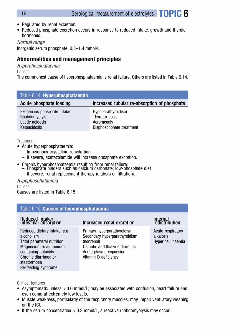
7/17/2019 0080451373
http://slidepdf.com/reader/full/0080451373 127/199
• Regulated by renal excretion.• Reduced phosphate excretion occurs in response to reduced intake, growth and thyroid
hormones.
Normal range Inorganic serum phosphate: 0.8–1.4 mmol/L.
Abnormalities and management principlesHyperphosphataemia Causes
The commonest cause of hyperphosphataemia is renal failure. Others are listed in Table 6.14.
Treatment
• Acute hyperphosphataemia:– Intravenous crystalloid rehydration– If severe, acetazolamide will increase phosphate excretion.
• Chronic hyperphosphataemia resulting from renal failure:– Phosphate binders such as calcium carbonate; low-phosphate diet– If severe, renal replacement therapy (dialysis or filtration).
Hypophosphataemia Causes
Causes are listed in Table 6.15.
Clinical features
• Asymptomatic unless <0.6 mmol/L; may be associated with confusion, heart failure andeven coma at extremely low levels.
• Muscle weakness, particularly of the respiratory muscles; may impair ventilatory weaningon the ICU.
• If the serum concentration <0.3 mmol/L, a reactive rhabdomyolysis may occur.
Table 6.14 Hyperphosphataemia
Acute phosphate loading Increased tubular re-absorption of phosphate
Exogenous phosphate intakeRhabdomyolyisLactic acidosisKetoacidosis
HypoparathyroidismThyrotoxicosis
AcromegalyBisphosphonate treatment
Table 6.15 Causes of hypophosphataemia
Reduced intake/ intestinal absorption Increased renal excretion
Internalredistribution
Reduced dietary intake, e.g.alcoholismTotal parenteral nutritionMagnesium or aluminium-containing antacidsChronic diarrhoea orsteatorrhoeaRe-feeding syndrome
Primary hyperparathyroidismSecondary hyperparathyroidism(nonrenal)Osmotic and thiazide diuretics
Acute plasma expansion Vitamin D deficiency
Acute respiratoryalkalosisHyperinsulinaemia
116 Serological measurement of electrolytes TOPIC 6

7/17/2019 0080451373
http://slidepdf.com/reader/full/0080451373 128/199
Test: Serum chloride
Indications1. Metabolic acidosis.2. Patients on intravenous fluid replacement therapy.3. Patients on total parenteral nutrition.4. Severe vomiting.
How it is doneChloride ion-sensitive electrode (uses the same reference electrode as that used for pHmeasurement).
InterpretationPhysiological principles • The body’s predominant extracellular anion.
• Maintains electroneutrality, mainly as a counter to sodium; serum chloride generallyincreases and decreases with serum sodium.
Normal range 96–106 mmol/L.
Abnormalities and management principlesHyperchloraemia • Hyperchloraemia accompanying metabolic acidosis may occur with excessive infusion of
normal saline and renal tubular acidosis.• Other causes include hyperparathyroidism and hypernatraemia of any aetiology, including
gastrointestinal upsets such as diarrhoea, dehydration, ileal loops and loss of pancreaticsecretion.
• Drug causes include acetazolamide, ammonium chloride and triamterine.
Hypochloraemia • Overhydration.• Congestive cardiac failure.• Syndrome of inappropriate secretion of ADH.• Vomiting.• Chronic respiratory acidosis or metabolic alkalosis of any aetiology.
• Addison’s disease.• Burns.• Some instances of diuretic therapy.
Management principlesIdentification and management of the underlying cause.
Test: Serum lactate
Indications1. Metabolic acidosis.2. Suspected or proven hypoperfusion (either generalized or specifically to the liver and/or
intestinal tract).
How it is done• Laboratory analysis using a venous or arterial blood sample.• Lactate is oxidized by L-lactate peroxidase, producing hydrogen peroxide.• This is then passed to a platinum electrode, which oxidizes the hydrogen peroxide,
producing a current, the size of which is proportional to the lactate concentration.
TOPIC 6 117Renal, metabolic and endocrine systems

7/17/2019 0080451373
http://slidepdf.com/reader/full/0080451373 129/199
InterpretationPhysiological principles • Lactate is a by-product of anaerobic metabolism of pyruvate.• Lactate levels may also rise as a result of reduced gluconeogenesis. Lactate is
metabolized by the liver to bicarbonate, so a failure of this metabolism may also cause a
rise in serum lactate.• If a rise in serum lactate is also accompanied by a metabolic acidosis this is termed lactic
acidosis.
Normal range 0.6–1.8 mmol/L.
AbnormalitiesThe causes of lactic acidosis are classified as either Type A (associated with tissue hypoxia)or Type B (without tissue hypoxia) (Table 6.16).
Management principles• Management of lactic acidosis predominantly involves identification and treatment of the
underlying cause.• Initial supportive management includes optimization of tissue oxygen delivery with
adequate oxygenation, cardiac output and haemoglobin concentration.
Test: Serum bicarbonate
Indications Any suspected acid-base disturbance.
How it is doneCalculated from the plasma pH and PCO2, as measured by the pH and Severinghauselectrodes in a blood gas analyzer.
Interpretation
Physiological principles • Bicarbonate is an anion that is a major component of the body’s predominant buffer system.• It is formed from the dissociation of carbonic acid as a part of a three-stage chemical
reaction, which is catalyzed by carbonic anhydrase.• Bicarbonate is completely filtered in the glomerulus and then reabsorbed, predominantly in
the proximal tubule, via the formation of carbonic acid; the degree of this reabsorption isdetermined by total body acid-base balance.
Table 6.16 Causes of lactic acidosis
Type A (tissue hypoxia) Type B (no tissue hypoxia)
Hypoxaemia AnaemiaHypoperfusion of any origin (e.g. haemorrhage,sepsis, hypovolaemia)Cardiac failureIschaemic bowel
Hepatic failureRenal failureDiabetes mellitusDrug-induced: e.g. salicylate overdose,biguanides, alcoholsTotal parenteral nutrition
Inborn errors of metabolismGlycogen storage disorders
118 Serological measurement of electrolytes TOPIC 6

7/17/2019 0080451373
http://slidepdf.com/reader/full/0080451373 130/199
Normal range 24–33 mmol/L.
Abnormalities and management principlesSee Chapter 2.
Limitations and complicationsSee Chapter 2.
Investigation of salt and water disturbance
Test: Serum osmolality
Indications1. Renal impairment.2. Serum sodium derangement.3. Suspicion of diabetes insipidus or SIADH.
How it is done1. Estimation using the formula:
Plasma osmolality ¼ [Glucose] þ [urea] þ 2 [sodium](mosmol/kg) (mmol/L) (mmol/L) (mmol/L)
2. Laboratory determination of depression of freezing point.
3. Laboratory measurement of osmotic pressure.4. Laboratory determination of serum ionic concentrations using flame photometry.
Data presented asNumerical data: units mosmol/kg solvent.
In the case of plasma, the solvent is water.
InterpretationPhysiological principles
• Osmolality – the number of osmoles per kilogram solvent.• Osmolarity – the number of osmoles per litre of solution
where osmole refers to the molecular weight of a substance divided by the number of freelymoving particles in solution.• In the plasma, the presence of proteins and fats mean that the values for osmolarity and
osmolality are slightly different.• In health, plasma osmolality is maintained within a narrow range by a number of
homeostatic mechanisms involving osmoreceptors, which respond to changes by thestimulation of antidiuretic hormone.
Normal range 280–305 mosmol/kg.
Abnormalities and management principlesSyndrome of inappropriate ADH secretion (SIADH) This is characterized by low plasma osmolality and sodium, high urine sodium andurine/plasma osmolality ratio >1.
TOPIC 6 119Renal, metabolic and endocrine systems

7/17/2019 0080451373
http://slidepdf.com/reader/full/0080451373 131/199
It may be caused by:• Ectopic vasopressin secretion (from malignancies, including lymphoma or carcinoma of
bronchus, pancreas, colon, prostate)• Pulmonary disease (e.g. tuberculosis, pneumonia)• Neurological disorders (e.g. meningitis, encephalitis, tumour, head injury, neurosurgery)
• Drugs (e.g. chlorpropamide, oxytocin, carbemazepine, certain antidepressants).Treatment
• Identify and treat underlying cause.• Water restrict; demeclocycline may be used to block the renal effects of ADH.
Diabetes insipidus (DI) This causes a high serum osmolality and presents with polyuria and polydipsia associatedwith reduced ADH activity.
This may be due to:• Cranial DI: hyposecretion of ADH – e.g. due to head injury, brain tumours, neurosurgery or,
rarely, familial cranial DI• Nephrogenic DI: renal unresponsiveness to the effects of ADH – e.g. related to drugs such
as lithium, demeclocycline and gentamicin. Patients are unable to concentrate their urinein response to water deprivation.
Treatment
Depends on the cause.• Cranial DI is treated with desmopressin, a synthetic ADH analogue.• Nephrogenic DI is treated with thiazide diuretics.
Limitations and complications• Pathologically high levels of alcohols, proteins, triglycerides and mannitol will make the
calculation of osmolality using the above formula inaccurate, as these molecules are notaccounted for.
• Urine osmolality should also be measured to confirm diagnosis of SIADH or DI.
Assessment of thyroid function
Test: Serum thyroid hormones measurement
IndicationsInvestigation of thyroid function.
How it is doneSerological immunoassay of thyroxine, thyronine and thyroid-stimulating hormone.
InterpretationPhysiological principles • Thyroxine (L-thyroxine, T4) is an amine hormone produced by the thyroid gland under the
influence of thyroid-stimulating hormone (TSH), which is produced in the pituitary. T4 hasa half life of 7 days.
• Thyroxine is enzymatically cleaved to T3 (L-thyronine) in the thyroid gland and in thecirculation; T3 is the active form and has a half life of one day.
Normal ranges • Serum thyroxine (T4) ¼ 4.6–12 mg/dL.• Free thyroxine (T4) ¼ 0.7–1.9 ng/dL.
120 Assessment of thyroid function TOPIC 6

7/17/2019 0080451373
http://slidepdf.com/reader/full/0080451373 132/199
• Serum T3 ¼ 80–180 ng/dL.• Free T3 ¼ 230–619 pg/dL.• Serum TSH ¼ 0.5–6 mmol/mL.
Abnormalities and management principles
Hyperthyroidism Causes
The commonest causes are characterized by an overactive gland and thus normal orincreased radio-iodine uptake; these are Graves’ disease, toxic (hot) nodule and toxicmultinodular goitre administration. Drugs that can induce hyperthyroidism includeamiodarone and oestrogens.
Clinical features are listed in Table 6.17.
It is characterized by an increase in serum T4 measurements; TSH may or may not besuppressed depending on the cause.• Medical management is with carbimazole or propyluracil, which prevent formation of
thyroid hormones.
• Iodine therapy temporarily inhibits hormone release and is often given for 2 weeks beforethyroidectomy.• Beta adrenoceptor antagonists have dual effect, by reducing the conversion of thyroxine to the
active tri-iodothyronine, and also by counteracting the systemic effects of hyperthyroidism.• Surgical removal of the thyroid gland requires adequate preoperative medical control of
hyperthyroidism, in order to prevent precipitation of a thyroid crisis.
Hypothyroidism Causes
• Total or partial thyroidectomy.• Drugs – e.g. amiodarone, aspirin, phenytoin, interferon, frusemide, lithium.• Autoimmune diseases are 10 times more common in females, and the incidence increases
with age.• It is characterized by a reduction in T4/T3 levels, and will be accompanied by a high TSH if
the hypothyroidism is due to intrinsic thyroid disease or drugs affecting the thyroid.• It may rarely be caused by pituitary disease, in which case the TSH will also be low.
Clinical features are listed in Table 6.18.• Hypothyroidism may present as a medical emergency in the form of a myxoedema coma.• It is treated with oral thyroxine replacement or, in the emergency setting, with intravenous T3.
Assessment of glycaemic control
Test: Serum glucose
Indications1. Diabetes mellitus.2. Any acute or critical illness, in particular, sepsis, acute pancreatitis.
Table 6.17 Clinical features of hyperthyroidism
Cardiovascular Dyspnoea; atrial fibrillation; high-output cardiac failure
Musculoskeletal Proximal myopathy, periodic paralysis; osteoporosis; hypercalcaemia
Blood Leucopenia; microcytic anaemia
TOPIC 6 121Renal, metabolic and endocrine systems

7/17/2019 0080451373
http://slidepdf.com/reader/full/0080451373 133/199
3. Reduced level of consciousness.4. Glycogen storage diseases.
How it is doneLaboratory assay Several methods exist. In one such method D-glucose is oxidized in the presence of glucoseoxidase, producing hydrogen peroxide and glucono-d-lactone. Hydrogen peroxide is thenpassed to a platinum electrode, where it is oxidized; the magnitude of the resultant current isproportional to the glucose concentration.
Glucose reagent sticks The same oxidation reaction is employed, producing hydrogen peroxide. In the case ofreagent sticks, the hydrogen peroxide then oxidizes a dye, which produces a colour changethat can be compared against a chart.
InterpretationPhysiological principles In health, blood sugar is maintained within a narrow range by various neuroendocrineresponse systems including the sympathetic nervous system, insulin and glucagon.
Normal range Fasting blood glucose: 4.1–7 mmol/L.
Abnormalities and management principlesHyperglycaemia Hyperglycaemia is defined as fasting plasma glucose greater than 7.0 mmol/L. Causes arelisted in Table 6.19.
Chronic hyperglycaemia
• Diabetes mellitus causes multisystem problems, and is a leading cause of morbidity andmortality in the Western world.
• Diabetes may be classified as:– Type 1: due to failure of insulin secretion; most commonly diagnosed in childhood or
adolescence– Type 2: due to insulin resistance and is usually associated with defective insulin
secretion
Table 6.18 Clinical features of hypothyroidism
General Serous effusions (ascites, pleural, pericardial or joint effusions);hypothermia
Cardiovascular Hypercholestrolaemia and ischaemic heart disease; bradycardia;
cardiomegalyECG changes include low-voltage complexes and T wave flattening/ inversion
Respiratory Hypoventilation
Musculoskeletal Muscular chest pain; muscular cramps; raised creatinine kinase
Blood Macrocytic anaemia; microcytic anaemia in context of menorrhagia inwomen
122 Assessment of glycaemic control TOPIC 6

7/17/2019 0080451373
http://slidepdf.com/reader/full/0080451373 134/199
– Type 3: encompasses all other specific forms of diabetes, including those related togenetic beta cell defects, genetically related insulin resistance, pancreatic disease,hormones or drugs.
– Type 4: gestational diabetes
Acute hyperglycaemia • Diabetic ketoacidosis is frequently precipitated by a concomitant illness.• Hyperosmolar nonketotic coma is a more common presentation of hyperglycaemia in
elderly patients. Progressive dehydration due to polyuria results in a very high plasmaosmolality accompanying severe hyperglycaemia, but with little or no ketonuria.
• Management of both conditions involves slow correction of fluid, glycaemic and electrolyteabnormalities.
Acute hypoglycaemia • Hypoglycaemia in diabetic patients may occur in those treated with either insulin or oral
sulphonylureas.
• Hypoglycaemia in nondiabetic patients is rare, but can occur in association with hepaticdisease, alcohol, hypoadrenalism and certain tumours (e.g. insulinomas, IGF-2 secretingtumours).
• Postprandial hypoglycaemia is well documented in patients post gastrectomy.
Tight glycaemic control • Evidence from several major studies has found there to be outcome benefit, both in terms
of morbidity and mortality from controlling blood glucose in surgical critically ill patients.• There is still some debate over whether ‘tight’ glycaemic control is more beneficial than
‘moderate’ glycaemic control; however, most studies use a target blood glucose range of
4–8 mmol/L.
Limitations and complications1. Glucose reagent sticks tend to be less accurate at lower glucose levels.2. Falsely high glucose results may occur due to contamination of the testing sample,
for example, with glucose-containing contaminants on the skin from where a bloodsample is obtained.
Table 6.19 Causes of hyperglycaemia
Causes Examples
Pancreatic insufficiency Diabetes mellitus (types 1 and 2) Acute or chronic pancreatitis or pancreatectomy
SepsisCystic fibrosis
Neuroendocrine response causingincreased circulatingcatecholamines, growth hormone,glucocorticoids, glucagon
Trauma, burns, critical illnessStress response to surgery
Insulin resistance Type 2 diabetes mellitusPolycystic ovary syndrome
Acromegaly, Cushing’s syndrome or glucagonomas
PhaeochromocytomaExogenous administration of glucose Total parenteral nutrition (TPN), enteral nutrition
Drugs Steroids, thiazide diuretics, octreotide
TOPIC 6 123Renal, metabolic and endocrine systems

7/17/2019 0080451373
http://slidepdf.com/reader/full/0080451373 135/199
Test: Glycosylated haemoglobin (HbA1C)
IndicationsMonitoring of long-term glycaemic control.
Data presented as A percentage of glycosylated haemoglobin relative to red cell haemoglobin.
InterpretationPhysiological principles • Red cell haemoglobin is nonenzymatically glycated at a rate that relates to overall level of
glucose.• It has been found to correlate well with the risk of microvascular complications and so is a
valuable tool in the assessment of long-term glycaemic control.
Normal range
There is no standardized assay of HbA1C and interpretation should be made in conjunctionwith local guidelines.
Abnormalities and management principles• The commonest cause of a high HbA1C is poor glycaemic control secondary to diabetes
mellitus of any aetiology.• However, abnormalities in HbA1C may occasionally be due to some haemoglobinopathies
and disorders of red cell metabolism.
Investigation of the hypothalamic–pituitary axis
Test: Short synacthen test
IndicationsSuspected adrenocortical insufficiency:1. Sepsis/critical illness2. Addison’s disease3. Long-term steroid usage4. Pituitary disease or surgery.
How it is done1. Baseline serum sample for cortisol assay.2. Intravenous administration of 250 mg synthetic adrenocorticotrophin (ACTH) –
tetracosactrin.3. Serum cortisol assay at 30 minutes following ACTH administration.
Serum cortisol samples are laboratory analyzed by radioimmunoassay.
Data presented as
Numerical value for cortisol assay; units nmol/L.
InterpretationPhysiological principles The administered dose of tetracosactrin should maximally stimulate the adrenal cortex. In thecase of adrenal insufficiency, the adrenal cortex is unable to respond to this stimulus,resulting in a subnormal rise in serum cortisol levels.
124 Investigation of the hypothalamic–pituitary axis TOPIC 6

7/17/2019 0080451373
http://slidepdf.com/reader/full/0080451373 136/199
Normal range Post-ACTH administration cortisol level >500 nmol/L.
AbnormalitiesThe causes of adrenocortical insufficiency may be divided into:
1. Primary adrenal failure (Addison’s disease). This may have a variety of aetiologiesincluding autoimmune (most common), infiltration from TB, malignancy or amyloidosis,critical illness, haemorrhage or infarction. Autoimmune Addison’s may be associated withother autoimmune disorders.
2. Secondary adrenal failure. Most commonly due to either abrupt withdrawal of long-termsteroid therapy, ACTH deficiency due to pituitary disease or surgery.
Adrenal insufficiency as demonstrated by an inadequate increment in serum cortisol inresponse to a short synacthen test may result in a variety of clinical, biochemical andhaematological abnormalities. The clinical features differ in acute and chronic adrenocorticalinsufficiency (Table 6.20).
Management principles• Severe acute adrenocortical insufficiency may present with circulatory collapse.• Longer term management involves steroid replacement therapy, initially with intravenous
hydrocortisone, and then subsequently with oral prednisolone if required.
Measurement of hormones: Phaeochromocytoma
Test: Plasma and urine catecholamines and their metabolites
IndicationsKnown or suspected phaeochromocytoma.
Table 6.20 Acute versus chronic adrenocortical insufficiency
Acute Chronic
Clinical features• Abdominal and muscular pain• Hypotension• Psychosis
Clinical features• Malaise and muscle weakness• Weight loss• Diarrhoea and vomiting• Postural hypotension
• Increased susceptibility to infection
Haematological features• Eosinophilia• Lymphocytosis• Normocytic anaemia
Biochemical features• Raised serum urea• Hyponatraemia• Hypoglycaemia
• Hyperkalaemia• Hypercalcaemia• Hypochloraemia
TOPIC 6 125Renal, metabolic and endocrine systems

7/17/2019 0080451373
http://slidepdf.com/reader/full/0080451373 137/199
How it is done1. Plasma assays Laboratory assay of venous blood sample for plasma epinephrine and norepinephrine levels.
2. Urine assays
Laboratory assay of 24-hour urine sample for urinary epinephrine, norepinephrine anddopamine levels. Their urinary metabolites, vanillymandelic acid (VMA), metanephrine andnormetanephrine, may also be measured.
InterpretationPhysiological principles • Phaeochromocytomas are rare tumours of the sympathetic nervous system that are found
in either the adrenal medulla or on sympathetic ganglia.• They may secrete one or a combination of epinephrine, norepinephrine and dopamine;
extraadrenal tumours do not secrete epinephrine.
• Symptoms include headache, sweating, palpitations, psychosis. Other symptoms or signsof hypertension, and sometimes hypotension, including postural hypotension, especially ifthe tumour secretes epinephrine.
• Patients may also present with signs or symptoms of glucose intolerance andcardiomyopathy.
Management principlesResection of phaeochromocytomas involves careful anaesthetic preparation.1. Complete preoperative alpha-blockade using phentolamine or phenoxybenzamine.2. Subsequently, complete beta-blockade.
3. Prevention of intraoperative hypertension using vasodilators such as GTN, sodiumnitroprusside or magnesium sulphate, and intraoperative tachy-arrhythmias using beta-blockers or amiodarone.
4. Goal-directed fluid resuscitation, particularly after tumour resection.5. Postoperative high-dependency unit (HDU)/intensive care unit (ICU) admission.
Limitations• Plasma catecholamine levels may be normal or only slightly elevated in up to a third of
patients with phaeochromocytoma.• Slight elevations of urinary and serum catecholamines and their metabolites may occur in
a number of patients with poorly controlled systemic hypertension that is not due tophaeochromocytoma.
• False-positive urinary VMA results may occur because of concurrent therapy withmethyldopa, levodopa or labetolol. It may also occur in patients with clonidine withdrawal,hypoglycaemia, raised intracranial pressure and after strenuous exercise.
• Various medications may cause false-positive results in urinary and serum catecholamineand metanephrine levels.
A variety of physiological stresses and pathophysiological processes may also causefalse-positive urinary catecholamine and plasma catecholamine and metanephrine
levels including the stress response to surgery, myocardial infarction, critical illness anddepression.
Further investigations• Clonidine suppression testing (i.e. the measurement of plasma free normetanephrine
before and after administration of clonidine) may aid in the diagnosis.• Preoperative tumour localization is carried out using CT scanning, radioactive meta-
iodobenzyl guanidine (MIBG) scintigraphy and/or selective venous catheterization.
126 Measurement of hormones: Phaeochromocytoma TOPIC 6

7/17/2019 0080451373
http://slidepdf.com/reader/full/0080451373 138/199
Measurement of hormones: Carcinoid tumours
Test: Urinary 5-hydroxyindole acetic acid (5-HIAA)
IndicationsKnown or suspected carcinoid tumour or carcinoid syndrome.
How it is doneLaboratory assay of 24-hour urine samples using high-performance liquid chromatographywith electrochemical detection.
InterpretationPhysiological principles • Carcinoid tumours are common, being an incidental finding in 1% of post-mortems. 85%
of carcinoid tumours are found in the terminal ileum; they are rarely found outside the GItract, in the lungs and gonads.• Carcinoid tumours secrete serotonin and kinins but remain asymptomatic until hepatic
metastases are present, resulting in carcinoid syndrome, which is a rare diagnosis. The presenceof hepatic metastases impair serotonin metabolism, resulting in a variety of symptoms andsigns, including flushing in association with wheezing, sweating, vasodilatation and resultanthypotension. They may also cause episodic diarrhoea and commonly lead to weight loss.
• Right-sided cardiac failure may result from endocardial fibrosis of the tricuspid and pulmonaryvalves (left-sided cardiac failure may occur in the presence of bronchial carcinoid or an AtrialSeptal Defect (ASD)).
Normal range 3–15 mg/day. There is a correlation between tumour mass and 5-HIAA production.
Management principles Anaesthetic management of carcinoid resection • Cardiostable induction and maintenance with invasive arterial and central venous pressure
monitoring.• Consider cardiac output monitoring using oesophageal Doppler or similar.• Intraoperative management revolves around the prevention of hypo- or hypertension,
and bronchospasm.• Postoperative ICU/HDU management.
Limitations• Bananas, kiwi fruit, pineapples and a variety of nuts, all of which are serotonin rich, may
cause false-positive results.• Certain drugs, including salicylates, paracetamol and L-dopa may also affect the assay.
Further investigations• Preoperative tumour localization is carried out using CT, MRI or PET scanning.
• Echocardiography is recommended if there are signs of cardiac involvement.
Investigation of allergic reactions
Test: Serum mast cell tryptase measurement
IndicationsSuspected anaphylactic or anaphylactoid reaction.
TOPIC 6 127Renal, metabolic and endocrine systems

7/17/2019 0080451373
http://slidepdf.com/reader/full/0080451373 139/199
How it is doneEnzyme immunoassay.
InterpretationPhysiological principles
Tryptase is a neutral protease that is found almost exclusively in mast cells. It exists in twostructural forms (a and b); b–tryptase is normally involved in airway homeostasis, vascularcontraction and relaxation, gastrointestinal motility and smooth muscle activity and incoagulation. Unlike histamine, tryptase remains elevated in the plasma for some hours afteran anaphylactic reaction.
Normal range
<11.4 ng/mL.
Abnormalities and management principles
• Anaphylaxis (a type I immune reaction) and anaphylactoid reactions (involving complementreleased as well as mast cell degranulation) both lead to the release of histamine,serotonin, kinins and leukotrienes amongst other vasoactive substances.
• An important clinical difference between the two types of reaction is that anaphylaxisrequires prior exposure and thus sensitization to the culprit agent, whereas anaphylactoidreactions do not.
• The management of suspected anaphylactic and anaphylactoid reactions is outlined inguidelines published by the Association of Anaesthetists of Great Britain and Ireland(AAGBI).
Limitations and complications• At least three serial mast cell tryptase samples are required to be able to diagnoseanaphylaxis, looking for an initial high value followed by serial decay.
• Mast cell tryptase assay does not distinguish between anaphylactic and anaphylactoidreactions.
Test: Serum and urine histamine assay
IndicationsSuspected anaphylactic or anaphylactoid reaction.
How it is doneEnzyme immunoassay of either a 2-mL blood sample (non-EDTA bottle), 5 mL urine sampleor 24-hour urine collection.
Data presented asNumerical value. Units: ng/mL for random urine and blood sample; mg/24 hours for 24-hoururine collection.
Interpretation
Physiological principles Histamine is a low-molecular-weight molecule produced by the decarboxylation of histidine.It is actively released from mast cells and basophils as part of anaphylactic andanaphylactoid reactions, but is also found throughout many body tissues and organs. Peak levels occur within 10 to 20 minutes of exposure, and then rapid decay to baseline occurswithin 60–90 minutes. Thereafter, histamine may be detected in the urine.
128 Investigation of allergic reactions TOPIC 6

7/17/2019 0080451373
http://slidepdf.com/reader/full/0080451373 140/199
Normal range • Whole blood: 20–200 ng/mL.• Random urine: 0–88 ng/mL.• 24-hour urine: 0–118 mg/24 hours.
Abnormalities and management principlesSee under mast cell tryptase.
Limitations and complicationsHistamine assay does not differentiate between anaphylactic and anaphylactoid reactions.
TOPIC 6 129Renal, metabolic and endocrine systems

7/17/2019 0080451373
http://slidepdf.com/reader/full/0080451373 141/199
TOPICl7Haematology and coagulation
Topic ContentsCommon first tests 130
Test: Full blood count and peripheralblood smear (PBS) 130
Test: Group and screen/crossmatch 135
Laboratory tests of coagulation 137
Tests: Prothrombin time (PT)/ international normalized ratio (INR),activated partial thromboplastin time(APTT) and thrombin time (TT) 137
Test: Fibrinogen 139
Test: D-dimers and fibrin degradation
products 140Test: Anti-Xa assay 141
Test: Factor inhibitor assay/mixingstudies 141
Test: Hypercoagulation screen 141
Point-of-care tests of coagulation 142
Test: Activated clotting time (ACT) 142
Test: High-dose thrombin time (HiTT) 142
Haemoglobinopathies 143
Test: Sickledex 143
Test: Haemoglobin electrophoresis/ high-performance liquid chromatography(HPLC) 144
Viscoelastic measurement of
haemostasis 144
Test: Thromboelastography/ thromboelastometry 144
Laboratory platelet function monitors 150
Test: Optical light transmission plateletaggregometry (LTA) 150
Point-of-care platelet function
monitors 151
Test: PFA-100 151
Common first tests
Test: Full blood count and peripheral blood smear (PBS)
Indications• To show abnormalities in the production, life span, and destruction of blood cells and aid
in the diagnosis of anaemia, polycythaemia, thrombocytosis, thrombocytopenia,leucopaenia and leucocytosis.• To help diagnose infection.• As a preoperative baseline in cases with expected significant blood loss (see NICE
guidelines at http://www.nice.org.uk).• As a guide to blood/platelet transfusion.

7/17/2019 0080451373
http://slidepdf.com/reader/full/0080451373 142/199
How it is done• Blood is taken into an EDTA tube and analyzed, ideally within 4 hours.• Automated blood counters using either forward angle light scatter or impedance analysis
to provide:– Enumeration of erythrocytes, leucocytes (5 part differential count) and platelets
– Quantification of haemoglobin (Hb) by spectrophotometry in lyzed sample– Erythrocyte mean cell volume (MCV), haematocrit (Hct), red cell distribution width
(RDW), a measure of cell size scatter– Derived parameters include mean cell haemoglobin (MCH) and mean corpuscular
haemoglobin concentration (MCHC).• The counter flags samples that need further analysis with a PBS and a manual count
or examination of morphology.
InterpretationNormal adult ranges.
See Table 7.1.
Abnormalities Anaemia: Low haemoglobin
See Table 7.2.
Polycythaemia: Increased haemoglobin/haematocrit
See Table 7.3.
Table 7.1 Normal adult ranges for blood values
Male Female*
Haemoglobin (g/dL) 13–18.0 11.5–16.5
RBCs (1012 /L) 4.5–6.5 4.0–5.8
Hct (%) 0.40–0.52 0.37–0.47
MCV (fL) 84–96 84–96
MCH (pg) 27.0–32.0
MCHC (g/dL) 27.0–32.0
Platelets (109 /L) 150–400
WBCs (109 /L) 4.0–11.0
Neutrophils (109 /L) 2.0–7.5
Lymphocytes (109 /L) 1.5–4.0
Monocytes (109 /L) 0.2–0.8
Eosinophils (109 /L) 0.04–0.4
Basophils (109 /L) 0.0–0.1
Reticulocytes (% or 109 /L) 0.5–2.5 or 20–80
*In pregnancy the Hb may fall as low as 9 g/dL in the third trimester. RBCs, red blood cells; Hct, haematocrit;
MCV, mean cell volume; MCH, mean cell haemoglobin; MCHC, mean corpuscular haemoglobin concentration; WBC, white blood cell.
TOPIC 7 131Haematology and coagulation

7/17/2019 0080451373
http://slidepdf.com/reader/full/0080451373 143/199
Table 7.2 The anaemias
Type of
anaemia
Differential
diagnosis
Peripheral blood
smear (PBS) Comment
MicrocyticMCV <80 fL
Iron deficiencyanaemia
Anisocytosis,Poikilocytosis,Elliptocytosis
Hypochromic (lowMCH)Low serum ferritin, lowiron and raised totaliron binding capacity(TIBC)
Thalassaemia Polychromasia,target cells,basophilicstippling
Anaemia of chronic
disease
Due to chronic
inflammatory andinfective conditions
NormocyticMCV 80–100 fL
Bleeding Polychromasia
Haemolysis;may be:• Hereditary or
acquired• Immune or
non-immune
• Extravascular orintravascular
Polychromasia,spherocytes,schistocytes, bitecells
Low haptoglobinRaised LDH, bilirubinand reticulocytes
Anaemia of chronicdisease
Especially associatedwith renal failure
MacrocyticmegaloblasticMCV >100 fL
Vitamin B12/folatedeficiency
Oval macrocytes,Howell-Jollybodies, basophilicstippling
Serum B12 lowRaised LDHPernicious anaemia:intrinsic factorantibodies
Reduced red cell folate(reflects total bodyfolate status)
MacrocyticnormoblasticMCV >100 fL
Liver disease andexcessive alcoholconsumption
Hypothyroidism
Drug induced e.g. hydroxyurea
Myelodysplasticsyndrome
Ringedsideroblasts (ironstain), nuclearabnormalities ofleucocytes
Markedreticulocytosis
e.g. in haemolysis

7/17/2019 0080451373
http://slidepdf.com/reader/full/0080451373 144/199
Thrombocytopenia: Platelet count <150 10 9 /L
See Table 7.4.
Thrombocytosis: Platelet count >450 10 9 /L
See Table 7.5.
Leucopenia: WBC <4.0 10 9 /L • It is uncommon for absolute leucopenia to be due to an isolated deficiency of any cell
other than the neutrophil.• The risk of infection is closely related to the absolute neutrophil count.
Table 7.3 Polycythaemia
True
polycythaemia Secondary polycythaemia
Apparent or spurious
polycythaemia
Polycythaemia
rubra vera, (PRV)
Inappropriate erythropoietin
secretion in benign & malignantrenal disorders and by sometumours
Secondary to cigarette
smoking, obesity, excessalcohol or hypertension
Table 7.4 Causes of thrombocytopenia
Failure ofproduction Increased consumption Pseudothrombocytopenia
Drugs & chemicals Viral infection,e.g. HIV Radiation
Aplastic anaemiaLeukaemiaMarrow infiltration
MegaloblasticanaemiaLiver disease
Idiopathic thrombocytopenicpurpura (ITP)Disseminated intravascularcoagulation (DIC)InfectionHaemorrhage & transfusionSystemic lupus
Some leukaemias & lymphomaHeparinHypersplenismThrombotic thrombocytopenicpurpura (TTP)Haemolytic uraemic syndrome
Caused by clumping: can beexcluded by examination ofPBS and citrate sample(clumping due to EDTA)
Table 7.5 Causes of thrombocytosis
Primary myeloproliferative disorder Secondary reactive disorder
Essential thrombocythaemiaPRV Idiopathic myelofibrosis
Haemorrhage Acute & chronic infectionInflammatory diseasePost splenectomyTrauma/surgeryIron deficiency anaemia
Malignancy
TOPIC 7 133Haematology and coagulation

7/17/2019 0080451373
http://slidepdf.com/reader/full/0080451373 145/199
• Neutropenia – neutrophils <2.0 109 /L.• Lymphopenia – lymphocytes <1.5 109 /L.
See Table 7.6.
Leucocytosis
Detection of a leucocytosis by the automated counter needs confirmation with a PBS andmanual count. See Table 7.7.
Table 7.6 LeucopeniasNeutropenia Lymphopenia
Congenital causes Acquired causes
Benign chronic neutropenia(Yemenite Jews & people of
African descent)Rare – Kostmann’ssyndrome, cyclic neutropenia
Drugs – cytotoxic agents,anticonvulsants, thyroidinhibitors, antibiotics, clozapine,procainamide,hydroxychloroquine,penicillamine, NSAIDs
Post-infectious – usually afterviral infection:• Severe sepsis• Autoimmune• Felty’s syndrome (rheumatoid
arthritis)• Splenomegaly
Acute infectionsCardiac failureTuberculosisUraemiaLymphoma,carcinoma
Systemic lupuserythematosusCorticosteroidsHIV
Table 7.7 Causes of leucocytosis
Neutrophilia
(>7.5 109 /L)
Lymphocytosis
(>4.0 109 /L) Eosinophilia Basophilia
Infection/
inflammationDrugs –corticosteroids,epinephrineMyocardialinfarctionExerciseStress includingafter surgeryCigarette smoking
‘Leukaemoidreaction’: responseto severe infection
Viral infection
InfectiousmononucleosisCMV, hepatitis A Chronic infection withTB, brucellosis,secondary syphilisCLL, ALL, occasionallyNHL
Drugs
Parasite infections Asthma andallergic reactions
VasculitidesMetastatic cancerHypereosinophilicsyndrome
Myeloproliferative
disordersInflammatorydisordersDrugs
Viral infectionHypothyroidism
ALL, acute lymphoblastic leukaemia; CLL, chronic lymphocytic leukaemia; NHL, non-Hodgkin lymphoma.
134 Common first tests TOPIC 7

7/17/2019 0080451373
http://slidepdf.com/reader/full/0080451373 146/199
Management principles• The full blood count is often performed unnecessarily as a preoperative screening tool.
Various studies have shown that this is a waste of resources although some would arguethat it is indicated in all premenopausal women because of the higher incidence ofanaemia.
• NICE guidelines are available indicating those patients who should have a full bloodcount performed, mainly those with significant comorbidity or undergoing majorsurgery.
• Abnormal results may need manual verification or a PBS to provide further information.
Limitations and complications• 5% of the population lie outside the ‘normal’ reference range.• People of Afro-Caribbean descent display significantly lower haemoglobin, WBC, neutrophil
and platelet count.• Individuals may show substantial change from their baseline without falling outside
‘normal range’.
Test: Group and screen/crossmatch
Indications• To determine a patient’s ABO blood group, and to screen serum for the presence of
antibodies to common red cell antigens.• To allow provision of appropriate red cell concentrate (RCC) and blood products in order to
avoid transfusion reactions.
How it is done• ‘Group’ – determines which ABO and Rh antigens are present on patient’s red blood cells(RBCs). RBCs are incubated with commercially available antibodies (anti-A, anti-B), whichreact with antigens if present and cause agglutination. The patient’s serum is thenincubated with A and B cells to determine the presence of anti-A and anti-B antibodies.(See Table 7.8)
• ‘Screen’ – utilizes the indirect Coomb’s test (indirect antiglobulin test). Serum is incubatedwith a wide range of RBCs that together exhibit a comprehensive range of surface
antigens. If antibodies are present they will cause agglutination.• Crossmatch – may be electronic or serological. An electronic crossmatch is used in
uncomplicated cases with a negative antibody screen. A serological crossmatch requiresRBCs from the donor unit to be incubated with the patient’s serum, which will causeagglutination if the unit is not compatible.
Table 7.8 ABO blood group antigens present on RBC and IgM antibodies
present in serum
Group A Group B Group AB Group O
Antigens present A antigen B antigen AB antigen No antigens
Antibodies present Anti-B Anti-A No antibodies Anti-A & Anti-B
UK incidence 42% 8% 3% 47%
TOPIC 7 135Haematology and coagulation

7/17/2019 0080451373
http://slidepdf.com/reader/full/0080451373 147/199
InterpretationData presented as
• A, B or O blood group and Rhesus (Rh) status, e.g. Oþ, AB.
Physiological principles
• Blood type is determined by the presence or absence of inherited antigenic material on thesurface of RBCs; the ABO blood group system and Rh blood group system are the twomost likely to cause harmful immunologically mediated reactions if noncompatible blood istransfused.
• ABO system. The A and B antigens are produced from a common precursor, the H antigen.Either, both or none may be present. Anti-A or anti-B IgM antibodies are produced in thefirst years of life possibly by sensitization to environmental substances such as food,viruses and bacteria.
• Rhesus system. The most significant Rh antigen is the Rh D antigen, which is eitherpresent (Rh D positive, RhDþ) or not (Rh D negative, RhD). IgG and IgM anti-D antibodies
are formed in RhD-negative individuals through a sensitizing event.
Management principles• ABO compatibility. When considering blood group compatibility for transfusion it is the
recipient antibodies and donor antigens that are important. For example, group ABindividuals have A and B antigens on the surface of their RBCs; their blood serumdoes not contain any antibodies against either A or B antigen so they can receive blood fromany group (in theory, AB being preferable). Blood group O individuals have neither antigenbut both anti-A and anti-B antibodies therefore will cause agglutination of the donor RBCsif they are given anything other than group O blood (Table 7.9).
• Rhesus compatibility. An RhD-negative patient with no antibodies could only receive
RhD-positive blood once, as this would lead to the formation of antibodies and potentiallyhazardous transfusion reactions if they were to receive it again. If an RhD-negative womandevelops antibodies and becomes pregnant with an RhD-positive child, these antibodies cancross the placenta and cause haemolytic disease of the newborn. Therefore rhesus-positiveblood or platelets should never be given to RhD-negative women of childbearing age orpatients with rhesus D antibodies. Platelets can be given with anti-D cover in emergencies.
• Plasma compatibility. Donor-recipient compatibility is the opposite of blood compatibility.Type O plasma (fresh frozen plasma, FFP) can only be given to type O recipients; Type ABcan be given to individuals of any blood group (Table 7.10).
Table 7.9 Red blood cell compatibility
Recipient
blood type Donor RBC must be:
ABþ O Oþ A A þ B Bþ AB ABþ
AB O A B AB
A þ O Oþ A A þ
A O A
Bþ O Oþ B Bþ
B O B
Oþ O Oþ
O O
136 Common first tests TOPIC 7

7/17/2019 0080451373
http://slidepdf.com/reader/full/0080451373 148/199
• There are usually local policies in place for the routine ordering of preoperative G&S orcrossmatched blood that take into account the planned surgery, estimated blood loss andneed for transfusion.
• Guidelines are available for the transfusion of blood and blood products published byThe Association of Anaesthetists of Great Britain and Ireland (AAGBI) and British Committeefor Standards in Haematology (BCSH) and advice should be sought from a haematologistwhere necessary.
Laboratory tests of coagulation
Tests: Prothrombin time (PT)/international normalized ratio
(INR), activated partial thromboplastin time (APTT) andthrombin time (TT)
Indications• Part of coagulation screen often ordered preoperatively.• Assessment of synthetic liver function.• Assessment of coagulopathy in disseminated intravascular coagulation (DIC) and massive
blood transfusions.• INR used to monitor warfarin therapy.• APTT used to monitor unfractionated heparin therapy.
• TT used in diagnosis of hypo/dysfibrinogenaemia.
How it is done• Platelet poor plasma (PPP) obtained from citrated blood sample (9:1 ratio).• PT/INR – thromboplastin and calcium added to PPP and time to fibrin formation
measured by a photo-optical or electromechanical device; this equals the prothrombintime (PT). To standardize results the PT is compared to a reference value, which givesthe INR.
• APTT – PPP, phospholipid and calcium are added to kaolin and the time to fibrin formationmeasured.
• TT – thrombin added to plasma and clotting time measured in seconds.
InterpretationNormal range/graph
• PT ¼ 10–14 seconds.• INR ¼ 0.9–1.2.• APTT ¼ 25–35 seconds.• TT ¼ <15 seconds.
Table 7.10 Plasma compatibility
Recipient blood type Donor plasma must be:
AB AB
A A or ABB B or AB
O O, A, B or AB
TOPIC 7 137Haematology and coagulation

7/17/2019 0080451373
http://slidepdf.com/reader/full/0080451373 149/199
Physiological principles
• Coagulation was traditionally thought to consist of ‘intrinsic’ and ‘extrinsic’ pathwayswith a final common pathway. This has now been replaced with the cell-based modelof haemostasis which comprises three stages of initiation, amplification andpropagation.
• At the site of vascular injury tissue factor (TF) expressed on extravascular cells forms aTF/VIIa complex that activates factors X to Xa and IX to IXa.
• Factor Xa converts factor V to Va and forms a Xa/Va complex on the TF cell, whichconverts prothrombin to thrombin (initiation).
• Thrombin activates platelets and factors VIII, V and XI.• Factor VIII circulates in combination with von Willebrand factor (vWF) which acts as a
carrier molecule to transport factor VIII to the platelet surface.• Activated platelets bind cofactors VIIIa and Va with their respective enzymes IXa and Xa to
form a prothrombinase complex, which causes a thrombin burst to take place on thesurface of the activated platelet (amplification).
• Thrombin converts fibrinogen to fibrin and the haemostatic plug comprised of platelets andfibrin strands is formed (propagation and stabilization).
• Control mechanisms exist to limit fibrin formation to the site of injury including TF pathwayinhibitor, the protein C and S system and antithrombin III.
• Disturbances in these mechanisms can lead to thrombotic disorders.
AbnormalitiesSee Table 7.11.
Table 7.11 Coagulation abnormalitiesProlonged INR Prolonged APTT Prolonged TT
Due to deficiency of factorI, II, V, VII or X:
• Warfarin anticoagulationtherapy
• Vitamin K deficiencyresulting in deficiency ofvitamin K dependentfactors II, VII, IX and X
• Liver disease• Coagulopathy secondary
to DIC*• Coagulopathy secondary
to massive bloodtransfusion
• Dilutional coagulopathy• High-dose unfractionated
heparin therapy• Fibrinogen deficiency
• Haemorrhagic diseasesof the newborn
Due to deficiency of Factor VIII, IX, XI or XII:• Unfractionated heparin
therapy• Haemophilia A, B or C
(factor VIII, IX or XIdeficiency, respectively)
• Lupus anticoagulant(actually prothromboticbut binds tophospholipid in testsample)
• Coagulopathysecondary to DIC
• Coagulopathysecondary to massiveblood transfusion
• Von Willebrand disease
Prolonged due tothrombin inhibition:• Heparin• Fibrin degradation
products (FDP)• Lupus anticoagulant
*Disseminated intravascular coagulopathy: DIC may complicate massive tissue injury, sepsis and some pregnancy-related complications. The normal anticoagulant and fibrinolytic systems are overwhelmed resulting in disseminated microvascular thrombi with consumption of platelets and coagulation factors leading to a haemorrhagic state. The fibrinolytic system is activated to dissolve the fibrin thrombi, resulting in the formation of D-dimers and fibrin degradation products (FDP), which have a further anticoagulant action.
138 Laboratory tests of coagulation TOPIC 7

7/17/2019 0080451373
http://slidepdf.com/reader/full/0080451373 150/199
Management principles• Preoperatively.• Determine cause for prolonged INR.• Stop warfarin and start heparin if anticoagulation essential.• Current guidelines for the emergency reversal of Warfarin therapy include vitamin K
and prothrombin complex concentrates (PCC) containing plasma-derived factors II, VII, IXand X.
• In the presence of bleeding or to correct the INR prior to surgery/invasive procedurestreatment options include:– FFP to maintain INR 1.5–1.8 unless there is evidence of microvascular ooze– PCC.
• Protamine for heparin overdosage.
Further investigations• Liver function tests.• Full blood count (FBC) (monitor platelet count on heparin).• Fibrinogen level to exclude hypofibrinogenaemia.• Mixing studies to check for possible factor deficiencies or inhibitors.• Specific factor deficiencies.• Lupus/anticardiolipin antibodies.• Fibrin degradation products (FDPs).
Limitations and complications• Underfilling sample tube alters the anticoagulant:blood ratio of 9:1.
• High or low haematocrit alters the APTT.• Heparin contamination prolongs PT and APTT.• Haemolysis, lipaemia, hyperbilirubinaemia and hyperproteinaemia produce errors in the
optical endpoint analysis.• APTT is not suitable for high-dose heparin, e.g. on cardiopulmonary bypass.
Test: Fibrinogen
Indications
• Diagnosis of acquired/hereditary fibrinogen deficiencies.
How it is done• Most automated analyzers produce a derived fibrinogen level from PT,
APTT and TT.• Clauss method uses a high concentration of thrombin, which is added to dilute patient
plasma, converting fibrinogen into a fibrin clot. The amount of fibrinogen in the sample isinversely proportional to the clotting time.
InterpretationNormal range
1.5–4.0 g/L.
AbnormalitiesSee Table 7.12.• <1.0 g/L may be associated with bleeding.
TOPIC 7 139Haematology and coagulation

7/17/2019 0080451373
http://slidepdf.com/reader/full/0080451373 151/199
Management principles• In a bleeding patient if fibrinogen level <1.0 g/L give cryoprecipitate, which contains
350 mg of fibrinogen, factor VIII, von Willebrand factor, factor XIII and fibronectin orfibrinogen concentrate.
• With massive transfusion fibrinogen levels fall rapidly and cryoprecipitate should always begiven as well as FFP.
Further investigations• Usually done in conjunction with a coagulation screen.• FDPs.• D-dimers.
Test: D-dimers and fibrin degradation products
Indications• Diagnosis of DIC.• Diagnosis of deep vein thrombosis/pulmonary embolism (DVT/PE).
How it is done• Latex agglutination assay.• Enzyme-linked immunosorbent (ELISA) assay.
InterpretationNormal range/graph
• D-dimers <0.5 mg/mL.• FDP <5 mg/mL.
Physiological principles
• Fibrinolysis is mediated by plasmin, which degrades fibrin clots into D-dimersand FDPs.
Abnormalities Elevated D-dimers/FDP:• Massive tissue injury, sepsis• Malignancy
• Intrauterine death• DVT/PE.
Management principles• DIC – identify cause, support coagulation with blood, FFP, cryoprecipitate and platelets as
necessary.• Antithrombin concentrates or recombinant activated protein C have also been used.
Table 7.12 Abnormalities of fibrinogen
Elevated fibrinogen Fibrinogen deficiencies
Pregnancy Acute phase reactions
Congenital Acquired:
• Advanced liver disease• DIC due to excessive thrombin generation• Thrombolytic therapy• Massive blood transfusion
140 Laboratory tests of coagulation TOPIC 7

7/17/2019 0080451373
http://slidepdf.com/reader/full/0080451373 152/199
• Heparin has been used in the treatment of DIC but is controversial.• DVT/PE – anticoagulation.
Further investigations• Coagulation screen.
• Fibrinogen.
Limitations and complications• Not specific.• Latex agglutination tests are not sensitive to exclude DVT/PE.• Moderate increases of D dimers in major surgery do not necessarily indicate a pathological
state.• Rheumatoid factor may cause false positives.
Test: Anti-Xa assay
Indications• Monitoring low-molecular-weight heparin (LMWH).• Monitoring unfractionated heparin when APTT cannot be used, e.g. if lupus anticoagulant
present.• Different from factor X activity assay, which tests for rare factor X deficiency.
InterpretationNormal range
• Unfractionated heparin: therapeutic 0.3–0.7 anti-Xa units/mL; prophylactic 0.1–0.4 anti-Xa
units/mL.• LMWH: therapeutic 0.5–1.0 anti-Xa units/mL; prophylactic 0.2–0.4 anti-Xa units/mL.
Test: Factor inhibitor assay/mixing studies
Indications• To identify specific factor deficiencies or the presence of inhibitors.
How it is done• Patient’s serum mixed with normal serum and APTT measured.
• A 50:50 mix will correct a factor deficiency.• A 50:50 mix will not correct the APTT in the presence of an inhibitor.
Test: Hypercoagulation screen
Indications• Diagnosis of acquired or congenital hypercoagulable conditions leading to venous or
arterial thromboses.
How it is done• Panel of tests including:
– Protein C & S levels– Activated protein C resistance & factor V Leiden mutation– Antiphospholipid antibody– Homocysteine.
Management principles• Patients at risk of venous or arterial thromboembolism may require anticoagulation.
TOPIC 7 141Haematology and coagulation

7/17/2019 0080451373
http://slidepdf.com/reader/full/0080451373 153/199
Point-of-care tests of coagulation
Test: Activated clotting time (ACT)
Indications• Monitor high doses of unfractionated heparin used in cardiopulmonary bypass (CPB),
interventional radiology, haemofiltration and critical care.• Assess adequacy of heparin reversal by protamine.
How it is done• Sample of whole blood added to a glass tube containing an activator and a small magnet
and slowly rotated at 37 in the analyzer.• As the blood clots the magnet is pulled away from a detection switch and this signals
the ACT.• Other devices use a cuvette, which only needs two drops of blood.
InterpretationNormal range/graph
• Normal ACT ¼ 107 13 seconds.• Prolonged ACT:
– Heparin– Hypothermia– Haemodilution– Excess protamine administration.
Management principles • Adjust dose of heparin to achieve an ACT of at least 4 times baseline (normally>480 seconds).
Limitations and complications• ACT is relatively insensitive to low doses of heparin.• High ACT values of >600 do not have a linear relationship with heparin dose.• Affected by hypothermia/haemodilution.• As protamine may affect the ACT, a prolonged ACT after the appropriate dose of protamine
should not generally be treated.• Aprotinin prolongs celite-activated ACT so ACT should be kept >750 seconds; kaolin-
activated ACT is unaffected by aprotinin.
Test: High-dose thrombin time (HiTT)
Indications• Monitor high doses of unfractionated heparin used in cardiopulmonary bypass (CPB),
interventional radiology, haemofiltration and critical care.
How it is done
• Assay contains high levels of thrombin to cleave fibrinogen directly and produce a fibrinclot independently of other plasma coagulation factors.• Thrombin is inhibited by heparin therefore the HiTT is prolonged in presence of heparin.
Limitations and complications• Hypo/dysfibrinogenaemia prolongs HiTT.• Limited shelf life of thrombin reagent.
142 Point-of-care tests of coagulation TOPIC 7

7/17/2019 0080451373
http://slidepdf.com/reader/full/0080451373 154/199
Haemoglobinopathies
Test: Sickledex
Indications• Screening test for sickle cell anaemia/sickle cell trait (see NICE guidelines).
How it is done• Blood is mixed with sodium metabisulphite, a reducing agent that induces sickling in
susceptible cells. These can be viewed under a microscope within 20 minutes.
InterpretationData presented as
• Positive or negative test.
Physiological principles • This is an autosomal recessive condition in which the beta chain of haemoglobin A has
valine substituted for glutamine at position 6 (HbS).• At low oxygen tensions deoxygenated HbS aggregates producing abnormal, rigid ‘sickle’-
shaped cells, which can impede blood flow.• Sickled cells adhere to the vascular endothelium resulting in chronic endothelial
inflammation and damage.• Sickle cells haemolyze resulting in anaemia.
Abnormalities
A positive test indicates presence of HbS but does not differentiate between:• Sickle cell trait – heterozygous condition HbS/HbA • Sickle cell anaemia – homozygous HbS/HbS• Other haemoglobinopathy in which patients are heterozygous for HbS and another
abnormal Hb, e.g HbSC.
Management principles• All patients of African and Afro-Caribbean descent should have screening by Sickledex test
prior to anaesthesia if sickle status unknown.
• Eastern Mediterranean, Middle Eastern and Asian people should also be considered.• Preoperative advice from haematologists with regards to the need for ‘top up’ or exchangetransfusion in patients with sickle cell disease. The hazards of exchange transfusion mayoutweigh the benefits unless the patient is considered high risk for developing a crisis andcomplications. Preoperative transfusion aims for a haematocrit of 30% and HbS less than 30%.
• Sickling can be induced by hypoxia, acidosis, dehydration, hypothermia and presence ofdesaturated HbS therefore perioperative management includes keeping patients warmand well-hydrated, administering supplemental oxygen and maintaining an adequatecardiac output.
• The use of tourniquets is controversial and most authorities advise against their use unless
absolutely necessary.• Antibiotic prophylaxis is important as risk of infection is higher.
Further investigations• FBC and PBS to look for anaemia (normocytic) and characteristic morphological changes.• Haemoglobin electrophoresis.• U&E, urinalysis, liver function tests and chest x-ray to investigate signs of end organ damage.
TOPIC 7 143Haematology and coagulation

7/17/2019 0080451373
http://slidepdf.com/reader/full/0080451373 155/199
Limitations and complications• Indicates presence of HbS not the diagnosis of Sickle cell disease.• Is positive if HbS is more than 20% of total haemoglobin, so has limited use if recently
heavily transfused or <1 year old.
Test: Haemoglobin electrophoresis/high-performance liquidchromatography (HPLC)
Indications• Diagnosis of haemoglobinopathy.
How it is done• Haemoglobin electrophoresis is performed at an alkaline pH. At this pH, haemoglobin, a
negatively charged protein, will migrate to the anode.• Normal and abnormal haemoglobins separate from each other on the basis of charge and
produce distinct bands that also allow quantification.
InterpretationData presented as • Type and percentage of haemoglobins present.
Abnormalities • Sickle cell disease – HbS present (see Sickledex test).• Thalassaemia – genetic disorder resulting in abnormal rates of production of alpha and
beta haemoglobin chains. The gene/promoter deletions result in a spectrum of clinical
conditions ranging from asymptomatic to death in utero.
Management principles• As above for sickle cell anaemia/trait.• Obtain haematology advice.• There are some case reports of difficult intubation associated with thalassaemia due to
ectopic marrow expansion in the bones of the face.
Further investigations• FBC: Sickle cell anaemia, normocytic anaemia.
• Thalassaemias: hypochromic, microcytic anaemia with normal iron levels.• Other tests may be required (e.g. echocardiogram to assess pulmonary hypertension) afterdiscussion with a haematologist.
Viscoelastic measurement of haemostasis
Test: Thromboelastography/thromboelastometry
Indications• Point-of-care test of global coagulation.
• Diagnosis of hypo- or hypercoagulability, platelet function and primary/secondary fibrinolysis.• To guide blood product and antifibrinolytic drug administration.
How it is done• Two main instruments perform similar assays:
– Thromboelastography (TEG, Haemoscope Corp, IL, USA; Fig. 7.1) results in athromboelastograph trace
144 Viscoelastic measurement of haemostasis TOPIC 7

7/17/2019 0080451373
http://slidepdf.com/reader/full/0080451373 156/199
– Thromboelastometry (ROTEM, Pentapharm, Milton Keynes, UK) results in athromboelastogram trace.
• A small quantity of blood is placed in a heated cup.
• A pin is suspended within the cup connected to a detector system, a torsion wire in theTEG system and an optical detector in the ROTEM system.
• The cup and pin are oscillated relative to each other.• Initially when no clot exists the motion of the cup does not affect the pin and the resulting
trace is a straight line. As fibrin forms characteristic traces are obtained.• Modifications:
– Addition of ‘activators’, e.g. kaolin, celite or tissue factor, which allow more rapidresults
– Heparinase-coated cups to evaluate the effects of exogenous or endogenous heparins– Functional fibrinogen tests.
InterpretationData presented as
• Graphical trace (Fig. 7.2) and descriptive data. See Table 7.13.
Normal range
• Normal range differs depending on instrument and whether activators are used.
Heating element,sensor and controller
Torsion wire
Pin
0.36 mL whole blood(clotted)
4°45
Cup
Fig. 7.1 TEG cup attached to pin and torsion wire.
TOPIC 7 145Haematology and coagulation

7/17/2019 0080451373
http://slidepdf.com/reader/full/0080451373 157/199
Physiological principles• The cell-based model of haemostasis involves the exposure of tissue factor (TF) and
formation of a TF/factor VIIa complex, which causes activation of other factors and theultimate conversion of prothrombin to thrombin.
20mm
Time
Coagulation Fibrinolysis
K timer time
2mm MA
60min
α angle
Aeo
Fig. 7.2 TEG trace showing common parameters.
Table 7.13 Nomenclature used for TEG and ROTEM
TEG ROTEM
Measurement period RT
Clot time-latency time fromplacing blood in cup until clotstarts to form (2 mm amplitude)
Reaction time r Clotting time (CT)
Period from 2 mm to 20 mm Amplitude
K Clot formation time (CFT)
Alpha angle a a
Maximum angle Clot formation rate (CFR)
Maximum strength Maximumamplitude (MA)
Maximum clot firmness (MCF)
Time to maximum strength TMA MCF-t
Amplitude at set time A30, A60 A5, A10. . .
Clot elasticity G Maximum clot elasticity (MCE)
Maximum lysis Maximum lysis (ML)
Lysis at fixed time LY30, LY60 CL30, CL60
146 Viscoelastic measurement of haemostasis TOPIC 7

7/17/2019 0080451373
http://slidepdf.com/reader/full/0080451373 158/199
• Thrombin activates platelets and further thrombin activation takes place on the plateletsurface.
• Control mechanisms exist to limit fibrin formation to the site of injury; disturbances inthese mechanisms can lead to thrombotic disorders.
NormalSee Fig. 7.3.
AbnormalitiesHypocoagulable See Fig. 7.4.
Rmin5.24–8
Kmin1.71–4
Angledeg68.0
47–74
MAmm66.5
55–73
PMA Gd/sc9.9K
6.0 –13.2K
EPL%*0*
0–15
A CI
1.5 –3–3
LY30%*0*
0–8
10 millimetres
Fig. 7.3 Normal TEG trace.
Rmin20.04–8
Kmin13.31–4
Angledeg16.5
47–74
MAmm38.0
55–73
PMA Gd/sc3.1K
6.0 –13.2K
EPL A CI
–19.9 –3–3
LY30%
*0.5*0–8
10 millimetres
Fig. 7.4 Hypocoagulable TEG trace.
TOPIC 7 147Haematology and coagulation

7/17/2019 0080451373
http://slidepdf.com/reader/full/0080451373 159/199
• R, K ¼ prolonged.• a, MA ¼ decreased.• Factor deficiency.• Anticoagulant therapy.• Thrombocytopenia.
Primary(hyper) fibrinolysis See Fig. 7.5.
• R ¼ normal.• MA ¼ continuous decrease.• LY60 >15%.• e.g. trauma, advanced liver disease.
Secondary fibrinolysis
See Fig. 7.6.
10 millimetres
Fig. 7.5 Primary fibrinolysis.
Rmin3.44–8
Kmin1.01–4
Angledeg79.0
47–74
MAmm82.5
55–73
PMA Gd/sc
23.6K6.0 –13.2K
EPL%
12.50–15
A CI
5.7 –3–3
LY30%
12.50 –8
10 millimetres
Fig. 7.6 Secondary fibrinolysis.
148 Viscoelastic measurement of haemostasis TOPIC 7

7/17/2019 0080451373
http://slidepdf.com/reader/full/0080451373 160/199
• Hypercoagulable state with secondary fibrinolysis.• LY60 >15%.• e.g. DIC.
Hypercoagulable
See Fig. 7.7.
• R, K ¼ decreased.• MA, a ¼ increased.
Management principles• Treatment algorithms based on thromboelastograph/thromboelastogram results have been
shown to reduce blood and blood product administration in cardiac surgery, livertransplantation and critical care.
• In general:– Prolonged R – give FFP 10–15 mL/kg
– Prolonged R, heparinase trace normal – give protamine– Reduced MA – give desmopressin (e.g. DDAVP), platelet transfusion– Primary fibrinolysis – give antifibrinolytic drugs, e.g. tranexamic acid, aprotinin– Secondary fibrinolysis – determine cause and treat appropriately– Low a angle/low fibrinogen – give cryoprecipitate.
• Hypercoagulable traces may be used to identify patients at risk of peri-/postoperativethrombotic events.
Further investigations
• FBC/Hb if patient bleeding.• Thrombotic screen may be indicated with hypercoagulable trace.
Limitations and complications• Native samples need to be analyzed within 4 minutes of venepuncture.• Citrated samples should be analyzed between 1 and 4 hours so are not always appropriate
for point of care testing when immediate results are needed.
Rmin4.84–8
Kmin0.81–4
Angledeg79.5
47–74
MAmm78.5
55–73
PMA Gd/sc
18.3K6.0 –13.2K
EPL%2.5
0–15
A CI
4.4 –3–3
LY30%2.50–8
10 millimetres
Fig. 7.7 Hypercoagulable TEG trace.
TOPIC 7 149Haematology and coagulation

7/17/2019 0080451373
http://slidepdf.com/reader/full/0080451373 161/199
• Citrated samples do not always show good correlation with native samples.• High interuser variability, therefore tests should be performed in as standardized a way as
possible and interpreted in the clinical setting.
Laboratory platelet function monitors
Test: Optical light transmission platelet aggregometry (LTA)
Indications• ‘Gold standard’ for assessment of platelet function.• Diagnosis of congenital and acquired platelet disorders.
How it is done• Anticoagulated blood is used to prevent thrombin-mediated activation of platelets and
fibrinogen cleavage.
• Blood is centrifuged to obtain platelet rich plasma.• Different platelet agonists are added to stimulate platelet aggregation; ADP, arachidonic
acid, collagen, ristocetin.• Changes in light transmittance after platelet stimulation are measured to give the degree
of platelet aggregation and compared to platelet poor plasma.
InterpretationData presented as • Percentage aggregation.
Physiological principles
• Platelets are involved in normal haemostasis and the haemostatic plug.• They adhere to exposed endothelial surfaces at the site of a vascular injury and become
activated.• Thrombin generation takes place on the platelet surface and converts fibrinogen to fibrin• Activated platelets undergo conformational change of GPIIb/IIIa receptors, which bind fibrin
to form the platelet/fibrin haemostatic plug.
Abnormalities
See Table 7.14.
Management principles• Stop antiplatelet drugs if indicated before a surgical procedure.• Platelet transfusion is needed if the patient is actively bleeding and has poor platelet function.
Table 7.14 Platelet abnormalities
Congenital platelet
dysfunction
Acquired platelet
dysfunction Antiplatelet drugs
Failure of productionor function(rare)
Renal failureLiver failurePost cardiopulmonarybypassMyeloproliferativedisorders
Cyclo-oxygenase inhibitors(e.g. aspirin)GPIIb/IIIa inhibitors(e.g. abciximab)Thienopyridines(e.g. clopidogrel, ticlopidine)
Phosphodiesterase inhibitors(e.g. cilostazol)
150 Laboratory platelet function monitors TOPIC 7

7/17/2019 0080451373
http://slidepdf.com/reader/full/0080451373 162/199
• DDAVP has been used when poor platelet function occurs in the context of renal failure orvon Willebrand disease.
Further investigations• Specialist haematological referral if appropriate.
Limitations and complications• Laboratory based; long sample preparation time.• Artificial activation of platelets during centrifugation.• Interference from lipaemia.• Does not take account of other cellular components of blood.
Point-of-care platelet function monitors
Test: PFA-100 Various point-of-care platelet function monitors are available. The PFA-100 is described indetail.
Indications• Assessment of adequacy of antiplatelet therapy.• Assessment of platelet function prior to invasive/surgical procedure.• Guide to platelet transfusion in bleeding patients.
How it is done• The PFA-100 simulates a vascular injury and measures the time taken for a platelet plug to
occlude an aperture in a membrane that is impregnated with collagen and either epinephrine(EPI) or adenosine diphosphate (ADP). This is the closure time C-EPI CT or C-ADP CT.
See Fig. 7.8.
InterpretationData presented as
See Table 7.15.
Physiological principles • See LTA.
Abnormalities See Table 7.16.
Management principles• As above.
Further investigations
• Confirmation by platelet aggregometry if necessary.
Limitations and complications• Results are not always consistent when compared to gold standard platelet aggregometry
and may give false negatives with clopidogrel.• Closure time linearly prolongs as platelet counts fall below 100 109.• Not accurate if haematocrit low.
TOPIC 7 151Haematology and coagulation

7/17/2019 0080451373
http://slidepdf.com/reader/full/0080451373 163/199
Table 7.15 PFA-100 data.
Closure time
Normal Nonclosure
C-EPI CT 77–163 seconds >300 seconds
C-ADP CT 64–114 seconds
Table 7.16 Causes of platelet abnormalitiesProlonged C-EPI CT Prolonged C-ADP CT
Aspirin Clopidogrelvon Willebrand diseaseRenal failure
Cross section of
vessel injury
Cross section of
PFA-100
Flow
Agonist
Collagen
Platelet plug
Fig. 7.8 Cross section of PFA-100 showing simulated vessel injury.
152 Point-of-care platelet function monitors TOPIC 7

7/17/2019 0080451373
http://slidepdf.com/reader/full/0080451373 164/199
TOPICl8The labour ward
Topic ContentsFetal well-being: Antenatal
investigations 153
Test: Cardiotocograph (CTG) 153
Test: Fetal scalp blood sampling 158
Test: Fetal lactate 159
Test: Fetal electrocardiogram (ECG)analysis 160
Fetal well-being: Postnatal
investigations 160
Test: Cord blood sampling 160
Test: APGAR scoring 161
Maternal well-being 162
Test: Electrocardiogram 162
Test: Antenatal tests in the UK 162
Test: Blood tests during pregnancy 163
Fetal well-being: Antenatal investigations
The main aim of fetal monitoring is to identify the fetus at risk in order to allow sufficient timeto intervene thereby preventing permanent injury or death from occurring. Currently theoptimal method for assessing fetal well-being during labour and delivery has not beendetermined.
Test: Cardiotocograph (CTG)
IndicationsFor factors in high-risk pregnancies see Table 8.1.
How it is done A cardiotocograph simultaneously records both the fetal heart rate (FHR) and uterinecontractions.
External monitoring
The FHR is derived from a Doppler ultrasound transducer that detects fetal heart movementsand a pressure transducer that records the frequency and duration (but not strength) ofuterine contractions. Both transducers are applied externally to the maternal abdominal wall.
Internal monitoring The FHR is monitored via an electrode that is inserted transcervically to penetrate the fetalscalp and the frequency, duration and strength of uterine contractions are recorded via afine intrauterine pressure catheter. Internal monitoring requires cervical dilatation and ruptureof the membranes. Internal FHR monitoring provides a more accurate measurement ofbeat to beat and baseline variability than external monitoring.

7/17/2019 0080451373
http://slidepdf.com/reader/full/0080451373 165/199
A hybrid of internal measurement of fetal heart rate with external monitoring of contractionsmay be performed.
Data presented asThe CTG trace (Fig. 8.1) shows two lines:• The upper line is a record of the fetal heart rate in beats per minute• The lower line is a recording of uterine contractions
InterpretationPhysiological principles
• An electronic method of determining fetal heart rate, presence and duration of uterine
contractions in addition to the reaction of the fetus to contractions.• Assessments are made of fetal heart rate, variability of heart rate, presence or absence of
accelerations and decelerations.
The fetus requires an adequate supply of oxygen via the placenta and umbilical vein.Interruption of this blood supply may compromise a fetus without adequate reserve. Uterinecontractions or cord compression may interrupt this blood supply. Fetal hypoxaemia maypresent itself as changes to the CTG tracing as discussed below.
100
80
60
40
20
0
100
80
60
40
20
0
100
80
60
40
20
0
100
80
60
40
20
035 36 37 38
240
210
180
150
120
90
60
30
240
210
180
150
120
90
60
30
240
210
180
150
120
90
60
30
240
210
180
150
120
90
60
30
Fig. 8.1 Normal CTG.
Table 8.1 Factors involved in high-risk pregnancies
Maternal factors Fetal factors
Pre-eclampsia/hypertensionDiabetes
Other significant maternal medical conditionsMaternal pyrexiaPrevious caesarean sectionPost-term pregnancy (>42 weeks)Prolonged membrane rupture (>24 hours)Induced/augmented labour
Antepartum haemorrhageEpidural analgesia
Fetal growth restrictionPrematurity
Oligohydramnios Abnormal Doppler artery velocimetryMultiple pregnanciesMeconium-stained liquorBreech presentation
154 Fetal well-being: Antenatal investigations TOPIC 8

7/17/2019 0080451373
http://slidepdf.com/reader/full/0080451373 166/199
DefinitionsBaseline FHR
The average FHR, excluding accelerations and decelerations, determined over a 5–10-minuteperiod. Normal valves are 110–160 beats per minute (bpm).
Bradycardia A baseline heart rate of less than 110 bpm (Fig. 8.2). A profound and sustained decrease inFHR is indicative of fetal distress.
Tachycardia
A suspicious tachycardia is defined as being between 160 and 180 bpm, whereas apathological recording is above 180 bpm. Fetal tachycardias may be associated with fetaldistress, maternal pyrexia and/or intrauterine infection.
Baseline variability
This is the minor fluctuation in baseline fetal heart rate. It is measured by estimating the differencein beats per minute between the highest peak and lowest trough of fluctuation in a 1-minutesegment of the trace. The normal baseline variability is>5 bpm between contractions. Decreasedor absent variability (Fig. 8.3) reflects decreased fetal central nervous system activity (associatedwith fetal sleep cycles or maternal drug administration, e.g. opioid, magnesium, benzodiazepines).However sustained reduction in variability may indicate fetal hypoxaemia/acidosis.
Accelerations
This is defined as a transient increase in heart rate >15 bpm for at least 15 seconds. Accelerations are normal indicating fetal responsiveness. Two accelerations in 20 minutes is
100
80
60
40
20
0
100
80
60
40
20
0
100
80
60
40
20
0
100
80
60
40
20
0
240
210
180
150
120
90
60
30
240
210
180
150
120
90
60
30
240
210
180
150
120
90
60
30
240
210
180
150
120
90
60
30
Fig. 8.3 CTG of loss of variability.
240
210
100
150
120
90
60
30
240
210
100
150
120
90
60
30
240
210
100
150
120
90
60
30
240
210
100
150
120
90
60
30
240
210
100
150
120
90
60
30
100
75
50
25
0
100
75
50
25
0
100
75
50
25
0
100
75
50
25
0
100
75
50
25
0
Fig. 8.2 CTG of fetal bradycardia.
TOPIC 8 155The labour ward

7/17/2019 0080451373
http://slidepdf.com/reader/full/0080451373 167/199
considered a reactive trace. The absence of accelerations with an otherwise normal CTG is ofuncertain significance.
Decelerations
These may either be normal or pathological:• Early decelerations (Fig. 8.4A) occur at the same time as uterine contractions and are
thought to result from fetal head compression, transient elevation of intracranial pressureand reflex increased vagal tone. They are considered benign.
• Late decelerations (Fig. 8.4B) start during the mid–end period of the contraction and end afterthe completion of the contraction. Their presence suggests fetal hypoxaemia/acidosis.
100
80
60
40
20
0
100
80
60
40
20
0
100
80
60
40
20
0
100
80
60
40
20
0
240
210
180
150
120
90
60
30
240
210
180
150
120
90
60
30
240
210
180
150
120
90
60
30
240
210
180
150
120
90
60
30
100
80
60
40
20
0
100
80
60
40
20
0
100
80
60
40
20
0
100
80
60
40
20
0
240
210
180
150
120
90
60
30
240
210
180
150
120
90
60
30
240
210
180
150
120
90
60
30
240
210
180
150
120
90
60
30
100
80
60
40
20
0
100
80
60
40
20
0
100
80
60
40
20
0
100
80
60
40
20
0
240
210
180
150
120
90
60
30
240
210
180
150
120
90
60
30
240
210
180
150
120
90
60
30
240
210
180
150
120
90
60
30
A
B
C
Fig. 8.4 CTGs of (A) early decelerations, (B) late decelerations and (C) variable decelerations.
156 Fetal well-being: Antenatal investigations TOPIC 8

7/17/2019 0080451373
http://slidepdf.com/reader/full/0080451373 168/199
• Variable decelerations (Fig. 8.4C) have a variable temporal relationship to uterinecontractions or occur in isolation. They are thought to result from umbilical cordcompression.
Normal values, abnormalities and management principles
See Table 8.2 and Table 8.3.
Management principlesRCOG guidelines recommend:• Those falling into the suspicious category should follow the conservative approach• Conservative measures include:
– Maternal supplemental oxygen– Maternal repositioning (thereby reducing aorto-caval compression or dislodgement of
occult cord prolapse)
Table 8.2 Categorization of fetal heart traces
Category Definition
Normal A CTG where all four features fall into the reassuring category
Suspicious A CTG whose features fall into one of the nonreassuring categories and theremainder of the features are reassuring
Pathological A CTG whose features fall into two or more nonreassuring categories or oneor more abnormal categories
From Royal College of Obstetricians and Gynaecologists (2001) The use of electronic fetal monitoring: the use and interpretation of cardiotocography in intrapartum fetal surveillance. http://www.rcog.org.uk
Table 8.3 Categorization of fetal heart rate (FHR) features (Table 2.3
RCOG guidelines)
Feature
Baseline
(bpm)
Variability
(bpm) Decelerations Accelerations
Reassuring 110–160 5 None Present
Nonreassuring 100–109161–180
<5 for 40but <90minutes
Early deceleration VariabledecelerationSingle prolongeddeceleration<3 minutes
Absent
Abnormal <100>180Sinusoidalpattern10 minutes
<5 for 90minutes
Atypical variabledecelerationsLatedecelerationsSingle prolongeddeceleration>3 minutes
Absent
From Royal College of Obstetricians and Gynaecologists (2001) The use of electronic fetal monitoring: the use and interpretation of cardiotocography in intrapartum fetal surveillance. http://www.rcog.org.uk
TOPIC 8 157The labour ward

7/17/2019 0080451373
http://slidepdf.com/reader/full/0080451373 169/199
– Administration of intravenous fluid bolus (non-glucose crystalloid 500 mL) unlessconcern about possible intravascular fluid overload (e.g. pre-eclampsia)
– Stopping/reducing oxytocin augmentation as blood flow to the placenta is reducedduring contractions
– Consideration of tocolysis (e.g. terbutaline 0.25 mg subcutaneously).
• Those patients in the pathological category should undergo further assessment with fetalblood sampling with a view to urgent delivery if necessary.• However if fetal compromise is present, this should not delay immediate delivery, ideally
within the accepted standard of 30 minutes.
Note that all CTG tracings should be kept as part of the birth record for a minimum of25 years for medicolegal reasons.
Limitations and complications• FHR monitoring is accurate at confirming a healthy non-acidotic fetus, however it is
poor at predicting a hypoxaemic acidotic fetus. This is due to the low prevalenceof asphyxia in labour and the fact that the positive predictive valve of any test isdirectly proportional to the prevalence of the disease it is predicting.
• Although continuous intrapartum FHR monitoring was introduced in order to reduceperinatal mortality and cerebral palsy, it has failed to achieve this aim. The likelyexplanation is that only 10% of cases of cerebral palsy are thought to have intrapartumcauses. Its use, however, does appear to have reduced the incidence of neonatalseizures.
• There is considerable intra- and inter-observer variation in interpretation of the CTGeven amongst experts, increasing likelihood of both false-positive and false-negative
results.• Maternal drugs affect FHR monitoring (e.g. beta-blockade and spinal opioids associated
with fetal bradycardia).• The mother is confined to bed (with application of monitor belts or fetal scalp electrode)
unless telemetry available.• The use of intrapartum CTG monitoring has been associated with an increase in
interventional/operative delivery, without necessarily being justified.• Early rupture of membranes may be required for internal monitoring via a fetal
scalp electrode and this may cause fetal distress (the procedure can causeincreased direct pressure of the fetal head on the cervix causing intense and prolonged
contractions).• Fetal scalp electrode application carries an infective risk.
Further investigations• Fetal blood sampling.• Fetal scalp lactate measurement.• Fetal electrocardiogram analysis.
Test: Fetal scalp blood samplingIndications• Assessment of fetal acid-base balance and adequacy of oxygenation.• Usually other markers of fetal hypoxia are present such as CTG abnormality.• The evidence supports that with pathological fetal heart rate the use of fetal scalp
blood sampling as an additional surveillance is associated with less vaginal operativedeliveries, and improved short-term neonatal outcome.
158 Fetal well-being: Antenatal investigations TOPIC 8

7/17/2019 0080451373
http://slidepdf.com/reader/full/0080451373 170/199
How it is done• Requires ruptured membranes, cervical dilatation >3 cm.• Left lateral or occasionally lithotomy position.• Small cut made in fetal scalp skin and 30–40 mL of blood collected.
InterpretationPhysiological principles
• Impaired fetal oxygen supply causes anaerobic metabolism of glucose.• This in turn causes a metabolic acidaemia via pyruvate and lactate.
Abnormalities and management principles
• Normal scalp blood pH 7.25–7.35.• Scalp pH >7.25 and fetal heart trace remains non-reassuring:
– Continue to observe labour– Repeat scalp sampling every 2–3 hours.
• Scalp pH 7.21–7.24 and fetal heart trace remains non-reassuring:– Repeat scalp sample in 15–30 minutes or consider rapid delivery if rapid decrease in pH
since last sample.• Scalp pH <7.20:
– Immediate delivery indicated.• Assessment of fetal pH should be in context of clinical scenario.
Limitations and complications• Breech presentations (debatable relationship between buttock and scalp pH).
• Avoid in parturient with HIV, hepatitis B, herpes simplex.• Avoid in fetal bleeding disorders and prematurity (<34 weeks’ gestation).• Often contaminated causing rejection of sample (amniotic fluid, etc).• Uncomfortable for patient.
Test: Fetal lactate
Indications• Assessment of fetal well-being.• Adjunct to CTG monitoring.
How it is done• See fetal blood sampling above.• Less blood needed – 5 mL versus 30–40 mL.• Lower failure rate than fetal blood sampling.• Hand-held devices available.
InterpretationPhysiological principles
• Lactate is produced as glucose is metabolized anaerobically in absence of oxygen.• Good correlation between fetal pH and lactate levels.• Lactate may be better at predicting poor outcomes than pH.
Abnormalities and management principles
• High lactate levels are more predictive of subsequent neurological disability than a low pH.• Levels >4.2 mmol/L have been suggested as a trigger for delivery.
TOPIC 8 159The labour ward

7/17/2019 0080451373
http://slidepdf.com/reader/full/0080451373 171/199
Management principles• Early delivery of the fetus to avoid hypoxic injury when lactate levels exceed 4.2 mmol/L.
Limitations and complications• Further work needed to ascertain validity of trigger values.
• May be nonhypoxic reasons for high lactate.
Test: Fetal electrocardiogram (ECG) analysis Assessment of fetal well-being using electrodes attached to the fetal scalp once membranesruptured. ECG analysis by computer specifically looking at ST changes, which may reflecthypoxia. The use of ST waveform analysis has been associated with neonates with severemetabolic acidosis at birth, fetal scalp blood sampling and operative deliveries.
Fetal well-being: Postnatal investigations
Test: Cord blood sampling
Indications• Postpartum – assessment of hypoxia, acid-base balance or for research purposes.• All caesarean sections, instrumental deliveries, meconium staining, deliveries where fetal
scalp blood testing has been performed or if the neonatal condition is poor.• Useful medicolegal tool.• In pregnancy – investigation and diagnosis of fetal abnormality (as with fetal blood
sampling and amniocentesis in utero).
How it is done• A 10–20-cm segment of cord should be double clamped immediately after delivery to
reduce erroneous alterations of pH and gas values resulting from continuing metabolismand gaseous diffusion.
• Postpartum samples must be taken from both umbilical artery and vein and promptlytested for pH, PO2, PCO2 and base excess.
InterpretationPhysiological principles
See arterial blood gas in Chapter 2, and Table 8.4.
Management principles• Umbilical arterial (rather than venous) blood pH and gas values provide the best
information on fetal acidosis.
Table 8.4 Normal ranges in fetal cord sampling
Venous Arterial
pH 7.20–7.41 7.15–7.38
PO2 (kPa) 3.7–4.2 2.1–2.6
PCO2 (kPa) 3.5–7.9 4.9–10.7
Base excess 0–5 0–10
160 Fetal well-being: Postnatal investigations TOPIC 8

7/17/2019 0080451373
http://slidepdf.com/reader/full/0080451373 172/199
• In association with APGAR scoring (see later) may be indicative of further hypoxia-relatedproblems and may guide need for postnatal care.
• Can be used to distinguish hypoxia-induced low APGAR scores from non-hypoxia causes.• Metabolic acidaemia is one of the essential criteria for establishing an intrapartum cause
for cerebral palsy.
• Severe metabolic acidosis (pH <
7.0–7.04) has been shown to correlate with neurologicaldysfunction and death. However the majority of neonates with pH <7.0 will befunctionally normal.
• Cord PO2 is not particularly useful as many newborns are initially hypoxaemic until normalextrauterine respiration is established.
• Respiratory acidosis alone is not predictive of long-term injury.• Normal postpartum cord gases virtually exclude intrapartum cause for hypoxia-related
problems.• A difference of PCO2 of >3.3 kPa indicates an acute rather than a chronic (prelabour)
acidosis.
• Note that records need to be kept for 25 years for medicolegal purposes.
Limitations and complications• An end-point that does not aid avoidance of hypoxia but only helps diagnosis.
Test: APGAR scoring
Indications• All newborn babies.
How it is done• Performed at 1 minute and 5 minutes after birth and assessing the newborn’s statusthrough muscle tone, heart rate, reflex response, colour and breathing. Scored 0–2 foreach variable (Table 8.5).
Table 8.5 APGAR data presented as
0 1 2
A ctivity(muscle tone)
Limp;no movement
Some flexion ofarms and legs
Active motion
P ulse(heart rate)
No heart rate Fewer than 100beats per minute
At least 100 beatsper minute
G rimace (reflexresponse)
No response toairways beingsuctioned
Grimace duringsuctioning
Grimace and pullaway, cough, orsneeze duringsuctioning
A ppearance(colour)
The baby’swhole body iscompletelybluish-grey orpale
Good colour in bodywith bluish hands orfeet
Good colour allover
R espiration(breathing)
Not breathing Weak cry; maysound likewhimpering, slow orirregular breathing
Good, strong cry;normal rate andeffort of breathing
TOPIC 8 161The labour ward

7/17/2019 0080451373
http://slidepdf.com/reader/full/0080451373 173/199
InterpretationNormal range
A score of 7 or above at 1 minute suggests good health.
Abnormalities and management principles
• Scores of 4–6 mean conservative measures such as suctioning or oxygen might be needed.• 3 or below at 1 minute suggest problems. Call a paediatrician – interventions may
be required such as tracheal intubation and/or resuscitation.• Look for an improvement in scores to above 7 at 5 minutes.
Management principles• Simple and repeatable bedside test.• Nonspecific test for fetal depression (e.g. due to maternal drug effects, fetal anomalies,
prematurity).• Commonly low in premature infants, emergency caesarean sections.• A low 1-minute score is not indicative of future problems.• An APGAR of 0–3 at 5 minutes correlates with neonatal mortality but is not predictive of
future neurological status.
Limitations and complications• Poor predictive value for long-term outcome.• Not useful in preterm infants.• 75% of children with subsequent cerebral palsy had normal APGAR scores at
5 minutes.
Further investigations• Cord blood sampling ascertains a hypoxic cause for low APGAR score.
Maternal well-being
Test: ElectrocardiogramChanges in the electrocardiogram (ECG) in pregnancy are due to alterations in ventricularmass and the position of the heart. The enlarging uterus causes upward movement of the
diaphragm causing the heart to shift to the left and anteriorly. These changes result in leftaxis deviation and nonspecific ST segment changes. Flow murmurs are also common.
Test: Antenatal tests in the UK • Blood group and antibody screen. It is recommended that routine antenatal anti-D
prophylaxis be offered to all nonsensitized pregnant women who are RhD negative. Womenshould be screened for atypical red cell alloantibodies in early pregnancy and again at 28weeks regardless of their RhD status. Haemoglobin levels outside the normal UK range forpregnancy (11 g/dL at first contact and 10.5 g/dL at 28 weeks) should be investigated andiron supplementation considered if indicated.
• Routine blood test include full blood count, urea and electrolytes, liver function tests andrandom glucose.
• Serological tests include rubella antibody status, syphilis, HIV and hepatitis B serology.• Midstream urine.• Screening for haemoglobinopathies.• Obstetric ultrasound scan. Pregnant women should be offered an ultrasound scan to
screen for structural anomalies and placental localization, ideally between 18 and 20weeks’ gestation.
162 Maternal well-being TOPIC 8

7/17/2019 0080451373
http://slidepdf.com/reader/full/0080451373 174/199
Test: Blood tests during pregnancy
Renal function testsInterpretation
• During pregnancy the serum sodium is about 3–5 mmol/L lower than normal because of
an increase in intravascular volume.• Cardiac output and renal blood flows are also increased.• Glomerular filtration rate (GFR) is increased with resultant decrease in concentrations of
serum urea, creatinine and uric acid.
Table 8.6 shows renal function values in pregnant and non-pregnant women.
Liver function testsInterpretation Physiological principles
• Serum albumin, transaminases (aspartate aminotransferase (AST) and alanineaminotransferase (ALT)) and total bilirubin are low compared with the non-pregnant statedue to the expansion of extracellular fluid (Table 8.7).
• Serum alkaline phosphatase (ALP) is elevated as it is secreted by the placenta.
Table 8.6 Renal function values in pregnant and nonpregnant women
Electrolyte Nonpregnant Pregnant Abnormalities
Sodium (mmol/L) 135–145 132–140 Depends on clinical state
Potassium (mmol/L) 3.5–5.5 3.2–4.6 Depends on clinical state
Creatinine (mmol/L) 0.06–0.1 0.04–0.08 Increased in: renal failure,pre-eclampsia
Urea (mmol/L) 2.5–6.8 1.0–3.8 Increased in: dehydration,pre-eclampsia (late stages), renalimpairment, hyperemesis gravidarum
Table 8.7 Comparison of liver enzymes between pregnant and nonpregnant
Liver enzyme Nonpregnant Pregnant Abnormalities
Albumin (g/L) 33–41 24–31 Increased in:Intrahepatic cholestasis of pregnancyHELLPLate stages of pre-eclampsia
Acute fatty liver Viral hepatitides
AST (U/L) 1–30 1–21 As above
ALT (U/L) 1–40 1–30 As above
Bilirubin (mol/L) 3–22 3–14 As above
Alkalinephosphatase (U/L)
25–100 125–250 Increased in metabolic bonedisorders (when placental serum
ALP excluded)
The use of electronic fetal monitoring: the use and interpretation of cardiotocography in intrapartum fetal surveillance. Published May 2001 by the Royal College of Obstetricians and Gynaecologists. http://www.rcog.org.uk
TOPIC 8 163The labour ward

7/17/2019 0080451373
http://slidepdf.com/reader/full/0080451373 175/199
Platelet countPlatelet count may be reduced by:• Pregnancy-induced causes (e.g. pre-eclampsia, gestational thrombocytopaenia,
haemolysis elevated liver enzymes low platelet (HELLP) syndrome)• Non-pregnancy-related causes (e.g. infection, immune thrombocytopaenic purpura,
leukaemia, etc.).In a normal pregnancy platelet count may be reduced, whilst in pre-eclampsia there may be areduction in platelet function as well as number. There is limited data to suggest a safelower limit for regional blockade. In our institution, an isolated platelet count of >80 109 /Lis acceptable for siting a regional block. An isolated platelet count between 50 and80 109 /L should be discussed with a haematologist and an obstetric anaesthetist.Consider the use of the TEG (thromboelastography) or PFA-100 (platelet function analyzer).
A platelet count of <50 109 /L is an absolute contraindication to a regional block.
164 Maternal well-being TOPIC 8

7/17/2019 0080451373
http://slidepdf.com/reader/full/0080451373 176/199
TOPICl9Intensive care
Topic ContentsCardiac output monitoring 165
Test: Pulmonary artery catheter 165
Test: Oesophageal Doppler monitor(ODM) 167
Test: Lithium dilution cardiac output(LiDCO plus) 168
Test: Pulse-induced contour cardiacoutput (PiCCO) 170
Test: Noninvasive cardiac output (NICO)monitor 170
Test: Impedance cardiography 171
Test: Echocardiography 171Perioperative optimization 171
Test: Central venous pressure(CVP) 172
Test: Invasive arterial pressuremonitoring 173
Monitors of organ perfusion 174
Indication 174
Test: Serum lactate 174
Test: Base deficit 175
Test: Anion gap 175
Test: Mixed venous oxygen saturation(SvO2) 176
Test: Gastric tonometry and theCO2 gap 177
Other investigations performed inintensive care 177
Test: Intra-abdominal pressure (IAP) 177
Test: Ultrasound in intensive care 178
Cardiac output monitoring
Indications
1. To aid management of patients with haemodynamic instability. This may be in the setting ofisolated cardiac dysfunction, e.g. myocardial infarction or acute left ventricular failure; moreoften in critically ill patients there is combined pathology – hypovolaemia and distributive shock as well as associated cardiac dysfunction. Rationalizing appropriate vasoconstrictor, inodilatorand fluid therapy is enhanced with knowledge of cardiovascular parameters.
2. To aid management of surgical patients at high risk due to intrinsic cardiac disease orother comorbidities. Cardiac output (CO) monitoring instituted in the perioperative periodwith goal-directed therapy has a growing body of clinical evidence of outcome benefit.
Method
New technology has resulted in a number of techniques, which range vary in invasiveness(Table 9.1).
Test: Pulmonary artery catheter• Considered by many to be the gold standard of cardiac output monitoring.• Position less assured with advent of newer techniques and uncertainty of outcome benefit
in addition to suggestion of possible harm.

7/17/2019 0080451373
http://slidepdf.com/reader/full/0080451373 177/199
• Balloon-tipped catheter is floated through right heart and ‘wedged’ in medium-sizedpulmonary artery.
• Thermodilution principle is used to measure CO using preterminal thermistor.• Two methods:
– Cold injectate – 10 mL normal saline or 5% dextrose solution (cold or roomtemperature) injected quickly through proximal port produces a temperature change inblood detected by thermistor. Area under curve of resultant temperature/time curve(Fig. 9.1) is inversely proportional to CO. Usually averaged over three measurements atend-expiration.
– Continuous cardiac output – uses same principle, but uses pulses of heat from afilament to produce the temperature/time curve.
Temperature change
Time
AUC ∞ 1/cardiac output
Fig. 9.1 Cold injectate temperature change. Area under curve inversely proportional to Cardiac
Output.
Table 9.1 Invasiveness of monitoring methods
Invasive Moderately invasive Noninvasive
Pulmonary arterycatheter
ODMLIDCO
PICCONICOTOE
Impedance cardiography (ICG)Transthoracic
echocardiography
166 Cardiac output monitoring TOPIC 9

7/17/2019 0080451373
http://slidepdf.com/reader/full/0080451373 178/199
Provides information regarding pulmonary as well as systemic vasculature.• Measured variables (normal values in brackets):
– Right atrial pressure (5–8 mmHg)– Pulmonary artery pressure (systolic 15–25 mmHg, diastolic 8–15 mmHg)– Pulmonary artery occlusion pressure (PAOP) (6–15 mmHg)
– Cardiac output – derived via Stewart Hamilton method (4–7 L/minute)– Mixed venous oxygen saturation (SvO2) drawn from distal port (70–75%) – see below.• Calculated variables:
– Stroke volume (60–100 mL/beat, varies with age)– Systemic vascular resistance (SVR) (1000–1500 dyne s/cm5)– Pulmonary vascular resistance (PVR) (<250 dyne s/cm5).
Interpretation• PAOP low in hypovolaemia, raised due to left ventricular failure or mitral valve disease.• Low SVR represents vasodilated state, high suggests vasoconstriction.
• CO low may reflect hypovolaemia or impaired cardiac function, CO is raised inhyperdynamic states (e.g. sepsis, thyrotoxicosis, inotrope therapy).
Limitations– Invasive– Requires technical skills– Arrhythmias result in unreliable data– Risk of:
• catheter-related infection (limit placement to maximum 72 hours)• Pulmonary infarction if continuously wedged
• Potential damage to cardiac and pulmonary structures (NB always deflate balloon priorto withdrawing catheter).
Test: Oesophageal Doppler monitor (ODM)• Flexible ultrasound probe placed in distal oesophagus under anaesthesia/sedation via
mouth or nose and focused to produce a Doppler waveform trace corresponding to flow ofblood in descending aorta (Fig. 9.2).
• Softer flexible nasal probes available for awake patients.• Doppler ultrasound is used to quantify velocity of blood in descending aorta during systole.• Combining this with estimated aortic cross-sectional area gives the stroke volume.• Stroke volume heart rate ¼ cardiac output.
PVMA
Velocity
Flow time (FT)
Time
Fig. 9.2 OCM waveform.
TOPIC 9 167Intensive care

7/17/2019 0080451373
http://slidepdf.com/reader/full/0080451373 179/199
• Flow time (FT) represents systolic time and is corrected (FTc) to a heart rate of 60 bpmwith a further correction factor for the unmeasured upper extremity blood flow (i.e. carotid/ subclavian flow).
• Measured variables: Mean acceleration.• Calculated variables: Stroke volume, peak velocity (PV), cardiac output, flow time, systemic
vascular resistance.InterpretationBased on pattern recognition using all the indices. Using one single parameter should not berelied upon for decision making (normal values in brackets):• FTc (330–360 ms) – reduced in hypovolaemia and vasoconstriction. High values may
represent cardiac failure or vasodilatation in a volume replete patient.• PV (varies with age, lower limit of normal approximately 120 age) – marker of
contractility.• MA (10–12 m/s2) – affected primarily by contractility.
• SV – low values occur in hypovolaemia, cardiac failure, increased afterload.
Contraindications and limitations• Pharyngo-oesophageal pathology.• Presence of intra-aortic balloon pump (IABP) makes data unreliable.• No pulmonary data obtained.• Difficult to interpret with arrhythmias.• User variability in focusing probe.
Suprasternal Doppler
• Similar principle to ODM, however ultrasound signal probe placed in suprasternal notch tomeasure flow in aortic arch.
• Direction of probe manipulated to achieve best audible signal and trace.• Not widely used partly due to concerns of reproducibility of measured results.
Test: Lithium dilution cardiac output (LiDCO plus)• Lithium chloride injected as a bolus via central (ideally) or peripheral line.• Blood is drawn from an arterial line, past a lithium sensor producing a lithium
concentration/time curve.• Mathematical application to the area under this curve provides an estimate of cardiac
output.• Pulse power cardiac output software (Pulse CO) uses this information to calibrate the
arterial waveform such that each pulse is a quantified estimate of the patient’s strokevolume (Fig. 9.3).
• Hence provides ‘beat to beat’ (time averaged) cardiac output monitoring.• Also has the facility to quantify the pulse pressure variation (PPV), systolic pressure
variation (SPV) and stroke volume variation (SVV) – each providing preload statusinformation.
• Calculates oxygen delivery (DO2).
Limitations• Contraindicated in lithium therapy.• Stroke volume variation interpretation difficult with arrhythmias.• Pregnancy (first trimester).• Regular recalibration recommended.• Muscle paralysis affects calibration.
168 Cardiac output monitoring TOPIC 9

7/17/2019 0080451373
http://slidepdf.com/reader/full/0080451373 180/199
• Requires arterial þ / central access.• Aortic valve regurgitation will cause inaccuracies.• Excessive positive end-expiratory pressure (PEEP) will overestimate SVV.• Intra-aortic balloon pump affects data.
Interpretation• CO/SV/SVR as above.• SVV, PPV, SPV values above 10% suggest hypovolaemia, predicting fluid responsiveness in
the fully mechanically ventilated patient.• DO2 (850–1200 mL/minute): low values suggest hypovolaemia, low cardiac output,
hypoxaemia or anaemia. Raised with hyperdynamic state.
UDCO
21:17 BP Data OK Data Stable BP OK CO OK Averaging Event
20.0
15.0
10.0
5.0
0
0
3000
2000
1000
250200
150
100
50
0150
100
50
0 –00:05 –00:04 –00:03 –00:02 –00:01 21:17:12
DO2
CO
Cl
SVR
SVR
MAP
DV
HR
1264
ml/min
l min
l min m
[RAP=5]
dyn 5 cm
1400
4.1
mmHg
mL
min
850
6.7
77
86
78
Cardiac output
Cardiac index
Systemic vascular
resistance
Mean arterial
pressure
Left ventricular
stroke volume
Heart rate
Actual or average data available Oxygen delivery
Blood pressure waveform
250
200
150
100
50
Systolic 150
Mean 112
Diastolic 74
SV ver 5.0%
2
–1
–1
–1
–1
2
–2
Fig. 9.3 Pulse power cardiac output software (Pulse CO) display.
TOPIC 9 169Intensive care

7/17/2019 0080451373
http://slidepdf.com/reader/full/0080451373 181/199
Test: Pulse-induced contour cardiac output (PiCCO)• Some similarities to cold injectate thermodilution pulmonary artery catheter (PAC) and
LIDCO method.• Bolus injectate given through central venous catheter .• Thermistor at tip of centrally placed arterial catheter – femoral (usual), brachial or axillary.
• Transpulmonary thermodilution curve produced and modified Stewart-Hamilton methodapplied to calculate cardiac output.
• Pulse contour analysis (Table 9.2) applies a calibrated algorithm to calculate the strokevolume from each pulse wave.
• Further mathematical algorithms are applied to obtain other volumetric parameters (listed).
Interpretation (normal values in brackets)• CO/SV/SVR/SVV/PVV as above.• Global end-diastolic volume index (GEDI) (680–800 mL/m2). Represents volume of blood in
the heart.• Extravascular lung water. (ELWI (3–7 mL/kg). Water content of lungs, i.e. degree of
pulmonary oedema (cardio- or noncardiogenic). If raised then further volume loading maylead to deterioration in oxygenation.
• Pulmonary vascular permeability index (PVPI) (1–3.0). May suggest aetiology of pulmonaryoedema. High values consistent with increased capillary permeability, i.e. noncardiogenic,normal values suggest cardiogenic origin.
• GEF/CFI/dPmax – markers of contractility.
Contraindications/limitations:– Requires regular recalibration (8-hourly)– Central access and large arterial access required– Some parameters difficult to interpret with arrhythmias– IABP makes data interpretation difficult
– Erroneous results with aortic aneurysms when using femoral thermistor.
Test: Noninvasive cardiac output (NICO) monitor• Requires tracheal intubation• Consists of a microprocessor system (connected to patient via ET tube) measuring
end-tidal CO2 partial pressure, (PETCO2) and calculating the total CO2 elimination over arespiratory cycle (VCO2).
Table 9.2 Variables in monitoring contour cardiac output
Thermodilution variables Pulse contour variables
Cardiac output Continuous cardiac output
Global end-diastolic volume index (GEDI) Sytemic vascular resistance
Extravascular lung water index (ELWI) Stroke volume
Intrathoracic blood volume index (ITBI) dP/dtmax
Global ejection fraction (GEF) Stroke volume/pulse pressure variation
Cardiac function index (CFI)
Pulmonary vascular permeabilty index (PVPI)
170 Cardiac output monitoring TOPIC 9

7/17/2019 0080451373
http://slidepdf.com/reader/full/0080451373 182/199
• System set up to allow intermittent partial rebreathing of CO2 for periods of 35 seconds byincreasing dead space every 3 minutes.
• Rebreathing results in higher alveolar CO2 (PETCO2) and overall decreased CO2 elimination(VCO2).
• Extrapolation of Fick principle suggests ratio of D VCO2 and DPETCO2 is proportional to
cardiac output.• Data provided: Cardiac output, SpO2, tidal volume, ETCO2, VCO2, dead space volume,respiratory rate, pulse rate, airway pressure.
Limitations• Only applicable to ventilated patients.• Blood flow through shunt area (Qs) is not incorporated into calculation. Instead Qs is
estimated from SpO2 and subsequently added.• May overestimate CO in spontaneously breathing patients.
Test: Impedance cardiography• Small current applied across thorax via four dual sensor electrodes on neck and chest.• Impedance (resistance) to current measured.• Impedance changes proportional to volume/flow of blood in thorax.• Thoracic blood volume changes with each cardiac contraction.• Mathematical modelling allows estimation of cardiac output from subsequent impedance
change curves.• Not widely used partly due to concerns regarding assumptions made in mathematical
derivation affecting accuracy.• There is increasing clinical evidence supporting its use with promising applications in
critically ill patients.
Test: Echocardiography• Transthoracic or transoesophageal.• Two-dimensional ultrasound visualization of ventricular chamber estimates volume at end
diastole and systole.• Difference between systolic and diastolic volume equates to stroke volume (see Chapter 3
for more information).• Also able to assess ventricular function and reveal related pathology, e.g. pericardial
effusion, regional wall motion abnormality, valvular lesions and estimate pulmonary arterypressure.
Limitations• Specialist skills required.• Noncontinuous data.• Potential for oesophageal injury with TOE.
Perioperative optimization
• Goal-directed therapy as distinct from resuscitation of ill patients priorto surgery.
• Patients able to mount a cardiovascular response to major surgery and achieveparticular goals, i.e. cardiac index >4.5 L/minute/m2 and tissue oxygen delivery>600 mL/minute/m2 have a better outcome in the perioperative period.
TOPIC 9 171Intensive care

7/17/2019 0080451373
http://slidepdf.com/reader/full/0080451373 183/199
• Manipulation of a patient’s physiology to match these parameters is the cornerstone ofoptimization.
• Mounting clinical evidence that this confers a positive outcome in the perioperativeperiod.
• Ideally goals targeted preoperatively, but can be done with benefit intra- or
postoperatively.• Advent of noninvasive cardiac output monitors described above has made assessing thegoals much easier.
• Well known physiological equations applied:– CI ¼ CO/BSA – DO2I ¼ CI oxygen content of arterial blood (CaO2)– CaO2 ¼ 1.34 Hb arterial saturation/100.
• Therefore parameters that can be manipulated are:– SaO2 – supplemental oxygen/ventilation (aim >95%)– Cardiac output – fluid therapy/cardiac inotropes (aim >4.5 L/minute/m2)
– Haemoglobin – blood transfusion (aim 8–10 g/dL).• Evidence suggests that if goal-directed therapy is to be initiated postoperatively, targets
should be met within the first hour and continued for 8 hours post op.
Test: Central venous pressure (CVP)
Indications1. To guide fluid resuscitation.2. Management of heart failure.3. Drug administration (e.g. inotropes, parenteral nutrition).
Method• Catheter can be placed via internal jugular (commonest), subclavian, femoral or
antecubital vein approach.• Multi lumen lines are inserted using Seldinger technique. Single lumen lines may be sited
using Seldinger or catheter over needle method.• NICE guidelines (2002) recommend use of 2D ultrasound to guide catheter placement
particularly via internal jugular route (see Chapter 3 for more information on thistechnique). Otherwise landmark method used.
• Correct thoracic placement should be confirmed with a chest x-ray, showing the catheterwithin the SVC and the tip at the level of the tracheal carina. Waveform and blood gasanalysis (although acceptable for femoral cannulation) will only confirm venousintrathoracic placement and not position.
• Measurements represent right atrial pressure and hence right ventricular end diastolicpressure (RVEDP).
• Commonly (and mistakenly) taken to represent preload, however preload is a function ofright ventricular end diastolic volume (RVEDV) not RVEDP, which will be dependent oncompliance of ventricle.
• Preload assessed by dynamic changes in response to a fluid challenge (see below).• Assuming normal anatomy and structure of heart and lungs this will also represent left
heart pressure.
Interpretation• CVP: (normal range 6–12 mmHg). Static measurement less helpful in guiding fluid
resuscitation since compliance of right heart will affect reading for any given RVEDV. Dynamicmeasurements with response to fluid challenges are more informative – see below. In thecontext of left ventricular failure higher values are more reliable since they reflect poorlycompliant ventricle and ‘off loading’ (diuresis/venodilatation) may be appropriate.
172 Perioperative optimization TOPIC 9

7/17/2019 0080451373
http://slidepdf.com/reader/full/0080451373 184/199
• Waveform interpretation (see Fig. 9.4)– Tricuspid regurgitation: CV waves.– Tricuspid or pulmonary stenosis: giant a waves– Complete heart block, junctional rhythm, VT: cannon a waves– Atrial fibrillation: absent a waves
Fluid challenge
Assess response to fast 250 mL fluid bolus:• CVP rises then falls over 10 minutes with overall increase <3 mmHg – underfilled• CVP rises then equilibrates at a value between 3 and 5 mmHg above baseline – optimal• CVP rises and equilibrates 5 mmHg above baseline – fluid overload, further fluid may result
in pulmonary oedema.
Test: Invasive arterial pressure monitoring
Indications1. Monitoring patients with actual or potential cardiovascular instability.2. Monitoring patients receiving inotropic/vasoconstrictor support.
3. Situations necessitating strict arterial pressure goals, e.g. head injury/cerebral oedema.4. Patients receiving ventilatory support or requiring frequent arterial blood gas analysis.5. To allow repeated assessment of metabolic abnormalities, e.g. acidaemia, lactataemia.
Method• 20 G or 22 G catheter sited in artery.• Most commonly sited in radial artery due to lower complication rates – brachial and
femoral arteries are other common sites.• ‘Allen test’ should ideally be performed prior to insertion to identify competent ulnar artery
blood flow.• Each pulse is transmitted through a column of fluid (usually heparinized saline) under
pressure, within optimally compliant tubing, to a transducer which converts this to give awaveform and numerical data.
• The transducer should be placed at the level of the heart, unless managing head injuredpatients, when it may be ‘zeroed’ at level of mastoid.
• Transducer system pressurized to stop blood passing beyond catheter into tubing andflushed 4 mL/hour to ensure patency.
Ventricular diastole (late)
Ventricular systole
Ventricular diastole (early)
a - atrial contractionc - tricuspid valve closurex - atrial relaxation
v - atrial filling against closed tricuspid valvey - atrial emptying (early diastole)
a
c
x
v
y
Fig. 9.4 CVP trace: right atrial pressure.
TOPIC 9 173Intensive care

7/17/2019 0080451373
http://slidepdf.com/reader/full/0080451373 185/199
InterpretationSee Fig. 9.5.• Systolic/diastolic/mean arterial pressure.• dp/dt – steep upstroke represents increased cardiac contractility against low systemic
vascular resistance.• Dicrotic notch – occurs low in vasodilatation and high in vasoconstriction (NB position also
shifts higher the more central the artery is catheterized).• DP –systolic pressure variation. Values greater than 10 mmHg may suggest hypovolaemia.• Pulse pressure is the systolic minus the diastolic pressure. Low in aortic stenosis. High in
aortic regurgitation and sepsis.• Area within pulse wave proportional to stroke volume.
Contraindications/limitations• Limb ischaemia.• Care with anticoagulated patients.• Avoid ulnar artery.• Measurements may be unreliable if trace not optimally ‘damped’ to ensure no over or
under reading.
Monitors of organ perfusion
Indication• Although monitors of cardiac output can tell us whether oxygen is being delivered to
the tissues, they cannot tell us whether that oxygen is being utilized. Failure to utilizeoxygen may be due to inadequate delivery at a global or microcirculatory level, or may bedue to mitochondrial failure at a cellular level, such as that seen with severe sepsis.
• Monitors of perfusion can help indicate whether a problem of oxygen delivery or utilizationexists, and help monitor it.
• Clinical monitors of perfusion adequacy include conscious level and urine output. In addition,capillary refill time and core–peripheral temperature difference may be used. However, all these
clinical signs are susceptible to changes independent of perfusion, and hence lack specificity.• Biochemical markers of global perfusion adequacy may be more sensitive and specific.
Test: Serum lactate• Under normal conditions, glucose is broken down to pyruvate, yielding two molecules of
ATP via anaerobic glycolysis. Pyruvate then enters the Krebs cycle within themitochondrion, undergoing aerobic glycolysis to yield a further 32 molecules of ATP.This process requires oxygen.
Pressure
Systolic
dP/dt - slope of upstrokereflects contractility
Dicrotic notch
Diastolic
Time
∆P
Fig. 9.5 Arterial trace.
174 Monitors of organ perfusion TOPIC 9

7/17/2019 0080451373
http://slidepdf.com/reader/full/0080451373 186/199
• Lactate is formed from pyruvate under conditions where there is insufficient oxygen for pyruvateto enter the Krebs cycle, and thus may act as a monitor of oxygenation at a cellular level.
• Normal serum level is <2 mmol/L, being produced at a rate of approximately 0.8 mmol/ kg/hour, a testament to the enormous capacity to metabolize lactate by the liver, skeletalmuscle, kidneys and brain.
• A serum level >
4 mmol/L is associated with a poor outcome in severe sepsis. However,there are many other causes of an increased lactate, and thus careful interpretation of anelevated lactate is necessary to rule these out (see Table 9.3).
Test: Base deficit• If the serum lactate cannot be measured, an assessment of the metabolic component
of any acidosis seen on arterial blood gas analysis can give an important estimate of thedegree of perfusion inadequacy. The base excess provides such an assessment.
• It is equivalent to the amount of base required to normalize the pH of a sample of patient’sblood, under standard conditions (a temperature of 37C and, most importantly,
a normal pCO2 of 5.3 kPa (40 mmHg), to exclude any respiratory component of the acidosis).• A high base deficit implies that the main physiological buffer, HCO3
, has been lost,either to neutralize excess acid production (e.g. lactate, ketones, or sulphate in renalfailure), or it has been lost directly via the kidneys (renal tubular acidosis) or GI tract(diarrhoea, ureteric diversion).
• To differentiate between these two classes of metabolic acidosis, calculation of the aniongap is required.
• Base deficit can be classified according to severity:– Mild 2–5 mmol/L– Moderate 6–14 mmol/L
– Severe >15 mmol/L.• A base deficit of 6 mmol/L or more has been shown to be a marker of injury severity and a
poor prognostic indicator.
Test: Anion gap• As well as lactate, a number of other metabolic acids are produced during ischaemic
states.• Their presence in increased amounts can be deduced via calculation of the anion gap,
which represents the difference between measured strong cations (Naþ, K þ) and stronganions (Cl–, HCO3–).
• Because electroneutrality must be maintained, this difference is made up of unmeasuredweak anions such as albumin, phosphate and metabolic acids, and is usually in therange of 8–16 mmol/L, depending on the albumin concentration.
Table 9.3 Causes of a raised lactate
Global tissue ischaemia – Shock, sepsis
Regional tissue ischaemia – Gut ischaemia, compartment syndrome
Mitochondrial inability to utilize oxygen – Severe sepsis, CO/CN poisoning
Reduced elimination – Primary liver disease/reduced hepaticblood flow
Compounds that affect lactate metabolism – Ketones, Metformin
Increased metabolic rate – Adrenaline, Salbutamol
Other compounds metabolized to lactate – Ethanol, methanol
Increased production by skeletal muscles – Exercise, fitting
TOPIC 9 175Intensive care

7/17/2019 0080451373
http://slidepdf.com/reader/full/0080451373 187/199
• If the concentration of metabolic acid increases due to inadequate perfusion, the anion gapwill also increase (see Table 9.4).
Test: Mixed venous oxygen saturation (SvO2)
Indications• As a surrogate marker of cardiac output and tissue oxygenation.
• As an endpoint in resuscitation.
How it is done• The mixed venous oxygen saturation (SvO2) represents the saturation of blood in the
pulmonary artery, sampled from the distal tip of a pulmonary artery catheter.• In practice, a blood sample from the distal lumen of a central line may be taken. This
does not require the insertion of a pulmonary artery catheter and represents the centralvenous oxygen saturation – ScvO2 (see below).
• Alternatively a central venous catheter that continuously monitors central venous oxygensaturation (ScvO2) may be used.
Interpretation• Values represent the balance between oxygen content and cardiac output (supply/delivery)
and extraction (demand/consumption).• Under constant oxygen content, changes in SvO2 reflect the relation between whole body
oxygen consumption and cardiac output.• The saturation is normally between 70% and 75%. A normal SvO2 can help to confirm that
oxygen delivery (and its primary determinant, cardiac output) is adequate for the body’s totaloxygen requirements and hence that perfusion is adequate. A reduced SvO2 represents:– Reduction in oxygen delivery, due to either a fall in cardiac output or a drop in
haemoglobin concentration– An increased oxygen consumption due to an increase in metabolic rate (e.g. caused by
sepsis, thyrotoxicosis).• An increased SvO2 represents tissue shunting or inadequate oxygen utilization, for example
in some forms of sepsis, cirrhosis or arteriovenous shunting. It can also point to overdoseof inotropic support.
Physiological principles• SvO2 is a far more sensitive indicator of likely perfusion inadequacy than routine
haemodynamic measurements such as heart rate and blood pressure. However, it requiresaccurate placement of a PA catheter, which may be quite difficult.
• A surrogate measure that is frequently used instead is the mixed central venous oxygensaturation (ScvO2), which represents the saturation of venous blood sampled from thesuperior vena cava (SVC), via the distal lumen of a central venous catheter.
• Although changes in ScvO2 broadly track changes in SvO2 in both health and illness, theexact relationship between the two values is complex. In health, the SvO2 is slightly higher
Table 9.4 Common causes of a raised anion gap
Lactate acidosisKetoacidosisRenal failureDrug overdose – particularly salicylatesToxins – methanol, ethylene glycol
176 Monitors of organ perfusion TOPIC 9

7/17/2019 0080451373
http://slidepdf.com/reader/full/0080451373 188/199
than the ScvO2 because of the high oxygen consumption of the brain, compared to the restof the body (ScvO2 represents venous drainage from the head and upper body). Duringshock states, however, there is physiological diversion of blood from the gut in an attemptto optimize oxygen delivery to more vital organs, making venous blood from the GI tractmore desaturated.
• Thus, the ScvO2, which does not incorporate this desaturated venous blood, will have ahigher value than the SvO2, which does.
Management principles• There is evidence that increasing ScvO2 to more than 70% (in addition to other measures)
with fluids, blood and inotropes/vasoactive drugs lowers mortality in an early sepsis.
Limitations• For correct interpretation of SvO2, it is preferable that arterial oxygen saturation is >97%.• Being a global measure, however, a normal value cannot exclude the presence of regional
hypoperfusion, e.g. gut ischaemia.• A normal or high SvO2 does not rule out pathological oxygen delivery, for example inperipheral shunting in sepsis due to inadequate oxygen utilization in the tissues.
• Requires insertion of a PA catheter (ScvO2) or central venous line (SvO2).
Test: Gastric tonometry and the CO2 gap• In the early stages of shock, one of the compensatory mechanisms to maintain oxygen
delivery to vital organs is the diversion of blood away from less essential organs such asthe gut.
• This may cause anaerobic respiration in the gastrointestinal (GI) tract wall, and a
commensurate acidosis. The additional Hþ
ions are buffered by HCO3 to form CO2.• This increase in CO2 within the GI tract wall equilibrates with the GI tract lumen, and can
be detected using a gastric tonometer. This is essentially a saline-filled balloon, which sitswithin the stomach.
• The CO2 tension within the stomach lumen equilibrates with the saline, which is thenrepeatedly aspirated and analyzed using a CO2 sensor connected to the tonometry tube.The difference between the gastric CO2 tension and arterial CO2 tension (termed ‘the CO2
gap’) will be proportional to the degree of gut ischaemia present.• Normal values are 15–20 kPa.• Although this technique has shown efficacy in many small trials, it has yet to be demonstrated
in a large, randomized controlled trial that measurement and correction of the CO2 gapimproves mortality. However, prevention of the development of a large CO2 gapintraoperatively, by optimizing haemodynamics in an attempt to maintain GI tract perfusion,has been shown to improve perioperative morbidity in certain surgical subgroups.
Other investigations performed in intensive care
Test: Intra-abdominal pressure (IAP)• The pressure within the abdominal cavity is normally 5–7 mmHg.
• In critically ill patients IAP may exceed this.• Intra-abdominal hypertension (IAH) is defined as IAP 12 mmHg and graded as shown in
Table 9.5.• Abdominal compartment syndrome is defined as sustained IAP >20 mmHg associated
with new organ dysfunction.• May be primary (pathology within the abdomen and/or pelvis) or secondary (extra-
abdominal pathology).
TOPIC 9 177Intensive care

7/17/2019 0080451373
http://slidepdf.com/reader/full/0080451373 189/199
Method• Most common technique involves measuring bladder pressure.• Urinary catheter placed within bladder of supine patient lying flat .• 25 mL of sterile saline instilled into bladder with catheter tubing clamped at distal end.
• A pressure transducer is inserted via the sampling port of the catheter and zeroed levelwith the symphysis pubis.
• Numerous commercial kits available. Alternatively after clamping, the catheter tubing canbe held vertically at right angles to the patient and used as a manometer. The heightof the meniscus gives IAP in cmH2O (1 mmHg ¼ 1.36 cmH2O).
• End expiratory value taken.
Test: Ultrasound in intensive care
Indications
• Improving line placement safety.• Aiding insertion of tracheostomy.• Evaluating haemodynamics using echocardiography (see Chapter 3).• Diagnosis and drainage of intrathoracic and intra-abdominal fluid collections.• Trauma: the FAST examination (see Chapter 2).
Diagnosis of deep vein thrombosis. For a more detailed description of the mechanism ofultrasound, refer to Chapter 3.
Table 9.5 Grades of intra-abdominal hypertension
Grade Intra-abdominal pressure (mmHg)
I 12–15
II 16–20III 21–25
IV >25
178 Other investigations performed in intensive care TOPIC 9

7/17/2019 0080451373
http://slidepdf.com/reader/full/0080451373 190/199
TOPICl10Therapeutic drug monitoring
Topic ContentsIntroduction 179
Gentamicin 180
Digoxin 180Phenytoin 180
Theophylline/aminophylline 184
Vancomycin 184
Commonly prescribed drugs needingTDM 184
Introduction
Drugs that are suitable for therapeutic drug monitoring (TDM) have a recognized desiredserum concentration range. Within this range the drug will produce its optimal effect with
minimal toxicity. TDM is thus necessary for two reasons:1. To determine the drug is at therapeutic plasma concentrations: for most drugs, the
easiest way to determine efficacy is by achieving a clinical endpoint. However for certaindrugs, TDM is the only way to ensure the drug is working.
2. To determine if the drug is at toxic levels: some drugs have a ‘narrow therapeutic range’,where the difference between efficacious and toxic concentrations is small. TDM aids drugdosing, keeping plasma levels within the desired efficacious range and out of the toxic range.
The drugs most likely to require TDM are gentamicin, vancomycin, phenytoin, digoxin andtheophylline/aminophylline. When considering measuring a drug level, the following needs tobe known.
Indication for TDM• This may include achieving a therapeutic target, trying to avoid the toxic range, or both.• Possible drug interaction. The drugs mentioned are prone to multiple drug interactions in
relation to enzyme induction and inhibition.• Change in renal or hepatic function. The drugs mentioned above are either renally excreted
(gentamicin) or hepatically metabolized (phenytoin, digoxin, theophylline).
When to measure the drug level
Drug levels need to be taken at specific times to make sense of the results. It takes about4–5 half-lives for a regularly administered drug to accumulate in the blood, at which pointthe drug is said to be in steady state. Ideally, one would wait for the drug to reachsteady state before taking a level, but in the acute situation this luxury is not possible. Ideallybefore the drug has reached steady state, one should take one of the following:• Trough level, i.e. sample immediately before the dose is due, when the drug concentration
is at its lowest; or

7/17/2019 0080451373
http://slidepdf.com/reader/full/0080451373 191/199
• Peak level: timing depends on the half-life of a drug – the longer the distribution half-life,the longer the time before the drug concentration is at its highest.
InterpretationThe sample needs to be labelled with the time of sampling and the time of dosing. Without it,
the result will be uninterpretable.How to act on what you findThis is not always simple. When in doubt, get help! But before considering anything complex,ask yourself some simple questions:• Was the patient compliant in the community?• Is there a possible drug interaction?
If possible, find an alternative to the offending drug. If there is no alternative, but theoffending drug is for a short course (e.g. an antibiotic), continue both drugs, monitor for signsof drug failure or toxicity and recheck levels when the offending drug is stopped. If there is no
alternative, but the offending drug is needed for a more chronic duration, seek advice.
Gentamicin
There are two methods of gentamicin dosing, the ‘conventional’ method and the ‘once-daily’method (Table 10.1). ‘Once-daily’ is extremely dangerous if used in patients with renaldysfunction, for this reason, some units only use ‘conventional’ dosing.
For obese patients, use corrected body weight:
• Ideal body weight (IBW) in kilograms:–IBW (male) ¼ 50 kg in weight þ (2.3 every inch over 5 ft in height)–IBW (female) ¼ 45 kg in weight þ (2.3 every inch over 5 ft in height)
• Excess body weight (EBW):
EBW ðkgÞ ¼ actual body weight IBW
% Obesity ¼actual body weight IBW 100
IBW
• Corrected body weight (CBW):
CBW ðkgÞ ¼ IBW þ ð0:4 EBWÞ
Use if patient is >15% obese.
Digoxin
See Table 10.2 for the dosing regimen, and Box 10.1 for drugs that can increase or reduce
levels of the drug.
Phenytoin
See Table 10.3 for the dosing regimen, and Box 10.2 for drugs that can increase or reducelevels of the drug.
180 Phenytoin TOPIC 10

7/17/2019 0080451373
http://slidepdf.com/reader/full/0080451373 192/199
Not suitable for this regimen
Every 48 h
Every 36 h
Every 24 h
Time (h) between start of infusion and sample draw
14
13
12
11
10
9
8
7
6
5
4
3
2
13121110987 146
C o n c e n t r a t i o n ( m
g / L )
Fig. 10.1 Hartford nomogram for gentamicin once-daily dosing regimen.
Table 10.1 Gentamicin dosing regimens
Conventionaldosing
1–1.5 mg/kg IV with frequency depending on estimated creatinineclearance (CrCl). If CrCl:• >70 mL/minute, 8-hourly• 30–70 mL/minute, 12-hourly• 10–30 mL/minute, 24-hourly
• 5–10 mL/minute, 48-hourly
Indication Avoid toxicity (trough) and efficacy (peak)
How it is done Take a peak and trough level before the third dose or on day 2
Target The trough level should be <2 mg/mL. The peak should be 4–8 mg/mL
Interpretation If the trough level is >2 mg/mL, withhold the next doses until the levelfalls to <2 mg/mL
Adjust the dosage interval rather than the actual doseIf the peak is too low consult a pharmacist or microbiologist for advice
Once-dailydosing
7 mg/kg IV If obese, do not use their actual weight, but calculate their corrected bodyweight
Contraindication Once-daily dosing is not appropriate for a CrCl <20 mL/minute, oliguria,haemofiltration, endocarditis, severe liver disease, cystic fibrosis, majorburns, prophylaxis or infants <6 months
Indication For safe and effective therapy
How it is done Levels any time between 6 and 14 hours postdose
Interpretation Refer to the Hartford nomogram (Fig. 10.1)If the levels are high, increase the interval between doses as per theHartford nomogram

7/17/2019 0080451373
http://slidepdf.com/reader/full/0080451373 193/199
Table 10.2 Digoxin dosing regimen
Loading dose Urgent: 750 mg to 1 mg IV over 2 hours or 1–1.5 mg PO over 24 hours individed doses (6-hourly loading allows for distribution and less nausea)Less urgent: 250–500 mg PO daily for 3 days
Maintenancedose
125–250 mg PO ODLess in the elderly or the renally impaired
Indication Avoid toxicityInvestigate treatment failure
How it isdone
Take levels 1 week after starting treatmentSample 6–9 hours postdose, or immediately predoseNot necessary in all patients starting therapy. Advisable in the elderly andthe renally impaired
Interpretation Recommended therapeutic range:
• 0.5–0.8 mg/L (0.6–1 nanomol/L) target in heart failure• 1.5–2 mg/L (1.9–2.6 nanomol/L) target in arrhythmias>3 mg/L (3.8 nanomol/L) is usually associated with signs of toxicityToxicity possible at levels as low as 1.5 mg/L (1.9 nanomol/L)
Action Patient toxic and levels raised:• If life-threatening, i.e. level >4 mg/L (5.1 mol/L), omit drug, treat with
Digibind (expensive)• Determine cause before restarting• If restarting, consider a lower dose. In general, to achieve a serum
concentration of half the value, reduce the maintenance dose by halfPatient toxic but levels within therapeutic range:• Exclude other causes of toxic symptoms• Check K þ
• Reduce dose, consider additional therapy for cardiac failure ofarrhythmia
Patient clinically undertreated, and levels low:• Increase dose, usually by 50–75 mg daily, and recheck levelsPatient clinically undertreated, but levels within therapeutic range:• Consider additional therapy
Important Hypokalaemia and hypothyroidism are commonly associated with digoxintoxicity, so in suspected toxicity check K þ and thyroid function!
Box 10.1 Drugs that affect digoxin levels in the body
Can increase levels Can reduce levels
Amiodarone
Bendroflumethiazide
Ciclosporin
Diltiazem
Furosemide
Itraconazole
Macrolides
Propafenone
Antacids
Cholestyramine
St John’s Wort
182 Phenytoin TOPIC 10

7/17/2019 0080451373
http://slidepdf.com/reader/full/0080451373 194/199
Table 10.3 Phenytoin dosing regimen
Loading dose 15 mg/kg IV: maximum rate of 50 mg/minute
Maintenance dose 100 mg IV 6- to 8-hourly150–300 mg PO OD. Gradually increasing to 200–500 mg PO ODDoses can be split if not tolerated
Indication Ensure achieving therapeutic target Avoid toxicityNarrow therapeutic index
A fit free patient, with no signs of toxicity, requires no levels
How it is done Steady state takes 2–4 weeks after starting the drug orchanging the dose. In the acute ICU setting take daily levelsFor IV regimen: loading dose 2–4 hours to assess peak,otherwise predose samplingFor PO regimen: trough level predose
In an emergency, take levels at any time
Interpretation Recommended therapeutic range 40–80 mmol/L or 10–20 mg/LPhenytoin is largely albumin bound, so in apparenthypoalbuminaemia, an apparently therapeutic level may actuallybe toxic. Hypoalbuminaemia will lead to an increased fractionof unbound drug. The free fraction is responsible for thepharmacological action of the drug. Total (free plus bound) levelsare open to misinterpretation because an apparently ‘normal’level in a hypoalbuminaemic patient may hide a toxic level offree phenytoin. A conceptual corrected level can be determined,
which reflects what the total phenytoin level would be if thepatient had normal protein levels. To adjust for a low albumin:
Adjusted phenytoin level ¼ reported level [(0.02 serumalbumin) þ 0.1]However, this equation depends on the accurate measurementof serum albumin. Some albumin assays are not reliablebelow 15 g/L
Action Patient showing toxic signs and raised level or level withinrange:• Seek expert advice on dose reduction
Patient fitting and levels low:• Seek advice on repeating a loading dose; and• Increase maintenance dose as follows:
– <28 mmol/L level, increase daily dose by 100 mg daily– 28–48 mmol/L level, increase daily dose by 50 mg daily– 48–64 mmol/L level, increase daily dose by 25 mg daily
Box 10.1 Continued
Quinidine
Spironolactone
Telmisartan
Verapamil
TOPIC 10 183Therapeutic drug monitoring

7/17/2019 0080451373
http://slidepdf.com/reader/full/0080451373 195/199
Theophylline/aminophylline
See Table 10.4 for the dosing regimen. Serum levels predict the type of adverse effects well,but are less good at predicting severity. Adverse effects of these drugs are outlined inTable 10.5.
Vancomycin
There are two methods of vancomycin therapy in use, ‘conventional’ and ‘continuous’(Table 10.6). When samples are sent off for measurement, it is essential that they are markedwith the method of use, as the interpretation of these levels is linked to this method.
Commonly prescribed drugs needing TDM
See Table 10.7.
Table 10.3 Continued
Loading dose 15 mg/kg IV: maximum rate of 50 mg/minute
Patient fitting and levels are in the therapeutic range:• Seek expert advice: patient might well need additional/
alternative therapyBefore you make a dose change, seek expert advice!
NG administration andIV to oral/ngconversion
Theoretically one should take account of the different salts of theIV and oral preparation but in practice you can use a 1 to 1conversion, but give the oral/ng as a single daily dose. Note thatenteral feed reduces the absorption of phenytoin liquid so stopfeed for 1 hour before and 2 hours after phenytoin administration
Box 10.2 Drugs that affect phenytoin levels in the body
Can increase levels Can reduce levels
Amiodarone
Chloramphenicol
Cimetidine
Clarithromycin
Fluconazole
Fluoxetine
Fluvoxamine
Isoniazid
Metronidazole
Trimethoprim
Voriconazole
Alcohol
Antacids
Carbamazepine
Rifampicin
St John’s Wort
Theophylline
184 Commonly prescribed drugs needing TDM TOPIC 10

7/17/2019 0080451373
http://slidepdf.com/reader/full/0080451373 196/199
Table 10.4 Dosing regimen for theophylline/aminophylline
Loading dose Aminophylline 5 mg/kg IV over 20 minutes
Maintenancedose
Aminophylline 500 mg/kg/hour IV titrated to plasma levelsOral therapy depends on the brandIn the obese patient, use IBW to calculate the dose, as aminophyllinedistributes poorly in adipose tissue
Indication Ensure achieving therapeutic target Avoid toxicityFor IV/PO therapy: TDM is essential, as the drug has a narrowtherapeutic range
How it is done IV infusion: take levels at 1 and 6 hours, repeating dailyOral therapy: take trough levels after 2–3 days of treatment
Interpretation Recommended therapeutic range55–110 mmol/L or 10–20 mg/L
Action In most patients, theophylline has linear kinetics, so doubling the dosewill double the serum concentration, but this is not always the case
Acute patient, with low/high level: • Adjust dose by desired fraction
Acute patient in range: • Repeat levels dailyChronic patient, with low/high level: • Having excluded a reversible cause, adjust the doseChronic patient in range: • Suspected toxicity: consider a second drug or pathology causing
toxic signs. You might need to stop theophylline regardlessSymptomatic patient: • Consider alternative therapy
Important Beware those in whom the theophylline half-life is increased, i.e.where a normal dose may cause toxicity: cardiac failure, liver failure,the elderly, those taking enzyme inhibitors (e.g. cimetidine,ciprofloxacin)Beware of those in whom the theophylline half-life is decreased, i.e.where a normal dose may be ineffective: smokers, chronic alcoholics,those taking enzyme inducers (e.g. rifampicin, phenytoin). The effect
of smoking can be maintained up to 6 months after cessationConcurrent systemic salbutamol use may reduce theophylline levels
IV to oral/ngconversion
Converting aminophylline IV to aminophylline oral/ng:• Calculate 24 hours IV aminophylline requirement to attain desired
level• Use a 1 to 1 conversion to convert IV to oral/ng aminophylline. For
ng administration, divide the total dose into 6-hourly doses(although unlicensed, you can give the IV product down the ngtube). For oral dosing use aminophylline modified release products;divide the total dose into 12-hourly dose (round dose up or down,
as the tablets are 225 mg)Converting aminophylline IV to oral/ng theophylline:• Calculate 24 hours IV aminophylline requirement to attain desired
level• Multiply this by 0.79 for equivalent theophylline dose. Round up or
down and convert to the nearest oral product available. Remembermodified release products will block the ng tube so are not suitable

7/17/2019 0080451373
http://slidepdf.com/reader/full/0080451373 197/199
Table 10.5 Levels associated with adverse effects of aminophylline
and theophylline
Level (mg/L) Adverse reaction Frequency (%)
<5 Usually absent N/A
5–20 Nausea and vomiting 5–10
20–35 As above þ diarrhoea, irritability, arrhythmias 25
>35 As above þ seizures 80
Table 10.6 Dosing regimens for vancomycin
Conventional dosing Usually start at 1 g IV BDFor the elderly or those with low body weight:
500 mg IV BDIn moderate/severe renal failure or haemofiltration:1 g IV once and repeat dose when trough is<15 mg/L every 48–72 hours
Indication for TDM Renally toxic and ototoxic at raised levels
How it is done Take trough level in the morning before third or fourthdose, i.e. day 2 of treatment. Repeat levels daily.
Interpretation Trough level: should be 10–15 mg/L and 15–20 for
less sensitive strains of MRSA Peak level: these are not required
Action Raised trough level: • 16–20 mg/L: reduce dose by 25% (e.g. to 750 mg
IV BD)• >20 mg/L: omit drug and repeat level daily
– when level <15 mg/L, give 1 g IV – repeat for each dose
Continuous dosing Some units are using continuous vancomycininfusions. The main advantage is the ease ofinterpretation of assays
Loading dose <70 kg: 1 g IV over 1hour70 kg: 1.25 g IV over 1hour
Continuous IV infusion follows straight after the loading dose
Serum
creatinine
(mmol/L)
Starting daily
vancomycin
dose
Starting
infusion rate
(mg/hour)
Normal renalfunction<
120 1500 mg 63
Impaired renalfunction
>120 1000 mg 42
Haemofiltration 1000 mg 42
Frequency oflevels
Daily
186 Commonly prescribed drugs needing TDM TOPIC 10

7/17/2019 0080451373
http://slidepdf.com/reader/full/0080451373 198/199
Vancomycin
level:
Dosage change
required Infusion rate adjustment
<15 mg/L Increase the dose by500 mg
Increase infusion rate to next level
15–25 mg/L No change
>25 mg/L Decrease the dose by500 mg*
Reduce infusion rate to next leveldown
>30 mg/L Stop infusion for at least6 hours
Restart at reduced dose agreed onward round
*If the patient is only receiving 500 mg/day, the dose should be decreased to 250 mg/day. Acknowledgement to Guy’s and St Thomas’s Critical Care Unit
Table 10.6 Continued
Table 10.7 Commonly prescribed drugs requiring therapeutic drugsmonitoring
Drug
Half-life
(hours) Target range
When to
sample
Sampling
notes
Carbamazepine Chronictherapy:5–27
17–51 mmol/L4–12 mg/L
Trough levelafter 2–4weeks
Aim for higherend-of-range ifmonotherapyHalf-lifedecreases with
chronic therapyCiclosporin Neoral
preps:8.4 (5–18)Sandimumpreps:19 (10–27)
Very broaddepending onindication,typically 200–600 mg/L (168–498 nanomol/L).Should notexceed toxiclevels of
600 mg/L(498 nanomol/L)
Trough Check withprimary teamfor the requiredtarget range.Beware ofmany druginteractions.Note there aretwo brands of
ciclosporin(Neoral PO andSandimmun POand IV). Care isrequired withconversion
Lithium 18 0.4–1.3 mmol/L15–48 mg/L
12 hourspostdose
Desired levelvaries withindication
Phenobarbital 120 65–172 mmol/L15–40 mg/L
Anytimeafter 3–4weeks, dueto longhalf-life
Poor correlationbetween leveland response
Sodiumvalproate
8–15 350–700 mmol/L50–100 mg/L
Take troughlevel after2–4 days
Correlationbetween leveland efficacyless reliable

7/17/2019 0080451373
http://slidepdf.com/reader/full/0080451373 199/199
This page intentionally left blank