29
Cerebral edema YOUMANS Neurological Surgery Sixth edition
| Date post: | 15-Apr-2017 |
| Category: |
Education |
| Upload: | neurosurgery-vajira |
| View: | 93 times |
| Download: | 1 times |

Cerebral edemaYOUMANS Neurological Surgery
Sixth edition

• Accumulation of excess fluid in the intracellular or extracellular spaces of the brain

The Blood-Brain Barrier


• Blood-Brain barrier (BBB) is exceptionally active system
• Endothelial cells can inactivate neuroactivate or neurotoxic substances
• Regulate microenvironment of the brain, fluid and ions between circulation and brain
• Interstitial fluid of the brain
• lower Ca2+ and K+ and higher Mg2+
Cerebral edema
• Four categories
• Cytotoxic edema
• Vasogenic edema
• Interstitial edema
• Osmotic edema
Cytotoxic edema

• Cause of cytotoxic edema
• Cerebral infarction or ischemia
• Meningitis
• Reye’s syndrome
• Trauma
• Seizure
• Water intoxication

• Mechanisms
• Osmotic gradient from metabolic failure of the Na+, K+-ATPase pump cause cellular swelling of neurons, glia and endothelial cells
• Loss of ATP and excess glutamate after cerebral ischemia or TBI cause influx of calcium into cell then apoptosis and sodium exchange (3 Na+ per Ca3+) occur
• Nitric oxide (NO) from nitric oxide synthase (NOS)
• Neuronal NOS produces toxic free radical (early after cytotoxic injury)
• Endothelial NOS cause vasodilatation and increase blood flow
• Inducible NOS produce NO and free radical at 24-48 hr after injury

• NCCa-ATP channel (nonselective cation channel) opened after depletion of ATP, cause cytotoxic edema after ischemia
• Regulated by sulfonylurea receptor 1 (can be blocked by low dose glibencamide)

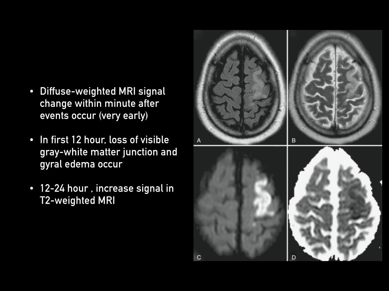
• Diffuse-weighted MRI signal change within minute after events occur (very early)
• In first 12 hour, loss of visible gray-white matter junction and gyral edema occur
• 12-24 hour , increase signal in T2-weighted MRI

Vasogenic edema
• Cause of vasogenic edema
• Primary or secondary brain tumour
• Brain abscess and encephalitis
• Trauma
• Lead poisoning
• Late stage of cerebral infarction

• Mechanisms
• Blood-tumor barrier has abnormal micro vessels that lacks of tight junctions cause plasma leakage into brain’s extracellular space
• Macromolecular protein produced by tumor has been identified as vascular permeability factor (VPF) and vascular endothelial growth factor (VEGF)

• Glucocorticoids can block permeability-enhancing effects of VPF and VEGF and inhibit tumor cell production of VPF and VEGF
• High VPF and VEGF gene expression found in glioblastomas, meningiomas and metastases

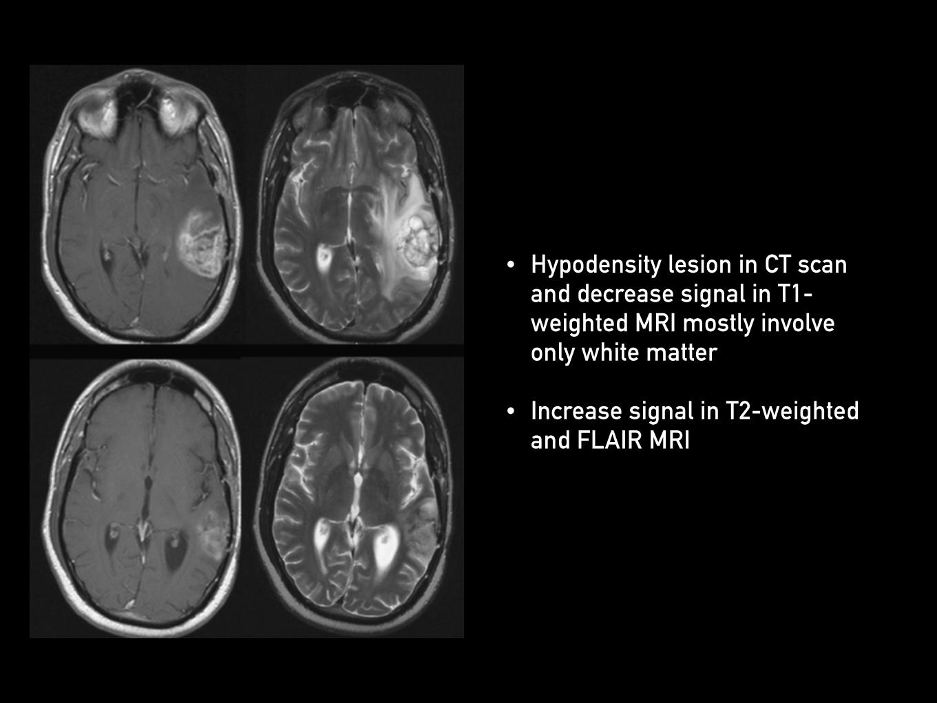
• Hypodensity lesion in CT scan and decrease signal in T1-weighted MRI mostly involve only white matter
• Increase signal in T2-weighted and FLAIR MRI

Interstitial edema

• Cause of interstitial edema
• Hydrocephalus
• Mechanism
• Transependymal flow of water and solute into periventricular extracellular space

• Hypodensity area around periventricular white matter in CT scan
• Increased signal in FLAIR MRI at interstitial brain surrounding ependyma

Osmotic edema

• Cause of osmotic edema
• Hemodialysis
• SIADH
• Hypertensive crisis
• Water intoxication
• Rapid reduction of blood glucose in hyperglycaemic crisis
• Mechanism
• Hyperosmolarity in brain relatively to circulatory then water move into brain along osmotic gradient


Treatment

• Specific treatment
• Direct treatment of causative disease or conditions
• Glucocorticoids have effect to peritumoral edema (mostly vasogenic edema) but less effect to cytotoxic and interstitial edema

• Diuretics can cause systemic dehydration and increase circulatory osmolarity and carbonic anhydrase inhibitor (acetazolamide) can reduce CSF production
• Mannitol and other osmotic agents temporality reduce cerebral edema (used in acute setting and prepare for definite treatments)

Thank you


![Acorus tatarinowii Schott extract reduces cerebral edema ......cerebral edema [11, 12]. Thus, the expression of glial fi-brillary acidic protein (GFAP), a marker of reactive astrogliosis,](https://static.documents.pub/doc/80x56/60f9fb03b1d27d0bb6581189/acorus-tatarinowii-schott-extract-reduces-cerebral-edema-cerebral-edema.jpg)

