33
HIP JOINT ANATOMY HIP JOINT ANATOMY Fahad zakwan Fahad zakwan MD5 MD5
| Date post: | 09-Aug-2015 |
| Category: |
Health & Medicine |
| Upload: | fahad-zakwan |
| View: | 38 times |
| Download: | 2 times |

HIP JOINT ANATOMYHIP JOINT ANATOMYFahad zakwanFahad zakwan
MD5MD5

ArticulationArticulation
•The hip joint is the articulation between The hip joint is the articulation between the hemispherical the hemispherical head of femurhead of femur and the and the cup shaped cup shaped acetabulumacetabulum of the hip bone of the hip bone•The articular surface of the acetabulum The articular surface of the acetabulum is horseshoe shaped and is deficient is horseshoe shaped and is deficient inferiorly at the acetabular notchinferiorly at the acetabular notch
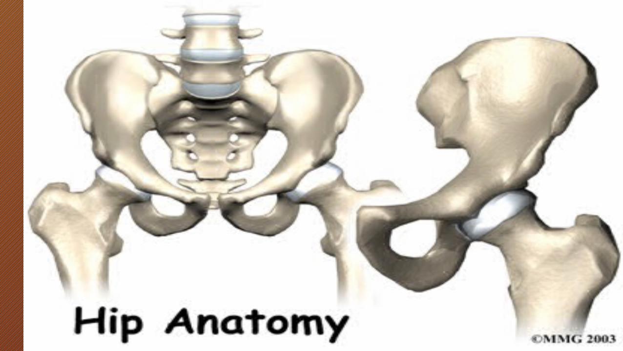

• Synovial ball and socket Synovial ball and socket joint.joint.• Femoral head: slightly Femoral head: slightly asymmetric, asymmetric, forms 2/3 forms 2/3 sphere.sphere.• Acetabulum: Acetabulum: inverted “U” inverted “U” shaped articular surface.shaped articular surface.• Ligamentum teresLigamentum teres, with , with artery to femoral head, artery to femoral head, passes through middle of passes through middle of inverted “U”. inverted “U”.


Joint Contact Joint Contact AreaArea
Throughout ROM:Throughout ROM:•40% of femoral head 40% of femoral head is in contact with is in contact with acetabular articular acetabular articular cartilage.cartilage.•10% of femoral head 10% of femoral head is in contact with is in contact with labrum.labrum.

Femoral neck angleFemoral neck angle

ACETABULUMACETABULUM
Strong fibrous ringStrong fibrous ringIncreases femoral head Increases femoral head coveragecoverageContributes to hip joint stabilityContributes to hip joint stability

ArticulationArticulation• The cavity of acetabulum is deepened by the The cavity of acetabulum is deepened by the presence of a presence of a fibrocartilaginous rim fibrocartilaginous rim called called acetabular labrumacetabular labrum• The labrum bridges across the acetabular The labrum bridges across the acetabular notch and is here called the notch and is here called the transverse transverse acetabular ligamentacetabular ligament• The articular surfaces are covered with The articular surfaces are covered with hyaline cartilagehyaline cartilage


Ligament and Other Ligament and Other StructuresStructures
• Joint capsuleJoint capsule• LigamentsLigaments
– IliofemoralIliofemoral– PubofemoralPubofemoral– IschiofemoralIschiofemoral
• Ligamentum teresLigamentum teres• Acetabular labrumAcetabular labrum• Inguinal ligamentInguinal ligament• Iliotibial band or tractIliotibial band or tract

Joint CapsuleJoint Capsule• Strong and thickStrong and thick• Covers hip cylindrically Covers hip cylindrically • AttachesAttaches
– Proximally - around lip of Proximally - around lip of acetabulumacetabulum
– Distally - neck of femurDistally - neck of femur
• Forms a cylindrical Forms a cylindrical sleeve sleeve – Encloses the joint and Encloses the joint and
most of the femoral neckmost of the femoral neck

LIGAMENTSLIGAMENTS

Iliofemoral Iliofemoral LigamentsLigaments
• It is a strong, inverted Y-shaped It is a strong, inverted Y-shaped ligamentligament
• Its base is attached to the Its base is attached to the anterior anterior inferior iliac spineinferior iliac spine above above
• Below the two limbs of Y are Below the two limbs of Y are attached to the attached to the upper and lower upper and lower parts of the intertrochanteric line parts of the intertrochanteric line of the femurof the femur
• The strong ligament prevents The strong ligament prevents overextension during standingoverextension during standing

Pubofemoral LigamentPubofemoral Ligament•It is a triangular It is a triangular ligamentligament•The base of the ligament The base of the ligament is attached to the is attached to the superior ramus of the superior ramus of the pubispubis•The apex is attached The apex is attached below to the lower part of below to the lower part of the intertrochanteric linethe intertrochanteric line•This ligament limits This ligament limits extension and abduction extension and abduction
Ischiofemoral Ischiofemoral LigamentLigament
•It is a spiral shaped It is a spiral shaped ligament ligament •Attached to the body of Attached to the body of the ischium near the the ischium near the acetabular marginacetabular margin•Fibers pass upward and Fibers pass upward and laterally and attached to laterally and attached to the greater trochanterthe greater trochanter•This ligament limits the This ligament limits the extensionextension


Ligamentum Ligamentum TeresTeres
• Small intracapsular ligamentSmall intracapsular ligament• Attaches:Attaches:
– Proximally in acetabulumProximally in acetabulum– Distally in fovea of the femoral Distally in fovea of the femoral
headhead
• Debatable importanceDebatable importance– Contains a blood vessel to Contains a blood vessel to
supply head of femursupply head of femur– ? Taut in adduction or lateral ? Taut in adduction or lateral
rotation when hip semiflexedrotation when hip semiflexed

Ligament of Head of FemurLigament of Head of Femur
• It is flat and triangular ligamentIt is flat and triangular ligament• It is attached by its apex to the pit on the It is attached by its apex to the pit on the head of the femur (fovea capitis)head of the femur (fovea capitis)•Attached by its base to the transverse Attached by its base to the transverse ligament and the margins of the acetabular ligament and the margins of the acetabular notchnotch• It lies within the joint and is ensheathed by It lies within the joint and is ensheathed by synovial membranesynovial membrane

Transverse Transverse Acetabular LigamentAcetabular Ligament
• It is It is formed by the formed by the acetabular labrum acetabular labrum as it bridges the as it bridges the acetabular notchacetabular notch• It It converts the converts the notch into a tunnel notch into a tunnel through which blood through which blood vessels and nerves vessels and nerves enter the jointenter the joint

Synovial MembraneSynovial Membrane
• The synovial membrane The synovial membrane lines the capsule lines the capsule • It is attached to the margins of the articular surfacesIt is attached to the margins of the articular surfaces• It covers the portion of the neck of the femur that lies within It covers the portion of the neck of the femur that lies within
the joint capsulethe joint capsule• It ensheathes the ligament of the head of the femurIt ensheathes the ligament of the head of the femur• It covers the pad of fat contained in the acetabular fossaIt covers the pad of fat contained in the acetabular fossa• A pouch of synovial membrane frequently protrudes A pouch of synovial membrane frequently protrudes
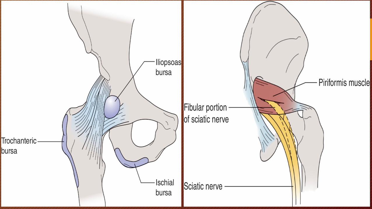
through a gap in the anterior wall of the capsulethrough a gap in the anterior wall of the capsule• Forms the psoas bursa beneath the psoas tendon Forms the psoas bursa beneath the psoas tendon


Surrounding Vital Structures:Surrounding Vital Structures:
• Blood Vessel & Blood Supply of the Blood Vessel & Blood Supply of the JointJoint
• femoral arteryfemoral artery passes by the front of passes by the front of the hip area, and has a deep branch, the hip area, and has a deep branch, called the called the profunda femorisprofunda femoris. The profunda . The profunda femoris sends two vessels that go femoris sends two vessels that go through the hip joint capsule. through the hip joint capsule.
• Lateral & Medial femoral circumflex Lateral & Medial femoral circumflex arteriesarteries
• These vessels are the main blood supply These vessels are the main blood supply for the femoral head, the ligamentum for the femoral head, the ligamentum teres (Ligament of the head of the femur) teres (Ligament of the head of the femur) contains a small blood vessel hat gives a contains a small blood vessel hat gives a very small supply of blood to the top of very small supply of blood to the top of the femoral head.the femoral head.

BLOOD SUPPLYBLOOD SUPPLY
• At age 4 years, the major blood supply to the femoral head comes At age 4 years, the major blood supply to the femoral head comes from the medial and lateral circumflex arteries .from the medial and lateral circumflex arteries .
• After age 4 years, the posterosuperior and posteroinferior arterial After age 4 years, the posterosuperior and posteroinferior arterial branches of the medial femoral circumflex bypass the growth plate branches of the medial femoral circumflex bypass the growth plate and form the main blood supply to the femoral head.and form the main blood supply to the femoral head.
• During adolescence, the growth plate fuses and the metaphyseal During adolescence, the growth plate fuses and the metaphyseal vessels again become significant, traveling along the femoral neck.vessels again become significant, traveling along the femoral neck.
• Fractures in this area can disrupt this delicate blood supply, Fractures in this area can disrupt this delicate blood supply, leading to AVN, the most severe complication of this fracture.leading to AVN, the most severe complication of this fracture.


NervesNerves::
•All of the nerves that travel All of the nerves that travel down the thigh pass by the down the thigh pass by the hip. The main nerves are hip. The main nerves are the the femoral nervefemoral nerve in front in front and the and the sciatic nervesciatic nerve in in back of the hip. A smaller back of the hip. A smaller nerve, called the nerve, called the obturator obturator nervenerve, also goes to the hip, also goes to the hip


Movements at the HipMovements at the Hip
1.1. FlexionFlexion2.2. ExtensionExtension3.3. AbductionAbduction4.4. AdductionAdduction5.5. Medial RotationMedial Rotation6.6. Lateral RotationLateral Rotation
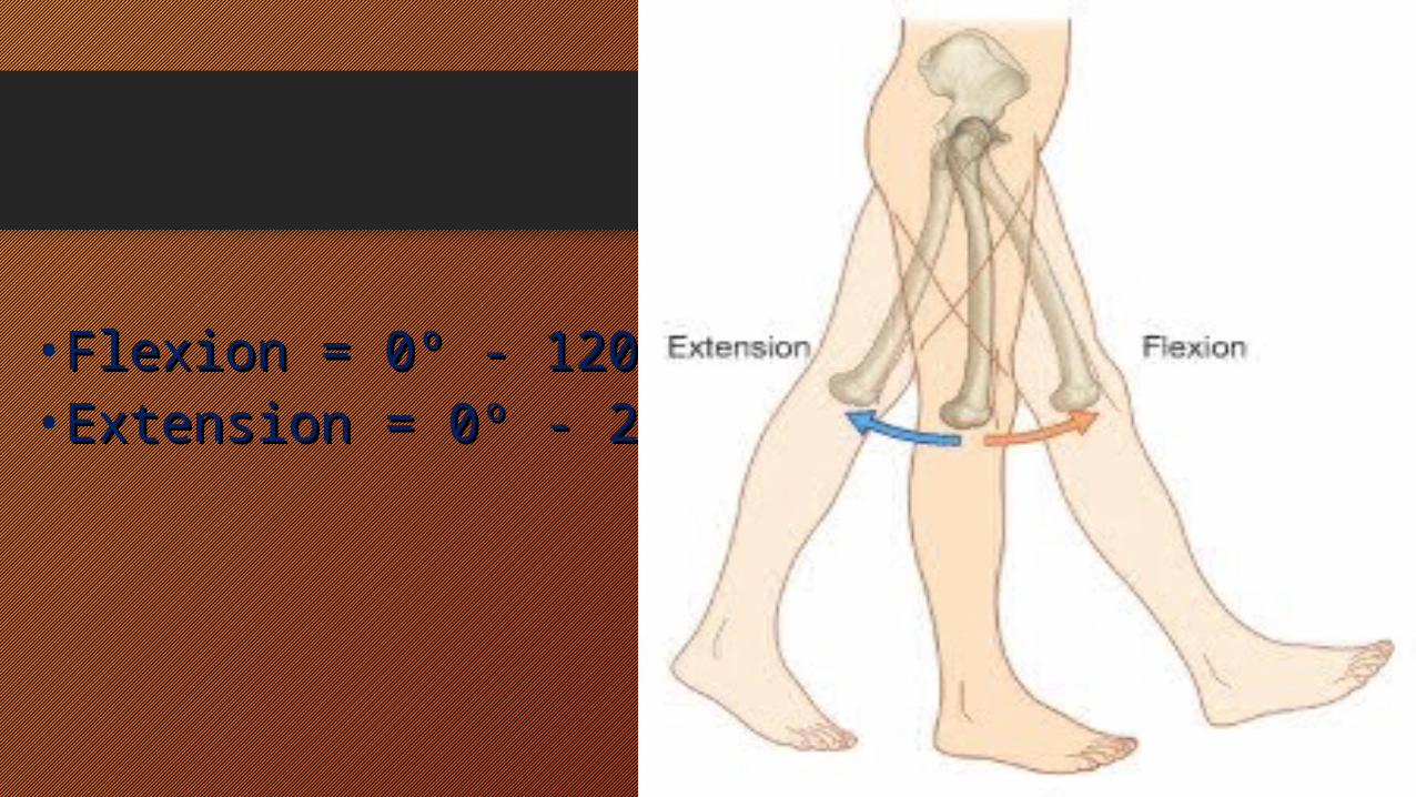
• Flexion = 0Flexion = 0ºº - 120 - 120ºº• Extension = 0Extension = 0ºº - 20 - 20ºº

• Abduction = 0Abduction = 0ºº - 45 - 45ºº• Adduction = 0Adduction = 0ºº - 25 - 25ºº

• Internal Rotation = 0Internal Rotation = 0ºº - 45 - 45ºº• External Rotation = 0External Rotation = 0ºº - 45 - 45ºº


