3,350+ OPEN ACCESS BOOKS 108,000+ INTERNATIONAL AUTHORS AND EDITORS 115+ MILLION DOWNLOADS BOOKS DELIVERED TO 151 COUNTRIES AUTHORS AMONG TOP 1% MOST CITED SCIENTIST 12.2% AUTHORS AND EDITORS FROM TOP 500 UNIVERSITIES Selection of our books indexed in the Book Citation Index in Web of Science™ Core Collection (BKCI) Chapter from the book Parasitology Downloaded from: http://www.intechopen.com/books/parasitology PUBLISHED BY World's largest Science, Technology & Medicine Open Access book publisher Interested in publishing with IntechOpen? Contact us at [email protected]
Transcript
3,350+OPEN ACCESS BOOKS
108,000+INTERNATIONAL
AUTHORS AND EDITORS115+ MILLION
DOWNLOADS
BOOKSDELIVERED TO
151 COUNTRIES
AUTHORS AMONG
TOP 1%MOST CITED SCIENTIST
12.2%AUTHORS AND EDITORS
FROM TOP 500 UNIVERSITIES
Selection of our books indexed in theBook Citation Index in Web of Science™
Core Collection (BKCI)
Chapter from the book Paras itologyDownloaded from: http://www.intechopen.com/books/paras itology
PUBLISHED BY
World's largest Science,Technology & Medicine
Open Access book publisher
Interested in publishing with IntechOpen?Contact us at [email protected]
Genotyping of Giardia intestinalis Isolates from Dogs by Analysis of gdh, tpi, and bg Genes
Enedina Jiménez-Cardoso, Leticia Eligio-García, Adrian Cortés-Campos and Apolinar Cano-Estrada
Laboratorio de Investigación en Parasitología Hospital Infantil de México Federico Gómez, México, D.F.
Mexico
1. Introduction
Giardia intestinalis, a flagellated protozoan parasite, is the most prevalent human intestinal
protozoan worldwide (Adam, 2001). About 200 million people in the world are infected
with giardiasis and each individual eliminates up to 900 million cysts per day (Minivielle ,
2008). Higher prevalence is found in tropical and subtropical areas, where Giardia affects up
to 30% of the population. In epidemiological studies carried out in Mexico and other
sudamerican countries, prevalence between of 10-16% has been found in urban areas and
34% in shantytowns (Gamboa “et al”, 2003; Giraldo-Gomez, 2005; Sulaiman, 2004). G.
intestinalis is a cosmopolitan pathogen with a very wide host range, including humans,
domestic animals, and wild animal species (Caccio, 2008; Thompson, et al 1993). The most
common cause of infection with Giardia is the consumption of contaminated food or water
(Ortega, 1997), although zoonotic transmission is also possible. Once a person is infected, the
parasite lives in the intestines and is passed in the stool of the infected person. Animals such
as cats, dogs and cattle can also be infected and spread the disease to humans.
Infections may be asymptomatic or include symptoms of chronic diarrhea, weight loss, and
malabsorption. When children infected with Giardia have no symptoms of giardiasis, the
parasite is present in their feces and they can pass the infection to others. Other symptoms
of chronic giardiasis include: Loose, soft, greasy stools, discomfort in the abdomen, general
feeling of discomfort or illness, weakness and fatigue.
The parasite has two interchangeable forms that guarantee a simple and efficient life cycle.
The cyst that contaminate the environment and the trophozoite, which attach to the
intestinal villi via a specialized microtubule structure, the ventral disc.
There are at least seven major genotypes referred to as assemblages (A-G) including 2 (A
and B) known to infect humans (Mcpherson, 2005; Monis, 2009).
Assemblages A and B, of clinical significance to humans, differ from each other by as much
as 20% at the DNA sequence level (Caccio, 2008). There is also evidence that genetic
exchange has resulted in hybrids, or mixed types, based on assemblage-specific PCR of
Giardia isolates from cases of human infection (Monis, 1999).
www.intechopen.com
Parasitology
68
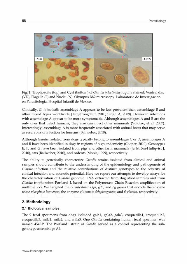
Fig. 1. Trophozoite (top) and Cyst (bottom) of Giardia intestinalis lugol´s stained. Ventral disc
(VD), Flagella (F) and Nuclei (N). Olympus Bh2 microscopy. Laboratorio de Investigacion
en Parasitologia. Hospital Infantil de Mexico.
Clinically, G. intestinalis assemblage A appears to be less prevalent than assemblage B and
other mixed types worldwide (Tungtrongchitr, 2010; Singh A, 2009). However, infections
with assemblage A appear to be more symptomatic. Although assemblages A and B are the
only ones that infect humans, they also can infect other mammals (Volotao, et al. 2007).
Interestingly, assemblage A is more frequently associated with animal hosts that may serve
as reservoirs of infection for humans (Ballweber, 2010).
Although Giardia isolated from dogs typically belong to assemblages C or D, assemblages A
and B have been identified in dogs in regions of high endemicity (Cooper, 2010). Genotypes
E, F, and G have been isolated from pigs and other farm mammals (Jerlström-Hultqvist J,
2010), cats (Ballweber, 2010), and rodents (Monis, 1999), respectively.
The ability to genetically characterize Giardia strains isolated from clinical and animal
samples should contribute to the understanding of the epidemiology and pathogenesis of
Giardia infection and the relative contributions of distinct genotypes to the severity of
clinical infection and zoonotic potential. Here we report our attempts to develop assays for
the characterization of Giardia genomic DNA extracted from dog stool samples and from
Giardia trophozoites Portland I, based on the Polymerase Chain Reaction amplification of
multiple loci. We targeted the G. intestinalis tpi, gdh, and bg genes that encode the enzyme
triose-phosphate isomerase, the enzyme glutamate dehydrogenase, and β-giardin, respectively.
2. Methodology
2.1 Biological samples
The 9 fecal specimens from dogs included gala1, gala2, gala3, croquetilla1, croquetilla2,
croquetilla3, mila1, mila2, and mila3. One Giardia containing human fecal specimen was
named 454LP. The Portland1 strain of Giardia served as a control representing the sub-
genotype assemblage AI.
www.intechopen.com
Genotyping of Giardia intestinalis Isolates from Dogs by Analysis of gdh, tpi, and bg Genes
69
2.2 Coproparasitoscopic analysis
Fecal specimens were stained with Lugol’s iodine (Faust, 1938) and examined by
microscopy to find cysts of Giardia intestinalis. Cysts were concentrated from dog feces by
repeated washing in distilled water and stored at 4 ºC until use.
2.3 DNA extraction
DNA was extracted from the fecal samples by use of the QIAamp DNA Stool Mini Kit (Qiagen Inc., Valencia, CA) according to the manufacturer’s instructions. All DNA concentrations were determined by using an Epoch spectrophotometer (Biotek, Winooski, VT).
2.4 PCR amplification
The gdh gene encoding glutamate dehydrogenase, the tpi gene encoding triose phosphate
isomerase, and the bg gene encoding β-giardin were each amplified using the Polymerase
Chain Reaction (PCR) as follows.
2.5 gdh gene amplification
The gdh gene was amplified by using 0.8 µM of each primer (578: 5´-
GAGAGGATCCTTGARCCNGAGCGCGTNATC-3´ and 579:5´-
CCGCGNTTGTADCGNCCNAAGATCTTCCA-3´) in 50 µL reactions containing 10 mM
Tris-HCl, 50 mM KCl, 4 mM MgCl2, 0.2 mM each dNTP, 1 U Taq DNA Polymerase and 200
ng of genomic DNA. Samples were subjected to 30 cycles of [94 °C for 30 s, 56 °C for 30 s,
and 72 °C for 2 min], with an initial denaturation step at 94 °C for 4 min, and a final
extension step at 72 °C for 6 min (Monis, 1996).
2.6 bg gene amplification
The bg gene was amplified in two steps via nested‐PCR. The first round of PCR was
conducted in a 25‐µL reaction containing 200 pmol each primer (G7:
5’‐AAGCCCGACGACCTCACCCGCACTGC‐3’ and G759:
5’‐GAGGCCGCCCTGGATCTTCGAGACGAC‐3’), 10 mM Tris‐HCl, 50 mM KCl, 1 mM
MgCl2, 0.2 mM each dNTP, 2.5 U Taq DNA Polymerase and 200 ng of genomic DNA. This
was amplified for 45 cycles of [95 °C 30 s, 65 °C 30 s and 72 °C 1 min]. The second round of
PCR, using the product of the first reaction as template, was performed in a 50‐µL reaction
with 200 pmol each primer (F: 5´‐GAACGAGATCGAGGTCCG‐3´; R:
5’‐CTCGACGAGCTTCGTT‐3’), 10 mM Tris‐HCl, 50 mM KCl, 1 mM MgCl2, 0.2 mM dNTPs,
2.5 U of Taq DNA Polymerase and 3 µL of template. Amplification was for 35 cycles of [94°C
30 s, 53°C 30 s and 72°C 1 min] (Lalle, 2005).
2.7 tpi gene amplification
The tpi gene was amplified by nested‐PCR in which the first round was a duplex reaction to
amplify two fragments corresponding to genotypes A and B simultaneously using four
primers (TPIAF 5´‐CGAGACAAGTGTTGAGATGC‐3´, TPIAR
5´‐GGTCAAGAGCTTACAACACG‐3´ and TPIBF 5´‐GTTGCTCCCTCCTTTGTGC‐3´, TPIBR
www.intechopen.com
Parasitology
70
5´‐CTCTGCTCATTGGTCTCGC‐3´). PCR amplification was performed in a volume of 50‐µL
with 500 ng of DNA in 1X PCR buffer, 2 mM MgCl2, 0.25 mM of dNTP and 1 U of Taq DNA
Polymerase. Amplification was achieved with 25 cycles of [94 ºC for 20 s, 50 ºC for 30 s and
72 ºC for 1 min]. The second round of PCR comprised two separate hemi-nested PCRs to
amplify internal fragments of 476 bp and 140 bp corresponding to the A and B genotypes
respectively.
To amplify genotype A, primers TPIAIF: 5´‐CCAAGAAGGCTAAGCGTGC‐3´ and TPIAR were used using 3 µL of the first round amplicon as template in a 50‐µL volume reaction. The amplification step used 33 cycles of [94 °C for 20 s, 56 °C for 30 s, and 72 °C for 1 min]. Alternatively, the 140 bp fragment corresponding to genotype B was amplified with primers TPIBIF: 5´‐GCACAGAACGTGTATCTGG‐3´ and TPIBR. Amplification was performed under the same conditions used for A except that the MgCl2 concentration in the PCR mixture was 1.5 mM. (Amar, 2003. Molina, 2005)
2.8 Restriction analysis
The amplicons generated by PCR were digested with restriction enzymes for the purpose of subtyping. The tpi gene amplicons were digested with restriction enzyme RsaI, the bg gene amplicons were digested with HaeIII, and the gdh amplicons were digested with BspHI. The products of restriction enzyme digestion were separated by 2% agarose gel electrophoresis, using 100bp DNA ladder (Promega, Madison,Wi. USA) as a size standard, and visualized by staining with ethidium bromide.
3. Results
We developed a molecular method to test stool samples for the presence of G. intestinalis genotypes that are of clinical significance to human infection possibly by zoonotic transmission from dogs. Giardia infection of dog stool samples was confirmed by coproparasitoscópico analysis (data not shown). Giardia cysts isolated from feces was genotyped by a combination of multi‐locus (gdh, bg, tpi) PCR followed by restriction analysis of the PCR amplicons. We analyzed nine samples of dog stool, one sample of human stool, and G. intestinalis cysts from the Portland‐1 control strain. The Portland‐1 standard is a control for the A‐I assemblage.
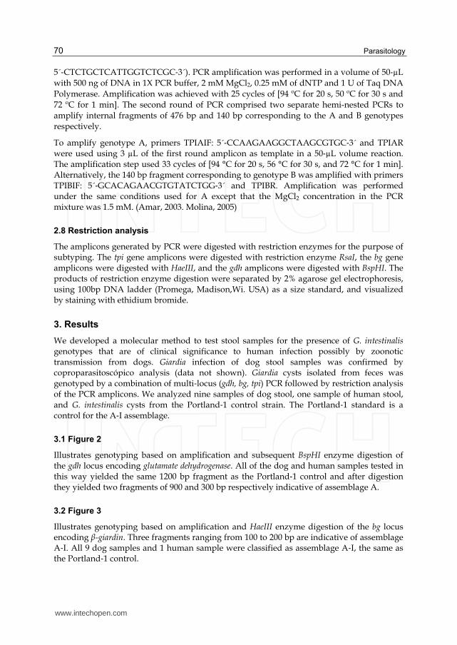
3.1 Figure 2
Illustrates genotyping based on amplification and subsequent BspHI enzyme digestion of the gdh locus encoding glutamate dehydrogenase. All of the dog and human samples tested in this way yielded the same 1200 bp fragment as the Portland‐1 control and after digestion they yielded two fragments of 900 and 300 bp respectively indicative of assemblage A.
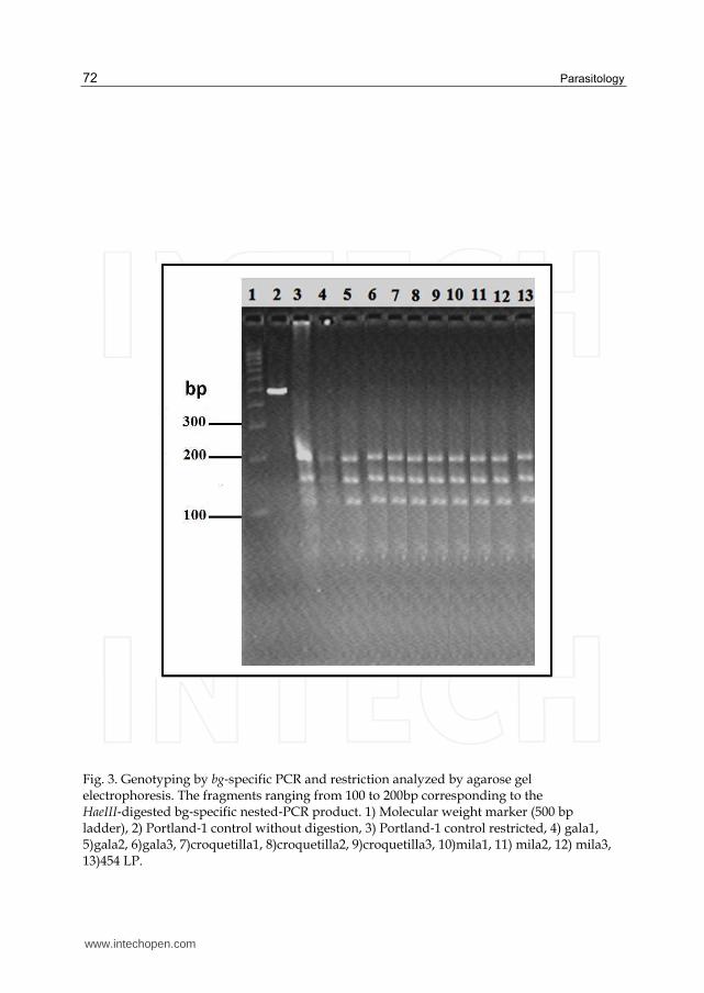
3.2 Figure 3
Illustrates genotyping based on amplification and HaeIII enzyme digestion of the bg locus encoding β-giardin. Three fragments ranging from 100 to 200 bp are indicative of assemblage A‐I. All 9 dog samples and 1 human sample were classified as assemblage A‐I, the same as the Portland‐1 control.
www.intechopen.com
Genotyping of Giardia intestinalis Isolates from Dogs by Analysis of gdh, tpi, and bg Genes
71
3.3 Figures 4
Illustrate genotyping based on amplification and RsaI enzyme digestion of the tpi locus
encoding triose‐phosphate isomerase. Again, all samples yielded products consistent with their
identification as belonging to G. intestinalis genotype AI.
Fig. 2. Genotyping by gdh‐specific PCR and restricted analyzed by agarose gel electrophoresis. The 900 bp band corresponds to the BspHI‐digested gdh‐specific PCR product. 1) Molecular weight marker (500 bp ladder), 2) Portland‐1 control without digestion, 3) Portland‐1 control restricted, 4) gala1, 5)gala2, 6)gala3, 7)croquetilla1, 8)croquetilla2, 9)croquetilla3, 10)mila1, 11) mila2, 12) mila3, 13) 454 LP.
www.intechopen.com
Parasitology
72
Fig. 3. Genotyping by bg‐specific PCR and restriction analyzed by agarose gel electrophoresis. The fragments ranging from 100 to 200bp corresponding to the HaeIII‐digested bg‐specific nested‐PCR product. 1) Molecular weight marker (500 bp ladder), 2) Portland‐1 control without digestion, 3) Portland‐1 control restricted, 4) gala1, 5)gala2, 6)gala3, 7)croquetilla1, 8)croquetilla2, 9)croquetilla3, 10)mila1, 11) mila2, 12) mila3, 13)454 LP.
www.intechopen.com
Genotyping of Giardia intestinalis Isolates from Dogs by Analysis of gdh, tpi, and bg Genes
73
Fig. 4. Genotyping by tpi‐specific PCR and restriction analyzed by agarose gel
electrophoresis. The 437 bp band corresponds to the RsaI‐digested amplicon representative
of assemblage A‐I. 1) Molecular weight marker (500 bp ladder), 2) Portland‐1 control
without digestion, 3) Portland‐1 control restricted, 4) gala1, 5)gala2, 6)gala3, 7)croquetilla1,
Human giardiasis is caused by two genetically distinct assemblages (A and B) of G.
intestinalis. A number of molecular assays have been developed for their specific detection in
stool and environmental samples (Caccio, 2008).
We have developed a method to detect Giardia based on the PCR amplification of three
genes (gdh, bg, tpi) used in prior genotyping studies.
Although DNA-based methods reported in the literature have been used with success to
genotype Giardia, we did not have observed differences among the analysis of different
genes in studied samples. Some researchers had found frequent mismatches, intra-
assemblage discordances and mixed positions, in tpi and in bg sequences, especially in
assemblage B (Bonhomme, 2011).
All of the fecal samples analyzed in this report (9 from dogs, 1 from humans) were
determined to belong to the sub-genotype A‐I assemblage. This predominance of
assemblage A‐I probably reflects the mechanism that led to infection of the animals from
which the fecal samples came. The dogs might have drunk from water that had been
www.intechopen.com
Parasitology
74
contaminated by livestock rather than by humans. This has important epidemiological
ramifications.
5. References
Adam RD. (2001). Biology of Giardia lamblia. Clinical Microbiology Review. ; 14 (3), pp.447-75.
Adam RD, Nigam A, Seshadri V, Martens CA, Farneth GA, Morrison HG, Nash TE, Porcella SF, Patel R. (2010). The Giardia lamblia vsp gene repertoire: characteristics, genomic organization, and evolution. BMC Genomics, 11 (9), pp. 424.
Almeida A, Pozio E, Cacciò SM. (2010). Genotyping of Giardia duodenalis cysts by new Real-Time PCR assays for detection of mixed infections in human samples. Applied and Environmental Microbiology, 76(6), pp. 1895–1901.
Almeida A, Moreira MJ, Soares S, Delgado ML, Figueiredo J, Silva Magalhães E, Castro A, Da Costa AV, Correia da Costa JM. (2010). Biological and genetic characterization of Cryptosporidium spp. and Giardia duodenalis isolates from five hydrographical basins in northern Portugal. Korean Journal of Parasitology, 48(2) pp. 105–111.
Ballweber LR, Xiao L, Bowman DD, Kahn G, Cama VA. (2010). Giardiasis in dogs and cats: update on epidemiology and public health significance. Trends in Parasitology, 26(4), pp. 180-9.
Bonhomme J, Le Goff L, Lemée V, Gargala G, Ballet JJ, Favennec L. (2011). Limitations of tpi and bg genes sub-genotyping for characterization of human Giardia duodenalis isolates. Parasitology International, 60(3), pp. 327-30.
Cacciò SM, Ryan U. (2008). Molecular epidemiology of giardiasis. Molecular and Biochemical Parasitology, 160(2), pp.75-80.
Cooper MA, Sterling CR, Gilman RH, Cama V, Ortega Y, Adam RD. (2010). Molecular analysis of household transmission of Giardia lamblia in a region of high endemicity in Peru. The Journal of Infectious Diseases, 202(11), pp. 1713-21.
Amar CF, Dear PH, McLauchlin J. (2003). Detection and genotyping by real-time PCR/RFLP analyses of Giardia duodenalis from human feces. Journal of Medical Microbiology, 52, pp. 681–683.
De Boer RF, Ott A, Kesztyüs B, Kooistra-Smid AMD. (2010). Improved Detection of Five Major Gastrointestinal Pathogens by Use of a Molecular Screening Approach. Journal of Clinical Microbiology, 48(11), pp. 4140–4146.
Faust EC, D´antonio JS, Odom V, Miller MJ, Peres C, Sawitz W, Thomen LF, Tobie J, Walker JH . (1938). A critical study of clinical laboratory techniques for the diagnosis of the protozoan cysts and helminthes egg in feces. The American Journal of Tropical Medicine and Hygiene, 18, pp. 169-83.
Jerlström-Hultqvist J, Franzén O, Ankarklev J, Xu F, Nohýnková E, Andersson JO, Svärd SG, Andersson B. (2010). Genome analysis and comparative genomics of a Giardia intestinalis assemblage E isolate. BMC Genomics, 11, pp.543.
Lalle M, Jimenez-Cardosa E, Cacciò SM, Pozio E. (2005). Genotyping of Giardia duodenalis from humans and dogs from Mexico using a beta-giardin nested polymerase chain reaction assay. The Journal of Parasitology, 91(1), pp. 203-5.
www.intechopen.com
Genotyping of Giardia intestinalis Isolates from Dogs by Analysis of gdh, tpi, and bg Genes
75
Macpherson CN. (2005). Human behaviour and the epidemiology of parasitic zoonoses. International Journal of Parasitology, 35(11-12), pp.1319-31.
Minvielle MC, Molina NB, Polverino D, Basualdo JA. (2008). First genotyping of Giardia lamblia from human and animal feces in Argentina, South America. Memorias do Instituto Oswaldo Cruz, 103(1), pp. 98-103.
Molina N, Polverino D, Minvielle M, Basualdo J. (2007). PCR amplification of triosephosphate isomerase gene of Giardia lamblia in formalin-fixed feces. Revista Latinoamericana de Microbiología, 49(1-2), pp. 6-11.
Monis PT, Mayrhofer G, Andrews RH, Homan WL, Limper L, Ey PL. (1996). Molecular genetic analysis of Giardia intestinalis isolates at the glutamate dehydrogenase locus. Parasitology, 112 (Pt 1), pp. 1-12.
Monis PT, Andrews RH, Mayrhofer G, Ey PL. (1999). Molecular systematics of the parasitic protozoan Giardia intestinalis. Molecular Biology and Evolution, 16(9), pp. 1135-44.
Ortega YR, Adam RD. (1997). Giardia: overview and update. Clinical of Infectious Diseases, 25, pp. 545-550.
Plutzer J, Ongerth J, Karanis P. (2010). Giardia taxonomy, phylogeny and epidemiology: Facts and open questions. International Journal of Hygiene and Environmental Health, 15(5), pp. 321-33.
Singh A, Janaki L, Petri WA Jr, Houpt ER. (2009). Giardia intestinalis assemblages A and B
infections in Nepal. The American Journal of Tropical Medicine and Hygiene, 81(3), pp.
538-9.
Sulaiman IM, Jiang J, Singh A, Xiao L. (2004). Distribution of Giardia duodenalis genotypes
and subgenotypes in raw Urban Wastewater in Milwaukee, Wisconsin. Applied
Environmental Microbiology, 70(6), pp. 3776–3780.
Ten Hove RJ, van Esbroeck M, Vervoort T, van den Ende J, van Lieshout L, Verweij JJ. (2009)
Molecular diagnostics of intestinal parasites in returning travelers. European Journal
of Clinical Microbiology and Infectious Diseases, 28(9), pp. 1045–1053.
Thompson RC, Reynoldson JA, Mendis AH. (1993). Giardia and giardiasis. Advances in
Parasitology, 32, pp. 71-160.
Tungtrongchitr A, Sookrung N, Indrawattana N, Kwangsi S, Ongrotchanakun J, Chaicumpa
W. (2010). Giardia intestinalis in Thailand: identification of genotypes. Journal of
Health, Population and Nutrition, 28(1), pp. 42-52.
van Keulen H, Macechko PT, Wade S, Schaaf S, Wallis PM, Erlandsen SL.(2002). Presence of
human Giardia in domestic, farm and wild animals, and environmental samples
suggests a zoonotic potential for giardiasis. Veterinary Parasitology, 108(2), pp.97-
107.
Volotão AC, Costa-Macedo LM, Haddad FS, Brandão A, Peralta JM, Fernandes O.
(2007). Genotyping of Giardia duodenalis from human and animal samples from
Brazil using beta-giardin gene: a phylogenetic analysis. Acta Tropica, 102(1),
pp.10-9.
Gamboa M, Basualdo J, Córdoba M, Pezzani B, Minvielle M, Lahitte H. (2003). Distribution
of intestinal parasitoses in relation to environmental and sociocultural parameters
in La Plata, Argentina. Journal of Helminthology, 77, pp. 15-20.
www.intechopen.com
Parasitology
76
Giraldo-GómezI J; LoraII F; Henao LH; Mejía S, Gómez-Marín J. (2005) Prevalence of
giardiasis and intestinal parasites in pre-school children from homes being
attended as part of a state programme in Armenia, Colombia. Revista de Salud
Pública, 7(3), pp. 327-338.
www.intechopen.com
ParasitologyEdited by Dr. Mohammad Manjur Shah
ISBN 978-953-51-0149-9Hard cover, 206 pagesPublisher InTechPublished online 14, March, 2012Published in print edition March, 2012
InTech ChinaUnit 405, Office Block, Hotel Equatorial Shanghai No.65, Yan An Road (West), Shanghai, 200040, China
Phone: +86-21-62489820 Fax: +86-21-62489821
Parasitology is an established discipline that covers a wide area of subjects, ranging from the basics (study oflife cycle, ecology, epidemiology, taxonomy, biodiversity, etc) to the advanced and applied aspects (humanand animal related, although control aspect remains the most important task). There is a great scarcity in theamount of available literature that is freely accessible to anyone interested in the subject. This book wasconceptualized with this in mind. The entire book is based on the findings of various studies performed bydifferent authors, comprising reviews and original scientific papers. I hope this book will be helpful to diverseaudiences like biologists, zoologists, nematologists, parasitologists, microbiologists, medical doctors,pathologists as well as the molecular biologists, by providing them with a better understanding of the subject.
How to referenceIn order to correctly reference this scholarly work, feel free to copy and paste the following:
Enedina Jiménez-Cardoso, Leticia Eligio-García, Adrian Cortés-Campos and Apolinar Cano-Estrada (2012).Genotyping of Giardia intestinalis Isolates from Dogs by Analysis of gdh, tpi, and bg Genes, Parasitology, Dr.Mohammad Manjur Shah (Ed.), ISBN: 978-953-51-0149-9, InTech, Available from:http://www.intechopen.com/books/parasitology/genotyping-of-giardia-intestinalis-isolates-from-dogs-by-analysis-of-gdh-tpi-and-b-giardin-genes-


![multiplexing stool MD short.ppt [Compatibiliteitsmodus] · 8 0394-0527 Giardia Entamoeba coli Giardia 9 0394-0526 Giardia Entamoeba coli Giardia 10 1032-1221 Giardia negative Giardia](https://static.documents.pub/doc/80x56/5e4182dad3a23a3d0b082f2c/multiplexing-stool-md-shortppt-compatibiliteitsmodus-8-0394-0527-giardia-entamoeba.jpg)


