02 Immunology and Oncology Research in the Department of Immunology and Oncology focusses on the molecular and cellular bases of immune system function and tumour development, to develop improved approaches for immune response modulation during infection and inflammatory reactions, to innovate in vaccination strategies, and to identify targets for the prevention, diagnosis and treatment of cancer. The various research groups in the department address many aspects of innate and adaptive immunity, with special emphasis on characterising the molecular mechanisms that underlie inflammation, the processes that drive tissue-specific tumour development, as well as tumour immunology and the relationships among stem cells, inflammation and cancer. From a methodological perspective, the department’s activities are multidisciplinary, combining advanced microscopy and flow cytometry, next-generation genomics technologies, the use of animal models, and the generation of complex genetically modified mouse lines.
Transcript
0202 Immunology and Oncology
Research in the Department of Immunology and Oncology focusses on the molecular and cellular bases of immune system function and tumour development, to develop improved approaches for immune response modulation during infection and inflammatory reactions, to innovate in vaccination strategies, and to identify targets for the prevention, diagnosis and treatment of cancer.
The various research groups in the department address many aspects of innate and adaptive immunity, with special emphasis on characterising the molecular mechanisms that underlie inflammation, the processes that drive tissue-specific tumour development, as well as tumour immunology and the relationships among stem cells, inflammation and cancer.
From a methodological perspective, the department’s activities are multidisciplinary, combining advanced microscopy and flow cytometry, next-generation genomics technologies, the use of animal models, and the generation of complex genetically modified mouse lines.
SELECTED PUBLICATIONS Differentiation and functional specialisation of dendritic cells during inflammatory, infectious and allergic processes
Our research explores the functional specialisation of dendritic cells derived from monocytes during inflammatory responses caused by infection by bacteria (Listeria monocytogenes), yeasts (Candida albicans) and parasites (Leishmania major) or by allergic reactions induced by plant- , fungi- or acaridae-derived allergens. Our current research interests are focussed on the following topics:
• Analysis of the effector functions of mouse monocytes in innate and adaptive immunity• Analysis of the differential migratory properties of mouse monocyte-derived dendritic cells and macrophages.• Regulation of mouse monocyte differentiation into dendritic cells and macrophages during in vivo immune response to Leishmania major.• Functional specialisation of mouse dendritic cells for the induction of Th2 responses against pathogens and allergens.• Gene expression profile of mouse monocytes and monocyte-derived dendritic cells
exposed to allergens and Th2-polarising mediators.• Effect of statin treatment on proinflammatory cytokine production and nitric oxide metabolism by LPS-activated or Listeria monocytogenes-infected mouse monocyte-derived dendritic cells.• Analysis of the splenic innate immune response to in vivo Listeria monocytogenes infection in statin-treated mice.• Role of type-I interferon in the induction of Th17 immune responses against Candida albicans in mice.
Domínguez PM, López-Bravo M, Kalinke U, Ardavín C. Statins inhibit iNOS-mediated microbicidal potential of activated monocyte-derived dendritic cells by an IFN-β-dependent mechanism. Eur J Immunol. 2011 Nov;41(11):3330-9
Leirião P, del Fresno C, Ardavín C. Monocytes as effector cells: activated Ly-6C(high) mouse monocytes migrate to the lymph nodes through the lymph and cross-present antigens to CD8+ T cells. Eur J Immunol. 2012 Aug;42(8):2042-51
Risco A, del Fresno C, Mambol A, Alsina-Beauchamp D, MacKenzie KF, Yang HT, Barber DF, Morcelle C, Arthur JS, Ley SC, Ardavin C, Cuenda A. p38γ and p38δ kinases regulate the Toll-like receptor 4 (TLR4)-induced cytokine production by controlling ERK1/2 protein kinase pathway activation. Proc Natl Acad Sci USA. 2012 Jul 10;109(28):11200-5
Martin Caballero J, Garzón A, González-Cintado L, Kowalczyk W, Jimenez Torres I, Calderita G, Rodriguez M, Gondar V, Bernal JJ, Ardavín C, Andreu D, Zürcher T, von Kobbe C. Chimeric infectious bursal disease virus-like particles as potent vaccines for eradication of established HPV-16 E7-dependent tumors. PLoS One. 2012;7(12):e52976
GROUP LEADER:Carlos Ardavín
POSTDOCTORAL SCIENTISTS:María López-BravoPilar DomínguezCarlos del FresnoPatricia LeiriaoDidier Soulat
Identification of regulators of activation, apoptosis and proliferation specific for controlling autoimmune T cell memory and inflammation
Novel p21 and Fas functions in normal and autoimmune T memory responses
Apoptosis is considered a basic mechanism for limiting the T cell memory expansion known as homeostasis. Activation and proliferation of memory T cells are also critical for homeostasis control. Autoimmune memory T cells present unique features compared to normal memory T cells, due to their repetitive encounter with autoantigens. Our work focusses on identifying the differences in the control of expansion between naïve, memory and autoimmune memory T cells.
The cell cycle inhibitor p21 suppresses autoimmunity and controls autoimmune but not normal T cell memory responses. We have shown that p21 is an autoimmunity
suppressor, since p21 deficiency leads to autoimmunity. Indeed, p21 overexpression in T cells of autoimmune lupus-prone Fas-deficient (B6-lpr) mice reduces autoimmunity and lymphadenopathy development (Fig. 1). p21 limits the expansion of autoreactive B6-lpr memory T cells but not of normal memory T cells. Our work identifies a p21 function in the regulation of TCR-dependent activation of memory T cells.
An alternative function for the Fas/FasL apoptosis system. Our analysis of memory T cell activation, apoptosis and cell cycle regulation events in immunity and autoimmune disease revealed a previously unknown function of Fas. We established that Fas also has a crucial attenuating role in the response of previously activated T cells, but not of primary T cells. We focussed on the mechanistic aspects of this role of the Fas/FasL system on preactivated T cells. As shown in Figure 2, Fas interaction with the TCR receptor following secondary stimulation T cell stimulation is evident.
p21 regulates the macrophage activation and
inflammation
Independently of its cell cycle inhibitory capacity, p21 regulates macrophage activation by controlling the NF-κB pathway. p21 regulation of NF-κB activation is critical for progression of in vivo inflammation, since it decreases sensitivity to LPS-induced septic shock. We are studying the mechanism that defines the role of p21 in NF-κB activation.
MASTER’S DEGREE STUDENTS: Martina Romagnoni María Jesus Piña
UNDERGRADUATE STUDENTS: Adrián Madrigal Avilés
Balomenos D. Cell Cycle regulation and systemic lupus erythematosus. Systemic Lupus Erythematosus (5th Edition). (2011) Chapter: 11 Págs:191-197. Editor:LAHITA/ELSEVIER.
Mavers M, Alexander V. Misharin, Carla M Cuda, Angelica K Gierut, Hemant Agrawal, Evan Weber, G. Kenneth Haines III, Balomenos D, Perlman H. The Cyclin Dependent Kinase Inhibitor p21(WAF1/CIP1/SDI1) Suppresses and Mediates Resolution of Inflammatory Arthritis via its C-Terminal Domain. Arthritis and Rheumatism (2012).
SELECTED PUBLICATIONS
1
2
1 Decreased lymph node size in the presence of the p21 transgene in B6/lpr-p21tg compared to B6/lpr mice at 8 months of age. 2 T cells from B6 (A) and Fas-deficient B6/lpr mice (B) were stained after
secondary TCR stimulation, to detect Fas (green) and TCR (red), and mounted with DAPI (blue) for confocal microscopy. Results show strong TCR interaction with Fas shown by color interaction (A), while B6/lpr T cells show only TCR expression and lack of Fas staining, as predicted (B).
Lymphocytes in physiological and pathological processes: autoimmune inflammatory diseases, cancer immunotherapy, and nanobiomedicine
The group’s research is organised around the study of molecular and cellular mechanisms that control the immune response and immune tolerance in the regulation of autoimmune disease onset and progression and in the anti-tumour immune response; we also study nanobiomedicine in autoimmunity and cancer.
Autoimmunity and immune-mediated diseases. We analysed atherosclerosis development in the absence ofp110γ. Atherosclerotic plaques in fat-fed LR-/-p110γ-/- mice were smaller than in controls, with less immune cell infiltration. This coincided with decreased macrophage proliferation in atherosclerotic lesions, and with higher intracellular cAMP levels.
Altered natural killer (NK) cell activity correlates with the pathogenesis of various autoimmune diseases, although their role in the immunopathogenesis of systemic lupus erythematosus (SLE) is poorly understood. Using MRL/MpJ and MRL/MpJlpr SLE-like murine models, we found greater infiltration of activated NK cells in kidneys of diseased mice, with increased glomerulonephritis. The NKG23D ligands Rae-1 and Mult-1 were expressed in all MRL mouse glomeruli, at higher levels in diseased mice, which correlated with increased NK and CD8+ cell infiltration in glomeruli.
Nanobiomedicine and cancer immunotherapy. We developed dimercaptosuccinic acid (DMSA)-coated monodisperse magnetic nanoparticles (MNP) as an IFNγ delivery system. In mouse cancer models, we targeted IFNγ -DMSA-MNP to the tumour site using an external magnetic field, and found a notable reduction in tumour size. We studied long-term biodistribution, toxicity, and in vivo biotransformation of DMSA-MNP, which could be a safe, efficient drug delivery system for tumour immunotherapy. We are developing MNP for controlled, localised release of siRNA, microRNA and antagomirs for specific gene silencing as a potential therapy in cancer and autoimmune disorders.
Mejias, R., Barber, D.F. (2012) Tumor targeting of drug-loaded magnetic nanoparticles by an external magnetic field for in vivo cytokine delivery in cancer immunotherapy. Hot Topics in Cell Biology, edited by José Becerra and Leonor Santos-Ruiz. Chartridge Books Oxford, Oxford, UK. ISBN 978 1 909287 00 6
Sánchez-Ruiz, J., Mejías, R., García-Belando, M., Barber, D.F., and González-García, A. (2011) Ral GTPases regulate cell-mediated cytotoxicity in NK cells. J Immunol 187: 2433-41.
Gutierrez, L., Mejias, R., Barber, D.F., Veintemillas-Verdaguer, S., Serna, C.J., Lazaro, F.J., and Morales, M.P. (2011) Ac magnetic susceptibility study of in vivo nanoparticle biodistribution. J Physics D-Appl Physics, 44, Article Number: 255002.
Mejías, R., Pérez-Yagüe, S., Gutiérrez, L., Cabrera, L.I., Spada, R., Acedo, P., Serna, C.J., Lázaro, F.J., Villanueva, A., Morales, M.P., and Barber, D.F. (2011) Dimercaptosuccinic acid-coated magnetite nanoparticles for magnetically guided in vivo delivery of interferon-gamma for cancer therapy. Biomaterials, 32:2938-2952.
SELECTED PUBLICATIONS
P201030138: Magnetic nanoparticles to be used in a pharmaceutical composition
PATENT
1 Magnetic nanoparticles inside vesicles of cells from a pancreatic adenocarcinoma(Photo: R Mejías/C Patiño).
2 Glomerular (purple) infiltration by NK cells (green) in an autoimmune disease process (Photo: R Spada)
1
2
33
GROUP LEADER:Yolanda R. Carrasco
PHD STUDENTS: Julia Sáez de Guinoa CorralLaura Barrio Cano
Sáez de Guinoa J, Barrio L, Mellado M, Carrasco YR. CXCL13/CXCR5 signaling enhances BCR-triggered B-cell activation by shaping cell dynamics. Blood. 2011 Aug 11;118(6):1560-9
Baixauli F, Martín-Cófreces NB, Morlino G, Carrasco YR, Calabia-Linares C, Veiga E, Serrador JM, Sánchez-Madrid F. The mitochondrial fission factor dynamin-related protein 1 modulates T-cell receptor signalling at the immune synapse. EMBO J. 2011 Apr 6;30(7):1238-50
Rincón E, Sáez de Guinoa J, Gharbi SI, Sorzano CO, Carrasco YR, Mérida I. Translocation dynamics of sorting nexin 27 in activated T cells. J Cell Sci. 2011 Mar 1;124(Pt 5):776-88
SELECTED PUBLICATIONS B cell dynamics
The regulated interplay between cell adhesion and cell motility is critical for B lymphocyte function. B cells explore entire follicles in secondary lymphoid organs, where antigens are collected and presented by antigen-presenting cells (APC); B cells migrate continuously in response to the chemokine CXCL13, assisted by integrin ligands. Specific B cell receptor (BCR) recognition of antigen leads B cells to establish the immune synapse (IS). This supramolecular structure arrests chemokine-mediated B cell motility and leads to long-lasting B cell adhesion to the APC; a ring-shaped LFA-1 integrin-rich domain surrounds the antigen/BCR cluster formed at the IS. The IS has an important role in B cell activation.
We study the molecular mechanisms used by the CXCL13 receptor CXCR5 and the BCR to coordinate integrin function and thus, B cell adhesion and motility. Our results identified a co-stimulatory function for CXCL13/CXCR5 signalling in shaping the cell dynamics that enhance BCR-triggered B cell activation. At limiting antigen conditions, B cells integrate BCR signals while migrating in response to CXCL13 (kinapse stage). When antigen abundance is sufficient, BCR signalling triggers IS formation (synapse stage) and CXCL13/CXCR5 assist antigen gathering by promoting membrane ruffling. B cells exploit both kinapse and synapse dynamic stages to integrate BCR signals; the use of one or the other will be determined mainly by antigen quality and abundance (Sáez de Guinoa et al., Blood 2011). We recently identified the actin-binding protein vinculin as a major controller of integrin-mediated adhesion dynamics in B cells. BCR recognition of membrane-tethered antigen recruits vinculin to the ring-shaped LFA-1-rich domain of the IS; this is dependent on Syk and actomyosin activity. Lack of vinculin localisation at the IS impairs firm adhesion to the APC and thus, motile B cell arrest. We also focussed on the interplay between CXCR5 and inflammatory/innate receptors of the Toll-like receptor (TLR) family to modulate B cell dynamics and ultimately, B cell fate. We found that TLR4 signalling primes the actin cytoskeleton of B cells for subsequent CXCL13 responses; this might improve B cell abilities to encounter cognate antigen.
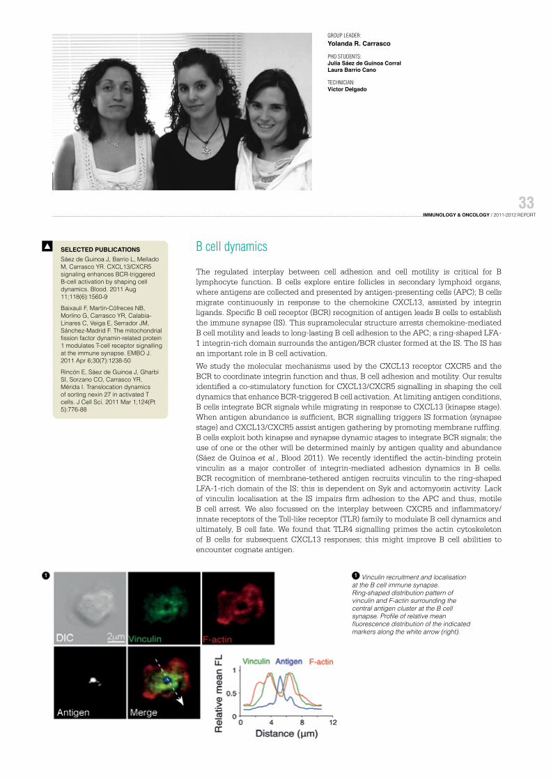
1 Vinculin recruitment and localisation at the B cell immune synapse. Ring-shaped distribution pattern of vinculin and F-actin surrounding the central antigen cluster at the B cell synapse. Profile of relative mean fluorescence distribution of the indicated markers along the white arrow (right).
1
34
Functional study of PI3K in survival, cell division and cancer
Our team is currently examining the function of kinases that affect the cell membrane lipid composition on cell behaviour, with special emphasis on phosphoinositide 3-kinases (PI3K).
These enzymes (class IA) phosphorylate the membrane phosphoinositides, giving rise to a lipid product (PIP3) that is normally found at low levels in quiescent cells. PIP3 levels increase transiently when normal cells are activated to execute a cell response, as PIP3 induces cell survival and contributes to triggering cell migration and division. In cancer cells, PIP3 levels are constitutively high (in ∼50% of all human cancers).
Although the PI3K enzymatic system is clearly a promising therapeutic target, very few therapeutic approaches are currently directed successfully to this target.
We study the molecular mechanisms by which PI3K isoforms control cell behaviour and its potential implications in disease.
In chronic inflammation, our recent findings show PI3K involvement in systemic lupus erythematosus, due in part to its capacity to mediate memory T cell survival. These studies help to show that haematopoietic PI3K isoforms are a potential target for chronic inflammatory disease treatment.
In cancer, we focus on a nuclear form of PI3K that controls DNA duplication, DNA repair and chromosome division, among other processes. Our goal is to define how this enzyme controls DNA homeostasis and the consequences of its amplification in cancer.
The second aspect of our recent studies is to understand why PI3K regulatory subunit usage changes during tumour progression, how this affects tumour progression and invasion, and the potential therapeutic applications of these findings.
P201031137: Biomarker for cancer diagnosis, prognosis and follow-up.
Kumar A, Fernandez-Capetillo O, Carrera AC. Nuclear phosphoinositide 3-kinase beta controls double-strand break DNA repair. Proc Natl Acad Sci USA. 2010 Apr 20;107(16):7491-6
Kumar A, Redondo-Muñoz J, Perez-García V, Cortes I, Chagoyen M, Carrera AC. Nuclear but not cytosolic phosphoinositide 3-kinase beta has an essential function in cell survival. Mol Cell Biol. 2011 May;31(10):2122-33
Suárez-Fueyo A, Barber DF, Martínez-Ara J, Zea-Mendoza AC, Carrera AC. Enhanced phosphoinositide 3-kinase δ activity is a frequent event in systemic lupus erythematosus that confers resistance to activation-induced T cell death. J Immunol. 2011 Sep 1;187(5):2376-85
Cortés I, Sánchez-Ruíz J, Zuluaga S, Calvanese V, Marqués M, Hernández C, Rivera T, Kremer L, González-García A, Carrera AC. p85β phosphoinositide 3-kinase subunit regulates tumor progression. Proc Natl Acad Sci USA. 2012 Jul 10;109(28):11318-23
Silió V, Redondo-Muñoz J, Carrera AC. Phosphoinositide 3-kinase β regulates chromosome segregation in mitosis. Mol Biol Cell. 2012 Dec;23(23):4526-42
SELECTED PUBLICATIONS
1
1 p85β-driven tumour lymphomagenesis model induced in SCID mice after transplant with control or p85β-infected bone marrow, followed by ENU treatment. Kaplan-Meier survival curves (right); (***) Mantel-Cox test P <0.001 (n = 15 p85β, n = 22 controls) and percentage of mice with spleen metastases of the thymic lymphoma (left).
Role of stress-activated protein kinase p38MAPK in human diseases
The aim of our group is to discover how members of the p38MAPK family regulate cell function in physiological conditions and in response to environmental stresses, infection and proinflammatory cytokines, and to understand how they are deregulated in several human disease situations such as oncogenic transformation and inflammation.
Our research is focussed on:
1. The discovery of new substrates, interacting proteins and inhibitors for these kinases, and the study of their physiological roles using mice transgenic for p38 isoforms, and
2. The study of p38MAPK as a link between chronic inflammation and cancer, and as mediators of chronic inflammatory diseases.
These studies use biochemical, cell biology and whole animal model approaches.
There are four p38MAPK family members (p38α, p38β, p38γ and p38δ), which are similar in amino acid sequence but differ in expression patterns, substrate specificities and sensitivity to inhibitors. In recent years, our group has centred on elucidating the regulation and roles of p38γ and p38δ. Using p38γ- or p38δ-deficient cells, we found that these kinases regulate processes involved in malignant cell transformation such as contact inhibition, migration, apoptosis, proliferation and tumourigenesis. Moreover, lack of p38γ in K-Ras-transformed fibroblasts causes an increase in cell proliferation in vitro and tumour formation in vivo, whereas lack of p38δ in fibroblasts blocks cell growth by inhibition contact and induces focus formation. Additionally, we recently showed that p38γ and p38δ are key components in innate immune responses, since their deletion impaired the innate immune response to septic shock by reducing the production of inflammatory cytokines such as TNFα and IL1β.
We are currently studying how p38γ and p38δ regulate the integrity of nuclear and intercellular-junctional complexes, cell adhesion, migration and polarity in response to many kinds of external stimuli. Little is known about the role of p38γ and p38δ isoforms in chronic inflammatory disease. We are currently undertaking further studies to investigate this as well as their role in the development of cancer associated to inflammation, using the genetically modified mice.
Cerezo-Guisado MI, del Reino P, Remy G, Kuma Y, Arthur JS, Gallego-Ortega D, Cuenda A. Evidence of p38γ and p38δ involvement in cell transformation processes. Carcinogenesis. 2011 Jul;32(7):1093-9
Risco A, Cuenda A. New insights into the p38γ and p38δ MAPK pathways. J Signal Transduct. 2012;2012:520289
Risco A, del Fresno C, Mambol A, Alsina-Beauchamp D, MacKenzie KF, Yang HT, Barber DF, Morcelle C, Arthur JS, Ley SC, Ardavin C, Cuenda A. p38γ and p38δ kinases regulate the Toll-like receptor 4 (TLR4)-induced cytokine production by controlling ERK1/2 protein kinase pathway activation. Proc Natl Acad Sci USA. 2012 Jul 10;109(28):11200-5
Chen CM, Bentham J, Cosgrove C, Braganca J, Cuenda A, Bamforth SD, Schneider JE, Watkins H, Keavney B, Davies B, Bhattacharya S. Functional significance of SRJ domain mutations in CITED2. PLoS One. 2012;7(10):e46256
1 Evidence of p38γ and p38δ as a tumour suppressor in cell transformation processes
1
The role of epigenetics in cancer
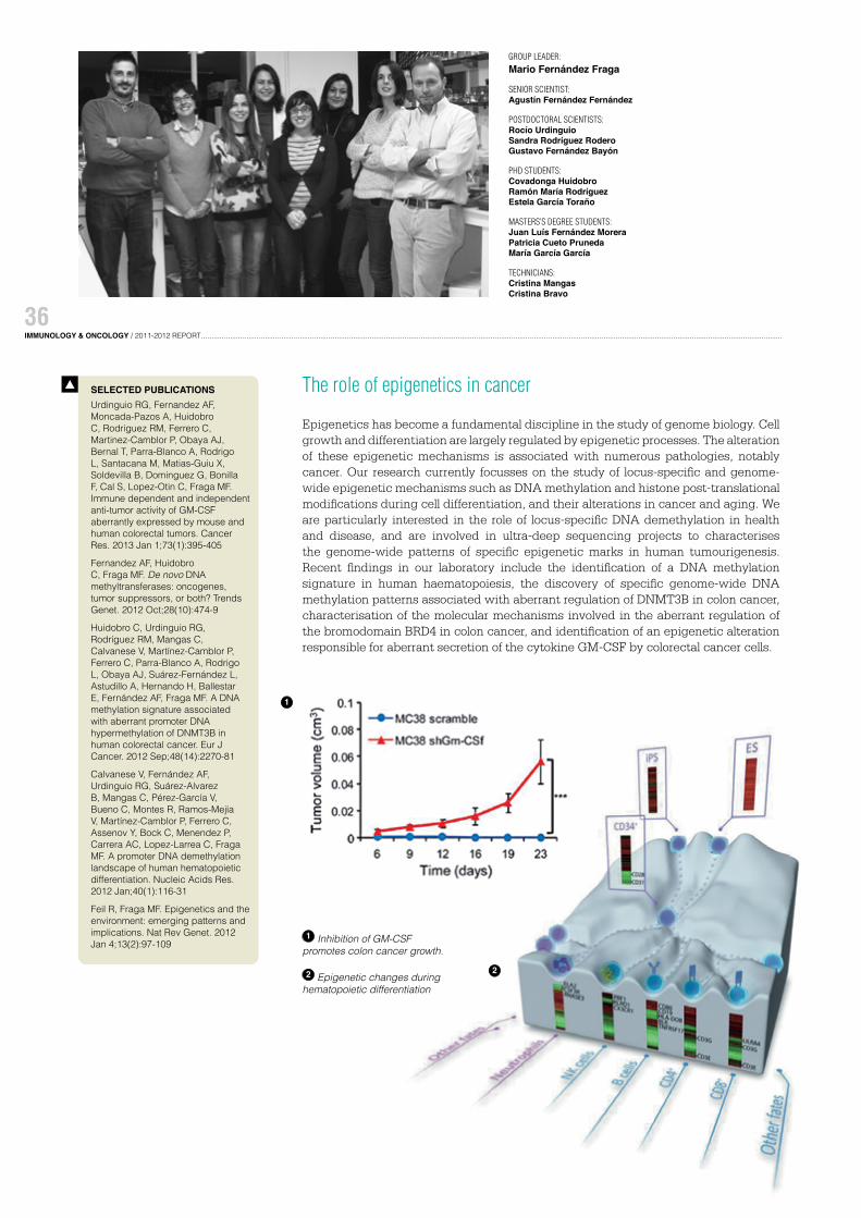
Epigenetics has become a fundamental discipline in the study of genome biology. Cell growth and differentiation are largely regulated by epigenetic processes. The alteration of these epigenetic mechanisms is associated with numerous pathologies, notably cancer. Our research currently focusses on the study of locus-specific and genome-wide epigenetic mechanisms such as DNA methylation and histone post-translational modifications during cell differentiation, and their alterations in cancer and aging. We are particularly interested in the role of locus-specific DNA demethylation in health and disease, and are involved in ultra-deep sequencing projects to characterises the genome-wide patterns of specific epigenetic marks in human tumourigenesis. Recent findings in our laboratory include the identification of a DNA methylation signature in human haematopoiesis, the discovery of specific genome-wide DNA methylation patterns associated with aberrant regulation of DNMT3B in colon cancer, characterisation of the molecular mechanisms involved in the aberrant regulation of the bromodomain BRD4 in colon cancer, and identification of an epigenetic alteration responsible for aberrant secretion of the cytokine GM-CSF by colorectal cancer cells.
PHD STUDENTS:Covadonga HuidobroRamón María RodríguezEstela García Toraño
MASTERS’S DEGREE STUDENTS:Juan Luís Fernández MoreraPatricia Cueto PrunedaMaría García García
TECHNICIANS:Cristina MangasCristina Bravo
Urdinguio RG, Fernandez AF, Moncada-Pazos A, Huidobro C, Rodriguez RM, Ferrero C, Martinez-Camblor P, Obaya AJ, Bernal T, Parra-Blanco A, Rodrigo L, Santacana M, Matias-Guiu X, Soldevilla B, Dominguez G, Bonilla F, Cal S, Lopez-Otin C, Fraga MF. Immune dependent and independent anti-tumor activity of GM-CSF aberrantly expressed by mouse and human colorectal tumors. Cancer Res. 2013 Jan 1;73(1):395-405
Fernandez AF, Huidobro C, Fraga MF. De novo DNA methyltransferases: oncogenes, tumor suppressors, or both? Trends Genet. 2012 Oct;28(10):474-9
Huidobro C, Urdinguio RG, Rodríguez RM, Mangas C, Calvanese V, Martínez-Camblor P, Ferrero C, Parra-Blanco A, Rodrigo L, Obaya AJ, Suárez-Fernández L, Astudillo A, Hernando H, Ballestar E, Fernández AF, Fraga MF. A DNA methylation signature associated with aberrant promoter DNA hypermethylation of DNMT3B in human colorectal cancer. Eur J Cancer. 2012 Sep;48(14):2270-81
Calvanese V, Fernández AF, Urdinguio RG, Suárez-Alvarez B, Mangas C, Pérez-García V, Bueno C, Montes R, Ramos-Mejía V, Martínez-Camblor P, Ferrero C, Assenov Y, Bock C, Menendez P, Carrera AC, Lopez-Larrea C, Fraga MF. A promoter DNA demethylation landscape of human hematopoietic differentiation. Nucleic Acids Res. 2012 Jan;40(1):116-31
Feil R, Fraga MF. Epigenetics and the environment: emerging patterns and implications. Nat Rev Genet. 2012 Jan 4;13(2):97-109
SELECTED PUBLICATIONS
1 Inhibition of GM-CSF promotes colon cancer growth.
2 Epigenetic changes during hematopoietic differentiation
COLLABORATORS: Lucio Gómez Mónica García-GalloMercedes LlorenteMaría Teresa Martín
González-Magaldi M, Postigo R, de la Torre BG, Vieira YA, Rodríguez-Pulido M, López-Viñas E, Gómez-Puertas P, Andreu D, Kremer L, Rosas MF, Sobrino F. Mutations that hamper dimerization of foot-and-mouth disease virus 3A protein are detrimental for infectivity. J Virol. 2012, 86:11013-23.
Cortés I, Sánchez-Ruíz J, Zuluaga S, Calvanese V, Marqués M, Hernández C, Rivera T, Kremer L, González-García A, Carrera AC. p85β phosphoinositide 3-kinase subunit regulates tumor progression. Proc Natl Acad Sci USA. 2012, 109: 11318-23.
Ruiz-Sáenz A, Kremer L, Alonso MA, Millán J, Correas I. Protein 4.1R regulates cell migration and IQGAP1 recruitment to the leading edge. J Cell Sci. 2011, 124:2529-38.
López-Santalla M, Salvador-Bernáldez M, González-Alvaro I, Castañeda S, Ortiz AM, García-García MI, Kremer L, Roncal F, Mulero J, Martínez-A C, Salvador JM. Tyr³²³-dependent p38 activation is associated with rheumatoid arthritis and correlates with disease activity. Arthritis Rheum. 2011, 63: 1833-42.
Aranda JF, Reglero-Real N, Kremer L, Marcos-Ramiro B, Ruiz-Sáenz A, Calvo M, Enrich C, Correas I, Millán J, Alonso MA. MYADM regulates Rac1 targeting to ordered membranes required for cell spreading and migration. Mol Biol Cell. 2011, 22:1252-62.
SELECTED PUBLICATIONS
PCT ES2010/070718: Monoclonal antibody against the protein GAPDH from Candida famata.
PCT ES09/070206: Antibody anti-Dectin-1, hybrodoma producer of this antibody and its application
PATENTS
Chemokine-receptor interactions in physiopathological processes
Our group is interested in understanding how chemokines and their receptors regulate the interaction between cancer cells and their microenvironment, specifically, how these proteins control tumour growth and progression. We also evaluate the potential of chemokine receptors as targets for anti-tumour therapy.
Chemokine receptors are a family of seven transmembrane domain proteins that are coupled to G proteins and regulate cell migration during homeostasis, inflammation and infection. These proteins are important in defining organ-specific metastatic localisation in cancer. The levels of certain chemokine receptors correlate with cell dissemination in lymph nodes, lung, bones, liver or other sites. We focus mainly on CCR9, a receptor expressed almost exclusively on lymphoid cells in the thymus, infiltrating cells in small bowel, a small subset of circulating memory T lymphocytes (CCR9+ α4β7hi), IgA-secreting plasma cells and plasmacytoid dendritic cells. The chemokine CCL25, the CCR9 ligand, is expressed in thymus and small bowel.
Recent reports showed that CCR9 overexpression increases the migratory and invasive capacity of prostate and ovarian cancer cells, directs melanoma cell metastases to the small intestine, and increases proliferation and resistance to apoptosis of acute lymphoblastic leukaemia-derived cell lines. CCR9-mediated intracellular signalling activates anti-apoptotic pathways and downregulates caspase activation, leading to
cell survival and increased proliferation.
Our laboratory focusses on determining the role of CCR9 in tumour physiopathology. We have generated mouse anti-human CCR9 monoclonal antibodies that recognise this receptor by flow cytometry, Western blot and immunocytochemistry. Using xenogeneic mouse models and human tumour lines, we study whether tumour cell cycling, survival, migration, invasiveness or growth are inhibited by these antibodies. CCR9-expressing human carcinoma cell lines and RNA interference approaches are also being used to study the molecular mechanisms that underlie CCR9-mediated effects. In addition, we are analysing whether these antibodies can be used for diagnostic imaging and for screening of low molecular weight antagonists.
1 Immunofluorescence staining of a human xenograft tumour. Paraffin sections were stained with anti-CD31 antibody (red), a specific endothelial marker suitable for the identification of blood vessels and angiogenesis. Cell nuclei were counterstained with DAPI (blue).
2 Bioluminescence images showing differential growth of human leukaemia cell tumours in an immunodeficient Rag2-/- mouse model, in response to treatment with two different monoclonal antibodies. Luciferase activity was measured and transformed to a pseudocoloured image.
1
2
Signalling networks in inflammation and cancer
Inflammation is a complex stereotypical response essential for effective defence of the organism against harmful stimuli such as pathogens, irritants or tissue damage. The detailed processes of inflammation indicate a close relationship between the inflammatory reaction and the immune response. A hallmark of inflammation is the directed migration (chemotaxis) of inflammatory cells (mostly leukocytes) through the walls of blood vessels to the site of injury. Once wound healing is complete, inflammation resolves and tissue homeostasis returns. A deregulated response to tissue damage can nonetheless lead to autoimmunity and chronic inflammatory diseases, and can also promote cancer.
Recent clinical and experimental evidence indicates that solid tumours increase inflammation to promote their progression. This leads to a tumour microenvironment largely controlled by inflammatory cells, which alters the metabolic needs of the tissue, promoting neo-angiogenesis, proliferation, survival, mutagenesis, migration and metastasis of malignant cells. Intriguingly, tumour-induced inflammation usually leads to immunosuppression, impeding the surveillance function of the immune system and clearance of the tumour. Breaking immunosuppression has been demonstrated as a useful, efficient means of eradicating cancers. Immune cells might thus provide both anti- and pro-tumourigenic signals, which might be harnessed or attacked for therapeutic purposes.
We aim to understand the critical cellular, molecular, and chemical mediators by which tumours promote inflammation and subvert the immune system to favour their own progression. We hope that comprehension of the mechanisms that balance pro- and anti-tumour immunity will lead to the design of more effective anti-cancer therapeutics.
The main achievements of the group in these two years have been
1. The study of signalling pathways elicited by the phosphatidylinositol-4-phosphate-5-kinase during acquisition of a motile phenotype in leukocytes.
2. The characterisation of the specific role of CCR5 and its ligands in antigen cross-presentation and the potentiation of anti-tumour responses.
3. The identification of SOD3 as a molecule involved in normalisation of the tumour vasculature and enhancement of chemo and immunotherapy of cancer.
POSTDOCTORAL FELLOWS: Emilia Mira Rosa Ana Lacalle Manuel Tardáguila
PHD STUDENTS: Juan Carlos de Karam Cristina Jiménez-Johansen Andrea Díaz Lorena Carmona-Rodríguez
TECHNICIANS: Rosa PeregilConcepción Gómez-MoutónAnna María Feijóo Freizer
VISITING SCIENTISTS: Noé Rodríguez (UAM, Madrid, Spain)Blanca López-Cuesta (Universidad de Navarra, Spain)Verena Haage (Konstanz University, Konstanz, Germany)Jez Marston (Yale University, New Haven, CT, USA)
González-Martín A, Gómez L., Lustgarten J, Mira E, Mañes S. Maximal T cell-mediated antitumor responses rely upon CCR5 expression in both CD4+ and CD8+ T cells. Cancer Res. 2011 Aug 15;71(16):5455-66
González-Martín A, Mira E, Mañes S. CCR5 in cancer immunotherapy: more than an “attractive” receptor for T cells. Oncoimmunology. 2012 Jan 1;1(1):106-108
Zonca M, Mancheño-Corvo P, De la Rosa O, Mañes S, Büscher D, Lombardo E, Planelles L. APRIL and BAFF proteins increase proliferation of human adipose-derived stem cells through activation of ERK1/2 MAP kinase. Tissue Eng Part A. 2012 Apr;18(7-8):852-9
González-Martín A, Mira E, Mañes S. CCR5 as a potential target in cancer therapy: inhibition or stimulation? Anticancer Agents Med Chem. 2012 Nov;12(9):1045-57
SELECTED PUBLICATIONS
P201031015. Use of SOD-3 as adjuvant for immunotherapy
PCT/EP2011/051237: Methods and compositions for the treatment of AIDS. Use of Des-1 inhibitors to prevent HIV infection
PATENTS
1 Model for interaction between the PDZ-binding motif in the type1 PIP5K-beta isoform and the tandem EBP50-PDZ domains (modelled based on syntein structure). The critical EBP50 residues involved in PIP5K binding are indicated.
Martínez-A C, van Wely KH. Centromere fission, not telomere erosion, triggers chromosomal instability in human carcinomas. Carcinogenesis. 2011 Jun;32(6):796-803
Meester-Smoor MA, Janssen MJ, ter Haar WM, van Wely KH, Aarnoudse AJ, van Oord G, van Tilburg GB, Zwarthoff EC. The ETS family member TEL binds to nuclear receptors RAR and RXR and represses gene activation. PLoS One. 2011;6(9):e23620
ter Haar WM, Meester-Smoor MA, van Wely KH, Schot CC, Janssen MJ, Geverts B, Bonten J, Grosveld GC, Houtsmuller AB, Zwarthoff EC. The leukemia-associated fusion protein MN1-TEL blocks TEL-specific recognition sequences. PLoS One. 2012;7(9):e46085
Fütterer A, Raya A, Llorente M, Izpisúa-Belmonte JC, de la Pompa JL, Klatt P, Martínez-A C. Ablation of Dido3 compromises lineage commitment of stem cells in vitro and during early embryonic development. Cell Death Differ. 2012 Jan;19(1):132-43
Muñoz LM, Holgado BL, Martínez-A C, Rodríguez-Frade JM, Mellado M. Chemokine receptor oligomerization: a further step toward chemokine function. Immunol Lett. 2012 Jul 30;145(1-2):23-9
SELECTED PUBLICATIONS Biology of stem cells: genomic instability, cancer and immunoregulation
We work with stem cells to try to understand the connections between chromosome instability and the stem cell theory of cancer. Most tumours have a combination of genetic defects termed chromosome instability (CIN), found in approximately 85% of non-hereditary carcinomas. We study two aspects of CIN, aneuploidy and the combination of translocations, duplications and deletions known collectively as segmental chromosome defects. The data from patient material and human cancer cell lines show that the majority of chromosome defects in human carcinomas comprise pericentromeric breaks that are captured by healthy telomeres; only a small proportion of chromosome fusions can be attributed to telomere erosion or random breakage. After comparing different DNA breakage pathways and by studying the role of mitotic spindle defects in the generation of aneuploidy and chromosome breakage, we postulate that a single mechanism, centromere fission, might be responsible for genetic instability on the cellular, individual and evolutionary scale. We hypothesise that centromere fission, rather than telomere erosion, is the most probable trigger of CIN and early carcinogenesis.
One of our model systems focusses on mutations in Dido, a locus identified by genome-wide screens as a potential regulator of stem cell function. The Dido locus is a gene complex described only in higher vertebrates; it encodes three splice variants, Dido1 (the smallest of these), Dido2 and Dido3. Dido3 also interacts with centrosomes and the synaptonemal complex. N-terminal truncation of Dido3 provokes aneuploidy, centrosome amplification and increased incidence of haematological myeloid neoplasms in the adult mouse. Misexpression of this splice variant is linked to myelodysplastic syndrome/myeloproliferative disease in humans.
In all attempts to date, deletion of the entire Dido locus or of Dido3-specific exons (exon XVI) in cell lines or mice has failed, indicating that its loss is incompatible with life. Stem cells generated from a targeted deletion of exon XVI show aneuploidy, centrosome amplification and inability to differentiate in vitro, as indicated by sustained
expression of Oct4 and other undifferentiated stem cell markers. The ability of exon XVI mutants to differentiate can be restored by retinoic acid, in vivo teratoma formation or expression of full-length Dido3. The role of Dido3 in the spindle assembly checkpoint, which senses and signals centrosome position and spindle tension, and its implication in an early cell-cell contact-induced
ES cell differentiation step, render Dido3 an appropriate candidate for future study of proteins that couple centrosome orientation to stem cell
f a t e decisions.
1 Merotelic attachments in Dido mutant cells. Dido mutant MEF were seeded on coverslips and double-labelled with antibodies to centromeres (green) and α-tubulin (red). DNA was stained with DAPI (blue), and cells were studied by confocal fluorescence microscopy. (A) Detection of merotelic centromere attachments in Dido mutant MEF. An individual anaphase centromere attached to both spindle poles is shown. The image of a whole cell is from maximum projection (A), and the three-fold amplification (B) is from a single confocal layer. (C-E) Appearance of centromeres in Dido mutant MEF. Centromeres in micronuclei (C) and in a nuclear protrusion (D) are indicated (arrowheads). (E) In some cases, an individual centromere is distorted (arrow), whereas neighbouring centromeres appear normal (arrowhead). From PNAS;107:4159-4164
1
Chemokine receptors: new targets for therapeutic intervention
The chemokines, a family of structurally related chemoattractant proteins that bind to specific seven transmembrane receptors linked to G proteins, trigger a broad array of biological responses that range from cell polarisation, movement, immune and inflammatory responses to prevention of HIV-1 infection. Chemokine-mediated cell activation was thought to be due to the binding of a monomeric chemokine to its monomeric receptor. Chemokine biology is nonetheless more complex than was initially predicted, as several studies suggest that chemokines can dimerise and that their receptors are found as dimers and/or higher order oligomers at the cell surface. These complexes form organised arrays that can be modified by receptor expression and ligand levels, indicating that they are dynamic structures. This oligomerisation could be important to different aspects of their biology, from ontogeny to the regulation of their pharmacological and signalling properties.
Using fluorescence resonance energy transfer (FRET) techniques, we confirmed the functional relevance and regulation of homo- and heterodimeric receptor complexes. CXCR5 and EBI2 form homo- and heterodimers. EBI2 expression induces conformational changes in CXCR5 homodimers, triggering a notable reduction in binding affinity of the ligand (CXCL13) for CXCR5; as a consequence, CXCR5-mediated responses in cell lines and primary B cells is altered. In addition to the receptors, the chemokines themselves oligomerise, which influences cell activation. HMGB1 (high mobility group box 1), a nuclear protein released by necrotic and severely stressed cells, binds CXCL12; the heterocomplexes promote conformational rearrangements
of CXCR4 different from those of CXCL12 alone. As a result, HMGB1 influences inflammatory cell recruitment to damaged tissues. We reported that chemokines activate the JANUS kinases (JAK), which associate to the chemokine receptor and promote its rapid tyrosine phosphorylation. Through the JAK/STAT pathway, the chemokines trigger suppressor of cytokine signalling (SOCS) expression. The SOCS intracellular proteins are thus key physiological regulators of cytokine and chemokine responses; they target ubiquitinated signalling intermediates for degradation by the proteasome pathway through ECS (elongin-Cullin-SOCS) E3 ligase formation. As a result of this effect, SOCS1 acts as a tumour suppressor, blocking cell cycling in human melanoma by affecting G1/S and mitosis.
Barroso R, Martínez Muñoz L, Barrondo S, Vega B, Holgado BL, Lucas P, Baíllo A, Sallés J, Rodríguez-Frade JM, Mellado M. EBI2 regulates CXCL13-mediated responses by heterodimerization with CXCR5. FASEB J. 2012 Dec;26(12):4841-54
Schiraldi M, Raucci A, Muñoz LM, Livoti E, Celona B, Venereau E, Apuzzo T, De Marchis F, Pedotti M, Bachi A, Thelen M, Varani L, Mellado M, Proudfoot A, Bianchi ME, Uguccioni M. HMGB1 promotes recruitment of inflammatory cells to damaged tissues by forming a complex with CXCL12 and signaling via CXCR4. J Exp Med. 2012 Mar 12;209(3):551-63
Vega B, Muñoz LM, Holgado BL, Lucas P, Rodríguez-Frade JM, Calle A, Rodríguez-Fernández JL, Lechuga LM, Rodríguez JF, Gutiérrez-Gallego R, Mellado M. Technical advance: Surface plasmon resonance-based analysis of CXCL12 binding using immobilized lentiviral particles. J Leukoc Biol. 2011 Aug;90(2):399-408
Muñoz LM, Lucas P, Holgado BL, Barroso R, Vega B, Rodríguez-Frade JM, Mellado M. Receptor oligomerization: a pivotal mechanism for regulating chemokine function. Pharmacol Ther. 2011 Sep;131(3):351-8
Lamana A, Martin P, de la Fuente H, Martinez-Muñoz L, Cruz-Adalia A, Ramirez-Huesca M, Escribano C, Gollmer K, Mellado M, Stein JV, Rodriguez-Fernandez JL, Sanchez-Madrid F, del Hoyo GM. CD69 modulates sphingosine-1-phosphate-induced migration of skin dendritic cells. J Invest Dermatol. 2011 Jul;131(7):1503-12
SELECTED PUBLICATIONS
1 In type I diabetes, immune cells invade the pancreas 2 In cerebral infarction, neural stem cells
VISITING SCIENTISTS: Naoaki Saito (University of Kobe, Japan)Yasuhito Shirai (University of Kobe, Japan)
Martínez-Moreno M, García-Liévana J, Soutar D, Torres-Ayuso P, Andrada E, Zhong XP, Koretzky GA, Mérida I, Ávila-Flores A. FoxO-dependent regulation of diacylglycerol kinase ζ gene expression. Mol Cell Biol. 2012 Oct;32(20):4168-80
Rincón E, Gharbi SI, Santos-Mendoza T, Mérida I. Diacylglycerol kinase ζ: at the crossroads of lipid signaling and protein complex organization. Prog Lipid Res. 2012 Jan;51(1):1-10
Gharbi SI, Rincón E, Avila-Flores A, Torres-Ayuso P, Almena M, Cobos MA, Albar JP, Mérida I. Diacylglycerol kinase ζ controls diacylglycerol metabolism at the immunological synapse. Mol Biol Cell. 2011 Nov;22(22):4406-14
Almena M, Mérida I. Shaping up the membrane: diacylglycerol coordinates spatial orientation of signaling. Trends Biochem Sci. 2011 Nov;36(11):593-603
Rincón E, Sáez de Guinoa J, Gharbi SI, Sorzano CO, Carrasco YR, Mérida I. HYPERLINK “http://www.ncbi.nlm.nih.gov/pubmed/21303929”Translocation dynamics of sorting nexin 27 in activated T cells. J Cell Sci. 2011 Mar 1;124(Pt 5):776-88
SELECTED PUBLICATIONS Role of diacylglycerol kinases in the control of immune response and cancer progression
We study the contribution of diacylglycerol kinases (DGK) to T cell functions and oncogenic transformation. We have focussed on two specific isoforms that are important modulators of DAG-dependent functions in T lymphocytes. We expect that our findings will help to assess the therapeutic potential of the DGK enzyme family as tools for better, more effective management of the immune response and treatment of cancer. The work in the laboratory is organised in two different, complementary areas:
1. DGK and as negative regulators of the adaptive immune response
We and others have characterised DGKα and ζ as negative regulators of the DAG that is generated during T cell activation, limiting Ras guanyl-releasing protein (RasGRP1)- dependent activation of Ras. A better understanding of the mechanisms that regulate the expression and activation of these two enzymes will allow T cell functions to be modulated, perhaps even harnessed, to provide new strategies in combating diseases such as autoimmunity and cancer. In the past years, we have contributed to better understand the mechanism that determines DGKα localisation/activation during immune synapse formation, as well as its contribution to DAG metabolism at this site. We also identified DGKα as a gene regulated by the AKT/FoxO axis, and characterised it as a negative regulator of TCR and IL2-regulated functions.
2. DGKα and ζ: the lipid restraint on tumour metabolism
Our studies intend to define the precise contribution of these DGK isoforms to the onset of malignant transformation. We use genetic and biochemical approaches to investigate the role of DGKα in the control of the transition from quiescence to proliferative states in untransformed cells, and to assess its function in the context of malignant transformation. Validation of the distinct regulation of expression as well as careful assessment of DGKα and ζ functions in normal and transformed cells is an important challenge to the full evaluation of the potential of these proteins as therapeutic targets.
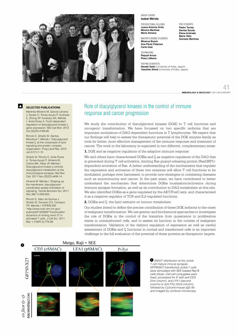
1 SNX27 distribution at the Jurkat T cell mature inmune synapse. GFPSNX27-transfected Jurkat T cells were stimulated with SEE-loaded Raji B cells (blue). Cell-cell conjugates werefixed, processed for IF with anti-CD3 (first column), anti-LFA1 (second column) or anti-PTyr (third column), followed by Cy3-anti-mouse IgG Ab and imaged by confocal microscopy.
1
PHD STUDENTS: Pedro TorresDenise SoutarElena AndradaMaría TelloGonzalo Martínez
During an immune response, activated B lymphocytes undergo a final stage of differentiation known as terminal B lymphocyte differentiation. This process is characterised by the generation of antibody-secreting cells (ASC) or plasma cells, and memory B cells. Antigen re-challenge can improve the function of the antibodies produced by these specialised cells through two mechanistically linked processes, immunoglobulin class switch recombination (CSR) and somatic hypermutation (SH). The transcriptional program driving terminal B cell differentiation is regulated by the transcriptional repressor Blimp-1. Among its transcriptional targets, Blimp-1 downregulates the gene expression of c-myc. c-Myc protein is a member of the MYC family of transcription factors involved in multiple biological processes such as cell proliferation, apoptosis and differentiation.
Despite extensive study of Myc, little is known of its function in terminal B cell differentiation. In recent years, our group has focussed on defining the role of c-Myc in this process. It is assumed that repression of c-myc by Blimp-1 during terminal differentiation is intended to cease cell proliferation exclusively. However, we has observed that c-Myc function goes beyond the regulation of cell proliferation during terminal B lymphocyte differentiation. We found that c-Myc is also essential for ASC function and differentiation in vivo using several genetically-modified mouse models. Our current efforts in the lab are focussed on the characterisation of the c-Myc-dependent mechanisms governing this process and how these functions might be altered in human pathologies.
GROUP LEADER: Ignacio Moreno de Alborán
POSTDOCTORAL FELLOW: David Fernández AntoránMaitane Ortiz
PHD STUDENT: Antonio Navarro
SELECTED PUBLICATIONSVallespinós M, Fernández D, Rodríguez L, Alvaro-Blanco J, Baena E, Ortiz M, Dukovska D, Martínez D, Rojas A, Campanero MR, Moreno de Alborán I. B lymphocyte commitment program is driven by the proto-oncogene c-myc. J Immunol. 2011 Jun 15;186(12):6726-36
Li DQ, Pakala SB, Reddy SD, Ohshiro K, Zhang JX, Wang L, Zhang Y, Moreno de Alborán I, Pillai MR, Eswaran J, Kumar R. P53-independent bidirectional autoregulatory mechanism of MTA1-ARF in oncogenesis. Proc Natl Acad Sci USA. 2011 May 24;108(21):8791-6
Calado DP, Sasaki Y, Godinho SA, Pellerin A, Köchert K, Sleckman BP, de Alborán IM, Janz M, Rodig S, Rajewsky K. The cell-cycle regulator c-Myc is essential for the formation and maintenance of germinal centers. Nat Immunol. 2012 Nov;13(11):1092-100
Fernandez D, Sanchez-Arevalo VJ, de Alboran IM. The role of the proto-oncogene c-myc in B lymphocyte differentiation. Crit Rev Immunol. 2012;32(4):321-34
1 Haematoxylin / Eosin-stained section of mouse spleen
2 Immunoglobulin class switch recombination (CSR) and cell division of activated B lymphocytes in vitro. Flow cytometry analysis of B lymphocytes were stained with CFSE and anti-IgG1 to monitor cell division and CSR, and analysed by Flow cytometry.
Function and regulation of APRIL, a TNF protein: implications in pathology
Our research studies the biological functions of the tumour necrosis factor proteins APRIL (a proliferation-inducing ligand) and BAFF (B cell-activator factor) in- and outside the immune system, and their involvement in disease processes such as cancer. We have three main projects in which we characterise the modulatory role of APRIL on B lymphocytes, examine the dialogue between hASC and APRIL in an inflammatory context, and assess the relevance of APRIL in solid tumours such as breast cancer. We generated MMTV-ErbB2-APRIL-Tg and MMTV-ErbB2-APRIL-KO mouse models and used a panel of human breast carcinoma cell lines to study the implication of APRIL in breast cancer. We found that APRIL is expressed in breast carcinoma cell lines; it supports their basal growth and activates MAP kinase signalling pathways. We are currently analysing the mouse models as well as the clinical potential of APRIL.
We recently characterised the APRIL/BAFF system in hASC. hASC are mesenchymal stem cells with reduced immunogenicity and the ability to modulate immune responses; hASC therapy is currently under clinical investigation. As APRIL and BAFF proteins are overexpressed in many inflammatory and autoimmune diseases, we addressed their potential association with hASC. We found that hASC express APRIL and BAFF as well
as their receptors TACI, BCMA and the BAFF-specific receptor (BAFF R). APRIL and BAFF secretion was differentially enhanced by CXCL12 and IFNγ, implicated in hASC-mediated migration and immunosuppression, respectively. In addition, APRIL and BAFF induced rapid phosphorylation of ERK1/2 and Akt kinases and promoted an increase in hASC proliferation, without affecting the immunosuppressive capacity of these cells. The use of specific chemical inhibitors indicated that the PI3K transduction pathway is involved in hASC basal growth, and that APRIL- and BAFF-mediated effects are ERK-dependent.
These results provide new information about the molecular mechanisms that underlie APRIL and BAFF secretion and signalling in hASC, and are of special relevance for the use of allogeneic hASC as therapeutic tools.
SELECTED PUBLICATIONS
PATENTEP10382244,PCT/EP11/065540. Stem cell culture media and methods
Zonca M, Mancheño-Corvo P, DelaRosa O, Mañes S, Büscher D, Lombardo E, Planelles L. APRIL and BAFF proteins increase proliferation of human adipose-derived stem cells through activation of Erk1/2 MAP kinase. Tissue Eng Part A. 2012 Apr;18(7-8):852-9
GROUP LEADER:Lourdes Planelles
PhD STUDENTS:Manuela ZoncaDouglas Florindo PinheiroAraceli García Castro
12
1 APRIL expression in MDA-MB231 breast carcinoma cells by IHQ
2 A) APRIL, BAFF, BCMA, TACI and BAFF-R transcripts are found in hASC. B) TACI and BCMA protein expression in hASC as determined by flow cytometry. C) APRIL and BAFF sustain hASC proliferation (MTS assay).
Receptor ligand interactions in immune responses to cancer and viruses
Current research in the laboratory addresses various issues related to the biology of NK cells and in particular, the receptor NKG2D and its ligands:
1. We have shown that shedding of molecules such as MICA and ICAM-1 is increased after human cytomegalovirus (HCMV) infection both in vitro and in transplant recipients who develop HCMV infection. This phenomenon depends on an increase in activity of the metalloproteases ADAM17 and MMP14 due to decreased expression of the endogenous inhibitor of metalloproteases TIMP3. Assay of the sheddase activity of ADAM17 could be of use as a biomarker in patients at risk of developing CMV disease.
2. We found that innate immune recognition of double-stranded RNA produced during viral infection plays a key role in the induction of expression of NKG2D ligands. These data suggest interesting parallels between the production of type I interferons and the induction of expression of NKG2D ligands. We identified proteins in both vaccinia and influenza virus whose expression markedly reduces the induction of both IFN-β and NKG2D ligands after infection.
3. The observation that stimulation via PRRs has a key role in the induction of NKG2D ligand expression prompted us to study the cell biology of these molecules in more detail. We have chosen to concentrated on inflammasome protein IFI16, that senses cytosolic DNA and stimulates production of both interferons and IL-1β.
4. Loss of immune function is commonly observed in NK cells isolated from patient tumours. We have established an in vitro system to induce hyporesponsiveness in primary human NK cells and have identified changes in specific cell surface receptors as being particularly important mechanistically. We are currently collaborating with the group of Dr. Eric Long (NIAID) to use live single-particle tracking to examine the diffusion properties of these receptors on normal and hyporesponsive NK cells.
Fernández-Messina L, Reyburn HT, Valés-Gómez M. Human NKG2D-ligands: cell biology strategies to ensure immune recognition. Front Immunol. 2012;3:299
Agüera-González S, Gross CC, Fernández-Messina L, Ashiru O, Esteso G, Hang HC, Reyburn HT, Long EO, Valés-Gómez M. Palmitoylation of MICA, a ligand for NKG2D, mediates its recruitment to membrane microdomains and promotes its shedding. Eur J Immunol. 2011 Dec;41(12):3667-76
Fernández-Messina L, Ashiru O, Agüera-González S, Reyburn HT, Valés-Gómez M. The human NKG2D ligand ULBP2 can be expressed at the cell surface with or without a GPI anchor and both forms can activate NK cells. J Cell Sci. 2011 Feb 1;124(Pt 3):321-7
Johnen H, González-Silva L, Carramolino L, Flores JM, Torres M and Salvador JM. Gadd45g is essential for primary sex determination, male fertility and testis development. PLoS ONE 8(3): e58751. doi:10.1371/journal.pone.0058751, 2013
López-Santalla, M., Salvador-Bernáldez, M., González-Álvaro, I., Castañeda, S., Ortiz, A.M., García, M.I., Eiró, N., Kremer, L., Roncal, F., Mulero, J., Martínez-A, C. and Salvador JM. Tyr323-dependent p38 activation is associated with rheumatoid arthritis and correlates with disease activity. Arthritis Rheum 63:1833-42, 2011
SELECTED PUBLICATIONS T cell signalling in autoimmune diseases and cancer
The main goal of our group is the identification of novel therapeutic targets in autoimmune diseases and cancer. We focus on the role of key proteins involved in the regulation of T cell activation and differentiation. We are studying the biological functions of Gadd45 and p38 MAPK in autoimmunity and cancer development. To address this goal, we generated mouse models deficient for each member of the Gadd45 family and have analysed T cells from patients with different autoimmune diseases including rheumatoid arthritis (RA) and ankylosing spondylitis.
The pathogenesis and progression of RA is complex and involves T-cell-mediated antigen-specific responses. The initial stages of RA-associated autoimmunity, which precede articular inflammation, are the results of breakdown in B and T cell tolerance. p38 MAPK is a key regulator of T cell signalling and controls immune system effector functions. Our group demonstrated that p38 is hyperphosphorylated on Tyr323 in T cells from patients with active RA and that this phosphorylation reflects the disease activity state of RA. We generated and patented a novel method to determine the disease activity state of RA. We designed an intracellular staining method to assess RA disease activity by flow cytometry analysis (Lopez-Santalla et al., 2011). In addition, we showed that the tyrosine kinase lck has a major role in p38 activation in pathological conditions in RA. Our data suggest that specific inhibition of lck can abolish p38 effector functions in T cells.
We recently showed that the Gadd45 family also has roles in growth and development of specific tissues in the embryo. In mice, Gadd45 genes are differentially expressed during embryonic development; Gadd45b is expressed in the chorion whereas Gadd45g is expressed in mouse brain. We found a critical role for Gadd45g in gonad development, male fertility and sex determination, as Gadd45g-deficient mice show an unexpected
male-to-female sex reversal phenotype. In male gonads, SRY expression triggers differentiation of a somatic supporting cell lineage into Sertoli cells, which direct the male developmental pathway. In the absence of SRY, SOX9 is downregulated, leading to differentiation of the somatic supporting lineage into granulosa cells, which aid oocyte development. Gadd45g, but not Gadd45a or Gadd45b, is necessary for activation of the male sex-determining pathway in mice; its absence leads to development of female gonads. Lack of Gadd45g decreases SRY expression and blocks SOX9 expression resulting in ovary and Müllerian duct development, whereas lack of Gadd45a and/or Gadd45b has no effect on testis development.
GROUP LEADER: Jesús María Salvador
POSTDOCTORAL FELLOWS: Mercedes López-SantallaHeiko JohnenSara Mateus FernándezBeatriz Dorado de la Corte
PHD STUDENTS: María Salvador BernáldezLaura González SilvaUmberto Rosato
TECHNICIANS: Vanesa Cano DaganzoCarmen Mireya Martínez García
1 Lack of Sertoli cell differentiation and testis cord formation in XY Gadd45g-/- gonads. (A-L) Confocal optical slices of whole mount immunostained B6 gonads (dashed outline), showing expression of Sertoli cell markers SOX9 (nuclear, blue) and AMH (cytoplasmic, red) and the germ/endothelial cell marker Pecam1 (membrane, green).
Biochemical characterisation of ligands for the immune receptor NKG2D: implications of heterogeneity for pathology and therapy
Our research aims to characterise the cellular routes that facilitate tumour elimination by the immune system and those that permit tumour escape. We have focussed recently on the study of the ligands for the NKG2D receptor, molecules that send stress signals to the immune system and that regulate effector cells such as T lymphocytes and natural killer (NK) cells. NKG2D is an activating immune receptor, constitutively expressed in humans in most cytotoxic lymphocytes, including NK and CD8+ T cells, whereas in mice it is expressed on NK cells, but on T cells only after activation. After binding to its ligands, NKG2D activates the mechanisms that lead to lysis and cytokine secretion by immune effector cells. In humans, NKG2D ligands (NKG2D-L) belong to two families of “stress-inducible” proteins: the polymorphic family of MHC-I-related chain A/B (MICA/B) and the multi-gene family of UL16-binding proteins (ULBP, now called RAET1A-E). Besides their activating role after ligand binding, NKG2D-L can be released to the extracellular media and interfere with immune recognition. Recent work from our laboratory focussed on the cellular and molecular differences that explain NKG2D-L heterogeneity. Our data demonstrate that, although both NKG2D-L
families, MICA/B and ULBP, are related to MHC molecules and their expression is increased after stress, there are many differences in terms of biochemical properties and cell trafficking. We propose that categorisation of NKG2D-L according to their biological features, rather than their genetic family, might help to achieve a better understanding of the association of these molecules with disease.
Fernández-Messina L, Reyburn HT, Valés-Gómez M. Human NKG2D-ligands: cell biology strategies to ensure immune recognition. Front Immunol. 2012;3:299
Agüera-González S, Gross CC, Fernández-Messina L, Ashiru O, Esteso G, Hang HC, Reyburn HT, Long EO, Valés-Gómez M. Palmitoylation of MICA, a ligand for NKG2D, mediates its recruitment to membrane microdomains and promotes its shedding. Eur J Immunol. 2011 Dec;41(12):3667-76
Fernández-Messina L, Ashiru O, Agüera-González S, Reyburn HT, Valés-Gómez M. The human NKG2D ligand ULBP2 can be expressed at the cell surface with or without a GPI anchor and both forms can activate NK cells. J Cell Sci. 2011 Feb 1;124(Pt 3):321-7
SELECTED PUBLICATIONS
GROUP LEADER:Mar Valés Gómez
POSTDOCTORAL SCIENTISTS:Lola Fernández Messina
PHD STUDENTS:Eva M. García CuestaSheila López CoboDaniela Dukovska
VISITING SCIENTISTS:Rachele Pandolfi
1 Some NKG2D ligands, in addition to being expressed at the cell surface, are recycled and accumulate in intracellular organelles.
2 NKG2D ligands belong to two polymorphic families of MHC-related proteins that respond to cell stress.