Department of Orthopaedics Chairperson& Moderator: Prof. & HOD: Dr. Kiran Kalaiah Presenter : Dr. Yashavardhan .T.M SEMINAR PRESENTATION ON : GAIT ANALYSIS WITH ABNORMALITIES IN GAIT PATTERN IN ORTHOPAEDICS.
Transcript
Department of Orthopaedics
Chairperson& Moderator: Prof. & HOD: Dr. Kiran Kalaiah
Presenter : Dr. Yashavardhan .T.M
SEMINAR PRESENTATION ON: GAIT ANALYSIS WITH ABNORMALITIES IN GAIT PATTERN IN
ORTHOPAEDICS.
INTRODUCTION:
A systematic approach to gait analysis i,e, looking at trunk & each joint moving in all three planes i,e. sagital, coronal & transverse.
It can yield valuable information about patient's condition & help in establishing a treatment plan.
The earliest work on gait was done by BORELLI in 1682.
The WEBBER brothers in Germany gave first clear description of GAIT CYCLE in1836.
In 1940 SCHERB from Switzerland studied various muscle activity during different parts of gait cycle, using treadmill & later by EMG.
Normal GaitDefinition
Human gait is bipedal, biphasic,
forward propulsion of center of gravity,
in which there is alternate sinousmovement of head and body,
with least expenditure of energy
Definition.
Continuous rhythmic alternative movements of lower limbs, in order to forward propulsion of the body, by moving centre of gravity in forward direction with minimal expenditure of energy
Normal walking requirementsEquilibrium-ability to assume upright
posture and maintain balance.
Locomotion-ability to initiate and maintain
rhythmic stepping.
Muskulo-skeletal integrity-normal bone
joint and muscle function.
Neurological control-visual ,auditory
vestibular and sensory motor input
GAIT CYCLE►The duration that occurs from the time when the heel of one foot strikes the
ground to the time at which the same foot contacts the ground again.
►Normally 1-2 sec.
►Two phases:
► 1.Stance phase-60%
► 2.Swing phase-40%
►The gait cycle consist of 2 phases for each foot
TEMPORAL PARAMETERS
Distance and time measurements calculated during gait analysis are referred to as CADENCE PARAMETERS.
It includes
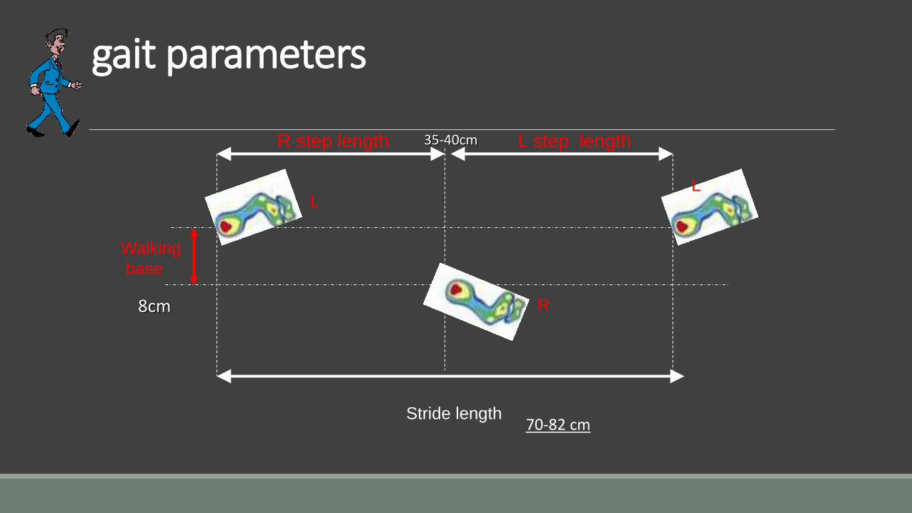
gait parameters
Stride length
L step lengthR step length
LL
R
Walking
base
8cm
70-82 cm
35-40cm
Cadence: ◦ It is the no of steps taken by a person per unit of time.
◦ It is measured as the no of steps / sec or per minute.
Cadence = Number of steps / Time
GAIT ANALYSIS The science of studying human gait is GAIT ANALYSIS , which is done in terms of◦ Movement in space
◦ Metabolic energy
◦ Functional muscle patterns
◦ Interaction of forces
STANCE PHASE :
Defined as the time during which the limb is in contact with the ground and supporting the weight of the body from heel strike to toe off.
SWING PHASE:
Defined as the time period during which the limb is off the ground and advancing forward, the body weight supported by contralateral limb. i.e acceleration to decelaration
STANCE Subphases
IC: Initial Contact (Heel Strike)
> Both limbs are in contact – Double stance> The heel strikes the ground> The stance knee begins to flex slightly.> The ankle is at the neutral position> The knee is close to full extension
> Hip 30° of flexion Femur externally rotated
STANCE Subphases
LR: Loading Response (Foot Flat)
>Flattening of the foot – reacting to impact of body weight
>Double stance ends
>Knee – 15o flexion, tibia internally rotates and then begins to externally rotate
>Hip – 30o flexion, femur internally rotating moving to neutral
>Maximum Impact Loading occurs>Foot rapidly moves into pronation>Weight has been shifted to the support leg
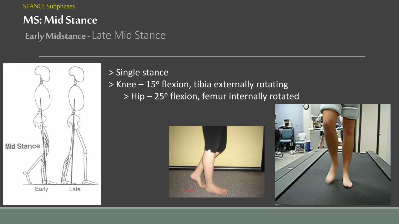
STANCE Subphases
MS: Mid StanceEarly Midstance - Late Mid Stance
> Single stance> Knee – 15o flexion, tibia externally rotating
> Hip – 25o flexion, femur internally rotated
STANCE Subphases
TS: Terminal Stance (Heel-off)
>Single stance – “Falling forward” forward fall of the body moves the vector further anterior to the ankle, creating a large dorsiflexion moment >Begins as COG passes over foot and ends when opposite foot touches ground
>Knee – 5o flexion to 0o, tibia externally rotates>Hip – 0 to 10o extension, femur externally rotates and begins abduction
STANCE Subphases
PS: Pre-Swing (Toe-Off/ Knee Break)
>Double stance – “Transition”>Limb is rapidly unloaded – “Toe-off”
>Knee – 0-30o flexion, tibia externally rotates>Hip – 20o extension, femur externally rotates with abduction>The ankle moves rapidly from its dorsiflexion position atterminal stance to 20 degrees of plantarflexion
SWING Subphases
IS: Initial Swing ( acceleration )
>From “toe-off” until maximum knee flexion>Knee – 30–60o flexion, tibia internally rotates>Hip – 0–20o flexion, femur moves frominternal rotation to neutral (externally rotating)
SWING Subphases
MS: Mid Swing
>Goal is for tibia to reach vertical position perpendicular to surface
>Knee – moves to 0o, tibia externally rotates
>Hip – 20-30o flexion, femur externally rotates
>Knee extension and hip flexion continue by inertia
SWING Subphases
TS: Terminal Swing
>Preparing for initial contact>Knee – 0o, tibia externally rotated
>Hip – 30o flexion, femur externally rotates
NEUROLOGICAL CONTROL OF GAIT
Motor CortexVoluntary modulation of gait.Eg:Alter in speed,change in direction.
•Cerebellum
•Extrapyramidal tract
Controlling Balance
Responsible for most complex unconscious pathways
Spinal Cord
Golgi Tendon UnitsMuscle Spindle,Joint
Reflex Stepping Movements
Produce neurologic feedback & serve as dampening devices for coordination of gait.
DETERMINANTS OF GAIT ►Six optimizations used to minimize excursion of CG in vertical & horizontal planes
►Reduce significantly energy consumption of ambulation
►The six determinants are
1. Pelvic rotation
2. Pelvic tilt
3. Knee flexion
4. Ankle mechanism
5. Foot mechanism
6. Physiological valgus of knee
DETERMINANTS OF GAIT
1) Pelvic rotation:
◦ Forward rotation of the pelvis in the horizontal plane approx. so on the swing-phase side
◦ Reduces the angle of hip flexion & extension
◦ Enables a slightly longer step-length w/o further lowering of CG
(2) Pelvic tilt:
◦5 degree dip of the swinging side (i.e. hip adduction)
◦ In standing, this dip is a positive Trendelenberg sign
◦Reduces the height of the apex of the curve of CG
(3) Knee flexion in stance phase:
◦ Approx. 20o dip
◦ Shortens the leg in the middle of stance phase
◦ Reduces the height of the apex of the curve of CG
(4) Ankle mechanism:
◦ Lengthens the leg at heel contact
◦ Smoothens the curve of CG
◦ Reduces the lowering of CG
(5) Foot mechanism:
◦Lengthens the leg at toe-off as ankle moves from dorsiflexion to plantar flexion
◦Smoothens the curve of CG◦Reduces the lowering of CG
Physiological valgus of kneeReduces the base of support, so only little lateral
motion of pelvis is necessary.
FACTORS AFFECTING GAIT
Age
Gender
Assistive devices
Disease states
Muscle weakness or paralysis
Asymmetries of the lower extremities
Injuries and malalignments
GAIT EXAMINATION
Take a history
Couch examination
Static examination
Allow patient time to relax
Reasonable length walkway - gait pattern changes before & after turn
COUCH EXAMINATIONObserve deformities & lesions
Check ROM’s
Check muscle tightness/strength
Neurological & vascular assessment
STATIC EXAMINATION
Feet non-weight bearing (hanging) with weight bearing
Standing from front◦ Shoulders, hips, knees, feet
◦ From behind
◦ Shoulders, hips, calcaneus
GENERAL POINTS
Is the gait fast or slow?
Is it smooth?
Does the patient appear relaxed & comfortable or pained?
Is it noisy?
KINETICS OF GAITIt is the study of forces that produces a change in motion.
It is concerned with internal forces developed within body by muscular action as well as forces acting in body.
External forces includes:
Centre of gravity
Ground reaction forces
Centre of gravity:
It is imaginary point at which all weight of body is concentrated at a given instant.
The body of gravity lies two inches in front of SECOND SACRAL VERTEBRA.
It follows up & down movements as well as side to side.
Due to complex interaction of muscular activity & joint motion in lower extremity it follows a SMOOTH SINUSOIDAL CURVE.
Ground reaction forces:
It is a line represents the direction & magnitude of force encountered by the body at heel strike.
The length of vector is proportionate to the magnitude of force.
The ground reaction force horizontal & vertical can be measured by force plateforms(force plates).
GAIT IN YOUNGMain ways in which gait of small children differs from that of adult are as follows:
The walking base is wider.
The stride length & speed are lower & the cycle time
shorter(higher cadence).
Infants have no heel strike, initial contact being made by flat foot.
There is very little stance phase knee flexion.
The whole leg is externally rotated during the swing phase.
There is an absence of reciprocal arm swinging.
The above list will change to adult pattern by age of 2 to 4yrs.
GAIT IN ELDERLYThe age related changes in gait takes place in decade from 60 to 70yrs.
There is a decreased stride length, increased cycle time(decreased cadence).
Relative increase in duration of stance phase of gait cycle.
An increase in walking base.
The speed almost always reduced in elderly people.
Reduction in total range of hip flexion & extension,a reduction in swing phase knee flexion & reduced ankle plantar flexion during the push off.
BENEFITS OF GAIT ANALYSIS
1. To diagnose mechanisms responsible for gait disorders.
2. To asses degree of disability.
3. To evaluate the improvement resulting from treatment.
4. Evaluation of the rate of deterioration in progressive disorders that affects gait.
5. Quantification for clinical & research.
Clinical gait analysis1. Observational gait data:
- Clinician watches a patient walk
- Results can be augmented by videotape
- It screening step for more analysis
Clinical gait analysis2. Gait parameters:
• Cadence, 90 to 120 steps/min
• Step length, 15 inches
• Walking velocity, 60 to 90 m/min
• Single limb support, 0.5 to 2 sec
3. kinematic data:
•Passive Marker Systems•use reflective markers and multiple cameras• cameras send out infra red light signals and detect the reflection from the markers placed on the body.
• The movement of the body in space without any reference to forces.
3. kinematic data:
Active Marker Systems are similar to the passive marker system but use "active“ markers.
The advantage is that individual markers work at predefined frequencies and therefore, have their own "identity". markers.
3. kinematic data:
Electromagnetic Systems track the position
• 4. force plate data• It represent the ground reaction force of walking
generate by force plate
• May combine with kinematic data to calculate joint force at any point in gait cycle.
5. Energetics Deals with measurement of oxygen consumption during a specific task
PATHOLOGIC GAIT Divided into neurovascular or musculoskeletal etiologies
LIMPING: In this, patient avoids weight bearing on affected side as far as possible. i,e. diminished stance phase.
It denotes a painful condition of affected side.
LURCHINNG: In this patient prolongs stance phase to improve the stability.
It denotes variable failure of abduction mechanism.
Abnormal gait may be due to:
MUSCLE WEAKNESS
STRUCTURAL DEFORMITIES OF BONE & JOINT
NEUROLOGICAL DISORDERS
MISCELLANEOUS
MUSCLE WEAKNESS CAUSING PATHOLOGICAL GAIT
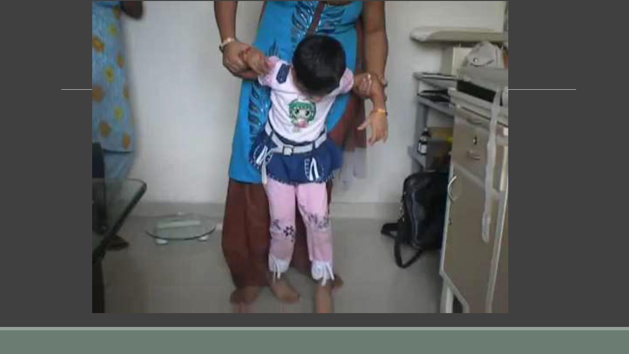
TRENDELENBURG GAIT:The stability of hip during walking provided by bony components of joint, muscles & around joint & normal alignment of center of gravity.
any disruption in the osseo muscular mechanism between pelvis & femur leads to lost of stability of hip joint.
The action of abductor in pulling downwards in stance phase become ineffective.
Usually unilateral
B/L =waddling gait
Causes
weak abductors
Fulcrum
Defective lever system
Abductor muscle function
Two limb stance
One limb stance
In 1895 Fredrich Trendelenburg described a clinical sign useful for detecting the function of hip abductor muscle with special referance to CDH and progressive muscular dystrophy
2
TRENDELENBERG GAITFunctional weakening of abductor mechanism.
Abductor muscles at mechanical disadvantage
Standing on affected side pelvis drop to normal side
To compensate pt lurch to affected side
steppage gait
No need to compensate – tilt to opposite side
TRENDELENBERG GAIT
Fulcrum – head .hip joint – DDH
arthritis
Lever arm - neck
Congenital coxa vara,
#neck, malunited # trochanter
Power – abductors (medius and minmus)
polio, myopathy etc.
As a result patient lurches on affected side & pelvis drops on opposite side of hip.
seen in polio, CDH, perthesdisease, coxa vara, muscular distrophies.
GLUTEUS MAXIMUS LURCH:
Gluteus maximus is the chief extensor & lateral rotator of hip.
Normally when body moves forward in mid stance phase, the hip is extended by gluteus maximus tilting pelvis backwards to retain centre of gravity over supporting leg.
When there is weakness of gluteus maximus muscle the stabilizing factor is lost & patient leans backwards at hip to passively extend it & keep centreof gravity over stance leg.
This causes backward lurch in gluteus maximus gait.
Patient walks with protruberant abdomen.
Seen in poliomyelities & above knee amputation with prosthesis.
Gait in bilateral hip diseases
Waddling gait
Bilateral trendelenberg
CDH
COXA VARA
Gait in bilateral ankylosisAnkylosis in abduction
Weight on one side
Lift other side
Foot as fulcrum
Rotate the whole body
Advance opp leg
Repeat on other side
‘a curious clockwork gait’– Herbert Sedden
Gait in bilateral ankylosis
◦Ankylosis in adduction
Knee close
cannot lift leg
walking not possible
GLUTEUS MEDIUS GAIT(Abduction Lurch):
Gluteus Medius is principal abductor of hip joint along with obturator internious & piriformis.
The weakened Gluteus Medius forces patient to lurch towards involved side to place center of gravity over hip.
This is called Gluteus Medius Gait.
MUSCLE WEAKNESS CAUSING PATHOLOGIC GAIT
QUADRICEPS GAIT:
Quadriceps muscle is the principal extensor of the knee joint.
Due to weakness of quadriceps muscle, the affected limb is put forward in stepping with the body leaning toward it anteriorly.
Patient gradually learns to stabilize his knee by directly transferring his body weight over lower thigh, through his ipsilateral hand.
Weakness of quadriceps is most apparent during heel strike through the stance phase.
The limp affects all phases of gait cycle.
Extension at femur results in flexion of the trunk & an extension movement at knee.
CALCANEAL GAIT:
It occurs due to weakness of the gastrocnemius-soleus muscle group.
As a result, reduced foot propulsion occurs during toe off period of the stance phase & patient walks on his broadened heel with a tendency of rotating foot outwards.
WEAKNESS OF HIP FLEXORS:
The patient will have difficulty in initiating swing through.
To compensate for this specific muscular weakness patient externally rotates leg & uses hip adductors for swing through.
This circumduction of hip exaggerates energy expenditure & produces extreme trunk & pelvis motion.
HIGH STEPPING GAIT:
Ankle dorsiflexors act during the swing phase of cycle.
The weakness of this group of muscles causes foot drop.
During walking foot slap in ground on heel strike & then drops in swing phase.
To prevent this patient flexes hip & knee excessively in order to clear the ground.
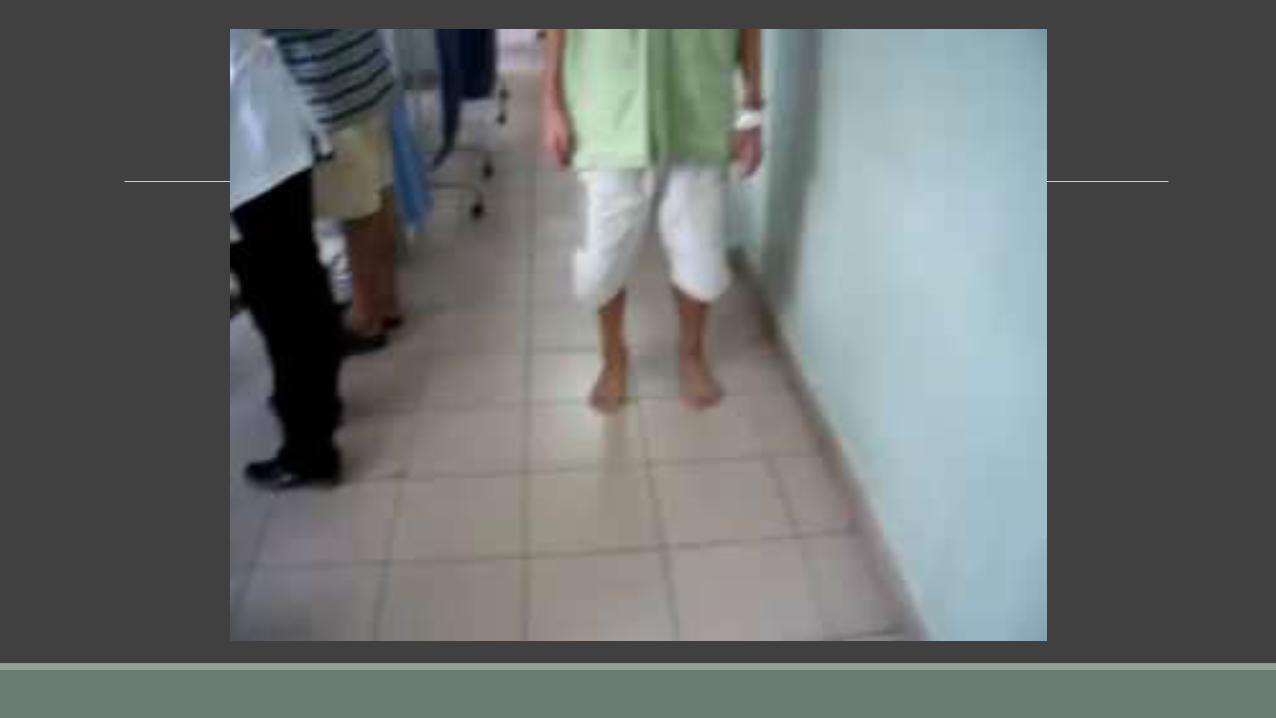
TOE-TIP GAIT
In persistent foot drop or contracture of heel cord the patient walks on toe tip and ball of toes(Meta tarsals head)
STRUCTURAL DEFORMITIES OF THE BONE AND JOINT CAUSING PATHOLOGICAL GAIT
STRUCTURAL DEFORMITIES OF BONE & JOINT
ANTALGIC GAIT:
Any pathology in lower extremity which causes during weight bearing result in antalgic gait.
To minimize pain on weight bearing, person shortens time duration of stance phase on painful side & quickly transfers weight on normal leg.
Longer stance on normal leg & shorter stance on painful leg.
STIFF HIP GAIT:
When the hip is ankylosed, it is not possible to flex at hip joint walking to clear the ground in stance phase,hence person with stiff hip lifts pelvis on that side & swings leg in circumduction to take the forward step.
STIFF KNEE GAIT Normally knee goes to flexion during early stage of swing phase to clear the foot from the ground
But in stiff knee gait the patient as to lift the pelvis of affected side to clear ground and swinging side ways with circumduction of limb to propel it foreword to reach the heel strike
SHORT LIMB GAIT STEP:
A limb length discripancy of 1 to 1.5 inch is compensated by tilt of the pelvis,which is demonstrated by a low shoulder,low iliac crest & low ASIS.
Another method to compensate shortening is to put foot & ankle at the affected side into equinus position & hip & knee of normal limb in flexion.
NEUROGENIC DISORDERS CAUSING PATHOLOGICAL GAIT
NEUROLOGICAL DISORDERS
HEMIPLEGIC/FLACCID GAIT:
In a hemiplegic gait,the shoulder is adducted & the elbow & wrist are flexed.
The pateint swings the paraplegic gait outwards & aheads in a circumduction to avoid foot scraping ground.
It is seen in cerebrovascular disease.
SCISSORING/SPASTIC GAIT:
This gait is characteristic of gait of a spastic child with marked b/l adductor spasm at hip & equinus in the ankle.
The child needs support to walk & leg goes into marked adduction in swing phase so that the foot with equinus goes across to opposite side.
Such repeated crossing of leg whle walking gives scissoring appearance called as scissor gait.
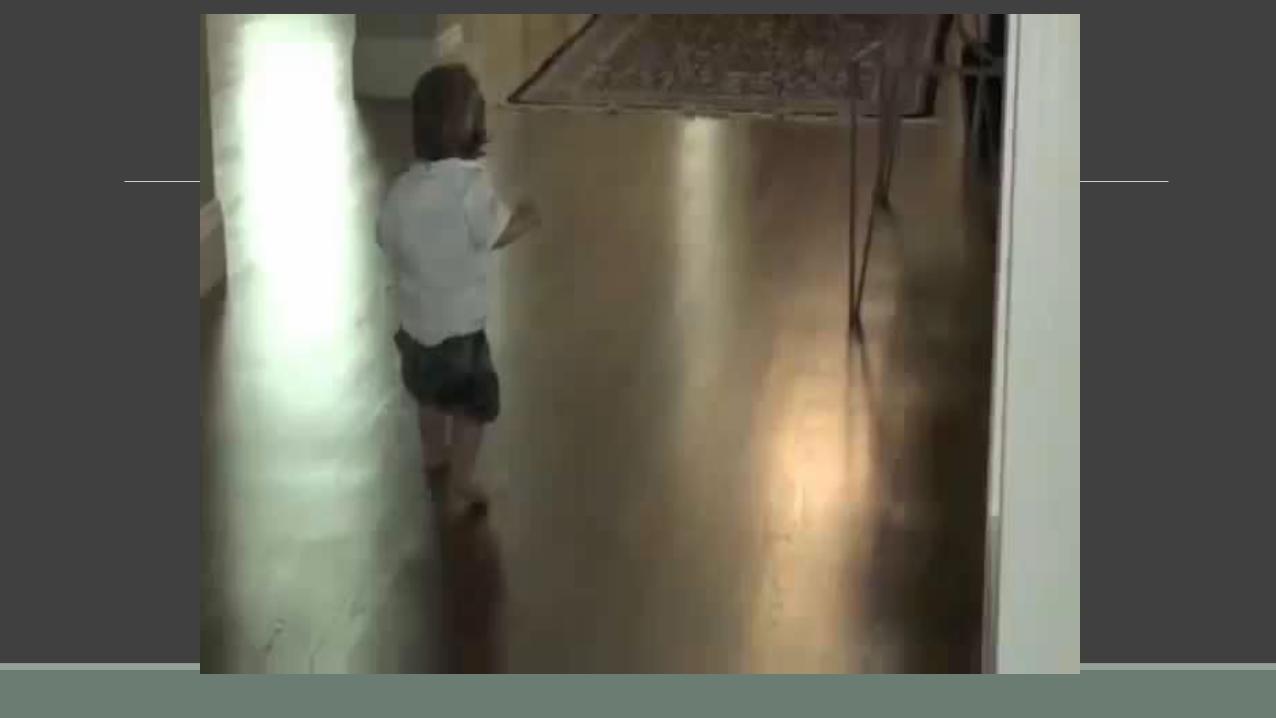
FESTINANT GAIT:
The steps are short so that feet barely clear floor.
If patient is pushed backward or forward, compensatory flexion or extension fails to occur & patients is forced to make a series of propulsive or retropulsive steps with forward locomotion.
Steps become successively more rapid as if trying to catch up with centreof gravity.
Seen in Parkinson's disease.
STAMPING/ATAXIC GAIT:
It occurs in sensory ataxia in which there is loss of sensation in lower extremity due to disease processes in peripheral nerves, dorsal roots, dorsal column of spinal cord.
Due to absence of deep position sense,the patient constantly observes placing of his feet.
Hip is hyperflexed & externally rotated & forefoot is dorsiflexed to strike ground with a Stamp. Seen in peripheral neuritis & brain stem lesion in children, tabes dorsalis in adults.
DRUNKARDS/REELING GAIT:
The patient tends to walk irregularly on a wide base sways from side to side with tendency of falling with each step.
It is seen in lesion of cerebellum, lesion connecting pathway to & from the cerebellum.
CHOREFORM GAIT:
In this patient will be having chorea in upper limbs & has a unstable gait.
Seen in patients having extrapyramidal symptoms.
MISCELLANEOUS GAIT
MISCELLANEOUS GAIT
ALDERMAN'S GAIT :
Patient walks with head & chest thrown backwards & protuberant & walks with legs thrown wide apart.
Seen in Tuberculosis spine of lower dorsal & lumbar vertebrae.
HYSTERICAL GAIT:
Patient walks in a bizzare as if going to fall on every step but seldom falls and walks cautiously.
KNOCK KNEE GAIT:
The patient flexes his hip slightly the knee joint opposes each other, the ankle & feet are kept apart with tendency of toe in.
GENU RECURVATUM GAITIn paralysis of hamstrings muscles knee goes to hyperextension
Due to lack of counteraction of hamstrings while transmitting the weight in mid stance phase
SENILE GAIT:
Changes in gait & difficulty with balance occurs with aging.
Elderly man develops forward of upper portion of trunk with flexion of arms & knees.
Decreasing arm swing & shortening of step length.
The In-Toeing Toddler/Child: Assessment
Rotational Profile: Evaluate in four steps
1.) Observe child walking and running.
Estimate the foot progression angle (FPA): angular difference between the axis of the foot and the line of progression
Assessing the Foot Progression Angle
Nonspecific estimation
Normal: usually -5 tp +20 degrees
In-toeing -5 to-10 degrees: mild
-10 to -15 degrees moderate
>-15 degrees severe
Internal Femoral Torsion/Anteversion
In standing position, patellae will point inwards when feet are forward
Compensatory external rotation of tibia
Femoral Anteversion: Clinical Assessment
“Kissing” patellae
Femoral Anteversion: Definitions
Femoral version defined as the angular difference between axis of femoral neck and the transcondylar axis of knee
Femoral anteversion ranges from 30-40 degrees at birth and decreases progressively to about 10-15 degrees at skeletal maturity
Usually first seen in the 3-5 year age group, usually most severe b/w 4-6 years
Almost always symmetrical
Mechanism unknown, genetic factors and position of fetus in uterus causing increased rotation
More common in females: approx. 2: 1 ratio, often familial
Gait/running described as awkward/clumsy by parents
OUT-TOEING GAIT
Normal range for out-toing is from 0-30 deg
It is most common in infant/toddler this will resolves spontaneously
How ever associated with lateral tibial torsion will worsens with growth should corrected surgically
TOE WALKING
Usually established in age 3 years
By the age of 3 years heal strike pattern of gait should establish
Causes of persistence beyond 3 years suspet
1. Cerebral palsy
2. Muscular dystrophies
3. Residual polio deformity
4. Post burn contracture
5. Spinal cord tumours
6. Idiopathic it common
CRUTCH WALKING-PATTERNS OF GAIT
There are 4 patterns of gait:
Swinging crutch gait - in paraplegics
Four point crutch gait - in unsteady pts.
Two point crutch gait - pts.balance good
Three point crutch gait
Swinging crutch gait :
There are two types of swinging crutch gaits, the swing to crutch gait & swing through crutch gait.
These gaits are when body weight can be taken through both lower limbs together but patient is incapable of moving lower limbs individually due to paralysis.
The lower limbs are moved by trunk muscles acting on the pelvis.
Swing Through Crutch Gait:
In this body is swing through beyond the crutches.
In this pt. advances the crutches & then swings his body to the crutuhes.
The sequence of events:
both crutches both lower limbs.
Four point crutch gait:
It is used when all or part of body weight can be taken on each foot.
Pt. is unsteady & requires a wide base of support.
As pt's balance improves he may progress to two point crutch gait.
The four points are two crutch tips & two limbs.
Sequence of events:
right crutch left foot left crutch right foot.
Two point crutch gait:
When two point crutch gait is used,the amount of body weight taken on both feet is reduced.
This type of gait used when pts. balance is good.
Sequence of events:
Right crutch & left foot simultaneously f/by
left crutch & right foot simultaneously.
Three point crutch gait:
In this gait, the amount of body weight taken by a foot can vary from none to partial or full.
This gait is commonly taught to orthopaedic patients who may have one painful or weak limb which cannot support the whole body weight & one lower limbs which can.
Both crutuhes support weaker lower limb, while the stronger limb takes whole body weight without any support from the crutches.
Sequence of events:
Both crutches & the weaker lower limb together, the stronger lower limb.


![[PPT]Abnormal Gait gait.ppt · Web viewAbnormal Gait Department of Physical Therapy NEW YORK UNIVERSITY Historical Perspective Tendency to classify gait according to disease or injury](https://static.documents.pub/doc/80x56/5a9f71187f8b9a76178cd32e/pptabnormal-gaitpptweb-viewabnormal-gait-department-of-physical-therapy-new-york.jpg)
