SPECTROSCOPIC EVALUATION OF THE CORROSION PRODUCTS ON THE TURRET OF THE AMERICAN CIVIL WAR IRONCLAD, USS MONITOR Desmond C. Cook (a) and Mark J. Shaw Department of Physics Old Dominion University Norfolk, VA 23529, USA Eric Schindelholz (b) The Mariners’ Museum 100 Museum Drive Newport News, VA 23606, USA Bruce L. Bramfitt, Samuel J. Lawrence and Robert C. Nester Mittal Steel USA-Steelton 215 Sth Front St. Steelton, PA 17113 ABSTRACT Spectroscopic analysis of artifacts from the American Civil War ironclad, USS Monitor, has been undertaken in order to determine the present state of degradation of the objects, and to identify foreign compounds that will require removal during the stages of conservation. Metallic artifacts consisting of wrought iron from the rotating gun turret have been studied by a variety of analytical techniques to determine the effect of long-term exposure to salt, and the anaerobic environment of the deep ocean. Mössbauer spectroscopy, and X-ray diffraction have been used to identify the corrosion products, concretion, and marine sediments attached to the turret when it was recovered in 2002, and subsequently during storage. Optical microscopy and Electron-Probe Micro-Analysis have been used to characterize the wrought iron morphology, and to locate and map the chlorine and other potentially detrimental elements in the iron. While submerged in the ocean, the corrosion of the turret has resulted in a thin coating of the reduced iron oxide, Corrosion Magnetite, which, when covered or incorporated with marine concretions, appears stable, and offers the wrought iron protection from accelerated corrosion often observed following recovery of metal artifacts from the ocean. However, if the concretion is breached and the rust or metal is exposed to air, further and immediate oxidation of the 1
Transcript
SPECTROSCOPIC EVALUATION OF THE CORROSION PRODUCTS ON THE TURRET OF THE AMERICAN CIVIL WAR IRONCLAD, USS MONITOR
Desmond C. Cook(a) and Mark J. Shaw
Department of Physics Old Dominion University Norfolk, VA 23529, USA
Eric Schindelholz(b)
The Mariners’ Museum 100 Museum Drive
Newport News, VA 23606, USA
Bruce L. Bramfitt, Samuel J. Lawrence and Robert C. Nester Mittal Steel USA-Steelton
215 Sth Front St. Steelton, PA 17113
ABSTRACT
Spectroscopic analysis of artifacts from the American Civil War ironclad, USS Monitor, has been undertaken in order to determine the present state of degradation of the objects, and to identify foreign compounds that will require removal during the stages of conservation. Metallic artifacts consisting of wrought iron from the rotating gun turret have been studied by a variety of analytical techniques to determine the effect of long-term exposure to salt, and the anaerobic environment of the deep ocean. Mössbauer spectroscopy, and X-ray diffraction have been used to identify the corrosion products, concretion, and marine sediments attached to the turret when it was recovered in 2002, and subsequently during storage. Optical microscopy and Electron-Probe Micro-Analysis have been used to characterize the wrought iron morphology, and to locate and map the chlorine and other potentially detrimental elements in the iron. While submerged in the ocean, the corrosion of the turret has resulted in a thin coating of the reduced iron oxide, Corrosion Magnetite, which, when covered or incorporated with marine concretions, appears stable, and offers the wrought iron protection from accelerated corrosion often observed following recovery of metal artifacts from the ocean. However, if the concretion is breached and the rust or metal is exposed to air, further and immediate oxidation of the
1
metal and existing rust occurs, and is believed to be detrimental to conservation processes. Elemental X-ray mapping by Wavelength Dispersive Spectroscopy shows that during ocean submersion, chloride ions diffuse deep into the inclusions in the wrought iron, where they are trapped and become responsible for significant and continued corrosion. Once exposed to air and dried, the trapped chlorides have a detrimental effect on the longevity of the artifact. Keywords: corrosion, USS Monitor, conservation, wrought iron, Civil War
(a)Corresponding Author: [email protected] (b)Now at National Park Service P.O. Box 50, Harpers Ferry, WV 25425.
INTRODUCTION
USS Monitor was the first ironclad warship to be constructed in the United States. Built in 1862, it was rushed into battle in Hampton Roads, Virginia where the infamous Civil War battle with the iron covered Confederate frigate, CSS Virginia, was waged on March 9, 1862. Built in only 147 days, USS Monitor was 173 feet in length and 41 feet wide. Its flat deck was armor plated with 1-inch thick wrought iron, and the waterline armor consisted of 5 layers of 1-inch wrought iron plates bolted together. Although outfitted with only two very large 11” Dahlgren guns, the revolutionary design of USS Monitor centered on the gun turret that could be rotated through 360 degrees in 10 minutes. The cylindrical turret was 20 feet in diameter, 9 feet high, and weighed 120 tons. It was constructed using 8 layers of 1-inch wrought iron plate bolted together. History records the 4-hour battle between the two ironclads in Hampton Roads as fierce and at close range. Although struck often, neither ship sustained serious damage, due to their protective armor, thus ending the era of wooden fighting ships. CSS Virginia was eventually scuttled in Hampton Roads, Virginia, and USS Monitor sank in a storm off Cape Hatteras, North Carolina, on December 31, 1862. Lost at sea were 16 of the 56 sailors on board.
The wreck of the Monitor was found in August 1973, 16 miles East of Cape Hatteras, and at a depth of 240 feet. It has now become one of the most historically significant warships of the US Navy. In what has turned out to be scientific good-fortune, the ironclad and the dislodged turret landed upside down on the ocean floor. The ship’s guns were found to be still inside the turret. Those and many other important items, including human remains, were retained inside the inverted turret by its wrought iron cap. The intact turret was recovered in August 2002 and is now being prepared for conservation at The Mariner’s Museum(1) in Newport News Virginia, less than 3 miles from the site of the original battle. It is heavily encrusted with marine growth, filled with ocean sediment, and shows signs of significant corrosion(2, 3). On the day of recovery from the ocean floor, the turret quickly changed color from dark brown to bright orange as it was exposed to the air, despite being continuously sprayed with seawater during its 20-hour voyage to land(2-3). Figure 1 shows corrosion, concretion, and ocean mud on the exterior turret wall two days after recovery in 2002.
Conservation of the wrought and cast iron artifacts requires considerable understanding of the effects of ocean exposure, and the subsequent exposure to air during the conservation process. Recently thickness loss measurements on the turret’s outer wall plates(4) showed a mean corrosion rate of 1.4 mpy, (38 μm/yr), which in comparative figures is about one half the corrosion rate of an uncoated weathering steel structure located in region of high de-icing salt usage, and about six times greater that the desired corrosion rate of bare steel structures(5). Corrosion rates were also estimated by
2
electrochemical methods through measurement of the corrosion potentials, and linear polarization resistance of the turret walls, and other artifacts(4). LPR measurements show localized instantaneous corrosion rates varied across the turret walls between 1.8-13.9 mpy, (46-353 μm/yr), for old and recently de-concreted surfaces respectively. Although such data is important for deciding if the artifacts require stabilization while they wait for conservation to begin, no information is provided concerning causes of the corrosion and the nature of the detrimental compounds that have formed on the wrought iron. Ultimately the corrosion material must be removed or stabilized, and any chlorides present need to be extracted from the artifacts to ensure that their historical and cultural importance is maintained indefinitely. In order to evaluate the nature of degradation of the iron artifacts due to prolonged seawater submersion, a spectroscopic study of the corrosion products, concretion, and marine sediments attached to the wrought iron turret has been completed. The stability of the rust has also been investigated following exposure to air. X-ray mapping of chlorine and other elements by Electron-Probe Micro-Analysis (EPMA), with WDS elemental analysis, has been performed in order to evaluate the nature and extent of localized corrosion and to help guide conservators in their treatment to extract the foreign impurities. Micro-Raman spectrometry has been used to identify the compounds formed in the wrought iron inclusions during iron production, as well as a result of the corrosive ocean exposure.
SAMPLE PREPARATION AND EXPERIMENTAL PROCEDURE
Since its recovery in 2002, the turret of USS Monitor has been kept submerged in a tank of fresh water at The Mariners’ Museum. There is debate amongst conservators in general as to whether or not concretions and rust should be removed from artifacts following recovery, to remove detrimental surface contaminants that may accelerate corrosion in the air. Identification of the materials attached to the wrought iron turret walls has been undertaken in a manner that has characterized the layering of the adherent material within minutes of sample collection, and then over a period of 12 months as those samples age and dry in air. The original marine sediments, concretions, and rust have been purposely kept on the turret until sufficient data exists to justify or not, their removal. During excavation and inspection of the turret, the tank is emptied of water to permit worker access. At these times the turret is sprayed with fresh water every 20 minutes to prevent drying of the concretion and rust.
Samples were collected from many locations on the outer wall of the turret, and included ocean mud, concretion, and rust. The ocean sediments covered the concretion and rust in many locations, especially where the turret had been imbedded into the ocean floor, Figure 1. The mud-like material was 8-10 cm thick in places, and the samples were collected from at least 2 cm below the surface. The sediments were fine and densely packed as a moist, sticky, gelatinous, clay-like solid, often streaked with orange color. The samples were initially black when first collected, and changed color to dark olive-green-brown if allowed to dry in air over a period of several weeks.
Figure 2 shows a typical sampling location where the outer turret wall is partially covered with 1-3 cm thick concretion, beneath which is a solid rust layer covering an inner moist or liquid suspension of black rust. Samples of rust were collected from beneath small regions of freshly removed concretion, as well as regions where concretion had become dislodged many months earlier. The rust covering the entire surface of the turret was about 0.5 cm thick, and was characterized by an outer layer, or crust of solid rust, whose outer surface exposed to the air, was light brown to bright orange in color. Beneath the outer solid rust surface, the rust consisted of a moist, fine packed black powder, which became a very
3
liquidous black suspension at the surface of the wrought iron, which itself was very shiny. When the solid orange rust was breached, the black material would flow through the void and down the turret wall. It would begin to change color within 5 minutes to dark brown and then to light brown to often orange after 12 hours, thus giving the turret its variable bronze-like color(1-3). Pieces of rust from the outer crust were collected, along with the black moist powder. Samples of the black, liquid suspension were collected in glass vials that were filled and sealed from the air. While sealed the powder remained black indefinitely, but if unsealed and kept in the liquid, the fine black powder slowly turned dark brown over 2-3 weeks.
X-ray diffraction patterns, (CuKα), and Mössbauer spectra were recorded for each type of sample described, including outer and inner surfaces of solid samples of concretion and rust. Spectra were also recorded of the black liquid-like rust beneath the crust, at different times after sample collection from 1 hour to 180 days. X-ray diffraction allowed the identification of all crystalline compounds in the samples whereas Mössbauer spectroscopy, which is ideal for analysis of rust, was used to identify the iron bearing compounds, their valence state, relative fraction, particle size, and possible presence of impurities.
A metallographic mount of wrought iron from the turret was studied by optical microscopy,
electron microprobe, and micro-Raman analysis, in order to characterize the inclusions, identify their composition, and document the extent of corrosion. Two sides of the polished iron sample were original surfaces and therefore showed significant corrosion. These examinations provided extensive information concerning the corrosion and chloride composition directly on the exposed surfaces of the wrought iron.
RESULTS AND ANALYSIS Ocean Sediments
Analysis of the sediments attached to the turret wall was completed in order to investigate any interaction with the corroded material on the turret. During removal, the mud had a strong smell of hydrogen sulfide, and was determined to have a low pH of 2-3 in many locations. Therefore the presence of microbial activity and the possibility of accelerated corrosion due to the sulfur reducing bacteria, was of concern. Chemical analysis of the mud, showed a high concentration of bound and unbound sulfur, averaging 1.5 wt%. X-ray diffraction identified the mud to consist of predominantly quartz, SiO2, (> 75%), a small amount of calcite, CaCO3, (5%), and three iron compounds, (20%), also identified by Mössbauer Spectroscopy. Figure 3 shows the Mössbauer spectra of the mud recorded at temperatures of 300 K and 77 K. The iron compounds in the mud consist of 63% siderite, FeCO3, 21% goethite, α-FeOOH, and 16% lepidocrocite, γ-FeOOH. The Mössbauer spectra show that goethite is magnetic at 77 K but not at 300 K, inferring that the particle size is very small, in the range of 5-20 nm. The formation of siderite is likely the result of corrosion of the wrought iron in contact with the forming concretions and subsequent diffusion into the mud. The lepidocrocite and nano-phase goethite are indicative of fresh rust that formed recently under oxic conditions. It probably became incorporated in the mud, seen as the orange streaks, due to bleeding of the turret following exposure to air.
Concretion
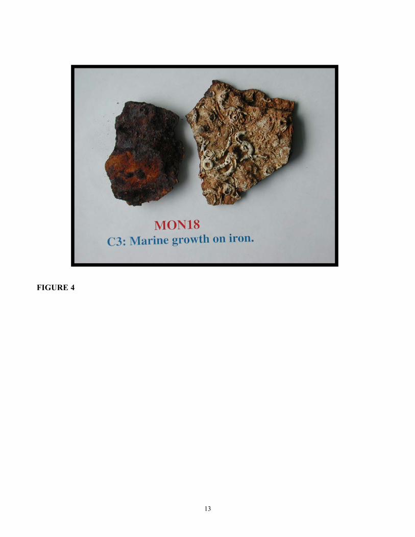
The concretion forms a thick barrier layer between the ocean and the corroding iron. It is therefore not unexpected that its composition varies through its thickness. Figure 4 shows the outer and
4
inner surfaces of concretion removed from the turret. The outer surface is medium brown in color and covered with various marine organisms. The inner surface is very dark brown due to contact with the rusting iron. Spectroscopic analysis of 5 samples of concretion showed the compounds present to be the same but in varying amounts depending on the location of sampling and the surface from which the sample was prepared. Typical Mössbauer spectra of one sample of concretion are shown in Figure 5. The composition of the iron containing compounds was determined to be siderite, (15%), lepidocrocite (8%), goethite, (41%), and Corrosion Magnetite, Fe3O4, (36%). At 300 K the goethite was comprised of a magnetic component (αm = 35%), (large particle > 20 nm), and a non-magnet component (αs = 6%), (nanophase, < 20 nm). The high fraction of large particle goethite indicates it was formed over a long time interval, (> 2 years), and is therefore may be the result of the iron corrosion during ocean submersion in fairly oxic conditions. X-ray diffraction also identified very small amounts of quartz (< 3%), and calcite, (7%). The fractions of siderite and corrosion magnetite varied through the thickness of the concretion with the former being higher at the outer surface, and the latter higher at the inner surface next to the rusting iron. It is clear from the high fraction of siderite in the concretion, that there is significant interaction between the rusting iron and the carbonate formation that is normally calcite.
Turret Rust: Outer Solid Rust Layer
Analysis of three regions of one piece of the orange and brown outer crusty rust layer on the outer turret wall was undertaken to determine the effects of exposure to air. The piece of rust was removed from region labeled B in Figure 2. It was orange-brown on the outside surface exposed to the air and black on the inside surface adjacent to the moist black rust. The pH on the inner black surface was 2-3. The concretion from this region was dislodged soon after the turret was raised, indicating that exposure to air and some drying of the outer surface has occurred at times when the turret tank was drained of water. Spectroscopic analysis was performed on the outer orange rust, inner black rust, and one piece comprising the full thickness of the rust. The Mössbauer spectra of the outer orange rust are shown in Figure 6. X-ray diffraction and Mössbauer spectroscopy showed the presence of goethite (54%), akaganeite, β-FeOOH, (9%), lepidocrocite (20%), Corrosion Magnetite (12%), and fayalite, FeSiO4, (5%). The goethite was comprised of a magnetic component (αm = 16%), and a non-magnetic component (αs = 38%) at 300 K. A very small amount of calcite was detected in the X-ray patterns. The composition of the rust varied slightly between the outer and inner surfaces of the rust with lepidocrocite decreasing to 13% and akaganeite increasing to 15% at the inner surface. The other three iron compounds maintained their percentage composition. Of interest, the goethite on the inner surface, (55%), was comprised of a magnetic component (αm = 28%), and a non-magnetic component (αs = 27%) at 300 K. The presence of more lepidocrocite and nano-phase goethite on the outer surface indicates significant oxidation of this rust layer has occurred as a result of exposure to air and/or drying. The presence of akaganeite indicates that chloride ions are present in the solid rust layer and additionally confirms the oxidation of the rust layer following the turret’s recovery. Typically the chloride ion would be soluble and easy to remove from the surface of the structure had it not been exposed to air. However, with the formation of akaganeite, the chloride is now bound and removal is perhaps more difficult because akaganeite is stable in air and has low solubility in water. This is especially a problem if the akaganeite forms on the surface of the wrought iron walls, rather than several millimeters away in the rust layer, as was true in this case. However, the results indicate that prolonged exposure of a freshly cleaned wrought surface to air, in the presence of chlorides from seawater, is likely to result in an additional surface treatment prior to final artifact conservation.
5
The Corrosion Magnetic formed in a very reduced environment and is the direct product of the anoxic corrosion of the iron due to 140 years of submersion in the ocean at a depth of 240 ft and perhaps the gradual build-up of concretion. Corrosion Magnetite is usually stable in air below a temperature of 80 C. It is therefore not expected that the three oxyhydroxides in the rust formed due to oxidation of the Corrosion Magnetite, leading to the suggestion that another, as yet unidentified, corrosion process is active at the metal surface. The presence of about 5-7% fayalite in the rust is a direct result of the corrosion of the wrought iron that is comprised of a very high density of inclusions formed during the production and rolling of the iron. Data presented below show the inclusions to be mainly FeSiO4, which will be stable during the corrosion processes, and is thereby incorporated into the rust without degradation.
Turret Rust: Inner Liquid Rust Layer
Located between the outer solid rust layer and the wrought iron, is a region of moist densely packed fine, black, powdered rust. The water content increases closer to the iron, to the extent that directly on the surface of the wrought iron, the rust is completely liquid. As mentioned earlier, this liquid can leak from any void or crack in the solid rust layer and quickly begin to change color as it runs down the turret walls(2). This instability was observed on exposure to air but not in sealed airtight containers. X-ray diffraction patterns and Mössbauer spectra were recorded on sample of the wet black powder. Data acquisition required about 24 hours for each run and spectra are therefore indicative of samples exposed to air for 1 day. Data was recorded periodically for about one year to observe changes in the samples. After about 4 weeks the samples appeared stable. Figure 7 shows the room temperature Mössbauer spectra for one sample recorded after 13 days and 68 days. The former spectrum is indicative of the fresh sample, (1day<age<30days), and the latter for times greater than 30 days. The Mössbauer and X-ray analysis show that the black rust consists of predominantly Corrosion Magnetite (91%), goethite (2%), and lepidocrocite (7%). A very small amount of fayalite, (<0.5%), was also detected. The spectra of several black powder samples collected from adjacent to the wrought iron on the turret walls showed consistency in their compositions. The Corrosion Magnetite component of the spectra can be seen to change as the sample ages. This is seen in Figure 7 by the two sextets plotted as subspectra for the Corrosion Magnetite. The inner sextet is less intense and broader in the aged sample. Spectral analysis however showed that the relative amount of magnetite remained at 91% for both spectra. The inner sextet decreased by 4% as the sample aged, while the outer sextet population of iron increased by the same amount. This change is attributed to an oxidation of 4% of iron in the Corrosion Magnetite from Fe2+ to Fe3+. It should be noted that the Corrosion Magnetite therefore shows good long-term stability with exposure to air, and does not oxidize to maghemite, γ-Fe2O3, at ambient temperatures as it does at 100 C.
The presence of lepidocrocite and goethite in the black powder samples collected from adjacent
to the wrought iron surface is of interest. These oxyhydroxides are commonly observed on iron surfaces as fresh rust due to iron oxidation in the presence of ample oxygen. Yet the environment in which these samples were collected has remained highly anoxic for probably the last 130 years, including the 4 years since the recovery of the turret. At the present time, we believe that the slow oxidation of the Corrosion Magnetite is not responsible for the formation of lepidococite or goethite, or the rapid color changes from black to brown/orange observed on the turret, as described above. We are planning now to spectroscopically analyze the fresh, black Corrosion Magnetite in a specially designed oxygen-free cell, and to rapidly record spectra sequentially at hourly intervals, to study any short-term instability in the liquidous black powder.
6
Wrought Iron Morphology
A piece of corroded wrought iron from the Turret, (NOAA artifact # MNMS.2002.WI001),
about 7 cm long, 4 cm wide and 2 cm thick was cut into six pieces, of which two were mounted and polished to permit study of the wrought iron inclusions in directions parallel and orthogonal to the direction of plate rolling during manufacture. The original wrought iron was assumed to be 1” thick, in agreement with the armor plating used to construct USS Monitor. The material remaining across the 2 cm thickness was comprised of a wrought iron core, 1-1.5 cm thick in a casing of Corrosion Magnetite. Detailed preparation and photographs of the samples are provided elsewhere(2). Figure 8 shows a photograph of the longitudinal mount with the direction of rolling being shown horizontally. The upper and lower sample surfaces are heavily corroded and represent the original iron surfaces exposed to the ocean for 160 years. Significant corrosion material was removed for analysis prior to mounting of the sample. The mounted sample dimensions are approximately 1.6 cm wide and 1.1 cm high, (corresponding to the thickness of the remaining iron). The wrought iron contains a very high density of inclusions running parallel to the direction of rolling. Significant corrosion of the iron, to depths up to about half the sample width can be seen. The inclusions have been labeled as internal or corroded. The majority of inclusions are internal and do not appear to connect in any way to an exposed and corroded surface, as seen by the regions 4, 5, and 6 in Figure 8. Corroded inclusions are typically connected to a region of the iron that has corroded, and are most commonly observed at the upper and lower corroded surfaces. All inclusions connected to the two original surfaces are corroded along part of their length. Typical regions are 1 and 2. Regions of very high iron corrosion, 3, are also seen in the sample and appear to preferentially follow the inclusions into the iron. As will be seen from the data presented, it is concluded that the corrosion of the wrought iron is dominated by the corrosion of the interior walls of the inclusions.
Figure 9 shows two expanded views of one corroded inclusion in Region 1 of Figure 8. Figure
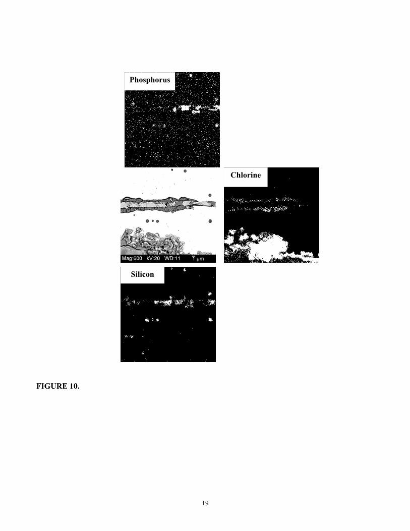
9(a) shows the inclusion has corroded along about 65% of its present length, corresponding to about 0.7 mm. Of course the original length of the inclusion is unknown due to the surface corrosion. It is clear from Figure 9(b) that a distinct boundary between the corroded and un-corroded regions of the inclusion exists. The width of the original inclusion is about 3 μm, whereas the corroded section tapers from 40 μm to about 20 μm at the corrosion boundary. The entire inclusion, on both sides of the corrosion boundary, still contains the original material, showing that the corrosion of only the iron has occurred due to the ocean submersion. This is clearly seen in Figure 10 showing a photograph of the same inclusion recorded during the Electron-Probe Micro-Analysis of the composition of the inclusion. The EPMA analysis with X-ray mapping of the un-corroded inclusions, along with micro-Raman spectrometry, identified the major composition to be the iron silicate, fayalite, FeSiO4, as was also identified in the rust by X-ray diffraction and Mössbauer spectroscopy. Most inclusions also contained phosphorous, in varying amounts, often greater than silicon, and often incorporated in the fayalite structure, Fe(Si1P1-x)O4. As part of all un-corroded inclusions and within the fayalite, nobules rich in iron, (62 at.%), and oxygen, (38 at.%), were observed. Raman spectra are indicative of high purity magnetite or wustite, FeO, but positive identification has not yet been made. X-ray mapping of the elemental composition of the un-corroded inclusions did not reveal the presence of any chloride ions, in un-corroded inclusions.
7
Analysis of the corroded sections of the inclusions showed that along the center of the corroded region, the same compounds were present. Surrounding this material was a layer of corrosion material filling the region of the inclusion. EPMA showed the elemental composition of the corrosion products to be iron, (52 at.%), oxygen, (41 at.%), phosphorus, (3 at.%), silicon, (0.5 at.%), chlorine, (2 at.%), and sulfur, (1 at.%). Micro-Raman analysis indicated the corrosion products to be magnetite, whose morphology is very different to the nodules in the un-corroded inclusions, and very similar to rust on corroded steel. Further work is continuing towards complete identification of the inclusion compounds. Of importance to the understanding of the corrosion of the wrought iron in the ocean, is the data obtained from the X-ray mapping of the elements in the corroded inclusions. Figure 10 includes the X-ray maps for phosphorus, silicon and chlorine, scaled to the same size as the photograph. This allows visual comparison by overlapping the mapped elements with the photograph of the inclusion. It is clear that the phosphorus and silicon correspond to the original composition of the inclusion, whereas the chlorine is situated in the surrounding corrosion product, most likely as the chloride ion. It is clear that chloride from the ocean exposure, has been able to diffuse deeply into the wrought iron along the inclusions, and has probably played a role in the original rates of corrosion of the iron at the surfaces of the inclusions. The presence of the chloride ions in the narrow inclusions, may begin to explain the difficulty, and often lack of success, reported by conservators attempting to removing the chloride from submerged iron artifacts, usually by electrolytic diffusion techniques(6). Work is continuing to obtain a better understanding of the chemistry and properties of the inclusions, the interaction of the chlorides, and the methods by which the chlorides can be completely extracted. Determining the location of chlorides in the wrought iron has established that spectroscopic analysis is important for characterizing metal artifacts prior to conservation treatments, and should also be used to establish the success of such treatments by applying the same methodology to treated samples.
CONCLUSIONS
The composition of the mud, concretions, and corrosion products formed on the outer surfaces of the wrought iron turret of USS Monitor following 140 years of exposure to seawater and ocean sediments has been completed. It is concluded from the dominating presence of corrosion magnetite in the rust adjacent to the iron, that the anoxic environment of the deep ocean has been maintained following recovery of the turret, at locations where the concretions and outer solid rust layer have remained intact. The low corrosion rates beneath the concretions provide additional evidence that artifacts recovered from the ocean and awaiting conservation, should be protected from any exposure to the air, especially if chlorides are present at the metal surface. The finding that chlorides are not only present at the surfaces of the artifacts, but diffuse deeply into wrought iron along the inclusions, shows evidence of the mechanism by which failure to reach stable conservation, through incomplete removal of chlorides, has been previously reported.
ACKNOWLEDGEMENTS
This work has been supported by the Old Dominion University Summer Faculty Research Program, the Old Dominion University Undergraduate Research Program, the National Oceanic and Atmospheric Administration, and The Mariners’ Museum.
8
REFERENCES
1. The Mariners’ Museum, www.Monitorcenter.org 2. Materials Physics web-site, Old Dominion University, www.physics.odu.edu/cmmp 3. RustDr web-site,www.RustDr.com 4. C. S. Brossia, M. Yunovich, D. Hill, K. M. Lawson, R. Denzine, J. T. Schmidt, E. Klechka, E. Schindelholz, E. Nordgren, D. Krop, R. Baboian, H. Hack, J. D. Flessas, D. C. Cook, “Corrosion Condition Assessment, Mitigation, and Preservation of USS Monitor Artifacts.” CORROSION 2007, paper no. 07239. (Nashville, TN: NACE, 2007). 5. P. Albrecht, T. T. Hall, J. Mater. Civil Eng., 15(1) (2003), 2-24. 6. M. O. Carlson, M. R. M. Bruce, W. C. Riess, Bulletin on the Research On MEtal Conservation, (BROMEC), Ed. C. Degrigny, Vol. 8, November 2003, 9-10. http://icom-cc.icom.museum/Documents/WorkingGroup/Metals/Bromec8.pdf
9
FIGURE 1. Photographs of the turret of USS Monitor two days after recovery in 2002. The wrought iron is covered in places with ocean mud, concretion, and rust. Photographs by T. Hartlove, and D. Cook.
10
FIGURE 2. Exterior turret wall showing locations of sample collection; A. concretion, B. outer solid rust layer, C. freshly exposed moist fine, black rust powder. Bleeding of the liquidous black rust, and subsequent oxidation results in the brown/orange color of the surface.
A B C
11
FIGURE 3. Mössbauer spectra recorded at (a) 300 K, and (b) 77 K, of the mud and ocean sediments attached to the outer wall of the turret. The iron compounds are mainly siderite (63%), and smaller fractions of goethite (21%), and lepidocrocite (16%). X-ray diffraction analysis also identified a large fraction of quartz, (>75%), and a small amount of calcite. The pH of the mud was 2-3 and the sulfur concentration was about 1.5 wt.%.
FIGURE 4. Photograph of the concretion on the outer wall of the turret, (location A on Figure 2). The outer surface is covered with marine organisms, and the inner surface, is dark brown due to contact with the corroding iron.
13
FIGURE 5. Mössbauer spectra of the concretion recorded at (a) 300 K, and (b) 77 K. It is comprised of siderite, (15%), lepidocrocite, (8%), goethite, (41%), and Corrosion Magnetite, (36%). X-ray diffractions also identified small amounts of quartz, (<3%), and calcite, (7%).
-10 -8 -6 -4 -2 0 2 4 6 8 10
Lepidocrocite Siderite
Magnetite Goethite
Rel
ativ
e In
tens
ity
Velocity (mm/s)
(a) Concretion: 300 K
(b) Concretion: T = 77 K
14
FIGURE 6. Mössbauer spectra of the outer layer of the solid orange rust on the outer wall of the turret, (location B on Figure 2, covering location C). The spectrum at (a) 300 K shows that the majority of the rust is comprised of non-magnetic iron oxyhydroxides, and a small amount of Corrosion Magnetite, (12%) and fayalite, (5%) . The spectrum at (b) 77 K shows the additional components are separately identified as goethite, (54%), and akaganeite, (9%), that are magnetically ordered, and lepidocrocite, (20%).
FIGURE 7. Room temperature Mössbauer spectra of Corrosion Magnetite formed under anoxic conditions under the concretion and solid rust layer on the outer turret wall. Spectra were recorded after 13 and 68 days exposure of the rust to air. Changes in the Corrosion Magnetite due to slow oxidation, can be seen for the peaks located between –6 to –8 mm/s.
-10 -8 -6 -4 -2 0 2 4 6 8 10
FeOOH: 9% Magnetite A1: 44% Magnetite B1: 47%
R
elat
ive
Inte
nsity
Velocity (mm/s)
FeOOH: 9% Magnetite A1: 40% Magnetite B1: 51%
(a) Monitor Turret Rust: 13 days
(b) Monitor Turret Rust: 68 days
16
FIGURE 8. Metallographic cross-section photograph of the wrought iron showing the high density of inclusions. Regions 1 and 2 show corroded inclusions that are connected to an exposed outer surface. Region 3 shows an area a high corrosion of the wrought iron. Regions 4, 5, and 6, show internal inclusions that are not corroded.
Direction of Rolling
3
Corrosion
45
Internal Inclusions
6
1
2
Corroded Inclusions
1000 μm
17
FIGURE 9. Metallographic cross-section photographs of the thin corroded inclusion in Region 1 of Figure 8. The partially corroded inclusion consists of a “backbone” of fayalite, on both sides, (left and right), of the corrosion boundary. In the corroded region the fayalite is surrounded by corrosion products that have high chloride, (2 at.%), and sulfur, (1 at.%), content. The rust at the bottom of the figures is at the wrought iron surface.
1A: Corroded 1B: Non-Corroded
Corrosion Products
(a)
(b)
18
FIGURE 10. EPMA (WDS) X-ray maps for phosphorus, silicon and chlorine, in the corroded inclusion. The phosphorus and silicon are in the fayalite “backbone” of the entire inclusion. The chlorides are found only in the corroded section of the inclusion in the rust surrounding the fayalite. Note the distinct absence of chlorides to the right of the corrosion boundary.