49
09 Digestive system 1 Oral cavity & Salivary Glands
| Date post: | 05-May-2019 |
| Category: |
Documents |
| Upload: | truonghanh |
| View: | 223 times |
| Download: | 0 times |
09Digestive system 1
Oral cavity &Salivary Glands
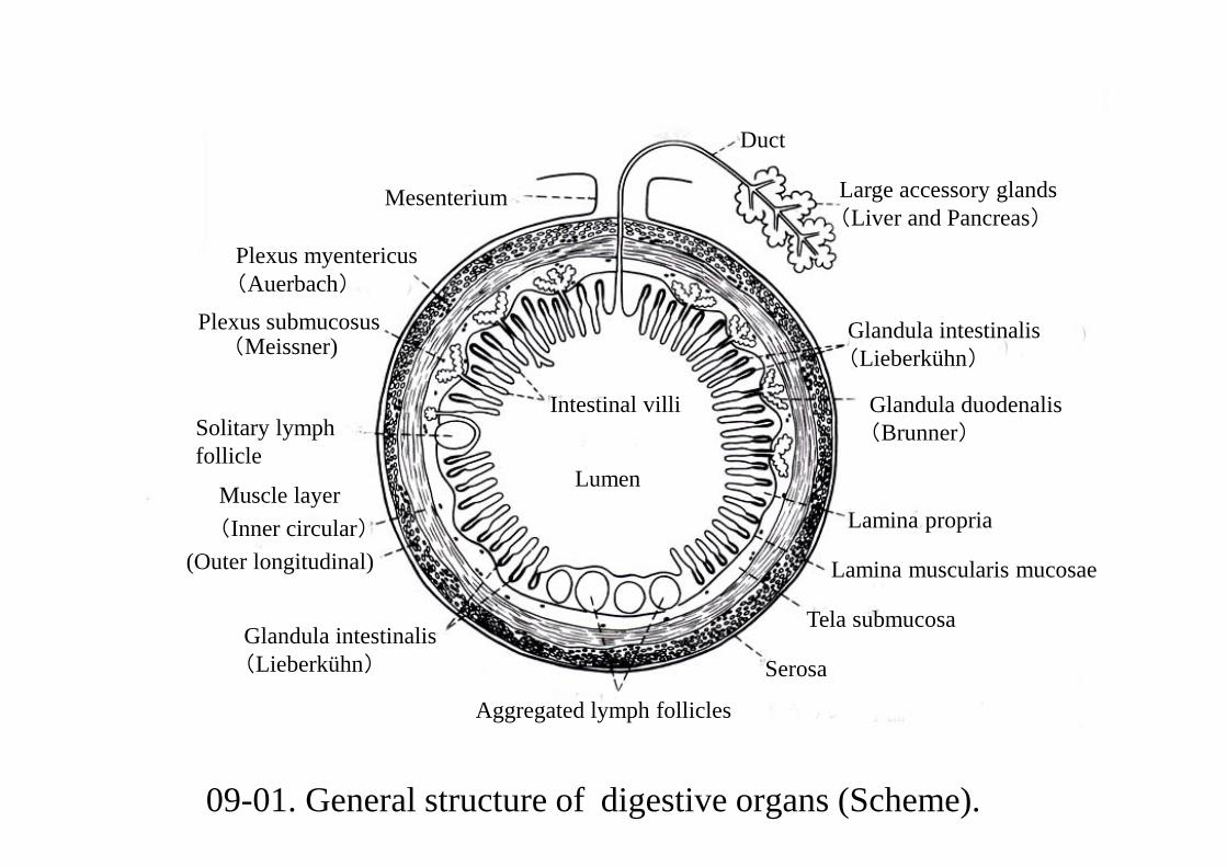
09-01. General structure of digestive organs (Scheme).
Aggregated lymph follicles
Serosa
Tela submucosa
Lamina muscularis mucosae
Lamina propria
Glandula duodenalis(Brunner)
Glandula intestinalis(Lieberkühn)
Large accessory glands(Liver and Pancreas)
Duct
Mesenterium
Plexus myentericus(Auerbach)
Plexus submucosus
Solitary lymph follicle
(Inner circular)
Glandula intestinalis(Lieberkühn)
Muscle layer
Intestinal villi
Lumen
(Meissner)
(Outer longitudinal)
09-001 Oral Cavity
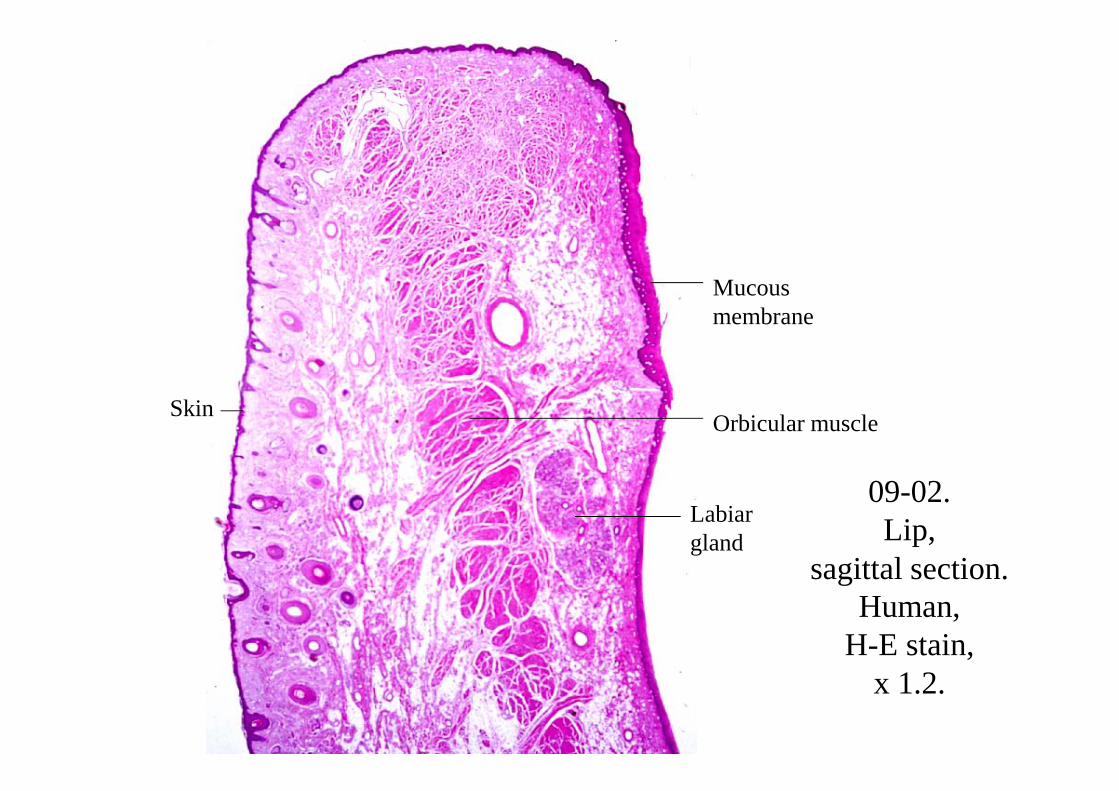
09-02. Lip,
sagittal section.Human,
H-E stain, x 1.2.
Skin
Mucous membrane
Labiar gland
Orbicular muscle
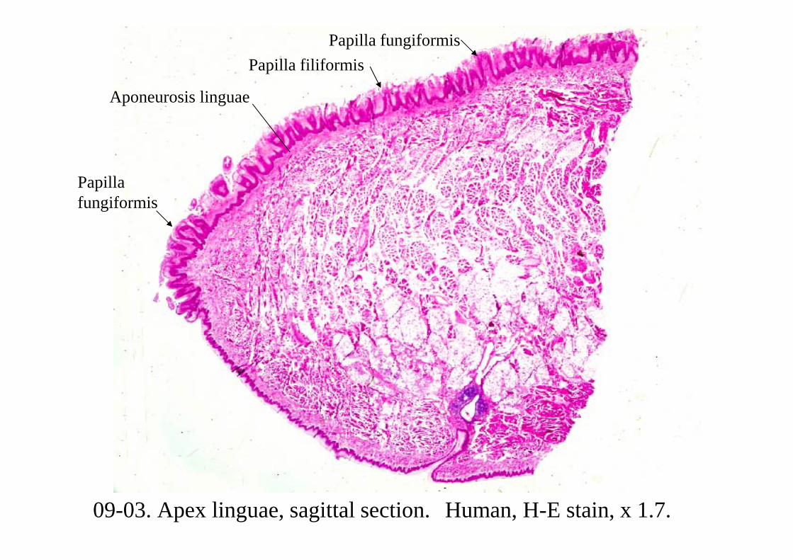
09-03. Apex linguae, sagittal section. Human, H-E stain, x 1.7.
Papilla fungiformis
Papilla fungiformis
Aponeurosis linguae
Papilla filiformis
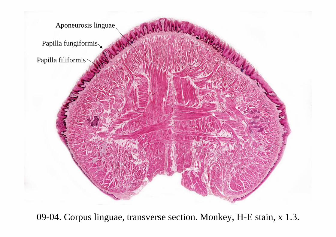
09-04. Corpus linguae, transverse section. Monkey, H-E stain, x 1.3.
Aponeurosis linguae
Papilla fungiformis
Papilla filiformis
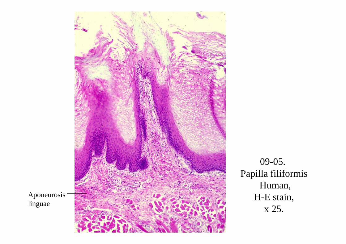
09-05. Papilla filiformis
Human, H-E stain,
x 25.Aponeurosis linguae
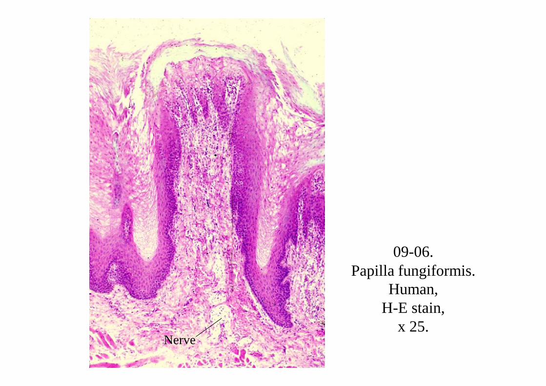
09-06. Papilla fungiformis.
Human, H-E stain,
x 25.Nerve
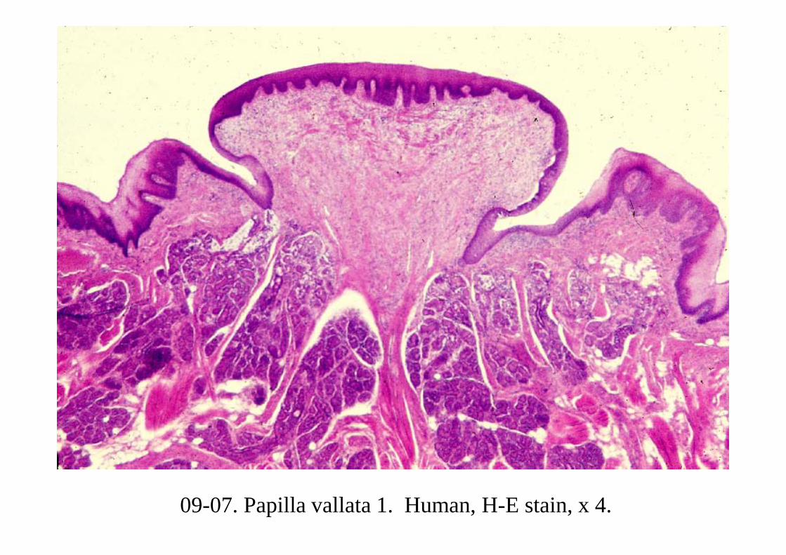
09-07. Papilla vallata 1. Human, H-E stain, x 4.
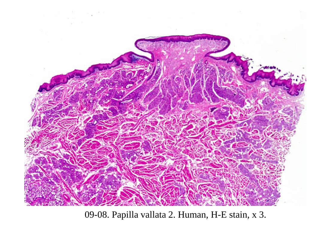
09-08. Papilla vallata 2. Human, H-E stain, x 3.
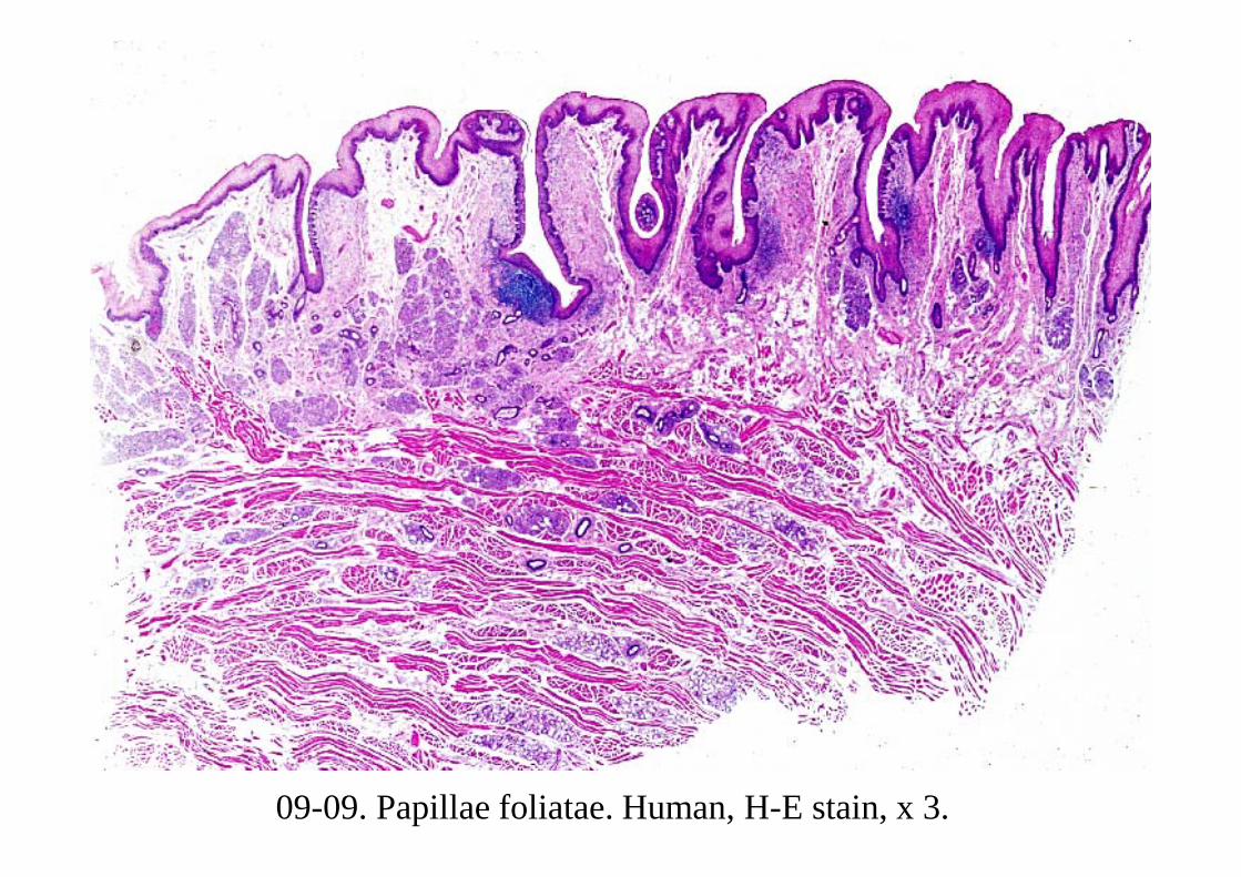
09-09. Papillae foliatae. Human, H-E stain, x 3.
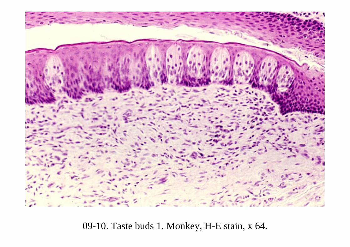
09-10. Taste buds 1. Monkey, H-E stain, x 64.

09-11. Taste buds 2. Monkey, H-E stain, x 160.
N
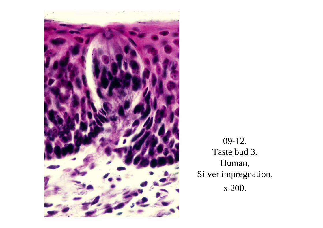
09-12. Taste bud 3.
Human, Silver impregnation,
x 200.
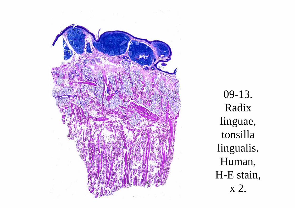
09-13. Radix
linguae, tonsilla
lingualis. Human,
H-E stain, x 2.
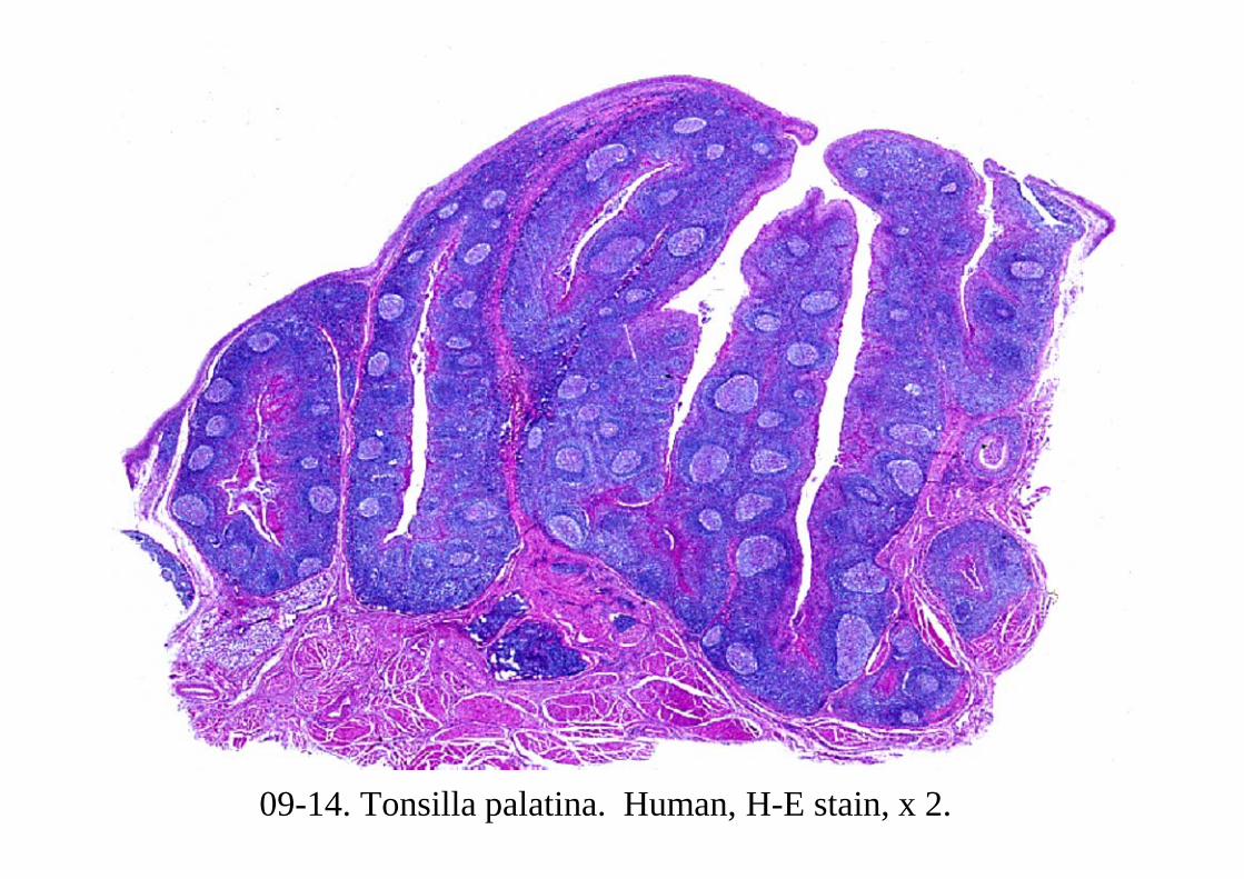
09-14. Tonsilla palatina. Human, H-E stain, x 2.
09-002Salivary Glands
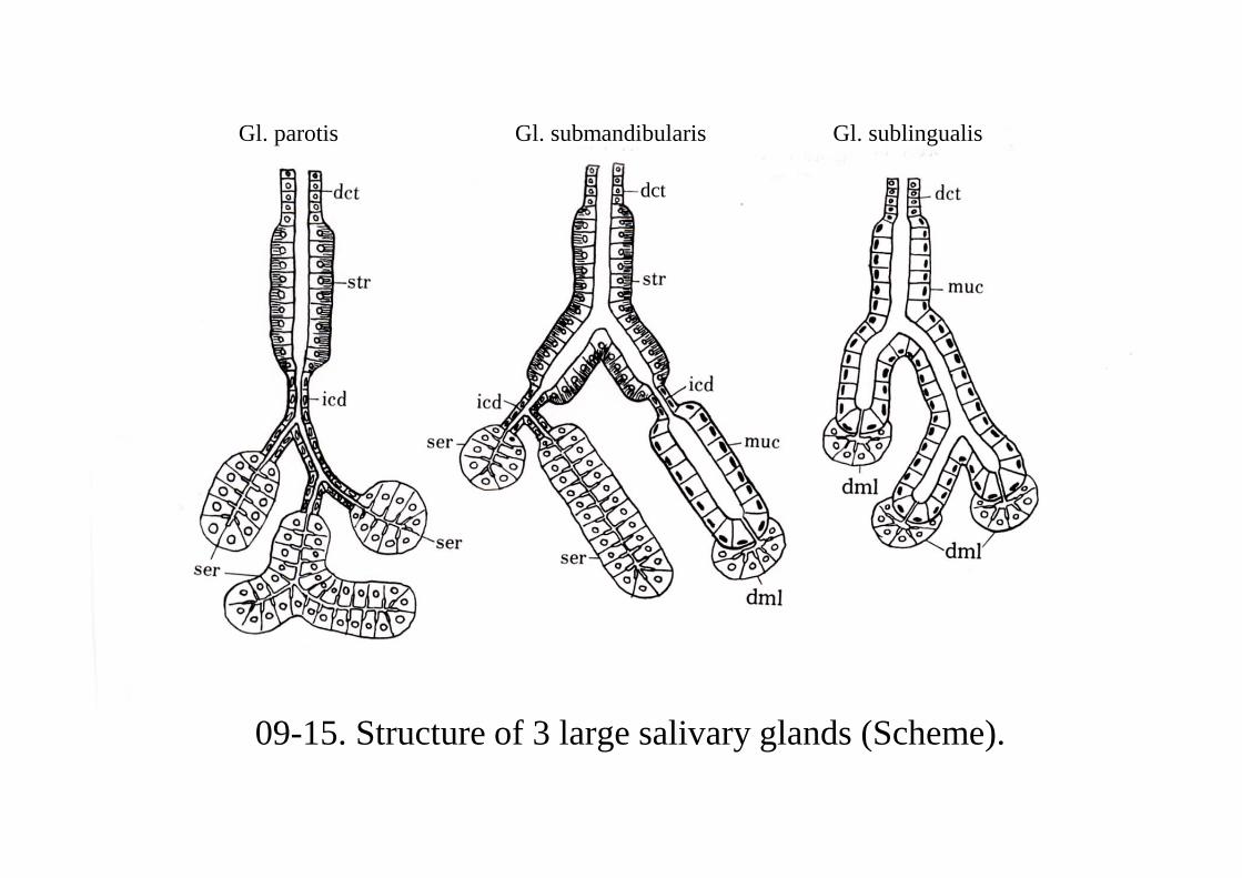
09-15. Structure of 3 large salivary glands (Scheme).
Gl. parotis Gl. submandibularis Gl. sublingualis
dct
09-0021Gl. parotis
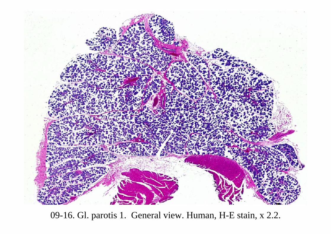
09-16. Gl. parotis 1. General view. Human, H-E stain, x 2.2.
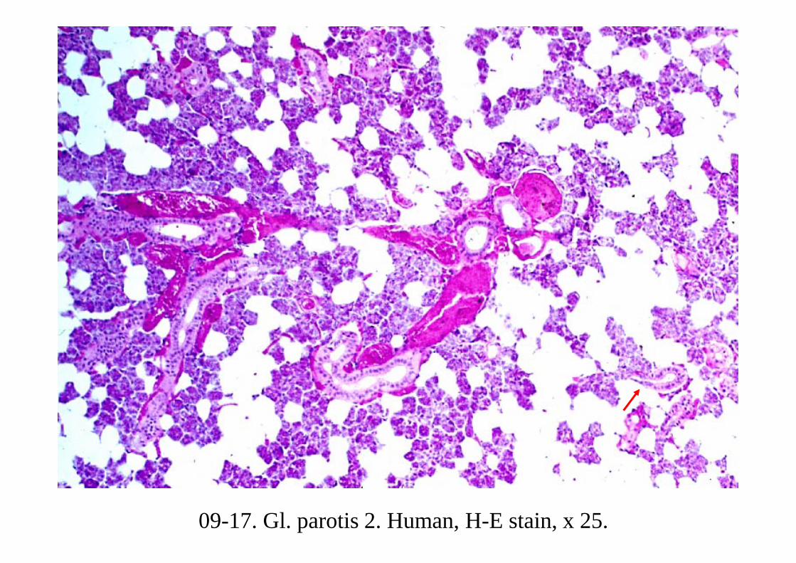
09-17. Gl. parotis 2. Human, H-E stain, x 25.

09-18. Gl. parotis 3. Intercalated duct and striated duct. Human, H-E stain, x 160.
Striated duct
Intercalated duct
Acinus
Acinus
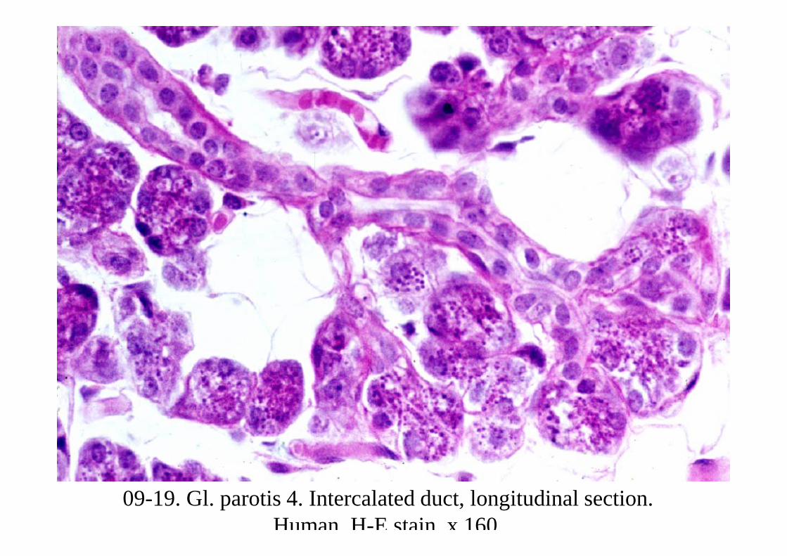
09-19. Gl. parotis 4. Intercalated duct, longitudinal section. Human, H-E stain, x 160.
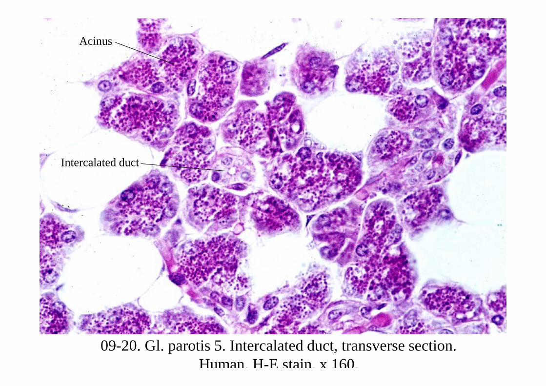
09-20. Gl. parotis 5. Intercalated duct, transverse section. Human, H-E stain, x 160.
Intercalated duct
Acinus
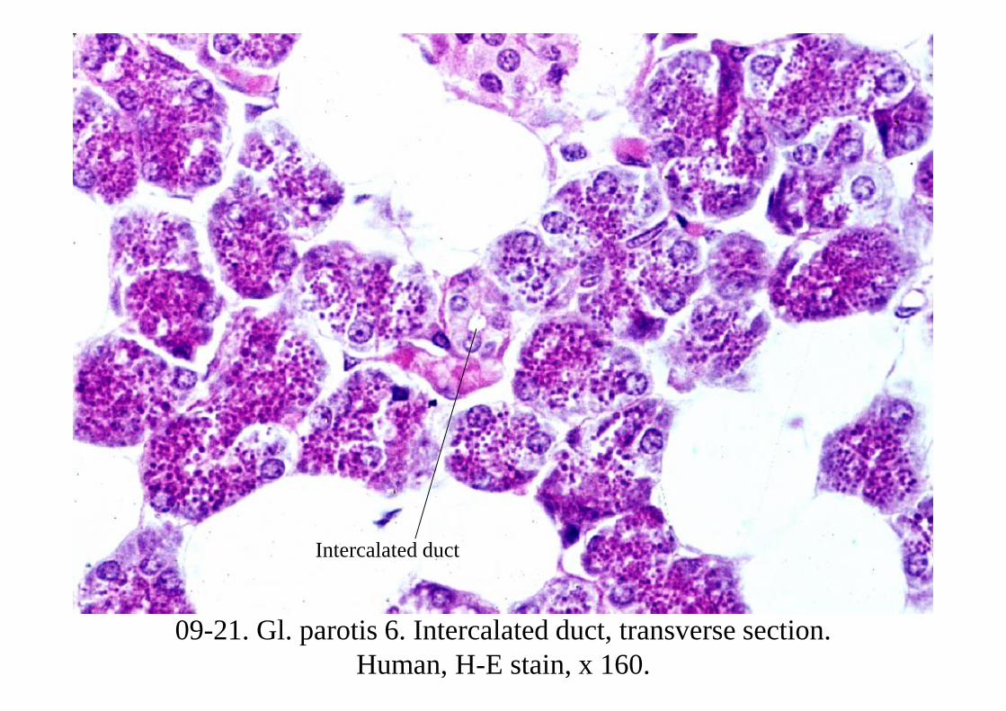
09-21. Gl. parotis 6. Intercalated duct, transverse section. Human, H-E stain, x 160.
Intercalated duct
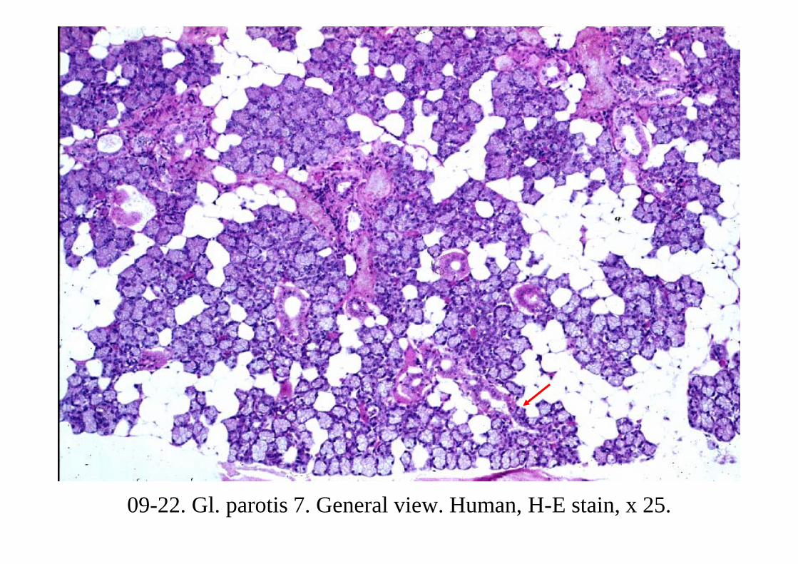
09-22. Gl. parotis 7. General view. Human, H-E stain, x 25.
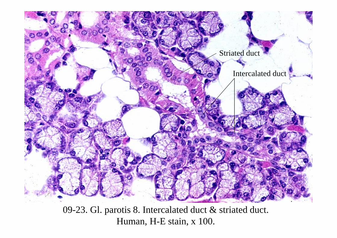
09-23. Gl. parotis 8. Intercalated duct & striated duct. Human, H-E stain, x 100.
Intercalated duct
Striated duct
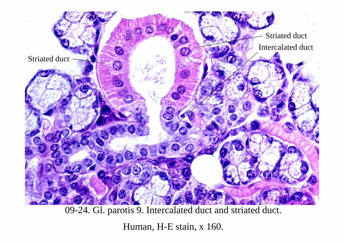
09-24. Gl. parotis 9. Intercalated duct and striated duct.
Human, H-E stain, x 160.
Striated ductIntercalated duct
Striated duct
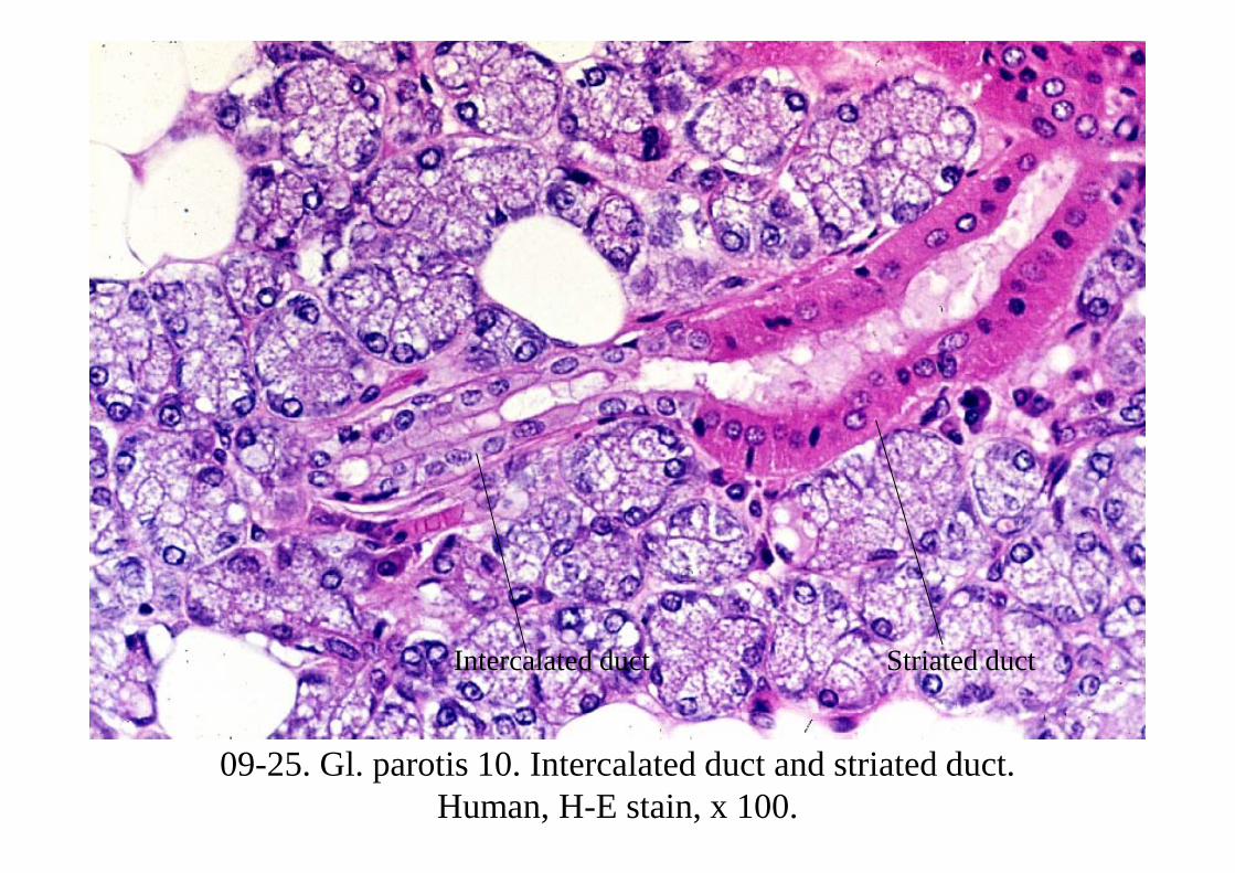
09-25. Gl. parotis 10. Intercalated duct and striated duct. Human, H-E stain, x 100.
Striated ductIntercalated duct
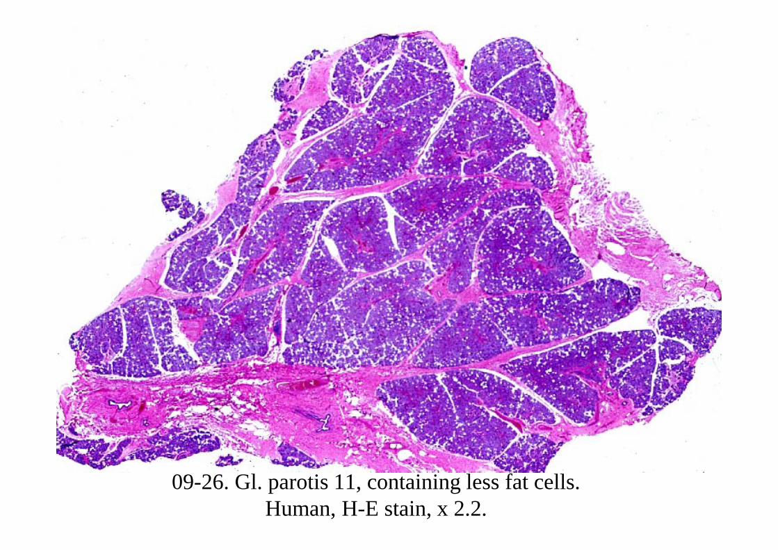
09-26. Gl. parotis 11, containing less fat cells. Human, H-E stain, x 2.2.
09-0022Gl. submandibularis
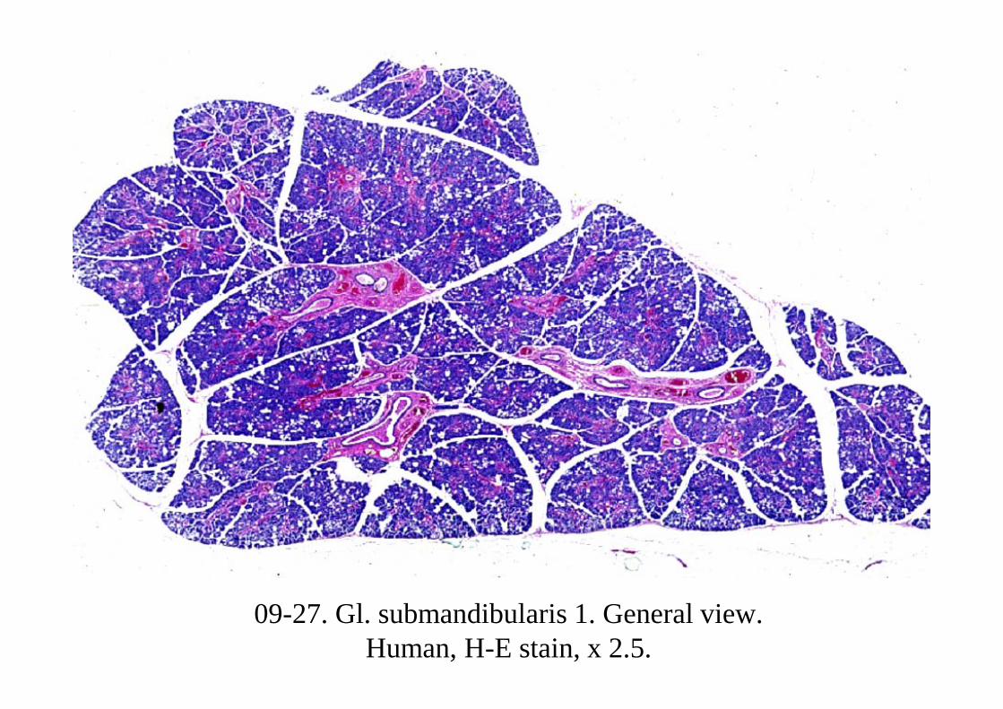
09-27. Gl. submandibularis 1. General view. Human, H-E stain, x 2.5.
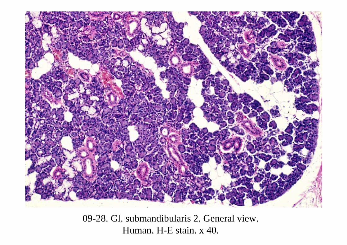
09-28. Gl. submandibularis 2. General view. Human, H-E stain, x 40.
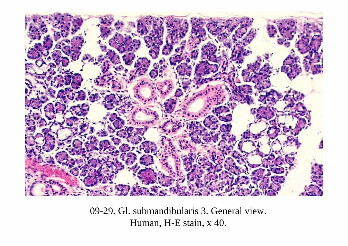
09-29. Gl. submandibularis 3. General view. Human, H-E stain, x 40.
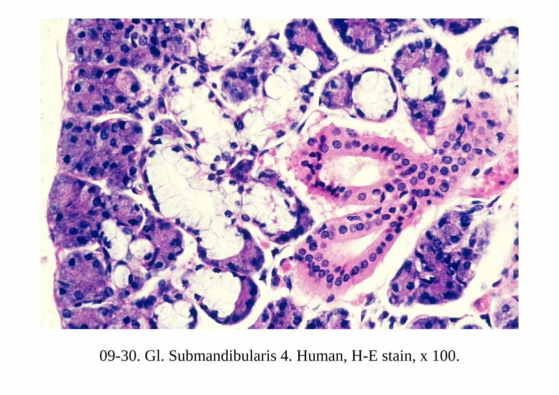
09-30. Gl. Submandibularis 4. Human, H-E stain, x 100.
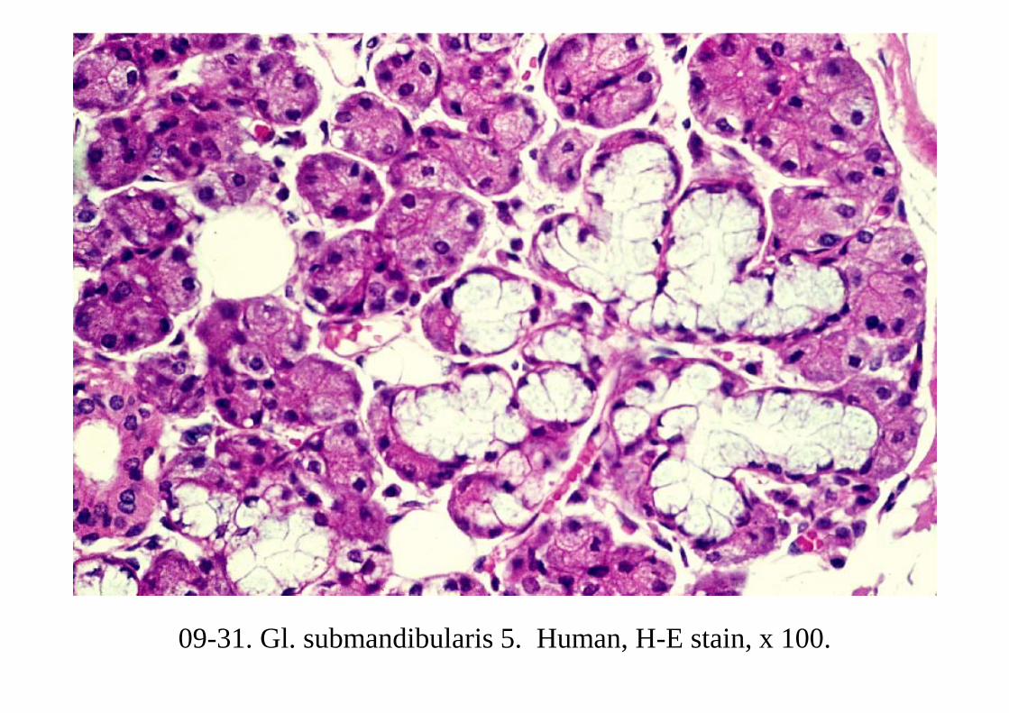
09-31. Gl. submandibularis 5. Human, H-E stain, x 100.
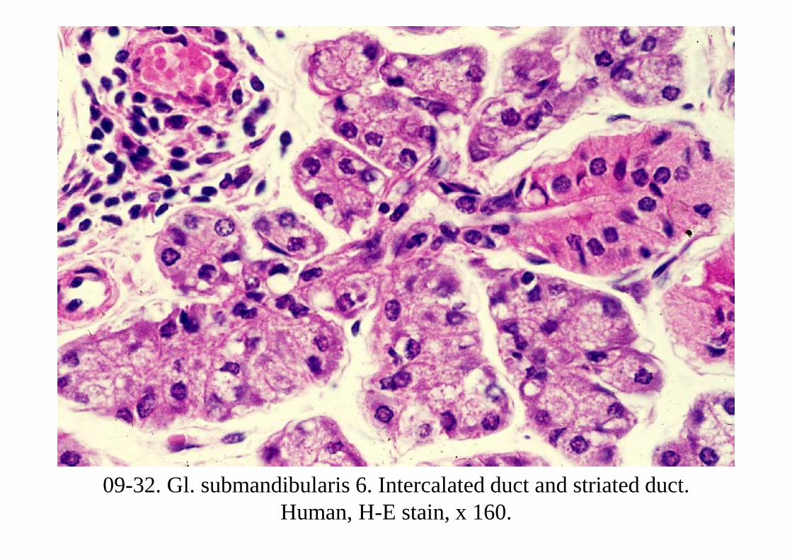
09-32. Gl. submandibularis 6. Intercalated duct and striated duct. Human, H-E stain, x 160.
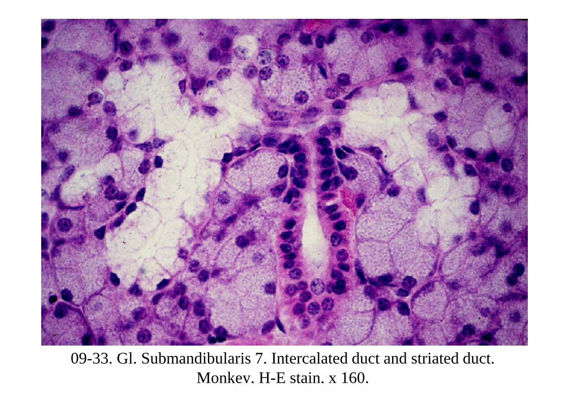
09-33. Gl. Submandibularis 7. Intercalated duct and striated duct. Monkey, H-E stain, x 160.
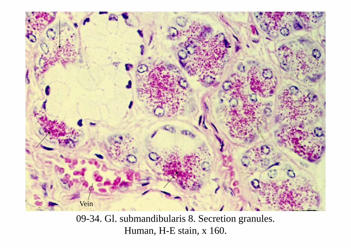
09-34. Gl. submandibularis 8. Secretion granules. Human, H-E stain, x 160.
Vein
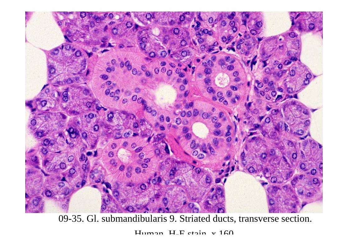
09-35. Gl. submandibularis 9. Striated ducts, transverse section. Human H-E stain x 160
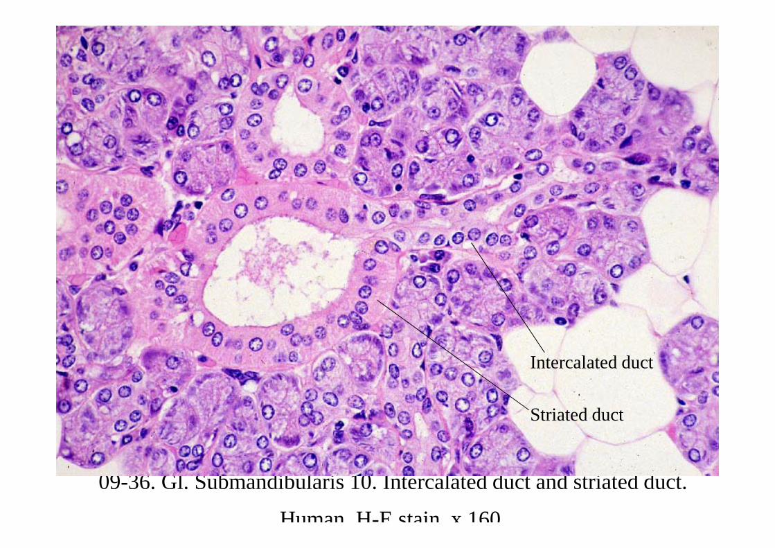
09-36. Gl. Submandibularis 10. Intercalated duct and striated duct.
Human H-E stain x 160
Striated duct
Intercalated duct
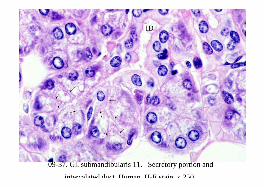
09-37. Gl. submandibularis 11. Secretory portion and
intercalated duct Human H-E stain x 250
ID
09-0023Gl. sublingualis
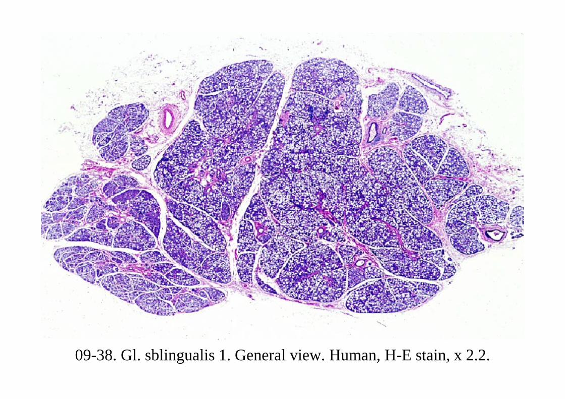
09-38. Gl. sblingualis 1. General view. Human, H-E stain, x 2.2.
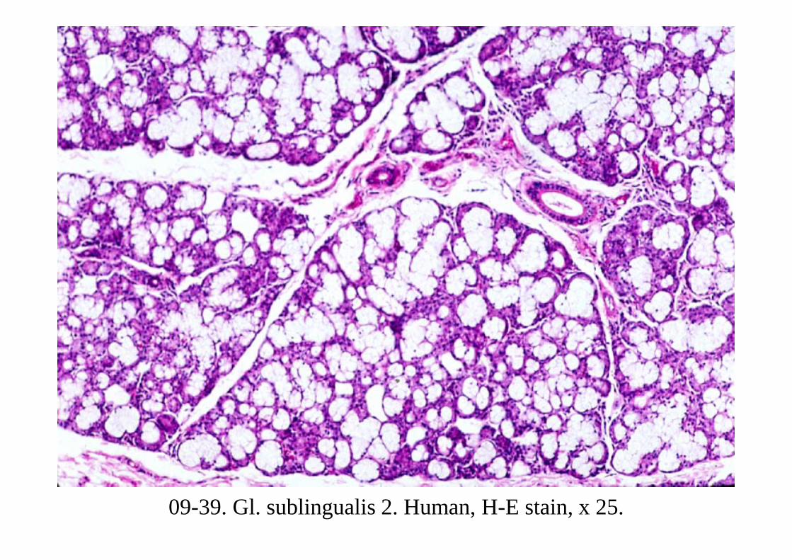
09-39. Gl. sublingualis 2. Human, H-E stain, x 25.
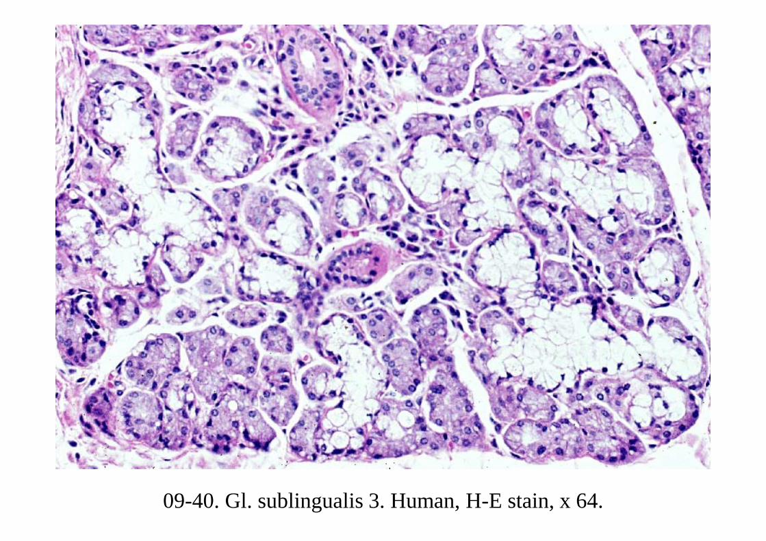
09-40. Gl. sublingualis 3. Human, H-E stain, x 64.
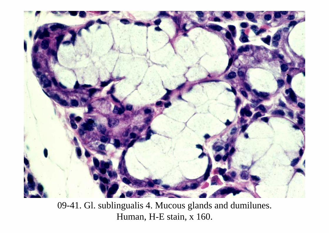
09-41. Gl. sublingualis 4. Mucous glands and dumilunes. Human, H-E stain, x 160.
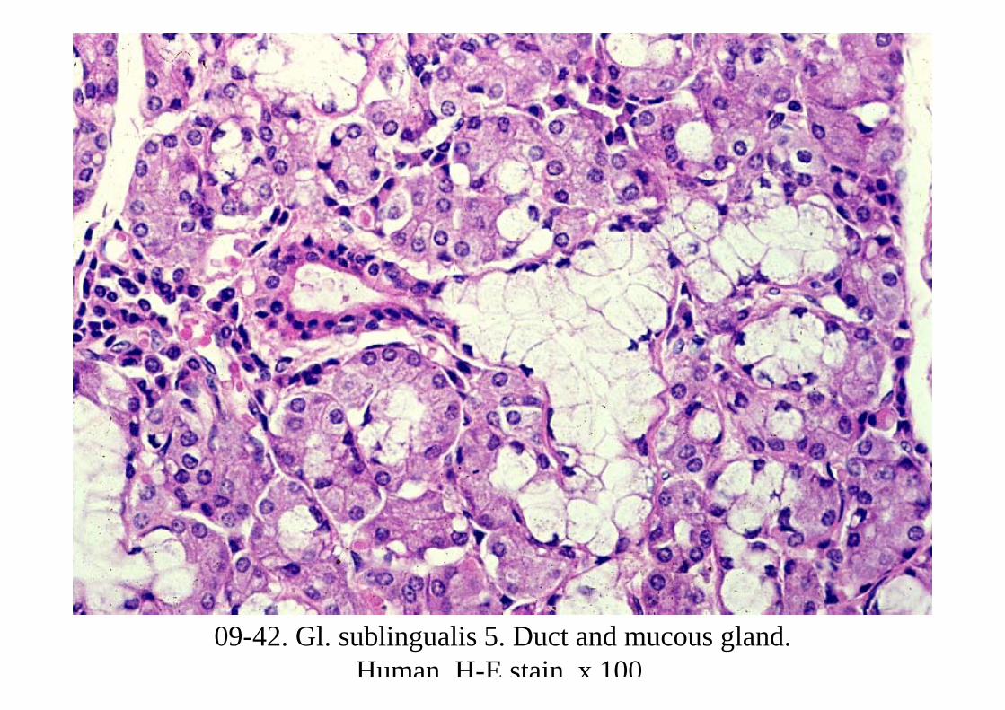
09-42. Gl. sublingualis 5. Duct and mucous gland. Human, H-E stain, x 100.
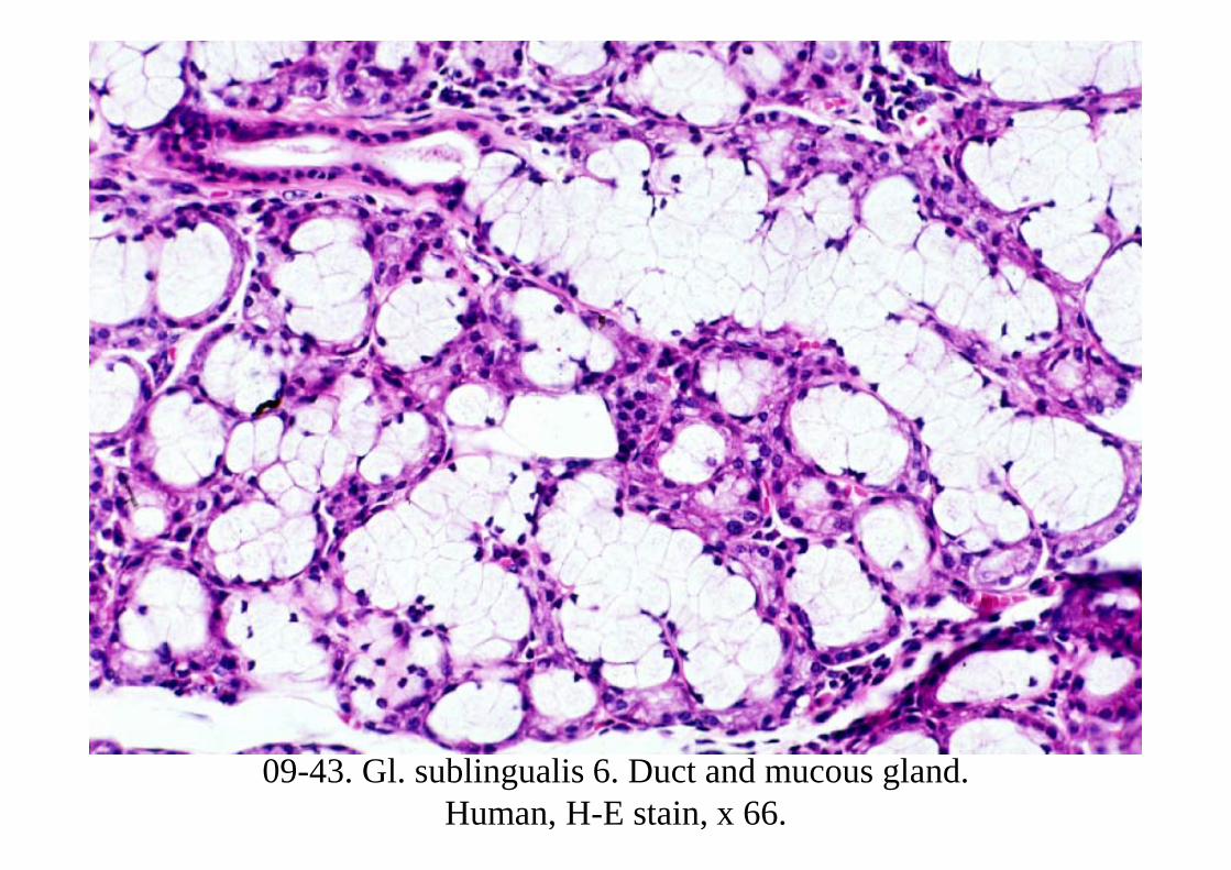
09-43. Gl. sublingualis 6. Duct and mucous gland. Human, H-E stain, x 66.


![Characterisation of DOG-1 Expression in Salivary Gland ...reported in salivary gland tumours [–68], in particular, as a marker for acinic and intercalated duct cells. Some stud-ies](https://static.documents.pub/doc/80x56/5f049c367e708231d40ed2b7/characterisation-of-dog-1-expression-in-salivary-gland-reported-in-salivary.jpg)
