molecules Article Cornus mas L. Stones: A Valuable by-Product as an Ellagitannin Source with High Antioxidant Potential Dominika Przybylska 1, * , Alicja Z. Kucharska 1 , Iwona Cybulska 2 , Tomasz Soza´ nski 3 , Narcyz Piórecki 4,5 and Izabela Fecka 6 1 Department ofFruit, Vegetable and Plant Nutraceutical Technology, Wroclaw University of Environmental and Life Sciences, Chelmo ´ nskiego 37, 51-630 Wroclaw, Poland; [email protected]2 Earth and Life Institute, Université Catholique de Louvain, Croix du Sud 2, 1348 Louvain-la-Neuve, Belgium; [email protected]3 Department of Pharmacology, Wroclaw Medical University, Jana Mikulicza-Radeckiego 2, 50-345 Wroclaw, Poland; [email protected]4 Arboretum and Institute of Physiography in Bolestraszyce, 37-700 Przemy´ sl, Poland; [email protected]5 Institute of Physical Culture Sciences, Medical College, University of Rzeszów, Towarnickiego 3, 35-959 Rzeszów, Poland 6 Department of Pharmacognosy and Herbal Medicines, Wroclaw Medical University, Borowska 211 A, 50-556 Wroclaw, Poland; [email protected]* Correspondence: [email protected]Academic Editor: Urszula Gawlik-Dziki Received: 9 September 2020; Accepted: 10 October 2020; Published: 12 October 2020 Abstract: The stone of Cornus mas L. remains the least known morphological part of this plant, whereas the fruit is appreciated for both consumption purposes and biological activity. The stone is considered to be a byproduct of fruit processing and very little is known about its phytochemical composition and biological properties. In this study, the complete qualitative determination of hydrolyzable tannins, their quantitative analysis, total polyphenolic content, and antioxidant properties of the stone of C. mas are presented for the first time. The 37 identified compounds included the following: various gallotannins (11), monomeric ellagitannins (7), dimeric ellagitannins (10), and trimeric ellagitannins (7). The presence of free gallic acid and ellagic acid was also reported. Our results demonstrate that C. mas stone is a source of various bioactive hydrolyzable tannins and shows high antioxidant activity which could allow potential utilization of this raw material for recovery of valuable pharmaceutical or nutraceutical substances. The principal novelty of our findings is that hydrolyzable tannins, unlike other polyphenols, have been earlier omitted in the evaluation of the biological activities of C. mas. Additionally, the potential recovery of these bioactive chemicals from the byproduct is in line with the ideas of green chemistry and sustainable production. Keywords: Cornus mas stones; UPLC-ESI-qTOF-MS/MS; gallotannins; ellagitannins; technological waste; bioactive compounds; antioxidants 1. Introduction Cornelian cherry (Cornus mas L.), which belongs to the Cornaceae family, is one of the two species from genus Cornus, which have been used in traditional ethnomedicine. It is native principally to Central and South Eastern Europe, while the second species, Cornus officinalis Torr. ex Dur., grows in Asia. These two species form a closely related phylogenetical pair [1]. They show numerous similarities of phytochemical profile and several differences, which are reflected by their therapeutic applications. Knowledge of the medicinal use of C. mas dates back to traditional medicine practices, for example, in Greece, Turkey, Slovakia, and China, where it served for prevention and treatment of gastrointestinal Molecules 2020, 25, 4646; doi:10.3390/molecules25204646 www.mdpi.com/journal/molecules
Transcript
molecules
Article
Cornus mas L. Stones: A Valuable by-Product as anEllagitannin Source with High Antioxidant Potential
Dominika Przybylska 1,* , Alicja Z. Kucharska 1 , Iwona Cybulska 2, Tomasz Sozanski 3,Narcyz Piórecki 4,5 and Izabela Fecka 6
1 Department of Fruit, Vegetable and Plant Nutraceutical Technology, Wrocław University of Environmentaland Life Sciences, Chełmonskiego 37, 51-630 Wrocław, Poland; [email protected]
2 Earth and Life Institute, Université Catholique de Louvain, Croix du Sud 2, 1348 Louvain-la-Neuve,Belgium; [email protected]
3 Department of Pharmacology, Wrocław Medical University, Jana Mikulicza-Radeckiego 2, 50-345 Wrocław,Poland; [email protected]
4 Arboretum and Institute of Physiography in Bolestraszyce, 37-700 Przemysl, Poland; [email protected] Institute of Physical Culture Sciences, Medical College, University of Rzeszów, Towarnickiego 3,
35-959 Rzeszów, Poland6 Department of Pharmacognosy and Herbal Medicines, Wroclaw Medical University, Borowska 211 A,
Academic Editor: Urszula Gawlik-DzikiReceived: 9 September 2020; Accepted: 10 October 2020; Published: 12 October 2020
�����������������
Abstract: The stone of Cornus mas L. remains the least known morphological part of this plant,whereas the fruit is appreciated for both consumption purposes and biological activity. The stone isconsidered to be a byproduct of fruit processing and very little is known about its phytochemicalcomposition and biological properties. In this study, the complete qualitative determination ofhydrolyzable tannins, their quantitative analysis, total polyphenolic content, and antioxidantproperties of the stone of C. mas are presented for the first time. The 37 identified compoundsincluded the following: various gallotannins (11), monomeric ellagitannins (7), dimeric ellagitannins(10), and trimeric ellagitannins (7). The presence of free gallic acid and ellagic acid was also reported.Our results demonstrate that C. mas stone is a source of various bioactive hydrolyzable tanninsand shows high antioxidant activity which could allow potential utilization of this raw materialfor recovery of valuable pharmaceutical or nutraceutical substances. The principal novelty of ourfindings is that hydrolyzable tannins, unlike other polyphenols, have been earlier omitted in theevaluation of the biological activities of C. mas. Additionally, the potential recovery of these bioactivechemicals from the byproduct is in line with the ideas of green chemistry and sustainable production.
Cornelian cherry (Cornus mas L.), which belongs to the Cornaceae family, is one of the two speciesfrom genus Cornus, which have been used in traditional ethnomedicine. It is native principally toCentral and South Eastern Europe, while the second species, Cornus officinalis Torr. ex Dur., grows inAsia. These two species form a closely related phylogenetical pair [1]. They show numerous similaritiesof phytochemical profile and several differences, which are reflected by their therapeutic applications.Knowledge of the medicinal use of C. mas dates back to traditional medicine practices, for example,in Greece, Turkey, Slovakia, and China, where it served for prevention and treatment of gastrointestinal
and circulatory disorders, diabetes, diarrhea, and flu. According to the literature reports, fruits wereused most frequently, whereas leaves, flowers, and fruit stones were applied to a minor extent [2,3].
Cornelian cherry bears single-stone fruits of mostly dark-red color. Their shape is oval or spherical,and they have an average length of 1.00–2.22 cm and weight of 0.39–3.78 g [4,5]. Fruits are attractivefor direct consumption and production of jams, juices, alcoholic beverages, and pickles which havebeen proven to be a rich source of health-promoting compounds, such as polyphenols (anthocyanins,flavonols, and phenolic acids), iridoids, terpenoids (ursolic acid), and vitamin C [2,6,7]. This factprobably justifies the traditional applications of cornelian cherry [2,8]. Recently, several in vitroand in vivo studies have confirmed antioxidant, anti-inflammatory, antidiabetic, hypolipidemic,anti-atherosclerotic, antimicrobial, and anticancer activity of the fruits [3,7–14].
Cornelian cherry stone forms 8.0–15.9% [5,14] or 5.7–11.0% [3] of total fruit weight and is consideredto be a waste material after fruit processing. Exploitation of fruit stones (technological wastes) is nota new issue for the food industry. The conventional and emerging technologies applied for thevalorization of technological wastes or for the recovery of nutraceuticals (e.g., fruit and vegetableantioxidants) have been reviewed previously [15–17]. One of the possible utilizations of discardedfruit stones is their conversion into biofuels. This solution was also proposed for cornelian cherrystones, to obtain bio-oil from hydrothermal liquefaction [18]. Similar uses can be found for the residualstones of cherry, plum, and peach [19–22].
In addition, fruit stones, as a morphological part, seem to be a promising raw material, in connectionwith bioactive phytochemicals. There are some papers from recent years which have discussed possibleuses of fruit stones or ingredients from them for nutritional and therapeutic purposes [19,23]. The aboverepresent some attempts to lower the food wastes generated at the processing plant. Re-utilizationof such byproducts meets the sustainable consumption and production (SDG 12) goals of the UnitedNations (UN) 2030 Agenda for Sustainable Development, announced in 2015 [24].
The stone of cornelian cherry continues to be the least known part of this plant in terms of thecomposition and biological properties. According to the available literature, only fatty acids andminerals (Ca, K, P, Mg, Na, and Cu) have been reported in the stones of cornelian cherry [14,25].Six fatty acids have been identified in the oil fraction of stones and, importantly, about 90% constitutedunsaturated fatty acids. According to Kucharska et al. [14] and Vidrih et al. [25], the highest content, inthe range of 64.8–75.0%, was noted for linoleic acid, followed by 15.0–22.9% for oleic acid, and 1.3–2.1%for linolenic acid. The remaining fatty acids were stearic, palmitic, and arachidic acid. Such fattyacids composition corresponds to that of commonly consumed vegetable oils from sunflower, corn,or pumpkin [25].
Very few reports concerning traditional curative uses of cornelian cherry stones state that oil fromstones showed a healing effect for wounds, stomach ulcers, and colitis (Iran, Azerbaijan, Armenia,Georgia, and Turkey) [2]. According to another source, a mixture of stones and honey was ingestedagainst diabetes [2]. Additionally, roasted cornelian cherry stones were used in traditional folk practiceas a coffee substitute due to similar aroma, but specific recipes for this infusion are not available.Despite the above, beneficial activities of cornelian cherry stones have not been proven in modernscientific experiments as far as we know.
Hydrolyzable tannins are polyphenolic compounds which form complex molecular structures.They are esters built of some phenolic acids, for example, galloyl (gall), hexahydroxydiphenoyl (HHDP),valoneoyl (val), and a sugar moiety, usually d-glucose [26,27]. When subjected to hydrolysis, the HHDPgroup is released and converted into ellagic acid [27]. The presence of hydrolyzable tannins has beenreported in certain fruits, for example, raspberries, blackberries, strawberries, and pomegranates,as well as in non-edible parts, i.e., leaves, roots, and seeds of raspberry, blackberry, strawberry,pomegranate, and some nuts [28]. In the case of the genus Cornus, hydrolyzable tannins havebeen previously identified by Okuda et al. [29] and Hatano et al. [30–32], in fruits of C. officinalis.Until recently, it was believed that tannins appeared only in C. officinalis and were perceived as themain difference between the two species. However, ellagic acid, a marker of hydrolyzable ellagitannins,
Molecules 2020, 25, 4646 3 of 18
was found in both Cornus species, and therefore it could possibly indicate the presence of ellagitanninsin C. mas [5,7,27]. Moreover, Efenberger-Szmechtyk et al. [33], identified some ellagitannins in theleaves of C. mas. To our knowledge, there are no studies on the identification of tannins in C. mas stonesand no works which have addressed their health promoting characteristics. For this reason, the aimof our research was to present the first complete determination of hydrolyzable tannins in corneliancherry stones and analyze their antioxidant activity.
2. Results
2.1. Qualitative Identification by Means of UPLC-ESI-qTOF-MS/MS
The compounds were identified on the basis of UPLC retention time (tR), elution order,spectra UV-Vis (200–600 nm), MS, MS/MS fragmentation (acquired in negative mode), and bycomparison with literature data when available. In our research, we determined 35 polyphenoliccompounds from two groups, i.e., gallotannins and ellagitannins, including different isomers.Common MS/MS fragments for the given compounds were at m/z 169 and 125 from the gallicacid residue characteristic for galloyl esters, and a fragment ion of ellagic acid at m/z 301 from theHHDP esters, by comparison with the literature [34–39]. Additionally, free gallic acid (2) and ellagicacid (33) were present in the extract.
A typical UPLC chromatogram of the analyzed aqueous solution of ethanolic extract at 280 nm isshown in Figure 1 and the complete results of qualitative identification of the polyphenolic compoundsof cornelian cherry stones are presented in Table 1 and Table S1.
Molecules 2020, 25, x 3 of 20
in fruits of C. officinalis. Until recently, it was believed that tannins appeared only in C. officinalis and were perceived as the main difference between the two species. However, ellagic acid, a marker of hydrolyzable ellagitannins, was found in both Cornus species, and therefore it could possibly indicate the presence of ellagitannins in C. mas [5,7,27]. Moreover, Efenberger-Szmechtyk et al. [33], identified some ellagitannins in the leaves of C. mas. To our knowledge, there are no studies on the identification of tannins in C. mas stones and no works which have addressed their health promoting characteristics. For this reason, the aim of our research was to present the first complete determination of hydrolyzable tannins in cornelian cherry stones and analyze their antioxidant activity.
2. Results
2.1. Qualitative Identification by Means of UPLC-ESI-qTOF-MS/MS
The compounds were identified on the basis of UPLC retention time (tR), elution order, spectra UV-Vis (200–600 nm), MS, MS/MS fragmentation (acquired in negative mode), and by comparison with literature data when available. In our research, we determined 35 polyphenolic compounds from two groups, i.e., gallotannins and ellagitannins, including different isomers. Common MS/MS fragments for the given compounds were at m/z 169 and 125 from the gallic acid residue characteristic for galloyl esters, and a fragment ion of ellagic acid at m/z 301 from the HHDP esters, by comparison with the literature [34–39]. Additionally, free gallic acid (2) and ellagic acid (33) were present in the extract.
A typical UPLC chromatogram of the analyzed aqueous solution of ethanolic extract at 280 nm is shown in Figure 1 and the complete results of qualitative identification of the polyphenolic compounds of cornelian cherry stones are presented in Table 1 and Table S1.
Figure 1. UPLC-PDA chromatogram of cornelian cherry stone extract at λ = 280 nm. Peak numbers refer to compounds listed in Table 1 and Table S1.
Tandem mass spectrometry (UPLC-ESI-qTOF-MS/MS) allowed us to identify eleven compounds from the group of gallotannins and molecular weight 332–940 Da.
Compounds 1 and 5 gave a deprotonated pseudomolecular ion [M − H]− at m/z 331.0639 and some fragment ions from gallic acid residue at m/z 169 [GA − H]−, and 125 (decarboxylated galloyl, −44 Da) characteristic for mono-O-galloyl-β-D-glucose. These isomers describe the simplest gallotannin included in our experiment. Compounds 4, 7, 9, and 13 provided an [M − H]− ion at 483.0763, which corresponded to the pseudomolecular ion of di-O-galloyl esters of glucose, and a fragment ion at m/z 331, after loss of one galloyl moiety (−152 Da). Thus, these were identified as four isomers of di-O-galloyl-β-D-glucose. Compound 16 showed the same [M − H]− ion at m/z = 635.0872 as compound 19, which indicated two isomers of tri-O-galloyl-β-D-glucose. These compounds showed the same fragment ion at m/z 465 after loss of one galloyl and one water molecule (−170 Da),
Figure 1. UPLC-PDA chromatogram of cornelian cherry stone extract at λ = 280 nm. Peak numbersrefer to compounds listed in Table 1 and Table S1.
Molecules 2020, 25, 4646 4 of 18
Table 1. UPLC-ESI-qTOF-MS/MS and HPLC-DAD identification of hydrolyzable tannins in the extractof cornelian cherry stones.
Tandem mass spectrometry (UPLC-ESI-qTOF-MS/MS) allowed us to identify eleven compoundsfrom the group of gallotannins and molecular weight 332–940 Da.
Compounds 1 and 5 gave a deprotonated pseudomolecular ion [M −H]− at m/z 331.0639 and somefragment ions from gallic acid residue at m/z 169 [GA − H]−, and 125 (decarboxylated galloyl, −44 Da)characteristic for mono-O-galloyl-β-d-glucose. These isomers describe the simplest gallotanninincluded in our experiment. Compounds 4, 7, 9, and 13 provided an [M − H]− ion at 483.0763,which corresponded to the pseudomolecular ion of di-O-galloyl esters of glucose, and a fragmention at m/z 331, after loss of one galloyl moiety (−152 Da). Thus, these were identified as fourisomers of di-O-galloyl-β-d-glucose. Compound 16 showed the same [M − H]− ion at m/z = 635.0872as compound 19, which indicated two isomers of tri-O-galloyl-β-d-glucose. These compoundsshowed the same fragment ion at m/z 465 after loss of one galloyl and one water molecule (−170 Da),and the ion at m/z 313 after loss of two galloyls and one water (−322 Da), which were equal tothe dehydrated fragment of di-O-galloyl-β-d-glucose and mono-O-galloyl-β-d-glucose, respectively.Compound 32 gave a pseudomolecular ion [M − H]− at m/z = 787.1022 and it was assigned totetra-O-galloyl-β-d-glucose. The most complex derivatives of 1 were compounds 36 and 37, which gavepseudomolecular ions [M − H]− at m/z = 939.1080 and 939.1143, respectively, that corresponded to twoisomers of penta-O-galloyl-β-d-glucose. Similarly, isomers of penta-O-galloyl-β-d-glucose liberatedfragment ions through the loss of consecutive galloyl units and water molecules, and provided theions at m/z 787, 769, 617, 465, 313, 295, 169, and 125 (Table S1).
The collected data show that fragmentation of mono- to penta-galloyl derivatives results in theloss of subsequent galloyl moieties and water molecules, among other fragments, which are derivedfrom the gallic acid residue (m/z 169) and decarboxylated gallic acid (m/z 125). Structures of the abovegallotannins are shown in Figure 2.
The second group of characterized compounds comprised ellagitannins. Among ellagitannins,we identified seven monomeric, ten dimeric, and seven trimeric compounds of molecular weight634–2506 Da. Identified ellagitannins consist of a glucose core, galloyl and HHDP groups attached atthe O-3 and O-4/O-6 of glucose, as well as valoneoyl or lactonized valoneoyl groups, which create O-2and O-4/O-6 linkages between two or three glucose cores.
Compounds 3 and 6 displayed the same pseudomolecular ion [M − H]− at m/z = 633.0718,corresponding to the molecular formula of gemin D [29,31,37,38], the simplest among the ellagitanninmolecules identified here, with the one group of HHDP and galloyl. Compound 11 had apseudomolecular ion at m/z = 953.0872 and fragment ions at 909 (decarboxylated, −44 Da), 785,783 (degalloylated and dehydrated, −170 Da), and 633 (galloyl-HHDP-glucose, e.g., gemin D),which indicated isorugosin B of the molecular weight of 954.0974 Da [28,35]. Compounds 14 and20 showed an [M − H]− ion at m/z = 785.0821, suggesting two isomeric forms of tellimagrandinI [31,37–40], which were derivatives of gemin D with an additional galloyl unit attached to the glucosecore. Compound 31 gave a pseudomolecular ion [M − H]− at m/z = 937.0892 corresponding to amolecular weight of 938.6629 Da of tellimagrandin II [32,38,40], ellagitannin with three galloyl unitsattached to glucose. Compound 22, with [M − H]− at m/z = 1085.0734 and fragment ions diminishedby galloyl (m/z 933) and HHDP (m/z 783), was identified as cornusiin B, the monomer of the highestmolecular weight (1086.7357 Da) [29,31,37].
Compounds 8, 10, 17, 23–25, 28–30, and 35 represented dimeric ellagitannins. Peaks 12, 15, 18, 21,26, 27, and 34 represented trimeric ellagitannins. Among these groups, MS spectra for each compound,except 34, displayed two typical pseudomolecular ions, [M −H]− and [M − 2H]−2 [36], and in all cases,the doubly charged ion showed higher abundance.
Molecules 2020, 25, 4646 6 of 18
Molecules 2020, 25, x 7 of 20
Figure 2. Chemical structures of the identified hydrolyzable tannins. Abbreviations: gall, galloyl; glc, β-D-glucose; gm D, gemin D; tell I, tellimagrandin I; tell II, tellimagrandin II; oen C, oenothein C; icr F, isocoriariin F; irg B, isorugosin B; crn B, cornusiin B; cmt A, camptothin A; cmt B, camptothin B; crn A, cornusiin A; crn D, cornusiin D; crn F, cornusiin F; crn C, cornusiin; trp A, trapanin A.
Figure 2. Chemical structures of the identified hydrolyzable tannins. Abbreviations: gall, galloyl; glc,β-d-glucose; gm D, gemin D; tell I, tellimagrandin I; tell II, tellimagrandin II; oen C, oenothein C; icr F,isocoriariin F; irg B, isorugosin B; crn B, cornusiin B; cmt A, camptothin A; cmt B, camptothin B; crn A,cornusiin A; crn D, cornusiin D; crn F, cornusiin F; crn C, cornusiin; trp A, trapanin A.
Molecules 2020, 25, 4646 7 of 18
Ellagitannins 8 and 10, provided [M − 2H]−2 at m/z = 708.0688 and [M −H]− at m/z = 1417.1549,which were identified as two anomers or other isomers of camptothin A, a dimer in which gemin D andisocoriariin F constitute monomers [30,33]. Compounds 17, 23, 24, 25, and 28 gave two ions, [M− 2H]−2
at m/z = 784.0729 and [M − H]− at m/z = 1569.1556, which indicated five isomers of galloyl-camptothinA known as cornusiin A [29,31,33]. In addition, two isomers of cornusiin A (17, 23) fragmented tothe ion at m/z 1417, after the loss of galloyl, but all its isomers detached a fragment of monomericellagitannins such as gemin D (at m/z 633) and tellimagrandin I (at m/z 785), which indicated thatthe compound consisted of these structures, as well as a fragment (at m/z 783) of oenothein C ordehydrated isocoriariin F (from 802 − 18 = 784) [26,29,31], the ion at m/z 935 corresponding to thefragment formed after loss of HHDP, galloyl, and glucose (e.g., gemin D). Peaks 29 (tR = 6.15 min),30 (tR = 6.34 min), and 35 (tR = 7.04 min), for ions [M − 2H]−2 at m/z = 860.0745 and [M − H]− atm/z = 1721.1445, were tentatively identified as three positional isomers or anomers of either cornusiinD or camptothin B, which are galloyl esters of cornusiin A (additional galloyl in the structure) andhave the same molecular weight but differ in the position of the one galloyl group, R1 vs. R3 at O-1 ofglucose 1 or glucose 2 (Figure 2) [26,29,32,37]. The fragmentation of cornusiin D or camptothin B wasidentical to other dimeric ellagitannins with the valoneoyl bridge.
Compounds 12, 15, 18, and 21, 26, and 27 describe isomers of trimeric ellagitannins, cornusiin Fand cornusiin C, respectively, both of which we identified in the extract at three spatial forms [31,33].Compound 34 was trapanin A [41]. The molecular weights of isomers of cornusiin F, cornusiinC, and trapanin A were 2203.5389, 2355.6434 Da, and 2506.2544 Da, respectively. Compounds 12and 15 gave doubly charged pseudomolecular ions at m/z = 1100.6101 and single-charge ions atm/z = 2201.1279. Similarly, compound 18 produced ions [M − 2H]−2 at 1101.1184 and [M − H]– at2201.1184. Two stereoisomers of cornusiin F (12, 15) fragmented to ions at m/z = 2031, after the lossof one galloyl and one water molecule, and all of them displayed an ion at m/z 1247 diminished byisorugosin B [M − 954 − H]−. Figure 2 depicts the structures of the dimeric and trimeric ellagitannins.
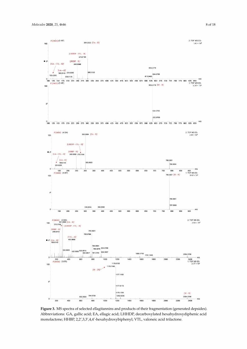
As shown inTable S1, oligomeric ellagitannins from cornelian cherry stones during MS/MSexperiments disconnected the fragment ions derived from their constituent monomers, for example,ions 953, 785, 783, 633, 331 and dehydrated monomers 765, 613, and 313. The detached HHDPgroup (−302 Da) was a source of ions at m/z 301, 275, and 249 derived from ellagic acid,and its degraded forms decarboxylated hexahydroxydiphenic acid monolactone (LHHDP) and2,2′,3,3′,4,4′-hexahydroxydiphenyl (HHBP). MS/MS analysis also revealed the presence of otherdepsides, derived from a valoneoyl group, at m/z 451 (valoneoic acid trilactone, VTL) and 425(decarboxylated valoneoic acid dilactone, DVDL) (Table S1). Although the fragment ion of valoneicacid dilactone (VDL) was not detected in the experiment, this structure occurred in oenothein C andcornusiin B (Figure 2), and the depsides VTL and DVDL were formed as its derivatives. When thesuitable fragment ions appear on the MS spectrum, it indicates whether the compound contains HHDPor a valoneoyl unit. Selected MS spectra are shown in Figure 3 and structures of the formed depsidesare shown in Figure 4.
Molecules 2020, 25, 4646 8 of 18
Molecules 2020, 25, x 9 of 20
hexahydroxydiphenyl (HHBP). MS/MS analysis also revealed the presence of other depsides, derived from a valoneoyl group, at m/z 451 (valoneoic acid trilactone, VTL) and 425 (decarboxylated valoneoic acid dilactone, DVDL) (Table S1). Although the fragment ion of valoneic acid dilactone (VDL) was not detected in the experiment, this structure occurred in oenothein C and cornusiin B (Figure 2), and the depsides VTL and DVDL were formed as its derivatives. When the suitable fragment ions appear on the MS spectrum, it indicates whether the compound contains HHDP or a valoneoyl unit. Selected MS spectra are shown in Figure 3 and structures of the formed depsides are shown in Figure 4.
Molecules 2020, 25, x 10 of 20
Figure 3. MS spectra of selected ellagitannins and products of their fragmentation (generated depsides). Abbreviations: GA, gallic acid; EA, ellagic acid; LHHDP, decarboxylated hexahydroxydiphenic acid monolactone; HHBP, 2,2’,3,3’,4,4’-hexahydroxybiphenyl; VTL, valoneic acid trilactone.
According to Dong et al. [42], thirty tannins have been identified in the fruit of C. officinalis,which is considered to be a rich source of gallotannins and ellagitannins. The groups of Hatano andOkuda have broadly investigated the hydrolyzable tannins as follows: their isolation, identification oftheir structures, and occurrence of several isomers as a result of anomerization at the glucose coresof tannins in C. officinalis [26–32,43]. In the comparative review of two Cornus species, C. mas andC. officinalis, Czerwinska and Melzig [8] indicated that none of the tannins identified in the fruit ofC. officinalis had been detected in the stones of C. mas.
The same gallotannins as determined here, except for penta-O-galloyl-β-d-glucose, were presentedin review papers by Czerwinska and Melzig [8] and Dong et al. [42] as constituents of C. officinalis fruits.
Four isomers of camptothin A and four isomers of cornusiin A have been identified previously inthe leaves of Camptotheca acuminata Decne. (Nyssaceae) and fruits of C. officinalis [29,31]. In the mostrecent paper, Efenberger-Szmechtyk et al. [33] reported the presence of four isomers of camptothin Aand two isomers of cornusiin A in the leaves of C. mas. Herein, we identified five isomers of cornusiinA. In our study, the signal with the greatest abundance in the UPLC-PDA chromatogram was assignedto cornusiin C, and compound 21 seems to be the predominant isomer.
2.2. Quantitative Identification of Compounds
The content of hydrolyzable tannins was calculated from the regression equation for gallic acid.Free gallic and ellagic acid were identified on the basis of retention time, elution order, spectra UV-Vis,and compared to commercial standards. The tannins and gallic acid were identified at 280 nm andellagic acid at 254 nm. Table 1 depicts the results of quantitative analysis. The total concentration
Molecules 2020, 25, 4646 10 of 18
of analyzed compounds (mean ± standard deviation) was 13,242.88 ± 37.07 mg/100 g of the extract,the total content of hydrolyzable tannins was 12,470.07 ± 66.53 mg/100 g of the extract and the contentsof individual tannins ranged from 53.55 (camptothin A (1)) to 1354.73 (cornusiin C (1)) mg/100 g ofthe extract.
For the comparison, the content of hydrolyzable tannins and ellagic acid in strawberries rangedfrom 6.53 to 52.38 mg/100 g of fresh weight fw [34], the ellagitannin content in raspberries was233.50 mg/100 g fw [44], and raspberry leaves contained 2.67–6.87% dw of tannins [45]. The dataconcerning ellagitannins in edible fruit stones is lacking and their content has been little discussed.However, Nowicka and Wojdyło [23] studied their presence in peach kernels and reported the presenceof ellagic acid between 0.77 and 9.42 mg/100 g dw. Taken together, these data suggest that C. mas stonescan be considered to be a novel rich source of ellagitannins, especially given that the stone of anotherfruit (peach) is a poor source of these compounds.
2.3. Antioxidant Properties and Total Phenolic Content (TPC)
Since the hydrolyzable tannins belong to polyphenols, many of which are antioxidants, we investigatedantioxidant properties of C. mas stones, rich in these components, using the spectrophotometric in vitromethods ABTS, FRAP, and DPPH, and total polyphenolic content assay (Table 2).
Table 2. The results of antioxidant activity and total phenolic content assays of the extract of stones.
So far, there were no papers which present antioxidant properties of C. mas stones, however,some authors claim strong antioxidant properties of fruit stones [23,46] and other sources of hydrolyzabletannins, for example, navy bean hull or strawberries [34,47,48]. According to Szajdek and Borowska [47],tannins are responsible for the antioxidant potency of navy bean seeds, which dominate among thepolyphenols in the hull.
In our experiment, the value obtained in ABTS•+ free radical-scavenging assaywas 255.99 ± 8.48 mmol TE/100 g. The reducing power measured in FRAP assay was210.62 ± 5.45 mmol TE/100 g and the value obtained on the DPPH free radical-scavenging assay was191.00 ± 0.04 mmol TE/100 g. The average content of compounds which reacted with the Folin–Ciocalteureagent (TPC) was 11,466.53± 1971.76 mg GAE/100 g, while the TPC in the kernels of apricot obtained byother authors ranged from 10.60 to 209.4 mg GAE/100 g dw [46]. According to Nowicka and Wojdyło [23],the antioxidative potential (ABTS•+) of the peach kernel ranged from 2.19 to 27.20 mmol TE/100 g.Chen et al. [46] indicated that apricot kernels had significant free radicals-scavenging activities(ABTS•+ and DPPH), however, they expressed the values as median effective dose (EC50) and theresults were difficult to compare. Nonetheless, our results indicate that stones of edible fruits,particularly the stones of cornelian cherry, are valuable raw materials in terms of antioxidant properties.
3. Discussion
The determined qualitative composition of hydrolyzable tannins in the stones of C. mas iscomparable to that presented by other authors, although mostly in C. officinalis. A similar fragmentationpathway, characteristic for galloyl-O-glucoses and for ellagitannins, was obtained by other authorsin previous studies concerning hydrolyzable tannins [34,35,38]. It is worth noting that the massspectrometry and fragmentation studies of hydrolyzable tannins carried out so far explain mainlygallotannins and monomeric ellagitannins in C. officinalis or other plant genera, whereas data onoligomeric hydrolyzable tannins are limited, especially in C. mas.
Molecules 2020, 25, 4646 11 of 18
According to available literature, ellagitannins detected in Cornus sp. demonstratediverse biological activities. Lavoie et al. [49] reported that hydrolyzable tannins1,2,3,6-tetra-O-galloyl-β-d-glucopyranose, 1,2,3,4,6-penta-O-galloyl-β-d-glucopyranose,tellimagrandin I, and tellimagrandin II possess antiviral properties against herpes simplexvirus type 1 (HSV-1). Various hydrolyzable tannins, including gemin D, showed activity against humanimmunodeficiency virus (HIV), diminishing the HIV-induced cytopathogenic effect and HIV-specificantigen expression, and inhibiting binding of viruses to the target cells [50–52]. Interestingly,condensed tannins and other lower molecular weight polyphenols revealed no detectable anti-HIVactivity [51,52]. Moreover, the ongoing SARS-CoV-2 pandemic crisis may increase the need fordelivering new sources of antiviral compounds to the market. Growing consumers’ awareness of thediet-disease relationship raises their interest in fortified foods or dietary supplements for support ofthe immune system. A solid argument in favor of the use of hydrolyzable tannins is their plant origin,desirable to consumers. These premises point to the opportunity for natural sources of hydrolyzabletannins [53].
Tellimagrandin I significantly enhanced activity ofβ-lactam antibiotics against methicillin-resistantStaphylococcus aureus (MRSA) and decreased the minimum inhibitory concentrations (MICs) ofoxacillin against the MRSA strains [26,54,55]. Tellimagrandin II showed antioxidant (DPPH) [50]and neuroprotective activity [42] and an inhibitory effect against yeast Candida parapsilosis ATCC22,019 [56]. Tellimagrandin I, alongside the galloyl-O-glucoses, exhibited antioxidant activity in theDPPH test [57]. Berdowska et al. [58] evaluated the effect of the ellagitannins containing the HHDPgroup (tellimagrandin I, rugosin D and A, sanguiin H-6, agrimoniin, and pedunculagin), isolated frommeadowsweet flowers (Filipendulae ulmariae flos, Filipendula ulmaria (L.) Maxim.), on the human breastcancer cell lines resistant to adriamycin (MCF-7/Adr) with reference to the wild type cell line (MCF-7/wt).The authors suggested that ellagitannins exhibited inhibitory activity towards the wild type cells and astimulatory effect in the adriamycin-resistant cell model, in the case of all of the tested compounds,except sanguiin H-6. Moreover, penta-O-galloyl-β-d-glucose was found to have antioxidant activity(DPPH) and to inhibit skin carcinogenesis [50,59]. Ellagitannins containing galloyl and HHDP groupsmay be considered to be hepatoprotective agents diminishing liver injuries caused by free radicals.These groups are considered to be responsible for both antioxidant and hepatoprotective activity [60].Cardullo et al. [61] studied the antidiabetic properties of selected ellagitannins and galloylated glucoses,including mono-O-galloyl-β-d-glucose, penta-O-galloyl-α-d-glucose, and penta-O-galloyl-β-d-glucosein the α-glucosidase and α-amylase inhibition tests. The tested compounds showed potentα-glucosidase inhibition. According to the authors, the gallotannins were more potent inhibitorstowards α-glucosidase than ellagitannins. Strong inhibition of α-glucosidase was indicated as favorableregarding their future application in functional foods dedicated to preventing diabetes mellitus throughthe control of postprandial hyperglycemia.
In turn, Lee et al. [57] indicated potential activity of galloyl esters of glucose from the EtOAc-solublefraction of C. officinalis stones against diabetic complications (e.g., cataracts). Other authors havereported that an 80% ethanolic extract from C. officinalis stones showed a stronger inhibitory effecton advanced glycation end-product (AGE) formation than the pericarp and fruits (pericarp withstones), and the most potent compounds found in the extract were 1,2,3-tri-O-galloyl-β-d-glucose,1,2,6-tri-O-galloyl-β-d-glucose, 1,2,3,6-tetra-O-galloyl-β-d-glucose, 1,2,4,6-tetra-O-galloyl-β-d-glucose,1,2,3,4,6-penta-O-galloyl-β-d-glucose, and tellimagrandin II [62]. Cornusiin A of C. officinalis fruitshas been reported to exhibit an inhibitory effect on the reverse transcriptase of the RNA tumor virusand enhance host-mediated antitumor activity [29,32,63,64]. Park et al. [65] showed that cornusiin Aexhibited an antiproliferative effect on androgen-sensitive human prostate cancer cells and this effectwas mediated by apoptosis and S-phase cell cycle arrest. Another group presented the applicationof ellagitannins as functional ingredients in edible food packaging film, which improved its physicalproperties. At the same time, the formulation showed effective antioxidant activity and inhibitoryactivity towards E. coli and S. aureus strains [66]. Firstly, the edible substituents of artificial food
Molecules 2020, 25, 4646 12 of 18
packaging are less harmful to the environment and human health. Secondly, they prolong the shelf lifeof a product and even enhance its nutritional value.
Up to now, tannins have not been considered to be among the target bioactive compounds isolatedfrom the species C. mas. Our study broadens the scope of knowledge about cornelian cherry stones’chemical composition, as no previous papers mentioned either the presence of hydrolyzable tannins inthis morphological part or their fragmentation pathways. Interestingly, the rich mixture of ellagitanninsin cornelian cherry stones and their relatively high content could decide on their future exploitation,and therefore turn this technological waste into valuable secondary raw material.
To summarize, the novelty of this study includes the innovative approach to discarded C. mas stones,as well as to hydrolyzable tannins from this species. These are undervalued among bioactive plantpolyphenols although, as indicated above, they exhibit numerous biological activities which should beexplored further.
4. Materials and Methods
4.1. Reagents and Standards
All reagents and organic solvents were of analytical grade. Acetonitrile and 98–100%formic acid were acquired from Merck (Darmstadt, Germany). The water was glass distilledand deionized. Authentic standards of ellagic acid (EA) and gallic acid (GA) were purchasedfrom Extrasynthese (Genay, France); 1,1-diphenyl-2-picrylhydrazyl (DPPH) ferrous chloride,tripyridyltriazine (TPTZ), kaliumperoxodisulfat, 2,2’-azino-bis(3-ethylbenzthiazoline-6-sulphonicacid) (ABTS), and 6-hydroxy-2,5,7,8-tetramethylchroman-2-carboxylic acid (Trolox) were obtainedfrom Sigma Chemical Co. (Steinheim, Germany); and Folin–Ciocalteu reagent, methanol, acetonitrile,formic acid, and hydrochloric acid were obtained from POCh (Gliwice, Poland).
4.2. Raw Material
Cornelian cherry (Cornus mas L.) stones of the cultivar “Kostia” originated from the Arboretumin Bolestraszyce, Przemysl, Poland. The relevant voucher specimen (“Kostia”–BDPA 14131) wasdeposited at the Herbarium of Arboretum in Bolestraszyce, Poland. After manual separation fromfruits, stones were air-dried at room temperature, and after milling (IKA A11 Basic, Staufen, Germany),immediately analyzed.
4.3. Sample Preparation
Total solids (TS) and content of ashes (Ash) were determined in the raw material (milled stones)prior to the extraction, to give mean values of TS = 92.98% (n = 2) and Ash 1.04%, (n = 2). Milled stoneswere subjected to Soxhlet extraction in the sample-to-solvent ratio 1:30 (m/V). The solvent used forextraction was ethanol (absolute). Homogeneous plant material (5 g) was inserted into previouslyignited (575 ◦C/12 h) and weighed ceramic thimbles. Extraction was carried out until the liquidin the Soxhlet chamber became transparent (3 h). Extraction was done in duplicate. Subsequently,the obtained ethanolic extract was concentrated by a vacuum rotary evaporator (Rotavapor R, Büchi,Flawil, Switzerland) and dried in a conventional dryer (40 ◦C) to obtain a powder. The extraction yieldedin the mean value of 0.464 g of powder (8.63% dw of milled stones). In order to prepare the analyzedaqueous solution, powdered ethanolic extracts were re-dissolved in distilled water (20 mg/mL),vortexed for 15 s, sonicated in an ultrasonic water bath for 15 min at room temperature, and centrifuged(13,900 rpm/5 min). The supernatant was filtered through a PTFE 0.22 µm (qualitative analysis) anda PTFE 0.45 µm membrane (quantitative analysis and antioxidant assays) (Millex Samplicity Filter,Merck, Darmstadt, Germany).
Molecules 2020, 25, 4646 13 of 18
4.4. Qualitative Identification by UPLC-ESI-qTOF-MS/MS
The method was previously described by Wyspianska et al. [67]. Identification of compounds wasperformed using the Acquity ultra-performance liquid chromatography (UPLC) system, coupled witha quadrupole-time of flight (Q-TOF) MS instrument (UPLC/Synapt Q-TOF MS, Waters Corp., Milford,MA, USA), with an electrospray ionization (ESI) source. Separation was achieved on an Acquity UPLCBEH C18 column (100 × 2.1 mm i.d., 1.7 µm; Waters Corp., Milford, MA, USA). The mobile phase wasa mixture of 2.0% aq. formic acid v/v (A) and acetonitrile (B). The gradient program was as follows:initial conditions, 1% B in A; 12 min, 25% B in A; 12.5 min, 100% B; 13.5 min, 1% B in A. The flow rate was0.45 mL/min, and the injection volume was 5 µL. The column was operated at 30 ◦C. UV-Vis absorptionspectra were recorded online during UPLC analysis, and the spectral measurements were made in thewavelength range of 200–600 nm, in steps of 2 nm. The major operating parameters for the Q-TOF MSwere set as follows: capillary voltage 2.0 kV, cone voltage 40 V, cone gas flow of 11 L/h, collision energy28–30 eV, source temperature 100 ◦C, desolvation temperature 250 ◦C, collision gas, argon; desolvationgas (nitrogen) flow rate, 600 L/h; data acquisition range, m/z 100–2500 Da. The compounds weremonitored at 280 nm and explored in the negative mode before and after fragmentation. The datawere collected with Mass-Lynx V 4.1 software (Waters Corp., Milford, MA, USA).
4.5. Quantitative Determination of Phenolic Compounds by HPLC-DAD
The HPLC analysis was performed according to Nowicka et al. [34] using a Dionex (Germering,Germany) system equipped with diode array detector Ultimate 3000, quaternary pump LPG-3400A,autosampler EWPS-3000SI, thermostated column compartment TCC-3000SD, and controlled byChromeleon v.6.8 software. Separation was achieved using a Hypersil GOLD C18-column(250 × 4.6 mm, 5 µm; Thermo Fisher Scientific Inc., UK). The following mixtures were used aseluents: C, water-FA (98.5:1.5, v/v) and D, acetonitrile-FA (98.5:1.5, v/v). The gradient profile was asfollows: initial conditions 100% C, 30 min; 30% D, 33 min; 70% D, 45 min; 70% D in C, 48 min; 100% D,55–60 min; 100% C. The flow rate of the mobile phase was 1.2 mL/min, and the injection volume was20 µL. The column was operated at 22 ◦C. The UV-Vis measurements were made in the wavelengthrange of 200–600 nm in steps of 2 nm. Gallotannins and ellagitannins were detected at 280 nm andquantified using linear regression equations based on an external standard of GA as mg of gallic acidequivalent per 100 g of the dried extract. Ellagic acid was detected at 254 nm and quantified usinglinear regression equations based on an external standard of EA, as mg of ellagic acid equivalent per100 g of the extract. Results are provided as the mean ± standard deviation of two replications andexpressed as milligrams per 100 g of the extract.
4.6. Total Phenolic Content and Antioxidant Activity
4.6.1. Total Phenolic Content
Total phenolic content (TPC) assay was based on the method of Gao et al. [68], with slightmodifications. Diluted stones extract (5 µL) was placed in each well of a 96-well plate and mixed with10 µL of Folin–Ciocalteu reagent, 100 µL of H2O and 50 µL of 10% sodium carbonate, the mixturewas shaken automatically for 30 s. Total polyphenols were determined after 1 h of incubation atroom temperature in the dark. The absorbance of the resulting blue color was measured at 765 nm.The standard curve was prepared using different concentrations of gallic acid. The results werecalculated as mg of gallic acid equivalent (GAE) per 100 g of the extract. The results were expressed asthe mean ± standard deviations of four replications.
4.6.2. ABTS, FRAP, and DPPH Assays
ABTS•+ (2,2’-azino-bis (3-ethyl benzothiazoline-6-sulfonic acid) assay was based on the methodof Re et al. [69] with slight modifications. Briefly, ABTS radical cation is generated by reacting 7 mmolABTS•+ and 2.45 mmol potassium persulfate via incubation at room temperature (23 ◦C) in the dark
Molecules 2020, 25, 4646 14 of 18
for 12–16 h. The ABTS•+ solution was diluted with an absorbance of 1.000 ± 0.200 at 734 nm. Then,10 µL of diluted stones extract was placed in each well of a 96-well plate in four replications and 200 µLof prepared ABTS•+ solution was added automatically to the wells. The mixture was shaken at roomtemperature and the absorbance reading was taken 6 min after at 734 nm.
Ferric reducing antioxidant power (FRAP) assay was based on the method of Benzie and Strain [70]with slight modifications. Briefly, the FRAP reagent was prepared by mixing 50 mL of acetate buffer(300 M, pH 3.6), a solution of 0.0156 g 2,4,6-Tris(2-pyridyl)-s-triazine (TPTZ) reagent in 5 mL of 40 mmolHCl, and a solution of 0.02703 g ferric chloride in 5 mL H2O at 10:1:1 (v/v/v). Then, 10 µL of dilutedstones extracts was placed in each well of a 96-well plate in four replications and 200 µL of preparedFRAP solution was added automatically to the wells. The mixture was shaken automatically for 30 sand the absorbance reading was taken 10 min after at 593 nm.
The DPPH free radical scavenging capacity of fruits extracts was measured from bleaching of thepurple color of (2.2-diphenyl-1-picrylhydrazyl) based on the method of Yen and Chen [71], with somemodifications. The DPPH ethanolic solution was diluted with an absorbance of 1.000 ± 0.200 at 517 nm.Then, 10 µL of diluted stones extracts was placed in each well of a 96-well plate in four replicationsand 200 µL of prepared DPPH solution was added automatically to the wells. The mixture was shaken,and the absorbance reading was taken 10 min after at 517 nm.
All measurements were recorded on a microplate reader Synergy H1 (BioTek, Winooski, VT,USA). The standard curve was prepared using different concentrations of Trolox. The results areexpressed as Trolox equivalents (TE) per 100 g of the extract (mmol TE/100 g), the values represent themean ± standard deviation.
4.7. Chemical Structures
The chemical structures of compounds were prepared using the ACD/ChemSketch 2018.1.1Freeware, Advanced Chemistry Development, Inc. (Toronto, ON, Canada).
5. Conclusions
In this paper we presented the first detailed identification of a total of 37 compounds of the stones ofCornus mas L., including gallotannins, ellagitannins, gallic acid, and ellagic acid. Many previous articlesdedicated to C. mas omitted hydrolyzable tannins, especially their nutritional and health-promotingproperties, given that tannins from plants were perceived as antinutrients and astringent agents.However, some of the gallotannins and ellagitannins have previously been proven t have beneficialproperties, such as antioxidant, antidiabetic, antiphlogistic, antibacterial, hepatoprotective, antiviral,neuroprotective, and cancer preventing. This study contributes to the characterization of corneliancherry stones and to overall knowledge about the genus Cornus. We showed that cornelian cherrystones, traditionally used for healing purposes, contain a number of different hydrolyzable tanninsand have significant antioxidant properties. Our findings could justify the reuse of this technologicalwaste for the recovery of bioactive substances. The extracts or specific isolated hydrolyzable tanninscould be used as ingredients of functional foods or applied for therapeutic purposes. The perspectivesfor further research on phytochemicals from this raw material, including in the context of tannins,should involve the optimization of extraction, evaluation of the stability under processing and storageconditions, and last but not least, advanced assessment of the biological properties.
Supplementary Materials: The following is available online. Table S1: UPLC-ESI-qTOF-MS/MS and HPLC-DADidentification of hydrolyzable tannins in the extract of cornelian cherry stones–other ions.
Author Contributions: Conceptualization, D.P., A.Z.K., I.C., and I.F.; Formal analysis, D.P., A.Z.K., and I.F.;Investigation, D.P.; Resources and preparation of specimens of stones deposited at the Herbariums of Arboretumin Bolestraszyce, N.P.; Supervision, A.Z.K. and I.F.; Writing—original draft, D.P.; writing—review and editing,A.Z.K., I.C., T.S., N.P., and I.F. All authors have read and agreed to the published version of the manuscript.
Funding: This research received no external funding.
Conflicts of Interest: The authors declare no conflict of interest.
Molecules 2020, 25, 4646 15 of 18
References
1. West, B.J.; Deng, S.; Jensen, C.J.; Palu, A.K.; Berrio, L.F. Antioxidant, toxicity, and iridoid tests of processedCornelian cherry fruits. Int. J. Food Sci. Technol. 2012, 47, 1392–1397. [CrossRef]
2. Dinda, B.; Kyriakopoulos, A.; Dinda, S.; Zoumpourlis, V.; Thomaidis, N.S.; Velegraki, A.; Markopoulos, C.;Dinda, M. Cornus mas L. (cornelian cherry), an important European and Asian traditional food andmedicine: Ethnomedicine, phytochemistry and pharmacology for its commercial utilization in drug industry.J. Ethnopharmacol. 2016, 193, 670–690. [CrossRef]
3. Szczepaniak, O.M.; Kobus-Cisowska, J.; Kusek, W.; Przeor, M. Functional properties of Cornelian cherry(Cornus mas L.): A comprehensive review. Eur. Food Res. Technol. 2019, 245, 2071–2087. [CrossRef]
4. Tural, S.; Koca, I. Physico-chemical and antioxidant properties of cornelian cherry fruits (Cornus mas L.)grown in Turkey. Sci. Hortic. 2008, 116, 362–366. [CrossRef]
5. Kucharska, A.Z.; Sokół-Łetowska, A.; Piórecki, N. Morphological, physical and chemical, and antioxidantprofiles of polish varieties of cornelian cherry fruit (Cornus mas L.). Zywnosc Nauka Technol. Jakosc (Poland)2011, 3, 78–89. [CrossRef]
6. Szumny, D.; Sozanski, T.; Kucharska, A.Z.; Dziewiszek, W.; Piórecki, N.; Magdalan, J.;Chlebda-Sieragowska, E.; Kupczynski, R.; Szelag, A.; Szumny, A. Application of Cornelian CherryIridoid-Polyphenolic Fraction and Loganic Acid to Reduce Intraocular Pressure. Evid. Based Complement.Altern. Med. 2015, 2015, 1–8. [CrossRef] [PubMed]
7. Kucharska, A.Z.; Szumny, A.; Sokół-Łetowska, A.; Piórecki, N.; Klymenko, S.V. Iridoids and anthocyanins incornelian cherry (Cornus mas L.) cultivars. J. Food Compos. Anal. 2015, 40, 95–102. [CrossRef]
8. Czerwinska, M.E.; Melzig, M.F. Cornus mas and Cornus Officinalis—Analogies and Differences of TwoMedicinal Plants Traditionally Used. Front. Pharmacol. 2018, 9, 894. [CrossRef]
9. Sozanski, T.; Kucharska, A.; Szumny, A.; Magdalan, J.; Bielska, K.; Merwid-Lad, A.; Wozniak, A.;Dzimira, S.; Piórecki, N.; Trocha, M. The protective effect of the Cornus mas fruits (cornelian cherry)on hypertriglyceridemia and atherosclerosis through PPARα activation in hypercholesterolemic rabbits.Phytomedicine 2014, 21, 1774–1784. [CrossRef]
10. Sozanski, T.; Kucharska, A.Z.; Rapak, A.; Szumny, D.; Trocha, M.; Merwid-Lad, A.; Dzimira, S.; Piasecki, T.;Piórecki, N.; Magdalan, J.; et al. Iridoid–loganic acid versus anthocyanins from the Cornus mas fruits(cornelian cherry): Common and different effects on diet-induced atherosclerosis, PPARs expression andinflammation. Atherosclerosis 2016, 254, 151–160. [CrossRef]
11. Sozanski, T.; Kucharska, A.Z.; Wisniewski, J.; Fleszar, M.G.; Rapak, A.; Gomulkiewicz, A.; Dziegiel, P.;Magdalan, J.; Nowak, B.; Szumny, D.; et al. The iridoid loganic acid and anthocyanins from the corneliancherry (Cornus mas L.) fruit increase the plasma l-arginine/ADMA ratio and decrease levels of ADMA inrabbits fed a high-cholesterol diet. Phytomedicine 2019, 52, 1–11. [CrossRef] [PubMed]
12. Danielewski, M.; Matuszewska, A.; Nowak, B.; Kucharska, A.Z.; Sozanski, T. The Effects of Natural Iridoidsand Anthocyanins on Selected Parameters of Liver and Cardiovascular System Functions. Oxidative Med.Cell. Longev. 2020, 2020, 1–12. [CrossRef] [PubMed]
13. Tiptiri-Kourpeti, A.; Fitsiou, E.; Spyridopoulou, K.; Vasileiadis, S.; Iliopoulos, C.; Galanis, A.; Vekiari, S.;Pappa, A.; Chlichlia, K.; Kourpeti, T. Evaluation of Antioxidant and Antiproliferative Properties of Cornusmas L. Fruit Juice. Antioxidants 2019, 8, 377. [CrossRef] [PubMed]
14. Kucharska, A.Z.; Szumny, A.; Sokół-Łetowska, A.; Zajac, K. Fatty Acid compositions of seed oils of corneliancherry (Cornus mas L.). Acta Biochim. Pol. 2009, 56 (Suppl. 2), 21–22.
15. Galanakis, C.M. Recovery of high added-value components from food wastes: Conventional,emerging technologies and commercialized applications. Trends Food Sci. Technol. 2012, 26, 68–87. [CrossRef]
16. Galanakis, C.M. Separation of functional macromolecules and micromolecules: From ultrafiltration to theborder of nanofiltration. Trends Food Sci. Technol. 2015, 42, 44–63. [CrossRef]
17. Galanakis, C.M. Emerging technologies for the production of nutraceuticals from agricultural by-products:A viewpoint of opportunities and challenges. Food Bioprod. Process. 2013, 91, 575–579. [CrossRef]
18. Akalın, M.K.; Tekin, K.; Karagöz, S. Hydrothermal liquefaction of cornelian cherry stones for bio-oilproduction. Bioresour. Technol. 2012, 110, 682–687. [CrossRef]
19. Kowalczyk, R.; Piwnicki, K. Pestki owoców jako cenny surowiec wtórny przemysłu spozywczego.Postepy Techniki Przetwórstwa Spozywczego 2007, 2, 62–66.
20. Mendu, V.; Harman-Ware, A.E.; Crocker, M.; Jae, J.; Stork, J.; Morton, S.; Placido, A.; Huber, G.; DeBolt, S.Identification and thermochemical analysis of high-lignin feedstocks for biofuel and biochemical production.Biotechnol. Biofuels 2011, 4, 43. [CrossRef]
21. Kostic, M.D.; Velickovic, A.V.; Jokovic, N.; Stamenkovic, O.S.; Veljkovic, V.B. Optimization and kineticmodeling of esterification of the oil obtained from waste plum stones as a pretreatment step in biodieselproduction. Waste Manag. 2016, 48, 619–629. [CrossRef] [PubMed]
22. Anwar, M.; Rasul, M.; Ashwath, N. Optimization of biodiesel production from stone fruit kernel oil.Energy Procedia 2019, 160, 268–276. [CrossRef]
23. Nowicka, P.; Wojdyło, A. Content of bioactive compounds in the peach kernels and their antioxidant,anti-hyperglycemic, anti-aging properties. Eur. Food Res. Technol. 2018, 245, 1123–1136. [CrossRef]
24. Home|Department of Economic and Social Affairs. Available online: https://sdgs.un.org/ (accessed on30 September 2020).
25. Vidrih, R.; Cejic, Ž.; Hribar, J. Content of certain food components in flesh and stones of the cornelian cherry(Cornus mas L.) genotypes. Croat. J. Food Sci. Technol. 2012, 4, 64–70.
26. Okuda, T.; Ito, H. Tannins of Constant Structure in Medicinal and Food Plants—Hydrolyzable Tannins andPolyphenols Related to Tannins. Molecules 2011, 16, 2191–2217. [CrossRef]
27. Okuda, T.; Yoshida, T.; Hatano, T. Correlation of oxidative transformations of hydrolyzable tannins and plantevolution. Phytochemistry 2000, 55, 513–529. [CrossRef]
29. Okuda, T.; Hatano, T.; Ogawa, N.; Kira, R.; Matsuda, M. Cornusiin A, a dimeric ellagitannin formingfour tautomers, and accompanying new tannins in Cornus officinalis. Chem. Pharm. Bull. 1984,32, 4662–4665. [CrossRef]
30. Hatano, T.; Ikegami, Y.; Shingu, T.; Okuda, T. Camptothins A and B, new dimeric hydrolyzable tannins fromCamptotheca acuminata DECNE. Chem. Pharm. Bull. 1988, 36, 2017–2022. [CrossRef]
31. Hatano, T.; Ogawa, N.; Kira, R.; Yasuhara, T.; Okuda, T. Tannins of cornaceous plants. I. Cornusiins A,B and C, dimeric monomeric and trimeric hydrolyzable tannins from Cornus officinalis, and orientation ofvaloneoyl group in related tannins. Chem. Pharm. Bull. 1989, 37, 2083–2090. [CrossRef]
32. Hatano, T.; Yasuhara, T.; Okuda, T. Tannins of cornaceous plants. II. Cornusiins D, E and F, new dimeric andtrimeric hydrolyzable tannins from Cornus officinalis. Chem. Pharm. Bull. 1989, 37, 2665–2669. [CrossRef]
33. Efenberger-Szmechtyk, M.; Nowak, A.; Czyzowska, A.; Kucharska, A.Z.; Fecka, I. Composition andAntibacterial Activity of Aronia melanocarpa (Michx.) Elliot, Cornus mas L. and Chaenomeles superba Lindl.Leaf Extracts. Molecules 2020, 25, 2011. [CrossRef] [PubMed]
34. Kucharska, A.Z.; Kucharska, A.Z.; Sokół-Łetowska, A.; Fecka, I. Comparison of polyphenol content andantioxidant capacity of strawberry fruit from 90 cultivars of Fragaria × ananassa Duch. Food Chem. 2019,270, 32–46. [CrossRef]
35. Yisimayili, Z.; Abdulla, R.; Tian, Q.; Wang, Y.; Chen, M.; Sun, Z.; Li, Z.; Liu, F.; Aisa, H.A.; Huang, C.A comprehensive study of pomegranate flowers polyphenols and metabolites in rat biological samples byhigh-performance liquid chromatography quadrupole time-of-flight mass spectrometry. J. Chromatogr. A2019, 1604, 460472. [CrossRef] [PubMed]
36. Del Bubba, M.; Checchini, L.; Chiuminatto, U.; Doumett, S.; Fibbi, D.; Giordani, E.Liquid chromatographic/electrospray ionization tandem mass spectrometric study of polyphenoliccomposition of four cultivars ofFragaria vescaL. berries and their comparative evaluation. J. Mass Spectrom.2012, 47, 1207–1220. [CrossRef]
37. Romani, A.; Campo, M.; Pinelli, P. HPLC/DAD/ESI-MS analyses and anti-radical activity of hydrolyzabletannins from different vegetal species. Food Chem. 2012, 130, 214–221. [CrossRef]
38. Hanhineva, K.; Rogachev, I.; Kokko, H.; Mintz-Oron, S.; Venger, I.; Kärenlampi, S.; Aharoni, A. Non-targetedanalysis of spatial metabolite composition in strawberry (Fragaria×ananassa) flowers. Phytochemistry 2008,69, 2463–2481. [CrossRef]
39. Barry, K.M.; Davies, N.W.; Mohammed, C.L. Identification of hydrolysable tannins in the reaction zoneof Eucalyptus nitens wood by high performance liquid chromatography-electrospray ionisation massspectrometry. Phytochem. Anal. 2001, 12, 120–127. [CrossRef]
40. Wilkins, C.K.; Bohm, B.A. Ellagitannins from Tellima grandiflora. Phytochemistry 1976, 15, 211–214. [CrossRef]41. Hatano, T.; Okonogi, A.; Yazaki, K.; Okuda, T. Trapanins A and B, oligomeric hydrolyzable tannins from
Trapa japonica Flerov. Chem. Pharm. Bull. 1990, 38, 2707–2711. [CrossRef]42. Dong, Y.; Feng, Z.-L.; Chen, H.-B.; Wang, F.-S.; Lu, J.-H. Corni Fructus: A review of chemical constituents
and pharmacological activities. Chin. Med. 2018, 13, 34. [CrossRef] [PubMed]43. Hatano, T.; Yasuhara, T.; Abe, R.; Okuda, T. A galloylated monoterpene glucoside and a dimeric hydrolysable
tannin from Cornus officinalis. Phytochemistry 1990, 29, 2975–2978. [CrossRef]44. De Ancos, B.; Gonzalez, E.M.; Cano, M.P. Ellagic acid, vitamin C, and total phenolic contents and radical
scavenging capacity affected by freezing and frozen storage in raspberry fruit. J. Agric. Food Chem. 2000,48, 4565–4570. [CrossRef] [PubMed]
45. Gudej, J.; Tomczyk, M. Determination of flavonoids, tannins and ellagic acid in leaves from Rubus L. species.Arch. Pharmacal Res. 2004, 27, 1114–1119. [CrossRef]
46. Chen, Y.; Al-Ghamdi, A.A.; Elshikh, M.S.; Shah, M.H.; Al-Dosary, M.A.; Abbasi, A.M. Phytochemicalprofiling, antioxidant and HepG2 cancer cells’ antiproliferation potential in the kernels of apricot cultivars.Saudi J. Biol. Sci. 2020, 27, 163–172. [CrossRef]
47. Szajdek, A.; Borkowska, J. Antioxidant properties of a plant-based food products. Food. Sci. Technol. Qual.2004, 4S, 5–28.
48. Onyeneho, S.; Hettiarachchy, N. Effect of Navy Bean Hull Extract on the Oxidative Stability of Soy andSunflower Oils. J. Agric. Food Chem. 1991, 39, 1701–1704. [CrossRef]
49. Lavoie, S.; Côté, I.; Pichette, A.; Gauthier, C.; Ouellet, M.; Nagau-Lavoie, F.; Mshvildadze, V.; Legault, J.Chemical composition and anti-herpes simplex virus type 1 (HSV-1) activity of extracts from Cornuscanadensis. BMC Complement. Altern. Med. 2017, 17, 123. [CrossRef]
50. Okuda, T. Systematics and health effects of chemically distinct tannins in medicinal plants. Phytochemistry2005, 66, 2012–2031. [CrossRef]
51. Nakashima, H.; Murakami, T.; Yamamoto, N.; Sakagami, H.; Tanuma, S.-I.; Hatano, T.; Yoshida, T.; Okuda, T.Inhibition of human immunodeficiency viral replication by tannins and related compounds. Antivir. Res.1992, 18, 91–103. [CrossRef]
52. Sakagami, H.; Satoh, K.; Ida, Y.; Koyama, N.; Premanathan, M.; Arakaki, R.; Nakashima, H.; Hatano, T.;Okuda, T.; Yoshida, T. Induction of Apoptosis and Anti-HIV Activity by Tannin- and Lignin-Related Substances.In Plant Polyphenols 2: Chemistry, Biology, Pharmacology, Ecology; Gross, G.G., Hemingway, R.W., Yoshida, T.,Branham, S.J., Eds.; Basic Life Sciences; Springer US: Boston, MA, USA, 1999; pp. 595–611. [CrossRef]
53. Galanakis, C.M. The Food Systems in the Era of the Coronavirus (COVID-19) Pandemic Crisis. Foods 2020,9, 523. [CrossRef] [PubMed]
54. Shiota, S.; Shimizu, M.; Mizusima, T.; Ito, H.; Hatano, T.; Yoshida, T.; Tsuchiya, T. Restoration ofeffectiveness of Î2-lactams on methicillin-resistantStaphylococcus aureusby tellimagrandin I from rosered. FEMS Microbiol. Lett. 2000, 185, 135–138. [CrossRef] [PubMed]
55. Shiota, S.; Shimizu, M.; Sugiyama, J.; Morita, Y.; Mizushima, T.; Tsuchiya, T. Mechanisms of Actionof Corilagin and Tellimagrandin I That Remarkably Potentiate the Activity of β-Lactams againstMethicillin-ResistantStaphylococcus aureus. Microbiol. Immunol. 2004, 48, 67–73. [CrossRef] [PubMed]
56. Yamaguchi, M.U.; Garcia, F.P.; Cortez, D.A.G.; Ueda-Nakamura, T.; Filho, B.P.D.; Nakamura, C.V.Antifungal effects of Ellagitannin isolated from leaves of Ocotea odorifera (Lauraceae).Antonie Van Leeuwenhoek 2010, 99, 507–514. [CrossRef] [PubMed]
57. Lee, J.; Jang, D.S.; Kim, N.H.; Lee, Y.M.; Kim, J.; Kim, J.S. Galloyl glucoses from the seeds of Cornusofficinalis with inhibitory activity against protein glycation, aldose reductase, and cataractogenesis ex vivo.Biol. Pharm. Bull. 2011, 34, 443–446. [CrossRef]
58. Berdowska, I.; Zielinski, B.; Saczko, J.; Sopel, M.; Gamian, A.; Fecka, I. Modulatory impact of selectedellagitannins on the viability of human breast cancer cells. J. Funct. Foods 2018, 42, 122–128. [CrossRef]
59. Yoshizawa, S.; Horiuchi, T.; Suganuma, M.; Nishiwaki, S.; Yatsunami, J.; Okabe, S.; Okuda, T.; Muto, Y.;Frenkel, K.; Troll, W.; et al. Penta-O-galloyl-β-d-glucose and (−)-epigallocatechin gallate. In PhenolicCompounds in Food and Their Effects on Health II; ACS Symposium Series; American Chemical Society:Washington, DC, USA, 1992; Volume 507, pp. 316–325.
60. Al-Sayed, E.; Korinek, M.; Esmat, A.; Chen, G.-Y.; Cheng, Y.-B.; Hsieh, P.-W.; Chen, B.-H.; Hwang, T.-L.Anti-inflammatory, hepatoprotective and antioxidant activity of ellagitannin isolated from Melaleucastyphelioides. Phytochemistry 2020, 177, 112429. [CrossRef]
61. Cardullo, N.; Muccilli, V.; Pulvirenti, L.; Cornu, A.; Pouységu, L.; Deffieux, D.; Quideau, S.; Tringali, C.C-glucosidic ellagitannins and galloylated glucoses as potential functional food ingredients with anti-diabeticproperties: A study of α-glucosidase and α-amylase inhibition. Food Chem. 2020, 313, 126099. [CrossRef]
62. Kim, J.S. Chapter 45-Seeds of Cornus officinalis and diabetic cataracts. In Handbook of Nutrition, Diet and theEye; Preedy, V.R., Ed.; Academic Press: San Diego, CA, USA, 2014; pp. 451–458. [CrossRef]
63. Okuda, T.; Yoshida, T.; Hatano, T.; Ito, H.; Quideau, S. Ellagitannins renewed the concept of tannins. In Chemistryand Biology of Ellagitannins; Quideau, S., Ed.; World Scientific: Singapore, 2009; pp. 1–54. [CrossRef]
64. Okuda, T.; Yoshida, T.; Hatano, T. Pharmacologically Active Tannins Isolated from Medicinal Plants.In Plant Polyphenols: Synthesis, Properties, Significance; Hemingway, R.W., Laks, P.E., Eds.; Basic Life Sciences;Springer US: Boston, MA, USA, 1992; pp. 539–569. [CrossRef]
65. Park, K.H.; Yin, J.; Yoon, K.H.; Hwang, Y.J.; Lee, M.W. Antiproliferative Effects of New Dimeric Ellagitanninfrom Cornus alba in Prostate Cancer Cells Including Apoptosis-Related S-Phase Arrest. Molecules 2016,21, 137. [CrossRef]
66. Chen, Y.; Xu, L.; Wang, Y.; Chen, Z.; Zhang, M.; Chen, H. Characterization and functional properties of apectin/tara gum based edible film with ellagitannins from the unripe fruits of Rubus chingii Hu. Food Chem.2020, 325, 126964. [CrossRef]
67. Wyspianska, D.; Kucharska, A.Z.; Sokół-Łetowska, A.; Kolniak-Ostek, J. Physico-chemical, antioxidant,and anti-inflammatory properties and stability of hawthorn (Crataegus monogyna Jacq.) procyanidinsmicrocapsules with inulin and maltodextrin. J. Sci. Food Agric. 2016, 97, 669–678. [CrossRef] [PubMed]
68. Gao, X.; Ohlander, M.; Jeppsson, N.; Björk, L.; Trajkovski, V. Changes in Antioxidant Effects and TheirRelationship to Phytonutrients in Fruits of Sea Buckthorn (Hippophae rhamnoides L.) during Maturation.J. Agric. Food Chem. 2000, 48, 1485–1490. [CrossRef] [PubMed]
69. Re, R.; Pellegrini, N.; Proteggente, A.; Pannala, A.; Yang, M.; Rice-Evans, C. Antioxidant activity applying animproved ABTS radical cation decolorization assay. Free. Radic. Biol. Med. 1999, 26, 1231–1237. [CrossRef]
70. Benzie, I.F.; Strain, J. The Ferric Reducing Ability of Plasma (FRAP) as a Measure of “Antioxidant Power”:The FRAP Assay. Anal. Biochem. 1996, 239, 70–76. [CrossRef] [PubMed]
71. Yen, G.-C.; Chen, H.-Y. Antioxidant Activity of Various Tea Extracts in Relation to Their Antimutagenicity.J. Agric. Food Chem. 1995, 43, 27–32. [CrossRef]