Page 1
1
Bacterial proteolysis of casein leading to UHT milk gelation: an applicative study 1
Paolo D’Incecco1, Milena Brasca2, Veronica Rosi1, Stefano Morandi2, Pasquale Ferranti3,4, 2
Gianluca Picariello4, Luisa Pellegrino1*. 3
4
1 Department of Food, Environmental and Nutritional Sciences, University of Milano, Milan, Italy 5
2 Institute of Sciences of Food Production, National Research Council of Italy (ISPA-CNR), Milan, Italy 6
3 Department of Agricultural Sciences, University of Naples Federico II, Naples, Italy 7
4Institute of Food Science and Technology, National Research Council of Italy (ISA-CNR), Avellino, Italy 8
9
*Corresponding author: [email protected] ; +39 0250316668 10
11
Abstract 12
Heat-stable peptidases released in refrigerated raw milk by psychrotrophic bacteria are responsible 13
for UHT milk gelation. K-casein-derived caseinomacropeptides, identified by mass spectrometry, 14
were constantly detected in gelled milk by capillary electrophoresis. Strains of Pseudomonas 15
fluorescens, Ps. poae and Chryseobacterium joostei, selected among aprX-positive strains from raw 16
milk, were incubated in milk up to 6 days at 4 °C before sterilization (98 °C/4 min). Samples were 17
then stored at 25 or 40 °C, visually observed for gelation, and analysed for presence of 18
caseinomacropeptides throughout 90 days of storage. Depending on cold pre-incubation time, 19
caseinomacropeptides accumulated well before gelation onset in milk stored at 25 °C. 20
Caseinomacropeptides were successively degraded, especially in milk stored at 40 °C, due to 21
extensive proteolysis, and an abundant sediment developed instead of a gel. The 22
caseinomacropeptides are here presented as an early indicator of UHT milk gelation and a 23
mechanism explaining this phenomenon is proposed. 24
25
Page 2
2
Keywords: milk gelation, caseinomacropeptides, AprX, psychrotrophic bacteria, capillary zone 26
electrophoresis 27
28
1. Introduction 29
The premature deterioration of consumption milk remains a major problem for the dairy industry 30
that increasingly points at providing high-quality products to gain and maintain consumer loyalty. 31
In this context, finding out effective measures to prevent the entry of spoilage microorganisms, 32
control their growth and predict their enzymatic activities represents a priority challenge. Microbial 33
community of raw milk is complex and variable depending on several factors including cattle health 34
status, feed, milking equipment and procedures, environmental conditions (Bava et al. 2011; 35
Vithanage et al. 2016). In addition, the refrigeration conditions raw milk is stored until processing 36
further define the composition of microbial population by creating selective conditions for the 37
growth and prevalence of psychrotrophic bacteria. Psychrotrophic bacteria are able to growth at low 38
temperatures, although the optimal growth temperature is above 15-20 °C. Pseudomonas is the 39
main genus responsible for spoilage of refrigerated raw milk. Nevertheless, presence of other Gram-40
negative bacteria belonging to Serratia liquefaciens, S. marcescens, Klebsiella oxytoca, Hafnia 41
alvei, H. paralvei, Enterobacter aerogenes, Chryseobacterium joostei, Stenotrophomonas spp., 42
Burkholderia spp., along with Gram-positives such as Bacillus cereus, B. licheniformis, B. subtilis, 43
Paenibacillus polymixa, Anoxybacillus spp., was reported as well (Mcphee and Griffiths, 2011; von 44
Neubeck et al., 2015; Brasca et al., 2017; Machado et al., 2017). Many of these bacteria are able to 45
produce extracellular peptidases and lipases that are highly heat-stable (withstanding UHT 46
temperatures) and can seriously impair technological performances of milk and cause sensory 47
deterioration of the dairy products (Decimo et al. 2014; Glück et al. 2016; Baglinière et al., 2017). It 48
is sufficient to keep raw milk at 6 °C for 48 h to observe an increase of two logs in the 49
psychrotrophic bacterial load (Stoeckel et al. 2016a), allowing the production of peptidases that 50
Page 3
3
generally occurs in the late exponential growth phase (Stevenson et al., 2003; Alves et al., 2018). 51
Proteolytic activity in milk has been related to development of off-flavours and visually detectable 52
alterations (sediment formation, gelation or coagulation), to decreased milk foaming properties, 53
reduced cheese yield, and shortening of the shelf-life (Stoeckel et al. 2016b; D’Incecco et al., 2018). 54
Extracellular thermostable peptidases are alkaline metallo-peptidases with molecular mass between 55
40-50 kDa and belong to the family of serralysin peptidases. In particular, AprX peptidases from 56
Pseudomonas species isolated from raw milk have been extensively studied and, although the 57
protein is highly conserved within species, differences in optimum pH and temperature as well as in 58
thermal stability were observed among species (Marchand et al., 2009; Matéos et al., 2015). 59
According to Machado et al. (2017), occurrence of peptidases from Ps. fluorescence group is likely 60
overestimated by current literature due to cases of misidentification of this species. The aprX gene, 61
which encodes this protein, is rather heterogeneous within Pseudomonas spp. and its expression and 62
regulation are very complex (Marchand et al., 2009; Caldera et al., 2016). 63
Proteolytic activity of AprX from strains of Ps. fluorescens was studied in model solutions of single 64
casein fractions (Recio et al., 2000a; Matéos et al., 2015; Stuknyté et al., 2016). This approach 65
allowed identification of numerous derived peptides and their assignment to the parent casein. 66
However, the kinetics of proteolysis was always very fast and the single intact fractions (β-, αs-, k-67
casein) disappeared within 1-4 hours of hydrolysis, depending on the study, with most of primarily 68
released peptides being subsequently further cleaved. Although these studies provided relevant 69
information on the enzyme specificity, actual capability of AprX to degrade casein is more 70
effectively assessed in milk, where casein fractions are associated into large micelles. According to 71
this consideration, studies aiming to elucidate mechanisms leading to destabilization of UHT milk 72
were mostly carried out by adding milk with either the cells of selected Pseudomonas strains 73
(Baglinière et al., 2012; Matéos et al., 2015) or thermostable proteases purified from their culture 74
broth (Alves et al., 2018; Zhang et al., 2018). Beside a non-specific proteolysis of casein, largely 75
differing in terms of extent among studies, the preferential cleavage of k-casein (k-CN) was a rather 76
Page 4
4
common finding (Machado et al., 2017; Zhang et al., 2018). In particular, k-casein cleavage at 77
bonds 103-104, 104-105 and 105-106 was observed (Matéos et al., 2015), suggesting that AprX 78
from Pseudomonas could have chymosin-like activity. Chymosin specifically cleaves the Phe105-79
Met106 bond of k-CN and releases the C-terminal casein-macropeptide (CMP), the hydrophilic 80
“brush” protruding from the surface of the micelles and stabilizing them against interactions. The 81
hydrophobic para-k-casein remains at the surface of the micelles that progressively aggregate to 82
form a three-dimensional network appearing like a continuous gel. 83
We repeatedly observed a typical HPLC pattern of soluble peptides in gelled UHT milks of 84
different origin. Based on this observation, the hypothesis of this work was that all gelled samples 85
contain peptides deriving from the specific action of AprX. The aim of this work was first to assess 86
the presence of CMP or pseudo-CMPs in gelled UHT milk, supporting the role of the chymosin-like 87
proteolysis. We developed an analytical method using the capillary electrophoresis for evaluating 88
these peptides with high reliability. Then a protocol to simulate the industrial manufacturing and 89
storage conditions of UHT milk was set up as a suitable tool for laboratory-scale studies. By using 90
this protocol, the accumulation of CMP or pseudo-CMPs and gel formation were monitored over 90 91
days of storage in sterilized milk obtained from milk intentionally inoculated with aprX-positive 92
bacterial strains. Selected strains were: Pseudomonas fluorescens LPF3, Pseudomonas poae LP5 93
and Chryseobacterium joostei LPR1, all isolated from local raw milk. The feasibility of using our 94
approach for early diagnosis of UHT milk gelation was studied. 95
96
2. Materials and methods 97
2.1 Milk samples 98
Twelve commercial samples of UHT milk occasionally recalled from the market due to gelation 99
problems were obtained from four manufacturers of Northern Italy between 2015 and 2017. When 100
analysed, milk samples were not more than 3-month old from manufacturing date. At the processing 101
Page 5
5
site of one of the manufacturers, six separate samples (100 mL) of raw bulk milk were aseptically 102
collected from the storage tank (4±1 °C) on different days, were brought to the laboratory under 103
refrigerated conditions (4 °C) and used within 24 h for bacterial strain isolation. For the trials of 104
milk inoculation and storage, partly-skimmed (1.5 g fat/100 mL) microfiltered pasteurized milk (25 105
L) was aseptically collected just after manufacturing at an industrial plant and brought to the 106
laboratory under refrigerated conditions (4 °C). 107
108
2.2 Bacterial strain isolation and identification 109
Fourteen psychrotrophic strains were isolated from the six samples of raw milk. Samples were 110
serially diluted in quarter-strength Ringer’s solution (Scharlau Microbiology, Barcelona, Spain), 111
inoculated into Penicillin-Pimaricin (PP) (Biolife, Milan, Italy) agar supplemented with PP 112
Pseudomonas supplement (Biolife) and incubated aerobically at 30 °C for 24-48 h. The colonies 113
with different morphologies were isolated and cultured in Brain Heart Infusion (BHI) broth 114
(Scharlau Microbiology) and purified by streaking repeatedly on PP agar. The 14 isolates were 115
cultivated routinely overnight at 30 °C in BHI broth and preserved in litmus milk at -18 °C. 116
Genomic DNA was extracted from overnight cultures using the Microlysis kit (Aurogene Rome, 117
Italy) following the manufacturer’s instructions. Strain identification was performed by partial 16S 118
rRNA gene and rpoB gene sequencing according to McCabe et al. (1995) and Sajben et al. (2011). 119
The obtained PCR products were sent to Macrogen Europe (Amsterdam, the Netherlands) for 120
sequencing and sequences were analyzed with NCBI BLAST search 121
(http://www.ncbi.nlm.nih.gov/BLAST). 122
123
2.3 Detection of the aprX gene and proteolytic activity of the strains 124
The 14 strains were screened for the presence of the aprX gene as reported by Marchand et al. 125
(2009). Proteolytic activity was evaluated according to Hull (1947) and Pinto et al. (2014). Briefly, 126
strains were inoculated (1%) in reconstituted sterile non-fat dry milk (10%, w/v) (Sacco srl, 127
Page 6
6
Cadorago, Italy) and incubated at 10 and 30 °C for 7 days. After incubation, the samples were 128
analyzed by measuring the absorbance at a wavelength of 650 nm. Results were expressed as mg 129
tyrosine released/5 mL milk. 130
131
2.4 Milk storage trials 132
2.4.1 Preparation of inocula 133
Three strains, one from each species, harboring the aprX gene and differing in proteolytic activity 134
were selected for the milk inoculation. The inocula were prepared as described by Stoeckel et al. 135
(2016a). Each strain was incubated in BHI broth at 30 °C and refreshed two times. The cell 136
suspension was then centrifuged (3,000 rpm, 10 min) and the pellet was resuspended in partially 137
skimmed UHT milk and incubated at 2 °C for 3 days to allow the bacteria to adapt to the milk 138
medium and to cold conditions. The final cell count was 108 CFU/mL for all of the three strains. 139
140
2.4.2 Cold incubation, sterilization and storage of inoculated milk 141
For each strain, 4 mL of an appropriate dilution of the adapted culture was aseptically inoculated in 142
4 L of microfiltered pasteurized milk in order to obtain a final concentration of 103-104 CFU/mL. 143
Inoculated milk was kept in a sealed bottle in the dark at 4 °C. Aliquots were aseptically collected 144
just after the preparation and, thereafter, daily until 6 days, for counting (PP agar incubated 145
aerobically at 30 °C for 48 h), for casein and peptide analysis, and for further processing. A blank 146
sample consisting of 1-L non-inoculated milk was processed the same way. On the day of sampling, 147
samples were aseptically filled into 10-mL sterile high-density polyethylene tubes (15 tubes per 148
sample) and sealed with screw cap. Tubes were immediately heated at 97-98 °C for 4 min (with 149
additional 4 min heating time) in a water bath and one tube was tested for sterility. Tubes were 150
randomly divided into two sets that were stored in an upright position in the dark at 25 and 40 °C, 151
respectively, and visually inspected daily by gentle inversion for gelation or sedimentation onset. 152
Page 7
7
Two tubes from each set were analyzed in duplicate after 1 week, 3 weeks, and 3 months of storage 153
or at gelation. 154
155
2.4.3 Protein and peptide analyses 156
Intact milk proteins in milk samples were analysed by capillary zone electrophoresis (CZE) as 157
previously described (D’Incecco et al., 2018). For sample preparation, 400 µL milk were added 158
with 800 µL of 10 mol/L urea buffer (pH 8.6) and kept at room temperature for 4 hours. Then the 159
sample was diluted 1:5 with the same buffer and filtered (0.22 µm PVDF membrane filter) 160
(Millipore, Italy) prior to CZE analysis. 161
The soluble milk proteins and peptides were analysed by both HPLC and CZE, adopting the same 162
sample preparation conditions. The milk sample was acidified to pH 4.6 using 2N HCl to precipitate 163
casein and then centrifuged at 3,000 g for 20 minutes at 10 °C. The supernatant was filtered through 164
a 0.22 μm filter before analysis. Conditions for HPLC analysis were those of the ISO Standard 165
13875:2005 with the minor modifications described by Pellegrino et al. (2015). The HPLC 166
equipment was an Alliance 2695 coupled with a DAD 2996 detector (Waters, Milford, MA, USA) 167
set at 205 nm and a Polymer PLRP-S column (250x4.6 mm, 300 Å pore size, 5 µm particle size) 168
(Varian Medical System, Milan, Italy) was used. Chromatographic data were processed using 169
Empower2 software (Waters). The same equipment and capillary described above were used for 170
CZE but the operating conditions were optimized for CMPs analysis as follows. An aliquot of 750 171
μL of the filtered supernatant was added with 700 μL of urea buffer (pH 8.6) and 50 μL of 172
tryptophan (5 mg/mL water) (Sigma Aldrich, Italy) as an internal standard. The mix was kept at 173
room temperature for 4 hours, then filtered through a 0.22 μm filter and separated by CZE at 45 °C 174
using a linear gradient from 0 to 30 KV in 4 min followed by constant voltage at 30 KV for 56 min. 175
Data of CMPs were expressed as corrected peak area counts. 176
177
2.5 Identification of CMP and pseudo-CMPs by LC-HR-MS/MS analysis 178
Page 8
8
Four main peaks eluting at retention time 7, 7.5, 8.2 and 10 min respectively were collected from 179
the HPLC eluate of repeated injections of the pH 4.6-soluble fraction of a gelled UHT milk sample. 180
The collected fractions were neutralized using ammonia and lyophilized. Mass spectrometry 181
analysis was performed using a Q Exactive Orbitrap mass spectrometer (Thermo Scientific, San 182
Jose, CA, USA), online coupled with an Ultimate 3000 ultra-high performance liquid 183
chromatography instrument (Thermo Scientific). Samples were resuspended in 0.1% (v/v) formic 184
acid solution, loaded through a 5mm long, 300 µm id pre-column (LC Packings, USA) and 185
separated by an EASY-Spray™ PepMap C18 column (2 µm, 15 cm x 75 µm) 3 µm particles, 100 Å 186
pore size (Thermo Scientific). Eluent A was 0.1% formic acid (v/v) in in Milli-Q water; eluent B 187
was 0.1% formic acid (v/v) in acetonitrile. The column was equilibrated at 5% B. Peptides were 188
separated applying a 4–40% gradient of B over 60 min. The flow rate was 0.3 µL/min. The mass 189
spectrometer operated in data-dependent mode and all MS1 spectra were acquired in the positive 190
ionization mode with an m/z scan range of 350 to 1600. A resolving power of 70,000 full width at 191
half maximum (FWHM), an automatic gain control (AGC) target of 1x10⁶ ions and a maximum ion 192
injection time (IT) of 256 ms were set to generate precursor spectra. MS/MS fragmentation spectra 193
were obtained at a resolving power of 17,500 FWHM. In order to prevent repeated fragmentation of 194
the most abundant ions, a dynamic exclusion of 10s was applied. Ions with one or more than six 195
charges were excluded. Spectra were processed using the Xcalibur Software 3.1 version (Thermo 196
Scientific). Mass spectra were processed using the Proteome Discoverer 2.1 software (Thermo 197
Scientific), restricting the search to Bos taurus extracted from the NCBI (downloaded on March 198
2017) and with no cleavage specificity. Identification was carried out on the basis of peptide 199
accurate MW. LC-HR-MS/MS analyses were run in duplicate. 200
201
2.6 Statistical treatment of data 202
Statistical evaluation of pH values was performed using the T-test Window 2010, Excel (Microsoft, 203
Redmond, USA). The level of significance was 95%. 204
Page 9
9
205
3. Results 206
3.1 Optimization of the analytical and experimental conditions 207
Twelve commercial packages of UHT milk with destabilization signs were collected from 208
manufacturers over two years. The samples were different in origin, processing conditions, fat 209
content and age but, although sterile and normal in pH value, at the opening all showed a rennet-210
like gel involving the whole product or, in a few cases, separated at the bottom of the package with 211
a clear liquid phase on top. Initially, the pH 4.6-soluble fraction of these samples was analysed by 212
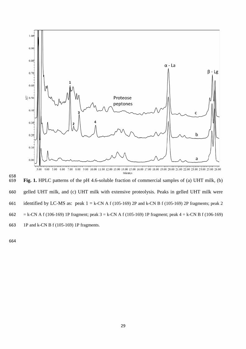
HPLC. Interestingly, besides the peaks of whey proteins, we systematically detected the presence of 213
few major peaks, eluting between 7 and 11 min in the HPLC chromatogram (Fig. 1, pattern b), that 214
we never detected in stable UHT milk samples from previous studies (Fig. 1, pattern a). This 215
suggested that a rather similar proteolytic pathway should bring UHT milk to gelation, regardless 216
the microbial species responsible. 217
Based on the HPLC-MS analysis, four peaks eluting at 7, 7.5, 8.2 and 10 min respectively proved to 218
contain fragments of k-CN, i.e. (f 105-169) from the genetic variants A and B and having either 219
single or double phosphorylation, and the canonical CMP (f 106-169) from the two variants, singly 220
phosphorylated only. The peak assignment is detailed in Table S1. 221
The identification of these peptides is consistent with previous findings reporting that, when 222
incubated with pure k-CN, AprX from different strains of Ps. fluorescens can cleave the peptide 223
bonds 104-105 and 105-106. These cleavages respectively generate the so-called pseudo-CMP and 224
pseudo-para-k-CN fragments, beside the true CMP and para-k-CN as also generated by the specific 225
action of chymosin (Baglinière et al., 2012; Stuknyté et al., 2016). In contrast with findings reported 226
by Recio et al., (2000a), we did not detect fragments (f 104-169), (f 107-169) and (f 108-169) in our 227
milk samples. The HPLC analysis of CMP in rennet whey samples was previously reported (Thoma 228
Page 10
10
et al., 2006; Pellegrino et al., 2015), with patterns comparable to those obtained here for gelled 229
UHT milk. 230
The four peaks became hardly distinguishable when an extensive proteolysis in milk gave rise to a 231
more complex HPLC pattern (Figure 1, pattern c). Such a pattern was observed for UHT milk 232
samples in which a compact sediment was present instead of a rennet-like gel. Consequently, we 233
discontinued using the HPLC for milk analyses, although it was a unique approach for peak 234
identification by MS, and preferred the CZE, which provides reliable and accurate separation of 235
milk proteins (Heck et al., 2008). 236
Considering the definite presence of different CMPs in all gelled UHT milks, an attempt was made 237
to evaluate the degradation of k-CN as a possible analytical approach not suffering from 238
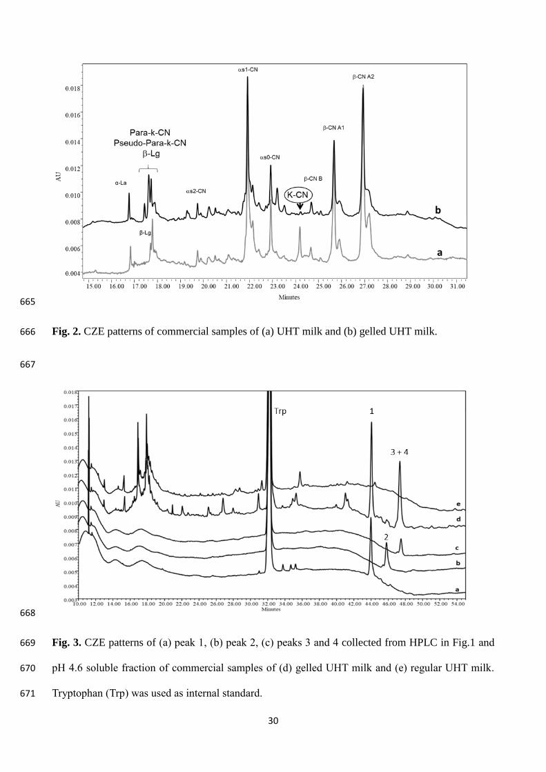
interferences of small peptides. Indeed, all the CZE patterns of gelled UHT milks showed the k-CN 239
degradation, and the presence of one or two peaks corresponding to para-k-CN and pseudo-para-k-240
CN fractions (Fig. 2, pattern b). These patterns also showed that no other casein fractions were 241
degraded. This allowed to exclude a residual plasmin activity and, most importantly, confirmed that 242
a chymosin-like cleavage of k-CN occurred in gelled milk. However, the obtained CZE patterns 243
were not satisfactory for a quantitative study since the tween peaks of β-lactoglobulin migrated very 244
close to those of para-k-CN and pseudo-para-k-CN fragments making the identification of these last 245
unreliable. Other authors reported the same difficulties when analysing milk added with rennet 246
whey solids (Recio et al., 2000b). To overcome these problems, we decided to develop novel CZE 247
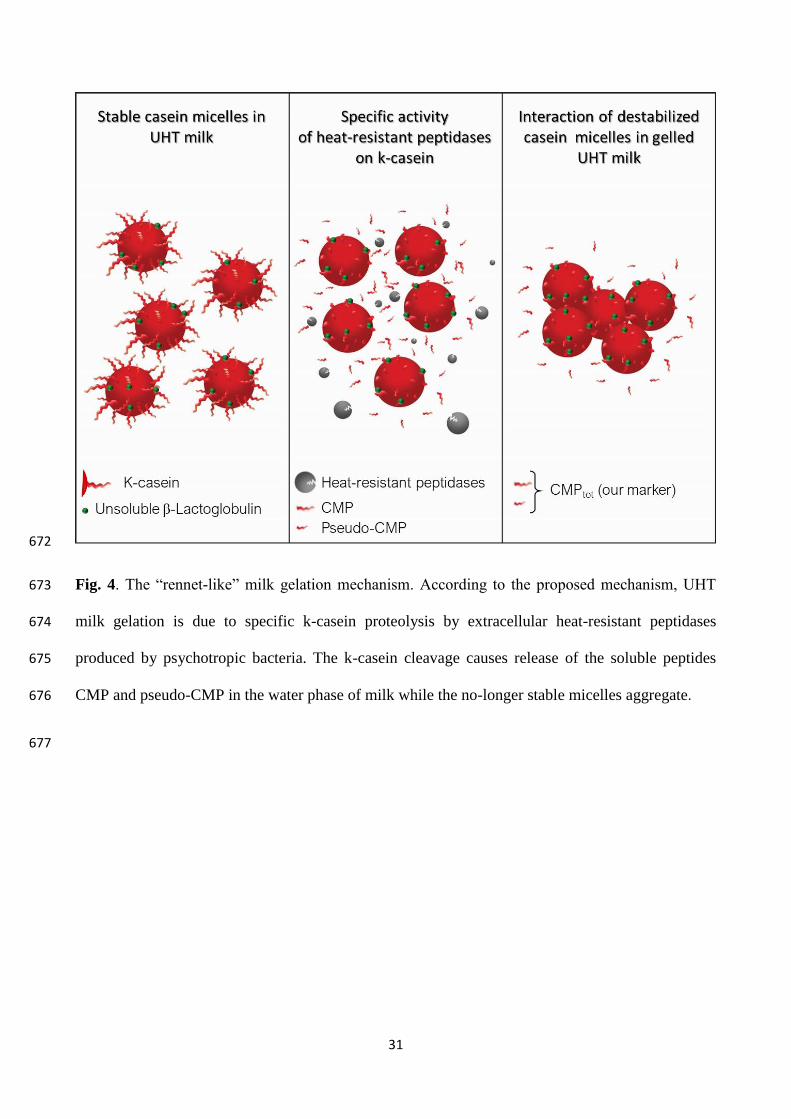
conditions intended to separate the target CMPs present in the pH 4.6-soluble fraction of milk. 248
These fragments migrate as three distinct peaks in the last part of the CZE pattern, which is free of 249
interferences (Fig. 3). A limitation of using CZE as an analytical technique is the difficulty of 250
adopting MS detection to achieve direct peak identification. Therefore, the CMP peaks were 251
identified by analysing the same fractions that were collected from the HPLC of a gelled UHT milk 252
and tested by HPLC-MS. The A and B genetic variants eluted as single peaks for both CMP and 253
pseudo-CMPs, as already observed by Recio et al., (2000a). Proteose peptones peaks were 254
Page 11
11
identified in a previous work (D’Incecco et al., 2018) and, like other peptides, migrated in the first 255
part of the pattern causing no interference with the peaks of CMPs. Tryptophan was added to the 256
samples as an internal standard to correct the peak area of target peptides for the instrumental error 257
in the injected volume. 258
The CZE of UHT milk inoculated with different counts of Ps. fluorescens were reported in previous 259
papers (Van Riel & Olieman, 1995; Recio et al., 2000a, b). However, these papers were focused on 260
the identification of rennet whey solids in adulterated milk and consequently no relation was 261
established between the presence of these fragments and occurrence of milk gelation. Nevertheless, 262
the peak assignments reported for CMP and pseudo-CMPs peaks by these authors were the same as 263
we found in commercial UHT milk samples where gelation occurred. The sum of corrected peak 264
area counts of CMP and pseudo-CMPs (f 105-169) peaks, hereafter named CMPtot, was thus 265
considered in the present study. Formation of CMPtot was monitored in milk samples inoculated 266
with selected bacterial strains, as it is discussed further, in a set of trials that we designed to best 267
simulate the conditions milk undergoes at the industrial manufacturing plant before the sterilization 268
treatment and during the successive shelf life. Contrary to previous studies using UHT milk (Datta 269
& Deeth, 2003) or sterile reconstituted milk (Alves et al., 2018) as a substrate, we used pasteurized 270
milk in order to make proteases released by the studied strains to act on casein micelles in a nearly 271
native state. Furthermore, interference of proteolytic activity from contaminating bacterial species 272
was avoided since these were preliminarily removed by milk microfiltration, as previously done by 273
other authors (Baglinière et al., 2012; Matéos et al., 2015). Thus, the microfiltered pasteurized (MP) 274
milk inoculated with the target strains could be stored at refrigeration conditions for some days 275
before the thermal processing, as it usually occurs at industrial plants for UHT milk processing. 276
277
3.2 Bacterial strain selection 278
Fourteen isolates were identified by partial 16S rDNA gene sequencing and rpoB gene as 279
Pseudomonas fluorescens (11 strains), Chryseobacterium joostei (2 strains), and Ps. poae (1 strain) 280
Page 12
12
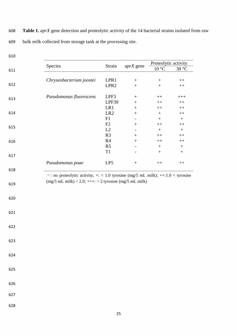
(Table 1). These findings were in agreement with those of Vithanage et al., (2016), that considered 281
these species as part of culturable psychrotrophic microbiota in refrigerate raw milk. The aprX gene 282
was widespread amongst the Pseudomonas strains, as only four strains out of the 11 tested did not 283
harbor this gene (Table 1). 284
All the strains exhibited proteolytic activity at both 10 and 30 °C, although to a different extent. At 285
30 °C, all the aprX positive strains were able to hydrolyze casein in the range 1.0 to 2.0 mg tyrosine 286
5 mL-1 milk and Ps. fluorescens LPF3 exhibited the highest proteolytic activity. At lower 287
temperature (10 °C) four strains out of the eight characterized by the highest activity at 30 °C 288
showed a decreased proteolytic activity, while Ps. poae LP5 showed values comparable with those 289
obtained at 30 °C (1.0 < OD650 < 2.0). C. joostei LPR1 and LPR2 showed a similar behavior, a 290
higher proteolytic activity being observed at 30 °C. For each species, the strain possessing the 291
highest proteolytic activity was selected for the subsequent experiments: C. joostei LPR1, Ps. 292
fluorescens LPF3, Ps. poae LP5. 293
294
3.3 Proteolysis and gelation in experimental milk samples 295
Three batches of MP milk were inoculated (final concentration 103 -104 CFU/mL) with LPF3, LP5 296
and LPR1 strains, respectively, and incubated at 6 °C for up to 6 days. Each day, an aliquot of 297
incubated milk was sterilized (97-98 °C/4 min) in sealed tubes and further stored at 25 and 40 °C, 298
the latter representing storage temperature abuse with respect to room temperature. The evaluation 299
of CMPtot by CE was carried out in milk just before sterilization and after 1 week, 3 weeks and 3 300
months of storage or at the gelling/instability onset when it happened at an intermediate time. The 301
results of this trial are compiled in Table 2. Concerning gelation, only samples entirely gelled were 302
referred to as gelled, depending on gel stability at the inversion of the tube (Fig. S1). Other 303
instability signs were the formation of a compact and robust sediment at the bottom of the tube and, 304
in a few cases, the flocculation of milk during the sterilization (Fig. S1). 305
Page 13
13
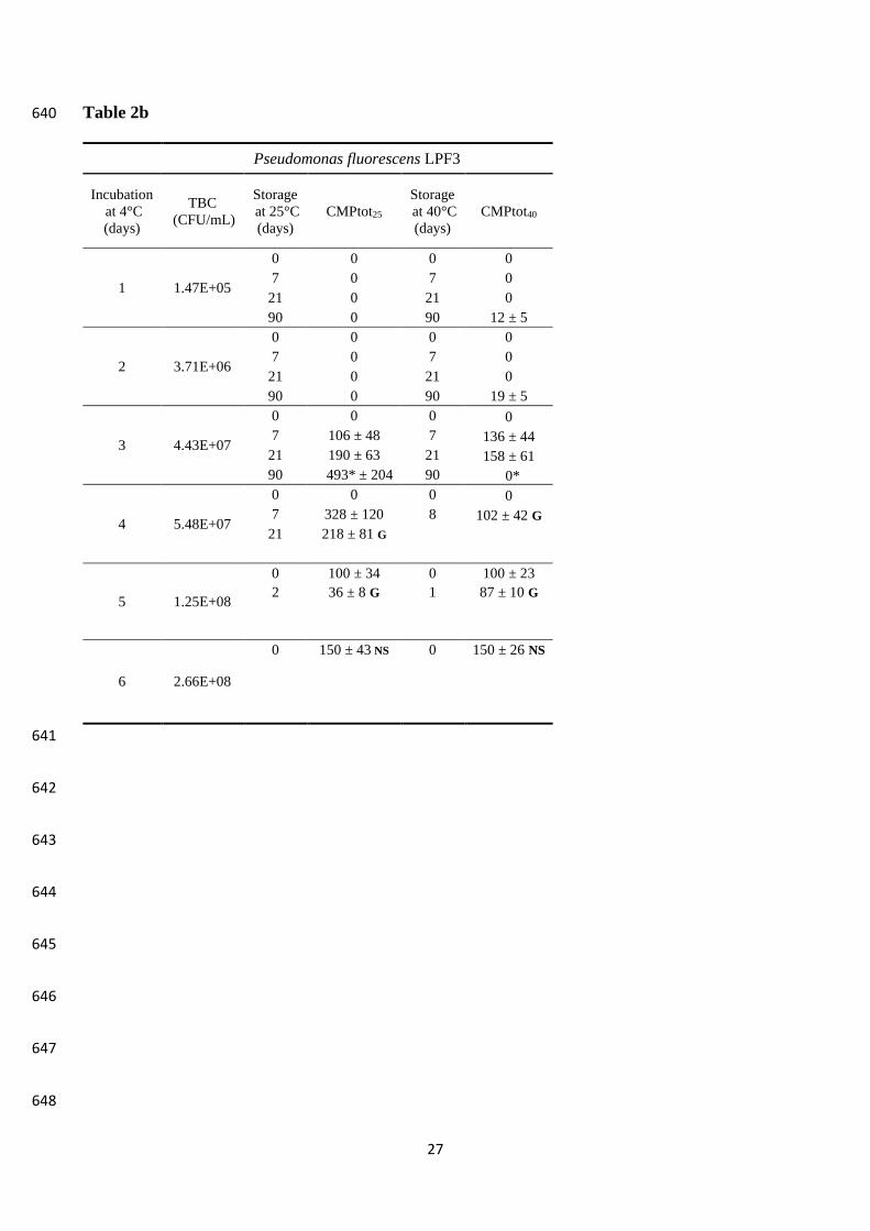
P. fluorescens LPF3 exhibited a faster growth rate than P. poe LP5, nevertheless both strains 306
approximately reached 108 cfu/mL after 6 days of incubation. 307
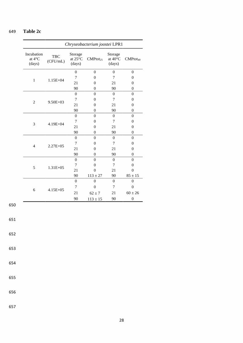
Differently, C. joostei LPR1 grew much more slowly since the beginning of incubation. Indeed, 308
after 6 days of cold incubation LPR1 reached counts comparable to those reached by the other two 309
strains after 1-2 days. In all of the samples, pH values were within the range 6.5-6.8 and were not 310
significantly different (P > 0.05) from that of the control (not inoculated) milk, indicating that no 311
milk acidification had happened during the cold storage. 312
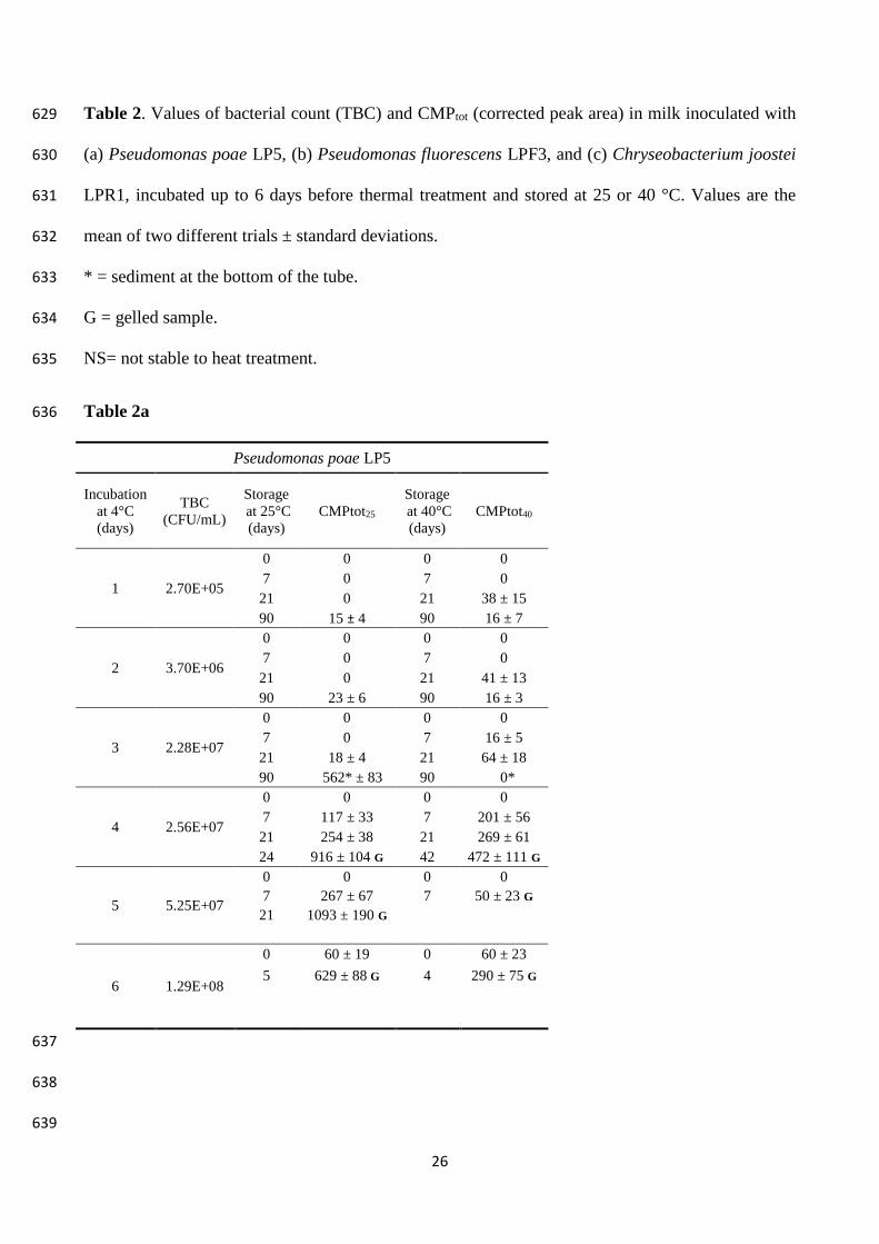
Production of CMPtot was markedly different among strains (Table 2), according to the differences 313
observed in the total proteolytic activity (as tyrosine equivalents) (Table 1). In this respect, LP5 314
proved to be the most active strain as CMPtot presence was detected even in milk samples that were 315
previously kept at low temperature for one or two days only. The strain LPR1 only produced small 316
amounts of CMPtot after 3 months of storage at 25 °C in the samples that were previously incubated 317
at low temperature for 5 days, consistently with the slow growth observed. Milk storage at 40 °C 318
dramatically anticipated the release of CMPtot, also with the lowest bacterial counts. As expected, 319
the proteolytic activity of AprX against k-CN was faster at higher temperature and the released 320
CMPtot accumulated. Optimum temperature for AprX from different strains of Ps. fluorescens 321
isolated from milk was reported to be 37-40 °C (Matéos et al., 2015; Alves et al., 2018). However, 322
when storage at 40 °C was prolonged, the degradation of CMPtot took place as well. 323
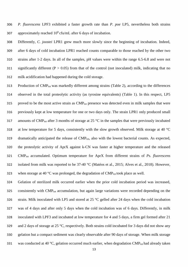
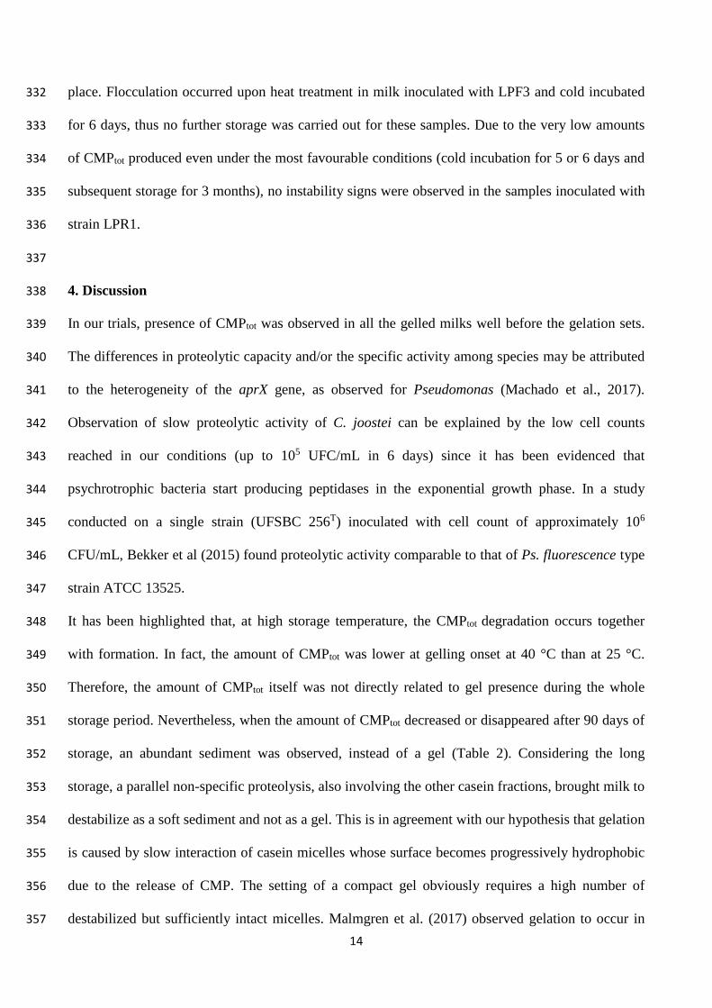
Gelation of sterilized milk occurred earlier when the prior cold incubation period was increased, 324
consistently with CMPtot accumulation, but again large variations were recorded depending on the 325
strain. Milk inoculated with LP5 and stored at 25 °C gelled after 24 days when the cold incubation 326
was of 4 days and after only 5 days when the cold incubation was of 6 days. Differently, in milk 327
inoculated with LPF3 and incubated at low temperature for 4 and 5 days, a firm gel formed after 21 328
and 2 days of storage at 25 °C, respectively. Both strains cold incubated for 3 days did not show any 329
gelation but a compact sediment was clearly observable after 90 days of storage. When milk storage 330
was conducted at 40 °C, gelation occurred much earlier, when degradation CMPtot had already taken 331
Page 14
14
place. Flocculation occurred upon heat treatment in milk inoculated with LPF3 and cold incubated 332
for 6 days, thus no further storage was carried out for these samples. Due to the very low amounts 333
of CMPtot produced even under the most favourable conditions (cold incubation for 5 or 6 days and 334
subsequent storage for 3 months), no instability signs were observed in the samples inoculated with 335
strain LPR1. 336
337
4. Discussion 338
In our trials, presence of CMPtot was observed in all the gelled milks well before the gelation sets. 339
The differences in proteolytic capacity and/or the specific activity among species may be attributed 340
to the heterogeneity of the aprX gene, as observed for Pseudomonas (Machado et al., 2017). 341
Observation of slow proteolytic activity of C. joostei can be explained by the low cell counts 342
reached in our conditions (up to 105 UFC/mL in 6 days) since it has been evidenced that 343
psychrotrophic bacteria start producing peptidases in the exponential growth phase. In a study 344
conducted on a single strain (UFSBC 256T) inoculated with cell count of approximately 106 345
CFU/mL, Bekker et al (2015) found proteolytic activity comparable to that of Ps. fluorescence type 346
strain ATCC 13525. 347
It has been highlighted that, at high storage temperature, the CMPtot degradation occurs together 348
with formation. In fact, the amount of CMPtot was lower at gelling onset at 40 °C than at 25 °C. 349
Therefore, the amount of CMPtot itself was not directly related to gel presence during the whole 350
storage period. Nevertheless, when the amount of CMPtot decreased or disappeared after 90 days of 351
storage, an abundant sediment was observed, instead of a gel (Table 2). Considering the long 352
storage, a parallel non-specific proteolysis, also involving the other casein fractions, brought milk to 353
destabilize as a soft sediment and not as a gel. This is in agreement with our hypothesis that gelation 354
is caused by slow interaction of casein micelles whose surface becomes progressively hydrophobic 355
due to the release of CMP. The setting of a compact gel obviously requires a high number of 356
destabilized but sufficiently intact micelles. Malmgren et al. (2017) observed gelation to occur in 357
Page 15
15
commercial UHT milk after 6 months of storage at 22 °C while a sediment developed consequent to 358
intense proteolysis when milk stored at 40 °C. 359
Overall, with respect to the studied strains, our data indicated a negative correlation between 360
bacterial counts in raw milk and time to gelation after the sterilization, since less days elapsed 361
before gelation onset when initial bacterial counts were higher. In fact, Pseudomonas spp. in 362
refrigerated milk produce peptidases in the late exponential, or early stationary, growth phase 363
(Stevenson et al., 2003; Alves et al., 2018). Stoeckel et al. (2016a) worked with three Pseudomonas 364
strains (Ps. weihenstephanensis, Ps. proteolytica and Pseusomonas R35698 W15a isolated from 365
raw milk) individually incubated in milk at 6 °C for 4 and 5 days before thermal treatment and 366
observed a complete milk gelation only after 4 months of subsequent storage at 20 °C. The related 367
degree of proteolysis in milk samples was measured as the amount of pH 4.6-soluble peptides 368
released during storage using the fluorescamine assay. Therefore, like in other similar studies 369
(Gaucher et al., 2011; Rauh et al., 2014), it was not possible to go deeper into the mechanism 370
leading to milk gelation. Baglinière et al. (2012) observed no gelation in milk inoculated with nine 371
strains of Ps. fluorescens and incubated at 4 °C for 3 days before thermal treatment and subsequent 372
storage at 20 °C up to 90 days. These authors identified many released peptides by HPLC-MS but, 373
since they did not work with gelled samples, no relation between presence of specific peptides and 374
gelation could be established. Based on the number of released peptides, they showed the casein 375
degradation to be β- > αs1- > k- > αs2-CN fractions, whereas more studies reported that AprX in 376
milk preferentially hydrolyses k- > β- > αs-CNs (Datta & Deeth, 2003; Zhang et al., 2018). 377
Although conducted under not always comparable experimental conditions, many studies showed 378
that both type and amount of AprX produced by Ps. fluorescens are strain-dependent, with different 379
response (enzyme expression) of strains to growth conditions (Marchand et al., 2009; Decimo et al., 380
2014; Caldera et al., 2016). In addition, the activity of AprX in UHT milk is regulated, both 381
qualitatively and quantitatively, by storage temperature. Consequently, milk gelation may take so 382
long time that is not observed during the studied storage period or may not settle at high storage 383
Page 16
16
temperature, when an intense proteolytic activity takes place and destabilization evolves into a 384
sediment accumulation. The number and complexity of these aspects suggested us checking for the 385
release of CMP or pseudo-CMPs rather than for the quantification of AprX activity or the total 386
proteolysis extent for predicting UHT milk stability. Due to the selectivity of the analytical 387
conditions, we were able to detected CMPtot in milk well before its gelation, in some cases even 388
before the sterilization treatment (not shown). Similarly, Matéos et al., (2015) observed 389
accumulation of these peptides in milk during storage at 6 °C before UHT treatment. This confirms 390
that these peptides can be useful markers for predicting the propensity of a milk to gel. 391
Different mechanisms have been proposed to explain UHT milk gelation, either enzymatic or non-392
enzymatic (McMahon, 1996; Datta and Deeth, 2001, Machado 2017, Anema 2018). Recently, 393
Machado et al., (2017) reported that AprX peptidases may hydrolyse either hydrophobic or 394
hydrophilic areas of casein micelles thus causing their aggregation and sedimentation in UHT milk. 395
In contrast, Anema (2018) proposed the interactions to occur via hydrophobic bonding between 396
para-k-casein either on micelles or in serum phase. Zhang et al., (2018) observed that, in UHT milk 397
intentionally added with AprX purified from Ps. fluorescens, the onset of gelation goes together 398
with an increase in particle size distribution above that expected for casein micelles and the specific 399
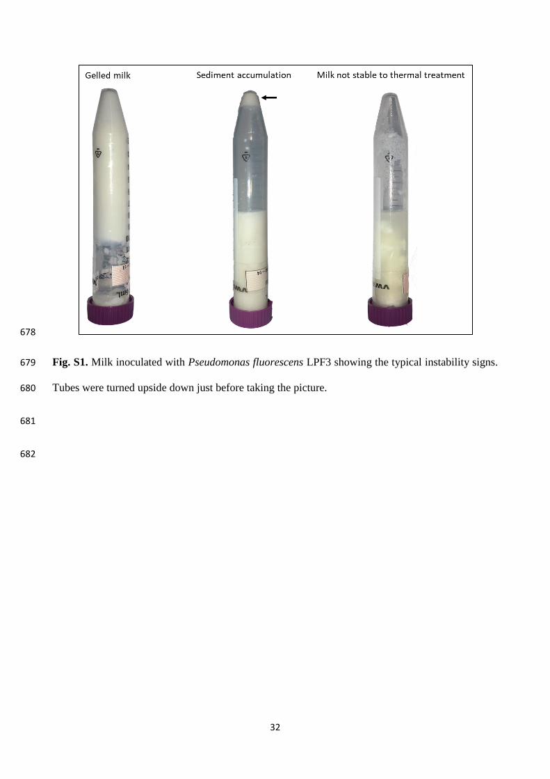
hydrolysis of k-CN. Based on the results of the present study and considering the most recent 400
literature, we hypothesized a “rennet-like” milk gelation mechanism (Fig. 4), in which the heat-401
stable bacterial peptidases cleave k-CN at the peptide bond 105-106 or in its proximity, depending 402
on the species and strains, and releases the soluble CMPtot. Consequently, the un-solvated micelles 403
slowly aggregate creating a gel that may occupy the whole milk volume when a critical number of 404
casein micelle is involved. Gelation onset is dependent on this event as the first necessary step and 405
our diagnostic approach relies on the detection of the soluble product (CMPtot) originated from this 406
step. Although gelled UHT milks typically contain higher amounts of pseudo-CMPs than CMP, this 407
does not seem to affect the phenomenon. In contrast, broad casein proteolysis plays against 408
gelation. Therefore, a parallel non-specific proteolytic activity that may occur during prolonged 409
Page 17
17
storage or when storage temperature is high impairs gel establishment and brings to its 410
solubilisation and the formation of a sediment. 411
412
5. Conclusions 413
The heat-stable AprX peptidase produced by psychrotrophic species has a chymosin-like activity as 414
it cleaves CMP and pseudo-CMPs from k-CN and promotes micelle destabilization. Therefore, 415
presence of AprX in milk is a challenge for UHT milk manufacturers. This study has shown that the 416
presence of CMPtot in milk represents a phenotypical character of strains in terms of their capability 417
of producing AprX peptidases. Due to the large variation in peptidase expression and activity 418
among bacterial species and strains, we have here proposed to evaluate the presence of CMPtot as a 419
useful indicator of milk susceptibility to gelation, irrespective of the responsible species. The 420
modern CZE equipment allows the implementation of this control for routine evaluation of raw 421
milk before processing. Based on the evidences collected during this study, a model supporting the 422
specific hydrolysis of k-CN as the first necessary step for milk gelation onset is here presented. 423
424
Acknowledgments 425
The authors wish to thank the graphic designer Dr. Nicolò Ughetti for the artwork of the gelation 426
mechanism. The authors declare no conflict of interest in this paper. 427
428
References 429
Alves, M. P., Salgado, R. L., Eller, M. R., Dias, R. S., de Paula, S. O., & de Carvalho, A. F. (2018). 430
Temperature modulates the production and activity of a metalloprotease from Pseudomonas 431
fluorescens 07A in milk. Journal of Dairy Science, 101(2), 992-999. 432
https://doi.org/10.3168/jds.2017-13238 433
434
Page 18
18
Anema, S. G. (2018). Age Gelation, Sedimentation, and Creaming in UHT Milk: A Review. 435
Comprehensive Reviews in Food Science and Food Safety, 0, 1-27. https://doi.org/10.1111/1541-436
4337.12407 437
438
Baglinière, F., Tanguy, G., Jardin, J., Matéos, A., Briard, V., Rousseau, F., Robert, B., Beaucher E., 439
Humbert G., Dary A., Gaillard J. L., Amiel C., & Gaucheron F. (2012). Quantitative and qualitative 440
variability of the caseinolytic potential of different strains of Pseudomonas fluorescens: 441
Implications for the stability of casein micelles of UHT milks during their storage. Food 442
Chemistry, 135(4), 2593-2603. https://doi.org/10.1016/j.foodchem.2012.06.099 443
444
Baglinière, F., Tanguy, G., Salgado, R. L., Jardin, J., Rousseau, F., Robert, B., Harel-Oger, M., 445
Dantas Vanetti, M. C., & Gaucheron, F. (2017). Ser2 from Serratia liquefaciens L53: A new heat 446
stable protease able to destabilize UHT milk during its storage. Food Chemistry, 229, 104-110. 447
https://doi.org/10.1016/j.foodchem.2017.02.054 448
449
Bava, L., Zucali, M., Sandrucci, A., Brasca, M., Vanoni, L., Zanini, L., & Tamburini, A. (2011). 450
Effect of cleaning procedure and hygienic condition of milking equipment on bacterial count of 451
bulk tank milk. Journal of Dairy Research, 78(2), 211-219. 452
https://doi.org/10.1017/S002202991100001X 453
454
Bekker, A., Steyn, L., Charimba, G., Jooste, P., & Hugo, C. (2015). Comparison of the growth 455
kinetics and proteolytic activities of Chryseobacterium species and Pseudomonas 456
fluorescens. Canadian Journal of Microbiology, 61(12), 977-982. https://doi.org/10.1139/cjm-457
2015-0236 458
459
Page 19
19
Brasca, M., Decimo, M., Morandi, S., Machado, S. G., Bagliniére, F., Vanetti, M. C. D. (2017) 460
Psychrotrophic bacteria. In Poltronieri P. (Ed.), Microbiology in dairy processing: challenges and 461
opportunities (pp. 37 – 61) IFT Press Series, Wiley-Blackwell, UK, 462
https://doi.org/10.1002/9781119115007.ch3 463
464
Caldera, L., Franzetti, L., Van Coillie, E., De Vos, P., Stragier, P., De Block, J., & Heyndrickx, M. 465
(2016). Identification, enzymatic spoilage characterization and proteolytic activity quantification of 466
Pseudomonas spp. isolated from different foods. Food Microbiology, 54, 142-153. 467
https://doi.org/10.1016/j.fm.2015.10.004 468
469
Datta, N., & Deeth, H. C. (2001). Age gelation of UHT milk—a review. Food and Bioproducts 470
processing, 79(4), 197-210. https://doi.org/10.1205/096030801753252261 471
472
Datta, N., & Deeth, H. C. (2003). Diagnosing the cause of proteolysis in UHT milk. LWT-Food 473
Science and Technology, 36(2), 173-182. https://doi.org/10.1016/S0023-6438(02)00214-1 474
475
Decimo M, Morandi S, Silvetti T, Brasca M. 2014. Characterization of gram-negative psy- 476
chrotrophic bacteria isolated from Italian bulk tank milk. Journal of Food Science 79(10):M2081–477
90. https://doi.org/10.1111/1750-3841.12645 478
479
D'Incecco, P., Rosi, V., Cabassi, G., Hogenboom, J. A., & Pellegrino, L. (2018). Microfiltration and 480
ultra-high-pressure homogenization for extending the shelf-storage stability of UHT milk. Food 481
Research International, 107, 477-485. https://doi.org/10.1016/j.foodres.2018.02.068 482
483
Gaucher, I., Tanguy, G., Fauquant, J., Jardin, J., Rousseau, F., Robert, B., Madec, M. N., & 484
Gaucheron, F. (2011). Proteolysis of casein micelles by Pseudomonas fluorescens CNRZ 798 485
Page 20
20
contributes to the destabilisation of UHT milk during its storage. Dairy Science & 486
Technology, 91(4), 413. 487
488
Glück, C., Rentschler, E., Krewinkel, M., Merz, M., von Neubeck, M., Wenning, M., Scherer, S., 489
Stoeckel, M., Hinrichs J., Stressler, T., & Fischer, L. (2016). Thermostability of peptidases secreted 490
by microorganisms associated with raw milk. International Dairy Journal, 56, 186-197. 491
https://doi.org/10.1016/j.idairyj.2016.01.025 492
493
Heck, J. M. L., Olieman, C., Schennink, A., Van Valenberg, H. J. F., Visker, M. H. P. W., 494
Meuldijk, R. C. R., & Van Hooijdonk, A. C. M. (2008). Estimation of variation in concentration, 495
phosphorylation and genetic polymorphism of milk proteins using capillary zone electrophoresis. 496
International Dairy Journal, 18(5), 548-555. https://doi.org/10.1016/j.idairyj.2007.11.004 497
498
Hull, M. E. (1947). Studies on milk proteins II. Colourimetric determination of the partial hydrolisis 499
of the proteins in milk. Journal of Dairy Science, 30, 881–884. 500
501
Machado, S. G., Baglinière, F., Marchand, S., Van Coillie, E., Vanetti, M. C., De Block, J., & 502
Heyndrickx, M. (2017). The biodiversity of the microbiota producing heat-resistant enzymes 503
responsible for spoilage in processed bovine milk and dairy products. Frontiers in microbiology, 8, 504
302. https://doi.org/10.3389/fmicb.2017.00302 505
506
Malmgren, B., Ardö, Y., Langton, M., Altskär, A., Bremer, M. G., Dejmek, P., & Paulsson, M. 507
(2017). Changes in proteins, physical stability and structure in directly heated UHT milk during 508
storage at different temperatures. International Dairy Journal, 71, 60-75. 509
https://doi.org/10.1016/j.idairyj.2017.03.002 510
511
Page 21
21
Marchand, S., Vandriesche, G., Coorevits, A., Coudijzer, K., De Jonghe, V., Dewettinck, K., De 512
Vos, P., Devreese, B., Heyndrickx, M., & De Block, J. (2009). Heterogeneity of heat-resistant 513
proteases from milk Pseudomonas species. International Journal of Food Microbiology, 133(1-2), 514
68-77. https://doi.org/10.1016/j.ijfoodmicro.2009.04.027 515
516
Matéos, A., Guyard-Nicodème, M., Baglinière, F., Jardin, J., Gaucheron, F., Dary, A., Humbert, G., 517
& Gaillard, J. L. (2015). Proteolysis of milk proteins by AprX, an extracellular protease identified 518
in Pseudomonas LBSA1 isolated from bulk raw milk, and implications for the stability of UHT 519
milk. International Dairy Journal, 49, 78-88. https://doi.org/10.1016/j.idairyj.2015.04.008 520
521
McMahon, D. J. (1996). Age-gelation of UHT milk: changes that occur during storage, their effect 522
on shelf life and the mechanism by which age-gelation occurs. In Heat treatments and alternative 523
methods. IDF Symposium, Vienna (Austria), 6-8 Sep 1995. International Dairy Federation. 524
525
Mcphee J. D., & Griffiths M. W. Psychrotrophic bacteria Pseudomonas spp. (2011). In: John WF, 526
editor. Encyclopedia of Dairy Sciences. Second Edition. Academic Press; San Diego. pp. 379–383. 527
528
McCabe, K. M., Khan, G., Zhang, Y. H., Mason, E. O., & McCabe, E. R. (1995). Amplification of 529
bacterial DNA using highly conserved sequences: automated analysis and potential for molecular 530
triage of sepsis. Pediatrics, 95(2), 165-169. 531
532
Pellegrino, L., Rosi, V., D'Incecco, P., Stroppa, A., & Hogenboom, J. A. (2015). Changes in the 533
soluble nitrogen fraction of milk throughout PDO Grana Padano cheese-making. International 534
Dairy Journal, 47, 128-135. https://doi.org/10.1016/j.idairyj.2015.03.002 535
536
Page 22
22
Pinto C. L O., Machado S. G., Cardoso R. R., Alves R. M., Vanetti M. C. D. (2014). Proteolytic 537
potential of Pseudomonas fluorescens isolated from refrigerated raw milk. Brazilian Journal of 538
Sustainable Agriculture, 4, 16-25. 539
540
Rauh, V. M., Sundgren, A., Bakman, M., Ipsen, R., Paulsson, M., Larsen, L. B., & Hammershøj, M. 541
(2014). Plasmin activity as a possible cause for age gelation in UHT milk produced by direct steam 542
infusion. International Dairy Journal, 38(2), 199-207. https://doi.org/10.1016/j.idairyj.2013.12.007 543
544
Recio, I., López-Fandiño, R., Olano, A., Olieman, C., & Ramos, M. (1996). Study of the formation 545
of caseinomacropeptides in stored ultra-high-temperature-treated milk by capillary 546
electrophoresis. Journal of Agricultural and Food Chemistry, 44(12), 3845-3848. 547
548
Recio, I., García-Risco, M. R., Ramos, M., & López-Fandiño, R. (2000a). Characterization of 549
peptides produced by the action of psychrotrophic proteinases on κ-casein. Journal of Dairy 550
Research, 67(4), 625-630. https://doi.org/10.1017/S002202990000443X 551
552
Recio, I., García-Risco, M. R., López-Fandiño, Olano, A., & Ramos, M. (2000b). Detection of 553
rennet whey solids in UHT milk by capillary electrophoresis. International Dairy Journal, 10, 333-554
338. https://doi.org/10.1016/S0958-6946(00)00076-5 555
556
Sajben, E., Manczinger, L., Nagy, A., Kredics, L., & Vágvölgyi, C. (2011). Characterization of 557
pseudomonads isolated from decaying sporocarps of oyster mushroom. Microbiological 558
research, 166(4), 255-267. https://doi.org/10.1016/j.micres.2010.05.002 559
560
Page 23
23
Stevenson, R. G., Rowe, M. T., Wisdom, G. B., & Kilpatrick, D. (2003). Growth kinetics and 561
hydrolytic enzyme production of Pseudomonas spp. isolated from pasteurized milk. Journal of 562
Dairy Research, 70(3), 293-296. https://doi.org/10.1017/S0022029903006204 563
564
Stoeckel, M., Lidolt, M., Achberger, V., Glück, C., Krewinkel, M., Stressler, T., von Neubeckc, M., 565
Wenning, M., Scherer, S., Fischer, L., & Hinrichs, J. (2016a). Growth of Pseudomonas 566
weihenstephanensis, Pseudomonas proteolytica and Pseudomonas sp. in raw milk: Impact of 567
residual heat-stable enzyme activity on stability of UHT milk during shelf-life. International Dairy 568
Journal, 59, 20-28. https://doi.org/10.1016/j.idairyj.2016.02.045 569
570
Stoeckel, M., Lidolt, M., Stressler, T., Fischer, L., Wenning, M., & Hinrichs, J. (2016b). Heat 571
stability of indigenous milk plasmin and proteases from Pseudomonas: a challenge in the 572
production of ultra-high temperature milk products. International Dairy Journal, 61, 250-261. 573
https://doi.org/10.1016/j.idairyj.2016.06.009 574
575
Stuknytė, M., Decimo, M., Colzani, M., Silvetti, T., Brasca, M., Cattaneo, S., Aldini, G., & De 576
Noni, I. (2016). Extracellular thermostable proteolytic activity of the milk spoilage bacterium 577
Pseudomonas fluorescens PS19 on bovine caseins. Journal of Dairy Science, 99(6), 4188-4195. 578
https://doi.org/10.3168/jds.2016-10894 579
580
Thomä, C., Krause, I., & Kulozik, U. (2006). Precipitation behaviour of caseinomacropeptides and 581
their simultaneous determination with whey proteins by RP-HPLC. International Dairy Journal, 582
16, 285-293. https://doi.org/10.1016/j.idairyj.2005.05.003 583
584
Page 24
24
Vasbinder A.J., Rollema, H.S., & de Kruif, C.G. (2003). Impaired rennetability of heated milk: 585
study of enzymatic hydrolysis and gelation kinetics. Journal of Dairy Science, 83, 1548-1555. 586
https://doi.org/10.3168/jds.S0022-0302(03)73740-0 587
588
Van Riel, J., & Olieman, C. (1995). Determination of caseinomacropeptide with capillary zone 589
electrophoresis and its application to the detection and estimation of rennet whey solids in milk and 590
buttermilk powder. Electrophoresis, 16(1), 529-533. https://doi.org/10.1002/elps.1150160187 591
592
Vithanage, N. R., Dissanayake, M., Bolge, G., Palombo, E. A., Yeager, T. R., & Datta, N. (2016). 593
Biodiversity of culturable psychrotrophic microbiota in raw milk attributable to refrigeration 594
conditions, seasonality and their spoilage potential. International Dairy Journal, 57, 80-90. 595
https://doi.org/10.1016/j.idairyj.2016.02.042 596
597
von Neubeck, M., Baur, C., Krewinkel, M., Stoeckel, M., Kranz, B., Stressler, T., Fischer, L., 598
Hinrichs, J., Scherer, S., & Wenning, M. (2015). Biodiversity of refrigerated raw milk microbiota 599
and their enzymatic spoilage potential. International journal of food microbiology, 211, 57-65. 600
https://doi.org/10.1016/j.ijfoodmicro.2015.07.001 601
602
Zhang, C., Bijl, E., & Hettinga, K. (2018). Destabilization of UHT milk by protease AprX from 603
Pseudomonas fluorescens and plasmin. Food Chemistry, 263, 127-134. 604
https://doi.org/10.1016/j.foodchem.2018.04.128 605
606
607
Page 25
25
Table 1. aprX gene detection and proteolytic activity of the 14 bacterial strains isolated from raw 608
bulk milk collected from storage tank at the processing site. 609
610
611
612
613
614
615
616
617
618
619
620
621
622
623
624
625
626
627
628
Species Strain aprX gene Proteolytic activity
10 °C 30 °C
Chryseobacterium joostei LPR1 + + ++
LPR2 + + ++
Pseudomonas fluorescens LPF3 + ++ +++
LPF39 + ++ ++
LR1 + ++ ++
LR2 + + ++
F1 - + +
F2 + ++ ++
L2 - + +
R3 + ++ ++
R4 + ++ ++
R5 - + +
T1 - + +
Pseudomonas poae LP5 + ++ ++
−: no proteolytic activity, +: < 1.0 tyrosine (mg/5 mL milk); ++:1.0 < tyrosine
(mg/5 mL milk) < 2.0; +++: > 2 tyrosine (mg/5 mL milk)
Page 26
26
Table 2. Values of bacterial count (TBC) and CMPtot (corrected peak area) in milk inoculated with 629
(a) Pseudomonas poae LP5, (b) Pseudomonas fluorescens LPF3, and (c) Chryseobacterium joostei 630
LPR1, incubated up to 6 days before thermal treatment and stored at 25 or 40 °C. Values are the 631
mean of two different trials ± standard deviations. 632
* = sediment at the bottom of the tube. 633
G = gelled sample. 634
NS= not stable to heat treatment. 635
Table 2a 636
Pseudomonas poae LP5
Incubation
at 4°C
(days)
TBC
(CFU/mL)
Storage
at 25°C
(days)
CMPtot25
Storage
at 40°C
(days)
CMPtot40
1 2.70E+05
0 0 0 0
7 0 7 0
21 0 21 38 ± 15
90 15 ± 4 90 16 ± 7
2 3.70E+06
0 0 0 0
7 0 7 0
21 0 21 41 ± 13
90 23 ± 6 90 16 ± 3
3 2.28E+07
0 0 0 0
7 0 7 16 ± 5
21 18 ± 4 21 64 ± 18
90 562* ± 83 90 0*
4 2.56E+07
0 0 0 0
7 117 ± 33 7 201 ± 56
21 254 ± 38 21 269 ± 61
24 916 ± 104 G 42 472 ± 111 G
5 5.25E+07
0 0 0 0
7 267 ± 67 7 50 ± 23 G
21 1093 ± 190 G
6 1.29E+08
0 60 ± 19 0 60 ± 23
5 629 ± 88 G 4 290 ± 75 G
637
638
639
Page 27
27
Table 2b 640
Pseudomonas fluorescens LPF3
Incubation
at 4°C
(days)
TBC
(CFU/mL)
Storage
at 25°C
(days)
CMPtot25
Storage
at 40°C
(days)
CMPtot40
1 1.47E+05
0 0 0 0
7 0 7 0
21 0 21 0
90 0 90 12 ± 5
2 3.71E+06
0 0 0 0
7 0 7 0
21 0 21 0
90 0 90 19 ± 5
3 4.43E+07
0 0 0 0
7 106 ± 48 7 136 ± 44
21 190 ± 63 21 158 ± 61
90 493* ± 204 90 0*
4 5.48E+07
0 0 0 0
7 328 ± 120 8 102 ± 42 G
21 218 ± 81 G
5 1.25E+08
0 100 ± 34 0 100 ± 23
2 36 ± 8 G 1 87 ± 10 G
6 2.66E+08
0 150 ± 43 NS 0 150 ± 26 NS
641
642
643
644
645
646
647
648
Page 28
28
Table 2c 649
Chryseobacterium joostei LPR1
Incubation
at 4°C
(days)
TBC
(CFU/mL)
Storage
at 25°C
(days)
CMPtot25
Storage
at 40°C
(days)
CMPtot40
1 1.15E+04
0 0 0 0
7 0 7 0
21 0 21 0
90 0 90 0
2 9.50E+03
0 0 0 0
7 0 7 0
21 0 21 0
90 0 90 0
3 4.19E+04
0 0 0 0
7 0 7 0
21 0 21 0
90 0 90 0
4 2.27E+05
0 0 0 0
7 0 7 0
21 0 21 0
90 0 90 0
5 1.31E+05
0 0 0 0
7 0 7 0
21 0 21 0
90 113 ± 27 90 85 ± 15
6 4.15E+05
0 0 0 0
7 0 7 0
21 62 ± 7 21 60 ± 26
90 113 ± 15 90 0
650
651
652
653
654
655
656
657
Page 29
29
658 Fig. 1. HPLC patterns of the pH 4.6-soluble fraction of commercial samples of (a) UHT milk, (b) 659
gelled UHT milk, and (c) UHT milk with extensive proteolysis. Peaks in gelled UHT milk were 660
identified by LC-MS as: peak 1 = k-CN A f (105-169) 2P and k-CN B f (105-169) 2P fragments; peak 2 661
= k-CN A f (106-169) 1P fragment; peak 3 = k-CN A f (105-169) 1P fragment; peak 4 = k-CN B f (106-169) 662
1P and k-CN B f (105-169) 1P fragments. 663
664
Page 30
30
665
Fig. 2. CZE patterns of commercial samples of (a) UHT milk and (b) gelled UHT milk. 666
667
668
Fig. 3. CZE patterns of (a) peak 1, (b) peak 2, (c) peaks 3 and 4 collected from HPLC in Fig.1 and 669
pH 4.6 soluble fraction of commercial samples of (d) gelled UHT milk and (e) regular UHT milk. 670
Tryptophan (Trp) was used as internal standard. 671
Page 31
31
672
Fig. 4. The “rennet-like” milk gelation mechanism. According to the proposed mechanism, UHT 673
milk gelation is due to specific k-casein proteolysis by extracellular heat-resistant peptidases 674
produced by psychotropic bacteria. The k-casein cleavage causes release of the soluble peptides 675
CMP and pseudo-CMP in the water phase of milk while the no-longer stable micelles aggregate. 676
677
Page 32
32
678
Fig. S1. Milk inoculated with Pseudomonas fluorescens LPF3 showing the typical instability signs. 679
Tubes were turned upside down just before taking the picture. 680
681
682