1. Bioactive Glass Development (industry linked) Supervisors: Dr Philip Boughton, Prof Andrew Ruys
Bioactive glasses are used in tissue engineering, bone putty, dental root therapy, implant coatings and bioabsorbable devices. This industry linked project aims to develop new applications and improve existing glass manufacturing processes. Opportunity to investigate and develop novel glass compositions and post-forming methods (microspheres/fibers/coatings) to address clinical needs will be provided. Bioglass science, process design, and analytical testing within a commercial context will provide invaluable device design and manufacturing experience. Contact: [email protected] | 0402890150 | Rm 242 J13 __ 2. Soft Tissue Scaffold Development (industry linked) Supervisors: Dr Philip Boughton, A Prof Andrew Ruys, Prof Sue McLennan
Variotis™ is a versatile bioactive soft tissue scaffold that can be used with a range of cells and tissues. New methods, modifications and applications will be investigated. Photo-activated capabilities and bioactive glass facilitated tissue adhesion are important areas for investigation. The project will also include refinement objectives for existing production and post-process routes for various scaffold forms. The final phase of the project will involve design customization of the scaffold form and type for a tissue engineering collaborator. Contact: [email protected] | 0402890150 | Rm 242 J13 __ 3. Tissue Engineering Bioreactor Systems (industry linked) Supervisors: Dr Philip Boughton, Dr Giang Tran, Prof Andrew Ruys
In vitro tissue engineering benefits from biomechanical stimulus. The novel iaxsys™ system has been designed to complement existing cell biology experimental methods and equipment constraints. This project aims to further develop and refine systems: actuation, sensors, feedback, interface, mechanical couplings, perfusion, plate-bank and in-situ microscopy. User requirement analysis, design and development, manufacturing and verification/validation aspects will be addressed. Ability and experience with design (CAD), cell testing, and software programming will be helpful. Contact: [email protected] | 0402890150 | Rm 242 J13 __
4. Working with the biomedical industry to develop 3D printed medical devices
Supervisors: Prof Julie Cairney, Dr Philip Boughton Working with the biomedical industry to develop 3D printed medical devices 3DMedical are an exciting new start up based in Melbourne. They recently listed with the ASX and are already Australia’s leading medical and healthcare specific technology provider. In an Australian first, they recently developed a 3D printed and customised titanium jaw joint which was used to correct a rare jaw deformity in a 32-year-old male (x-ray shown below). In this project, you will work closely with the 3D Medical to develop new 3D printed products for orthopaedics. By undertaking a thorough review of the current orthopaedic consumables, you will be expected to identify the top 5 applications in which 3D printing could ‘disrupt’ the market for existing technologies. From there, you will be design and print a prototype product. The student undertaking this honours project will have the opportunity to undertake an industry placement in Melbourne over summer with 3DMedical.
http://3dmedical.com.au/ Contact: [email protected] | 0402890150 | Rm 242 J13 __ 5. Valve Biomaterials Optimization (Industry Linked) Supervisors: Dr Philip Boughton, Dr Giang Tran, Prof Andrew Ruys
Bovine pericardium is the outer membrane of the heart that is widely used in bioengineering of variety of cardiovascular applications including heart valve leaflet, patches for pericardial for cardiovascular reconstructive procedure as well as in general surgery. Calcification of these tissues can lead to structural dysfunction, tissue degeneration and catastrophic implant failure. The onset of calcification and its effects will be studied by a range of techniques. Existing and novel methods to prevent calcification will be investigated. Other opportunities to further enhance heart valve materials and valve configurations are also available. Contact: [email protected] | 0402890150 | Rm 242 J13 __
6. Optimization of Collagenous Implant Materials (Industry Linked). Supervisors: Dr Philip Boughton, Dr Giang Tran, Prof Andrew Ruys
Collagenous tissue such as bovine pericardium and porcine aortic wall have been used successfully in bioprosthetics for the past 40 years. The established route for collagenous tissue production utilizes glutaraldehyde crosslinking agent. A variety of processing conditions are employed by manufacturers. Concentration of glutaraldehyde, thickness of tissues, and strain conditions during crosslinking can be varied to enhance the mechanical performance of the bioprosthetic materials. This industry-sponsored study will provide opportunities to improve manufacturing processes, develop new approaches, engage in mechanical verification and analytical methods. This project is focussed on delivering process design, manufacturing and test recommendations. Contact: [email protected] | 0402890150 | Rm 242 J13 __ 7. 3D Printed Titanium Biomaterials Characterization Supervisors: Dr Philip Boughton, Prof Julie Cairney 3DMedical are an exciting new start up based in Melbourne. They recently listed with the ASX and are already Australia’s leading medical and healthcare specific technology provider. In an Australian first, they recently developed a 3D printed and customised titanium jaw joint which was used to correct a rare jaw deformity in a 32-year-old male (x-ray shown below). In this project, you will work closely with the 3D Medical to characterize SLM printed Titanium for mechanical properties, microstructural and fibroblast cell response. Titanium samples from a conventional manufacturing route will be compared against. Anisotropic printed structures will also be investigated. Recommendations for optimal 3d printing parameters for orthopaedic relevant outcomes will be established. The student undertaking this honours project will have the opportunity to undertake an industry placement in Melbourne over summer with 3DMedical.
Contact: [email protected] | 0402890150 | Rm 242 J13 __
8. Skin Tissue Engineering (RPA & Industry Linked). Supervisors: Prof Sue McLennan, A Prof Karen Vickery, Dr Philip Boughton, Prof Andrew Ruys
Diabetes and diabetic ulcers is a growing problem in aging populations and among remote indigenous communities. A novel resorbable scaffold for treating serious diabetic ulcers is currently being developed. Dermal chronic wounds are typically necrotic, apoxic, compromised by entrenched infection, and poor in mechanical integrity. An elastic highly interconnective porous scaffold laden with antibiotics and antibacterial agents is being developed. This project will focus on further biologic verification testing and design improvement of this scaffold with particular focus on resorption rate optimization. Exposure to production methods, invitro cell testing, analytical methods, mechanical testing will be provided. Contact: [email protected] | 0402890150 | JO7 Rm S428 __
9. Development of an App for Clinical Research, Rehabilitation Engineering, and Bioinformatics (industry linked) Supervisors: Dr Philip Boughton, Dr Simon Poon, Tamer Sabet, Prof Andrew Ruys Popular mobile devices contain a variety of sensors and integrated systems that can be applied to rehabilitation engineering, clinical research and bioinformatics. A thorough review of published and patented methods will be conducted. Broad design opportunities will be mapped out. A new app for use in conjunction with a treatment for frozen shoulder will be developed for mainstream mobile device platforms. The app will track patient joint biomechanics, have capacity to detect treatment abnormalities to allow immediate intervention if necessary, while remotely transponding data for centralized bioinformatic analysis. The prototype app will be verified and validated to ensure mitigation of risks identified in a design risk analysis and safety risk matrix. Candidates will need good software and hardware engineering experience. Contact: [email protected] | 0402890150 | Rm 242 J13 __ 10. Height-Adjustable Pillow System for Optimal Cervical Support (The Sydney Spine Institute) Supervisors: Specialist Physio Tamer Sabet, Dr Philip Boughton
The project will involve development of a pillow-augmenting system to provide cervical spine near-neutral zone positioning in varied positions. In addition to biomechanical design – materials selection, fabrication, user-friendliness, aesthetics, life-cycle, and business case summary will be important aspects to be addressed by this project. Contact: [email protected] | 0402890150 | Rm 242 J13 __
11. Supine Spine Manipulator (The Sydney Spine Institute) Supervisors: Specialist Physio Tamer Sabet, Dr Philip Boughton The aim is to develop a system to induce controlled amounts of displacement to select portions of the spine while supine. The system will incorporate a pressure sensor array and act via a pressure transducer system. The system will effectively provide manipulation therapy similar to that provided by a musculotskeletal physiotherapist, but in a quantified, repeatable, accessible manner. This system will also provide another method by which to track back pain foci with time. Contact: [email protected] | 0402890150 | Rm 242 J13 __ 12. Minimally-Invasive Trans-segmental Device for Treating Spondylolisthesis (The Sydney Spine Institute) Supervisors: Dr James Van Gelder, Dr Philip Boughton
“Slipped disk” is a major cause of serious low back pain. Surgical approaches to treating this condition A minimally invasive trans-segmental device design for treating spondylolisthesis is under development. Design process, prototype fabrication, specimen testing, biomechanical validation will be the mainstay of this project. Experience with CAD, FEA, mechanical testing, is preferred. Contact: [email protected] | 0402890150 | Rm 242 J13 __ 13. Intracranial Pressure Monitoring System (Concord Hospital, Iosys Pty Ltd) Supervisors: Dr Philip Boughton, Dr Simon Poon, Dr James Van Gelder
Like ECG, Intercranial Pressure (ICP) is an important vital sign used in intensive care. It is often too costly to be employed outside of ICU. Intracranial pressure monitoring systems provide a lower cost possibility to obtain important relative measurements (RAP) to assist with clinical planning, particularly in geriatric medicine. A compact mobile intracranial pressure monitoring system concept is under development and if transferable to a smartphone APP would also become an important M-health resource. Contact: [email protected] | 0402890150 | Rm 242 J13
__ 14. Development of a Neural Engineering Conduit (Cochlear Pty Ltd) Supervisors: Dr Philip Boughton, Prof Andrew Ruys, Prof Sri Bandyopadhyay, Dr Paul Carter The development of electrospun nerve conduits for peripheral repair is a relatively new area. Prototype conduit specimens (of a variety of conductivities) will be fabricated and cell tested. Cell culture will be conducted with and without electrical stimulation. Verification and validation testing will be undertaken to confirm specification requirements. Medical science background and/or cell culture experience is preferred. Contact: [email protected] | 0402890150 | Rm 242 J13 __
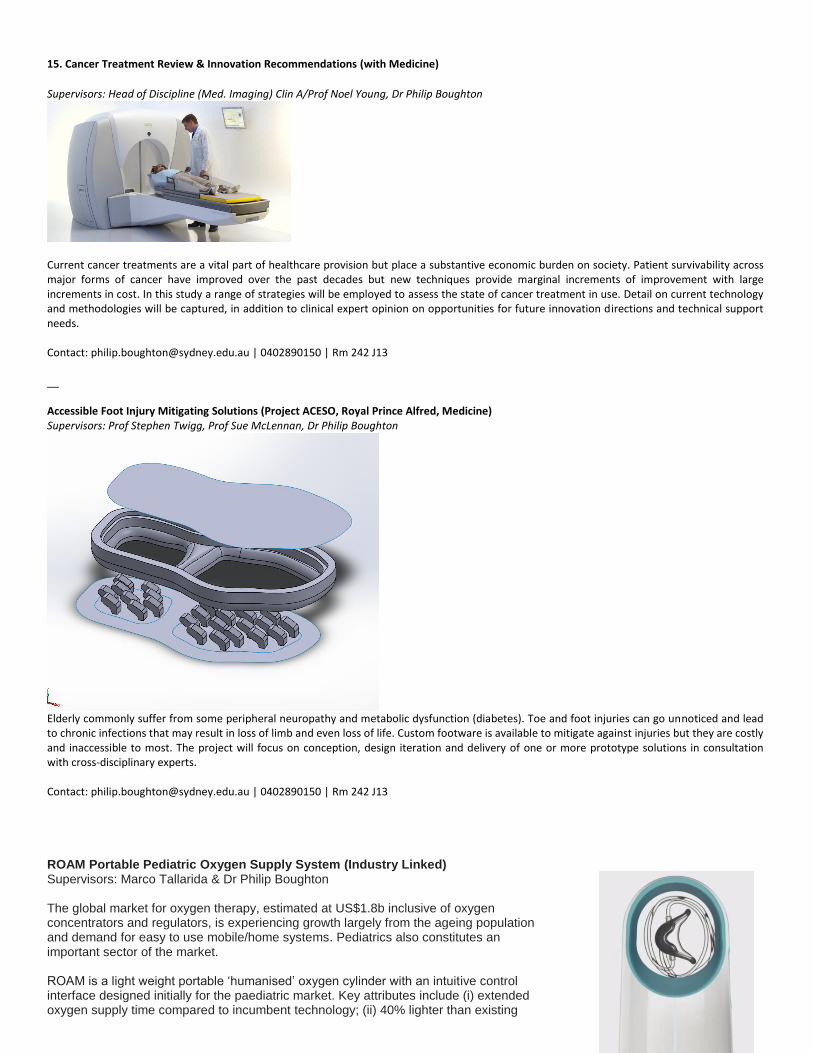
15. Cancer Treatment Review & Innovation Recommendations (with Medicine)
Supervisors: Head of Discipline (Med. Imaging) Clin A/Prof Noel Young, Dr Philip Boughton
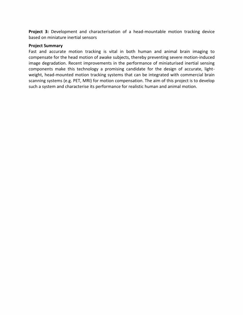
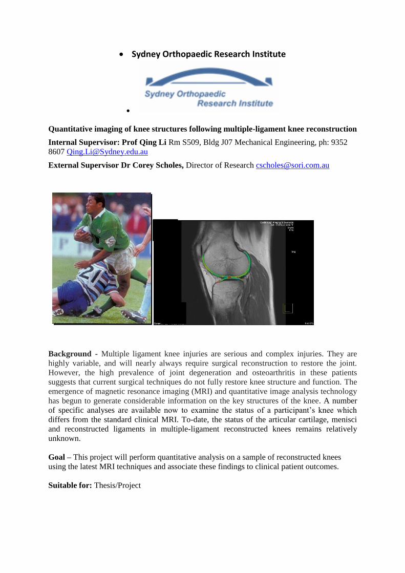
Current cancer treatments are a vital part of healthcare provision but place a substantive economic burden on society. Patient survivability across major forms of cancer have improved over the past decades but new techniques provide marginal increments of improvement with large increments in cost. In this study a range of strategies will be employed to assess the state of cancer treatment in use. Detail on current technology and methodologies will be captured, in addition to clinical expert opinion on opportunities for future innovation directions and technical support needs. Contact: [email protected] | 0402890150 | Rm 242 J13 __ Accessible Foot Injury Mitigating Solutions (Project ACESO, Royal Prince Alfred, Medicine) Supervisors: Prof Stephen Twigg, Prof Sue McLennan, Dr Philip Boughton
Elderly commonly suffer from some peripheral neuropathy and metabolic dysfunction (diabetes). Toe and foot injuries can go unnoticed and lead to chronic infections that may result in loss of limb and even loss of life. Custom footware is available to mitigate against injuries but they are costly and inaccessible to most. The project will focus on conception, design iteration and delivery of one or more prototype solutions in consultation with cross-disciplinary experts. Contact: [email protected] | 0402890150 | Rm 242 J13 ROAM Portable Pediatric Oxygen Supply System (Industry Linked) Supervisors: Marco Tallarida & Dr Philip Boughton The global market for oxygen therapy, estimated at US$1.8b inclusive of oxygen concentrators and regulators, is experiencing growth largely from the ageing population and demand for easy to use mobile/home systems. Pediatrics also constitutes an important sector of the market. ROAM is a light weight portable ‘humanised’ oxygen cylinder with an intuitive control interface designed initially for the paediatric market. Key attributes include (i) extended oxygen supply time compared to incumbent technology; (ii) 40% lighter than existing
metal tanks; (iii) nasal mask specifically designed for paediatric use; and (iv) a design aesthetic of appeal to young patients. This medical device is being developed in line with ISO13485/IEC60601. Design & development projects on offer include: 1. Regulator control and safety systems 2. Hardware – software systems integration with smarhphone control 3. Chassis and composite storage system verification and validation Contact: [email protected] | 0402890150 | Rm 242 J13
Intraoccular Lens Implant System (Sydney Eye Hospital & Save Sight Institute) Supervisors: Prof John Griff, Dr Philip Boughton, Prof Stepanie Watson
Prototype intraoccular lens prototype with clliary tethered haptics. The World Health Organisation estimates there were 161 million visually impaired people worldwide in 2002, cataract accounting for 47.8%. Over the next 20 years, there will be a doubling in the incidence of cataract, visual morbidity, and need for cataract surgery. The Global Intraocular Lens (IOL) Market is forecast to reach $3.1 Billion by 2017;compounded annual growth rate of 4%; due to: increase of cataracts in the aging global population; increase of risk factors such as diabetes and increase of new and available technologies. Current IOL designs are not appropriate for pediatrics, require a significant surgical portal for delivery, can migrate and misalign due to lack of appropriate fixation methods, and have significant chance of post capsule opacification. There may be opportunities to address some of these issues and develop a biomimetic compliant IOL that can be coupled to the ciliary for improved restoration of sight. In conjunction with opthamology specialists, this project seeks to identify priority IOL requirements and design risks to then lead to development of an IOL prototype proof of concept. Contact: [email protected] | 0402890150 | Rm 242 J13
Dr Andre Kyme (AMME Biomedical Engineering) Research Location AMME Brain & Mind Centre
Contact Dr Andre Kyme (AMME) E: [email protected] T: 9351 0612 Project 1: Use of machine learning for uninterrupted head motion tracking in medical imaging
Project Summary We have developed some key technologies that enable the brain of a rodent to be imaged while the animal moves freely inside a positron emission tomography (PET) scanner. This technique has enormous potential to improve our understanding of how brain function and behavior relate to each other in mammals. A vital component of the technique is using motion tracking to accurately estimate the animal’s head motion during a scan. The current method relies on optical markers attached to the animal, but this has a crucial line-of-sight limitation which results in intermittent drop-out of motion tracking. In this project you will investigate the feasibility of applying machine learning methods to a head phantom under highly controlled robotic motion to solve this problem and maintain consistent tracking.
Machine learning is a booming field impacting a diverse range of applications from internet searching to weather prediction to financial modelling. Skills in machine learning are highly sought after by many employers. This project will help you to develop valuable knowledge and experience in this important area. Project 2: A fast and reproducible robot manipulator for use inside an MRI scanner
Project summary Correcting for human head motion during magnetic resonance imaging (MRI) studies is extremely important to avoid distortion and corruption of brain images. This is especially true for studies involving children and patients with dementia-related movement disorders. Although many novel motion compensation methods are being developed for MRI, there is currently no reliable ‘ground truth’ against which these methods can be evaluated and compared. The aim of this project is to investigate suitable actuation approaches and robotic designs to achieve rapid and highly reproducible six degree-of-freedom manipulation of phantoms inside an MRI scanner, and to begin prototyping such a system.
Project 3: Development and characterisation of a head-mountable motion tracking device based on miniature inertial sensors
Project Summary Fast and accurate motion tracking is vital in both human and animal brain imaging to compensate for the head motion of awake subjects, thereby preventing severe motion-induced image degradation. Recent improvements in the performance of miniaturised inertial sensing components make this technology a promising candidate for the design of accurate, light-weight, head-mounted motion tracking systems that can be integrated with commercial brain scanning systems (e.g. PET, MRI) for motion compensation. The aim of this project is to develop such a system and characterise its performance for realistic human and animal motion.
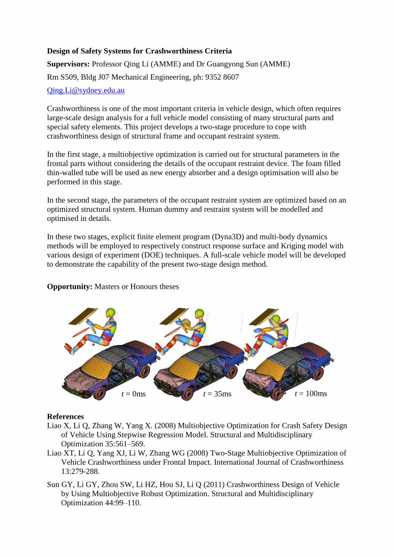
Design and Additive Manufacturing (3D Printing) for Scaffold Tissue Engineering
Supervisor: Professor Qing Li (AMME)
Rm S509, Bldg J07 Mechanical Engineering, ph: 9352 8607
Each year an estimated millions of patients suffer from bone fracture, while hundreds of
thousands of patients have conditions where large segments of bone are destroyed or must be
removed. As such new clinical treatment schemes are necessary to augment the body’s natural
healing process. As a fast emerging interdisciplinary technology, tissue engineering provides
alternative therapeutic strategies for repair of damaged tissue and organs, which shows
enormous potential to generate host-grown tissue in sufficient quantity and quality.
A milestone in the load-bearing tissue (e.g. bone/cartilage) engineering has been the
development of 3D scaffold technique that guides cells to generate desirable functional tissue
under appropriate mechanical and biological conditions. The success of tissue regeneration lies
heavily on the architecture design of the scaffold and its bio-reaction with the seeding cells.
Permeability has been recognised as one critical criterion for scaffold design in ensuring cell
migration and nutrient delivery. This project aims to (1) characterise the effective permeability
of different scaffold architecture; (2) develop finite element based homogenisation technique for
permeable problem; (3) design optimisation for tailored effective permeability. The student is
expected to closely work with the Research Fellow in the group and redevelop Matlab code for
finite element analysis and homogenisation for the permeable problem. The results will be
prototyped in commercial free-form solid fabrication facility.
Opportunity: Masters or Honours theses
Reference
Chen YH, Schellekens M, Zhou SW, Cadman J, Li W, Appleyard R, Li Q (2011) Design of
Tissue Scaffolds Using Wall Shear Stress Criterion for Flow Induced Erosion, ASME
Journal of Biomechanical Engineering - Transactions of the ASME, 133(8):081008.
Chen YH, Zhou SW, Li Q (2011) Microstructure Design of Biodegradable Scaffold and Its
Effect on Tissue Regeneration, Biomaterials 32: 5003-5014.
Chen YH, Zhou SW, Li Q (2011). Mathematical modeling of degradation for bulk-erosive
polymers: Applications in tissue engineering scaffolds and drug delivery systems. Acta
Biomaterialia 7:1140–1149。
Chen YH, Zhou SW, Cadman J, Li Q (2010) Design of Cellular Porous Biomaterials for Wall
Shear Stress Criterion, Biotechnology & Bioengineering 107(4):737-746.
Sturm S, Zhou SW, Mai YW, Li Q (2010) “On Stiffness of Scaffolds for Bone Tissue
Engineering – A Numerical Study”. Journal of Biomechanics 43:1738–1744.
Design of periodic scaffold
Base cell
FEA model
Scaffold
structure
Design of Safety Systems for Crashworthiness Criteria
Supervisors: Professor Qing Li (AMME) and Dr Guangyong Sun (AMME)
Rm S509, Bldg J07 Mechanical Engineering, ph: 9352 8607
Crashworthiness is one of the most important criteria in vehicle design, which often requires
large-scale design analysis for a full vehicle model consisting of many structural parts and
special safety elements. This project develops a two-stage procedure to cope with
crashworthiness design of structural frame and occupant restraint system.
In the first stage, a multiobjective optimization is carried out for structural parameters in the
frontal parts without considering the details of the occupant restraint device. The foam filled
thin-walled tube will be used as new energy absorber and a design optimisation will also be
performed in this stage.
In the second stage, the parameters of the occupant restraint system are optimized based on an
optimized structural system. Human dummy and restraint system will be modelled and
optimised in details.
In these two stages, explicit finite element program (Dyna3D) and multi-body dynamics
methods will be employed to respectively construct response surface and Kriging model with
various design of experiment (DOE) techniques. A full-scale vehicle model will be developed
to demonstrate the capability of the present two-stage design method.
Opportunity: Masters or Honours theses
References
Liao X, Li Q, Zhang W, Yang X. (2008) Multiobjective Optimization for Crash Safety Design
of Vehicle Using Stepwise Regression Model. Structural and Multidisciplinary
Optimization 35:561–569.
Liao XT, Li Q, Yang XJ, Li W, Zhang WG (2008) Two-Stage Multiobjective Optimization of
Vehicle Crashworthiness under Frontal Impact. International Journal of Crashworthiness
13:279-288.
Sun GY, Li GY, Zhou SW, Li HZ, Hou SJ, Li Q (2011) Crashworthiness Design of Vehicle
by Using Multiobjective Robust Optimization. Structural and Multidisciplinary
Optimization 44:99–110.
t = 0ms t = 35ms t = 100ms
Design Optimisation for Road Safety System
Supervisors: Professor Qing Li (AMME) and Dr Guangyong Sun (AMME)
Rm S509, Bldg J07 Mechanical Engineering, ph: 9352 8607
As the main safety facility on the highway, a guardrail system is essential for the vehicle
safety. In this study, the 3D Finite Element (FE) models of the vehicle and the corrugated
beam guardrail system will be created. Two types of widely used corrugated beam semi-rigid
guardrails will be considered, which are the W-beam guardrail and the Thrie-beam guardrail.
The collision between the corrugated beam guardrail systems and the vehicle body will be
analyzed. In the collision process, the snagging effect of the post to the vehicle body is also
taken into account. The multiobjective optimization problem will be used to determine
dimensional sizes of guardrails. Response surface method (RSM) is applied to construct the
surrogate models for the objective and constraint functions. The Pareto set and the optimal
solution will be obtained.
The student is expected to have background in finite element method and will be trained for
highly nonlinear finite element analysis in LS-Dyna. Surrogate models based design will be
applied for seeking multiobjective optimisation.
Opportunity: Masters or Honours theses
Reference
S Hou, W Tan, Y Zheng, X Han, Q Li (2014) Optimization design of corrugated beam
guardrail based on RBF-MQ surrogate model and collision safety consideration,
Advances in Engineering Software 78, 28-40.
Topology Optimisation for Connection Design with Crashworthiness Criteria
Supervisors: Professor Qing Li (AMME) and Dr Guangyong Sun (AMME)
Rm S509, Bldg J07 Mechanical Engineering, ph: 9352 8607
Crashworthiness is one of the most important criteria in vehicle design, which often requires
large-scale design analysis for a full vehicle model consisting of many structural parts and
special safety elements. The major challenge facing to engineering community is how to
design multicomponent system for a range of crashworthiness criteria. Previous researches
had found that different connection patterns will have different crashworthiness performances
during the crush.
In the past, evolutionary structural optimization (ESO) had been applied to find out the
locations with the highest stress concentration at the overlap area of connection and therefore
to determine the proper locations for placing the connection elements. However, it has not
proven that applying the connections on the highest stress concentration area will give the
best performance on energy absorption under crashing scenarios and the connection patterns
where connections are not in stress concentration area are not being concerned.
The genetic algorithm (GA) will be used in this study for determining optimal pattern of
connection elements so that the highest energy absorption capacity can be obtained. This
thesis study will tackle topology optimization problems for connection pattern in
multicomponent system. The nonlinear FEA will be used as a solution engine for modelling,
data collection, and optimisation.
The student is expected to have background in finite element method and will be trained for
highly nonlinear finite element analysis in LS-Dyna. Surrogate models based design will be
applied for seeking multiobjective optimisation.
Initial design at initial stage Optimised design at the final stage
Opportunity: Masters or Honours theses
References
Q Li, GP Steven, YM Xie (2001) Evolutionary structural optimization for connection
topology design of multi-component systems. Engineering Computations 18, 460-479.
Sydney Concord Hospital
Design Analysis and Optimization for Novel Arterial Stents
Supervisors: Professors Qing Li (AMME) and Harry Lowe (Sydney Concord Hospital and
Faculty of Medicine)
Rm S509, Bldg J07 Mechanical Engineering, ph: 9352 8607
Cardiovascular diseases (CVD) are one of the leading causes of death in western countries due
to lifestyle and an increasingly ageing population. In Australia, more than 176,000 patients are
hospitalised and 180,000 cardiovascular procedures are performed each year. Open-heart
bypass surgery is one of the most widely used procedures for CVD, and carries with it the
major problem of long waiting lists due to the large number of hospitalised patient-days (> 1.64
million totally in 2004–05) in the country. As a fast emerging interdisciplinary technology in
interventional cardiology, stenting treatment provides an alternative therapeutic strategy, which
uses a mechanical device (called a stent) to compress the plaque against the artery wall opening
the lumen of the obstructed artery for restoring blood flow. This technology demonstrates the
enormous potential to minimise surgical invasion/risk and shorten the hospital days.
This research aims at developing a computational framework for stent design analysis and
optimization. In this study, a newly designed cardiovascular stent with adapting the aorta stent
geometry will be modelled and evaluated by three-dimensional finite element analysis.
Compared with the existing conventional stents in the market, a series of novel designs will
be assessed to characterise the stiffness, damage to the arterial wall, fatigue life, and other
biomechanical behaviours. The geometry sharpness and increase the manufacturability.
Opportunity: Masters or Honours theses
a) Deliver crimped stent across a narrowing artery
b) Deploy stent & push plaque against arterial wall by inflating balloon.
c) Remove deflated balloon and leave stent on-site to support vessel wall.
Plastic deformation
Cardio-vascular diseases
Recoil??
PUC before deploying
R
Z
Sydney Dental Hospital
Finite Element Modelling of Orthodontic Tooth Movement
Supervisors: Professor Qing Li (AMME) and Professor A. Darendeliler (Sydney Dental
Hospital and Faculty of Dentistry)
Work Experience: Sydney Dental Hospital
Rm S509, Bldg J07 Mechanical Engineering, ph: 9352 8607
The ratio of patients who request orthodontic therapy to the total population is surprisingly
high. Unofficial data reports that every year 60% of all adolescents in Australia undergo
orthodontic treatment to improve their healthy start to life. Orthodontic tooth movement
(OTM) within the dentoalveolar bone is based on the ability of surrounding bone and
periodontal ligament (PDL) to react to a mechanical stimulus (e.g. stress/damage/strain
energy) with remodelling processes. Orthodontic forces generate a complex set of mechanical
stimuli triggering biological reactions in dentoalveolar and PDL, thereby causing teeth to
move to ideal positions in the jaw. Although it is recognised that the change in biomechanical
environment leads to OTM, it is unclear which of the mechanical signals are dominating the
initiation of the bone remodelling and how to quantify a dynamic tooth movement process in
response to the orthodontic force.
This project aims to (1) develop a precise model of the orthodontic treatment based on CT
images (NewTom – Sydney Dental Hospital in USyd Faculty of Dentistry), which may
involve uses of Rhinoceros/ScanFE – Solidworks – Ansys or Strand7); (2) quantify the
biomechanical responses in several different stages of OTM; (3) correlate the mechanical
stimuli to the OTM rate measured in clinic. In this project, the student will use his/her
CAD/FEA skills to an interdisciplinary topic, and he/she is expected to closely work with
USyd dental specialists (Prof Darendeliler) at Department of Orthodontics.
References
Field C, Ichim I, Swain MV, Chan E, Darendeliler MA, Li W, Li Q (2009) “Mechanical
Responses to Orthodontic Loading: A Three-Dimensional Finite Element Multi-Tooth
Model”. American Journal of Orthodontics And Dentofacial Orthopedics 135:174-181.
Chen JN, Li W, Swain MV, Darendeliler MA, Li Q (2014) A Periodontal Ligament Driven
Bone Remodeling Algorithm for Orthodontic Tooth Movement, Journal of Biomechanics
47:1689–1695.
Sydney Dental Hospital
Biomechanical Investigation into Orthodontic Root Resorption
Supervisors: Professor Qing Li (AMME) and Professor A. Darendeliler (Sydney Dental
Hospital and Faculty of Dentistry)
Rm S509, Bldg J07 Mechanical Engineering, ph: 9352 8607
Orthodontic root resorption (ORR) is described as the loss of hard tissue especially of
cementum and dentine of tooth tissue which can be an irreversible sequel of orthodontic
treatment. It has been documented that the phenomenon of root resorption is a very common
disorder, affecting up to 100% of all treated cases, and after treatment, 41% of adult patients
had increased root resorption of over 2.5mm in one or more teeth. Under severe
circumstances root resorption may potentially jeopardise the longevity and functional capacity
of the treated teeth; and may result in ending the treatment and greatly compromising the
outcome of a successful orthodontic therapy.
This project aims to (1) elucidate the biomechanical pattern of orthodontic force distribution
along the tooth root and its surrounding tissues by creating 3D finite element model and to
develop a numerical prediction of ORR (by using micro-CT scanner SkyScan 1172 at
Electronic Microscopic Unit and uses of Rhinoceros/ScanFE – Solidworks – Ansys or
Strand7); (2) correlate the root stress/strain to the change in cementum properties; (3) To
validate the numerical prediction through a clinical trial where the occurrence of orthodontic
root resorption (ORR) is predicted and therefore may be prevented. In this project, the student
will is expected to closely work with USyd dental specialists (Prof Darendeliler) at
Department of Orthodontics.
Opportunity: Masters or Honours theses
Buccal
Root
resorption
Stress
Sydney Dental Hospital
Modelling of Cracking in Dental Ceramic Restorations.
Supervisors: Professor Qing Li (AMME) and Prof Michael Swain (Sydney Dental Hospital
and Faculty of Dentistry)
Rm S509, Bldg J07 Mechanical Engineering, ph: 9352 8607 [email protected]
Ceramics become more and more popular as dental restorative materials. However sintering
and curing of such materials induces considerable residual stress of layered structure,
resulting in tensile stresses at the margin that may induce fracture. The aim of this project is to
utilise ABAQUS software to quantify the residual stresses and predict the conditions for the
onset of failure and follow the extent of fracture.
The student is expected to (1) create 3D FE model of a tooth with caries and in-lay ceramic
filling; (2) model shrinkage of materials in different temperature; (3) simulate the crack
initiation and propagation around the filling region; and (4) optimise the filling shape to
minimize potential fracture failure. The student is also expected to work with PhD students in
dental clinical and experimental studies.
Opportunity: Masters or Honours theses
References:
1. Ichim I, Li Q, Li W, Kieser J, Swain M (2007) “Modelling of Fracture Behaviour in
Biomaterials, A Leading Opinion Article”. Biomaterials, 28:1317-1326.
2. Ichim I, Li Q, Loughran JG, Kieser J and Swain MV (2007) “Restoration of Non-Carious
Cervical Lesions: Part I - Modelling of Restorative Fracture”. Dental Materials 23 (12):
1562-1569.
3. Ichim I, Schmidlin PR, Li Q, Swain MV, Kieser J (2007) “Restoration of Non-Carious
Cervical Lesions: Part II - Restorative Material Selection to Minimise Fracture”. Dental
Materials 23 (12): 1553-1561.
4. Zhang ZP, Guazzato M, Sornsuwan T, Scherrer SS, Rungsiyakull C, Li W, Swain MV,
Li Q (2013) Thermally induced fracture for core-veneered dental ceramic structures, Acta
Biomaterialia 9 (2013) 8394-8402.
Composite
filling
Fracture
School of Advanced Medicine, Macquarie University
Modelling of Femoral Fracture after Hip Replacement Surgery
Supervisors: Professors Qing Li (AMME) and Richard Appleyard (School of Advanced
Medicine, Macquarie University)
Rm S509, Bldg J07 Mechanical Engineering, ph: 9352 8607
Hip replacement surgery enables patients who once suffered from osteoarthritis to walk pain-
free. However, there is a high incidence of failure due to bone fracture, resulting in huge
additional public and private health costs, and a reduced quality of life for ageing Australians.
This project aims to develop the science for a computer-based technology that will enable
surgeons to optimise the match between a patient’s individual needs and a standard implant
device.
The student is expected to work closely with the group in Murray Maxwell Biomechanics Lab
at Sydney Royal North Shore Hospital on both FEA modelling and experimental studies. S/he
will be trained to use Simpleware and ABAQUS for 3D modelling and fracture analysis in the
following steps: (1) CT/MRI scanning of femur and hip replacement implants, segmentation
of the images and modelling in Simpleware; (2) FE modelling of 3D femur and implants
immediately after surgery; (3) modelling of the osseointegration process; (4) fracture
modeling of the system in different time steps.
Reference
Brad Miles, Elizabeth Kolos, William L. Walter, Richard Appleyard, Angela Shi, Qing Li,
Andrew J. Ruys (2015) Subject specific finite element modeling of periprosthetic femoral
fracture using element deactivation to simulate bone failure. Medical Engineering &
Physics 37(6): 567–573.
Experimental studies FE modelling of femur fracture
Design Optimisation for Metamaterials
Supervisor: Professor Qing Li (AMME)
Rm S509, Bldg J07 Mechanical Engineering, ph: 9352 8607, [email protected]
Metamaterials signify a new class of periodic materials and directly gain unusual properties
from their special wavelength-scale microstructures. These properties have great potential to
many electromagnetic applications and technological innovations such as super-lens and
invisible cloaks. This project aims to develop a systematic method by formulating
microstructural design in the topology optimisation framework. The evolutionary structural
optimisation will be used to determine the novel configurations of unit cells for desirable
physical properties.
The student is expected to conduct (1) modelling of unit-cell of periodic metamaterials; (2)
sensitivity analysis; (3) design optimisation for unit-cell configuration. S/he will closely work
with the research fellow in the AMME School and School of Physics.
References
Zhou SW; Li W; Sun GY; Li Q (2010) “A level-set procedure for the design of
electromagnetic metamaterials” Optics Express 18(7): 6693-6702.
Zhou SW, Li W, Li Q (2010) Level-Set Based Topology Optimization for Electromagnetic
Dipole Antenna Design, Journal of Computational Physics 229 (2010) 6915–6930.
Zhou SW, Li W, Chen YH, Sun GY, Li Q (2011) Topology Optimization for Negative
Permeability Metamaterials Using Level Set Algorithm, Acta Materialia 59:2624–2636.
Zhou SW, Li W, Li Q (2010) “Design of 3D Periodic Metamaterials for Electromagnetic
Properties”. IEEE Transactions on Microwave Theory and Techniques 58(4):910-916.
Metamaterials unit-cells
Invisible cloak cloth
Sydney Westmead Dental Hospital
Oral Biomechanics – Building Right Mouth via 3D Modelling and 3D Printing
External supervisors: Professor Chris Peck: [email protected]
Internal supervisor: Qing Li: [email protected] Rm S509, Bldg J07 Mechanical
Engineering, ph: 9352 8607
Work Experience: Internship can be offered in Sydney Westmead Dental Hospital
Background: The relationship of the upper and lower jaws is critically important in just about
every part of clinical dentistry. It is important to determine the optimal jaw relationships when
placing a dental filling, inserting crowns or bridges or removable dentures.
Unfortunately the determination of clinical jaw positions is often an art rather than based on
science. Dentists will debate fiercely the correct jaw positions and the way to obtain it.
One common method to determine the jaw position is to electrically stimulate the jaw closing
muscles to determine the “neuromuscular jaw position”
Proponents of this method suggest it produces a “functional jaw position” because it is
determined by activation, albeit external stimulation, of the jaw muscles.
Opponents to this method suggest it does not produce the optimal jaw position because the
electrical stimulation is directed at the outer part of the jaw closing muscles, and not entire
muscle. These outer fibres, when activated, produce an anteriorly and superiorly directed
force vector which would consequently position the lower jaw in a position anterior to where
it should be.
Aim: In this study, you will aim to better understand the difference in jaw positions created by
normal jaw closing (asking the subject to bring the teeth together) and neuromuscular jaw
closing.
The hypothesis is that the neuromuscular jaw position is anterior to the normal/habitual jaw
position.
Methods: The jaw positions of 200 human subjects have been recorded in position (1) the
normal jaw closing position and in position (2) the neuromuscular position. For each subject,
these positions have been digitised. Using position (1) as the global reference, you will
determine the relative displacement of position (2). You will need to develop a way to
describe the rotational and translational difference to dentists and others who are not familiar
with transformation matrices, vectors or other descriptors of displacement.
Opportunity: Masters or Honours theses
Cochlear Pty Ltd
Mechanical Finite Element Modelling of Intracochlear Electrode Arrays
Cochlear Project 1: XFEM modelling of brazing process induced cracking
External Supervisors: Dr Anthony Powell ([email protected]) (Cochlear Pty Ltd).
Internal Supervisor: Professor Qing Li (AMME) Rm S509, Bldg J07 Mechanical
Engineering, ph: 9351 8607 [email protected]
Background
Like many biomedical devices, cochlear implant comprises a number of different materials
through different manufacturing methods to bond different components and materials
together. One of the methods used is brazing. Since different materials have different
mechanical properties and the mismatch may cause considerable residual stress during the
manufacturing process. High residual stress together with external mechanical loading may
possibly lead to cracking and even failure of the device. This project aims to simulate
cracking inside cochlear implant system.
Aims
To develop finite element model of bi-material system for simulating crack initiation and
propagation under both thermal residual stress induced by the brazing process and additional
mechanical loading during application.
Methods
The project would involve
Development of a XFEM model of the bi-material system with realistic mechanical
properties and thermal and mechanical loading used in-vitro tests.
Some level of validation of the model by correlating cracking initiation site and length
of visual/invisible cracks
Simulation of various cooling processes and the effect on cracking process.
Requirement: Hons thesis project
Cracking under “thermal + mechanical” loadingThermal residual stresses
Cracking initiation
Cochlear Pty Ltd
Mechanical Finite Element Modelling of Intracochlear Electrode Arrays
Cochlear Project 2: Modelling of intracochlear electrode arrays to predict insertion and
removal dynamics
External Supervisors: Dr Anthony Powell ([email protected]) (Cochlear Pty Ltd). Dr
Nick Pawsey ([email protected]) (Cochlear Pty Ltd).
Internal Supervisor: Professor Qing Li (AMME) Rm S509, Bldg J07 Mechanical
Engineering, ph: 9351 8607 [email protected]
Background
Cochlear implants provide the sensation of hearing to moderate to profoundly deaf recipients
with sensorineural hearing loss. An array of electrodes is inserted into the scala tympani of
the cochlea in order to provide electrical stimulation to auditory neurons. Electrode arrays
must be designed to facilitate reliable insertion into the desired position in the cochlea,
minimising the risk of complications such as tip foldover or buckling. In addition, arrays
need to be flexible to minimise contact forces with cochlea structures to prevent damage that
may degrade hearing performance.
Arrays may be straight, or pre-curved to match the spiral of the cochlea. Different designs
have different insertion methods and risks. All must accommodate a range of cochlea sizes.
Aims
To develop a finite element model of electrode arrays, in order to simulate insertion into the
cochlea and predict contact pressures between the electrode and the cochlea, both during the
insertion and in the electrode’s final position.
Methods
The project would involve
Continuing the development of an
existing finite element model to achieve
successful simulation of electrode
insertion and prediction of contact
pressures.
Preliminary experimental validation of
the model against insertion force measurements.
If above is completed, begin on a sensitivity study of effect of cochlea duct geometry
on contact pressure on basilar membrane.
Requirement: Hons thesis project
Cochlear Pty Ltd
Mechanical Finite Element Modelling of Intracochlear Electrode Arrays
Cochlear Project 3: Modelling of intracochlear electrode arrays to predict mechanical
properties and robustness
External Supervisors: Dr Nick Pawsey ([email protected]) (Cochlear Pty Ltd). Dr
Anthony Powell ([email protected]) (Cochlear Pty Ltd).
Internal Supervisor: Professor Qing Li (AMME) Rm S509, Bldg J07 Mechanical
Engineering, ph: 9351 8607 [email protected]
Background
Cochlear implants provide the sensation of hearing to moderate to profoundly deaf recipients
with sensorineural hearing loss. An array of electrodes is inserted into the scala tympani of
the cochlea in order to provide electrical stimulation to auditory neurons. Arrays need to be
flexible to conform to the spiral shape of the cochlea, accommodate a range of individual
cochlea sizes, and to minimise contact forces with cochlea structures to prevent damage that
may degrade hearing performance. The fine wires within the array need to accommodate
large deformations during the insertion or removal. The mechanical properties, dimensions
and position of the wires within the silicone carrier are critical to the robustness and flexibility
of the array.
Aims
To develop a mechanical finite element model of an intracochlear electrode array, in order to
predict overall flexibility of the array, as well as the stresses experienced by its components.
This model could be used to investigate the sensitivity of these to various design parameters,
such as wire sizes, mechanical properties, wire placement and silicone grades.
Methods
The project would involve
Development of a detailed 3D CAD model of an electrode array
Use of a commercial FEA software package to develop a model of the electrode using
realistic material properties
Recommendations for design guidelines for electrode flexibility and reliability
Requirement: Hons thesis project
http://www.optimizedortho.com/
Work experience: will be offered in Optimized Ortho Company
Supervision team:
External supervisor: Dr Brad Miles ([email protected]), Michael Topham
([email protected]), Jim Pierrepont ([email protected] )
Internal supervisor: Prof Qing Li (AMME), email: [email protected]
1. Development of an Automated 3D Implant Positioning Tool for Total Hip
Replacement Planning
The student will use Simpleware ScanIP +CAD to develop an automatic method of
positioning hip implants within the femur and acetabulum using patient-specific landmarks.
This topic will require the student to learn programming languages, in particular Python and
MS VBA.
2. Validation of a Patient-Specific Neck Osteotomy Guide for the Direct Anterior
and Anterolateral Approaches
A patient-specific neck osteotomy guide has been developed by Optimized Ortho for posterior
approaches in Total Hip Arthroplasty. The guide is designed to assist the surgeon intra-
operatively and increase the likelihood of achieving a desirable leg length and offset for each
patient. The student will use Materalise Mimics Research software suite to validate the
osteotomy level of Optimized Ortho’s direct anterior and anterolateral femoral cutting guides.
3. Development and Validation of an Analytical Model for Determining Optimal
Combined Alignment
The effect of combined alignment of the femoral and acetabular components in Total Hip
Arthroplasty on the Range of Motion of the patient is not well understood. The student will be
tasked with developing an existing analytical model created by Hisatome (2011). in Matlab.
The final model will predictively measure the impingement and therefore Range of Motion of
a patient by demonstrating the maximum functional movements a patient can perform before
prosthetic impingement occurs. The analytical model will be validated in Solidwork. The
student should be experienced in programming, no particular language is preferred.
4. Development and Validation of a 2D Registration Technique for Intra-Operative
Femoral Stem Anteversion Using a Smartphone Camera
Stem anteversion is an important clinical factor when considering impingement within a hip
prosthesis. A 2D registration technique will allow for intra-operative feedback on the stem
anteversion to the surgeon. The student will develop a technique to capture a 2D image of the
stem/femur during the operation and register the image to a virtual pre-operative plan.
5. Determining the Patient-Specific Changes in Functional Combined Anteversion
The combined orientation of both femoral and acetabular components in Total Hip Replacements is not well understood. Throughout functional movements patients experience a
change in orientation of both these components, affecting their Range of Motion and chance
of dislocation. The student would be tasked with exploring the effects of combined acetabular
and femoral anteversion of patient outcome.
6. Use of Predictive Analytics to Determine Postoperative Changes in Functional
Pelvic Tilt
Following Total Hip Arthroplasty, the pelvic tilt of a patient frequently changes, resulting in a
varied functional orientation of the acetabular and femoral components. The student will
investigate the differences between pre- and post-operative pelvic tilts to create a predictive
model.
7. Can a Patient-Specific Guide be used to Control Femoral Stem Anteversion
Stem anteversion is an important clinical factor when considering impingement within a hip
prosthesis. Controlling stem anteversion using a patient-specific guide will be crucial in the
long term outcome of the patient’s prosthesis. The student will need a good knowledge of
solidworks in order to develop the model to be 3D printed/manufactured.
360 Knee Systems Pty Ltd
https://www.linkedin.com/company/360-knee-systems?trk=top_nav_home
Work experience: will be offered in 360 Knee Systems Pty Ltd
Supervision team:
External supervisor: Dr Brad Miles ([email protected]), Willy Theodore
Internal supervisor: Prof Qing Li (AMME), email: [email protected]
1. Investigation of post-operative kinematics with tibia component rotation
variation
There are numerous references used to define tibia component rotation and there is yet a
consensus which definition showed the strongest relationship with clinical assessment. This
topic will require the student to understand various definitions of tibia rotation as well as run
simulations to compare kinematics observed with various tibia component rotation placement.
2. Investigation the relationship of patella component placement achieved to post-
operative Patient Reported Outcome Measures (PROMS)
Total Knee Arthroplasty (TKA) is considered to be one of the most successful arthroplasty
surgery. However, there are still debate whether resurfaced patella can achieve better post-
operative outcomes than non-resurfaced patella. Additionally, there is no consistent
anatomical references used in placing patella button in a resurfaced patella. The student will
process post-operative CT scans and developed an analytical relationship between the
measured patella button positions relative to various anatomical references and post-operative
PROMS.
3. Development and validation of a 2D-3D registration technique for pre-operative
knee X-ray in functional positions
Understanding the pre-operative state of a Total Knee Arthroplasty (TKA) patient is
important for surgical planning. One method to assess it is through pre-operative X-ray in
functional positions. The X-ray need to be objectively assessed, i.e. measure the limits of the
knee in the positions when the X-ray is taken. This topic will require the student to develop a
2D-3D registration technique (using 3D geometry of the patient and register it against a 2D
image, e.g. X-ray) to measure the position of the distal femur relative to the tibia and develop
a process to validate the registration technique.
Sydney Eye Hospital
Novel Design and Fabrication of Eye Drop Delivery Devices
Supervisors: Prof Qing Li (AMME), Dr Kenneth Ooi [email protected], and Professor
Stephanie Watson ([email protected]) Sydney Eye Hospital, University of
Sydney,
Rm S509, Bldg J07 Mechanical Engineering, ph: 9352 8607 [email protected]
Introduction:
Dry eye, the most common eye disorder, is frequently accompanied by blepharitis
(eyelid inflammation) which has an overall prevalence of nearly 40%. Patients with dry eye
and blepharitis have their daily activities disrupted and work productivity lowered due to
recurrent blurred vision and ocular discomfort. Moderate dry eye damages the ocular surface
and can lead to blindness following infection. The consequences of these common conditions
occur despite maximal use of lubricating drops and ointments which address only the
symptoms and not the underlying cause. We have developed the first patented statin-based
eye drop that safely and successfully treats the causes and symptoms of dry eye and
blepharitis.
Aims:
We aim to develop new intellectual property by developing a novel delivery device that
will combine both statin eye drop and gel or ointment administration to increase patient
convenience of use. Medicine that is problematic to administer leads to poor compliance.
Currently, different forms of ocular medications (i.e. eye drops, gels, ointments) come in
separate delivery devices. Patients typically carry more than one form of medication at a time.
This is because eye drops do not tend to blur the vision and may be preferred if the patient is
reading or driving, whereas gels and ointments last longer, providing greater lubrication, but
blur the vision. Current ocular therapeutics in drop/gel/ointment forms are in separate delivery
devices, which is inconvenient. Further, current delivery mechanisms can be difficult to use,
particularly for elderly patients with arthritis. At least 50% of patients have reported difficulty
with self-administration, frequently saying they have trouble squeezing the bottle. The goal of
this work is to manufacture a combination topical Atorvastatin eye drop and gel/ointment
device that allows tailored drop and gel/ointment use according to vision, cosmetic, and
duration of action needs.
Methods:
Delivery device prototypes have already been conceptualised and will be designed
according to viscous and surface tension properties of the topical Atorvastatin formulations
which will determine optimal tip length and opening(s), dose dispensing time, size and weight
to reduce overflow, drainage and incidence of any systemic side effects. Force requirements
will be factored according to known force generating capacities of the 3 most-used handgrips
and applicator plastic rigidity. Computer-simulated models will be run according to the above,
and also account for dispensing angles. 3-D printed prototypes will then be built according to
established protocols.
Expected outcomes:
A more convenient and novel combination topical Atorvastatin delivery system. Incorporating
modern ergonomics, it will be easier to use than existing delivery devices and with improved
aesthetics it will assist with product marketing.
Sydney Eye Hospital
Design of Biomedical Device for Topical Ophthalmic Use
Supervisors: Dr. Kenneth Ooi, Ophthalmologist and Clinical Senior Lecturer, Save Sight Institute
Dr. Aleksey Valyaev, Commercial Development Officer, CDIP
Prof. Stephanie Watson, Clinical Professor of Ophthalmology, Save Sight Institute
Prof. Qing Li, Professor of Biomechanical Engineering, School of Aerospace, Mechanical
and Mechatronic Engineering
Expressions of interest are sought for the continued development of a novel topical
ophthalmic delivery device. A working prototype has already been manufactured and the
student would be involved in the refinement of the design. The student is expected to be of
interest and strengths in design analysis and fabrication with 3D printing. S/he is also
expected to be self-motivated in driving the project and communicating effectively with the
supervision team effectively. The project is biomedical industry based and there is the
opportunity for an industrial placement at the Sydney Eye Hospital. Further details will be
provided under a confidentiality disclosure agreement to interested candidates.
Inquiries can be made to Prof Qing Li +61 93518607, Email: [email protected]
Opportunity: Masters or Honours theses
Sydney Eye Hospital
Biomechatronic Development of Novel Topical Ophthalmic Devices
Supervisors:
Dr. Kenneth Ooi, Ophthalmologist and Clinical Senior Lecturer, Save Sight Institute
Dr. Aleksey Valyaev, Commercial Development Officer, CDIP
Prof. Stephanie Watson, Clinical Professor of Ophthalmology, Save Sight Institute
Prof. Qing Li, Professor of Biomechanical Engineering, School of Aerospace, Mechanical
and Mechatronic Engineering
Expressions of interest are sought for the development of a novel topical ophthalmic delivery
device. A working prototype has already been manufactured and the student would be
involved in the refinement of the design towards a new application. The candidate is expected
from a mechatronic background with interests in biomedical engineering applications. S/he
should have some knowledge and skills in circuit board and mechanism design for bioMEMS.
The candidate is also expected to be self-motivated in driving the project and communicating
with the supervision team effectively. The project is based on biomedical industry and there is
an opportunity for an industrial placement at the Sydney Eye Hospital. Further details will be
provided under a confidentiality disclosure agreement to interested candidates.
Inquiries can be made to Prof Qing Li +61 93518607, Email: [email protected]
Opportunity: Masters or Honours theses
Sydney Orthopaedic Research Institute
Sydney Orthopaedic Research Institute is a not-for-profit organisation dedicated to the study
and research of orthopaedic disorders, in particular those associated with the knee joint. The
Institute uses high quality research methods to investigate the causes and development of
common knee disorders, as well as treatments and rehabilitative procedures employed for
these disorders. In addition, the Institute also conducts research on arthritis, cartilage
pathologies, ligament injuries, knee trauma, and methods for clinical assessment of these
conditions.
External Supervisor Details
Dr Corey Scholes, PhD
Director of Research
02 9904 7182
Suite 12, Level 1, 445 Victoria Avenue, Chatswood, 2067
Prerequisites
Open to all disciplines
Curiosity in how the human body works
Interest in patient-centred research
Pride and confidence in their work
Willingness to learn new concepts and skills
Modelling, Matlab or general computer programming skills an advantage
Sydney Orthopaedic Research Institute
Quantitative imaging of knee structures following multiple-ligament knee reconstruction
Internal Supervisor: Prof Qing Li Rm S509, Bldg J07 Mechanical Engineering, ph: 9352
8607 [email protected]
External Supervisor Dr Corey Scholes, Director of Research [email protected]
Background - Multiple ligament knee injuries are serious and complex injuries. They are
highly variable, and will nearly always require surgical reconstruction to restore the joint.
However, the high prevalence of joint degeneration and osteoarthritis in these patients
suggests that current surgical techniques do not fully restore knee structure and function. The
emergence of magnetic resonance imaging (MRI) and quantitative image analysis technology
has begun to generate considerable information on the key structures of the knee. A number
of specific analyses are available now to examine the status of a participant’s knee which
differs from the standard clinical MRI. To-date, the status of the articular cartilage, menisci
and reconstructed ligaments in multiple-ligament reconstructed knees remains relatively
unknown.
Goal – This project will perform quantitative analysis on a sample of reconstructed knees
using the latest MRI techniques and associate these findings to clinical patient outcomes.
Suitable for: Thesis/Project
Sydney Orthopaedic Research Institute
Functional correlates of patient outcome during level walking following multiple-
ligament knee reconstruction
Internal Supervisor: Prof Qing Li Rm S509, Bldg J07 Mechanical Engineering, ph: 9352
8607 [email protected]
External Supervisor Dr Corey Scholes, Director of Research [email protected]
Background - The relationship between joint disease, joint forces and muscle control is well
illustrated. Abnormal forces acting in one part of the knee are related to degeneration of key
structures and the severity of symptoms, such as pain and stiffness. This can be caused by
traumatic injuries which cause a rupture of one or more knee ligaments. Patients alter muscle
control, at the affected joint and across the body, to relieve pain during functional activities
such as walking. However, functional recovery after surgery is dependent on the pattern of
muscle activity used by the patient during movement. The timing and magnitude of muscle
forces regulates the forces acting on the knee and an optimal balance of forces is crucial to
maintain long-term joint health.
Goal – This project will use the latest techniques in biomechanics to analyse joint function in
a clinical population to identify at-risk individuals for future knee degeneration.
Suitable for: Thesis/Project
Sydney Orthopaedic Research Institute
FE analysis of the tibiofemoral joint following multiple-ligament knee reconstruction
during locomotion
Internal Supervisor: Prof Qing Li Rm S509, Bldg J07 Mechanical Engineering, ph: 9352
8607 [email protected]
External Supervisor Dr Corey Scholes, Director of Research [email protected]
Background - The relationship between joint disease, joint forces and muscle control is well
illustrated. Importantly, the pattern of loading is known to vary within and between
individuals. This is particularly apparent in patients suffering multiple-ligament knee injuries.
However, it remains unclear if reconstruction is able to restore normal loading, with emphasis
on the articular cartilage and menisci. These structures are vulnerable to overload during
locomotion and their dysfunction is thought to initiate joint degeneration leading
osteoarthritis. FEA has provided considerable insight into other clinical problems such as
joint replacement, but also has the potential to determine the efficacy of current surgical
reconstruction techniques in this context.
Goal – This project will use the latest techniques in FEA to analyse articular cartilage and
menisci loading in knee reconstructions to develop a method of identifying individuals at risk
of future knee degeneration.
Suitable for: Thesis/Project
Sydney Orthopaedic Research Institute
Three-dimensional reconstruction of menisci during weight-bearing knee flexion
Internal Supervisor: Prof Qing Li Rm S509, Bldg J07 Mechanical Engineering, ph: 9352
8607 [email protected]
External Supervisor Dr Corey Scholes, Director of Research [email protected]
Background - Menisci are important cartilaginous structures within the knee that absorb
load, guide joint kinematics and stabilise the joint. Due to their function, the menisci are
vulnerable to traumatic tears and degeneration. Innovations in surgical repair have increased
the ability of surgeons to preserve the structure despite considerable damage, which
previously would have required removal to restore overall joint function. However, there is a
lack of objective evidence linking meniscal repair with reduced incidence of osteoarthritis.
Furthermore, there remains a lack of information regarding the ability of repaired menisci to
replicate the function of uninjured structures. This project will use the latest modelling
techniques to compare the loading response of uninjured and surgically repaired menisci.
Goal – This project will utilise Matlab and other image-processing platforms to generate 3-
dimensional surface models of the menisci and track their deformation and translation during
knee flexion.
Suitable for: Thesis/Project
Sydney Orthopaedic Research Institute
The effect of acute knee injury and surgical reconstruction on tibiofemoral kinematics
during locomotion
Internal Supervisor: Prof Qing Li Rm S509, Bldg J07 Mechanical Engineering, ph: 9352
8607 [email protected]
External Supervisor Dr Corey Scholes, Director of Research [email protected]
Background – Rupture of the anterior cruciate ligament is a common knee injury, which has
considerable impact on joint function. A key role of the ACL is to provide sensory
information such as limb position and movement velocity as well providing mechanical
restraint. However, there is limited information in the literature regarding the sensory
mechanisms that are affected by ACL rupture and whether individuals are able to compensate
following injury. Importantly, it is not known whether surgical reconstruction helps to restore
some sensory input during functional movements such as locomotion. Biomechanical analysis
of the knee during locomotion has revealed some information in this regard; however there
remains considerable potential for further research to examine this issue using emerging
analytical techniques.
Goal – This project will examine the effects of locomotion speed after ACL reconstruction
surgery on knee motion using the latest analytical techniques.
Suitable for: Thesis/Project
Passive mechanical properties of muscle in Parkinson’s disease and rigidity
External Supervisor: Dr Joanna Diong, School of Medical Sciences, University of Sydney,
External Supervisor: Associate Professor Kay Double.
Internal Supervisor: Prof Qing Li Rm S509, Bldg J07 Mechanical Engineering, ph: 9352
8607 [email protected]
People with Parkinson’s disease often experience ankle rigidity or stiffness during walking.
Rigidity in Parkinson’s disease makes normal movement difficult and prolonged rigidity may
lead to loss of joint flexibility over time by changing the length and stiffness of muscles. The
effects of rigidity on muscle length and stiffness in Parkinson’s disease are not well
understood.
This study aims to determine how passive (relaxed) muscle length and stiffness are different
in people who have Parkinson’s disease and rigidity compared to healthy people. Specifically,
passive ankle joint stiffness will be measured at different knee angles in people with
Parkinson’s disease who have ankle rigidity and healthy people. A biomechanical model will
be used to calculate passive gastrocnemius muscle length and stiffness, and these data will be
compared between groups.
This study will be the first clinical application of this biomechanical method in people with
Parkinson’s disease to identify how rigidity changes muscle length and stiffness. Findings
from this study may guide the clinical management of rigidity in rehabilitation for
Parkinson’s disease.
Opportunity
Masters or Honours theses
Quantification of the forces exerted during operative
vaginal delivery
Supervisors: Graham Brooker, ACFR [email protected]
Dr Jon Hyett, RPAH [email protected]
Sarah McDonald, RPAH [email protected]
A significant (10-15%) proportion of babies require assistance during the process of
vaginal delivery. This involves application of either forceps or ventouse (vacuum),
allowing the acoucher (the person who assists at a birth) to guide the presenting part
through the birth canal, onto the perineum and through to delivery.
Women and babies who have an operative vaginal delivery have higher risks of
traumatic injury related to the delivery process. The forces involved in delivery are
poorly understood. We hypothesize that injury would be more likely in circumstances
where higher forces are developed during the delivery process. Real time
measurement of these forces would provide the acoucher with information about the
risks associated with delivery
Research Proposal This project involves the development of an instrument to quantify the forces exerted
during operative vaginal delivery. The point of contact between the instrument and
the acoucher (i.e. handle of instrument to gloves) is sterile, and this device needs to
be able to be applied in this environment. The device should be able to quantify the
amount of force and record the direction of force during the delivery process. The
device will first be used in simulations of the birthing process, as proof of concept,
prior to being applied to clinical studies.
Required Skills
Some biology (or an interest in the subject)
Electronics & sensing
Mechanical design (SolidWorks)
Signal processing
Birth Simulator
Continuing Project
Supervisors: Graham Brooker, ACFR [email protected]
Sarah McDonald, RPAH [email protected]
The use of simulation in medicine is becoming increasingly used to educate and train
clinicians in a range of skills otherwise difficult to gain access to. Overall simulators are
becoming more realistic however obstetrics birthing simulators are still unable to provide a
simulation capable of training clinicians in some of the more fine motor skills and techniques
of delivery. This project originally focused on training clinicians in manual rotation. Aicha
started the project by developing a Pneumatic Artificial Muscle (PAM) based simulator that
actuated a foetal head. Ashan developed a tablet based interface and Sarah continued the
project by developing the soft tissue interfaces and alternate sensing methods.
A greater need for a more accurate birthing simulator has now been identified and a
completely revised system is required. The new system is to model the contraction of the
uterus and corresponding forces on the uterus, the fetus and the birth canal.
Research Proposal
Based on a human mannequin frame, develop a method to simulate the contraction
of the uterus in such a way that will facilitate the expulsion of a model fetus through
the birth canal and vagina.
Develop a suitable model for the birth canal and the vagina that will provide an
accurate interface for obstetricians and midwives practising manual rotation of the
fetal head.
Instrument the fetal head to identify forces imposed by the birth canal and also by
fingers or forceps during manual intervention.
Required Skills
Some biology
Control
SolidWorks
Mechanical and electronics skills
Forces on sutures after cervical cerclage
Supervisors: Graham Brooker, ACFR [email protected]
Dr Jon Hyett, RPAH [email protected]
Sarah McDonald, RPAH [email protected]
If a woman’s cervix is weak it may open too early and lead to premature labour.
Treatment for this is often a surgical procedure called cervical cerclage in which the
cervix is sewn closed during pregnancy. As the stitches are internal it is difficult to
know the ongoing success of the procedure without examinations. A method of
being able to measure the forces exerted on the sutures would allow for more
prompt detection of a failing or failed cervical cerclage and any resulting premature
dilation. With such detection clinicians would be more capable of acting to reduce
preterm delivery or detrimental outcome.
Research Proposal
Develop a suture material that is sensitive to longitudinal force
Develop an interface to the sutures that will allow them to interface to an external monitor.
The interface will probably be based on magnetic coupled RFID technology
Required Skills
Some biology (or interest in area)
SolidWorks
Interest in sensors
Mechanical and electronics skills
Medical scan to 3D print
Supervisors: Graham Brooker, ACFR [email protected]
Sarah McDonald, RPAH [email protected]
Maternal and fetal scans are used as key diagnostic tools during pregnancy. While
scanning imagery and reconstruction tools have improved and provide a strong tool
to assist clinicians in identifying and understanding problems that present there still
exists a tangible gap. The ability to easily build a 3D tangible model within clinics
presents a range of benefits for both research and clinical practice. Ultimately
clinicians would be better able to visualise and explain any issues that present to
patients and family in a way that may assist these patients further understanding why
recommendations are made. Patients would then be in a better position to make an
educated decision on whether they choose to accept the recommendations.
Clinicians will be in a better position to best help patients regardless of their decision.
Research Proposal
Identify materials and resolutions to best produce 3D replicas and corresponding printer
Design an intuitive interface that can be operated by a nontechnical operator
Develop a method of converting scan files into an object map ready for print
Use the developed interface to action a print of a preselected area of the scan
Required Skills
Some biology (or interest in area)
SolidWorks/ 3D printing
Mechanical and software skills