17
1 Dental X-ray Machine 118 Radiology
| Date post: | 16-Dec-2015 |
| Category: |
Documents |
| Upload: | holden-deemer |
| View: | 221 times |
| Download: | 0 times |

1
Dental X-ray Machine
118 Radiology

2
• The Dental x-ray machine consists of 3 visible component parts:– The Control Panel
– The Extension Arm
– The Tubehead
Component Parts

3
Control Panels
Older style Newer Design

4
• The Control Panel contains:– On-off switch and an indicator light– The exposure button and an indicator light– Control devices to regulate the x-ray beam
• Time setting• kVp setting• mA setting
The Control Panel

5
Miliamperage and Kilovoltage • Miliamperage = MA• controls number of electrons produced at tungsten filament quantity• 7-15 are average dental x-ray unit mA numbers
• Kilovoltage = kV• controls penetrating power of electrons and x-ray quality• how fast they travel from cathode to anode• creates contrast on resulting film image• usually 70 or 90 kVp kilovoltage power

6
Extension Arm

7
Extension Arm
• Hollow, holds electrical wires connecting tubehead with control panel
• Used to position tubehead
• Tubehead attached to arm by yoke, which can be moved 360 degrees horizontally
• Folds up when not in use

8
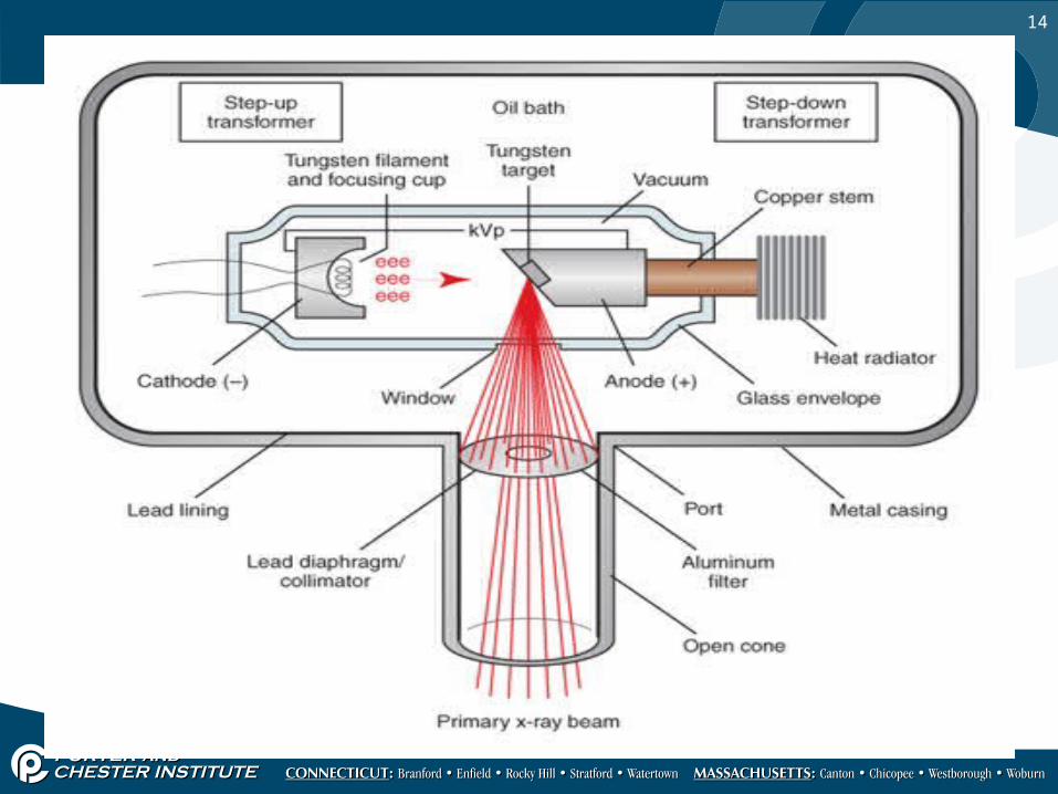
Tubehead
• Houses the x-ray tube
• Made of metal; lined with lead to prevent radiation leakage
• Filled with oil to absorb heat which is generated during x-ray production

9

10
X-ray Tube
• Made of glass• 6” long x 1 ½” diameter• Air is removed to create
vacuum to allow electrons to flow freely between cathode and anode

11

12
Cathode
• Tungsten filament gets hot when electricity is sent to tube
• Creates electron cloud
• Focusing cup: Keeps electrons suspended at cathode
• When exposure button is pushed, electrons shoot across tube to anode
Negative end of tube

13
Anode
Postive end of tube• Tunsten target• Electrons strike target and
product x-rays• 99% get absorbed by oil• 1% exits the tube at the area
towards the patient• Center of this beam of x-
rays is called the Central Ray
14

15
PID
• Position indicating device• Lead lined• Aims x-ray beam at film in
pt mouth OR at extra oral cassette
• Open end placed against pt face
• 8,12,16 inches long

16
Filter• Aluminum disc at port
where the PID is connected to the Tubehead
• Removes (absorbs) low-energy, long wavelength x-rays
• Only allows high energy, short wave length x-rays to pas through
• Both kinds of x-rays are absorbed by the pt’s tissues; only the short wave length , high energy x-rays create image on film

17
Collimator
• Metal disc (lead) with small opening in center to control size and shape of x-ray beam
• Further reduces pt exposure• 2 ¾” diameter; round shape
and size• Also available in
rectangular shape which exactly fits #2 film size exposes over ½ less tissue with rectangular collimator
![DENTAL X-RAY · [ 1 ] INTRODUCTION *(1(5$/ This manual provides information for the operation and maintenance prodedures and technical specifications for PHOT-X IIs 505 dental x-ray.](https://static.documents.pub/doc/80x56/5e8c4a93acf46e57d6589a48/dental-x-1-introduction-15-this-manual-provides-information-for-the-operation.jpg)


