Page 1
1
Hepatocyte Nuclear Factor-4 alpha: a key factor for homeostasis, cell architecture and 1
barrier function of the adult intestinal epithelium 2
ANNE-LAURE CATTIN♦, §, #
, JOHANNE LE BEYEC♦, §, #
, FREDERICK BARREAU‼, 3
SUSAN SAINT-JUST♦, §, #, ‡
, ANNE HOULLIER♦, §, #
, FRANK J. GONZALEZ†, SYLVIE 4
ROBINE¶, MARTINE PINÇON-RAYMOND
♦, §, #, PHILIPPE CARDOT
♦, §, #, MICHEL 5
LACASA♦, §, #
, AGNES RIBEIRO*, ♦, §, #
6
♦Centre de Recherche des Cordeliers, Université Pierre et Marie Curie – Paris 6, UMRS 872, 7
Paris, F-75006 France 8
§INSERM, U872, Paris, F-75006 France 9
#Université Paris Descartes, UMRS 872, Paris, F-75006 France 10
‡Laboratoire de Pharmacologie Cellulaire et Moléculaire, Ecole Pratique des Hautes Etudes, 11
Paris, F-75006 France 12
¶Morphogenesis and Intracellular Signaling, UMR 144, Institut Curie-CNRS, Paris, France 13
†Laboratory of Metabolism, Center for Cancer Research, National Cancer Institute, National 14
Institutes of Health, Bethesda, Maryland USA 15
‼UMR-S843 INSERM, Université Paris Diderot, Service de Gastroentérologie, Hôpital R. 16
Debré, AP-HP, Paris, France 17
18
Running title: Pivotal role of HNF-4α in adult intestine 19
20
*Corresponding author: Agnès Ribeiro, UMRS 872, 15 rue de l’école de médecine, 75006 21
Paris, FRANCE – e-mail : [email protected] – fax : 33 1 43 25 16 15 – tel : 22
33 1 42 34 69 18 23
Copyright © 2009, American Society for Microbiology and/or the Listed Authors/Institutions. All Rights Reserved.Mol. Cell. Biol. doi:10.1128/MCB.00939-09 MCB Accepts, published online ahead of print on 5 October 2009
on February 15, 2018 by guest
http://mcb.asm
.org/D
ownloaded from
Page 2
2
Abstract 1
HNF-4α is a transcription factor, which is highly expressed in the intestinal epithelium 2
from duodenum to colon and from crypt to villus. The homeostasis of this constantly 3
renewing epithelium relies on an integrated control of proliferation, differentiation and 4
apoptosis as well as on the functional architecture of the epithelial cells. In order to determine 5
the consequences of the HNF-4α loss in the adult intestinal epithelium, we used a tamoxifen-6
inducible Cre-loxP system to inactivate the Hnf-4a gene. In the intestine of adult mice the loss 7
of HNF-4α led to an increased proliferation in crypts and to an increased expression of several 8
genes controlled by the Wnt/β-catenin system. This control of the Wnt/β-catenin signaling 9
pathway by HNF-4α was confirmed in vitro. Cell lineage was affected, as indicated by an 10
increased number of goblet cells and an impairment of enterocyte and enterendocrine cell 11
maturation. In the absence of HNF-4α, cell/cell junctions were destabilized and the 12
paracellular intestinal permeability increased. Our results showed that HNF-4α modulates 13
Wnt/β-catenin signaling and controls intestinal epithelium homeostasis, cell function and cell 14
architecture. This study indicates that HNF-4α regulates the intestinal balance between 15
proliferation and differentiation and we hypothesize that it might act as a tumor suppressor. 16
on February 15, 2018 by guest
http://mcb.asm
.org/D
ownloaded from
Page 3
3
INTRODUCTION 1
HNF-4 belongs to the superfamily of nuclear receptors (8). In mammals, it is 2
represented by two paralogs: HNF-4α, which is expressed in liver, kidney, pancreas and 3
intestine and HNF-4γ, which is expressed in the same organs except the liver (14, 42, 50). 4
HNF-4γ has not been extensively studied. However, mice lacking expression of the Hnf-4g 5
gene do not present an overt phenotype (19). HNF-4α has been essentially studied in liver and 6
hepatocyte cell lines. Mutations in the human Hnf-4a gene are associated with Maturity Onset 7
Diabetes of the Young (MODY)1, a disorder that is characterised by the early onset of type 2 8
diabetes (20). Disruption of the Hnf-4a gene expression leads to embryonic death at the 9
gastrulation stage (E6.5) (15). HNF-4α is dispensable for hepatic specification but necessary 10
for hepatoblast differentiation into hepatocytes (29). Inactivation of the Hnf-4a gene in fetal 11
liver revealed that HNF-4α also controls epithelial morphology (41) and transcriptome 12
analysis of Hnf-4a knocked-out in fetal liver showed that genes encoding proteins of all 13
categories of cell junctions (adherens, desmosomes, tight) are targets of HNF-4α (5). 14
Expression of HNF-4α in dedifferentiated hepatocytes provokes the re-expression of 15
epithelial markers, such as E-cadherin (48). Its ectopic expression in mesenchymal cells 16
induces the expression and the proper localization of tight and adherens junction proteins (10, 17
41), as well as the formation of microvilli (12), suggesting that HNF-4α is a central regulator 18
of epithelial morphogenesis. In adult mouse liver, conditional disruption of the Hnf-4a gene 19
revealed a key role for HNF-4α in the control of genes involved in amino acid, glucose and 20
lipid metabolisms (21). All together these studies show that HNF-4α is at the crossroads 21
between liver morphogenesis and function (43). By contrast, the role of HNF-4α in intestine 22
is less well understood. 23
The intestinal epithelium establishes a physical barrier between the external and 24
internal compartments and plays a key role in the absorption and transfer of nutrients. These 25
on February 15, 2018 by guest
http://mcb.asm
.org/D
ownloaded from
Page 4
4
functions, in a constantly renewing tissue, rely on the functional architecture of the epithelial 1
cells as well as on a complex and precise control of proliferation, differentiation and apoptosis 2
to ensure the homeostasis of the intestinal epithelium (46). The permanent renewal of the 3
epithelial cells is accomplished by the stem cells located at the bottom of the crypts that 4
generate four differentiated cell types in strictly controlled proportions: Paneth cells, goblet 5
cells, entero-endocrine cells and the absorptive enterocytes which represent 90% of the 6
intestinal epithelial cells. The last three cell types differentiate during their migration upward, 7
from crypts toward the tip of the villi where they die by apoptosis and are shed into the lumen 8
(17, 33), the entire process being completed within 3 to 5 days in mice for enterocytes. The 9
fourth cell type, i.e. Paneth cells, is located in the bottom of the crypts. The precise molecular 10
mechanisms sustaining these proliferation/differentiation/apoptosis events are not well 11
understood. Besides cell/cell contacts and epithelium/mesenchyme interactions, different 12
transcription factors have been implicated in these events, among which are cdx 1, cdx 2 and 13
GATA 4, 5, 6. Results from our group (2, 9, 45) and from studies based on transcriptome, 14
metabolome and bioinformatic analyses have suggested an important role for HNF-4α in the 15
regulation of the enterocyte phenotype (30, 35, 49). Recently, it has been shown that HNF-4α 16
is essential for the normal embryonic development of the mouse colon (18). 17
The aim of the present study was to determine the role of HNF-4α in the adult mouse 18
small intestine. Using an inducible and tissue specific Cre-loxP system to circumvent the 19
early embryonic lethality of HNF-4α knockout mice, we generated adult mice lacking HNF-20
4α in the intestinal epithelium and demonstrated that HNF-4α plays a pivotal role in the 21
homeostasis of the intestinal epithelium, in the epithelial cell architecture and in the barrier 22
function of intestine. 23
24
on February 15, 2018 by guest
http://mcb.asm
.org/D
ownloaded from
Page 5
5
MATERIALS AND METHODS 1
Generation of HNF-4α Mutant Mice. The Hnf-4aloxP/loxP
mice harbouring a Hnf-4a 2
allele in which exons 4 and 5 were flanked by loxP sites (Fig. 1A), have been described (21). 3
The tamoxifen dependent 9-kb-Villin-Cre-ERT2 recombinase mouse transgenic line (16) was 4
used to produce inducible and intestinal epithelium-specific inactivation of the Hnf-4a gene. 5
Villin-CreERT2 mice were mated with Hnf-4aloxP/+
mice to obtain Hnf-4aloxP/loxP
;villin-6
CreERT2 mice. Mice were genotyped by PCR of tail biopsy DNA with previously described 7
primers (16, 21). The Cre recombinase activation in intestinal epithelial cells was induced by 8
daily intraperitoneal injection of 600µg/15µL (EtOH/PBS v/v) tamoxifen (Sigma) for 5 days, 9
as previously described (53). Mice were euthanized on day 8 following the first injection. 10
Two strains were utilized in this study, Hnf-4aloxP/loxP
, as control mice and Hnf-11
4aloxP/loxP
;villin-CreERT2, as Hnf-4aint∆
mice (where “int” stands for intestinal and “∆” for 12
deletion), both receiving tamoxifen treatment. The recombined allele was detected with the R 13
primer described in (21) and the ∆bis primer 5’-TGCTCCGTAGGAAGTCACAGG-3’ (Fig. 14
1A) after a rough scrapping of the intestine to prepare the epithelial sample for genotyping. 15
Experimental mice were 2-month-old males, fed ad libitum with a standard diet. Animal care 16
and experimental procedures used in this study conform to the French guidelines for animal 17
studies. 18
Tissue Isolation and Histology. Mice were euthanized and the intestines were 19
removed and flushed gently with phosphate buffer saline (PBS). The small intestine was taken 20
out after the duodenum and cut in three equal parts named i1, i2 and i3 (Fig. 1B). For 21
histological analyses, pieces of proximal jejunum (1cm) (Fig. 1B) were immediately 22
embedded in tissue-tek or fixed overnight at 4°C in alcohol-formalin-acetic acid (AFA) 23
before embedding in paraffin. Immuno-staining with anti-chromogranine A, anti-BrdU and 24
anti caspase-3 were performed on 5µm paraffin sections. Histological colorations and other 25
on February 15, 2018 by guest
http://mcb.asm
.org/D
ownloaded from
Page 6
6
immuno-stainings were performed with 6µm cryosections. Periodic Acid Schiff (32) staining 1
was performed by using a standard histological protocol (28). Immuno-staining was 2
performed as previously described (38). The primary antibodies used were as follows: goat 3
anti-HNF-4α (1/250), goat anti-HNF-4γ (1/5000) (C-19 and C-18 respectively, Santa Cruz 4
Biotechnology), rabbit anti-lysozyme (1/200) (A0099, Dako), rabbit anti-active caspase-3 5
(1/100) (557035, BD Pharmingen), rabbit anti chromogranin-A (1/200), rabbit anti-KI67 6
(1/100) (ab45138 and ab15580 respectively, Abcam), rat anti-BrdU (1/50) (ab6326, Abcam) 7
rabbit anti-EBP50 (1/100) (PA1-090, Affinity Bioreagents), mouse anti-E-cadherin (1/250) 8
(ECCD-2, Zymed laboratories). The secondary antibodies were the Alexa-fluorophore-9
conjugated donkey-anti-goat-546 or goat-anti-rat-488 or donkey-anti-rabbit-488 (Molecular 10
Probes). Nuclear counter-staining was performed with DAPI. Immuno-staining was examined 11
by epifluorescence microscopy (Axiophot microscope connected to an Axiocam camera using 12
the Axiovision 4.5 software; Carl Zeiss). The β-catenin staining was performed according to 13
standard procedures described previously (44) with mouse anti-β-catenin at 1/50 (610154, 14
Transduction laboratories). A peroxidase-labeled (HRP) secondary antibody (Amersham 15
Biosciences) and DAB were employed for revelation. 16
Electron microscopy. Fragments of the proximal jejunum (1cm) were prepared as 17
previously described (38). Observations were made with a JEOL CX100 electronic 18
microscope equiped with a Gatan Digital camera (3.11.0) and the micrographs were processed 19
with Gatan software. 20
Cell Proliferation. Eight days after the first tamoxifen injection, mice were injected 21
intraperitoneally with 120 mg bromodeoxyuridine (BrdU) (Sigma) per kg of body weight and 22
sacrificed 90 minutes later. Paraffin sections of AFA-fixed jejunum were incubated for 30 23
min in 2.5 N HCl before processing for immuno-staining with anti-BrdU antibody (Abcam). 24
Antigen retrieval was performed by boiling slides in 10 mM citrate buffer (pH 6) for 10 min. 25
on February 15, 2018 by guest
http://mcb.asm
.org/D
ownloaded from
Page 7
7
After washes in PBS, immuno-stainning was as described above with 1/50 dilution of a BrdU-1
directed monoclonal antibody. 2
Epithelial Cell Recovery. The epithelium from villi and crypts was isolated as 3
previously described (2, 45), using the Cell Recovery Solution (BD Biosciences) and a 4
chelating buffer, respectively. The enrichment of epithelial cells of villi versus the epithelial 5
cells of crypts, and reciprocally, was measured by the mRNA levels of apo A4, a gene 6
specific of enterocytes and those of Lgr5, a gene specifically expressed in the stem cells of 7
crypts. The ratio of the mRNA levels between the two compartments expresses the 8
enrichment factor. The level of apo A-IV mRNA is 7 fold higher in the villus fraction than in 9
the crypt fraction and that of Lgr5 is reciprocally 7.36 fold higher in the crypt fraction than in 10
the villus fraction, indicating a 7-fold enrichment in each fraction. Normalization was done 11
with cyclophilin mRNA. 12
RNA Extraction and Gene Expression Analysis. Total RNA from villus or crypt 13
epithelial cells from mice jejunum (i1) was isolated using RNAplus reagent (qBiogene) 14
according to the manufacturer’s instructions. Reverse Transcription was performed using 1 µg 15
of RNA in a 20 µL reaction. Semi-quantitative real-time polymerase chain reaction was 16
performed using the Light Cycler System using SYBR Green according to the manufacturer’s 17
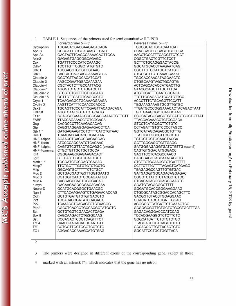
procedures (Roche Molecular Biochemicals). Primer sequences are reported in Table 1. After 18
tamoxifen injection the activated Cre recombinase deleted exons 4 and 5 in the Hnf4a gene. 19
The loss of normal HNF-4α mRNA was quantified with primers located in exons 3 and 5. 20
Results are expressed as the ratio between the mRNA of interest and cyclophilin mRNA. 21
Protein Extraction and Western-Blot Analysis. Total proteins from the villus 22
epithelial cells from mouse jejunum (i1) were isolated using a lysis buffer (20mM Tris, 23
150mM NaCl, 5mM EDTA, 1% triton 100X, 0.5% sodium deoxycholate) containing a 24
protease inhibitor cocktail (Sigma). Nuclear and cytoplasmic proteins from villus or crypt 25
on February 15, 2018 by guest
http://mcb.asm
.org/D
ownloaded from
Page 8
8
epithelial cells of the jejunum were isolated using the NE-PER® nuclear and cytoplasmic 1
extraction reagents kit (Pierce). Protein concentrations were measured using the Bio-Rad DC 2
protein assay (Bio-Rad). For SDS-PAGE, samples with equal amounts of protein were boiled 3
for 10 min in SDS-reducing buffer and separated on a 0.1% SDS-containing polyacrylamide 4
gel. Proteins were transferred onto nitrocellulose membranes and probed with primary 5
antibodies. The primary antibodies used were as follows: goat anti-HNF-4α at 1/5000, rabbit 6
anti-p21 at 1/1000 (C-19 and H-164 respectively, Santa Cruz Biotechnology), rabbit anti-7
Claudin-7 (1/2000), rabbit anti Claudin-2 (1/2000), rabbit anti-ZO-1 (1/100) (34-9100 32-8
5600 and 61-7300 respectively, Zymed laboratories), rabbit anti-EBP50 (1/2000) (PA1-090, 9
Affinity Bioreagents), rat anti-E-cadherin (1/2500) (ECCD-2, Takara), mouse anti-β-catenin 10
(1/2000) (610154, BD Transduction Laboratories), mouse anti-actin (1/2000) (MAB1501R, 11
Chemincon), rabbit anti-LDH (lactate dehydrogenase) (1/20000) (ab52488, Abcam) and 12
rabbit anti-SP1 (1/1000) (PEP-2, Santa Cruz Biotechnology). A peroxidase-labeled (HRP) 13
secondary antibody (Amersham Biosciences) was used and detected by chemoluminescent 14
method (ECL, Amersham Biosciences). The quantitative analyses were performed with a high 15
performance calibrated imaging densitometer (Bio-Rad GS-800) using PD Quest and Image 16
Quant 5.2 software. Results are expressed as the ratio between the protein of interest and Sp1 17
for nuclear proteins or actin for cytoplasmic proteins. 18
Measurement of Intestinal Permeability. In vivo paracellular permeability was 19
performed as previously described after mice were given an oral bolus of fluorescein-20
isothiocyanate-labelled dextran 4 kDa (FITC-dextran, Sigma) at 60mg/100g body weight 21
(40). Paracellular permeability of ileal mucosa was assessed in 0.196 cm2
Ussing chambers by 22
measuring the mucosal-to-serosal flux of FITC-dextran as previously described (4). FITC-23
dextran was assayed by fluorimetry (λex: 490 nm; λem: 520 nm). Mucosal-to-serosal flux was 24
expressed as picomoles per hour per square centimeter of ileal mucosa. 25
on February 15, 2018 by guest
http://mcb.asm
.org/D
ownloaded from
Page 9
9
Plasmids, transfection experiments and Tcf/β-catenin reporter assay. 1
The vectors encoding rat HNF-4α2 (pMT2-HNF-4α) and β-Galactosidase (pRSV-β-2
Gal) were as previously described (2). The TCF-responsive TOPflash vector (Millipore) 3
expressing the luciferase driven by multi TCF-responsive elements, was utilized to evaluate 4
the activity of β-catenin. TCF-responsive elements were mutated in the FOPflash vector 5
(Millipore) used as a negative control. HCT116 human colorectal cancer cells were plated 6
into 12-well plates with 105 cells per well and grown in DMEM supplemented with 10% 7
bovine fetal serum (Invitrogen) maintained at 37°C in a humidified 5% CO2-containing 8
atmosphere. Transfection was performed 72h after plating using LipofectAMINTM
2000 9
(Invitrogen) according to the manufacturer’s instructions. Cells were transfected with 200ng 10
of TOP or FOP vector along with 300ng RSV-βGal plasmid and increasing amounts of 11
PMT2-HNF-4α. Cells were then harvested 48h post-transfection. β-galactosidase and 12
luciferase assays were performed using a luminometer and a microplate reader. The luciferase 13
activity was normalized with the β-galactosidase activity. 14
Co-Immunoprecipitation. The plasmid encoding Flag-Tcf4 was a generous gift from Dr M. 15
Idogawa (23, 24). Caco-2/TC7 cells were cultured in DMEM supplemented with 20% heat-16
inactivated fetal bovine serum and antibiotics. HCT116 were cultivated and transfected with 17
plasmid encoding HNF-4α and Flag-Tcf4 as described previously. Cells were lysed on ice in 18
50 mM Tris HCl pH 8.0, 150 mM NaCl, 5mM EGTA pH 8.0, 50 mM NaF pH 8.0, 10% 19
Glycerol, 1.5 mM MgCl2, 1% Triton X-100 containing freshly added protease inhibitor 20
cocktail (Sigma). Lysates were clarified by centrifugation at 4°C; protein concentrations were 21
determined using a BCA protein quantification kit (Interchim). Immunoprecipitations were 22
performed overnight at 4°C with rabbit, mouse and goat control IgG (Santa Cruz 23
Biotechnology), mouse anti-β-catenin (610154, BD Transduction Laboratories), rabbit anti-24
HNF-4 (H-171, Santa Cruz Biotechnology), mouse anti-Flag (M2, Sigma) or rabbit anti-Tcf4 25
on February 15, 2018 by guest
http://mcb.asm
.org/D
ownloaded from
Page 10
10
(H-125, Santa Cruz Biotechnology). Five µg of each antibody or control IgG were used for 1 1
mg of transfected HCT116 and CaCo-2/TC7 cells extracts. Immunocomplexes were collected 2
on protein A-sepharose beads and protein G-sepharose beads (GE Healthcare) the last 45 3
minutes. The beads were pelleted by centrifugation, washed three times with Tris HCl pH 8.0, 4
150 mM NaCl, 5 mM EGTA, 1% NP40 and then boiled 10 minutes in SDS sample buffer. 5
Western blots were probed with goat anti- HNF-4α (1/5000)(C-19, Santa Cruz 6
Biotechnology), goat anti-Tcf4 (1/2000) (N-20, Santa Cruz Biotechnology) and mouse anti-β-7
catenin (1/2000). 8
Statistical Tests. Results were expressed as means ± SEM. The statistical significance 9
of differences was determined by an unpaired Student’s t test using Excel software. 10
RESULTS 11
Elimination of HNF-4αααα in the intestinal epithelium of adult mice. To examine the role of 12
HNF-4α in the intestinal epithelium of adult mice, floxed Hnf-4a mice (21) containing two 13
loxP sites flanking exons 4 and 5 of the Hnf-4a gene (Hnf-4aloxP/loxP
mice) (Fig. 1A top), were 14
crossed with the villin-Cre-ERT2 deletor line (16) to generate Hnf-4aloxP/loxP
;villin-CreERT2 15
mice. The expression of HNF-4α was quantified by semi quantitative RT-PCR on mRNA 16
isolated from 2 month-old wild type mice, Hnf-4aloxP/loxP
mice and Hnf-4aloxP/loxP
;villin-17
CreERT2 mice without CRE recombinase activation. Whatever the genotype, the levels of 18
Hnf-4a mRNA were identical and we demonstrated an absence of hypomorphism for the Hnf-19
4a gene expression. There was no detectable background recombination in the presence of the 20
villin-Cre-ERT2 transgene and no phenotypic differences between these strains. The different 21
genotypes were equally viable and were obtained at the expected ratios in all crosses (data not 22
shown). 23
on February 15, 2018 by guest
http://mcb.asm
.org/D
ownloaded from
Page 11
11
The CRE recombinase activity was induced by daily injection of tamoxifen during five 1
consecutive days in 2 month-old mice. The deletion of exons 4 and 5 by the induction of CRE 2
recombinase activity in Hnf-4aloxP/loxP
;villin-CreERT2 mice generated Hnf-4aint∆
mice (Fig. 3
1A, top). Hnf-4aloxP/loxP
mice, i.e. lacking the Cre-ERT2 transgene, were used as controls and 4
injected with tamoxifen under the same conditions as the Hnf-4aint∆
mice. Mice were 5
euthanized on day 8 (D8) following the first injection, a time lapse corresponding at least to 6
one complete renewal cycle of the intestinal epithelium. The removal of exons 4 and 5 was 7
specific for all segments of the intestine and was not observed in the other organs expressing 8
HNF-4α i.e. liver, kidney, stomach or pancreas (Fig. 1A, bottom). The inactivation was very 9
efficient as the drop in Hnf-4a mRNA expression reached 90% and 87% in villus and crypt 10
epithelium of jejunum, respectively (Fig. 1C). Similar results were obtained in ileum and 11
colon (data not shown). Accordingly, the level of HNF-4α protein was decreased by 90% in 12
the jejunum epithelium of Hnf-4a intestine-null mice (Fig. 1D). As previously shown for wild 13
type mice (45), HNF-4α was expressed in the nucleus of all epithelial cells along the crypt-to-14
villus axis in control Hnf-4aloxP/loxP
mice (Fig. 1E). HNF-4α was not detectable in epithelial 15
cells in Hnf-4aint∆
mice (Fig.1E) and remained at very low levels up to 30 days after 16
tamoxifen injections (Fig. 2A). Together, these results indicated that injections of tamoxifen 17
resulted in an intestine-specific recombination of the floxed Hnf-4a gene in Hnf-4a 18
loxP/loxP;villin-CreERT2 mice and that the loss of HNF-4α protein was observed in all 19
epithelial cells along the crypt-to-villus axis. 20
Loss of HNF-4α enhances the proliferation of epithelial cells in crypts. Histological 21
examination of intestinal sections from Hnf-4a loxP/loxP
and Hnf-4a int∆
mice showed that the 22
general morphology of the intestinal epithelium was conserved in Hnf-4a int∆
mice (Fig. 3A), 23
but that the depth of the crypts was significantly increased in the Hnf-4aint∆
mice (Fig. 3B and 24
C) whereas the size of villi remained similar in the two groups of mice. Within the crypt 25
on February 15, 2018 by guest
http://mcb.asm
.org/D
ownloaded from
Page 12
12
compartment of Hnf-4aint∆
mice, the size of the cells appeared normal suggesting that crypt 1
hypertrophy was the consequence of hyperplasia and of aberrant proliferation. To test this 2
hypothesis, Hnf-4a int∆
and Hnf-4aloxP/loxP
mice were injected with BrdU and euthanized 90 3
min later. The average number of BrdU-positive cells per crypt was two-fold higher in Hnf-4
4aint∆
mice than in Hnf-4aloxP/loxP
control mice (Fig. 3D and E). In colon, where the 5
inactivation of HNF-4α was efficient, the number of the proliferative cells was also increased 6
(Fig. 4A and B). The proliferative index (i.e. the ratio between the number of proliferative 7
cells and the number of total cells in the crypt) was higher in Hnf-4aint∆
mice (Fig. 3F), 8
suggesting a loss of the intestinal homeostasis. Immuno-staining of the Ki67 protein 9
confirmed the increased epithelial proliferation in crypts of Hnf-4aint∆
mice (Fig. 3G). As the 10
length of the villi was not affected, we measured the frequency of apoptosis by immuno-11
staining of the activated-caspase 3 in the intestinal epithelium. Hnf-4aint∆
epithelium presented 12
four fold more activated-caspase 3-positive cells than that of Hnf-4aloxP/loxP
controls (Fig. 3H 13
and I). This observation could explain the normal length of the villi despite the increased 14
number of proliferative cells. 15
Interestingly, 30 days after tamoxifen injection, the effects observed on the crypt size 16
and proliferation were still significant but less pronounced when compared to those observed 17
8 days after tamoxifen injection (Fig. 2B, C, and D) 18
Loss of HNF-4α activates the Wnt/β-catenin signaling pathways. The canonical Wnt 19
pathway was shown to play an important role in maintaining the proliferation capacity of the 20
intestinal epithelium (46). We observed an enhancement of intracellular β-catenin staining in 21
crypts of Hnf-4aint∆
mice as compared to Hnf-4aloxP/loxP
control mice (Fig. 5A). We further 22
analysed the level of nuclear and cytoplasmic β-catenin in crypt cells. The amount of 23
cytoplasmic β-catenin was unchanged (Fig. 5B, top) whereas that of nuclear β-catenin was 24
increased by 50% (Fig. 5B, bottom) in Hnf-4aint∆
mice as compared to Hnf-4aloxP/loxP
control 25
on February 15, 2018 by guest
http://mcb.asm
.org/D
ownloaded from
Page 13
13
mice. The cytoplasmic and nuclear fraction purity was assessed through LDH (cytoplasmic) 1
and Sp1 (nuclear) immunoblots. Contamination of the nuclear fraction by cytoplasm was 2
negligible (Fig. 5B). The accumulation of cytoplasmic and nuclear β-catenin is the hallmark 3
of an active Wnt signaling, we analyzed the expression of several classical target genes of the 4
Wnt pathway activation in crypt-enriched epithelial cells. The expression of Tcf-4, Axin-2, c-5
Myc and Lgr-5 was significantly increased in Hnf-4aint∆
mice (Fig. 5C). In parallel, the 6
expression of the cell cycle inhibitor p21 was significantly decreased in crypt- (data not 7
shown) and in villi-enriched epithelial cells from Hnf-4aint∆
mice, at both mRNA (Fig. 5C) 8
and protein levels (Fig. 5D). No significant change was observed in the expression of p27 9
(data not shown) or cyclin D1 (Fig. 5C), which are also regulators of the cell cycle. 10
To further address the relationships between HNF-4α and the Wnt/β-catenin pathway, 11
we transfected the TOP/FOP®
luciferase reporter vectors, a TCF/β-catenin-directed 12
transcription system (47), together with increasing amount of a HNF-4α expressing vector, in 13
the HCT116 colon carcinoma cell line, which does not express endogenous HNF-4α (22). 14
HNF-4α strongly (80%) decreased the β-catenin/TCF–dependent luciferase activity in a dose-15
dependent manner (Fig. 5E). An interaction between HNF-4α and Tcf4 or β-catenin has been 16
investigated. Through co-immunoprecipation experiments in HCT116 cells (Fig.6A) and in 17
Caco-2/TC7 cells (Fig. 6B), we showed that HNF-4α interacts with Tcf-4 but not with β-18
catenin. All together these results, which were obtained in vivo and in vitro, indicated that 19
HNF-4α interfered with the Wnt/β-catenin pathway to control proliferation of the crypt 20
epithelial cells. 21
Loss of HNF-4αααα alters the secretory cell lineage in the adult intestinal epithelium. We 22
analyzed the effect of HNF-4α loss on the intestinal epithelial cell lineage. The four 23
differentiated cell types were reported to originate from the intestinal stem cells. The 24
activation of the Notch signaling pathway is known to induce Hes1 expression and 25
on February 15, 2018 by guest
http://mcb.asm
.org/D
ownloaded from
Page 14
14
commitment of epithelial cells into the absorptive lineage leading to differentiated 1
enterocytes, through repression of Math1, a transcription factor necessary for cell 2
commitment into the secretory lineage (46). In Hnf-4aint∆
crypts, we observed a 1.8-fold 3
increase in Math-1 mRNA level and no significant modification in Hes-1 mRNA level (Table 4
2). 5
Consistent with this observation, the average number of goblet cells, detected by 6
Periodic Acid-Schiff (32) staining, was significantly increased (1.3 fold in villi and 1.7-fold in 7
crypts) in Hnf-4aint∆
mice (Fig. 7A and C). The mRNA encoding trefoil factor 3 (Tff3), a 8
marker of differentiated goblet cells, was also increased as well as that of Muc 2, the major 9
gel-forming mucin in goblet cells (Table 2). Goblet cells were mature as confirmed by PAS 10
staining and electron microscopy (Fig. 7A and B). The loss of HNF-4α affected neither the 11
number of Paneth cells, stained with an anti-lysozyme (Fig. 7F), nor the level of MMP7, or 12
Sox9 mRNAs (Table 2). The cryptdin mRNA level was only moderately affected (Table 2). 13
We examined and counted the enteroendocrine cells after immunostaining of chromogranin A 14
(Fig. 7D and E). In Hnf-4a int∆
mice, we observed a 1.4-fold decrease of the enteroendocrine 15
cell number in villi and a 1.4-fold increase in crypts, resulting in an unchanged total (crypts 16
plus villi) enteroendocrine cell number. The expression level of genes encoding gut hormones 17
GIP, somatostatin and glucagon (Gcg) was significantly decreased (Table 2), suggesting that 18
the corresponding endocrine subtypes were reduced. The transcription factors Ngn3 and 19
NeuroD were reported to be involved in enteroendocrine specification and differentiation 20
(31). The mRNA encoding Ngn3 was significantly increased in Hnf-4aint∆
mice, whereas that 21
encoding NeuroD remained unchanged (Table 2). 22
In intestine, our previous work (2, 9) and a bioinformatic analysis (49) showed that 23
HNF-4α influences the enterocyte function by regulating genes involved in lipid metabolism. 24
Thus we analyzed the expression of several genes involved in the function of lipid transfer in 25
on February 15, 2018 by guest
http://mcb.asm
.org/D
ownloaded from
Page 15
15
enterocytes. In Hnf-4aint∆
mice, there was a significant reduction of the mRNA coding for 1
microsomal triglyceride transfer protein (Mttp), apolipoproteins (apos) B and A4 and 2
intestinal fatty acid binding protein (i-FABP) (Table 2). 3
In villi, the expression of some transcription factors, such as HNF-1α and cdx2 was 4
unaffected by the loss of HNF-4α whereas that of Klf4, a Krüppel-like factor (KLF) 5
expressed in terminally differentiated mucosecretory cells, was decreased by 30% (Table 2). 6
In crypts, HNF-1β (or Tcf2) expression was significantly increased (Table 2). Notably, the 7
absence of HNF-4α induced a strong expression of HNF-4γ in crypts (Table 2 and Fig. 8A) 8
which was still observed 30 days after tamoxifen injection (Fig. 8B), without modifications of 9
HNF-4γ expression in villi (Table 2). 10
All together, these results showed that the intestinal disruption of the Hnf-4a gene 11
perturbed the homeostasis of the intestinal epithelium lineage and the terminal differentiation 12
of enterocytes and of some endocrine subtypes. 13
HNF-4αααα controls the epithelial cell architecture. HNF-4α has been shown to be essential 14
for morphological differentiation of hepatocytes by regulating the expression of cell/cell 15
junction-associated proteins (41). We thus investigated the impact of the intestinal HNF-4α 16
loss on cell architecture of adult small intestine epithelium and particularly on the 17
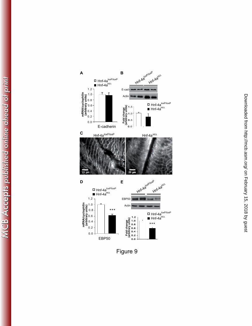
organization of the cell/cell junctions. E-cadherin (adherens junctions) mRNA and protein 18
expression was unchanged in Hnf-4aint∆
mice (Fig. 9A and B), but the protein was not 19
properly located to membranes and was abundantly present in the cytoplasm (Fig. 9C). 20
EBP50 (ERM (Ezrin-Radixin-Meosin) binding protein, also named Na+/H
+ exchanger 21
regulatory factor 1 or NHERF1) is a scaffolding protein which organizes these ERM proteins 22
at the apical membrane (37) and is also a target gene of HNF-4α (12). Accordingly, the 23
mRNA and protein levels of EBP50 were significantly decreased in Hnf-4aint∆
mice (Fig. 9D 24
and E). 25
on February 15, 2018 by guest
http://mcb.asm
.org/D
ownloaded from
Page 16
16
The expression of some genes encoding junctional proteins was further analyzed. 1
Desmocollin 2 and plakophilin2 (desmosomes) remained unchanged in Hnf-4aint∆
mice, 2
whereas the mRNA encoding Gjb1 (gap junctions) was 1.7-fold increased in mutant mice 3
(Fig. 10A). Important modifications in the expression of tight junction proteins were observed 4
in Hnf-4aint∆
mice, namely a significant decrease in claudins -4, -7 and ZO-1 mRNA levels 5
associated with a marked increase in that of claudin-2 (Fig. 10A). These variations correlated 6
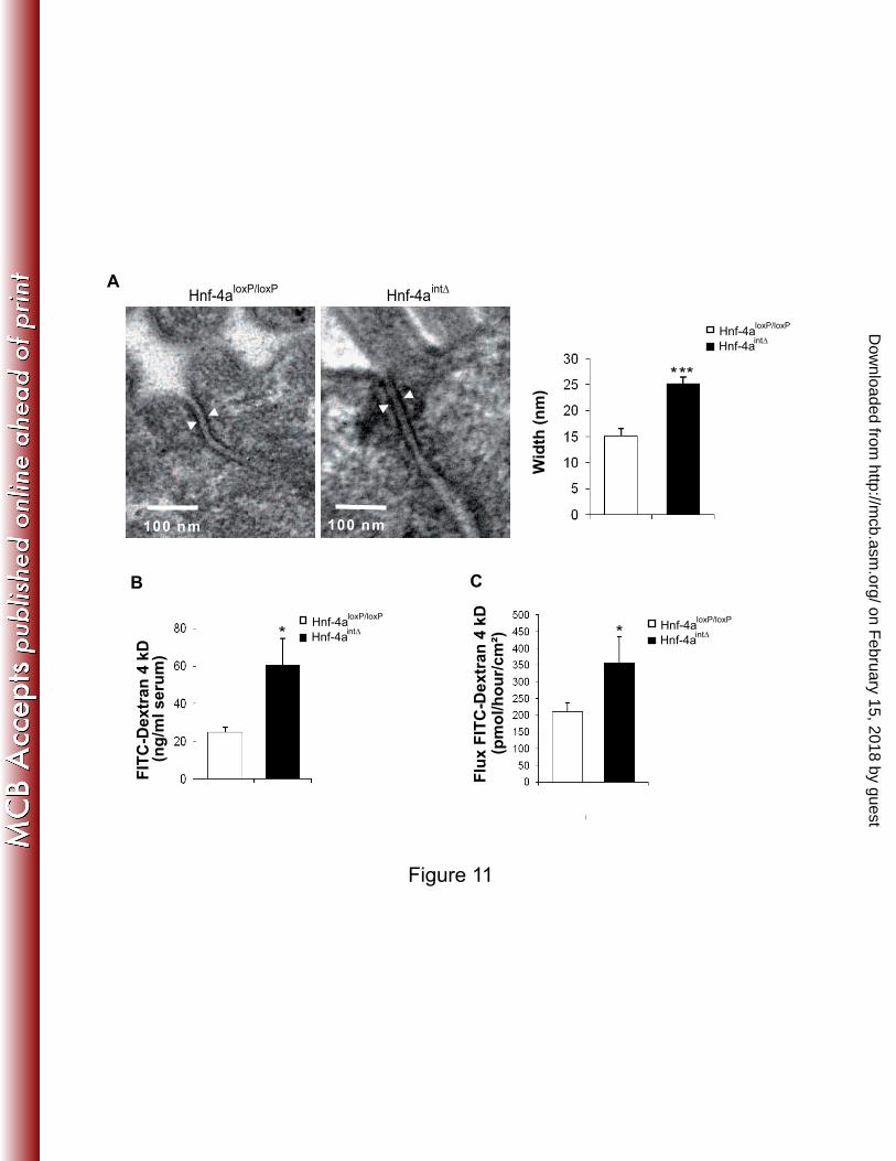
with those observed at the protein levels (Fig. 10B and C). Examination of epithelial cell-cell 7
contacts at the ultrastructural level showed that the intercellular space of tight junctions was 8
1.7-fold wider in hnf4αint∆
mice than in control mice (Fig. 11A). This structural change might 9
account for the increased paracellular permeability that we quantified both in vivo (Fig. 11B) 10
and ex vivo in Ussing chambers by measuring the mucosal-to-serosal flux of FITC-dextran 11
(Fig. 11C). 12
DISCUSSION 13
The results presented here show that the nuclear receptor HNF-4α plays a critical role 14
in the homeostasis of intestinal epithelium, in the epithelial cell architecture and in the barrier 15
function of the intestine. We report that loss of HNF-4α in adult small intestine affects the 16
Wnt signaling pathway, leads to an enhanced proliferation in crypts and, induces an increased 17
number of mucosecretory cells. In absorptive cells, HNF-4α exerts a major role in the 18
activation of genes involved in lipid transfer by fully differentiated enterocytes (6). Moreover 19
loss of HNF-4α also disturbs the epithelial cell architecture by modifying the expression of 20
proteins involved in cell/cell adhesion complexes, specifically in the tight junctions, leading 21
to increased in vivo paracellular permeability in Hnf-4aint∆
mice. From these results, we 22
hypothesize that HNF-4α plays a dual role in adult intestinal epithelium: HNF-4α controls the 23
functional differentiation of epithelial cells and, finely modulates the homeostasis of the 24
epithelium. 25
on February 15, 2018 by guest
http://mcb.asm
.org/D
ownloaded from
Page 17
17
The importance of HNF-4α as a master regulator of development and differentiation 1
has been established in liver (15, 29, 41) and in embryonic developing colon (18). In addition, 2
an increasing amount of evidence suggests that HNF-4α plays a role in regulating 3
proliferation. Indeed, HNF-4α was shown to inhibit cell proliferation in vitro (11, 22, 32) and 4
to slow the progression of hepato-cellular carcinoma in mice (27). We report here that loss of 5
HNF-4α in the adult intestinal epithelium results in an increased cell proliferation in the crypt 6
compartment. This is correlated with down-regulation of p21 and over-expression of c-Myc. 7
It was recently reported that HNF-4α increases the expression of the p21 gene by competing 8
with c-Myc on the p21 promoter (11, 22), a mechanism that could account for the down-9
regulation of p21 expression observed in Hnf-4aint∆
mice. The canonical Wnt pathway was 10
shown to play an important role in maintaining the proliferation capacity of the intestinal 11
epithelium (46). Tcf-4, whose intestinal inactivation leads to an absence of proliferating cells, 12
is the most prominent effector of Wnt/β-catenin signaling in the gastrointestinal tract (25, 51). 13
The expression of Tcf-4 is up-regulated in the absence of HNF-4α, concomitantly with that of 14
β-catenin/Tcf4 target genes, c-Myc, Axin-2, Lgr-5 and Claudin 2. Furthermore, we 15
demonstrate that HNF-4α is able to decrease the Wnt-Tcf4/β-catenin-driven transcription 16
activity. This modulation of the Wnt signaling pathway could be due to an interaction 17
between HNF-4α and Tcf4 (7) or β-catenin, as shown for other members of the nuclear 18
receptor family (39). Through immunoprecipation experiments in Caco-2/TC7 and HCT116 19
cells, we showed that HNF-4α interacts with Tcf-4 but not with β-catenin. One can 20
hypothesized that HNF-4α acts in the crypt to sequester Tcf4 and thereby represses its 21
transcriptional activity. All together, these observations imply that HNF-4α could regulate the 22
balance between proliferation and differentiation by interfering directly with the Wnt 23
signaling pathway in intestinal epithelium. 24
on February 15, 2018 by guest
http://mcb.asm
.org/D
ownloaded from
Page 18
18
We show here that disrupting Hnf-4a gene expression in intestinal epithelium of adult 1
mice is associated with a marked increase of mucus-secreting cells. In intestinal epithelium, 2
cells commit either into the absorptive lineage when the Notch signal is activated or into the 3
secretory lineage (enteroendocrine, goblet and Paneth cells) when the Notch signal is off (46). 4
Math1, which is repressed by the Notch signaling effector Hes1, is expressed at an early 5
progenitor stage in cells that are committing into the secretory lineage. Hnf-4a int∆
mice show 6
a marked increase of Math1 expression, which might explain the higher number of mucus-7
secreting cells as well as the upregulation of their differentiation markers Tff3 and Muc2. 8
Inducible inactivation of the Notch signaling pathway in adult intestinal epithelium leads to 9
an increased expression of Math1 with a massive conversion of proliferative crypt cells into 10
goblet cells without impact on the other secreting cells (52). Consistent with these 11
observations, there is no global change in the number of enteroendocrine and Paneth cells in 12
Hnf-4aint∆
mice. However, the number of enteroendocrine cells along the crypt to villus axis 13
reveals a paradox: when compared with controls, Hnf-4aint∆
mice exhibit more 14
enteroendocrine cells in crypts and less in villi as well as an increased expression of Ngn3, 15
which is involved in enteroendocrine specification (31), and a decreased expression of some 16
differentiation markers, such as Gcg, Gip and somatostatin. We hypothesize that the higher 17
level of Ngn3 in Hnf-4aint∆
mice drives more cells toward the enteroendocrine lineage but that 18
the absence of HNF-4α prevents their full terminal differentiation. Commitment into the 19
absorptive lineage is driven by the Notch signal and the transcriptional factor Hes1. In Hnf-20
4aint∆
mice, the expression of Hes1 and the number of enterocytes, estimated by the size of 21
villi, are unchanged. Thus, the main role of HNF-4α in the absorptive cells is to control genes 22
specifically expressed in fully differentiated enterocytes, notably those involved in transfer of 23
dietary lipids. 24
on February 15, 2018 by guest
http://mcb.asm
.org/D
ownloaded from
Page 19
19
It is noteworthy that Hnf-4g, the paralog of Hnf-4a, is expressed in intestinal villi (45) 1
but not in liver (50). Our results suggest that HNF-4γ, whose expression is similar in the villus 2
compartment and induced in the crypt compartment of Hnf-4a int∆
mice, may act to partly 3
compensate the lack of HNF-4α . This hypothesis is supported by the observation that HNF-4
4γ is still present in crypts at 30 days after tamoxifen injections. At this time the size of crypts 5
and the proliferation status are closer to those of control mice. A recent paper from Babeu et 6
al (3) reported the results obtained in mice where the same Hnf-4aloxP/loxP
gene was inactivated 7
in the intestine but using a constitutively active Cre recombinase driven by a 12.5 kbp villin 8
promoter which is active at E 12.5-13.5 (34). The animals were viable and did not present any 9
overt phenotype or intestinal dysfuntion. The results we obtained in adults 30 days after 10
tamoxifen injection are similar to those of Babeu et al (3) i.e. a subttle and statistical increase 11
of crypt cells proliferation and of the number of goblet cells. Our observations support the 12
hypothesis of a functional compensation by HNF-4γ. It may be hypothesized that in the study 13
of Babeu et al., the absence of an overt phenotype could be due to the presence of HNF-4γ, 14
even if a slight decrease in the expression of Hnf-4g was observed in the whole intestinal 15
epithelium of their mice. We have previously reported that, in vitro, the two isoforms have 16
similar properties, i.e. affinity for their binding site and transactivation of a reporter gene (2). 17
However, mice lacking expression of HNF-4γ are viable and have no evident phenotype 18
indicating that the two proteins are not fully redundant. Deciphering the different roles of the 19
two isoforms is still a challenging task. 20
HNF-4α plays an essential role in the architecture of epithelial cells (5, 10, 12, 41, 48). 21
In adult intestinal epithelium, the loss of HNF-4α induces a distension of tight junctions, 22
associated with changes in the expression of tight junction proteins such as claudin-2, -4, -7 23
and ZO-1. These modifications could be due to the increased expression of Tcf-4, which is a 24
negative regulator of claudin-7 expression and a positive regulator of claudin-2 expression 25
on February 15, 2018 by guest
http://mcb.asm
.org/D
ownloaded from
Page 20
20
(13). Claudins are responsible for the gate function of tight junctions, which control the 1
paracellular permeability. An increased claudin-2 expression was reported responsible for an 2
increased epithelial permeability (54). Accordingly, we observed an increased paracellular 3
permeability of Hnf-4aint∆
mouse intestine in vivo and in vitro. Interestingly, HNF-4α has 4
recently been shown to have a protective role against inflammatory bowel diseases, a 5
pathology where the epithelial barrier is impaired (1). Furthermore, loss of tight junction 6
structure and function is frequently observed in epithelium-derived cancers and an over-7
expression of claudin-2 can stimulate the invasion and migration activities of cancer cells 8
(36). The loss of HNF-4α in intestinal epithelium also induces a drastic decrease of EBP50 9
expression. ERM proteins regulate the organization and function of specific cortical structures 10
in polarized epithelial cells and EBP50 organizes these ERM proteins at the apical membrane 11
(37). The observed down-regulation of EBP50 expression in Hnf-4aint∆
mice could explain the 12
concomitant E-cadherin membrane delocalization. Indeed, a low expression of EBP50 13
decreased the interaction between β-catenin and E-cadherin, leading to disorganization of 14
adherens junctions and increased cell proliferation and motility (26). 15
Our work in vivo demonstrates for the first time that HNF-4α is a transcription factor 16
at the crossroads between the intestinal epithelium homeostasis and the epithelial cell 17
architecture in adult mouse intestine by controlling the cell proliferation in crypts and the 18
functional differentiation in villi. Indeed, HNF-4α interferes with the Wnt/β-catenin signaling 19
pathway and its loss destabilizes adherens and tight junctions. It is currently admitted that the 20
deregulation of the Wnt/β-catenin signaling pathway is an early event in the colorectal cancer 21
progression cascade and that the destabilization of cell/cell contacts takes part in the 22
epithelial-to-mesenchymal transition, a crucial process in tumour progression. Since HNF-4α 23
regulates the balance between proliferation and differentiation, we hypothesize that it might 24
act as a tumor suppressor. 25
on February 15, 2018 by guest
http://mcb.asm
.org/D
ownloaded from
Page 21
21
ACKNOWLEDGMENTS 1
We thank N. Comuce, C. Lasne and V. Chauffeton for animal care. Electronic 2
microscopy analysis was performed using facilities of the Centre de Recherche des 3
Cordeliers, UMRS 872. We thank Dr M. Pontoglio for helpful scientific discussions and 4
access to unpublished results, Drs JM Lacorte and V Carriere for their help during genotyping 5
and RT-PCR experiments, respectively, and Dr M. Rousset for critical reading of the 6
manuscript. 7
This work was supported by INSERM, France and Pierre & Marie Curie University, 8
Paris, France. A-L Cattin is the recipient of a doctoral fellowship from the Ministère de 9
l’Enseignement Supérieur et de la Recherche 10
REFERENCES 11
1. Ahn, S. H., Y. M. Shah, J. Inoue, K. Morimura, I. Kim, S. Yim, G. Lambert, R. 12
Kurotani, K. Nagashima, F. J. Gonzalez, and Y. Inoue. 2008. Hepatocyte nuclear 13
factor 4alpha in the intestinal epithelial cells protects against inflammatory bowel 14
disease. Inflamm Bowel Dis 14:908-20. 15
2. Archer, A., D. Sauvaget, V. Chauffeton, P. E. Bouchet, J. Chambaz, M. Pincon-16
Raymond, P. Cardot, A. Ribeiro, and M. Lacasa. 2005. Intestinal apolipoprotein A-17
IV gene transcription is controlled by two hormone-responsive elements: a role for 18
hepatic nuclear factor-4 isoforms. Mol Endocrinol 19:2320-34. 19
3. Babeu, J. P., M. Darsigny, C. R. Lussier, and F. Boudreau. 2009. Hepatocyte 20
nuclear factor 4alpha contributes to an intestinal epithelial phenotype in vitro and 21
plays a partial role in mouse intestinal epithelium differentiation. Am J Physiol 22
Gastrointest Liver Physiol 297:G124-34. 23
4. Barreau, F., U. Meinzer, F. Chareyre, D. Berrebi, M. Niwa-Kawakita, M. 24
Dussaillant, B. Foligne, V. Ollendorff, M. Heyman, S. Bonacorsi, T. Lesuffleur, 25 G. Sterkers, M. Giovannini, and J. P. Hugot. 2007. CARD15/NOD2 is required for 26
Peyer's patches homeostasis in mice. PLoS ONE 2:e523. 27
5. Battle, M. A., G. Konopka, F. Parviz, A. L. Gaggl, C. Yang, F. M. Sladek, and S. 28
A. Duncan. 2006. Hepatocyte nuclear factor 4alpha orchestrates expression of cell 29
adhesion proteins during the epithelial transformation of the developing liver. Proc 30
Natl Acad Sci U S A 103:8419-24. 31
6. Beaslas, O., F. Torreilles, P. Casellas, D. Simon, G. Fabre, M. Lacasa, F. Delers, 32
J. Chambaz, M. Rousset, and V. Carriere. 2008. Transcriptome response of 33
enterocytes to dietary lipids: impact on cell architecture, signaling, and metabolism 34
genes. Am J Physiol Gastrointest Liver Physiol 295:G942-52. 35
7. Benahmed, F., I. Gross, S. J. Gaunt, F. Beck, F. Jehan, C. Domon-Dell, E. 36
Martin, M. Kedinger, J. N. Freund, and I. Duluc. 2008. Multiple regulatory regions 37
on February 15, 2018 by guest
http://mcb.asm
.org/D
ownloaded from
Page 22
22
control the complex expression pattern of the mouse Cdx2 homeobox gene. 1
Gastroenterology 135:1238-1247, 1247 e1-3. 2
8. Benoit, G., A. Cooney, V. Giguere, H. Ingraham, M. Lazar, G. Muscat, T. 3
Perlmann, J. P. Renaud, J. Schwabe, F. Sladek, M. J. Tsai, and V. Laudet. 2006. 4
International Union of Pharmacology. LXVI. Orphan nuclear receptors. Pharmacol 5
Rev 58:798-836. 6
9. Carriere, V., R. Vidal, C. Lazou, M. Lacasa, F. Delers, A. Ribeiro, M. Rousset, J. 7
Chambaz, and J. M. Lacorte. 2005. HNF-4-dependent induction of apolipoprotein 8
A-IV gene transcription by an apical supply of lipid micelles in intestinal cells. J Biol 9
Chem 280:5406-5413. 10
10. Chiba, H., T. Gotoh, T. Kojima, S. Satohisa, K. Kikuchi, M. Osanai, and N. 11
Sawada. 2003. Hepatocyte nuclear factor (HNF)-4alpha triggers formation of 12
functional tight junctions and establishment of polarized epithelial morphology in F9 13
embryonal carcinoma cells. Exp Cell Res 286:288-97. 14
11. Chiba, H., T. Itoh, S. Satohisa, N. Sakai, H. Noguchi, M. Osanai, T. Kojima, and 15
N. Sawada. 2005. Activation of p21CIP1/WAF1 gene expression and inhibition of 16
cell proliferation by overexpression of hepatocyte nuclear factor-4alpha. Exp Cell Res 17
302:11-21. 18
12. Chiba, H., N. Sakai, M. Murata, M. Osanai, T. Ninomiya, T. Kojima, and N. 19
Sawada. 2006. The nuclear receptor hepatocyte nuclear factor 4alpha acts as a 20
morphogen to induce the formation of microvilli. J Cell Biol 175:971-80. 21
13. Darido, C., M. Buchert, J. Pannequin, P. Bastide, H. Zalzali, T. Mantamadiotis, 22
J. F. Bourgaux, V. Garambois, P. Jay, P. Blache, D. Joubert, and F. Hollande. 23 2008. Defective claudin-7 regulation by Tcf-4 and Sox-9 disrupts the polarity and 24
increases the tumorigenicity of colorectal cancer cells. Cancer Res 68:4258-68. 25
14. Drewes, T., S. Senkel, B. Holewa, and G. U. Ryffel. 1996. Human hepatocyte 26
nuclear factor 4 isoforms are encoded by distinct and differentially expressed genes. 27
Mol Cell Biol 16:925-31. 28
15. Duncan, S. A., A. Nagy, and W. Chan. 1997. Murine gastrulation requires HNF-4 29
regulated gene expression in the visceral endoderm: tetraploid rescue of Hnf-4(-/-) 30
embryos. Development 124:279-87. 31
16. el Marjou, F., K. P. Janssen, B. H. Chang, M. Li, V. Hindie, L. Chan, D. 32
Louvard, P. Chambon, D. Metzger, and S. Robine. 2004. Tissue-specific and 33
inducible Cre-mediated recombination in the gut epithelium. Genesis 39:186-93. 34
17. Fouquet, S., V. H. Lugo-Martinez, A. M. Faussat, F. Renaud, P. Cardot, J. 35
Chambaz, M. Pincon-Raymond, and S. Thenet. 2004. Early loss of E-cadherin 36
from cell-cell contacts is involved in the onset of Anoikis in enterocytes. J Biol Chem 37
279:43061-9. 38
18. Garrison, W. D., M. A. Battle, C. Yang, K. H. Kaestner, F. M. Sladek, and S. A. 39
Duncan. 2006. Hepatocyte nuclear factor 4alpha is essential for embryonic 40
development of the mouse colon. Gastroenterology 130:1207-20. 41
19. Gerdin, A. K., V. V. Surve, M. Jonsson, M. Bjursell, M. Bjorkman, A. Edenro, 42
M. Schuelke, A. Saad, S. Bjurstrom, E. J. Lundgren, M. Snaith, R. Fransson-43 Steen, J. Tornell, A. L. Berg, and Y. M. Bohlooly. 2006. Phenotypic screening of 44
hepatocyte nuclear factor (HNF) 4-gamma receptor knockout mice. Biochem Biophys 45
Res Commun 349:825-32. 46
20. Gupta, R. K., and K. H. Kaestner. 2004. HNF-4alpha: from MODY to late-onset 47
type 2 diabetes. Trends Mol Med 10:521-4. 48
on February 15, 2018 by guest
http://mcb.asm
.org/D
ownloaded from
Page 23
23
21. Hayhurst, G. P., Y. H. Lee, G. Lambert, J. M. Ward, and F. J. Gonzalez. 2001. 1
Hepatocyte nuclear factor 4alpha (nuclear receptor 2A1) is essential for maintenance 2
of hepatic gene expression and lipid homeostasis. Mol Cell Biol 21:1393-403. 3
22. Hwang-Verslues, W. W., and F. M. Sladek. 2007. Nuclear Receptor HNF4{alpha}1 4
Competes with Oncoprotein c-Myc for Control of the p21/WAF1 Promoter. Mol 5
Endocrinol. 6
23. Idogawa, M., M. Masutani, M. Shitashige, K. Honda, T. Tokino, Y. Shinomura, 7
K. Imai, S. Hirohashi, and T. Yamada. 2007. Ku70 and poly(ADP-ribose) 8
polymerase-1 competitively regulate beta-catenin and T-cell factor-4-mediated gene 9
transactivation: possible linkage of DNA damage recognition and Wnt signaling. 10
Cancer Res 67:911-8. 11
24. Idogawa, M., T. Yamada, K. Honda, S. Sato, K. Imai, and S. Hirohashi. 2005. 12
Poly(ADP-ribose) polymerase-1 is a component of the oncogenic T-cell factor-4/beta-13
catenin complex. Gastroenterology 128:1919-36. 14
25. Korinek, V., N. Barker, P. Moerer, E. van Donselaar, G. Huls, P. J. Peters, and 15
H. Clevers. 1998. Depletion of epithelial stem-cell compartments in the small 16
intestine of mice lacking Tcf-4. Nat Genet 19:379-83. 17
26. Kreimann, E. L., F. C. Morales, J. de Orbeta-Cruz, Y. Takahashi, H. Adams, T. 18
J. Liu, P. D. McCrea, and M. M. Georgescu. 2007. Cortical stabilization of beta-19
catenin contributes to NHERF1/EBP50 tumor suppressor function. Oncogene 20
26:5290-9. 21
27. Lazarevich, N. L., O. A. Cheremnova, E. V. Varga, D. A. Ovchinnikov, E. I. 22
Kudrjavtseva, O. V. Morozova, D. I. Fleishman, N. V. Engelhardt, and S. A. 23 Duncan. 2004. Progression of HCC in mice is associated with a downregulation in the 24
expression of hepatocyte nuclear factors. Hepatology 39:1038-47. 25
28. Le Beyec, J., V. Chauffeton, H. Y. Kan, P. L. Janvier, C. Cywiner-Golenzer, F. P. 26
Chatelet, A. D. Kalopissis, V. Zannis, J. Chambaz, M. Pincon-Raymond, and P. 27 Cardot. 1999-a. The -700/-310 fragment of the apolipoprotein A-IV gene combined 28
with the -890/-500 apolipoprotein C-III enhancer is sufficient to direct a pattern of 29
gene expression similar to that for the endogenous apolipoprotein A-IV gene. J Biol 30
Chem 274:4954-61. 31
29. Li, J., G. Ning, and S. A. Duncan. 2000. Mammalian hepatocyte differentiation 32
requires the transcription factor HNF-4alpha. Genes Dev 14:464-74. 33
30. Li, X., B. B. Madison, W. Zacharias, A. Kolterud, D. States, and D. L. Gumucio. 34
2007. Deconvoluting the intestine: molecular evidence for a major role of the 35
mesenchyme in the modulation of signaling cross talk. Physiol Genomics 29:290-301. 36
31. Lopez-Diaz, L., R. N. Jain, T. M. Keeley, K. L. VanDussen, C. S. Brunkan, D. L. 37
Gumucio, and L. C. Samuelson. 2007. Intestinal Neurogenin 3 directs differentiation 38
of a bipotential secretory progenitor to endocrine cell rather than goblet cell fate. Dev 39
Biol 309:298-305. 40
32. Lucas, B., K. Grigo, S. Erdmann, J. Lausen, L. Klein-Hitpass, and G. U. Ryffel. 41
2005. HNF4alpha reduces proliferation of kidney cells and affects genes deregulated 42
in renal cell carcinoma. Oncogene 24:6418-31. 43
33. Lugo-Martinez, V. H., C. S. Petit, S. Fouquet, J. Le Beyec, J. Chambaz, M. 44
Pincon-Raymond, P. Cardot, and S. Thenet. 2009. Epidermal growth factor 45
receptor is involved in enterocyte anoikis through the dismantling of E-cadherin-46
mediated junctions. Am J Physiol Gastrointest Liver Physiol 296:G235-44. 47
34. Madison, B. B., L. Dunbar, X. T. Qiao, K. Braunstein, E. Braunstein, and D. L. 48
Gumucio. 2002. Cis elements of the villin gene control expression in restricted 49
on February 15, 2018 by guest
http://mcb.asm
.org/D
ownloaded from
Page 24
24
domains of the vertical (crypt) and horizontal (duodenum, cecum) axes of the 1
intestine. J Biol Chem 277:33275-83. 2
35. Mariadason, J. M., C. Nicholas, K. E. L'Italien, M. Zhuang, H. J. Smartt, B. G. 3
Heerdt, W. Yang, G. A. Corner, A. J. Wilson, L. Klampfer, D. Arango, and L. H. 4 Augenlicht. 2005. Gene expression profiling of intestinal epithelial cell maturation 5
along the crypt-villus axis. Gastroenterology 128:1081-8. 6
36. Mima, S., M. Takehara, H. Takada, T. Nishimura, T. Hoshino, and T. 7
Mizushima. 2008. NSAIDs Suppress the Expression of Claudin-2 to Promote 8
Invasion Activity of Cancer Cells. Carcinogenesis. 9
37. Morales, F. C., Y. Takahashi, E. L. Kreimann, and M. M. Georgescu. 2004. 10
Ezrin-radixin-moesin (ERM)-binding phosphoprotein 50 organizes ERM proteins at 11
the apical membrane of polarized epithelia. Proc Natl Acad Sci U S A 101:17705-10. 12
38. Morel, E., S. Fouquet, C. Strup-Perrot, C. P. Thievend, C. Petit, D. Loew, A. M. 13
Faussat, L. Yvernault, M. Pincon-Raymond, J. Chambaz, M. Rousset, S. Thenet, 14 and C. Clair. 2008. The cellular prion protein PrP is involved in the proliferation of 15
epithelial cells and in the distribution of junction-associated proteins. PLoS ONE 16
3:e3000. 17
39. Mulholland, D. J., S. Dedhar, G. A. Coetzee, and C. C. Nelson. 2005. Interaction of 18
nuclear receptors with the Wnt/beta-catenin/Tcf signaling axis: Wnt you like to know? 19
Endocr Rev 26:898-915. 20
40. Napolitano, L. M., M. J. Koruda, A. A. Meyer, and C. C. Baker. 1996. The impact 21
of femur fracture with associated soft tissue injury on immune function and intestinal 22
permeability. Shock 5:202-7. 23
41. Parviz, F., C. Matullo, W. D. Garrison, L. Savatski, J. W. Adamson, G. Ning, K. 24
H. Kaestner, J. M. Rossi, K. S. Zaret, and S. A. Duncan. 2003. Hepatocyte nuclear 25
factor 4alpha controls the development of a hepatic epithelium and liver 26
morphogenesis. Nat Genet 34:292-6. 27
42. Plengvidhya, N., A. Antonellis, L. T. Wogan, A. Poleev, M. Borgschulze, J. H. 28
Warram, G. U. Ryffel, A. S. Krolewski, and A. Doria. 1999. Hepatocyte nuclear 29
factor-4gamma: cDNA sequence, gene organization, and mutation screening in early-30
onset autosomal-dominant type 2 diabetes. Diabetes 48:2099-102. 31
43. Ribeiro, A., A. Archer, J. Le Beyec, A.-L. Cattin, S. Saint-Just, M. Pinçon-32
Raymond, J. Chambaz, M. Lacasa, and P. Cardot. 2007. Hepatic Nuclear Factor-4, 33
a key transcription factor at the crossroads between architecture and function of 34
epithelia. Recent Patents on Endocrine, Metabolic & Immune Drug Discovery 1:176-35
181 36
44. Sansom, O. J., V. S. Meniel, V. Muncan, T. J. Phesse, J. A. Wilkins, K. R. Reed, 37
J. K. Vass, D. Athineos, H. Clevers, and A. R. Clarke. 2007. Myc deletion rescues 38
Apc deficiency in the small intestine. Nature 446:676-9. 39
45. Sauvaget, D., V. Chauffeton, D. Citadelle, F. P. Chatelet, C. Cywiner-Golenzer, J. 40
Chambaz, M. Pincon-Raymond, P. Cardot, J. Le Beyec, and A. Ribeiro. 2002. 41
Restriction of apolipoprotein A-IV gene expression to the intestine villus depends on a 42
hormone-responsive element and parallels differential expression of the hepatic 43
nuclear factor 4alpha and gamma isoforms. J Biol Chem 277:34540-8. 44
46. Scoville, D. H., T. Sato, X. C. He, and L. Li. 2008. Current view: intestinal stem 45
cells and signaling. Gastroenterology 134:849-64. 46
47. Shitashige, M., R. Satow, K. Honda, M. Ono, S. Hirohashi, and T. Yamada. 2008. 47
Regulation of Wnt signaling by the nuclear pore complex. Gastroenterology 48
134:1961-71, 1971 e1-4. 49
on February 15, 2018 by guest
http://mcb.asm
.org/D
ownloaded from
Page 25
25
48. Spath, G. F., and M. C. Weiss. 1998. Hepatocyte nuclear factor 4 provokes 1
expression of epithelial marker genes, acting as a morphogen in dedifferentiated 2
hepatoma cells. J Cell Biol 140:935-46. 3
49. Stegmann, A., M. Hansen, Y. Wang, J. B. Larsen, L. R. Lund, L. Ritie, J. K. 4
Nicholson, B. Quistorff, P. Simon-Assmann, J. T. Troelsen, and J. Olsen. 2006. 5
Metabolome, transcriptome, and bioinformatic cis-element analyses point to HNF-4 as 6
a central regulator of gene expression during enterocyte differentiation. Physiol 7
Genomics 27:141-55. 8
50. Taraviras, S., T. Mantamadiotis, T. Dong-Si, A. Mincheva, P. Lichter, T. Drewes, 9
G. U. Ryffel, A. P. Monaghan, and G. Schutz. 2000. Primary structure, 10
chromosomal mapping, expression and transcriptional activity of murine hepatocyte 11
nuclear factor 4gamma. Biochim Biophys Acta 1490:21-32. 12
51. van de Wetering, M., E. Sancho, C. Verweij, W. de Lau, I. Oving, A. Hurlstone, 13
K. van der Horn, E. Batlle, D. Coudreuse, A. P. Haramis, M. Tjon-Pon-Fong, P. 14
Moerer, M. van den Born, G. Soete, S. Pals, M. Eilers, R. Medema, and H. 15 Clevers. 2002. The beta-catenin/TCF-4 complex imposes a crypt progenitor 16
phenotype on colorectal cancer cells. Cell 111:241-50. 17
52. van Es, J. H., M. E. van Gijn, O. Riccio, M. van den Born, M. Vooijs, H. Begthel, 18
M. Cozijnsen, S. Robine, D. J. Winton, F. Radtke, and H. Clevers. 2005. 19
Notch/gamma-secretase inhibition turns proliferative cells in intestinal crypts and 20
adenomas into goblet cells. Nature 435:959-63. 21
53. Xie, Y., E. P. Newberry, S. G. Young, S. Robine, R. L. Hamilton, J. S. Wong, J. 22
Luo, S. Kennedy, and N. O. Davidson. 2006. Compensatory increase in hepatic 23
lipogenesis in mice with conditional intestine-specific Mttp deficiency. J Biol Chem 24
281:4075-86. 25
54. Zeissig, S., N. Burgel, D. Gunzel, J. Richter, J. Mankertz, U. Wahnschaffe, A. J. 26
Kroesen, M. Zeitz, M. Fromm, and J. D. Schulzke. 2007. Changes in expression 27
and distribution of claudin 2, 5 and 8 lead to discontinuous tight junctions and barrier 28
dysfunction in active Crohn's disease. Gut 56:61-72. 29
on February 15, 2018 by guest
http://mcb.asm
.org/D
ownloaded from
Page 26
26
FIGURE LEGENDS 1
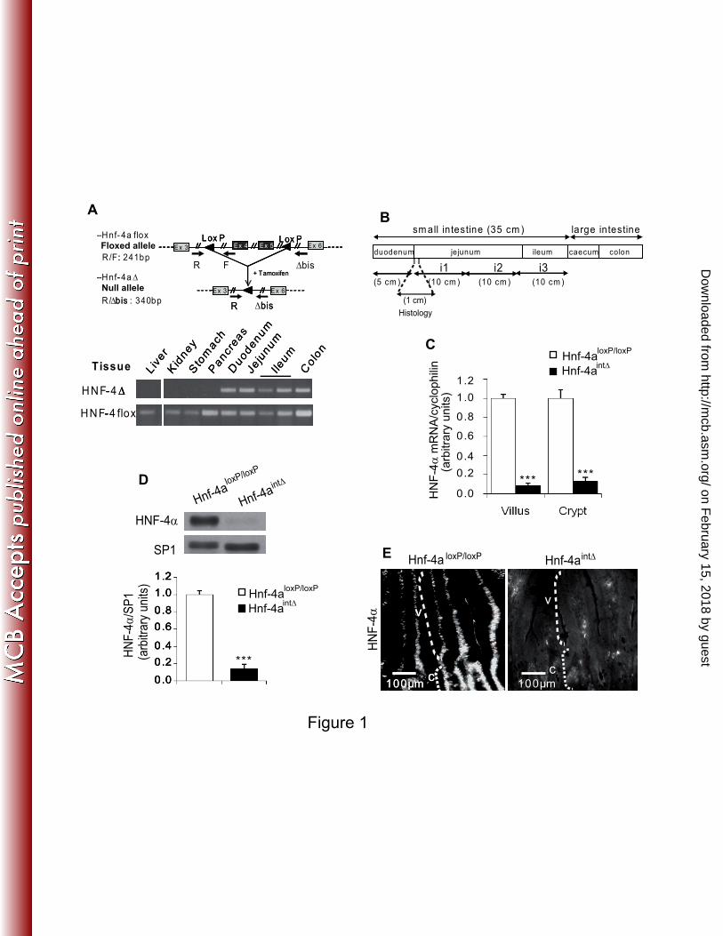
FIG. 1. The induction of CreERT2 recombinase leads to a specific loss of HNF-4α in the 2
epithelium of adult mouse small intestine. (A) Schematic representation of the gene with the 3
positions of the three primers used for genotyping (top) and identification of the 4
recombination of the Hnf-4a gene by PCR analysis of genomic DNA from various tissues 5
(bottom). The 340 bp fragment and the 241 bp fragment originate respectively from the 6
recombined and the intact alleles. The epithelium was obtained by scrapping and the 7
contaminating mesenchyme, where the villin promoter is not active, is at the origin of the 241 8
bp fragment in the five intestinal samples. Recombination is only detectable in the intestine. 9
(B) Schematic representation of the intestinal cephalo-caudal axis. The i1 part of the jejunum 10
was taken to perform RNA and protein analyses. About 1cm of the proximal jejunum was 11
removed for histological analyses. (C) Quantification of Hnf-4a mRNA level by RT-qPCR in 12
epithelial cells from villi and crypts of the jejunum. The mRNA levels are expressed relative 13
to the amount present in Hnf-4aloxP/loxP
mice and were normalized to that of cyclophilin. (D) 14
Representative western-blot and quantification analysis of HNF-4α protein in the epithelial 15
cells from the jejunum of control Hnf-4aloxP/loxP
and Hnf-4aint∆
mice. The level of HNF-4α is 16
expressed relative to the amount present in Hnf-4aloxP/loxP
mice and is normalized to that of 17
SP1. (E) Immunofluorescence staining of HNF-4α in the jejunum of Hnf-4aloxP/loxP
and Hnf-18
4aint∆
mice. Crypts and villi are indicated by C and V, respectively. For each graph an 19
unpaired Student’s t test was used to measure significance. ***, P<0.001. Error bars indicate 20
the standard error of the mean. 21
FIG 2: Cell proliferation in adult intestinal epithelium 30 days after tamoxifen injection. (A) 22
Representative western-blot of HNF-4α protein in epithelial cells from the jejunum of control 23
Hnf-4aloxP/loxP
and Hnf-4aint∆
mice 30 days following the first injection. (B) BrdU (green) and 24
DAPI (blue) staining in the crypts of jejunum from Hnf-4aint∆
and Hnf-4aloxP/loxP
mice. The 25
on February 15, 2018 by guest
http://mcb.asm
.org/D
ownloaded from
Page 27
27
white dashed bracket delimits the crypt compartment. (C) Representation of the average 1
length of crypts as determined by measuring 40 crypts from Hnf-4aint∆
or from Hnf-4aloxP/loxP
2
mice (n=3 animals per genotype). (D) Representation of the average number of BrdU positive 3
epithelial cells per crypt of Hnf-4aint∆
and Hnf-4aloxP/loxP
mice (n=40 crypts from 3 animals per 4
genotype). 5
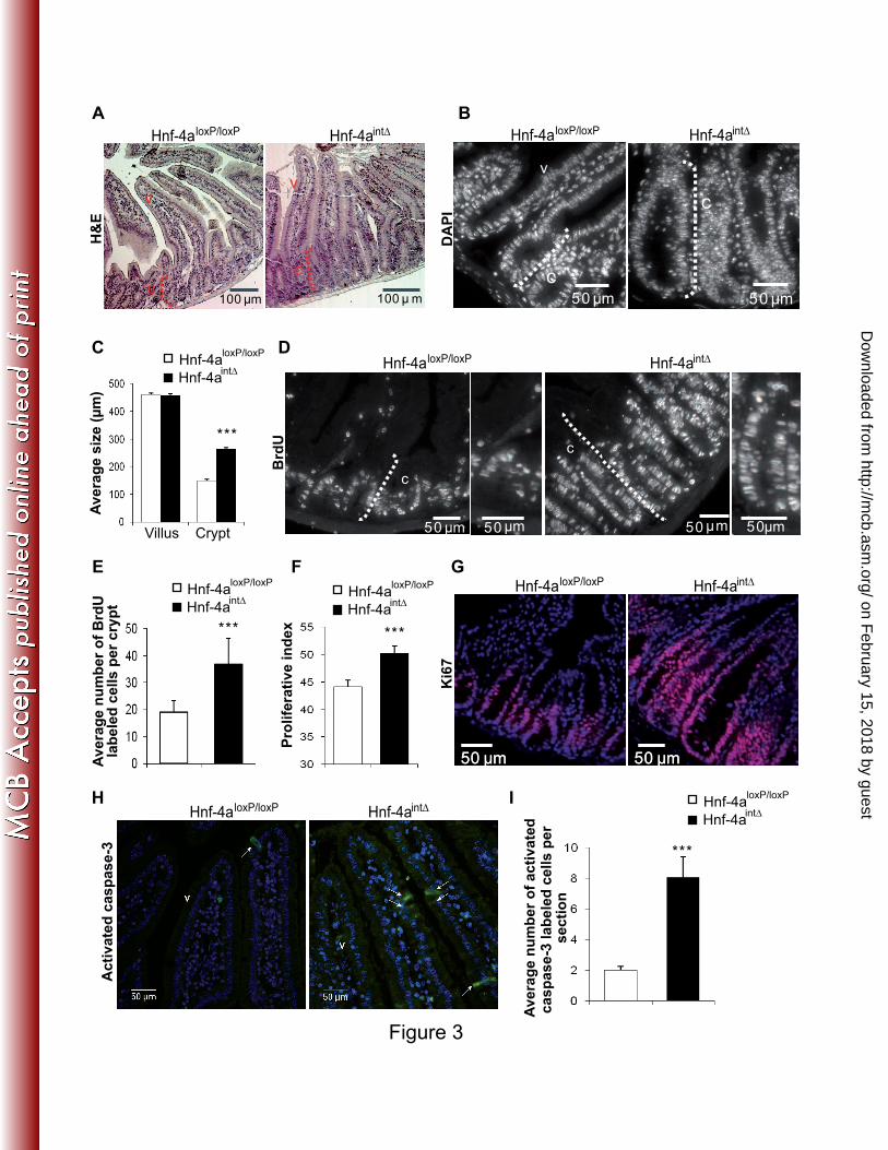
FIG. 3. Increased cell proliferation and death in the absence of HNF-4α in adult intestinal 6
epithelium. (A) Hematoxylin and eosin staining of Hnf-4aint∆
and Hnf-4aloxP/loxP
jejunum 7
sections shows the general structure of crypt-to-villus axis of Hnf-4aint∆
and Hnf-4aloxP/loxP
8
mice. Note the size of the crypt of Hnf-4aint∆
mice compared to that of control mice Hnf-9
4aloxP/loxP
. The dashed line indicates the crypt (c) and v marks the villus. (B) DAPI staining to 10
visualize nuclei in the crypts of Hnf-4aint∆
and Hnf-4aloxP/loxP
mice. The white dashed bracket 11
delimits the crypt compartment. (C) Representation of the average length of crypts and villi as 12
determined by measuring 40 crypts or villi in each Hnf-4aint∆
and Hnf-4aloxP/loxP
mouse (n=9 13
animals per genotype). (D) BrdU staining of Hnf-4aint∆
and Hnf-4aloxP/loxP
jejunum sections 14
showing the progression rate of cells in S phase. The crypt compartment is delimitated by a 15
white dashed bracket. (E) Representation of the average number of BrdU positive epithelial 16
cells per crypt of Hnf-4aint∆
and Hnf-4aloxP/loxP
mice (n=70 crypts from 9 animals per 17
genotype). (F) Graphic representation of the proliferative index in crypts of Hnf-4aint∆
and 18
Hnf-4aloxP/loxP
mice, i.e. percentage of BrdU-positive epithelial cells per number of crypt cells 19
visualized by DAPI staining. (G) Immunostaining of Ki67 in Hnf-4aint∆
and Hnf-4aloxP/loxP
20
jejunum sections for the identification of epithelial cells undergoing cell cycle progression. 21
(H) Immunostaining of activated caspase-3 in Hnf-4aint∆
and Hnf-4aloxP/loxP
jejunum sections 22
for detection of the apoptosis status of epithelial cells. Positive cells are indicated by an 23
arrow. (I) Representation of the average number of activated caspase-3-positive cell number 24
per section of jejunum of Hnf-4aint∆
and Hnf-4aloxP/loxP
mice. For each graph an unpaired 25
on February 15, 2018 by guest
http://mcb.asm
.org/D
ownloaded from
Page 28
28
Student’s t test was used to measure significance. ***, P<0.001. Error bars indicate the 1
standard error of the mean. Crypts and villi are indicated by C and V, respectively. 2
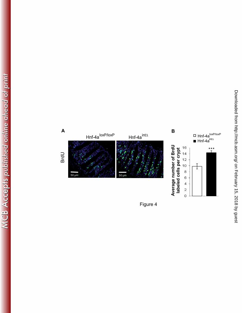
FIG. 4. Proliferation in colon epithelium of mice lacking HNF-4α. (A) Representative BrdU 3
staining in colon of Hnf-4aloxP/loxP
and Hnf-4aint∆
mice. (B) Average number of BrdU-positive 4
nuclei in epithelium of Hnf-4aloxP/loxP
and Hnf-4aint∆
mice. For each graph an unpaired 5
Student’s t test was used to measure significance. ***, P<0.001. Error bars indicate the 6
standard error of the mean. 7
FIG. 5. Loss of HNF-4α enhances the Wnt/β-catenin activity in the adult mouse small 8
intestine. (A) Immunohistochemistry of β-catenin of Hnf-4aint∆
and Hnf-4aloxP/loxP
jejunum 9
sections. Membrane and cytosol immunostained β-catenin is detected by a brown precipitate. 10
Sections were counterstained with hematoxylin (blue). Magnifications of the insets (upper 11
panels), delimitated by a black dashed square, are shown (lower panels). Cytosolic 12
accumulation of β-catenin is indicated by arrows. Crypts and villi are indicated by C and V, 13
respectively. (B) The level of nuclear β-catenin is enhanced in absence of HNF-14
4α. Immunoblot analysis with an anti-β-catenin antibody in crypts of jejunum revealed the 15
nuclear and cytoplasmic steady state levels of β-catenin. The cytoplasmic and nuclear fraction 16
purity was assessed through LDH and SP1 immunoblots, respectively. The level of protein 17
expression is expressed relative to the amount present in Hnf-4aloxP/loxP
mice and is 18
normalized to that of SP1 in nuclear fraction. Value represent the fold change in mean 19
expression between Hnf-4aint∆
and Hnf-4aloxP/loxP
mice (n=3 per genotype). (C) RT-qPCRs for 20
Wnt target gene expression in adult Hnf-4aint∆
and Hnf-4aloxP/loxP
jejunum. A significant up-21
regulation of TCF-4, Axin2, c-Myc, and LGR-5 and a significant down-regulation of p21 22
were seen after HNF-4α loss. The mRNA levels are expressed relative to the amount present 23
in Hnf-4aloxP/loxP
mice and were normalized to that of cyclophilin. (D) The p21 level is 24
on February 15, 2018 by guest
http://mcb.asm
.org/D
ownloaded from
Page 29
29
regulated by HNF-4α in the adult jejunum. Immunoblot analysis with an anti-p21 antibody 1
revealed the steady state levels of p21 and actin (loading control). The value represents the 2
fold change in the level of p21 expression between Hnf-4aint∆
and Hnf-4aloxP/loxP
mice and was 3
normalized to that of actin. (E) Overexpression of HNF-4α in HCT116 inhibits the TOPflash 4
activity. HCT116 cells were transfected with TOPflash, a β-catenin/TCF/LEF-responsive 5
luciferase reporter plasmid containing TCF-4-binding sites, or the corresponding negative 6
control FOPflash, along with HNF-4α and β-galactosidase vectors. Luciferase activity was 7
normalized to the corresponding β-galactosidase activity to obtain the relative luciferase 8
activity TOP/FOP. The graph represents the average luciferase activity (n=3 independent 9
experiments performed in triplicate). For each graph an unpaired Student’s t test was used to 10
measure significance. ***, **, *, P<0.001, P<0.01, and P<0.05, respectively. Error bars 11
indicate the standard error of the mean. 12
FIG. 6: HNF-4α interacts with Tcf-4 in intestinal epithelial cells. Co-immunoprecipitation 13
analysis of HNF-4α, Tcf-4 and β-catenin. (A) Lysates from HCT116 transfected with the 14
plasmids encoding HNF-4α and Flag-Tcf4 were subjected to immunoprecipitation with anti-15
HNF-4α, or anti-Flag, or anti-β-catenin antibodies as indicated in the top line. Input (four 16
percent of the total cell extracts), specific (IP) and non specific (IgG) immunoprecipitates 17
were analysed by western blot with the indicated antibodies. (B) Western blot detection of 18
endogenous HNF-4α in Caco-2/TC7 cells after immunoprecipitation with the indicated 19
antibodies. 20
FIG. 7. Altered number of goblet cells in the adult small intestine of Hnf-4aint∆
mice. (A) 21
Periodic Acid-Shiff (32) (32) staining (pink) of goblet cells in adult Hnf-4aint∆
and Hnf-22
4aloxP/loxP
jejunum sections. (B) Electron microscopy showing a mature goblet cell in adult 23
Hnf-4aint∆
jejunum. Optical microscopy shows that the goblet cells are opened at the luminal 24
on February 15, 2018 by guest
http://mcb.asm
.org/D
ownloaded from
Page 30
30
side and electron microscopy shows that the secretory granules occupy a large portion of the 1
cytoplasm. (C) Average number of PAS positive cells of adult Hnf-4aint∆
and Hnf-4aloxP/loxP
2
jejunum per villus or per crypt (n=8 crypt-to-villus axes and 3 mice per genotype). (D) 3
Chromogranin-A immunostainining (brown) and hematoxylin (blue) counterstaining of adult 4
Hnf-4aint∆
and Hnf-4aloxP/loxP
jejunum sections to visualize enteroendocrine cells. Arrows 5
indicate specific staining of enteroendocrine cells. (E) Average number of chromogranin-A 6
positive cells per villus or per crypt of adult Hnf-4aint∆
and Hnf-4aloxP/loxP
jejunum (n=40 7
crypt-to-villus axes from 8 animals per genotype). (F) Immunostaining of lysozyme of adult 8
Hnf-4aint∆
and Hnf-4aloxP/loxP
jejunum sections to visualize Paneth cells. Crypts and villi are 9
indicated by C and V, respectively. For each graph an unpaired Student’s t test was used to 10
measure significance. ***, *, P<0.001 and P<0.05, respectively. Error bars indicate the 11
standard error of the mean. 12
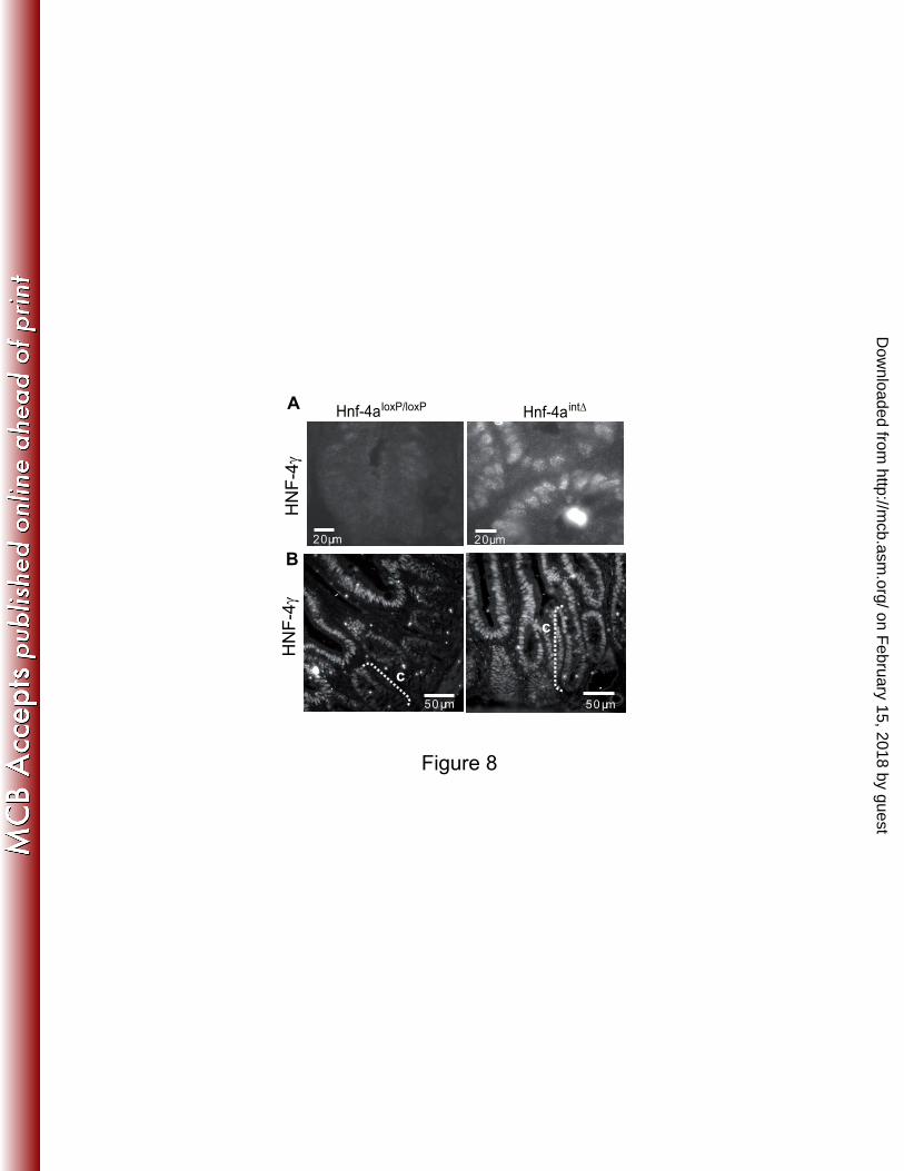
FIG. 8. Expression of HNF-4γ in the absence of HNF-4α. Representative HNF-4γ staining 13
observed in jejunum of Hnf-4aloxP/loxP
and Hnf-4aint∆
mice 8 days (A) or 30 days (B) after the 14
first tamoxifen injection. 15
FIG. 9. Expression of E-cadherin and EBP50 in the absence of HNF-4α. (A) The E-cadherin 16
mRNA level from villus epithelium is analyzed by RT-qPCRs in Hnf-4aloxP/loxP
and Hnf-4aint∆
17
mice. The mRNA level was expressed relative to the amount present Hnf-4aloxP/loxP
mice and 18
was normalized to that of cyclophilin. (B) Representative western-blot of E-cadherin in villus 19
epithelium of Hnf-4aloxP/loxP
and Hnf-4aint∆
mice. Histogram represents the quantification of 20
the protein level of E-cadherin (n=5 per genotype). The level of E-cadherin expression is 21
expressed relative to the amount present in Hnf-4aloxP/loxP
mice and is normalized to that of 22
actin. (C) Immunostaining of E-cadherin in the jejunum of Hnf-4aloxP/loxP
and of Hnf-4aint∆
23
mice. Note the diffuse staining of E-cadherin in the Hnf-4aint∆
mice. (D) The EBP50 mRNA 24
level from villus epithelium was analyzed by real-time qPCR in Hnf-4aloxP/loxP
and Hnf-4aint∆
25
on February 15, 2018 by guest
http://mcb.asm
.org/D
ownloaded from
Page 31
31
mice. The mRNA level was expressed relative to the amount present in Hnf-4aloxP/loxP
mice 1
and was normalized to that of cyclophilin. (E) Representative western-blot of EBP50 in villus 2
epithelium of Hnf-4aloxP/loxP
and Hnf-4aint∆
mice. Histogram represents the quantification of 3
the protein level of EBP50 (n=7 per genotype). The level of EBP50 expression is expressed 4
relative to the amount present in Hnf-4aloxP/loxP
mice and is normalized to that of actin. For 5
each graph an unpaired Student’s t test was used to measure significance. ***, P<0.001. Error 6
bars indicate the standard error of the mean. 7
FIG. 10. Expression of tight junction-associated proteins in the absence of HNF-4α. (A) The 8
mRNA levels encoding different junction proteins from villus epithelium are analyzed by RT-9
qPCR in Hnf-4aloxP/loxP
and Hnf-4aint∆
mice. The mRNA level was expressed relative to that of 10
Hnf-4aloxP/loxP
mice and was normalized to that of cyclophilin. (B) Representative western-blot 11
of Claudin-2, Claudin-7, and ZO-1 in jejunum of Hnf-4aloxP/loxP
and Hnf-4aint∆
mice. (C) 12
Quantification of the expression of tight junction-associated proteins by scanning western-13
blots. The level of protein expression is expressed relative to the amount present in Hnf-14
4aloxP/loxP
mice and is normalized to that of actin. Values represent the fold changes in mean 15
expression between Hnf-4aint∆
and Hnf-4aloxP/loxP
mice. For each graph an unpaired Student’s t 16
test was used to measure significance. ***, **, P<0.001 and P<0.01, respectively. Error bars 17
indicate the standard error of the mean. 18
FIG. 11. Distension of tight junctions and increased paracellular permeability in the absence 19
of HNF-4α. (A) Electron microscopy showing the intercellular space in tight junctions of Hnf-20
4aloxP/loxP
and Hnf-4aint∆
mice. The histogram represents the average width of tight junctions, 21
measured between the two external membranes, as indicated by arrowheads. (8 to 12 22
junctions and 3 animals per genotype). (B) In vivo paracellular permeability analysis by 23
measuring the FITC-Dextran-4Kd in serum from Hnf-4aloxP/loxP
and Hnf-4a
int∆ mice. (n=9 24
animals per genotype). (C) Paracellular permeability analysis by measuring the FITC-25
on February 15, 2018 by guest
http://mcb.asm
.org/D
ownloaded from
Page 32
32
Dextran-4Kd flux in Ussing chambers with Hnf-4aloxP/loxP
and Hnf-4aint∆
mice intestine. (n=12 1
animals per genotype.). For each graph an unpaired Student’s t test was used to measure 2
significance. ***, *, P<0.001 and P<0.05, respectively. Error bars indicate the standard error 3
of the mean. 4
on February 15, 2018 by guest
http://mcb.asm
.org/D
ownloaded from
Page 33
33
TABLE 1. Sequences of the primers used for semi quantitative RT-PCR 1 Forward primer 5’-> 3’ Reverse Primer 5’-> 3’
Cyclophilin Apo B Apo A4 Axin2 Cck Cdh-1 Cdx 1 Cdx 2 Claudin-2 Claudin-3 Claudin-4 Claudin-7 Claudin-12 Claudin-15 Crypt 1 Cyclin D1 Dsk2 EBP50 F11r FABP-I Gcg Gip Gjb 1 * Hes 1 HNF-1alpha HNF-1beta HNF-4alpha HNF-4gamma Klf4 Lgr5 Math 1 * Mmp7 Mttp Muc 2 Muc 3 Muc 4 c-myc Neuro-D Ngn 3 Ocln P21 P27 Pkp2 Sct Sox 9 Sst Tcf 4 Tff3 ZO1
TGGAGAGCACCAAGACAGACA GCCCATTGTGGACAAGTTGATC GACTACTTCAGCCAAAACAGTTGGA GAGAGTGAGCGGCAGAGC TGATTTCCCCATCCAAAGC TCCTTGTTCGGCTATGTGTC TCCGAGCTGGCTGCTAAC CACCATCAGGAGGAAAAGTGA GGCTGTTAGGCACATCCAT AAGCCGAATGGACAAAGAA CGCTACTCTTGCCATTACG AGGGTCTGCTCTGGTCCTT GTCCTCTCCTTTCTGGCAAC GCTTCTTCATGTCAGCCCTG TCAAGAGGCTGCAAAGGAAGA AAGTTCATTTCCAACCCACCC TGCAGTTCCCATTCGAGTTACAGACAGA AGGTCAATGGTGTCTGCATG CGAGGGGAAAGCCGGGAGGAAACTGTTGTT TTACCAGAAACCTCTCGGACA CACGCCCTTCAAGACACAG CAGGTAGGAGGAGAAGACCTCA GATGAGAAGTCCTCTTTCATCTGTAAC TCAACACGACACCGGACAAA AGAACCTCAGCCCAGAGGAG ATCCCCAGCAATCTCAGAAC CAGTGTCGTTACTGCAGGC (exon3) CTGCTGTTGCTGCTGCCA CGGGAAGGGAGAAGACACT CTTCACTCGGTGCAGTGCT TGCGATCTCCGAGTGAGAG TTCTGCTTTGTGTGTCTGCTG GGCAGTGCTTTTTCTCTGCT GCTGACGAGTGGTTGGTGAATG CGTGGTCAACTGCGAGAATGG CAGCAGCCAGTGGGGACAG GACAAGAGGCGGACACACAA GCATGCACGGGCTGAACGC CTTCACAAGAAGTCTGAGAACACCAG GCTGTGATGTGTGTGAGCTG TCCACAGCGATATCCAGACA TCAAACGTGAGAGTGTCTAACGG CGCCTCACCCTGCCACGCTATGCTC GCTGTGGTCGAACACTCAGA CAGCAAGACTCTGGGCAAG CCCAGACTCCGTCAGTTTCT CAACGAACACAGCGAATGTT CCTGGTTGCTGGGTCCTCTG AGGACACCAAAGCATGTGAG
TGCCGGAGTCGACAATGAT CCAGGACTTGGAGGTCTTGGA AAGCTGCCTTTCAGGTTCTCCT CGGCTGACTCGTTCTCCT GCTTCTGCAGGGACTACCG GGCATGCACCTAAGAATCAG CGGTTCTGGAACCAGATCTTT CTGCGGTTCTGAAACCAAAT TGGCACCAACATAGGAACTC CTGGCAAGTAGCTGCAGTG ACTCAGCACACCATGACTTG GTACGCAGCTTTGCTTTCA ATGTCGATTTCAATGGCAGA TTCTTGGAGAGATCCATGTTGC ACCCTTTCTGCAGGTTCCATT TGGAAAGAAAGTGCGTTGTGC TTGATGCCCGGGAAACACTACAGACTAAT TGCTGAAGGGCTCAGGCAAG CCGCATAGGGAGCTGTGATCTGGCTGTTAT TTACCAGAAACCTCTCGGACA GTCCTCATGCGCTTCTGTC CCTAGATTGTGTCCCCTAGCC GGTCATAGCAGACGCTGTTG TTATTCTTGCCCTTCGCCTC TGTGCTGCTGCAAGTACGA GCTTGGGAGGTGTTGAGG GATGGGAGAGGTGATCTGTTG (exon5) CAGTGTGGACATGGGACC GAGTTCCTCACGCCAACG CAGCCAGCTACCAAATAGGTG CTCTTCTGCAAGGTCTGATTTTT CCTTCTTTGTTTTAGAGTCATGAGG TGAGAGGCCAGTTGTGTGAC GATGAGGTGGCAGACAGGAGAC CGGCTCTATCTCTACGCTCTCC CTCAGACACGCCAGGGAACTC GGATGTAGGCGGCTTTT GGGATGCACCGGGAAGGAAG CTGCGCATAGCGGACCACAGCTTC GACGGTCTACCTGGAGGAAC GGACATCACCAGGATTGGAC AGGGGCTTATGATTCTGAAAGTCG GCGGGCGGTTCTGCTCTGCGTGCTTTGA GAGACAGGGACCCATCCAG TCCACGAAGGGTCTCTTCTC GGGCATCATTCTCTGTCTGG TTAGGAGCGCTCAGGTCTGT GCCACGGTTGTTACACTGTC GGCATTCCTGCTGGTTACA
2
The primers were designed in different exons of the corresponding gene, except in those 3
marked with an asterisk (*), which indicates that the gene has no intron. 4
on February 15, 2018 by guest
http://mcb.asm
.org/D
ownloaded from
Page 34
34
TABLE 2. Gene expression analysis in the jejunum of Hnf4αloxP/loxP mice and Hnf4α
int∆ mice. 1
Gene C or V Change in expression
Transcription factors
Hes-1 C 1.23ns
Math-1 C 1.86d
Ngn-3 V 2.3b
NeuroD V 1.14ns
Sox-9 C 1.29ns
HNF-4γ C 2.46b
V 1.10ns
HNF-1α V -1.16ns
HNF-1β C 1.80c
cdx-2 V -1.09ns
Klf-4 V -1.35c
Enteroendocrine cell markers
Gcg V -1.56c
Gip V -1.72d
Somatostatin V -1.72d
Secretin V 1.09ns
Paneth cell markers
MMP-7 C 1.13ns
Cryptdin-1 C -1.33a
Goblet cell markers
Muc 2 V 1.97b
Muc 3 V 1.01ns
Tff-3 V 1.35a
Enterocyte cell markers
Mttp V -1.27b
Apo B V -1.69d
Apo A4 V -1.39b
i-FABP V -1.45d
Quantitative real-time PCR from crypt (C) and from villus epithelium (V). All values are the 2
ratio (Hnf-4aint∆
/ Hnf-4aloxP/loxP
) of normalized expression levels. Negative values indicate 3
reduced gene expression in Hnf-4aint∆
compared to Hnf-4aloxP/loxP
intestines. 4
ap < 0.05.
bp < 0.01.
cp < 0.001.
dp < 0.0001. ns, not significant. 5
on February 15, 2018 by guest
http://mcb.asm
.org/D
ownloaded from
Page 35
R/F: 241 bp
R/ bis : 340 bpEx 3 Ex 6
bisR
Ex 3 Ex 4 Ex 6Ex 5Lox P Lox P
+ Tamoxifen- - 4a
-Hnf- 4a flox
:
∆bisEx 3 Ex 6
∆bisR
Ex 3 Ex 4 Ex 6Ex 5Lox P Lox P
Ex 3 Ex 4 Ex 6Ex 5Lox P Lox P
R F ∆bis+ Tamoxifen
- Hnf- ∆
-
HNF-4 ∆
HNF-4 flox
Liv
er
Kid
ney
Sto
mach
Pancr
eas
Duodenum
Jeju
num
Ileum
Colo
n
Tissue
- ∆
-
- ∆
-
Liv
er
Kid
ney
Sto
mach
Pancr
eas
Duodenum
Jeju
num
Ileum
Colo
n
A
C
*** ***
0.0
0.2
0.4
0.6
0.8
1.0
1.2
******
Hnf-4aloxP/loxP
int∆
Hnf-4a
HNF-4α
SP1
0.0
0.2
0.4
0.6
0.8
1.0
1.2
***
0.0
0.2
0.4
0.6
0.8
1.0
1.2
0.0
0.2
0.4
0.6
0.8
1.0
1.2
HN
F-4α
/SP
1(a
rbitr
ary
un
its)
HN
F-4α
Hnf-4a loxP/loxP Hnf-4aint∆
HN
F-4α
mR
NA
/cyclo
ph
ilin
(arb
itra
ry u
nits)
Hnf-4a
Hnf-4a
loxP/loxP
int∆
Hnf-4a
Hnf-4a
loxP/loxP
int∆
D
E
100 µm
v
c100 µm
c
v
100 µm
v
c100 µm
c
v
Floxed allele
Null allele
Figure 1
duodenum jejunum ileum caecum colon
in testine (35 cm )
(10 cm )
i1 i2 i3(5 cm )
sm all large intestine
(10 cm ) (10 cm )
Histology
B
(1 cm)
on February 15, 2018 by guest
http://mcb.asm
.org/D
ownloaded from
Page 36
Hnf-4aloxP/loxP
Hnf-4aint∆
50 µm 50 µm
cc
50 µm 50 µm
cc
Hnf-4aloxP/loxP
Hnf-4aint∆
Hnf-4a
Hnf-4a
loxP/loxP
int∆
******
Ave
rag
e c
ryp
t siz
e (
µm
)
Ave
rag
e n
um
be
r o
f B
rdU
lab
ele
d c
ells
pe
r cry
pt
A
B
C D
Figure 2
Hnf-4a
Hnf-4a
loxP/loxP
int∆
on February 15, 2018 by guest
http://mcb.asm
.org/D
ownloaded from
Page 37
50 µm 50 µm
c
50 µm 50 µm 50 µm 50 µm
c
50 µm
c
50 µm 50 µm 50 µm
c
100 µ 100 µ m
vv
cc
100 µm 100 µ m
vv
cc
50 µm
c
c
v
50 µm
c
c
v
50 µm50 µm 50 µm50 µm
Hnf-4aloxP/loxP Hnf-4aint∆ Hnf-4aloxP/loxP Hnf-4aint∆
Hnf-4aloxP/loxP Hnf-4aint∆
Hnf-4aloxP/loxP Hnf-4aint∆
Hnf-4aloxP/loxP Hnf-4aint∆
H&
E
DA
PI
Brd
U
Ki6
7
Ac
tiv
ate
d c
as
pa
se
-3
Hnf-4a
Hnf-4a
loxP/loxP
int∆
Hnf-4a
Hnf-4a
loxP/loxP
int∆Hnf-4a
Hnf-4a
loxP/loxP
int∆
Hnf-4a
Hnf-4a
loxP/loxP
int∆
Av
era
ge
siz
e (
µm
)A
ve
rag
e n
um
be
r o
f B
rdU
lab
ele
d c
ells
pe
r c
ryp
t
Pro
life
rati
ve
in
de
x
Av
era
ge
nu
mb
er
of
ac
tiv
ate
dc
as
pa
se
-3 la
be
led
ce
lls
pe
r s
ec
tio
n
A B
C D
E F G
H I
***
*** ***
***
50 µm
Villus Crypt
Figure 3
on February 15, 2018 by guest
http://mcb.asm
.org/D
ownloaded from
Page 38
50 µm50 µm 50 µm50 µm50 µm
Hnf-4aloxP/loxP
Hnf-4aint∆
Brd
U
Av
era
ge
nu
mb
er
of
Brd
U
lab
ele
d c
ells
pe
r c
ryp
t ***
A BHnf-4a
Hnf-4a
loxP/loxP
int∆
Figure 4
on February 15, 2018 by guest
http://mcb.asm
.org/D
ownloaded from
Page 39
50 µm
50 µm
50 µm
50 µm
cc
v v
β-c
ate
nin
50 µm
50 µm
50 µm
50 µm
cc
v v
Hnf-4aloxP/loxP
Hnf-4aint∆
Hnf-4aloxP/loxP
Hnf-4aint∆
Hnf-4aloxP/loxP
Hnf-4aint∆
β-cat
β-cat
SP1
SP1
LDH
LDH
cytoplasm
nucleus
1.50 0.014
Fold change
p value
Fold change
p value
A BHnf-4aloxP/loxP Hnf-4aint∆
0.0
0.5
1.0
1.5
2.0
2.5
3.0
0.0
0.5
1.0
1.5
2.0
2.5
3.0
Tcf4 c-myc Axin-2 Lgr-5 CyclinD1 p21
p21
actin
0.68 0.005
C
D
0.2
0.4
0.6
0.8
1.0
1.2
0.2
0.4
0.6
0.8
1.0
1.2
0.0
***
***
***
***
*
Empty vector (ng)
HNF-4α vector (ng)
1000 900 500
0 100 500
Re
lati
ve
TO
P/F
OP
Lu
cif
era
se
ac
tiv
ity
E
***
*
mR
NA
/cy
clo
ph
ilin
Hnf-4a
Hnf-4a
loxP/loxP
int∆
(a
rbit
rary
un
its
)
Figure 5
on February 15, 2018 by guest
http://mcb.asm
.org/D
ownloaded from
Page 40
Figure 6
on February 15, 2018 by guest
http://mcb.asm
.org/D
ownloaded from
Page 41
c
v
c
v
c
v
c
v
vv
50 µm
vv
50 µm
c c
50 µm50 µm
c c
50 µm50 µm
m
50 µm
cc
50 µm
m
50 µm
cc
50 µm
0.0
0.1
0.2
0.3
0.4
0.8
1.0
0.5
0.6
0.7
0.9
0.0
0.1
0.2
0.3
0.4
0.8
1.0
0.5
0.6
0.7
0.9
PA
SC
hro
mo
gra
nin
AL
ys
ozy
me
Ave
rag
e n
um
be
r o
fg
ob
let ce
lls
Ave
rag
e n
um
be
r o
fe
nte
roe
nd
ocrin
e c
ells
Hnf-4a
Hnf-4a
loxP/loxP
int∆
A B
C
D
E
Villus Crypt
Villus Crypt
Hnf-4aloxP/loxP Hnf-4aint∆
*** ***
*
100 µm 100 µm
50 µm
20 µm 20 µm20 µm 20 µm
2 µm2 µm
EM
F
Hnf-4aint∆
Figure 7
on February 15, 2018 by guest
http://mcb.asm
.org/D
ownloaded from
Page 42
20 µm20 µm 20 µm20 µm
50 µm 50 µm
c
c
50 µm 50 µm
c
c
Hnf-4aloxP/loxP Hnf-4aint∆
HN
F-4γ
HN
F-4γ
A
B
Figure 8
on February 15, 2018 by guest
http://mcb.asm
.org/D
ownloaded from
Page 43
0.0
0.2
0.4
1.2
0.6
0.8
1.0
0.0
0.2
0.4
1.2
0.6
0.8
1.0
0.0
0.5
1.0
1.5
0.0
0.5
1.0
1.5
20 µm20 µm
0.0
0.2
0.4
0.8
0.6
1.2
1.0
0.0
0.2
0.4
0.8
0.6
1.2
1.0
0.0
0.2
0.4
0.8
0.6
1.2
1.0
0.0
0.2
0.4
0.8
0.6
1.2
1.0
20 µm
Hnf-4a loxP/loxP Hnf-4a int∆
Hnf-4aloxP/lo
xP
Hnf-4aint∆
Hnf-4aloxP/lo
xP
Hnf-4aint∆
E-cadherin
E-cad
Actin
Fo
ld c
ha
ng
e(a
rbit
rary
un
its
)F
old
ch
an
ge
(arb
itra
ry u
nit
s)
EBP50
EBP50
Actin
Hnf-4a
Hnf-4a
loxP/loxP
int∆
Hnf-4a
Hnf-4a
loxP/loxP
int∆
Hnf-4a
Hnf-4a
loxP/loxP
int∆
Hnf-4a
Hnf-4a
loxP/loxP
int∆
A B
C
D E
***
***
mR
NA
/cy
clo
ph
ilin
(arb
itra
ry u
nit
s)
mR
NA
/cy
clo
ph
ilin
(arb
itra
ry u
nit
s)
Figure 9
on February 15, 2018 by guest
http://mcb.asm
.org/D
ownloaded from
Page 44
0.0
0.5
1 .5
1 .0
2.5
2 .0
0 .0
0 .5
1 .5
1 .0
2 .5
2 .0
Claudin
-2
Claudin
-3
Claudin
-4
Claudin
-7
Claudin
-12
Claudin
-15
Occ
ludin
JAM
-AZO
-1
Plako
philin
Desmoco
llinG
jb-1
mR
NA
/cy
clo
ph
ilin
Hnf-4a
Hnf-4a
loxP/loxP
int∆
***
***
***
** **
0.0
0.5
1.5
1.0
2.0
0.0
0.5
1.5
1.0
2.5
2.0
0.0
0.2
0.4
0.6
0.8
1.0
0.0
0.2
0.4
0.6
0.8
1.0
1.2
0.0
0.2
0.4
0.6
0.8
1.0
0.0
0.2
0.4
0.6
0.8
1.0
1.2
Hnf-4aloxP/lo
xP
Hnf-4aint∆
Claudin-2
Claudin-7
ZO-1
Actin
Claudin-2 Claudin-7 ZO-1
Fo
ld c
ha
ng
e
***
******
n=10 per genotype n=5 per genotype n=5 per genotype
Hnf-4a
Hnf-4a
loxP/loxP
int∆
A
B C
(a
rbit
rary
un
its
)
(a
rbit
rary
un
its
)
Figure 10
on February 15, 2018 by guest
http://mcb.asm
.org/D
ownloaded from
Page 45
100 nm100 nm100 nm 100 nm
100 nm100 nm100 nm 100 nm
Hnf-4aloxP/loxP
Hnf-4aint∆
FIT
C-D
ex
tra
n 4
kD
(ng
/ml s
eru
m)
Flu
x F
ITC
-De
xtr
an
4 k
D(p
mo
l/h
ou
r/c
m²)
Hnf-4a
Hnf-4a
loxP/loxP
int∆Hnf-4a
Hnf-4a
loxP/loxP
int∆
A
B C
* *
Wid
th (
nm
)
Hnf-4a
Hnf-4a
loxP/loxP
int∆
***
Figure 11
on February 15, 2018 by guest
http://mcb.asm
.org/D
ownloaded from