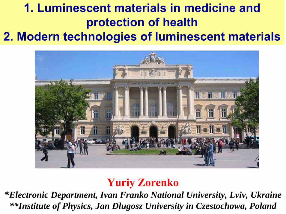
Yuriy Zorenko *Electronic Department, Ivan Franko National University, Lviv, Ukraine **Institute of Physics, Jan Dlugosz University in Czestochowa, Poland 1. Luminescent materials in medicine and protection of health 2. Modern technologies of luminescent materials
Transcript
Yuriy Zorenko*Electronic Department, Ivan Franko National University, Lviv, Ukraine
**Institute of Physics, Jan Dlugosz University in Czestochowa, Poland
1. Luminescent materials in medicine and protection of health
2. Modern technologies of luminescent materials
Topics of main part of lectures1. Milestone of luminescent materials in medicine and protection
of health2. Luminescence in solid state: types of emission centers,
luminescent mechanisms and energy transfer processes (2+2). 3. Technology of luminescent materials (2+2)4. Interaction of ionization radiation with organic and non-
organic materials5. Scintillators in medicine and monitoring of radiation (2+2);6. Computer tomography and positron-emission tomography7. Luminescent materials for dosimetry8. Luminescent materials for digital roentgenography9. Luminescent materials in raster scanning optical microscopy10. Luminescent materials for lighting11. Luminescent markers in biology and medicine12. Laser materials in medicine
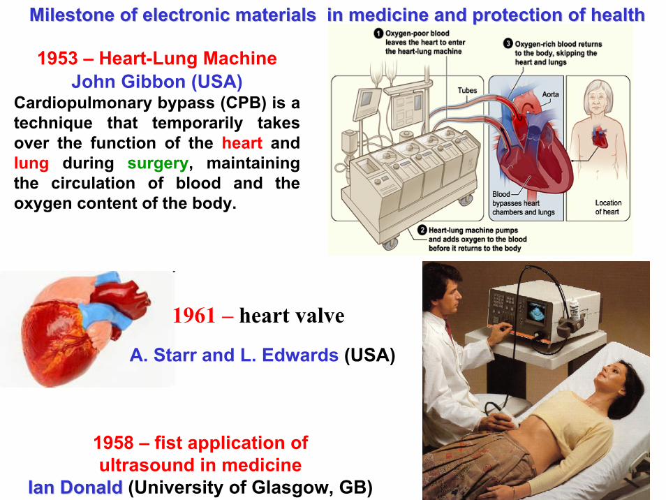
Milestone of Electronic in Medicine and Heals Milestone of Electronic in Medicine and Heals ProtectionProtection1895 – Intention of X- RaysWilhelm Conrad Röntgen, Nobelpreise in Physics, 19011895–1903 – Intention of Electrocardiogram (ECG)Willem von Eindhoven, Nobelpreis in Psychology and Medicine, 19241924 – invention of Electroencephalogram (EEG)Hans von Berger (University Jena, 1897)end of 40-th – Construction of the first electronic digital computerIt was built by John Vincent Atanasoff and Clifford Berry at Iowa State University during 1937-19421953 – Heart-Lung Machine
John Gibbon (USA)
1958 – Fist application of ultrasound in medicineIanIan DonaldDonald (University of Glasgow, GB)
1953 – Heart-Lung MachineJohn Gibbon (USA)
Cardiopulmonary bypass (CPB) is a technique that temporarily takesover the function of the heart andlung during surgery, maintainingthe circulation of blood and theoxygen content of the body.
1958 – fist application ofultrasound in medicine
IanIan DonaldDonald (University of Glasgow, GB)
Milestone of electronic materials in medicine and protection ofMilestone of electronic materials in medicine and protection of healthhealth
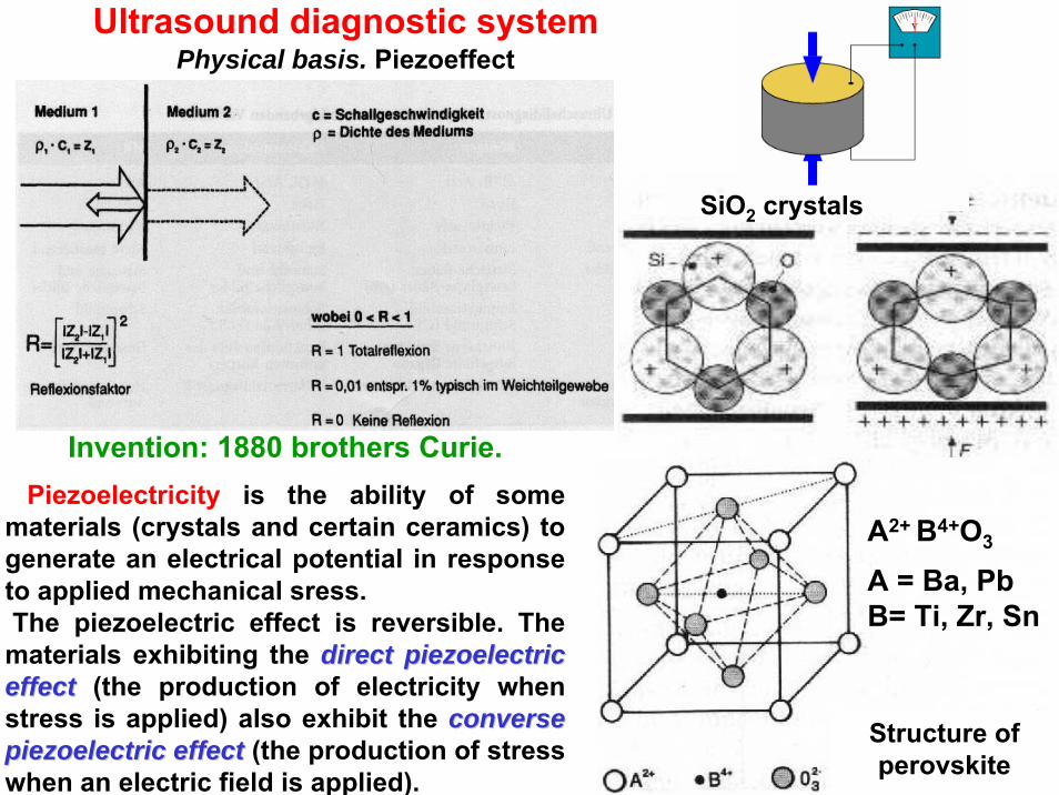
Invention: 1880 brothers Curie.Piezoelectricity is the ability of some
materials (crystals and certain ceramics) togenerate an electrical potential in responseto applied mechanical sress.The piezoelectric effect is reversible. Thematerials exhibiting the directdirect piezoelectricpiezoelectriceffecteffect (the production of electricity whenstress is applied) also exhibit the converseconversepiezoelectricpiezoelectric effecteffect (the production of stresswhen an electric field is applied).
Structure of perovskite
SiO2 crystals
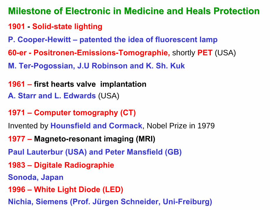
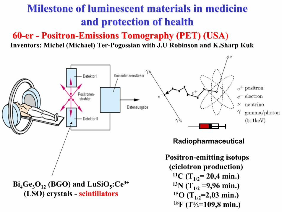
Milestone of Electronic in Medicine and Heals ProtectionMilestone of Electronic in Medicine and Heals Protection1901 - Solid-state lightingP. Cooper-Hewitt – patented the idea of fluorescent lamp60-er - Positronen-Emissions-Tomographie, shortly PET (USA)
M. Ter-Pogossian, J.U Robinson and K. Sh. Kuk
1961 – first hearts valve implantationA. Starr and L. Edwards (USA)
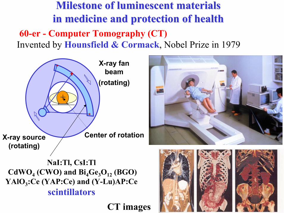
1971 – Computer tomography (CT)Invented by Hounsfield and Cormack, Nobel Prize in 1979
1977 – Magneto-resonant imaging (MRI)Paul Lauterbur (USA) and Peter Mansfield (GB)1983 – Digitale RadiographieSonoda, Japan1996 – White Light Diode (LED)Nichia, Siemens (Prof. Jürgen Schneider, Uni-Freiburg)
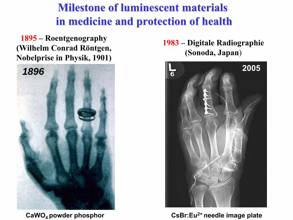
Milestone of luminescent materialsMilestone of luminescent materialsin medicine and protection of healthin medicine and protection of health
1895 – Roentgenography(Wilhelm Conrad Röntgen, Nobelprise in Physik, 1901)
CaWO4 powder phosphor
20051896
1983 – Digitale Radiographie (Sonoda, Japan)
CsBr:Eu2+ needle image plate
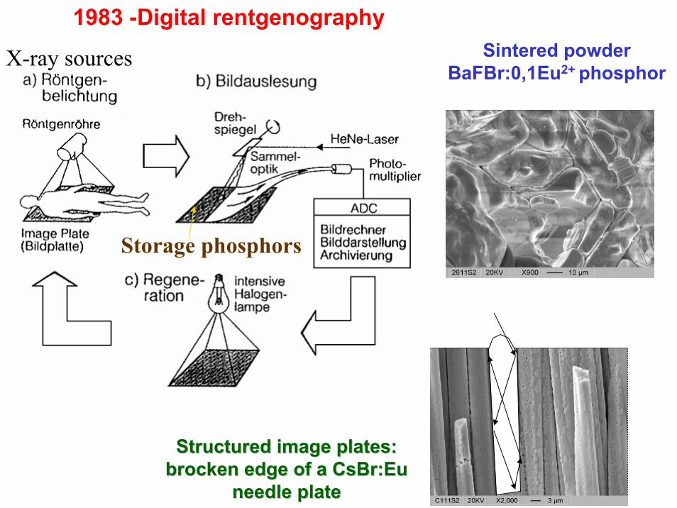
1983 -Digital rentgenography
Structured image plates: Structured image plates: brocken edge of a CsBr:Eu brocken edge of a CsBr:Eu
needle plateneedle plate
Sintered powder BaFBr:0,1Eu2+ phosphor
Storage phosphors
X-ray sources
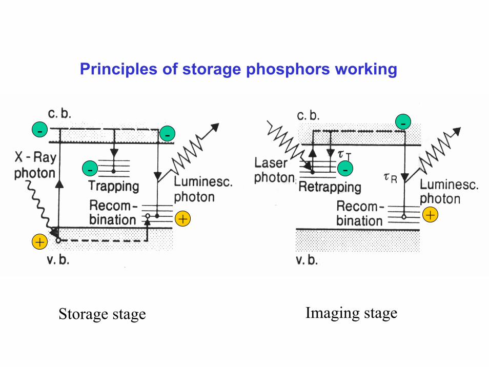
Principles of storage phosphors working
Storage stage Imaging stage
+
- -
+
-
+
- -
1. Irradiation2. Storage
3. Read-out
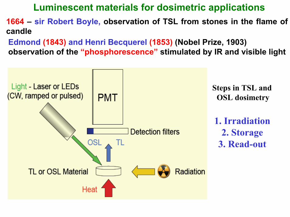
Luminescent materials for dosimetric applications1664 – sir Robert Boyle, observation of TSL from stones in the flame of candleEdmond (1843) and Henri Becquerel (1853) (Nobel Prize, 1903)observation of the “phosphorescence” stimulated by IR and visible light
Steps in TSL and OSL dosimetry
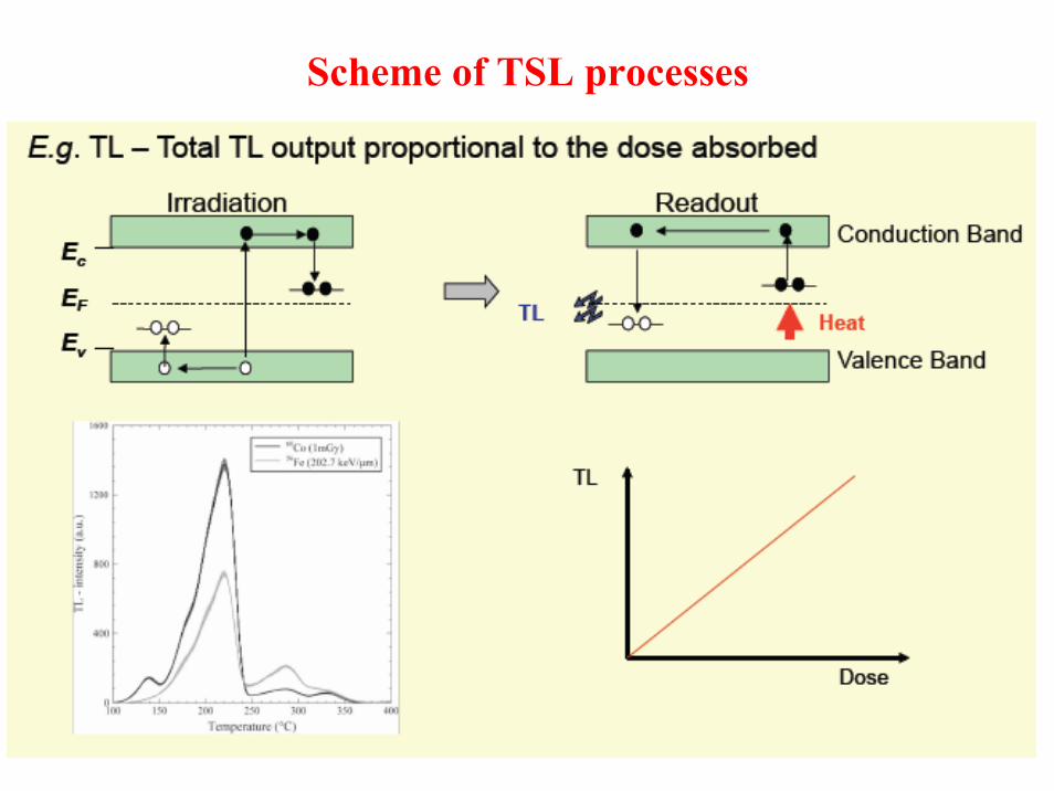
Scheme of TSL processes
Tissue equivalence for Photons
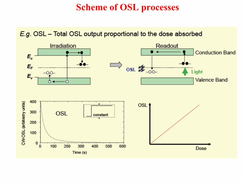
Scheme of OSL processes
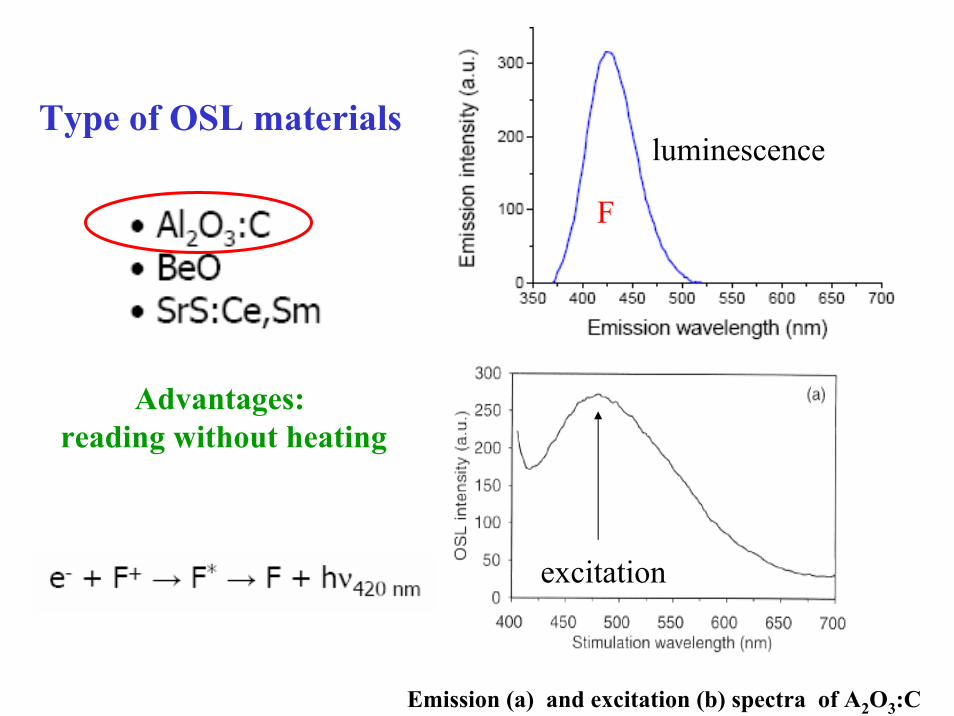
Type of OSL materials
Emission (a) and excitation (b) spectra of A2O3:C
F
excitation
luminescence
Advantages: reading without heating
Milestone of luminescent materials Milestone of luminescent materials in medicine and protection of healthin medicine and protection of health
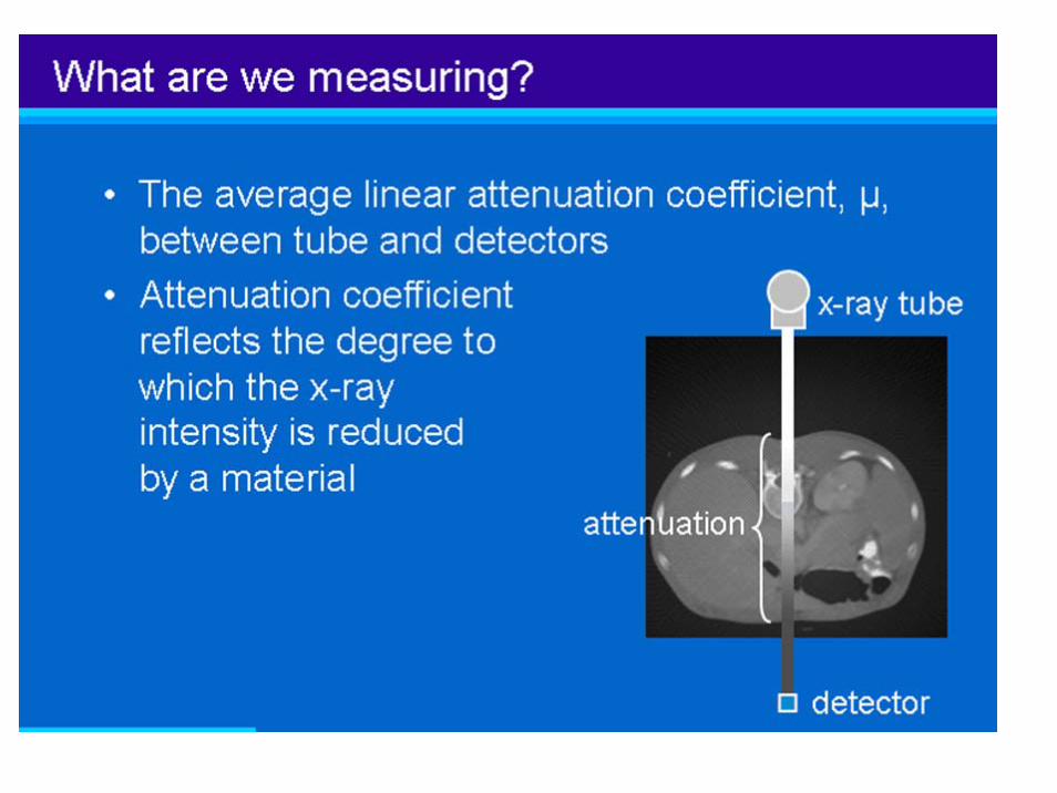
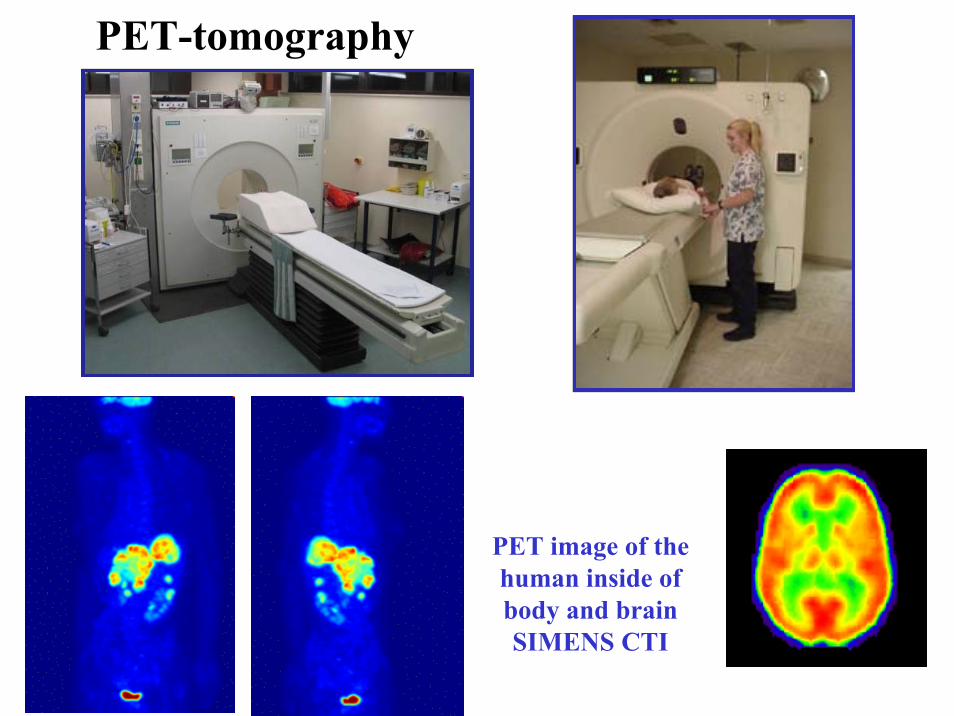
60-er - Computer Tomography (CT)
NaI:Tl, CsI:TlCdWO4 (CWO) and Bi4Ge3O12 (BGO)
YAlO3:Ce (YAP:Ce) and (Y-Lu)AP:Cescintillators
CT images
X-ray fan beam
(rotating)
X-ray source(rotating)
Center of rotation
+
Invented by Hounsfield & Cormack, Nobel Prize in 1979
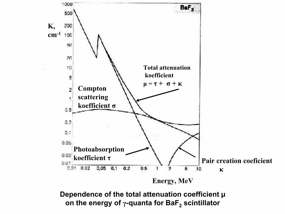
Energy, MeV
K,cm-1
Total attenuationkoefficientμ = τ + σ + κ
Photoabsorption koefficient τ
Compton scatteringkoefficient σ
Pair creation coeficientκ
Dependence of the total attenuation coefficient µon the energy of γ-quanta for BaF2 scintillator
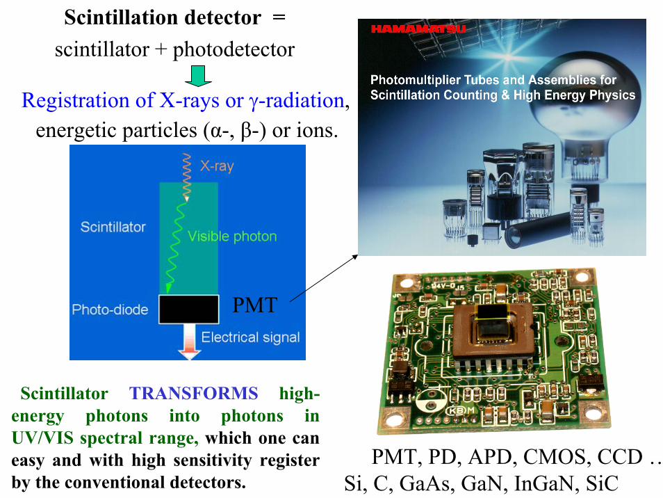
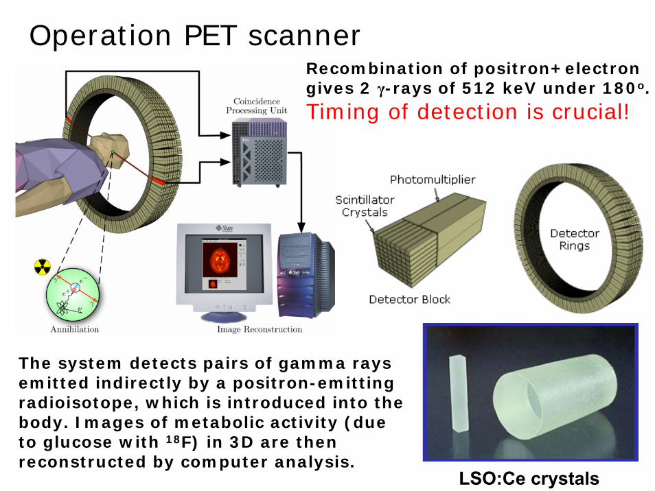
Registration of X-rays or γ-radiation, energetic particles (α-, β-) or ions.
Scintillator TRANSFORMS high-energy photons into photons in UV/VIS spectral range, which one can easy and with high sensitivity register by the conventional detectors.
The system detects pairs of gamma rays emitted indirectly by a positron-emitting radioisotope, which is introduced into the body. Images of metabolic activity (due to glucose with 18F) in 3D are then reconstructed by computer analysis.
Recombination of positron+electrongives 2 γ-rays of 512 keV under 180o.
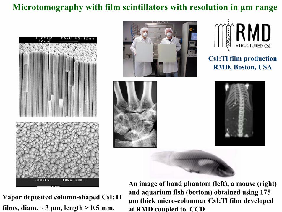
Microtomography with film scintillators with resolution in µm range
CsI:Tl film production RMD, Boston, USA
An image of hand phantom (left), a mouse (right) and aquarium fish (bottom) obtained using 175 μm thick micro-columnar CsI:Tl film developed at RMD coupled to CCD
Film screen
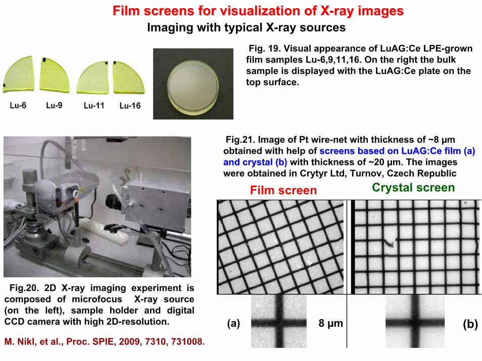
FilmFilm screens for visualization of Xscreens for visualization of X--ray imagesray images
Fig.21. Image of Pt wire-net with thickness of ~8 µm obtained with help of screens based onscreens based on LuAG:CeLuAG:Ce filmfilm (а)(а)and crystal and crystal ((bb)) with thickness of ~20 µm. The images were obtained in Crytyr Ltd, Turnov, Czech Republic
8 µm(а) (b)
Crystal screen
Fig. 19. Visual appearance of LuAG:Ce LPE-grown film samples Lu-6,9,11,16. On the right the bulk sample is displayed with the LuAG:Ce plate on the top surface.
Fig.20. 2D X-ray imaging experiment is composed of microfocus X-ray source (on the left), sample holder and digital CCD camera with high 2D-resolution.
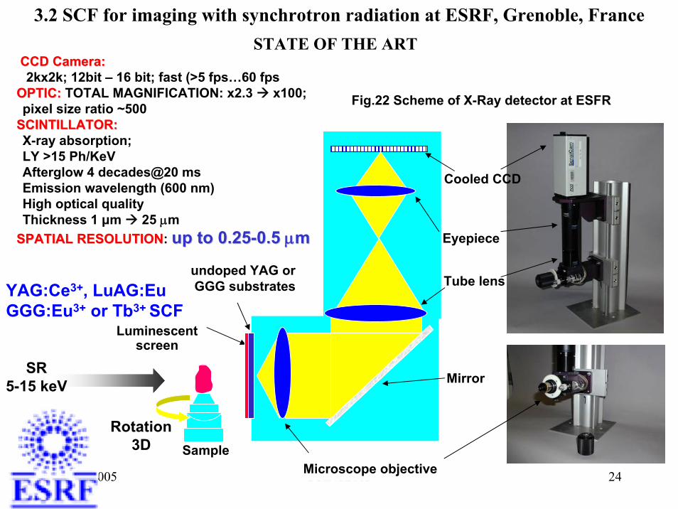
SPATIAL RESOLUTIONSPATIAL RESOLUTION: up to 0.25up to 0.25--0.5 0.5 μμmm
Cooled CCD
Sample
undoped YAG or GGG substratesYAG:Ce3+, LuAG:Eu
GGG:Eu3+ or Tb3+ SCF
3.2 SCF for imaging with synchrotron radiation at ESRF, Grenoble, FranceSTATE OF THE ART
Fig.22 Scheme of X-Ray detector at ESFR
Rotation3D
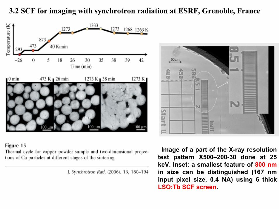
3.2 SCF for imaging with synchrotron radiation at ESRF, Grenoble, France
Image of a part of the X-ray resolutiontest pattern X500–200-30 done at 25 keV. Inset: a smallest feature of 800 nmin size can be distinguished (167 nminput pixel size, 0.4 NA) using 6 thickLSO:Tb SCF screen.
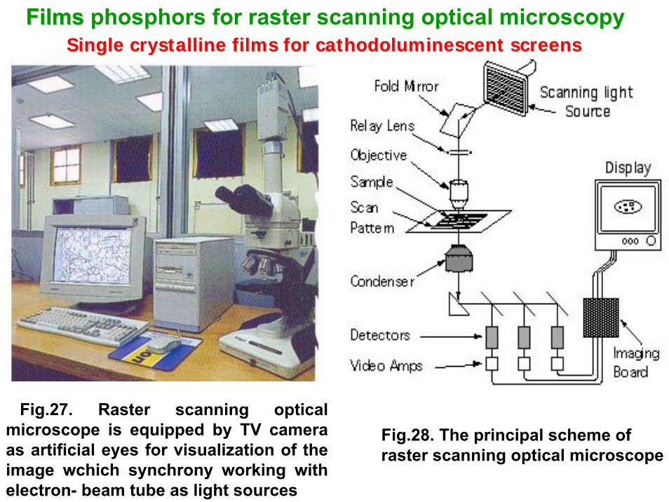
Fig.28. The principal scheme of raster scanning optical microscope
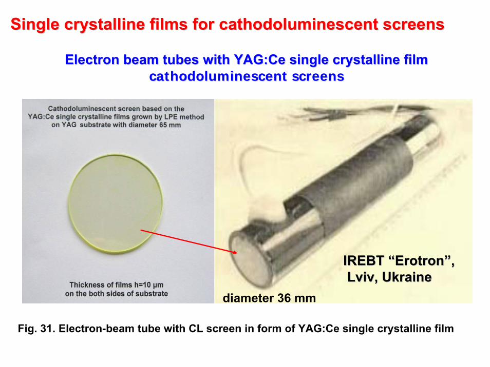
Single crystalline films for cathodoluminescent screensSingle crystalline films for cathodoluminescent screensFilmsFilms phosphors for raster scanning optical microscopy
Fig.27. Raster scanning optical microscope is equipped by TV camera as artificial eyes for visualization of the image wchich synchrony working with electron- beam tube as light sources
Electron beam tubes with Electron beam tubes with YAG:CeYAG:Ce single crystalline film single crystalline film cathodoluminescent screenscathodoluminescent screens
Fig. 31. Electron-beam tube with CL screen in form of YAG:Ce single crystalline film
Single crystalline films for cathodoluminescent screensSingle crystalline films for cathodoluminescent screens
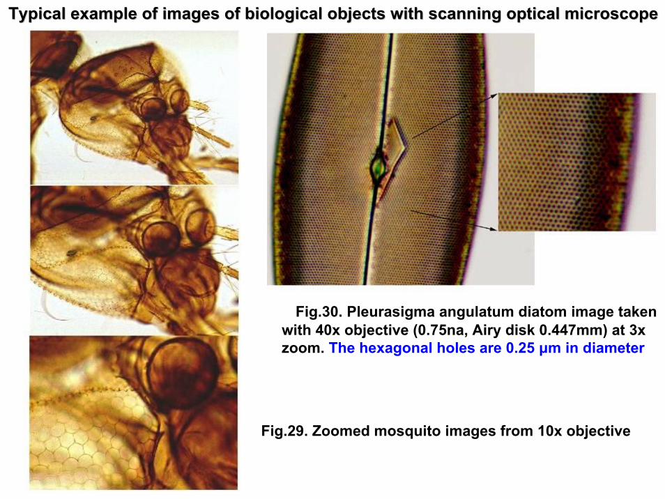
Fig.29. Zoomed mosquito images from 10x objective
Fig.30. Pleurasigma angulatum diatom image takenwith 40x objective (0.75na, Airy disk 0.447mm) at 3x zoom. The hexagonal holes are 0.25 μm in diameter
Typical example of images of biological objects with scanning opTypical example of images of biological objects with scanning optical microscopetical microscope
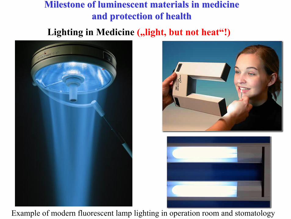
Lighting in Medicine („light, but not heat“!)
Example of modern fluorescent lamp lighting in operation room and stomatology
Milestone of luminescent materials in medicine Milestone of luminescent materials in medicine and protection of healthand protection of health
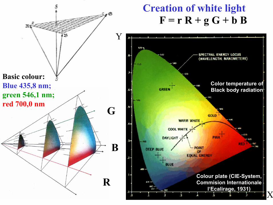
Colour plate (CIE-System, Commision Internationale
l‘Ecalirage, 1931)X
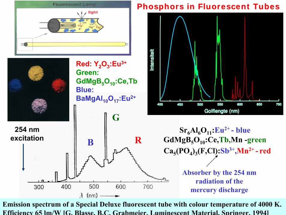
Emission spectrum of a Special Deluxe fluorescent tube with colour temperature of 4000 K. Efficiency 65 lm/W [G. Blasse, B.C. Grabmeier, Luminescent Material, Springer, 1994]
Sr6Al6O11:Eu2+ - blueGdMgB5O10:Ce,Tb,Mn -greenCa5(PO4)3(F,Cl):Sb3+,Mn2+ - red
RB
G
Absorber by the 254 nm radiation of the
mercury discharge
254 nmexcitation
Phosphors in Fluorescent TubesPhosphors in Fluorescent Tubes
Red: Y2O3:Eu3+
Green: GdMgB5O10:Ce,TbBlue: BaMgAl10O17:Eu2+
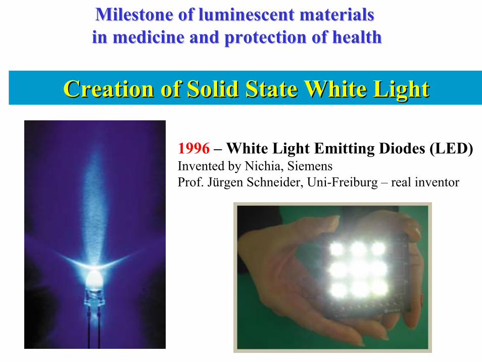
Creation of Solid State White LightCreation of Solid State White Light
1996 – White Light Emitting Diodes (LED) Invented by Nichia, Siemens Prof. Jürgen Schneider, Uni-Freiburg – real inventor
Milestone of luminescent materialsMilestone of luminescent materialsin medicine and protection of healthin medicine and protection of health
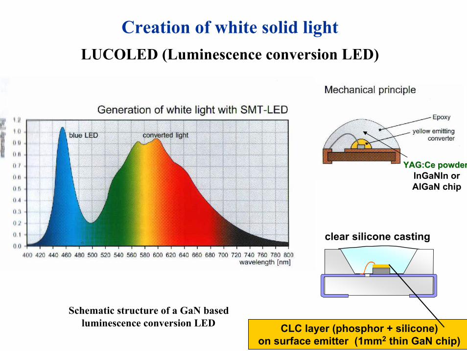
Creation of white solid lightLUCOLED (Luminescence conversion LED)
InGaNIn orAlGaN chip
Schematic structure of a GaN basedluminescence conversion LED
YAG:CeYAG:Ce powderpowder
clear silicone casting
CLC layer (phosphor + silicone) on surface emitter (1mm2 thin GaN chip)
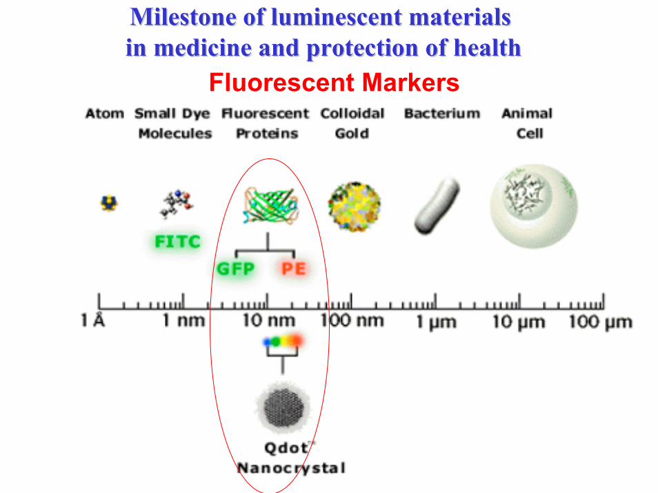
Fluorescent Markers
Milestone of luminescent materialsMilestone of luminescent materialsin medicine and protection of healthin medicine and protection of health
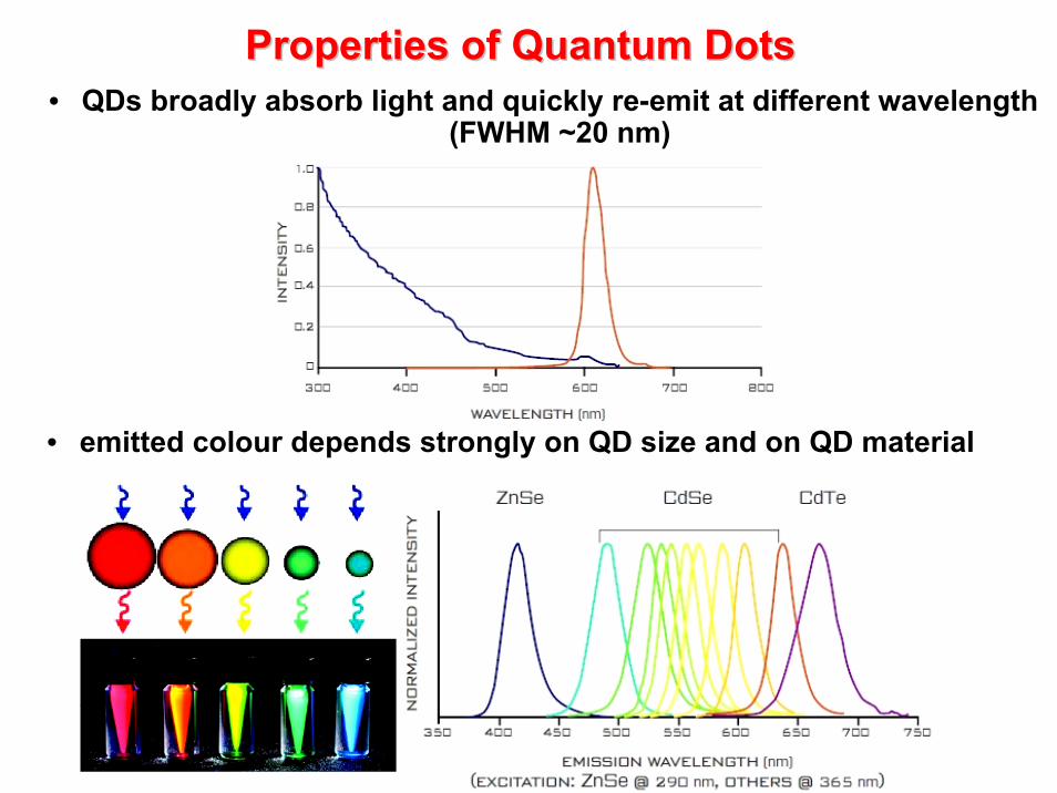
Properties of Quantum Properties of Quantum DotsDots• QDs broadly absorb light and quickly re-emit at different wavelength
(FWHM ~20 nm)
• emitted colour depends strongly on QD size and on QD material
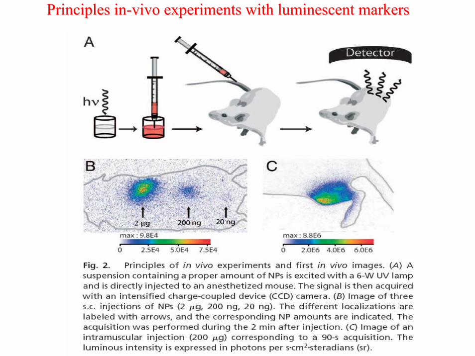
Principles inPrinciples in--vivo experiments with luminescent markersvivo experiments with luminescent markers
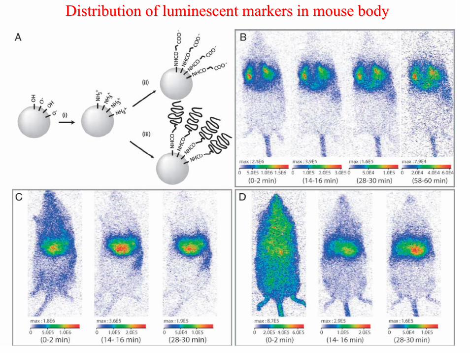
Distribution of luminescent markers in mouse bodyDistribution of luminescent markers in mouse body
Informationsverarbeitung. 2. Auflage, Springer-Verlag.[3] A. Winnacker. X-ray Imaging with Photostimulable Storage Phosphors and
Future Trends. Physica Medica – Vol. IX, No. 2-3 (1993).[4] J.F. Ziegler, J.P. Biersack, and U. Littmark, The Stopping and Ranges of Ions
in Solids, Pergamon Press, New York (1985).[5] L. H. Brixner: New X-ray phosphors, Mater. Chem. Phys. 16, 253-281 (1987)[6] M. Bruchez Jr. et al., Semiconductor Nanocrystals as Fluorescent Biological
Labels, science 281 (1998)[7] P. A. Rodnyi. Physical processes in inorganic scintillators. Science. 1997[8] G. F. Knoll: Radiation Detection and Measurement (Wiley, New York 2000)[9] M. J. Weber: Inorganic scintillators: today and tomorrow, J. Lumin. 100, 35-45
(2002)[10] A. A. Kaminsky, Laser Crystals, Dordrecht: Kluwer, 1990.[11] Phosphor Handbook. Edited by Sh. Shionoya and W. Yen. CRC press. NY.
1999. [12] S. Tavernier, B. Grinyov. Radiation detectors for medical applications .
Springer. 2006. [13] M. Nikl Scintillation detectors for x-rays, Meas. Sci. Technol. 17, R37-R54
(2006)[14] M. Nikl, V. Laguta, A. Vedda, Complex oxide scintillators: Material defects
and scintillation performance. Phys. Stat. sol. (b) 245, 1701-1722 (2008).