+ All Categories
Home > Documents > 1 Molecular Chaperones and the Ubiquitin– Proteasome · PDF fileIn some cases, mis- ......

1 Molecular Chaperones and the Ubiquitin– Proteasome · PDF fileIn some cases, mis- ......

Date post: 12-Mar-2018
Category:
Upload: ledieu
View: 219 times
Download: 1 times
Share this document with a friend
30
1 Molecular Chaperones and the Ubiquitin– Proteasome System Cam Patterson and Jo ¨rg Ho ¨hfeld Abstract A role for the ubiquitin–proteasome system in the removal of misfolded and ab- normal proteins is well established. Nevertheless, very little is known about how abnormal proteins are recognized for degradation by the proteasome. Recent ad- vances suggest that substrate recognition and processing require a close coopera- tion of the ubiquitin–proteasome system with molecular chaperones. Chaperones are defined by their ability to recognize nonnative conformations of other proteins and are therefore ideally suited to distinguish between native and abnormal pro- teins during substrate selection. Here we discuss molecular mechanisms that underlie the cooperation of molecular chaperones with the ubiquitin–proteasome system. Advancing our knowledge about such mechanisms may open up opportu- nities to modulate chaperone–proteasome cooperation in human diseases. 1.1 Introduction The biological activity of a protein is defined by its unique three-dimensional struc- ture. Attaining this structure, however, is a delicate process. A recent study sug- gests that up to 30% of all newly synthesized proteins never reach their native state [1]. As protein misfolding poses a major threat to cell function and viability, mo- lecular mechanisms must have evolved to prevent the accumulation of misfolded proteins and thus aggregate formation. Two protective strategies appear to be fol- lowed. Molecular chaperones are employed to stabilize nonnative protein confor- mations and to promote folding to the native state whenever possible. Alterna- tively, misfolded proteins are removed by degradation, involving, for example, the ubiquitin–proteasome system. For a long time molecular chaperones and cellular degradation systems were therefore viewed as opposing forces. However, recent evidence suggests that certain chaperones (in particular members of the 70- and 90-kDa heat shock protein families) are able to cooperate with the ubiquitin– Protein Degradation, Vol. 2: The Ubiquitin-Proteasome System. Edited by R. J. Mayer, A. Ciechanover, M. Rechsteiner Copyright 8 2006 WILEY-VCH Verlag GmbH & Co. KGaA, Weinheim ISBN: 3-527-31130-0 1
Transcript

1

Molecular Chaperones and the Ubiquitin–

Proteasome System

Cam Patterson and Jorg Hohfeld

Abstract

A role for the ubiquitin–proteasome system in the removal of misfolded and ab-

normal proteins is well established. Nevertheless, very little is known about how

abnormal proteins are recognized for degradation by the proteasome. Recent ad-

vances suggest that substrate recognition and processing require a close coopera-

tion of the ubiquitin–proteasome system with molecular chaperones. Chaperones

are defined by their ability to recognize nonnative conformations of other proteins

and are therefore ideally suited to distinguish between native and abnormal pro-

teins during substrate selection. Here we discuss molecular mechanisms that

underlie the cooperation of molecular chaperones with the ubiquitin–proteasome

system. Advancing our knowledge about such mechanisms may open up opportu-

nities to modulate chaperone–proteasome cooperation in human diseases.

1.1

Introduction

The biological activity of a protein is defined by its unique three-dimensional struc-

ture. Attaining this structure, however, is a delicate process. A recent study sug-

gests that up to 30% of all newly synthesized proteins never reach their native state

[1]. As protein misfolding poses a major threat to cell function and viability, mo-

lecular mechanisms must have evolved to prevent the accumulation of misfolded

proteins and thus aggregate formation. Two protective strategies appear to be fol-

lowed. Molecular chaperones are employed to stabilize nonnative protein confor-

mations and to promote folding to the native state whenever possible. Alterna-

tively, misfolded proteins are removed by degradation, involving, for example, the

ubiquitin–proteasome system. For a long time molecular chaperones and cellular

degradation systems were therefore viewed as opposing forces. However, recent

evidence suggests that certain chaperones (in particular members of the 70- and

90-kDa heat shock protein families) are able to cooperate with the ubiquitin–

Protein Degradation, Vol. 2: The Ubiquitin-Proteasome System.Edited by R. J. Mayer, A. Ciechanover, M. RechsteinerCopyright 8 2006 WILEY-VCH Verlag GmbH & Co. KGaA, WeinheimISBN: 3-527-31130-0

1

proteasome system. Protein fate thus appears to be determined by a tight interplay

of cellular protein-folding and protein-degradation systems.

1.2

A Biomedical Perspective

The aggregation and accumulation of misfolded proteins is now recognized as

a common characteristic of a number of degenerative disorders, many of which

have neurological manifestations [2, 3]. These diseases include prionopathies, Alz-

heimer’s and Parkinson’s diseases, and polyglutamine expansion diseases such as

Huntington’s disease and spinocerebellar ataxia. At the cellular level, these dis-

eases are characterized by the accumulation of aberrant proteins either intracellu-

larly or extracellularly in specific groups of cells that subsequently undergo death.

The precise association between protein accumulation and cell death remains in-

completely understood and may vary from disease to disease. In some cases, mis-

folded protein accumulations may themselves be toxic or exert spatial constraints

on cells that affect their ability to function normally. In other cases, the sequester-

ing of proteins in aggregates may itself be a protective mechanism, and it is the

overwhelming of pathways that consolidate aberrant proteins that is the toxic

event. In either case, lessons learned from genetically determined neurodegenera-

tive diseases have helped us to understand the inciting events of protein aggrega-

tion that ultimately lead to degenerative diseases.

Mutations resulting in neurodegenerative diseases fall into two broad classes.

The first class comprises mutations that affect proteins, irrespective of their native

function, and cause them to misfold. The classic example of this is Huntington’s

disease [4, 5]. The protein encoded by the huntingtin gene contains a stretch of

glutamine residues (or polyglutamine repeat), and the genomic DNA sequence

that codes for this polyglutamine repeat is subject to misreading and expansion.

When the length of the polyglutamine repeat in huntingtin reaches a critical

threshold of approximately 35 residues, the protein becomes prone to misfolding

and aggregation [6]. This appears to be the proximate cause of neurotoxicity in

this invariably fatal disease [7, 8]. A number of other neurodegenerative diseases

are caused by polyglutamine expansions [9, 10]. For example, spinocerebellar

ataxia is caused by polyglutamine expansions in the protein ataxin-1 [11]. In other

diseases, protein misfolding occurs due to other mutations that induce misfolding

and aggregation; for example, mutations in superoxide dismutase-1 lead to aggre-

gation and neurotoxicity in amyotrophic lateral sclerosis [12, 13].

Other mutations that result in neurodegenerative diseases are instructive in that

they directly implicate the ubiquitin–proteasome system in the pathogenesis of

these diseases [14]. For example, mutations in the gene encoding the protein par-

kin are associated with juvenile-onset Parkinson’s disease [15, 16]. Parkin is a

RING finger–containing ubiquitin ligase, and mutations in this ubiquitin ligase

cause accumulation of target proteins that ultimately result in the neurotoxicity

and motor dysfunction associated with Parkinson’s disease [17–20].

2 1 Molecular Chaperones and the Ubiquitin–Proteasome System

Repressor screens of neurodegeneration phenotypes in animal models have also

linked the molecular chaperone machinery to neurodegeneration [21–24]. Taken

together, the pathophysiology of neurodegenerative diseases provides a compelling

demonstration of the importance of the regulated metabolism of misfolded pro-

teins and provides direct evidence of the role of both molecular chaperones and

the ubiquitin–proteasome system in guarding against protein misfolding and its

consequent toxicity.

1.3

Molecular Chaperones: Mode of Action and Cellular Functions

Molecular chaperones are defined by their ability to bind and stabilize nonnative

conformations of other proteins [25, 26]. Although they are an amazingly diverse

group of conserved and ubiquitous proteins, they are also among the most abun-

dant intracellular proteins. The classical function of chaperones is to facilitate

protein folding, inhibit misfolding, and prevent aggregation. These folding events

are regulated by interactions between chaperones and ancillary proteins, the co-

chaperones, which in general assist in cycling unfolded substrate proteins on and

off the active chaperone complex [25, 27, 28]. In agreement with their essential

function under normal growth conditions, chaperones are ubiquitously expressed

and are found in all cellular compartments of the eukaryotic cell (except for perox-

isomes). In addition, cells greatly increase chaperone concentration as a response

to diverse stresses, when proteins become unfolded and require protection and sta-

bilization [29]. Accordingly, many chaperones are heat shock proteins (Hsps). Four

main families of cytoplasmic chaperones can be distinguished: the Hsp70 family,

the Hsp90 family, the small heat shock proteins, and the chaperonins.
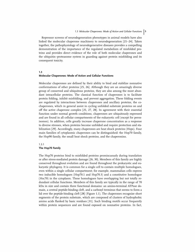
1.3.1

The Hsp70 Family

The Hsp70 proteins bind to misfolded proteins promiscuously during translation

or after stress-mediated protein damage [26, 30]. Members of this family are highly

conserved throughout evolution and are found throughout the prokaryotic and eu-

karyotic phylogeny. It is common for a single cell to contain multiple homologues,

even within a single cellular compartment; for example, mammalian cells express

two inducible homologues (Hsp70.1 and Hsp70.3) and a constitutive homologue

(Hsc70) in the cytoplasm. These homologues have overlapping but not totally re-

dundant cellular functions. Members of this family are typically in the range of 70

kDa in size and contain three functional domains: an amino-terminal ATPase do-

main, a central peptide-binding cleft, and a carboxyl terminus that seems to form a

lid over the peptide-binding cleft [28] (Figure 1.1). The chaperones recognize short

segments of the protein substrate, which are composed of clusters of hydrophobic

amino acids flanked by basic residues [31]. Such binding motifs occur frequently

within protein sequences and are found exposed on nonnative proteins. In fact,

1.3 Molecular Chaperones: Mode of Action and Cellular Functions 3

mammalian Hsp70 binds to a wide range of nascent and newly synthesized pro-

teins, comprising about 15–20% of total protein [32]. This percentage is most

likely further increased under stress conditions. Hsp70 proteins apparently prevent

protein aggregation and promote proper folding by shielding hydrophobic seg-

ments of the protein substrate. The hydrophobic segments are recognized by the

central peptide-binding domain of Hsp70 proteins (Figure 1.1). The domain is

composed of two sheets of b strands that together with connecting loops form a

cleft to accommodate extended peptides of about seven amino acids in length, as

revealed in crystallographic studies of bacterial Hsp70 [33]. In the obtained crystal

structure, the adjacent carboxyl-terminal domain of Hsp70 folds back over the b

sandwich, suggesting that the domain may function as a lid in permitting entry

and release of protein substrates (Figure 1.1). According to this model, ATP bind-

ing and hydrolysis by the amino-terminal ATPase domain of Hsp70 induce confor-

mational changes of the carboxyl terminus, which lead to lid opening and closure

[28]. In the ATP-bound conformation of Hsp70, the peptide-binding pocket is

open, resulting in rapid binding and release of the substrate and consequently in

a low binding affinity (Figure 1.1). Stable holding of the protein substrate requires

closing of the binding pocket, which is induced upon ATP hydrolysis and conver-

sion of Hsp70 to the ADP-bound conformation. The dynamic association of Hsp70

with nonnative polypeptide substrates thus depends on ongoing cycles of ATP

binding, hydrolysis, and nucleotide exchange. Importantly, ancillary co-chaperones

are employed to regulate the ATPase cycle [27, 30]. Co-chaperones of the Hsp40

family (also termed J proteins due to their founding member bacterial DnaJ) stim-

ulate the ATP hydrolysis step within the Hsp70 reaction cycle and in this way pro-

mote substrate binding [34] (Figure 1.1). In contrast, the carboxyl terminus of

Hsp70-interacting protein CHIP attenuates ATP hydrolysis [35]. Similarly, nucleo-

Fig. 1.1. Schematic presentation of the

domain architecture and chaperone cycle of

Hsp70. Hsp70 proteins display a characteristic

domain structure comprising an amino-

terminal ATPase domain (ATP), a peptide-

binding domain (P), and a carboxyl-terminal

domain (C) that is supposed to form a lid over

the peptide-binding domain. In the ATP-bound

conformation, the binding pocket is open,

resulting in a low affinity for the binding of a

chaperone substrate. ATP hydrolysis induces

stable substrate binding through a closure of

the peptide-binding pocket. Substrate release

is induced upon nucleotide exchange. ATP

hydrolysis and nucleotide exchange are

regulated by diverse co-chaperones.

4 1 Molecular Chaperones and the Ubiquitin–Proteasome System

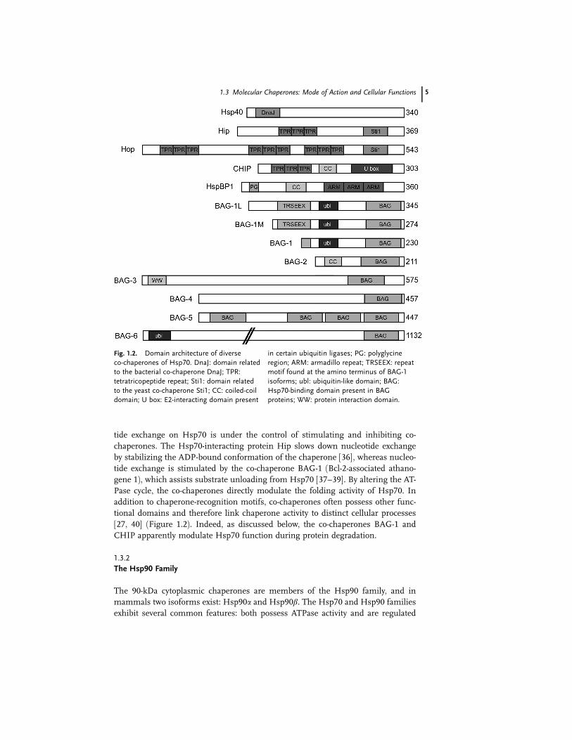
tide exchange on Hsp70 is under the control of stimulating and inhibiting co-

chaperones. The Hsp70-interacting protein Hip slows down nucleotide exchange

by stabilizing the ADP-bound conformation of the chaperone [36], whereas nucleo-

tide exchange is stimulated by the co-chaperone BAG-1 (Bcl-2-associated athano-

gene 1), which assists substrate unloading from Hsp70 [37–39]. By altering the AT-

Pase cycle, the co-chaperones directly modulate the folding activity of Hsp70. In

addition to chaperone-recognition motifs, co-chaperones often possess other func-

tional domains and therefore link chaperone activity to distinct cellular processes

[27, 40] (Figure 1.2). Indeed, as discussed below, the co-chaperones BAG-1 and

CHIP apparently modulate Hsp70 function during protein degradation.

1.3.2

The Hsp90 Family

The 90-kDa cytoplasmic chaperones are members of the Hsp90 family, and in

mammals two isoforms exist: Hsp90a and Hsp90b. The Hsp70 and Hsp90 families

exhibit several common features: both possess ATPase activity and are regulated

Fig. 1.2. Domain architecture of diverse

co-chaperones of Hsp70. DnaJ: domain related

to the bacterial co-chaperone DnaJ; TPR:

tetratricopeptide repeat; Sti1: domain related

to the yeast co-chaperone Sti1; CC: coiled-coil

domain; U box: E2-interacting domain present

in certain ubiquitin ligases; PG: polyglycine

region; ARM: armadillo repeat; TRSEEX: repeat

motif found at the amino terminus of BAG-1

isoforms; ubl: ubiquitin-like domain; BAG:

Hsp70-binding domain present in BAG

proteins; WW: protein interaction domain.

1.3 Molecular Chaperones: Mode of Action and Cellular Functions 5

by ATP binding and hydrolysis, and both are further regulated by ancillary co-

chaperones [41–48]. Unlike Hsp70, however, cytoplasmic Hsp90 is not generally

involved in the folding of newly synthesized polypeptide chains. Instead it plays a

key role in the regulation of signal transduction networks, as most of the known

substrates of Hsp90 are signaling proteins, the classical examples being steroid

hormone receptors and signaling kinases. On a molecular level, Hsp90 binds to

substrates at a late stage of the folding pathway, when the substrate is poised for

activation by ligand binding or associations with other factors. Consequently,

Hsp90 accepts partially folded conformations from Hsp70 for further processing.

In the case of the chaperone-assisted activation of the glucocorticoid hormone

receptor and also of the progesterone receptor, the sequence of events leading to

attaining an active conformation is fairly well understood [49–53]. It appears that

the receptors are initially recognized by Hsp40 and are then delivered to Hsp70

[54] (Figure 1.3). Subsequent transfer onto Hsp90 requires the Hsp70/Hsp90-

organizing protein Hop, which possesses non-overlapping binding sites for Hsp70

and Hsp90 and therefore acts as a coupling factor between the two chaperones

[55]. In conjunction with p23 and different cyclophilins, Hsp90 eventually medi-

Fig. 1.3. Cooperation of Hsp70 and Hsp90

during the regulation of signal transduction

pathways. The inactive signaling protein, e.g., a

steroid hormone receptor, is initially recognized

by Hsp40 and delivered to Hsp70. Subsequently,

a multi-chaperone complex assembles that

contains the Hsp70 co-chaperone Hip and the

Hsp70/Hsp90-organizing protein Hop. Hop

stimulates recruitment of an Hsp90 dimer that

accepts the substrate from Hsp70. At the final

stage of the chaperone pathway, Hsp90

associates with p23 and diverse cyclophilins

(cycloph.) to mediate conformational changes

of the signaling protein necessary to reach

an activatable state. Upon activation, i.e.,

hormone binding in the case of the steroid

receptor, the signaling protein is released

from Hsp90. In the absence of an activating

stimulus, the signaling protein folds back to

the inactive state when released and enters a

new cycle of chaperone binding.

6 1 Molecular Chaperones and the Ubiquitin–Proteasome System

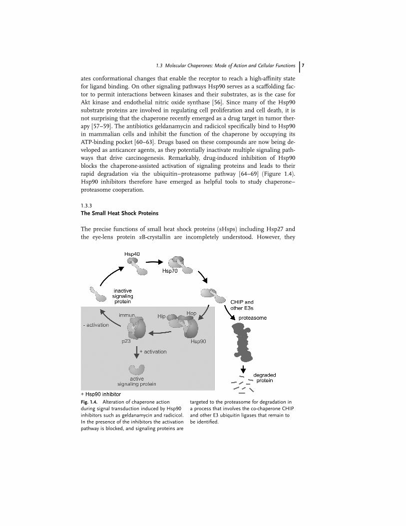
ates conformational changes that enable the receptor to reach a high-affinity state

for ligand binding. On other signaling pathways Hsp90 serves as a scaffolding fac-

tor to permit interactions between kinases and their substrates, as is the case for

Akt kinase and endothelial nitric oxide synthase [56]. Since many of the Hsp90

substrate proteins are involved in regulating cell proliferation and cell death, it is

not surprising that the chaperone recently emerged as a drug target in tumor ther-

apy [57–59]. The antibiotics geldanamycin and radicicol specifically bind to Hsp90

in mammalian cells and inhibit the function of the chaperone by occupying its

ATP-binding pocket [60–63]. Drugs based on these compounds are now being de-

veloped as anticancer agents, as they potentially inactivate multiple signaling path-

ways that drive carcinogenesis. Remarkably, drug-induced inhibition of Hsp90

blocks the chaperone-assisted activation of signaling proteins and leads to their

rapid degradation via the ubiquitin–proteasome pathway [64–69] (Figure 1.4).

Hsp90 inhibitors therefore have emerged as helpful tools to study chaperone–

proteasome cooperation.

1.3.3

The Small Heat Shock Proteins

The precise functions of small heat shock proteins (sHsps) including Hsp27 and

the eye-lens protein aB-crystallin are incompletely understood. However, they

Fig. 1.4. Alteration of chaperone action

during signal transduction induced by Hsp90

inhibitors such as geldanamycin and radicicol.

In the presence of the inhibitors the activation

pathway is blocked, and signaling proteins are

targeted to the proteasome for degradation in

a process that involves the co-chaperone CHIP

and other E3 ubiquitin ligases that remain to

be identified.

1.3 Molecular Chaperones: Mode of Action and Cellular Functions 7

seem to play a major role in preventing protein aggregation under conditions of

cellular stress [70–73]. All members investigated so far form large oligomeric com-

plexes of spherical or cylindrical appearance [74, 75]. Complex formation is inde-

pendent of ATP binding and hydrolysis, but appears to be regulated by tempera-

ture and phosphorylation. The structural analysis of wheat Hsp16.9 suggested

that the oligomeric complex acts as a storage form rather than an enclosure for

substrates, as the active chaperone appears to be a dimer [75]. In agreement with

this notion, dissociation of the oligomeric complex formed by yeast Hsp26 was

found to be a prerequisite for efficient chaperone activity [76]. Subsequent refold-

ing may occur spontaneously or may involve cooperation with other chaperones

such as Hsp70 [77].

1.3.4

Chaperonins

The chaperone proteins best understood with regard to their mode of action are

certainly the so-called chaperonins, which are defined by a barrel-shaped, double-

ring structure [25, 28]. Members include bacterial GroEL, Hsp60 of mitochondria

and chloroplasts, and the TriC–CCT complex localized in the eukaryotic cytoplasm.

Based on their characteristic ring structure, a central cavity is formed, which ac-

commodates nonnative proteins via hydrophobic interactions. Conformational

changes of the chaperonin subunits induced through ATP hydrolysis change the

inner lining of the cavity from a hydrophobic to a hydrophilic character [78–80].

As a consequence the unfolded polypeptide is released into the central chamber

and can proceed on its folding pathway in a protected environment [81]. The chap-

eronins are therefore capable of folding proteins such as actin that cannot be prop-

erly folded via other mechanisms [82].

1.4

Chaperones: Central Players During Protein Quality Control

Due to their ability to recognize nonnative conformations of other proteins, molec-

ular chaperones are of central importance during protein quality control. This was

elegantly revealed in studies on the influence of the Hsp70 chaperone system on

polyglutamine diseases using the fruit fly Drosophila melanogaster as a model or-

ganism (reviewed in Refs. [23] and [83]). Hallmarks of the polyglutamine disease

spinocerebellar ataxia type 3 (SCA3), for example, were recapitulated in transgenic

flies that expressed a pathological polyQ tract of the ataxin-3 protein in the eye disc

[84]. Transgene expression caused formation of abnormal protein inclusions and

progressive neuronal degeneration. Intriguingly, co-expression of human cytoplas-

mic Hsp70 suppressed polyQ-induced neurotoxicity. In a similar experimental

approach, Hsp40 family members protected neuronal cells against toxic polyQ ex-

pression [22]. Enhancing the activity of the Hsp70/Hsp40 chaperone system appar-

ently mitigates cytotoxicity caused by the accumulation of aggregation-prone pro-

8 1 Molecular Chaperones and the Ubiquitin–Proteasome System

teins. These findings obtained in Drosophila were confirmed in a mouse model

of spinocerebellar ataxia type 1 (SCA1) [85, 86]. Unexpectedly, however, the Hsp70

chaperone system was unable to prevent the formation of protein aggregates in

these models of polyglutamine diseases and upon polyQ expression in yeast and

mammalian cells [84, 85, 87–89]. Elevating the cellular levels of Hsp70 and of

some Hsp40 family members affected the number of protein aggregates and their

biochemical properties, but did not inhibit the formation of polyQ aggregates. No-

tably, Hsp70 and Hsp40 profoundly modulated the aggregation process of polyQ

tracts in biochemical experiments; this led to the formation of amorphous, SDS-

soluble aggregates, instead of the ordered, SDS-insoluble amyloid fibrils that form

in the absence of the chaperone system [88]. These biochemical data were con-

firmed in yeast and mammalian cells [88, 90]. Although unable to prevent the for-

mation of protein aggregates, the Hsp70 chaperone system apparently prevents the

ordered oligomerization and fibril growth that is characteristic of the disease pro-

cess. In an alternate but not mutually exclusive model to explain their protective

role, the chaperones may cover potentially dangerous surfaces exposed by polyQ-

containing proteins during the oligomerization process or by the final oligomers.

Intriguingly, elevated expression of Hsp70 also suppresses the toxicity of the non-

polyQ-containing protein a-synuclein in a Drosophila model of Parkinson’s disease

without inhibiting aggregate formation [24]. Hsp70 may thus exert a rather general

function in protecting cells against toxic protein aggregation. This raises the excit-

ing possibility that treatment of diverse forms of human neurodegenerative dis-

eases may be achieved through upregulation of Hsp70 activity.

The mentioned examples illustrate that one does not have to evoke the refolding

of an aberrant protein to the native state in order to explain the protective activity

of Hsp70 observed in models of amyloid diseases. In some cases it might be suffi-

cient for Hsp70 to modulate the aggregation process or to shield interaction sur-

faces of the misfolded protein to decrease cytotoxic effects. Another option may in-

volve presentation of the misfolded protein to the ubiquitin–proteasome system for

degradation.

1.5

Chaperones and Protein Degradation

Hsp70 and Hsp90 family members as well as small heat shock proteins have all

been implicated to participate in protein degradation. For example, the small heat

shock protein Hsp27 was recently shown to stimulate the degradation of phos-

phorylated IkBa via the ubiquitin–proteasome pathway, which may account for

the antiapoptotic function of Hsp27 [91]. Similarly, Hsp27 facilitates the proteaso-

mal degradation of phosphorylated tau, a microtubule-binding protein and compo-

nent of protein deposits in Alzheimer’s disease [92]. Hsp70 participates in the deg-

radation of apolipoprotein B100 (apoB), which is essential for the assembly and

secretion of very low-density lipoproteins from the liver [93]. Under conditions of

limited availability of core lipids, apoB translocation across the ER membrane is

1.5 Chaperones and Protein Degradation 9

attenuated, resulting in the exposure of some domains of the protein into the cyto-

plasm and their recognition by Hsp70. This is followed by the degradation of apoB

via the ubiquitin–proteasome pathway. Elevating cellular Hsp70 levels stimulated

the degradation of the membrane protein, suggesting that the chaperone facilitates

sorting to the proteasome. Genetic studies in yeast indicate that cytoplasmic Hsp70

may fulfill a rather general role in the degradation of ER-membrane proteins that

display large domains into the cytoplasm [94]. In agreement with this notion,

Hsp70 also takes part in the degradation of immaturely glycosylated and aberrantly

folded forms of the cystic fibrosis transmembrane conductance regulator (CFTR)

[95–98]. CFTR is an ion channel localized at the apical surface of epithelial cells.

Its functional absence causes cystic fibrosis, the most common fatal genetic dis-

ease in Caucasians [99, 100]. The disease-causing allele, DF508, which is expressed

in more than 70% of all patients, drastically interferes with the protein’s ability to

fold, essentially barring it from functional expression in the plasma membrane.

However, wild-type CFTR also folds very inefficiently, and less than 30% of the pro-

tein reaches the plasma membrane [99]. While trafficking from the endoplasmic

reticulum (ER) to the Golgi apparatus, immature forms of CFTR are recognized

by quality-control systems and are eventually directed to the proteasome for degra-

dation [101–104]. A critical step during CFTR biogenesis is the inefficient folding

of the first of two cytoplasmically exposed nucleotide-binding domains (NBD1) of

the membrane protein [105, 106]. The disease-causing DF508 mutation localizes

to NBD1 and further decreases the folding propensity of this domain. During the

co-translational insertion of CFTR into the ER membrane, cytoplasmic Hsp70 and

its co-chaperone Hdj-2 bind to NBD1 and facilitate intramolecular interactions

between the domain and another cytoplasmic region of CFTR, the regulatory R-

domain [96, 107]. However, Hsp70 is also able to present CFTR to the ubiquitin–

proteasome system [97], and heterologous expression of CFTR in yeast revealed an

essential role of cytoplasmic Hsp70 in CFTR turnover [98]. Hsp70 is thus a key

player in the cellular surveillance system that monitors the folded state of CFTR

at the ER membrane.

Interestingly, CFTR and the disease form DF508 are deposited in distinct peri-

centriolar structures, termed aggresomes, upon overexpression or proteasome inhi-

bition [108]. Subsequent studies established that aggresomes are induced upon ec-

topic expression of many different aggregation-prone proteins (reviewed in Refs.

[109] and [110]). Aggresomes form near the microtubule-organizing center in a

manner dependent on the microtubule-associated motor protein dynein, and are

surrounded by a ‘‘cage’’ of filamentous vimentin [108, 111]. Aggresome formation

is apparently a specific and active cellular response when production of misfolded

proteins exceeds the capacity of the ubiquitin–proteasome system to tag and re-

move these proteins. They likely serve to protect the cell from toxic ‘‘gain-of-

function’’ activities acquired by misfolded proteins. Aggresomes are also of clinical

relevance as they share remarkable biochemical and structural features, for exam-

ple, with Lewy bodies, the cytoplasmic inclusion bodies found in neurons affected

by Parkinson’s disease [112]. The pathways that regulate aggresome assembly are

only now being explicated. Histone deacetylase 6 (HDAC6) appears to be a key reg-

10 1 Molecular Chaperones and the Ubiquitin–Proteasome System

ulator of aggresome assembly [113]. HDAC6 is a microtubule-associated deacety-

lase that has the capacity to bind both multi-ubiquitinated proteins and dynein mo-

tors and is believed to recruit misfolded proteins to the pericentriolar region for

aggresome assembly. Deletion of HDAC6 prevents aggresome formation and sen-

sitizes cells to the toxic effects of misfolded proteins, which supports the hypothe-

sis that aggresomes sequester misfolded proteins to protect against their toxic ac-

tivities. Components of the ubiquitin–proteasome system and chaperones such as

Hsp70 are abundantly present in and are actively recruited to aggresomes [114–

116]. Furthermore, elevating cellular Hsp70 levels can reduce aggresome forma-

tion by stimulating proteasomal degradation [117]. It appears that these subcellular

structures are major sites of chaperone–proteasome cooperation to mediate the

metabolism of misfolded proteins.

The formation of aggresome-like structures is also observed in dendritic cells

that present foreign antigens to other immune cells [118]. Immature dendritic

cells are located in tissues throughout the body, including skin and gut. When

they encounter invading microbes, the pathogens are endocytosed and processed

in a manner that involves the generation of antigenic peptides by the ubiquitin–

proteasome system. Upon induction of dendritic cell maturation, ubiquitinated

proteins transiently accumulate in large cytosolic structures that resemble aggre-

somes and were therefore termed DALIS (dendritic cell aggresome-like induced

structures). It was speculated that DALIS formation may enable dendritic cells

to regulate antigen processing and presentation. DALIS contain components of

the ubiquitin–proteasome machinery as well as Hsp70 and the co-chaperone

CHIP [118, 119]. Again, an interplay of molecular chaperones and the ubiquitin–

proteasome system during regulated protein turnover is suggested.

The cellular function of molecular chaperones is apparently not restricted to me-

diating protein folding; instead, chaperones emerge also as vital components on

protein-degradation pathways. Remarkably, the balance between folding and degra-

dation activities of chaperones can be manipulated. In cells treated with Hsp90

inhibitors, for example, with geldanamycin (see above), the chaperone-assisted acti-

vation of signaling proteins is abrogated and chaperone substrates such as the pro-

tein kinases Raf-1 and ErbB2 are rapidly degraded by the ubiquitin–proteasome

system [64–69, 120]. This appears to be due, in part, to transfer of the sub-

strates back to Hsp70 and progression toward the ubiquitin-dependent degradation

pathway.

Substrate interactions with chaperones – and consequently their commitment

either toward the folding pathway or to their degradation via the ubiquitin–

proteasome machinery – apparently serve as an essential post-translational protein

quality-control mechanism within eukaryotic cells. The partitioning of proteins to

either one of these mutually exclusive pathways is referred to as ‘‘protein triage’’

[121]. Although some misfolded proteins may be directly recognized by the protea-

some [122], specific pathways within the ubiquitin–proteasome system are proba-

bly relied on for the degradation of most misfolded and damaged proteins. For ex-

ample, E2 enzymes of the Ubc4/5 family selectively mediate the ubiquitylation of

abnormal proteins as revealed in genetic studies in Saccharomyces cerevisiae [123].

1.5 Chaperones and Protein Degradation 11

It is well accepted that chaperones play a central role in the triage decision; how-

ever, less well understood are the events that lead to the cessation of efforts to fold

a substrate, and the diversion of the substrate to the terminal degradative pathway.

It is possible that chaperones and components of the ubiquitin–proteasome path-

way exist in a state of competition for these substrates and that repeated cycling of

a substrate on and off a chaperone maintains the substrate in a soluble state and

increases, in a stochastic fashion, its likelihood of interactions with the ubiquitin

machinery (Figure 1.5A). However, some data argue for a more direct role of the

chaperones in the degradation process. Hsp70 plays an active and necessary role

in the ubiquitylation of some substrates [124]; this activity of Hsp70 requires its

chaperone function, indicating that conformational changes within substrates

may facilitate recognition by the ubiquitylation machinery. Plausible hypotheses

to explain these observations include direct associations between the chaperone

and ubiquitin–proteasome machinery to facilitate transfer of a substrate from one

pathway to the other, or conversion of the chaperone itself to a ubiquitylation com-

plex (Figure 1.5B). It is also entirely possible that several quality-control pathways

may exist and that the endogenous triage decision may involve aspects of each of

these hypotheses.

Fig. 1.5. Interplay of molecular chaperones

with the ubiquitin–proteasome system. (A)

Chaperones and the degradation machinery

(i.e., ubiquitylation systems) compete with

each other in the recognition of folding

intermediates. Interaction with the chaperones

directs the substrate towards folding. However,

when the protein substrate is unable to attain

a folded conformation, the chaperones

maintain the folding intermediate in a soluble

state that can be recognized by the

degradation machinery. (B) The chaperones

are actively involved in protein degradation.

Through an association with certain compo-

nents of the ubiquitin conjugation machinery

(degrading partner), the chaperones participate

in the targeting of protein substrates to the

proteasome. A competition between degrading

partners and folding partners determines

chaperone action and the fate of the protein

substrate.

12 1 Molecular Chaperones and the Ubiquitin–Proteasome System

1.6

The CHIP Ubiquitin Ligase: A Link Between Folding and Degradation Systems

Major insights into molecular mechanisms that underlie the cooperation of molec-

ular chaperones with the ubiquitin–proteasome system were obtained through the

functional characterization of the co-chaperone CHIP (reviewed in Ref. [40]).

CHIP was initially identified in a screen for proteins containing tetratricopeptide

repeat (TPR) domains, which are found in several co-chaperones – including Hip,

Hop, and the cyclophilins – as chaperone-binding domains [27, 55] (Figure 1.2).

CHIP contains three TPR domains at its amino terminus, which are used for bind-

ing to Hsp70 and Hsp90 [35, 125]. Besides the TPR domains, CHIP possesses a U-

box domain at its carboxyl terminus [35] (Figure 1.2). U-box domains are similar to

RING finger domains, but they lack the metal-chelating residues and instead are

structured by intramolecular interactions [126]. The predicted structural similarity

suggests that U boxes, like RING fingers, may also play a role in targeting proteins

for ubiquitylation and subsequent proteasome-dependent degradation, and this

possibility is borne out in functional analyses of U box–containing proteins [127,

128]. The TPR and U-box domains in CHIP are separated by a central domain

rich in charged residues. The charged domain of CHIP is necessary for TPR-

dependent interactions with Hsp70 [35] and is also required for homodimerization

of CHIP [129].

The tissue distribution of CHIP supports the notion that it participates in pro-

tein folding and degradation decisions, as it is most highly expressed in tissues

with high metabolic activity and protein turnover: skeletal muscle, heart, and brain.

Although it is also present in all other organs, including pancreas, lung, liver, pla-

centa, and kidney, the expression levels are much lower. CHIP is also detectable in

most cultured cells, and is particularly abundant in muscle and neuronal cells and

in tumor-derived cell lines [35]. Intracellularly, CHIP is primarily localized to the

cytoplasm under quiescent conditions [35], although a fraction of CHIP is present

in the nucleus [97]. In addition, cytoplasmic CHIP traffics into the nucleus in re-

sponse to environmental challenge in cultured cells, which may serve as a protec-

tive mechanism or to regulate transcriptional responses in the setting of stress

[130].

CHIP is distinguished among co-chaperones in that it is a bona fide interaction

partner with both of the major cytoplasmic chaperones Hsp90 and Hsp70, based

on their interactions with CHIP in the yeast two-hybrid system and in vivo bindingassays [35, 125]. CHIP interacts with the terminal-terminal EEVD motifs of Hsp70

and Hsp90, similar to other TPR domain–containing co-chaperones such as Hop

[55, 131, 132]. When bound to Hsp70, CHIP inhibits ATP hydrolysis and therefore

attenuates substrate binding and refolding, resulting in inhibition of the ‘‘forward’’

Hsp70 substrate folding/refolding pathway, at least in in vitro assays [35]. The cel-

lular consequences of this ‘‘anti-chaperone’’ function are not yet clear, and in fact

CHIP may actually facilitate protein folding under conditions of stress, perhaps by

slowing the Hsc70 reaction cycle [130, 133]. CHIP interacts with Hsp90 with ap-

proximately equivalent affinity to its interaction with Hsp70 [125]. This interaction

1.6 The CHIP Ubiquitin Ligase: A Link Between Folding and Degradation Systems 13

results in remodeling of Hsp90 chaperone complexes, such that the co-chaperone

p23, which is required for the appropriate activation of many, if not all, Hsp90 cli-

ent proteins, is excluded. The mechanism for this activity is unclear – p23 and

CHIP bind Hsp90 through different sites – yet the consequence of this action is

predictable: CHIP should inhibit the function of proteins that require Hsp90 for

conformational activation. The glucocorticoid receptor is an Hsp90 client that

undergoes activation through a well-described sequence of events that depend on

interactions of the glucocorticoid receptor with Hsp90 and various Hsp90 co-

chaperones, including p23, making it an excellent model to test this prediction.

Indeed, CHIP inhibits glucocorticoid receptor substrate binding and steroid-

dependent transactivation ability [125]. Surprisingly, this effect of CHIP is accom-

panied by decreased steady-state levels of glucocorticoid receptor, and CHIP in-

duces ubiquitylation of the glucocorticoid receptor in vivo and in vitro, as well as

subsequent proteasome-dependent degradation. This effect is both U-box- and

TPR-domain-dependent, suggesting that CHIP’s effects on GR require direct inter-

action with Hsp90 and direct ubiquitylation of GR and delivery to the proteasome.

These observations are not limited to the glucocorticoid receptor. ErbB2, another

Hsp90 client, is also degraded by CHIP in a proteasome-dependent fashion [120].

Nor are they limited to Hsp90 clients. For example, CHIP cooperates with Hsp70

during the degradation of immature forms of the CFTR protein at the ER mem-

brane and during the ubiquitylation of phosphorylated forms of the microtubule-

binding protein tau, which is of clinical importance due to its role in the pathology

of Alzheimer’s disease [97, 134]. The effects of CHIP are dependent on both the

TPR domain, indicating a necessity for interactions with molecular chaperones,

and the U box, which suggests that the U box is most likely the ‘‘business end’’

with respect to ubiquitylation. The means by which CHIP-dependent ubiquityla-

tion occurs is not clear. In the case of ErbB2, ubiquitylation depends on a transfer

of the client protein from Hsp90 to Hsp70 [120], indicating that the final ubiquity-

lation complex consists of CHIP, Hsp70 (but not Hsp90), and the client protein.

In any event, the studies are consistent in supporting a role for CHIP as a key com-

ponent of the chaperone-dependent quality-control mechanism. CHIP efficiently

targets client proteins, particularly when they are partially unfolded (as is the

case for most Hsp90 clients when bound to the chaperone) or frankly misfolded

(as is the case for most proteins binding to Hsp70 through exposed hydrophobic

residues).

Once the ubiquitylation activity of CHIP was recognized, it was logical to specu-

late that its U box might function in a manner analogous to that of RING fingers,

which have recently been appreciated as key components of the largest family of

ubiquitin ligases. If CHIP is a ubiquitin ligase, then its ability to ubiquitylate a

substrate should be reconstituted in vitro when a substrate is added in the presence

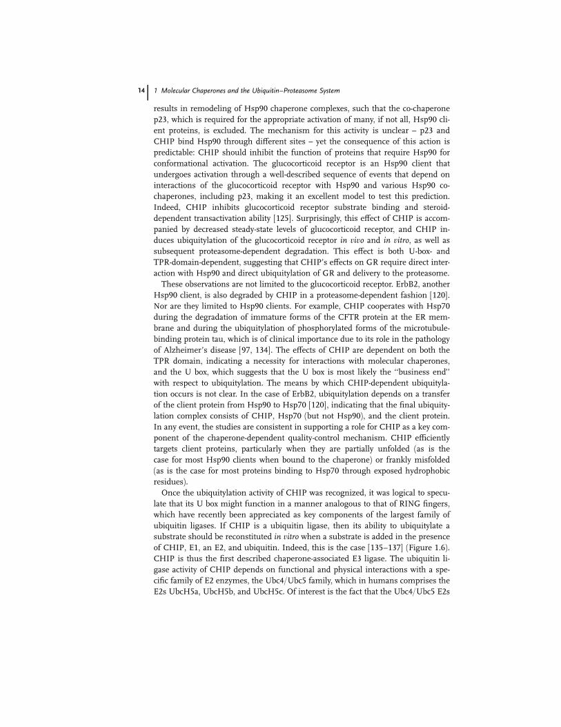
of CHIP, E1, an E2, and ubiquitin. Indeed, this is the case [135–137] (Figure 1.6).

CHIP is thus the first described chaperone-associated E3 ligase. The ubiquitin li-

gase activity of CHIP depends on functional and physical interactions with a spe-

cific family of E2 enzymes, the Ubc4/Ubc5 family, which in humans comprises the

E2s UbcH5a, UbcH5b, and UbcH5c. Of interest is the fact that the Ubc4/Ubc5 E2s

14 1 Molecular Chaperones and the Ubiquitin–Proteasome System

are stress-activated, ubiquitin-conjugating enzymes [123]. CHIP can therefore be

seen as a co-chaperone that, in addition to inhibiting traditional chaperone activity,

converts chaperone complexes into chaperone-dependent ubiquitin ligases. Indeed,

the chaperones themselves seem to act as the main substrate-recognition compo-

nents of these ubiquitin ligase complexes, as efficient ubiquitylation of chaperone

substrates by CHIP depends on the presence of Hsp70 or Hsp90 in reconstituted

systems [136, 137] (Figure 1.6). The chaperones apparently function in a manner

analogous to F-box proteins, which are required as substrate recognition modules

in many RING finger–containing ubiquitin ligase complexes [138–140].

Recently, another surprising function for CHIP has been identified, that of acti-

vation of the stress-responsive transcription factor heat shock factor-1 (HSF1)

[130]. Through this association, CHIP regulates the expression of chaperones

such as Hsp70 independently of its ability to modify their function through direct

interactions. The mechanisms through which CHIP activates HSF1 are not en-

tirely clear, but they are dependent in part on the induction of HSF1 trimerization,

which is required for nuclear import and DNA binding. In addition, activation of

HSF1 by CHIP seems to be independent of CHIP’s ubiquitin ligase activity. The

consequences of this activation are important for the response to stress, in that

cells lacking CHIP are prone to stress-dependent apoptosis and mice deficient in

CHIP (through homologous recombination) succumb rapidly to thermal chal-

lenge. These data indicate that CHIP plays a heretofore unsuspected role in coordi-

nating the response to stress, not only by serving as a rate-limiting step in the deg-

radation of damaged proteins but also by increasing the buffering capacity of the

chaperone system to guard against stress-dependent proteotoxicity.

Fig. 1.6. Characterization of CHIP as a

chaperone-associated ubiquitin ligase. Purified

CHIP, UbcH5b, the ubiquitin-activating

enzyme E1, ubiquitin, and the Hsp70–Hsp40

chaperone system were incubated with the

bacterially expressed protein kinase Raf-1 (for

details, see Ref. [137]). Raf-1 and ubiquitylated

forms of the kinase (ubðnÞ-Raf-1) were detected

by immunoblotting using a specific anti-Raf-1

antibody. Efficient ubiquitylation of Raf-1

through the CHIP conjugation machinery

depends on the recognition of the chaperone

substrate by Hsp70, which presents the kinase

to the conjugation machinery.

1.6 The CHIP Ubiquitin Ligase: A Link Between Folding and Degradation Systems 15

1.7

Other Proteins That May Influence the Balance Between Chaperone-assisted Folding

and Degradation

CHIP is ideally suited to mediate chaperone–proteasome cooperation, as it

combines a chaperone-binding motif and a domain that functions in ubiquitin-

dependent degradation within its protein structure (Figure 1.2). Some other co-

chaperones display a similar structural arrangement [40]. For example, BAG-1

contacts Hsp70 through a BAG-domain located at its carboxyl terminus and, in

addition, possesses a central ubiquitin-like domain that is used for binding to the

proteasome [141] (Figure 1.2). The co-chaperone thus belongs to a family of ubiq-

uitin domain proteins (UDPs), many of which were shown to be associated with

the proteasome [142]. This domain architecture enables BAG-1 to provide a physi-

cal link between Hsp70 and the proteasome, and elevating the cellular levels of

BAG-1 results in a recruitment of the chaperone to the proteolytic complex. Nota-

bly, BAG-1 and CHIP occupy distinct domains on Hsp70 (Figure 1.7). Whereas

BAG-1 associates with the amino-terminal ATPase domain, CHIP binds to the

carboxyl-terminal EEVD motif of Hsp70 [35, 37]. Ternary complexes that comprise

both co-chaperones associated with Hsp70 can be isolated from mammalian cells,

suggesting a cooperation of BAG-1 and CHIP in the regulation of Hsp70 activity

on certain degradation pathways. In fact, BAG-1 is able to stimulate the CHIP-

induced degradation of the glucocorticoid hormone receptor [137]. A cooperation

of diverse co-chaperones apparently provides additional levels of regulation to alter

chaperone-assisted folding and degradation pathways.

Interestingly, BAG-1 and also Hsp70 and Hsp90 are themselves substrates of the

CHIP ubiquitin ligase [135, 143] (J.H. unpublished). Yet, CHIP-mediated ubiquity-

lation of the chaperones and the co-chaperone does not induce their proteasomal

degradation. Instead, it seems to provide additional means to regulate the associa-

tion of the chaperone systems with the proteasome. In the case of BAG-1, ubiqui-

tylation mediated by CHIP indeed stimulates the binding of the co-chaperone to

the proteasome [143]. It remains to be elucidated, however, why Hsp70 and BAG-

1 are not degraded when sorted to the proteasome through CHIP-induced ubiqui-

Fig. 1.7. Schematic presentation of the BAG-

1–Hsp70–CHIP complex. BAG-1 associates

with the ATPase domain of Hsp70, while CHIP

is bound to the carboxyl terminus. BAG-1

mediates an association of Hsp70 with the

proteasome via its ubiquitin-like domain (ubl),

whereas CHIP acts in conjunction with Ubc4/5

as a chaperone-associated ubiquitin ligase to

mediate the attachment of a polyubiquitin

chain to the chaperone substrate.

16 1 Molecular Chaperones and the Ubiquitin–Proteasome System

tylation, in contrast to chaperone substrates such as the glucocorticoid hormone re-

ceptor. Possibly, the folded state of the proteins may serve to distinguish targeting

factors and substrates doomed for degradation.

Efficient ubiquitylation of BAG-1 mediated by CHIP is dependent on the forma-

tion of the ternary BAG-1–Hsp70–CHIP complex [143]. The formed chaperone

complex would thus expose multiple signals for sorting to the proteasome, e.g.,

the integrated ubiquitin-like domain of BAG-1 and polyubiquitin chains attached

to BAG-1, Hsp70, and the bound protein substrate. Such a redundancy of sorting

information might be considered unnecessary. Intriguingly, however, several sub-

units of the regulatory 19S particle of the proteasome are currently thought to act

as receptors for polyubiquitin chains and integrated ubiquitin-like domains, in-

cluding Rpn1, Rpn2, Rpt5, and Rpn10. The Rpn10 subunit was initially identi-

fied as a polyubiquitin chain receptor and was later shown to also bind integrated

ubiquitin-like domains presented by UDPs [144–146]. Rpn10 possesses two dis-

tinct ubiquitin-binding domains, of which only one is used for UDP recognition

[145–147]. However, conflicting data exist as to whether the subunit acts as a ubiq-

uitin receptor in the context of the assembled 19S complex [148, 149]. More re-

cently, Rpn1 was identified as a receptor for integrated ubiquitin-like domains

[149], and a similar function may be fulfilled by the Rpn1-related subunit Rpn2

[150]. Polyubiquitin chains seem to be recognized by the Rpt5 subunit, one of the

AAA ATPases present in the ring-like base of the regulatory 19S complex [151]. Its

receptor function was revealed when tetraubiquitin was cross-linked to intact pro-

teasomes [148]. Multiple docking sites for ubiquitin-like domains and polyubiqui-

tin chains are apparently displayed by the regulatory particle of the proteasome.

This may provide a structural basis for the recognition of multiple sorting signals

exposed by the CHIP–chaperone complex (Figure 1.8). A similar mechanism in-

volving multiple-site binding at the proteasome was recently proposed based on

the observation that two unrelated yeast ubiquitin ligases associate with specific

subunits of the 19S regulatory complex [152]. In these cases substrate delivery in-

volves interactions of proteasomal subunits with the substrate-bound ubiquitin

ligase, with the polyubiquitin chain attached to the substrate, and with the sub-

strate itself. Multiple-site binding may function to slow down dissociation of the

substrate from the proteasome and to facilitate transfer into the central proteolytic

chamber through ATP-dependent movements of the subunits of the 19S particle.

Human cells contain several BAG-1-related proteins: BAG-2, BAG-3 (CAIR-1;

Bis), BAG-4 (SODD), BAG-5, and BAG-6 (Scythe, BAT3) [153] (Figure 1.2). It ap-

pears that BAG proteins act as nucleotide-exchange factors to induce substrate

unloading from Hsp70 on diverse protein folding, assembly, and degradation path-

ways. Notably, BAG-6 is another likely candidate for a co-chaperone that regulates

protein degradation via the ubiquitin–proteasome pathway. Similar to BAG-1,

BAG-6 contains a ubiquitin-like domain that is possibly used for proteasome bind-

ing [154]. However, experimental data verifying a role of BAG-6 in protein degra-

dation remain elusive so far.

The cooperation of diverse co-chaperones not only may allow promotion of

chaperone-associated degradation but also may provide the means to confine the

1.7 Other Proteins That May Influence the Balance Between Chaperone-assisted Folding 17

destructive activity of CHIP. The Hsp70-binding protein 1 (HspBP1) seems to ful-

fill such a regulatory function [155]. HspBP1 was initially identified in a screen for

proteins that associate with the ATPase domain of Hsp70 and was shown to stim-

ulate nucleotide release from the chaperone [156, 157]. Notably, association of

HspBP1 with the ATPase domain blocks binding of BAG-1 to Hsp70 and at the

same time promotes an interaction of CHIP with Hsp70’s carboxyl terminus. In

the formed ternary HspBP1–Hsp70–CHIP complex, the ubiquitin ligase activity

of CHIP is attenuated and Hsp70 as well as a chaperone substrate are no longer

efficiently ubiquitylated [155]. By interfering with CHIP-mediated ubiquitylation,

HspBP1 stimulates the maturation of CFTR and promotes the sorting of the mem-

brane protein to the cell surface. HspBP1 apparently functions as an antagonist of

the CHIP ubiquitin ligase to regulate Hsp70-assisted folding and degradation path-

ways (Figure 1.8).

The HspBP1-mediated inhibition of the ubiquitin ligase activity may enable

CHIP to modulate the Hsp70 ATPase cycle without inducing degradation. In fact,

degradation-independent functions of CHIP have recently emerged [130, 133, 158,

159]. CHIP was shown to regulate the chaperone-assisted folding and sorting of

Fig. 1.8. The co-chaperone network that

determines folding and degradation activities

of Hsp70. BAG-1 and CHIP associate with

Hsp70 to induce the proteasomal degradation

of a Hsp70-bound protein substrate. When

BAG-1 is displaced by binding of HspBP1 to

the ATPase domain of Hsp70, the ubiquitin

ligase activity of CHIP is attenuated in the

formed complex, enabling CHIP to modulate

Hsp70 activity without inducing degradation.

The ATPase domain can also be occupied by

Hip, which stimulates the chaperone activity of

Hsp70 and participates in the Hsp70/Hsp90-

mediated regulation of signal transduction

pathways. At the same time, Hop displaces

CHIP from the carboxyl terminus of Hsp70 and

recruits Hsp90 to the chaperone complex.

18 1 Molecular Chaperones and the Ubiquitin–Proteasome System

the androgen receptor and of endothelial nitric oxide synthase without inducing

degradation [158, 159]. Moreover, CHIP fulfills an essential role in the chaperone-

mediated regulation of the heat shock transcription factor, independent of its

degradation-inducing activity [130]. It remains to be seen, however, whether

HspBP1 cooperates with CHIP in these instances, as HspBP1 displayed a certain

specificity with regard to chaperone substrates. The co-chaperone interfered with

the degradation of CFTR, but did not influence the CHIP-mediated turnover of

the glucocorticoid hormone receptor. Such a client specificity may arise in part

from the fact that HspBP1 inhibits the ubiquitin ligase activity of CHIP in a com-

plex with Hsc70, but leaves Hsp90-associated ubiquitylation unaffected [155]. In

addition, direct interactions between HspBP1 and a subset of chaperone substrates

may contribute to substrate selection. In any case, the cooperation of CHIP with

other co-chaperones apparently provides a means to regulate chaperone-assisted

protein degradation.

It is likely that there are multiple degradation pathways for misfolded proteins in

the eukaryotic cytoplasm. Although CHIP participates in the degradation of chap-

erone substrates induced by applying Hsp90 inhibitors to cell cultures (see above),

drug-induced degradation is not abrogated in cells that lack the CHIP ubiquitin

ligase [120]. Furthermore, CHIP cooperates with Hsp70 in the ER-associated deg-

radation of CFTR, but the Hsp70-assisted degradation of apoB at the cytoplasmic

face of the ER membrane does not involve CHIP [97]. Taken together, these data

strongly argue for the existence of other, yet to be identified, ubiquitin ligases that

are able to target chaperone substrates to the proteasome. A likely candidate in this

regard is Parkin, a RING finger ubiquitin ligase, whose activity is impaired in juve-

nile forms of Parkinson’s disease [17]. Hsp70 and CHIP were found to be associ-

ated with Parkin in neuronal cells, suggesting an involvement of Parkin in the pro-

teasomal degradation of chaperone substrates [160]. Interestingly, a-synuclein, the

main component of protein deposits observed in dopaminergic neurons of Parkin-

son patients, and synphilin, a protein that binds a-synuclein and induces deposit

formation, both associate with yet other ubiquitin ligases: Siah-1 and Siah-2 [161,

162]. In the case of Siah-1, a link to cytoplasmic chaperone systems is suggested by

the finding that the Hsp70 co-chaperone BAG-1 is a binding partner of the ubiqui-

tin ligase and suppresses some of the cellular activities of Siah-1 [163]. Taken to-

gether, it is tempting to speculate about a role of Parkin and Siah on chaperone-

assisted degradation pathways; yet, this remains to be explored in detail.

1.8

Further Considerations

Although the appreciation of interplay between molecular chaperones and

ubiquitin-dependent proteolysis has greatly expanded over the past decade, a num-

ber of critical issues remain to be resolved. It is not entirely clear what determines

whether a misfolded protein will undergo repeated attempts at misfolding versus

1.8 Further Considerations 19

diversion to the ubiquitin–proteasome pathway. Recruitment of CHIP into chaper-

one complexes appears to be a critical component of this reaction, which therefore

begs the question as to what regulates this step. Since this step in protein quality

control must be both rapidly activated and easily reversible, it is likely that regula-

tion occurs at the post-translational level rather than through changes in steady-

state protein levels. The precise sorting mechanisms for ubiquitinated proteins

are also unclear. BAG-1 is a player, and it is also likely that overlap exists to some

extent for sorting of the cytoplasmic and endoplasmic reticulum quality-control

pathways. Nevertheless, much remains to be learned about these steps.

From a broader perspective, it is now also imperative to understand the

pathophysiological roles of cytoplasmic quality-control mechanisms regulated by

chaperone–proteasome interactions. As mentioned previously, there is a strong as-

sociation between chaperone dysfunction and accumulations of misfolded proteins

that characterizes genetic neurodegenerative diseases. An imbalance between pro-

tein folding and degradation may also contribute to some features of senescence

and organismal aging. The link between chaperone systems and aging is based

on increasing appreciation that modified, misfolded, and aggregated proteins accu-

mulate with age [164]. Dysregulation of chaperone expression has been observed

with aging and is therefore implicated in aging-related changes [165]; in general,

it is accepted that induction of the major chaperones is impaired with aging, a

fact confirmed by recent gene-profiling experiments in vivo [166], although given

the diversity of chaperones it is probably not surprising that age-related changes

in expression are fairly complicated [167]. The mechanism underlying this dys-

regulation is not entirely clear, but seems to be due in part to impaired activation

of the stress-responsive transcription factor HSF1. Overexpression of heat shock

proteins in yeast, C. elegans, and Drosophila leads to increased longevity [168–170].

More recently, conclusive genetic evidence from C. elegans indicates that mutation

of HSF1 causes a dramatic and significant reduction in lifespan [170, 171], further

implicating the accumulation of misfolded proteins in age-related phenotypes.

1.9

Conclusions

The associations between molecular chaperones and the ubiquitin–proteasome

system represent a critical step in the response to proteotoxic damage. Whether at-

tempts should be made to refold damaged proteins (thus conserving cellular re-

sources) or degrade them instead (to prevent the possibility of protein aggregation

and concomitant toxicity) requires a consideration of cellular economy. Defects in

the quality-control mechanisms may have enormous consequences even if only

slight imbalances occur between protein folding and degradation, as these im-

balances can cause accumulated toxicity over time. The relationship between

chaperone–proteasome interactions and pathophysiological events is only now be-

ing unraveled. Modulation of this system may provide a unique therapeutic target

for degenerative diseases and pathologies associated with aging.

20 1 Molecular Chaperones and the Ubiquitin–Proteasome System

References

1 U. Schubert, L.C. Anton, J. Gibbs,

C.C. Norbury, J.W. Yewdell, J.R.

Bennink, Rapid degradation of a

large fraction of newly synthesized

proteins by proteasomes, Nature 404

(2000) 770–774.

2 J.P. Taylor, J. Hardy, K.H. Fisch-

beck, Toxic proteins in neurodegenera-

tive disease, Science 296 (2002) 1991–

1995.

3 A. Horwich, Protein aggregation in

disease: a role for folding inter-

mediates forming specific multimeric

interactions, J. Clin. Invest. 110 (2002)

1221–1232.

4 E. Scherzinger, R. Lurz, M.

Turmaine, L. Mangiarini, B.

Hollenbach, R. Hasenbank, G.P.

Bates, S.W. Davies, H. Lehrach,

E.E. Wanker, Huntingtin-encoded

polyglutamine expansions form

amyloid-like protein aggregates in vitroand in vivo, Cell 90 (1997) 549–558.

5 E. Scherzinger, A. Sittler, K.

Schweiger, V. Heiser, R. Lurz, R.

Hasenbank, G.P. Bates, H. Lehrach,

E.E. Wanker, Self-assembly of

polyglutamine-containing huntingtin

fragments into amyloid-like fibrils:

implications for Huntington’s disease

pathology, Proc. Natl. Acad. Sci. USA

96 (1999) 4604–4609.

6 A. Kazantsev, E. Preisinger, A.

Dranovsky, D. Goldgaber, D.

Housman, Insoluble detergent-

resistant aggregates form between

pathological and nonpathological

lengths of polyglutamine in mam-

malian cells, Proc. Natl. Acad. Sci.

U S A 96 (1999) 11404–11409.

7 D. Martindale, A. Hackam, A.

Wieczorek, L. Ellerby, C.

Wellington, K. McCutcheon, R.

Singaraja, P. Kazemi-Esfarjani, R.

Devon, S.U. Kim, D.E. Bredesen, F.

Tufaro, M.R. Hayden, Length of

huntingtin and its polyglutamine tract

influences localization and frequency

of intracellular aggregates. Nat. Genet.

18 (1998) 150–154.

8 I. Sanchez, C. Mahlke, J. Yuan,

Pivotal role of oligomerization in

expanded polyglutamine neuro-

degenerative disorders, Nature 421

(2003) 373–379.

9 M.F. Perutz, Glutamine repeats and

neurodegenerative diseases: molecular

aspects, Trends Biochem. Sci. 24

(1999) 58–63.

10 H.Y. Zoghbi, H.T. Orr, Glutamine

repeats and neurodegeneration, Annu.

Rev. Neurosci. 23 (2000) 217–247.

11 H.T. Orr, M.Y. Chung, S. Banfi, T.J.

Jr. Kwiatkowski, A. Servadio, A.L.

Beaudet, A.E. McCall, L.A. Duvick,

L.P. Ranum, H.Y. Zoghbi, Expansion

of an unstable trinucleotide CAG

repeat in spinocerebellar ataxia type 1,

Nat. Genet. 4 (1993) 221–226.

12 D.R. Rosen, T. Siddique, D.

Patterson, D.A. Figlewicz, P. Sapp,

A. Hentati, D. Donaldson, J. Goto,

J.P. O’Regan, H.X. Deng, et al.

Mutations in Cu/Zn superoxide

dismutase gene are associated with

familial amyotrophic lateral sclerosis.

Nature 362 (1993) 59–62.

13 J.A. Johnston, M.J. Dalton, M.E.

Gurney, R.R. Kopito, Formation of

high molecular weight complexes of

mutant Cu, Zn-superoxide dismutase

in a mouse model for familial

amyotrophic lateral sclerosis, Proc.

Natl. Acad. Sci. USA 97 (2000) 12571–

12576.

14 B.I. Giasson, V.M. Lee, Are

ubiquitination pathways central to

Parkinson’s disease? Cell 114 (2003)

1–8.

15 T. Kitada, S. Asakawa, N. Hattori,

H. Matsumine, Y. Yamamura, S.

Minoshima, M. Yokochi, Y.

Mizuno, N. Shimizu, Mutations in

the parkin gene cause autosomal

recessive juvenile parkinsonism,

Nature 392 (1998) 605–608.

16 C.B. Lucking, A. Durr, V. Bonifati,

J. Vaughan, G. De Michele, T.

Gasser, B.S. Harhangi, G. Meco, P.

Denefle, N.W. Wood, Y. Agid, A.

Brice, Association between early-

onset Parkinson’s disease and

mutations in the parkin gene, N. Engl.

J. Med. 342 (2000) 1560–1567.

References 21

17 H. Shimura, N. Hattori, S. Kubo, Y.

Mizuno, S. Asakawa, S. Minoshima,

N. Shimizu, K. Iwai, T. Chiba, K.

Tanaka, T. Suzuki, Familial

Parkinson disease gene product,

parkin, is a ubiquitin-protein ligase,

Nat. Genet. 25 (2000) 302–305.

18 K.K. Chung, Y. Zhang, K.L. Lim, Y.

Tanaka, H. Huang, J. Gao, C.A.

Ross, V.L. Dawson, T.M. Dawson,

Parkin ubiquitinates the a-synuclein-

interacting protein, synphilin-1:

implications for Lewy-body formation

in Parkinson disease, Nat. Med. 7

(2001) 1144–1150.

19 H. Shimura, M.G. Schlossmacher,

N. Hattori, M.P. Frosch, A.

Trockenbacher, R. Schneider, Y.

Mizuno, K.S. Kosik, D.J. Selkoe,

Ubiquitination of a new form of a-

synuclein by parkin from human

brain: implications for Parkinson’s

disease, Science 293 (2001) 263–269.

20 Y. Imai, M. Soda, H. Inoue, N.

Hattori, Y. Mizuno, R. Takahashi,

An unfolded putative transmembrane

polypeptide, which can lead to

endoplasmic reticulum stress, is a

substrate of Parkin, Cell 105 (2001)

891–902.

21 P. Fernandez-Funez, M.L. Nino-

Rosales, B. de Gouyon, W.C. She,

J.M. Luchak, P. Martinez, E.

Turiegano, J. Benito, M. Capovilla,

P.J. Skinner, A. McCall, I. Canal,

H.T. Orr, H.Y. Zoghbi, J. Botas,

Identification of genes that modify

ataxin-1-induced neurodegeneration,

Nature 408 (2000) 101–106.

22 P. Kazemi-Esfarjani, S. Benzer,

Genetic suppression of polyglutamine

toxicity in Drosophila, Science 287

(2000) 1837–1840.

23 N.M. Bonini, Chaperoning brain

degeneration, Proc. Natl. Acad. Sci.

U S A 99 Suppl. 4 (2002) 16407–

16411.

24 P.K. Auluck, H.Y. Chan, J.Q.

Trojanowski, V.M. Lee, N.M.

Bonini, Chaperone suppression of a-

synuclein toxicity in a Drosophila

model for Parkinson’s disease, Science

295 (2002) 865–868.

25 F.U. Hartl, M. Hayer-Hartl,

Molecular chaperones in the cytosol:

from nascent chain to folded protein,

Science 295 (2002) 1852–1858.

26 J. Frydman, Folding of newly

translated proteins in vivo: the role of

molecular chaperones, Annu. Rev.

Biochem. 70 (2001) 603–647.

27 J. Frydman, J. Hohfeld, Chaperones

get in touch: the Hip-Hop connection,

Trends Biochem. Sci. 22 (1997) 87–92.

28 B. Bukau, A.L. Horwich, The Hsp70

and Hsp60 chaperone machines, Cell

92 (1998) 351–366.

29 R.I. Morimoto, Regulation of the heat

shock transcriptional response: cross

talk between a family of heat shock

factors, molecular chaperones, and

negative regulators. Genes Dev. 12

(1998) 3788–3796.

30 M.P. Mayer, D. Brehmer, C.S.

Gassler, B. Bukau, Hsp70 chaperone

machines, Adv. Protein Chem. 59

(2001) 1–44.

31 S. Rudiger, L. Germeroth, J.

Schneider-Mergener, B. Bukau,

Substrate specificity of the DnaK

chaperone determined by screening

cellulose-bound peptide libraries,

EMBO J. 16 (1997) 1501–1507.

32 V. Thulasiraman, C.F. Yang, J.

Frydman, In vivo newly translatedpolypeptides are sequestered in a

protected folding environment, EMBO

J. 18 (1999) 85–95.

33 X. Zhu, X. Zhao, W.F. Burkholder,

A. Gragerov, C.M. Ogata, M.E.

Gottesman, W.A. Hendrickson,

Structural analysis of substrate

binding by the molecular chaperone

DnaK, Science 272 (1996) 1606–1614.

34 Y. Minami, J. Hohfeld, K. Ohtsuka,

F.U. Hartl, Regulation of the heat-

shock protein 70 reaction cycle by

the mammalian DnaJ homolog,

Hsp40, J. Biol. Chem. 271 (1996)

19617–19624.

35 C.A. Ballinger, P. Connell, Y. Wu,

Z. Hu, L.J. Thompson, L.Y. Yin, C.

Patterson, Identification of CHIP,

a novel tetratricopeptide repeat-

containing protein that interacts with

heat shock proteins and negatively

regulates chaperone functions, Mol.

Cell. Biol. 19 (1999) 4535–4545.

22 1 Molecular Chaperones and the Ubiquitin–Proteasome System

36 J. Hohfeld, Y. Minami, F.U. Hartl,

Hip, a novel cochaperone involved in

the eukaryotic Hsc70/Hsp40 reaction

cycle, Cell 83 (1995) 589–598.

37 J. Hohfeld, S. Jentsch, GrpE-like

regulation of the hsc70 chaperone by

the anti-apoptotic protein BAG-1,

Embo J. 16 (1997) 6209–6216.

38 H. Sondermann, C. Scheufler, C.

Schneider, J. Hohfeld, F.U. Hartl,

I. Moarefi, Structure of a Bag/Hsc70

complex: convergent functional

evolution of Hsp70 nucleotide

exchange factors, Science 291 (2001)

1553–1557.

39 C.S. Gassler, T. Wiederkehr, D.

Brehmer, B. Bukau, M.P. Mayer,

Bag-1M accelerates nucleotide release

for human Hsc70 and Hsp70 and can

act concentration-dependent as

positive and negative cofactor, J. Biol.

Chem. 276 (2001) 32538–32544.

40 J. Hohfeld, D.M. Cyr, C. Patterson,

From the cradle to the grave: molec-

ular chaperones that may choose

between folding and degradation,

EMBO Rep. 2 (2001) 885–890.

41 W.M. Obermann, H. Sondermann,

A.A. Russo, N.P. Pavletich, F.U.

Hartl, In vivo function of Hsp90 is

dependent on ATP binding and ATP

hydrolysis, J. Cell Biol. 143 (1998)

901–910.

42 J. Buchner, Hsp90 & Co. – a holding

for folding, Trends Biochem. Sci. 24

(1999) 136–141.

43 J.C. Young, I. Moarefi, F.U. Hartl,

Hsp90: a specialized but essential

protein-folding tool, J. Cell Biol. 154

(2001) 267–273.

44 L.H. Pearl, C. Prodromou, Structur,

function and mechanism of the

Hsp90 molecular chaperone, Adv.

Protein Chem. 59 (2001) 157–186.

45 B. Panaretou, G. Siligardi, P.

Meyer, A. Maloney, J.K. Sullivan, S.

Singh, S.H. Millson, P.A. Clarke, S.

Naaby-Hansen, R. Stein, R. Cramer,

M. Mooapour, P. Workman, P.W.

Piper, L.H. Peal, C. Prodromou,

Activation of the ATPase activity of

hsp90 by the stress-regulated

cochaperone aha1, Mol. Cell 10 (2002)

1307–1318.

46 J.C. Young, N.J. Hoogenraad, F.U.

Hartl, Molecular chaperones Hsp90

and Hsp70 deliver preproteins to the

mitochondrial import receptor Tom70,

Cell 112 (2003) 41–50.

47 A. Brychzy, T. Rein, K.F.

Winklhofer, F.U. Hartl, J.C.

Young, W.M. Obermann, Cofactor

Tpr2 combines two TPR domains and

a J domain to regulate the Hsp70/

Hsp90 chaperone system, EMBO J. 22

(2003) 3613–3623.

48 S.M. Roe, M.M. Ali, P. Meyer, C.K.

Vaughan, B. Panaretou, P.W. Piper,

C. Prodromou, L.H. Pearl, The

mechanism of Hsp90 regulation by

the protein kinase-specific cochaper-

one p50 (cdc37), Cell 116 (2004) 87–

98.

49 K.D. Dittmar, W.B. Pratt, Folding

of the glucocorticoid receptor be

the reconstituted Hsp90-based

chaperone machinery. The initial

hsp90.p60.hsp70 dependent step is

sufficient for creating the steroid

binding conformation, J. Biol. Chem.

272 (1997) 13047–13054.

50 K.D. Dittmar, D.R. Demady, L.F.

Stancato, P. Krishna, W.B. Pratt,

Folding of the glucocorticoid receptor

by the heat shock protein (hsp) 90-

based chaperone machinery. The role

of p23 is to stabilize receptor.hsp90

heterocomplexes formed by

hsp90.p60.hsp70, J. Biol. Chem. 272

(1997) 21213–21220.

51 S. Chen, V. Prapapanich, R.A.

Rimerman, B. Honore, D.F. Smith,

Interactions of p60, a mediator of

progesterone receptor assembly, with

heat shock proteins hsp90 and hsp70.

Mol. Endocrinol. 10 (1996) 682–693.

52 H. Kosano, B. Stensgard, M.C.

Charlesworth, N. McMahon, D.

Toft, The assembly of progesterone

receptor-hsp90 complexes using

purified proteins, J. Biol. Chem. 273

(1998) 32973–32979.

53 W.B. Pratt, D.O. Toft, Regulation

of signaling protein function and

trafficking by the hsp90/hsp70-based

chaperone machinery, Exp. Biol. Med.

228 (2003) 111–133.

54 M.P. Hernandez, A. Chadli, D.O.

References 23

Toft, Hsp40 binding is the first step

in the Hsp90 chaperoning pathway for

the progesterone receptor, J. Biol.

Chem. 277 (2002) 11873–11881.

55 C. Scheufler, A. Brinker, G.

Bourenkov, S. Pegoraro, L.

Moroder, H. Bartunik, F.U. Hartl,

I. Moarefi, Structure of TPR domain-

peptide complexes: critical elements in

the assembly of the Hsp70-Hsp90

multichaperone machine, Cell 101

(2000) 199–210.

56 J. Fontana, D. Fulton, Y. Chen, T.A.

Fairchild, T.J. McCabe, N. Fujita,

T. Tsuruo, W.C. Sessa, Domain

mapping studies reveal that the M

domain of hsp90 serves as a molecular

scaffold to regulate Akt-dependent

phosphorylation of endothelioal nitric

oxide synthase and NO release, Circ.

Res. 90 (2002) 866–873.

57 D.B. Solit, H.I. Scher, N. Rosen,

Hsp90 as a therapeutic target in

prostate cancer, Semin. Oncol. 30

(2003) 709–716.

58 L. Neckers, S.P. Ivy, Heat shock

protein 90, Curr. Opin. Oncol. 15

(2003) 419–424.

59 A. Kamal, L. Thao, J. Sensintaffar,

L. Zhang, M.F. Boehm, L.C. Fritz,

F.J. Burrows, A high-affinity

conformation of Hsp90 confers

tumour selectivity on Hsp90

inhibitors, Nature 425 (2003) 407–

410.

60 C. Prodromou, S.M. Roe, R.

O’Brien, J.E. Ladbury, P.W. Piper,

L.H. Pearl, Identification and

structural characterization of the

ATP/ADP-binding site in the Hsp90

molecular chaperone, Cell 90 (1997)

65–75.

61 C.E. Stebbins, A.A. Russo, C.

Schneider, N. Rosen, F.U. Hartl,

N.P. Pavletich, Crystal structure of

an Hsp90-geldanamycin complex:

targeting of a protein chaperone by an

antitumor agent, Cell 89 (1997) 239–

250.

62 S.V. Sharma, T. Agatsuma, H.

Nakano, Targeting of the protein

chaperone, Hsp90, by the transforma-

tion suppressing agent, radicicol,

Oncogene 16 (1998) 2639–2645.

63 T.W. Schulte, S. Akinaga, T.

Murakata, T. Agatsuma, S.

Sugimoto, H. Nakano, Y.S. Lee, B.B.

Simen, Y. Argon, S. Felts, D.O.

Toft, L.M. Neckers, S.V. Sharma,

Interaction of radicicol with members

of the heat shock protein 90 family of

molecular chaperones, Mol.

Endocrinol. 13 (1999) 1435–1448.

64 C. Schneider, L. Sepp-Lorenzino,

E. Nimmesgern, O. Ouerfelli, S.

Danishefsky, N. Rosen, F.U. Hartl,

Pharmacologic shifting of a balance

between protein refolding and

degradation mediated by Hsp90, Proc.

Natl. Acad. Sci. U S A 93 (1996)

14536–14541.

65 E.G. Mimnaugh, C. Chavany, L.

Neckers, Polyubiquitination and

proteasomal degradation of the p185c-

erbB-2 receptor protein-tyrosine kinase

induced by geldanamycin, J. Biol.

Chem. 271 (1996) 22796–22801.

66 M.G. Marcu, T.W. Schulte, L.

Neckers, Novobiocin and related

coumarins and depletion of heat

shock protein 90-dependent signaling

proteins, J. Natl. Cancer Inst. 92

(2000) 242–248.

67 W. Xu, E. Mimnaugh, M.F. Rosser,

C. Nicchitta, M. Marcu, Y. Yarden,

L. Neckers, Sensitivity of mature

Erbb2 to geldanamycin is conferred by

its kinase domain and is mediated by

the chaperone protein Hsp90, J. Biol.

Chem. 276 (2001) 3702–3708.

68 D.B. Solit, F.F. Zheng, M.

Drobnjak, P.N. Munster, B.

Higgins, D. Verbel, G. Heller, W.

Tong, C. Cordon-Cardo, D.B. Agus,

H.I. Scher, N. Rosen, 17-Allylamino-

17-demethoxygeldanamycin induces

the degradation of androgen receptor

and HER-2/neu and inhibits the

growth of prostate cancer xenografts,

Clin. Cancer Res. 8 (2002) 986–993.

69 A. Citri, I. Alroy, S. Lavi, C. Rubin,

W. Xu, N. Grammatikakis, C.

Patterson, L. Neckers, D.W. Fry, Y.

Yarden, Drug-induced ubiquitylation

and degradation of ErbB receptor

tyrosine kinases: implications for

cancer therapy, Embo J. 21 (2002)

2407–2417.

24 1 Molecular Chaperones and the Ubiquitin–Proteasome System

70 R. van Montfort, C. Slingsby, E.

Vierling, Structure and function of

the small heat shock protein/alpha-

crystallin family of molecular

chaperones, Adv. Protein Chem. 59

(2001) 105–156.

71 M. Haslbeck, N. Braun, T. Stromer,

B. Richter, N. Model, S. Weinkauf,

J. Buchner, Hsp42 is the general

small heat shock protein in the cytosol

of Saccharomyces cerevisiae, EMBO J.

23 (2004) 638–649.

72 T. Stromer, E. Fischer, K. Richter,

M. Haslbeck, J. Buchner, Analysis

of the regulation of the molecular

chaperone Hsp26 by temperature-

induced dissociation: the N-terminal

domain is important for oligomer

assembly and the binding of unfold-

ing proteins. J. Biol. Chem. 279 (2004)

11222–11228.

73 E. Basha, G.J. Lee, L.A. Breci, A.C.

Hausrath, N.R. Buan, K.C. Giese, E.

Vierling, The identity of proteins

associated with a small heat shock

protein during heat stress in vivoindicates that these chaperones protect

a wide range of cellular functions,

J. Biol. Chem. 279 (2004) 7566–7575.

74 K.K. Kim, R. Kim, S.H. Kim, Crystal

structure of a small heat-shock

protein, Nature 394 (1998) 595–599.

75 R.L. van Montfort, E. Basha, K.L.

Friedrich, C. Slingsby, E. Vierling,

Crystal structure and assembly of a

eukaryotic small heat shock protein.

Nat. Struct. Biol. 8 (2001) 1025–1030.

76 M. Haslbeck, S. Walke, T. Stromer,

M. Ehrnsperger, H.E. White, S.

Chen, H.R. Saibil, J. Buchner,

Hsp26: a temperature-regulated

chaperone, EMBO J. 18 (1999) 6744–

6751.

77 G.J. Lee, A.M. Roseman, H.R. Saibil,

E. Vierling, A small heat shock

protein stably binds heat-denatured

model substrates and can maintain a

substrate in a folding-competent state,

EMBO J. 16 (1997) 659–671.

78 Z. Xu, A.L. Horwich, P.B. Sigler,

The crystal structure of the asym-

metric GroEL-GroES-(ADP)7 chapero-

nin complex, Nature 388 (1997) 741–

750.

79 H.R. Saibil, N.A. Ranson, The

chaperonin folding machine, Trends

Biochem. Sci. 27 (2002) 627–632.

80 A.S. Meyer, J.R. Gillespie, D.

Walther, I.S. Millet, S. Doniach,

J. Frydman, Closing the folding

chamber of the eukaryotic chaperonin

requires the transition state of ATP

hydrolysis, Cell 113 (2003) 369–381.

81 V. Thulasiraman, C.F. Yang, J.

Frydman, In vivo newly translated

polypeptides are sequestered in a

protected folding environment, EMBO

J. 18 (1999) 85–95.

82 S.A. Lewis, G. Tian, I.E. Vainberg,

N.J. Cowan, Chaperonin-mediated

folding of actin and tubulin, J. Cell

Biol. 132 (1996) 1–4.

83 H. Sakahira, P. Breuer, M.K. Hayer-

Hartl, F.U. Hartl, Molecular

chaperones as modulators of

polyglutamine protein aggregation

and toxicity, Proc. Natl. Acad. Sci.

USA 99 Suppl. 4 (2002) 16412–16418.

84 J.M. Warrick, H.Y. Chan, G.L.

Gray-Board, Y. Chai, H.L. Paulson,

N.M. Bonini, Suppression of

polyglutamine-mediated neuro-

degeneration in Drosophila by the

molecular chaperone Hsp70. Nat.

Genet. 23 (1999) 425–428.

85 C.J. Cummings, M.A. Mancini, B.

Antalffy, D.B. DeFranco, H.T. Orr,

H.Y. Zoghbi, Chaperone suppression

of aggregation and altered subcellular

proteasome localization imply protein

misfolding in SCA1, Nat. Genet. 19

(1998) 148–154.

86 C.J. Cummings, Y. Sun, P. Opal, B.

Antalffy, R. Mestril, H.T. Orr,

W.H. Dillmann, H.Y. Zoghbi,

Over-expression of inducible HSP70

chaperone suppresses neuropathology

and improves motor function in SCA1

mice, Hum. Mol. Genet. 10 (2001)

1511–1518.

87 S. Krobitsch, S. Lindquist,

Aggregation of huntingtin in yeast

varies with the length of the

polyglutamine expansion and the

expression of chaperone proteins,

Proc. Natl. Acad. Sci. U S A 97 (2000)

1589–1594.

88 P.J. Muchowski, G. Schaffar, A.

References 25

Sittler, E.E. Wanker, M.K. Hayer-

Hartl, F.U. Hartl, Hsp70 and hsp40

chaperones can inhibit self-assembly

of polyglutamine proteins into

amyloid-like fibrils, Proc. Natl. Acad.

Sci. U S A 97 (2000) 7841–7846.

89 J.Z. Chuang, H. Zhou, M. Zhu, S.H.

Li, X.J. Li, C.H. Sung, Characteriza-

tion of a brain-enriched chaperone,

MRJ, that inhibits Huntingtin

aggregation and toxicity indepen-

dently, J. Biol. Chem. 277 (2002)

19831–19838.

90 A. Sittler, R. Lurz, G. Lueder, J.

Priller, H. Lehrach, M.K. Hayer-

Hartl, F.U. Hartl, E.E. Wanker,

Geldanamycin activates a heat shock

response and inhibits huntingtin

aggregation in a cell culture model of

Huntington’s disease, Hum. Mol.

Genet. 10 (2001) 1307–1315.

91 A. Parcellier, E. Schmitt, S.

Gurbuxani, D. Seigneurin-Berny,

A. Pance, A. Chantome, S.

Plenchette, S. Khochbin, E.

Solary, C. Garrido, Hsp27 is a

ubiquitin-binding protein involved in

IkBa proteasomal degradation, Mol.

Cell Biol. 23 (2003) 5790–5802.

92 H. Shimura, Y. Miura-Shimura,

K.S. Kosick, Binding of tau to heat

shock protein 27 leads to decreased

concentration of hyperphosphorylated

tau and enhanced cell survival, J. Biol.

Chem. 279 (2004) 17957–17962.

93 V. Gusarova, A.J. Caplan, J.L.

Brodsky, E.A. Fisher, Apoprotein B

degradation is promoted by the

molecular chaperones hsp90 and

hsp70, J. Biol. Chem. 276 (2001)

24891–24900.

94 C. Taxis, R. Hitt, S.H. Park, P.M.

Deak, Z. Kostova, D.H. Wolf, Use of

modular substrates demonstrates

mechanistic diversity and reveals

differences in chaperone requirement

of ERAD, J. Biol. Chem. 278 (2003)

35903–35913.

95 Y. Yang, S. Janich, J.A. Cohn, J.M.

Wilson, The common variant of

cystic fibrosis transmembrane

conductance regulator is recognized

by hsp70 and degraded in a pre-Golgi

nonlysosomal compartment, Proc.

Natl. Acad. Sci. U S A 90 (1993) 9480–

9484.

96 G.C. Meacham, Z. Lu, S. King, E.

Sorscher, A. Tousson, D.M. Cyr,

The Hdj-2/Hsc70 chaperone pair

facilitates early steps in CFTR

biogenesis, Embo J. 18 (1999) 1492–

1505.

97 G.C. Meacham, C. Patterson, W.

Zhang, J.M. Younger, D.M. Cyr,

The Hsc70 co-chaperone CHIP targets

immature CFTR for proteasomal

degradation, Nat. Cell Biol. 3 (2001)

100–105.

98 Y. Zhang, G. Nijbroek, M.L.

Sullivan, A.A. McCracken, S.C.

Watkins, S. Michaelis, J.L. Brodsky,

Hsp70 molecular chaperone facilitates

endoplasmic reticulum-associated

protein degradation of cystic fibrosis

transmembrane conductance regulator

in yeast, Mol. Biol. Cell 12 (2001)

1303–1314.

99 R.R. Kopito, Biosynthesis and

degradation of CFTR, Physiol. Rev. 79

(1999) S167–173.

100 J.R. Riordan, Cystic fibrosis as a

disease of misprocessing of the cystic

fibrosis transmembrane conductance

regulator glycoprotein, Am. J. Hum.

Genet. 64 (1999) 1499–1504.

101 C.L. Ward, S. Omura, R.R. Kopito,

Degradation of CFTR by the

ubiquitin–proteasome pathway, Cell

83 (1995) 121–127.

102 T.J. Jensen, M.A. Loo, S. Pind,

D.B. Williams, A.L. Goldberg, J.R.

Riordan, Multiple proteolytic

systems, including the proteasome,

contribute to CFTR processing, Cell 83

(1995) 129–135.

103 M.S. Gelman, E.S. Kannegaard,

R.R. Kopito, A principal role for the

proteasome in endoplasmic reticulum-

associated degradation of misfolded

intracellular cystic fibrosis

transmembrane conductance

regulator, J. Biol. Chem. 277 (2002)

11709–11714.

104 Z. Kostova, D.H. Wolf, For whom

the bell tolls: protein quality control of

the endoplasmic reticulum and the

ubiquitin–proteasome connection,

EMBO J. 22 (2003) 2309–2317.

26 1 Molecular Chaperones and the Ubiquitin–Proteasome System

105 S. Sato, C.L. Ward, M.E. Krouse, J.J.

Wine, R.R. Kopito, Glycerol reverses

the misfolding phenotype of the most

common cystic fibrosis mutation,

J. Biol. Chem. 271 (1996) 635–638.

106 B.H. Qu, P.J. Thomas, Alteration of

the cystic fibrosis transmembrane

conductance regulator folding

pathway, J. Biol. Chem. 271 (1996)

7261–7264.

107 E. Strickland, B.H. Qu, L. Millen,

P.J. Thomas, The molecular

chapeorne Hsc70 assists the in vitrofolding of the N-terminal nucleotide-

binding domain of the cystic fibrosis

transmembrane conductance

regulator, J. Biol. Chem. 272 (1997)

25421–25424.

108 J.A. Johnston, C.L. Ward, R.R.

Kopito, Aggresomes: a cellular

response to misfolded proteins, J. Cell

Biol. 143 (1998) 1883–98.

109 R.R. Kopito, Aggresomes, inclusion

bodies and protein aggregation,

Trends Cell Biol. 10 (2000) 524–530.

110 S. Waelter, A. Boeddrich, R. Lurz,

E. Scherzinger, G. Lueder, H.

Lehrach, E.E. Wanker, Accumula-

tion of mutant huntingtin fragments

in aggresome-like inclusion bodies as

a result of insufficient protein degra-

dation, Mol. Biol. Cell 12 (2001) 1393–

1407.

111 R. Garcia-Mata, Z. Bebok, E.J.

Sorscher, E.S. Sztul, Characteriza-

tion and dynamics of aggresome

formation by a cytosolic GFP-chimera,

J. Cell Biol. 146 (1999) 1239–1254.

112 K.S. McNaught, P. Shashidharan,

D.P. Perl, P. Jenner, C.W. Olanow,

Aggresome-related biogenesis of Lewy

bodies, Eur. J. Neurosci. 16 (2002)

2136–2148.

113 Y. Kawaguchi, J.J. Kovacs, A.

McLaurin, J.M. Vance, A. Ito, T.P.

Yao, The deacetylase HDAC6

regulates aggresome formation and

cell viability in response to misfolded

protein stress, Cell 115 (2003) 727–738.

114 R. Garcia-Mata, Z. Bebok, E.J.

Sorscher, E.S. Sztul, Characteriza-

tion and dynamics of aggresome

formation by a cytosolic GFP-chimera,

J. Cell Biol. 146 (1999) 1239–1254.

115 W.C. Wigley, R.P. Fabunmi, M.G.

Lee, C.R. Marino, S. Muallem, G.N.

DeMartino, P.J. Thomas, Dynamic

association of proteasomal machinery

with the centrosome, J. Cell Biol. 145

(1999) 481–490.

116 A. Wyttenbach, J. Carmichael, J.

Swartz, R.A. Furlong, Y. Narain, J.

Rankin, D.C. Rubinsztein, Effects of

heat shock, heat shock protein 40

(HDJ-2), and proteasome inhibition on

protein aggregation in cellular models

of Huntington’s disease, Proc. Natl.

Acad. Sci. USA 97 (2000) 2898–2903.

117 J.L. Dul, D.P. Davis, E.K. William-

son, F.J. Stevens, Y. Argon, Hsp70

and antifibrillogenic peptides promote

degradation and inhibit intracellular

aggregation of amyloidogenic light

chains, J. Cell Biol. 152 (2001) 705–

716.

118 H. Lelouard, E. Gatti, F. Cappello,

O. Gresser, V. Camosseto, P. Pierre,

Transient aggregation of ubiquitinated

proteins during dendritic cell

maturation, Nature 417 (2002) 177–

182.

119 H. Lelouard, V. Ferrand, D.

Marguet, J. Bania, V. Camosseto, A.

David, E. Gatti, P. Pierre, Dendritic

cell aggresome-like induced structures

are dedicated areas for ubiquitination

and storage of newly synthesized

defective proteins, J. Cell Biol. 164

(2004) 667–675.

120 W. Xu, M. Marcu, X. Yuan, E.

Mimnaugh, C. Patterson, L.

Neckers, Chaperone-dependent E3

ubiquitin ligase CHIP mediates a

degradative pathway for c-ErbB2/Neu,

Proc. Natl. Acad. Sci. USA 99 (2002)

12847–12852.

121 S. Wickner, M.R. Maurizi, S.

Gottesman, Posttranslational quality

control: folding, refolding, and

degrading proteins, Science 286 (1999)

1888–1893.

122 C.-W. Liu, M.J. Corboy, G.N.

DeMartino, P.J. Thomas, Endo-

proteolytic activity of the proteasome,

Science 299 (2003) 408–411.

123 W. Seufert, S. Jentsch, Ubiquitin-

conjugating enzymes UBC4 and

UBC5 mediate selective degradation of

References 27

short-lived and abnormal proteins,

EMBO J. 9 (1990) 543–550.

124 B. Bercovich, I. Stancovski, A.

Mayer, N. Blumenfeld, A. Laszlo,

A.L. Schwartz, A. Ciechanover,

Ubiquitin-dependent degradation of

certain protein substrates in vitrorequires the molecular chaperone

Hsc70, J. Biol. Chem. 272 (1997)

9002–9010.

125 P. Connell, C.A. Ballinger, J. Jiang,

Y. Wu, L.J. Thompson, J. Hohfeld,

C. Patterson, The co-chaperone

CHIP regulates protein triage

decisions mediated by heat-shock

proteins, Nat. Cell Biol. 3 (2001) 93–

96.

126 L. Aravind, E.V. Koonin, The U box

is a modified RING finger – a

common domain in ubiquitination,

Curr. Biol. 10 (2000) R132–R134.

127 S. Hatakeyama, M. Yada, M.

Matsumoto, N. Ishida, K.I.

Nakayama, U box proteins as a new

family of ubiquitin-protein ligases, J.

Biol. Chem. 276 (2001) 33111–33120.

128 E. Pringa, G. Martinez-Noel, U.

Muller, K. Harbers, Interaction of

the ring finger-related U-box motif of

a nuclear dot protein with ubiquitin-

conjugating enzymes, J. Biol. Chem.

276 (2001) 19617–19623.

129 R. Nikolay, T. Wiederkehr, W. Rist,

G. Kramer, M.P. Mayer, B. Bukau,

Dimerization of the human E3 ligase

CHIP via a coiled-coil domain is

essential for its activity, J. Biol. Chem.

279 (2004) 2673–2678.

130 Q. Dai, C. Zhang, Y. Wu, H.

McDonough, R.A. Whaley, V.

Godfrey, H.H. Li, N. Madamanchi,

W. Xu, L. Neckers, D. Cyr, C.

Patterson, CHIP activates HSF1 and

confers protection against apoptosis

and cellular stress, EMBO J. 22 (2003)

5446–5458.

131 J. Demand, J. Luders, J. Hohfeld,

The carboxyl-terminal domain of

Hsc70 provides binding sites for a

distinct set of chaperone cofactors,

Mol. Cell. Biol. 18 (1998) 2023–2028.

132 A. Brinker, C. Scheufler, F. Von

Der Mulbe, B. Fleckenstein, C.

Herrmann, G. Jung, I. Moarefi,

F.U. Hartl, Ligand discrimination by

TPR domains. Relevance and

selectivity of EEVD-recognition in

Hsp70�Hop�Hsp90 complexes, J.

Biol. Chem. 277 (2002) 19265–19275.

133 H.H. Kampinga, B. Kanon, F.A.

Salomons, A.E. Kabakov, C.

Patterson, Overexpression of the

cochaperone CHIP enhances Hsp70-

dependent folding activity in

mammalian cells, Mol. Cell. Biol. 23

(2003) 4948–4958.

134 H. Shimura, D. Schwartz, S.P.

Gygi, K.S. Kosik, CHIP-Hsc70

complex ubiquitinates phosphorylated

tau and enhances cell survival, J. Biol.

Chem. 279 (2004) 4869–4876.

135 J. Jiang, C.A. Ballinger, Y. Wu, Q.

Dai, D.M. Cyr, J. Hohfeld, C.

Patterson, CHIP is a U-box-

dependent E3 ubiquitin ligase:

identification of Hsc70 as a target for

ubiquitylation, J. Biol. Chem 276

(2001) 42938–42944.

136 S. Murata, Y. Minami, M. Minami,

T. Chiba, K. Tanaka, CHIP is a

chaperone-dependent E3 ligase that

ubiquitylates unfolded protein, EMBO

Rep. 2 (2001) 1133–1138.

137 J. Demand, S. Alberti, C. Patterson,

J. Hohfeld, Cooperation of a

ubiquitin domain protein and an E3

ubiquitin ligase during chaperone/

proteasome coupling, Curr. Biol. 11

(2001) 1569–1577.

138 P.K. Jackson, A.G. Eldridge, E.

Freed, L. Furstenthal, J.Y. Hsu,

B.K. Kaiser, J.D. Reimann, The lore

of the RINGs: substrate recognition

and catalysis by ubiquitin ligases,

Trends Cell. Biol. 10 (2000) 429–439.

139 P.K. Jackson, A.G. Eldridge, The

SCF ubiquitin ligase: an extended

look, Mol. Cell 9 (2002) 923–925.

140 N. Zheng, B.A. Schulman, L. Song,

J.J. Miller, P.D. Jeffrey, P. Wang,

C. Chu, D.M. Koepp, S.J. Elledge,

M. Pagano, R.C. Conaway, J.W.

Conaway, J.W. Harper, N.P.

Pavletich, Structure of the Cul1-

Rbx1-Skp1-F boxSkp2 SCF ubiquitin

ligase complex, Nature 416 (2002)

703–709.

141 J. Luders, J. Demand, J. Hohfeld,

28 1 Molecular Chaperones and the Ubiquitin–Proteasome System

The ubiquitin-related BAG-1 provides

a link between the molecular

chaperones Hsc70/Hsp70 and the

proteasome, J. Biol. Chem. 275 (2000)

4613–4617.

142 S. Jentsch, G. Pyrowolakis,

Ubiquitin and its kin: how close are

the family ties? Trends Cell. Biol. 10

(2000) 335–342.

143 S. Alberti, J. Demand, C. Esser, N.

Emmerich, H. Schild, J. Hohfeld,

Ubiquitylation of BAG-1 suggests a

novel regulatory mechanism during

the sorting of chaperone substrates

to the proteasome, J. Biol. Chem.

277 (2002) 45920–45927; Erratum in

J. Biol. Chem. 278 (2003) 15702–

15703.

144 Q. Deveraux, V. Ustrell, C. Pickart,

M. Rechsteiner, A 26 S protease

subunit that binds ubiquitin

conjugates, J. Biol. Chem. 269 (1994)

7059–7061.

145 P. Young, Q. Deveraux, R.E. Beal,

C.M. Pickart, M. Rechsteiner,

Characterization of two polyubiquitin

binding sites in the 26 S protease

subunit 5a, J. Biol. Chem. 273 (1998)

5461–5467.

146 H. Hiyama, M. Yokoi, C. Masutani,

K. Sugasawa, T. Maekawa, K.

Tanaka, J.H. Hoeijmakers, F.

Hanaoka, Interaction of hHR23 with

S5a. The ubiquitin-like domain of

hHR23 mediates interaction with S5a

subunit of 26 S proteasome, J. Biol.

Chem. 274 (1999) 28019–28025.

147 K.J. Walters, M.F. Kleijnen, A.M.

Goh, G. Wagner, P.M. Howley,

Structural studies of the interaction

between ubiquitin family proteins and

proteasome subunit S5a, Biochemistry

41 (2002) 1767–1777.

148 Y.A. Lam, T.G. Lawson, M.

Velayutham, J.L. Zweier, C.M.

Pickart, A proteasomal ATPase

subunit recognizes the polyubiquitin

degradation signal, Nature 416 (2002)

763–767.

149 S. Elsasser, R.R. Gali, M.

Schwickart, C.N. Larsen, D.S.

Leggett, B. Muller, M.T. Feng, F.

Tubing, G.A. Dittmar, D. Finley,

Proteasome subunit Rpn1 binds

ubiquitin-like protein domains, Nat.

Cell Biol. 4 (2002) 725–730.

150 Y. Saeki, T. Sone, A. Toh-e, H.

Yokosawa, Identification of ubiquitin-

like protein-binding subunits of the

26S proteasome, Biochem. Biophys.

Res. Commun. 296 (2002) 813–819.

151 M.H. Glickman, D.M. Rubin, O.

Coux, I. Wefes, G. Pfeifer, Z. Cjeka,

W. Baumeister, V.A. Fried, D.

Finley, A subcomplex of the

proteasome regulatory particle

required for ubiquitin-conjugate

degradation and related to the COP9-

signalosome and eIF3, Cell 94 (1998)

615–623.

152 Y. Xie, A. Varshavsky, UFD4 lacking

the proteasome-binding region

catalyses ubiquitination but is

impaired in proteolysis, Nat. Cell Biol.

4 (2002) 1003–1007.

153 S. Takayama, J.C. Reed, Molecular

chaperone targeting and regulation by

BAG family proteins, Nat. Cell Biol. 3

(2001) E237–241.

154 K. Thress, J. Song, R.I. Morimoto,

S. Kornbluth, Reversible inhibition

of Hsp70 chaperone function by

Scythe and Reaper, EMBO J. 20 (2001)

1033–1041.

155 S. Alberti, K. Bohse, V. Arndt, A.

Schmitz, J. Hohfeld, The co-

chaperone HspBP1 inhibits the CHIP

ubiquitin ligase and stimulates the

maturation of the cystic fibrosis

transmembrane conductance

regulator, Mol. Biol. Cell (2004), in

press.

156 D.A. Raynes, V. Guerriero,

Inhibition of Hsp70 ATPase activity

and protein renaturation by a novel

Hsp70-binding protein, J. Biol. Chem.

273 (1998) 32883–32888.

157 M. Kabani, C. McLellan, D.A.

Raynes, V. Guerriero, J.L. Brodsky,

HspBP1, a homologue of the yeast

Fes1 and Sls1 proteins, is an Hsc70

nucleotide exchange factor, FEBS Lett.

531 (2002) 339–342.

158 C.P. Cardozo, C. Michaud, M.C.

Ost, A.E. Fliss, E. Yang, C.

Patterson, S.J. Hall, A.J. Caplan, C-

terminal Hsp-interacting protein slows

androgen receptor synthesis and

References 29

reduces its rate of degradation, Arch.

Biochem. Biophys. 410 (2003) 134–140.

159 J. Jiang, D. Cyr, R.W. Babbitt, W.C.

Sessa, C. Patterson, Chaperone-

dependent regulation of endothelial

nitric-oxide synthase intracellular

trafficking by the co-chaperone/

ubiquitin ligase CHIP, J. Biol. Chem.

278 (2003) 49332–49341.

160 Y. Imai, M. Soda, S. Hatakeyama, T.

Akagi, T. Hashikawa, K.I. Nakayama,

R. Takahashi, CHIP is associated

with Parkin, a gene responsible for

familial Parkinson’s disease, and

enhances its ubiquitin ligase activity,

Mol. Cell 10 (2002) 55–67.

161 Y. Nagano, H. Yamashita, T. Taka-

hashi, S. Kishida, T. Nakamura, E.

Iseki, N. Hattori, Y. Mizuno, A.

Kikuchi, M. Matsumoto, Siah-1

facilitates ubiquitination and

degradation of synphilin-1, J. Biol.

Chem. 278 (2003) 51504–51514.

162 E. Liani, A. Eyal, E. Avraham, R.

Shermer, R. Szargel, D. Berg, A.

Bornemann, O. Riess, C.A. Ross, R.

Rott, S. Engelender, Ubiquitylation

of synphilin-1 and alpha-synuclein by

SIAH and its presence in cellular

inclusions and Lewy bodies imply a

role in Parkinson’s disease, Proc. Natl.

Acad. Sci. USA 101 (2004) 5500–5505.

163 S. Matsuzawa, S. Takayama, B.A.

Froesch, J.M. Zapata, J.C. Reed, p53-

inducible homologue of Drosophila

seven in absentia (Siah) inhibits cell

growth: suppression by BAG-1, EMBO

J. 17 (1998) 2736–2747.

164 C. Soti, P. Csermely, Aging and

molecular chaperones, Exp. Gerontol.

38 (2003) 1037–1040.

165 J. Fargnoli, T. Kunisada, A.J. Jr.

Fornace, E.L. Schneider, N.J.

Holbrook, Decreased expression of

heat shock protein 70 mRNA and

protein after heat tretament in cells of

aged rats, Proc. Natl. Acad. Sci. USA

87 (1990) 846–850.

166 C.K. Lee, R.G. Klopp, R. Weindruch,

T.A. Prolla, Gene expression profile

of aging and its retardation by caloric

restriction, Science 285 (1999) 1390–

1393.

167 S.X. Cao, J.M. Dhahbi, P.L. Mote,

S.R. Spindler, Genomic profiling of

short- and long-term caloric restriction

effects in the liver of aging mice, Proc.

Natl. Acad. Sci. USA 98 (2001) 10630–

10635.

168 M. Tatar, A.A. Khazaeli, J.W.

Curtsinger, Chaperoning extended

life, Nature 390 (1997) 30.

169 G. Morrow, R.M. Tanguay, Heat

shock proteins and aging in

Drosophila melanogaster, Semin. Cell.

Dev. Biol. 14 (2003) 291–299.

170 J.F. Morley, R.I. Morimoto,

Regulation of longevity in Caenor-

habditis elegans by heat shock factor

and molecular chaperones, Mol. Biol.

Cell 15 (2004) 657–664.

171 A.L. Hsu, C.T. Murphy, C. Kenyon,

Regulation of aging and age-related

disease by DAF-16 and heat-shock

factor, Science 300 (2003) 1142–

1145.

30 1 Molecular Chaperones and the Ubiquitin–Proteasome System


Recommended