1
Running Title: Plant nuclear envelope and nuclear pore
Mailing address: Iris Meier
Department of Molecular Genetics
The Ohio State University
520 Aronoff Laboratory
318 W 12th Avenue
Columbus, OH 43210
USA
+1 614 292 8323 (phone)
+1 614 292 6345 (fax)
[email protected]
Research category: Focus Issue, Cell Biology
Plant Physiology Preview. Published on September 26, 2011, as DOI:10.1104/pp.111.185256
Copyright 2011 by the American Society of Plant Biologists
www.plantphysiol.orgon January 21, 2019 - Published by Downloaded from Copyright © 2011 American Society of Plant Biologists. All rights reserved.
2
Dynamics of the plant nuclear envelope and nuclear pore
Joanna Boruc, Xiao Zhou, Iris Meier *
Department of Molecular Genetics, The Ohio State University, 520 Aronoff Laboratory,
318 W 12th Avenue, Columbus, OH 43210, USA.
www.plantphysiol.orgon January 21, 2019 - Published by Downloaded from Copyright © 2011 American Society of Plant Biologists. All rights reserved.
3
Footnotes
Work on this topic in the author’s lab is supported by grants from the National
Science Foundation (MCB-0641271 and MCB-0919880) to I.M.
* Corresponding author, Iris Meier email: [email protected]
Acknowledgements
We would like to thank Thushani Rodrigo-Peiris for help in generating the GFP-NDC1-
expressing Arabidopsis line.
www.plantphysiol.orgon January 21, 2019 - Published by Downloaded from Copyright © 2011 American Society of Plant Biologists. All rights reserved.
4
The nucleus is the most prominent compartment of any eukaryotic cell, and home to its
genetic information. The nucleoplasm is surrounded by a double membrane system, the
nuclear envelope (NE). The outer nuclear membrane (ONM) and the inner nuclear
membrane (INM) are separated by the perinuclear space (or periplasmic space) (Hetzer et
al., 2005). The lipid bilayer of the ONM is continuous with the endoplasmic reticulum
(ER), thus allowing for direct insertion of NE membrane proteins and translocation of
proteins into the perinuclear space (Hetzer and Wente, 2009), however, the ONM protein
composition differs from the ER (Hetzer et al., 2005). The INM has a distinct protein
composition and specialized functions.
The INM and ONM are fused at specific sites to form aqueous pores. Inserted at these
sites are the nuclear pore complexes (NPCs), large protein conglomerates responsible for
the selective nuclear import and export of macromolecules (D'Angelo and Hetzer, 2008;
Brohawn et al., 2009). Chromatin association with the nuclear pores and the NE is
involved in gene activation and repression, respectively (Akhtar and Gasser, 2007;
Kalverda et al., 2008; Capelson and Hetzer, 2009). In higher organisms, the NE plays a
role in the dissociation and re-formation of the nucleus during cell division (Kutay and
Hetzer, 2008). Proteins that interact in the perinuclear space connect the nucleoplasm and
cytoplasm through the NE, thereby transmitting information from the cytoskeleton and
giving rise to nuclear mobility (Burke and Roux, 2009). Like the ER, the NE lumen acts
as a repository of calcium, and ion transporters in both the ONM and INM are involved
in signal transduction (Erickson et al., 2006; Bootman et al., 2009).
Together, the NE and NPCs are at the crossroad of communication between the nucleus
and cytoplasm. Recent reviews have discussed the mechanism and relevance of nuclear
import and export in plants (Merkle, 2009), the regulation of plant nuclear import in the
context of signal transduction (Meier and Somers, 2011) and the plant NE during the cell
cycle (Evans et al., 2011). Here, we focus on the dynamic organization of the NE and
nuclear pore in quiescent and dividing plant cells.
Components of the Nuclear Periphery
www.plantphysiol.orgon January 21, 2019 - Published by Downloaded from Copyright © 2011 American Society of Plant Biologists. All rights reserved.
5
The nuclear Lamina
A mesh of intermediate filament proteins, the nuclear lamina, lines the mammalian INM.
Lamins mediate the attachment of chromatin to the NE during interphase and chromatin
detachment during mitosis (Gant and Wilson, 1997; Dechat et al., 2010). Lamin
mutations cause a variety of human diseases which are collectively termed laminopathies
(Andres and Gonzalez, 2009). Lamins have not been found outside the metazoan lineage,
however early electron microscopy and immunohistochemistry suggested a nuclear
lamina and lamin-like proteins in plants (Galcheva-Gargova and Stateva, 1988; Li and
Roux, 1992; McNulty and Saunders, 1992; Minguez and Moreno Diaz de la Espina,
1993). In contrast, no lamin-coding genes were found in the complete plant genome
sequences (Meier, 2007).
New ultrastructural studies now suggest that a lamina-like structure does indeed exist in
plants. The meshwork of filaments underlying the inner NE in tobacco BY-2 cells had
been recently revealed, closely resembling the animal nuclear lamina both in terms of
organization and filament thickness (Fiserova et al., 2009). The best candidates for plant
lamin-like proteins are currently a family of coiled-coil proteins about twice the size of
lamins, but with similar overall structure. First identified as Nuclear Matrix Constituent
Protein 1 (NMCP1) in carrot (Daucus carota) (Masuda et al., 1997), NMCP1-like
proteins have been found in many plant species and some localize exclusively to the
nuclear periphery (Moriguchi et al., 2005) (Fig. 1A). Mutants in two NMCP1-related
proteins in Arabidopsis, LITTLE NUCLEI1 (LINC1) and LINC2, have reduced nuclear
size and changes in nuclear morphology, suggesting an involvement in plant nuclear
organization (Dittmer et al., 2007).
It is conceivable that NMCP1-like proteins or other, unknown proteins form a lamina-like
protein meshwork underneath the plant NE. It will be well worth to unravel the function
of plant lamin-like proteins, given the exciting emerging connection between the animal
nuclear lamina and gene regulation (see below).
www.plantphysiol.orgon January 21, 2019 - Published by Downloaded from Copyright © 2011 American Society of Plant Biologists. All rights reserved.
6
Nuclear Envelope Proteins
Proteins of the animal INM have been related to several human genetic diseases (Ellis,
2006; Worman and Bonne, 2007; Wheeler and Ellis, 2008). They include lamin B
receptor (LBR), lamina-associated polypeptide-1 (LAP1), the LEM-domain protein
family (LAP2, emerin, MAN1) as well as the Sad1/UNC84 (SUN) domain proteins
(Wilson, 2010). Proteome analyses have added more proteins that are not yet functionally
investigated (Schirmer and Gerace, 2005). Surprisingly, very few INM proteins have
homologs in plants.
There is no plant LBR, but a GFP-LBR fusion protein locates at the plant INM,
suggesting that the INM targeting signal is conserved (Irons et al., 2003). The first bona
fide plant INM proteins have recently been reported in Arabidopsis and maize (Graumann
et al., 2010; Graumann and Evans, 2011; Oda and Fukuda, 2011; Murphy et al., 2010).
While the maize genome encodes at least five different SUN-domain proteins, there are
only two of them in the Arabidopsis. AtSUN1 and AtSUN2 are the Arabidopsis
homologs of animal and yeast INM proteins containing a conserved SUN (Spindle
architecture defective 1/UNC84 homology) domain. In animals, SUN proteins interact in
the perinuclear space with KASH-domain proteins (located at the outer NE) to form
protein bridges that connect the nucleus to the cytoplasmic cytoskeleton. SUN-KASH
protein bridges are involved in attaching centrosomes to the nuclear periphery, alignment
of homologous chromosomes, and their pairing and recombination in meiosis. They have
been implicated in the regulation of apoptosis, the maturation and survival of the
germline, nuclear location, and in human diseases such as laminopathies and Emery
Dreifuss muscular dystrophy (Burke and Roux, 2009; Fridkin et al., 2009; Hiraoka and
Dernburg, 2009).
AtSUN1 and AtSUN2 form dimers and are located at the INM in tobacco BY-2 cells
(Graumann et al., 2010) and at the NE in different cell types of Arabidopsis plants (Oda
and Fukuda, 2011). Their only currently known in planta role is an involvement in root
hair nuclear shape. Nuclei in mature root hairs, which are normally elongated, appear
round in the mutant, suggesting an involvement of plant SUN proteins in nuclear
www.plantphysiol.orgon January 21, 2019 - Published by Downloaded from Copyright © 2011 American Society of Plant Biologists. All rights reserved.
7
morphology. No KASH proteins are known in plants and it is thus of great interest to
identify plant interaction partners of SUN proteins.
There is now a significant number of proteins available to serve as markers for NE
dynamics in plants: NMCP1/2 (LINC1/2), SUN1/2, WIP1/2/3, WIT1/2, and NUA
(Dittmer et al., 2007; Jacob et al., 2007; Xu et al., 2007; Xu et al., 2007; Zhao et al.,
2008; Graumann et al., 2010) (Fig.1A). Together with the nucleoporins (see below), this
should allow for the first thorough investigation of the order of disassembly/reassembly
of plant NE/NPC components, similar to the impressive studies performed in other model
organisms (Onischenko et al., 2009). In addition to dual and multi-color labeling for real-
time imaging, the requirement of individual proteins, protein families, and protein
domains for the dynamic behavior of other NE/NPC components can now be tested.
Nuclear Pore Complexes
NPCs are 40-60 MD multi-protein complexes embedded in the NE and involved in
nucleocytoplasmic trafficking of macromolecules. They consist of multiple copies of
about 30 different nucleoporins (Nups) organized in a structure of eight-fold symmetry
(Brohawn et al., 2009; Brohawn and Schwartz, 2009; Elad et al., 2009). The actual
transport barrier in the core is composed of unfolded, hydrophobic repeat regions (FG-
repeats) of FG-Nups which bind to shuttling transport receptors moving through the NPC
(Frey et al., 2006; Frey and Gorlich, 2007; Jovanovic-Talisman et al., 2009). For recent
reviews on the different models of passage through the nuclear pore, see (Walde and
Kehlenbach, 2010) and (Kahms et al., 2011).
For many years, plant biologists have relied on high-resolution images of yeast and
vertebrate NPCs and on one early study of the plant NPC (Roberts and Northcote, 1970).
An in-depth view of the tobacco BY2 cell and onion NPC structure and organization has
recently been provided, demonstrating that the plant NPC closely resembles the known
yeast and vertebrate NPCs (Fiserova et al., 2009). Plant NPCs appear to be surprisingly
densely spaced (~ 50 NPCs per µm2 compared to 60 NPCs per µm2 for X. laevis oocytes,
considered very rich in NPCs). Interestingly, the NPCs are not randomly distributed but
www.plantphysiol.orgon January 21, 2019 - Published by Downloaded from Copyright © 2011 American Society of Plant Biologists. All rights reserved.
8
rather aligned in rows, similar to other higher eukaryotes, but different from yeast
(Belgareh and Doye, 1997; Maeshima et al., 2006).
Several proteins with significant similarity to animal and yeast Nups have been identified
in forward genetic screens for diverse pathways. In addition, reverse genetic approaches
with Nup homologs have been performed (Zhang and Li, 2005; Dong et al., 2006;
Kanamori et al., 2006; Jacob et al., 2007; Saito et al., 2007; Wiermer et al., 2007; Xu et
al., 2007; Zhao and Meier, 2011). In general, however, it has been proven difficult to
assign plant Nup identity solely based on sequence similarity.
A comprehensive proteomic study of the Arabidopsis nuclear pore has now added several
additional plant Nups (Tamura et al., 2010). Using nuclear pore-associated GFP-Rae1 as
their starting point, the authors performed a series of immunoprecipitations coupled with
mass spectrometry, added more thorough sequence similarity searches, and identified
together 8 known and 22 novel Nups (Fig.1A). Only the homologs for human Nup358,
Nup188, Nup153, Nup45, Nup37, NDC1, and Pom121 were absent in both the IPs and
the genome data.
A candidate for Arabidopsis NDC1, however, had been proposed by Stavru and
colleagues (Stavru et al., 2006). AtNDC1 (At1g73240) has sequence similarity to yeast
Ndc1p and is predicted to contain six transmembrane domains shared by all NDC1
proteins (Stavru et al., 2006). When fused N-terminally to GFP, AtNDC1 is localized at
the NE in Arabidopsis root tip cells (Fig. 1B), thus adding AtNDC1 to the list of likely
Arabidopsis Nups (Fig. 1A).
An FG-Nup identified both by Tamura et al. as Nup136 (Tamura et al., 2010) and by Lu
et al. as Nup1 (Lu et al., 2010) appears to be unique to plants. Its cell-cycle dynamics
include dispersal at metaphase, accumulation around the chromosomes in late
anaphase/early telophase and re-establishment at the NE in late telophase. Nup136
mutants have complex developmental phenotypes reminiscent of other Nup mutant
(Zhang and Li, 2005; Parry et al., 2006; Xu et al., 2007; Zhao and Meier, 2011).
www.plantphysiol.orgon January 21, 2019 - Published by Downloaded from Copyright © 2011 American Society of Plant Biologists. All rights reserved.
9
Together, Tamura and colleagues provide a copious amount of new and confirmatory
data about the plant NPC and has the potential to spark the much-needed systematic,
multi-prong functional investigation of the plant nuclear pore.
Dynamic interaction of chromatin with the NE and NPC
Electron micrographs have long shown that heterochromatin accumulates under the NE,
with gaps at the NPCs, while euchromatin is more centrally localized. This is true for
most of higher eukaryotes, including plants (Solovei et al., 2009). Large areas of gene-
poor chromatin in humans are associated with the nuclear lamina (lamina-associated
domains, LADs). Thousands of genes are present in LADs in a low-density arrangement,
and most genes within LADs have very low expression levels (Guelen et al., 2008). The
mammalian histone deacetylase HDAC3 accumulates at the nuclear periphery, binds to
lamina-associated proteins, and induces histone deacetylation (Somech et al., 2005).
Histone methylation marks involved in silencing are enriched at the NE (Yokochi et al.,
2009). Depletion of lamins causes large-scale mis-regulation of gene expression (Malhas
et al., 2007). Several transcription factors directly interact with proteins of the nuclear
lamina. The transcription factor Oct1, for example, binds Lamin B1 and is enriched at the
NE, dependent on Lamin B1. In a Lamin B1 mutant, expression of Oct1-dependent genes
is de-regulated, suggesting that physical association of Oct1 with lamins is involved in
gene regulation (Malhas et al., 2009; Malhas and Vaux, 2009). Interestingly, artificial
tethering of genes to the NE has resulted in repression of some, but not all tested genes,
suggesting that while the NE environment can be sufficient to repress genes, active
transcription also can occur at the NE (Finlan et al., 2008; Kumaran and Spector, 2008;
Reddy et al., 2008).
In contrast to the NE, the NPC has been recognized as a site of transcriptional activation
(Gerber et al., 2004; Akhtar and Gasser, 2007). In yeast, a connection between the
chromatin-bound SAGA transcriptional co-activator complex, the nuclear pore protein
Mlp1, and the RNA export complex TREX-2 is implied in this activation. The SAGA
histone acetyl transferase component Gcn5, the plant Mlp1 homolog NUA, and subunits
www.plantphysiol.orgon January 21, 2019 - Published by Downloaded from Copyright © 2011 American Society of Plant Biologists. All rights reserved.
10
of TREX-2 have all been identified in Arabidopsis, making it worthwhile to test if a
similar connection might be involved in regulating plant gene expression (Stockinger et
al., 2001; Xu et al., 2007; Lu et al., 2010; Yelina et al., 2010). Nucleoporins are bound to
hundreds of genomic sites, as identified by chromatin-IP experiments and fusion of Nups
to micrococcal nuclease (Schmid et al., 2006; Capelson et al., 2010; Vaquerizas et al.,
2010; Walde and Kehlenbach, 2010). Genes associated with Nups are typically highly to
moderately expressed, in contrast to the LAD-located genes. Nups also contact chromatin
away from the NPC and interactions with the most highly active genes actually occur in
the nucleoplasm (Kalverda and Fornerod, 2010; Kalverda et al., 2010).
The rich and growing evidence on the regulation of gene expression by both NE and NPC
components should encourage the plant community to also investigate this so far
untouched question in plant model systems. Specifically, addressing whether the putative
lamin-like plant proteins affect gene expression, investigating the spatial distribution of
histone marks and of gene-rich and gene-poor areas of the genome, and testing Nup-
chromatin interactions could open up a new area of investigation into the spatial
organization of gene expression in plants.
Dual roles of NE components during mitosis
Plants, like all higher eukaryotes, undergo open mitosis when the NE breaks down and
the separation of the nucleoplasm from the cytosol vanishes, until the NE reforms after a
cell completes the division. A cell needs to accurately segregate not only the genetic
material and all the organelles, but also the NE membranes with its specific protein
components. According to the ER-retention model (Collas and Courvalin, 2000), some
NE components are retained in the mitotic ER network during cell division, but numerous
other ones localize to diverse mitotic structures and play crucial roles in the consecutive
stages of the division process (Rabut and Ellenberg, 2001; Griffis et al., 2004; Xu et al.,
2008; Lee et al., 2009). Both the localization patterns and a variety of developmental
phenotypes point to these functions.
www.plantphysiol.orgon January 21, 2019 - Published by Downloaded from Copyright © 2011 American Society of Plant Biologists. All rights reserved.
11
Preprophase/Prophase
One of the canonical mitotic functions of the plant NE is to acts as a microtubule (MT)
organizing center (MTOC) (Stoppin et al., 1994; Canaday et al., 2000). Plant cells
undergo drastic MT array rearrangements during cell division, forming cortical and radial
MTs, the pre-prophase band (PPB), the spindle and phragmoplast structures. At the onset
of mitosis, the cortical MTs depolymerize and rearrange into the PPB surrounding the
nucleus. This initial cytoskeletal change is crucial for the fate of a dividing cell, since this
transient MT array demarcates the future cortical division site, where a cell will separate
into two daughter cells (Van Damme and Geelen, 2008; Muller et al., 2009). RanGAP1 is
a NE-associated protein that is delivered to the PPB in an MT-dependent manner and it
remains associated with the cortical division site during mitosis and cytokinesis,
constituting a continuous positive marker of the plant division plane (Xu et al., 2008).
RanGAP1 is thus a molecular landmark left behind by the PPB, which later guides the
phragmoplast and the forming cell plate, since the silencing of RanGAP1 in Arabidopsis
roots leads to mispositioned cell walls similar to other mutants with division plane
defects (Smith et al., 2001; Xu et al., 2008). At this stage, another NE-associated protein,
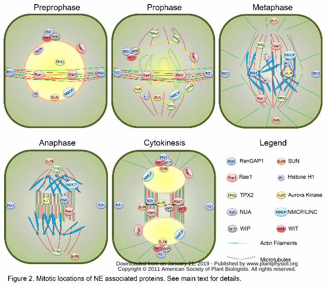
Rae1, is targeted to the PPB (Lee et al., 2009) (Fig. 2). This localization of Rae1 reflects
its association with mitotic MTs throughout mitosis, as well as at least partial
involvement of the PPB in spindle assembly, since the RNAi inhibition of Nicotiana
Benthamiana Rae1 (NbRae1) in BY-2 cells led to formation of disorganized or
multipolar spindles and defects in chromosome segregation (Lee et al., 2009). Indeed, in
plants the PPB marks the plane perpendicular to the axis of symmetry, the spindle (Lloyd
and Chan, 2006). The PPB is linked to and cross-communicates with the nucleus through
bridging MTs that partly mediates the establishment of the bipolarity of a cell and the
central positioning of the nucleus (Granger and Cyr, 2001; Ambrose and Wasteneys,
2008). This arrangement facilitates the formation of the prophase spindle perpendicular to
the PPB.
At this stage, the NE acting as an MTOC promotes nucleation of MTs on its surface
(Stoppin et al., 1994; Stoppin et al., 1996; Canaday et al., 2000). An essential factor of
the MT-nucleating complex is gamma-TuRC (gamma-tubulin ring complex), conserved
www.plantphysiol.orgon January 21, 2019 - Published by Downloaded from Copyright © 2011 American Society of Plant Biologists. All rights reserved.
12
among the kingdoms (Schmit, 2002). In mammals, the minimal complex functioning as
an MTOC is composed of gamma-tubulin, GCP2 and GCP3, which all have orthologs in
the Arabidopsis genome (Canaday, 2004). Besides their sequence similarity, gamma-
tubulin, AtGCP2 and AtGCP3 were detected in the same complex in vivo, localized at
the NE and the cell cortex, and were required for MT nucleation in Arabidopsis,
corroborating the conserved function of plant gamma-TuRC (Erhardt et al., 2002; Seltzer
et al., 2007). Interestingly, a nuclear rim-associated fraction of histone H1 was shown to
have MT-organizing activity in BY2 cells and to promote MT nucleation through
formation of complexes with tubulin and elongation of radial MTs (Hotta et al., 2007;
Nakayama et al., 2008) (Fig. 2). Recently, a biophysical interaction between Ran and
histone H1 and their co-localization at the nuclear rim have indicated a possible role for
histone H1 in organization of MTs adjacent to the NE in Leishmania donovani (Smirlis et
al., 2009).
Prior to the disappearance of the PPB in plant prophase, a rapid NE breakdown occurs
(Dixit and Cyr, 2002). Both processes seem to require phosphorylation events carried out
by a cyclin-dependent kinase (CDK) and its regulatory protein, cyclin B. The
CDK/CYCB complex promotes the PPB disassembly in plants (Hush et al., 1996), as
well as depolymerization of nuclear lamins in vertebrates, C. elegans, and yeast (Nigg,
1992; Daigle et al., 2001; Galy et al., 2008) and disassembly of nucleoporins in animal
cells (Macaulay et al., 1995; Favreau et al., 1996). This mitotic phosphorylation releases
lamins and some nuclear membrane and nuclear pore proteins, enabling progression
through the NE breakdown. Among plant nuclear pore proteins with dynamic mitotic
relocalization there is, for instance, NUA (Nuclear Pore Anchor, the Arabidopsis
Homolog of Tpr/Mlp1/Mlp2/Megator) (Jacob et al., 2007; Xu et al., 2007) and Rae1 (Lee
et al., 2009) (Fig. 2).
Metaphase
The Ran gradient controls the spindle assembly in animal cells. High concentrations of
RanGTP around chromosomes (and high RanGDP concentration at the cell periphery)
attract importins and release NLS-containing cargo proteins (Dasso, 2001; Weis, 2003).
www.plantphysiol.orgon January 21, 2019 - Published by Downloaded from Copyright © 2011 American Society of Plant Biologists. All rights reserved.
13
These cargos are, for instance, spindle assembly factors (SAFs), such as TPX2, Rae1 and
NuMA (Carazo-Salas et al., 1999; Kalab et al., 1999; Ohba et al., 1999; Wilde and
Zheng, 1999; Wiese et al., 2001; Caudron et al., 2005; Kalab et al., 2006). Arabidopsis
TPX2 is nuclear in interphase, but it is actively exported in prophase, enriched around the
NE, and then accumulates in the vicinity of the pro-spindle (Vos et al., 2008) (Fig. 2).
After its release from importin-dependent inhibition, TPX2 promotes spindle formation
around chromosomes through MT nucleation (Gruss and Vernos, 2004; Vos et al., 2008).
Simultaneously, human TPX2 targets Aurora A to the spindle and activates it (Bayliss et
al., 2003; Gruss and Vernos, 2004; Kawabe et al., 2005; Vos et al., 2008). In plant and
animal cells, the coordination of chromosomal and cytoskeletal events in mitosis is partly
mediated by the chromosomal passenger complex. Aurora kinases (in Arabidopsis
Aurora1 and 2) are thought to play this role through mediating the positioning
information of the PPB to the formation of the bipolar prophase spindle (Carmena and
Earnshaw, 2003; Vagnarelli and Earnshaw, 2004; Demidov et al., 2005). At the onset of
prophase, AtAurora1 and AtAurora2 are associated with the ONM and then gradually
migrate to the poles of the pro-spindle as mitosis progresses (Demidov et al., 2005) (Fig.
2).
Tobacco NbRae1, a homolog of Rae1/mrnp41 in metazoans, Gle2p in S. cerevisiae, and
Rae1 in S. pombe, exhibits a mitotic function, besides its role as an mRNA export factor
associated with the NPC (Whalen et al., 1997; Pritchard et al., 1999; Griffis et al., 2004;
Lee et al., 2009). Mammalian Rae1 is a mitotic spindle checkpoint component in
conjunction with Bub3 and forms a complex with Nup98 and the Cdh1-activated
anaphase promoting complex, preventing degradation of Securin before anaphase
(Whalen et al., 1997; Babu et al., 2003; Jeganathan et al., 2005). NbRae1 associates with
the spindle and was shown to function in the proper spindle organization and
chromosome segregation (Lee et al., 2009) (Fig. 2). NbRae1 silencing resulted in delayed
progression of mitosis, which led to plant growth arrest, reduced cell division activities in
the shoot apex and the vascular cambium, and increased ploidy levels in mature leaves.
Together, these results suggest a conserved function of the Rae1 proteins in spindle
organization among eukaryotes, which is distinct from their roles at the interphase NE.
www.plantphysiol.orgon January 21, 2019 - Published by Downloaded from Copyright © 2011 American Society of Plant Biologists. All rights reserved.
14
In metaphase, while histone H1 re-localizes along the condensed chromosomes
(Nakayama et al., 2008), Aurora 3 and 1 are associated with centromeric regions of
chromosomes (Demidov et al., 2005; Kawabe et al., 2005), RanGAP1 localizes to
kinetochores and the spindle (Joseph et al., 2002; Xu et al., 2008). Mammalian RanGAP1
is targeted to kinetochores in a SUMO-dependent manner (Joseph et al., 2002; Joseph et
al., 2004). Thus, it remains enigmatic how Arabidopsis RanGAP1, which lacks the
SUMOylation domain, is targeted to kinetochores. In view of human RanGAP1 found
only on the attached sister chromatids (Joseph et al., 2004), the exact timing of
kinetochore association and the function of plant RanGAP1 at this cellular location
remains to be verified.
Recently, the cell cycle dynamics of Apium graveolus NMCP1 and NMCP2 (AgNMCP1
and AgNMCP2) were investigated (Kimura et al., 2010). Both proteins associate with the
NE in interphase, disassemble simultaneously during prometaphase, and re-accumulate
around the reforming nuclei (Fig. 2). However, while AgNMCP1 was mainly localized to
the spindle and accumulated on segregating chromosomes, AgNMCP2 dispersed in the
mitotic cytoplasm in vesicular structures that could be distinguished from the bulk
endomembrane system. This vesicular signal might represent the NE membranes
absorbed into the ER network upon the NE breakdown.
Two Arabidopsis homologs of the spindle pole body protein Sad1 were initially
discovered in a survey for cytokinesis-related genes (Hagan and Yanagida, 1995; Van
Damme et al., 2004). These Arabidopsis SUN-domain proteins are NE markers in plants
(Graumann et al., 2010). Both Oda et al. and Graumann et al. carefully followed the
localization dynamics of both proteins through the cell cycle using transgenic
Arabidopsis plants and stably transformed BY-2 cells, respectively (Graumann and
Evans, 2011; Oda and Fukuda, 2011). Both groups reported the localization of SUNs in
mitotic ER membranes and an asymmetric re-association with the decondensing
telophase chromatin, with an envelope-like structure first appearing at the surface next to
the spindle poles and a delayed re-appearance of the envelope at the surface close to the
www.plantphysiol.orgon January 21, 2019 - Published by Downloaded from Copyright © 2011 American Society of Plant Biologists. All rights reserved.
15
phragmoplast (Fig. 2). This might indicate that NE assembly lags behind at the
phragmoplast-proximal surface of the daughter nuclei, and potentially this area remains
open longer to non-restricted exchange between nucleus and cytoplasm. Alternatively,
because SUN1/2 are nuclear proteins, it might indicate that nuclear pores at the
phragmoplast-proximal surface lag behind in regaining full import capacity. These
scenarios can be distinguished by also following ONM and NPC proteins as well as
generic markers for active nuclear import.
Anaphase/Telophase
As chromosomes migrate to opposing spindle poles, a plant-specific MT structure, the
phragmoplast, is formed to allow completion of cell division through assembly of a new
cell wall between the separating sister nuclei (Verma, 2001; Jurgens, 2005). Besides the
proteins involved in vesicular trafficking and fusion (reviewed in (Van Damme and
Geelen, 2008), some NE-associated proteins have been found to mark the phragmoplast
and/or the cell plate as well. The localization of Rae1 and SUN1/2 at the cell plate (and
the phragmoplast for Rae1) (Fig. 2) suggests a tight linkage between the NE components
and the cytoskeleton during mitosis. Thus, it would be of utmost interest to identify plant
interactors of SUN proteins both at the NE and at the cell plate. Such data would shed
more light on molecular bridges across the perinuclear space, linking the nucleoskeleton
to cytoskeleton, as well as on functions of NE proteins in cell division.
Apart from Rae1, other nuclear rim-associated proteins co-localize with SUNs at the cell
plate as well. For instance, Arabidopsis ONM proteins, WIP1, WIP2, WIT1 and WIT2,
are redistributed to the cell plate during cytokinesis (Patel et al., 2004; Xu et al., 2007;
Zhao et al., 2008) (Fig. 2). Both WITs and WIPs are required for RanGAP1 anchoring to
the NE in the root meristem, but only one of the protein families, either WIPs or WITs, is
sufficient to target RanGAP1 to the NE in differentiated cells (Zhao et al., 2008). The cell
plate localization of RanGAP1 (as well as its PPB and cortical division site association),
on the other hand, is independent on both WIPs and WITs, suggesting that interphase and
mitotic targeting of RanGAP1 require different mechanisms. Therefore, identification of
www.plantphysiol.orgon January 21, 2019 - Published by Downloaded from Copyright © 2011 American Society of Plant Biologists. All rights reserved.
16
molecular players involved in RanGAP1 localization and function(s) during plant cell
division would be of great importance.
Outlook
Over the past years, much progress has been made to unravel molecular players residing
at the nuclear periphery in animal, yeast and plant cells. Numerous INM, ONM, as well
as nuclear lamina and nuclear pore proteins have been brought to the stage via either
homology-based reverse genetics, forward genetics or proteomics approaches. The NE
components have been shown not only to separate the nucleoplasm from the cytosol and
to constitute a selective barrier for the nucleocytoplasmic transport, but they are also
involved in nuclear mobility, signal transduction, chromatin attachment and
transcriptional activation and repression. Subcellular localization, as well as thorough
phenotypic analyses has delivered additional spatio-temporal information regarding NE-
associated proteins. Namely, in plants these molecular players have been implicated in
such mitotic events as spindle assembly, chromosome segregation, MTOC-like function,
cortical division site demarcation and NE reformation upon cytokinesis. The concept of
NE components having additional roles throughout cell division is fascinating, but very
challenging to dissect experimentally. Therefore, certain biological questions remain to
be addressed. In vivo “fishing expeditions” using NE molecules as baits, would possibly
elucidate protein interactors involved in particular processes of cell division, as well as
targeting mechanisms of these molecules to diverse cellular addresses. Furthermore, the
precise dynamic localization of a given protein, and the order of disassembly/reassembly
of plant NE/NPC components, could be tackled with high-resolution imaging techniques,
such as multicolor confocal laser scanning microscopy (CLSM), in-lens field emission
scanning electron microscopy (feSEM) or three-dimensional structured illumination
microscopy (3D-SIM).
www.plantphysiol.orgon January 21, 2019 - Published by Downloaded from Copyright © 2011 American Society of Plant Biologists. All rights reserved.
17
Literature Cited Akhtar A, Gasser SM (2007) The nuclear envelope and transcriptional control.
Nature Reviews Genetics 8: 507-517
Ambrose JC, Wasteneys GO (2008) CLASP modulates microtubule-cortex
interaction during self-organization of acentrosomal microtubules. Molecular
Biology of the Cell 19: 4730-4737
Andres V, Gonzalez JM (2009) Role of A-type lamins in signaling, transcription, and
chromatin organization. Journal of Cell Biology 187: 945-957
Babu JR, Jeganathan KB, Baker DJ, Wu X, Kang-Decker N, van Deursen JM
(2003) Rae1 is an essential mitotic checkpoint regulator that cooperates with
Bub3 to prevent chromosome missegregation. Journal of Cell Biology 160:
341-353
Bayliss R, Sardon T, Vernos I, Conti E (2003) Structural basis of Aurora-A
activation by TPX2 at the mitotic spindle. Molecular Cell 12: 851-862
Belgareh N, Doye V (1997) Dynamics of nuclear pore distribution in nucleoporin
mutant yeast cells. Journal of Cell Biology 136: 747-759
Bootman MD, Fearnley C, Smyrnias I, MacDonald F, Roderick HL (2009) An
update on nuclear calcium signalling. journal of Cell Science 122: 2337-2350
Brohawn SG, Partridge JR, Whittle JR, Schwartz TU (2009) The nuclear pore
complex has entered the atomic age. Structure 17: 1156-1168
Brohawn SG, Schwartz TU (2009) Molecular architecture of the Nup84-Nup145C-
Sec13 edge element in the nuclear pore complex lattice. Nature Structural &
Molecular Biology 16: 1173-1177
Burke B, Roux KJ (2009) Nuclei take a position: managing nuclear location.
Developmental Cell 17: 587-597
Canaday J, Brochot, A.L., Seltzer, V., Herzog, E., Evrard, J.L. and Schmit, A.C.
(2004) Microtubule assembly in higher plants. In SG Pandalai, ed, Recent
Research Developments in Molecular Biology, Vol 2. Research Signpost,
Trivandrum, India, pp 103–119
Canaday J, Stoppin-Mellet V, Mutterer J, Lambert AM, Schmit AC (2000) Higher
plant cells: gamma-tubulin and microtubule nucleation in the absence of
centrosomes. Microscopy Research and Technique 49: 487-495
Capelson M, Hetzer MW (2009) The role of nuclear pores in gene regulation,
development and disease. EMBO reports 10: 697-705
Capelson M, Liang Y, Schulte R, Mair W, Wagner U, Hetzer MW (2010)
Chromatin-bound nuclear pore components regulate gene expression in
higher eukaryotes. Cell 140: 372-383
Carazo-Salas RE, Guarguaglini G, Gruss OJ, Segref A, Karsenti E, Mattaj IW
(1999) Generation of GTP-bound Ran by RCC1 is required for chromatin-
induced mitotic spindle formation. Nature 400: 178-181
Carmena M, Earnshaw WC (2003) The cellular geography of aurora kinases.
Nature Reviews: Molecular Cell Biology 4: 842-854
Caudron M, Bunt G, Bastiaens P, Karsenti E (2005) Spatial coordination of spindle
assembly by chromosome-mediated signaling gradients. Science 309: 1373-
1376
www.plantphysiol.orgon January 21, 2019 - Published by Downloaded from Copyright © 2011 American Society of Plant Biologists. All rights reserved.
18
Collas P, Courvalin JC (2000) Sorting nuclear membrane proteins at mitosis.
Trends in Cell Biology 10: 5-8
D'Angelo MA, Hetzer MW (2008) Structure, dynamics and function of nuclear pore
complexes. Trends in Cell Biology 18: 456-466
Daigle N, Beaudouin J, Hartnell L, Imreh G, Hallberg E, Lippincott-Schwartz J,
Ellenberg J (2001) Nuclear pore complexes form immobile networks and
have a very low turnover in live mammalian cells. Journal of Cell Biology
154: 71-84
Dasso M (2001) Running on Ran: nuclear transport and the mitotic spindle. Cell
104: 321-324
Dechat T, Adam SA, Taimen P, Shimi T, Goldman RD (2010) Nuclear lamins. Cold
Spring Harbor Perspectives in Biology 2: a000547
Demidov D, Van Damme D, Geelen D, Blattner FR, Houben A (2005)
Identification and dynamics of two classes of aurora-like kinases in
Arabidopsis and other plants. Plant Cell 17: 836-848
Dittmer TA, Stacey NJ, Sugimoto-Shirasu K, Richards EJ (2007) LITTLE NUCLEI
genes affecting nuclear morphology in Arabidopsis thaliana. Plant Cell 19:
2793-2803
Dixit R, Cyr RJ (2002) Spatio-temporal relationship between nuclear-envelope
breakdown and preprophase band disappearance in cultured tobacco cells.
Protoplasma 219: 116-121
Dong CH, Hu X, Tang W, Zheng X, Kim YS, Lee BH, Zhu JK (2006) A putative
Arabidopsis nucleoporin, AtNUP160, is critical for RNA export and required
for plant tolerance to cold stress. Molecular and Cellular Biology 26: 9533-
9543
Elad N, Maimon T, Frenkiel-Krispin D, Lim RY, Medalia O (2009) Structural
analysis of the nuclear pore complex by integrated approaches. Current
Opinion in Structural Biology 19: 226-232
Ellis JA (2006) Emery-Dreifuss muscular dystrophy at the nuclear envelope: 10
years on. Cellular and Molecular Life Sciences 63: 2702-2709
Erhardt M, Stoppin-Mellet V, Campagne S, Canaday J, Mutterer J, Fabian T,
Sauter M, Muller T, Peter C, Lambert AM, Schmit AC (2002) The plant
Spc98p homologue colocalizes with gamma-tubulin at microtubule
nucleation sites and is required for microtubule nucleation. Journal of Cell
Science 115: 2423-2431
Erickson ES, Mooren OL, Moore D, Krogmeier JR, Dunn RC (2006) The role of
nuclear envelope calcium in modifying nuclear pore complex structure.
Canadian Journal of Physiology and Pharmacology 84: 309-318
Evans DE, Shvedunova M, Graumann K (2011) The nuclear envelope in the plant
cell cycle: structure, function and regulation. Annals of Botany 107: 1111-
1118
Favreau C, Worman HJ, Wozniak RW, Frappier T, Courvalin JC (1996) Cell cycle-
dependent phosphorylation of nucleoporins and nuclear pore membrane
protein Gp210. Biochemistry 35: 8035-8044
www.plantphysiol.orgon January 21, 2019 - Published by Downloaded from Copyright © 2011 American Society of Plant Biologists. All rights reserved.
19
Finlan LE, Sproul D, Thomson I, Boyle S, Kerr E, Perry P, Ylstra B, Chubb JR,
Bickmore WA (2008) Recruitment to the nuclear periphery can alter
expression of genes in human cells. PLoS Genetics 4: e1000039
Fiserova J, Kiseleva E, Goldberg MW (2009) Nuclear envelope and nuclear pore
complex structure and organization in tobacco BY-2 cells. Plant Journal
Frey S, Gorlich D (2007) A saturated FG-repeat hydrogel can reproduce the
permeability properties of nuclear pore complexes. Cell 130: 512-523
Frey S, Richter RP, Gorlich D (2006) FG-rich repeats of nuclear pore proteins form
a three-dimensional meshwork with hydrogel-like properties. Science 314:
815-817
Fridkin A, Penkner A, Jantsch V, Gruenbaum Y (2009) SUN-domain and KASH-
domain proteins during development, meiosis and disease. Cellular and
Molecular Life Sciences 66: 1518-1533
Galcheva-Gargova Z, Stateva L (1988) Immunological identification of two lamina-
like proteins in Saccharomyces cerevisiae. Bioscience Reports 8: 287-291
Galy V, Antonin W, Jaedicke A, Sachse M, Santarella R, Haselmann U, Mattaj I
(2008) A role for gp210 in mitotic nuclear-envelope breakdown. Journal of
Cell Science 121: 317-328
Gant TM, Wilson KL (1997) Nuclear Assembly. Annual Review of Cell and
Developmental Biology 13: 669-695
Gerber AP, Herschlag D, Brown PO (2004) Extensive association of functionally
and cytotopically related mRNAs with Puf family RNA-binding proteins in
yeast. PLoS Biology 2: E79
Granger C, Cyr R (2001) Use of abnormal preprophase bands to decipher division
plane determination. Journal of Cell Science 114: 599-607
Graumann K, Evans DE (2011) Nuclear envelope dynamics during plant cell
division suggest common mechanisms between kingdoms. Biochemical
Journal 435: 661-667
Graumann K, Evans DE (2011) Nuclear envelope dynamics during plant cell
division suggest common mechanisms between kingdoms. Biochemical
Journal 435: 661-667
Graumann K, Runions J, Evans DE (2010) Characterization of SUN-domain
proteins at the higher plant nuclear envelope. Plant Journal 61: 134-144
Graumann K, Runions J, Evans DE (2010) Characterization of SUN-domain
proteins at the higher plant nuclear envelope. Plant Journal 61: 134-144
Griffis ER, Craige B, Dimaano C, Ullman KS, Powers MA (2004) Distinct
functional domains within nucleoporins Nup153 and Nup98 mediate
transcription-dependent mobility. Molecular Biology of the Cell 15: 1991-
2002
Gruss OJ, Vernos I (2004) The mechanism of spindle assembly: functions of Ran
and its target TPX2. Journal of Cell Biology 166: 949-955
Guelen L, Pagie L, Brasset E, Meuleman W, Faza MB, Talhout W, Eussen BH, de
Klein A, Wessels L, de Laat W, van Steensel B (2008) Domain organization
of human chromosomes revealed by mapping of nuclear lamina interactions.
Nature 453: 948-951
www.plantphysiol.orgon January 21, 2019 - Published by Downloaded from Copyright © 2011 American Society of Plant Biologists. All rights reserved.
20
Hagan I, Yanagida M (1995) The product of the spindle formation gene sad1+
associates with the fission yeast spindle pole body and is essential for
viability. Journal of Cell Biology 129: 1033-1047
Hetzer MW, Walther TC, Mattaj IW (2005) Pushing the envelope: structure,
function, and dynamics of the nuclear periphery. Annual Review of Cell and
Developmental Biology 21: 347-380
Hetzer MW, Wente SR (2009) Border control at the nucleus: biogenesis and
organization of the nuclear membrane and pore complexes. Developmental
Cell 17: 606-616
Hiraoka Y, Dernburg AF (2009) The SUN rises on meiotic chromosome dynamics.
Developmental Cell 17: 598-605
Hotta T, Haraguchi T, Mizuno K (2007) A novel function of plant histone H1:
microtubule nucleation and continuous plus end association. Cell Structure
and Function 32: 79-87
Hush J, Wu L, John PC, Hepler LH, Hepler PK (1996) Plant mitosis promoting
factor disassembles the microtubule preprophase band and accelerates
prophase progression in Tradescantia. Cell Biology International 20: 275-
287
Irons SL, Evans DE, Brandizzi F (2003) The first 238 amino acids of the human
lamin B receptor are targeted to the nuclear envelope in plants. Journal of
Experimental Botany 54: 943-950
Jacob Y, Mongkolsiriwatana C, Veley KM, Kim SY, Michaels SD (2007) The
nuclear pore protein AtTPR is required for RNA homeostasis, flowering time,
and auxin signaling. Plant Physiology 144: 1383-1390
Jeganathan KB, Malureanu L, van Deursen JM (2005) The Rae1-Nup98 complex
prevents aneuploidy by inhibiting securin degradation. Nature 438: 1036-
1039
Joseph J, Liu ST, Jablonski SA, Yen TJ, Dasso M (2004) The RanGAP1-RanBP2
complex is essential for microtubule-kinetochore interactions in vivo.
Current Biology 14: 611-617
Joseph J, Tan SH, Karpova TS, McNally JG, Dasso M (2002) SUMO-1 targets
RanGAP1 to kinetochores and mitotic spindles. Journal of Cell Biology 156:
595-602
Jovanovic-Talisman T, Tetenbaum-Novatt J, McKenney AS, Zilman A, Peters R,
Rout MP, Chait BT (2009) Artificial nanopores that mimic the transport
selectivity of the nuclear pore complex. Nature 457: 1023-1027
Jurgens G (2005) Cytokinesis in higher plants. Annual Review of Plant Biology 56:
281-299
Kahms M, Huve J, Wesselmann R, Farr JC, Baumgartel V, Peters R (2011)
Lighting up the nuclear pore complex. European Journal of Cell Biology
Kalab P, Pralle A, Isacoff EY, Heald R, Weis K (2006) Analysis of a RanGTP-
regulated gradient in mitotic somatic cells. Nature 440: 697-701
Kalab P, Pu RT, Dasso M (1999) The ran GTPase regulates mitotic spindle
assembly. Current Biology 9: 481-484
www.plantphysiol.orgon January 21, 2019 - Published by Downloaded from Copyright © 2011 American Society of Plant Biologists. All rights reserved.
21
Kalverda B, Fornerod M (2010) Characterization of genome-nucleoporin
interactions in Drosophila links chromatin insulators to the nuclear pore
complex. Cell Cycle 9: 4812-4817
Kalverda B, Pickersgill H, Shloma VV, Fornerod M (2010) Nucleoporins directly
stimulate expression of developmental and cell-cycle genes inside the
nucleoplasm. Cell 140: 360-371
Kalverda B, Roling MD, Fornerod M (2008) Chromatin organization in relation to
the nuclear periphery. FEBS Letters 582: 2017-2022
Kanamori N, Madsen LH, Radutoiu S, Frantescu M, Quistgaard EM, Miwa H,
Downie JA, James EK, Felle HH, Haaning LL, Jensen TH, Sato S, Nakamura
Y, Tabata S, Sandal N, Stougaard J (2006) A nucleoporin is required for
induction of Ca2+ spiking in legume nodule development and essential for
rhizobial and fungal symbiosis. Proceedings of the National Academy of
Sciences, USA 103: 359-364
Kawabe A, Matsunaga S, Nakagawa K, Kurihara D, Yoneda A, Hasezawa S,
Uchiyama S, Fukui K (2005) Characterization of plant Aurora kinases
during mitosis. Plant Molecular Biology 58: 1-13
Kimura Y, Kuroda C, Masuda K (2010) Differential nuclear envelope assembly at
the end of mitosis in suspension-cultured Apium graveolens cells.
Chromosoma 119: 195-204
Kumaran RI, Spector DL (2008) A genetic locus targeted to the nuclear periphery
in living cells maintains its transcriptional competence. Journal of Cell
Biology 180: 51-65
Kutay U, Hetzer MW (2008) Reorganization of the nuclear envelope during open
mitosis. Current Opinion in Cell Biology 20: 669-677
Lee JY, Lee HS, Wi SJ, Park KY, Schmit AC, Pai HS (2009) Dual functions of
Nicotiana benthamiana Rae1 in interphase and mitosis. Plant Journal 59:
278-291
Li H, Roux SJ (1992) Casein kinase II protein kinase is bound to lamina-matrix and
phosphorylates lamin-like protein in isolated pea nuclei. Proceedings of the
National Academy of Sciences, USA 89: 8434-8438
Lloyd C, Chan J (2006) Not so divided: the common basis of plant and animal cell
division. Nature Reviews: Molecular Cell Biology 7: 147-152
Lu Q, Tang X, Tian G, Wang F, Liu K, Nguyen V, Kohalmi SE, Keller WA, Tsang
EW, Harada JJ, Rothstein SJ, Cui Y (2010) Arabidopsis homolog of the yeast
TREX-2 mRNA export complex: components and anchoring nucleoporin.
Plant Journal 61: 259-270
Macaulay C, Meier E, Forbes DJ (1995) Differential mitotic phosphorylation of
proteins of the nuclear pore complex. Journal of Biological Chemistry 270:
254-262
Maeshima K, Yahata K, Sasaki Y, Nakatomi R, Tachibana T, Hashikawa T,
Imamoto F, Imamoto N (2006) Cell-cycle-dependent dynamics of nuclear
pores: pore-free islands and lamins. Journal of Cell Science 119: 4442-4451
Malhas A, Lee CF, Sanders R, Saunders NJ, Vaux DJ (2007) Defects in lamin B1
expression or processing affect interphase chromosome position and gene
expression. Journal of Cell Biology 176: 593-603
www.plantphysiol.orgon January 21, 2019 - Published by Downloaded from Copyright © 2011 American Society of Plant Biologists. All rights reserved.
22
Malhas AN, Lee CF, Vaux DJ (2009) Lamin B1 controls oxidative stress responses
via Oct-1. Journal of Cell Biology 184: 45-55
Malhas AN, Vaux DJ (2009) Transcription factor sequestration by nuclear envelope
components. Cell Cycle 8: 959-960
Masuda K, Xu ZJ, Takahashi S, Ito A, Ono M, Nomura K, Inoue M (1997)
Peripheral framework of carrot cell nucleus contains a novel protein
predicted to exhibit a long alpha-helical domain. Experimental Cell Research
232: 173-181
McNulty AK, Saunders MJ (1992) Purification and immunological detection of pea
nuclear intermediate filaments: evidence for plant nuclear lamins. Journal of
Cell Science 103 ( Pt 2): 407-414
Meier I (2007) Composition of the plant nuclear envelope: theme and variations.
Journal of Experimental Botany 58: 27-34
Meier I, Somers DE (2011) Regulation of nucleocytoplasmic trafficking in plants.
Current Opinion in Plant Biology [online first]. PMID: 21764628. Merkle T (2009) Nuclear Export of Proteins and RNA. In I Meier, ed, Functional
Organization of the Plant Nucleus, Vol 14. Springer Berlin / Heidelberg, pp
55-77
Minguez A, Moreno Diaz de la Espina S (1993) Immunological characterization of
lamins in the nuclear matrix of onion cells. Journal of Cell Science 106: 431-
439
Moriguchi K, Suzuki T, Ito Y, Yamazaki Y, Niwa Y, Kurata N (2005) Functional
isolation of novel nuclear proteins showing a variety of subnuclear
localizations. Plant Cell 17: 389-403
Muller S, Wright AJ, Smith LG (2009) Division plane control in plants: new players
in the band. Trends in Cell Biology 19: 180-188
Murphy SP, Simmons CR, Bass HW (2010) Structure and expression of the maize
(Zea mays L.) SUN-domain protein gene family: evidence for the existence of
two divergent classes of SUN proteins in plants. BMC Plant Biology 10: 269
Nakayama T, Ishii T, Hotta T, Mizuno K (2008) Radial microtubule organization
by histone H1 on nuclei of cultured tobacco BY-2 cells. Journal of Biological
Chemistry 283: 16632-16640
Nigg EA (1992) Assembly and cell cycle dynamics of the nuclear lamina. Seminars in
Cell Biology 3: 245-253
Oda Y, Fukuda H (2011) Dynamics of Arabidopsis SUN proteins during mitosis and
their involvement in nuclear shaping. Plant Journal 66: 629-641
Oda Y, Fukuda H (2011) Dynamics of Arabidopsis SUN proteins during mitosis and
their involvement in nuclear shaping. Plant Journal 66: 629-641
Ohba T, Nakamura M, Nishitani H, Nishimoto T (1999) Self-organization of
microtubule asters induced in Xenopus egg extracts by GTP-bound Ran.
Science 284: 1356-1358
Onischenko E, Stanton LH, Madrid AS, Kieselbach T, Weis K (2009) Role of the
Ndc1 interaction network in yeast nuclear pore complex assembly and
maintenance. Journal of Cell Biology 185: 475-491
Parry G, Ward S, Cernac A, Dharmasiri S, Estelle M (2006) The Arabidopsis
SUPPRESSOR OF AUXIN RESISTANCE proteins are nucleoporins with an
www.plantphysiol.orgon January 21, 2019 - Published by Downloaded from Copyright © 2011 American Society of Plant Biologists. All rights reserved.
23
important role in hormone signaling and development. Plant Cell 18: 1590-
1603
Patel S, Rose A, Meulia T, Dixit R, Cyr RJ, Meier I (2004) Arabidopsis WPP-domain
proteins are developmentally associated with the nuclear envelope and
promote cell division. Plant Cell 16: 3260-3273
Pritchard CE, Fornerod M, Kasper LH, van Deursen JM (1999) RAE1 is a shuttling
mRNA export factor that binds to a GLEBS-like NUP98 motif at the nuclear
pore complex through multiple domains. Journal of Cell Biology 145: 237-
254
Rabut G, Ellenberg J (2001) Nucleocytoplasmic transport: Diffusion channel or
phase transition? Current Biology 11: R551-R554
Reddy KL, Zullo JM, Bertolino E, Singh H (2008) Transcriptional repression
mediated by repositioning of genes to the nuclear lamina. Nature 452: 243-
247
Roberts K, Northcote DH (1970) Structure of the nuclear pore in higher plants.
Nature 228: 385-386
Saito K, Yoshikawa M, Yano K, Miwa H, Uchida H, Asamizu E, Sato S, Tabata S,
Imaizumi-Anraku H, Umehara Y, Kouchi H, Murooka Y, Szczyglowski K,
Downie JA, Parniske M, Hayashi M, Kawaguchi M (2007)
NUCLEOPORIN85 is required for calcium spiking, fungal and bacterial
symbioses, and seed production in Lotus japonicus. Plant Cell 19: 610-624
Schirmer EC, Gerace L (2005) The nuclear membrane proteome: extending the
envelope. Trends in Biochemical Sciences 30: 551-558
Schmid M, Arib G, Laemmli C, Nishikawa J, Durussel T, Laemmli UK (2006) Nup-
PI: the nucleopore-promoter interaction of genes in yeast. Molecular Cell 21:
379-391
Schmit AC (2002) Acentrosomal microtubule nucleation in higher plants.
International Review of Cytology 220: 257-289
Seltzer V, Janski N, Canaday J, Herzog E, Erhardt M, Evrard JL, Schmit AC (2007)
Arabidopsis GCP2 and GCP3 are part of a soluble gamma-tubulin complex
and have nuclear envelope targeting domains. Plant Journal 52: 322-331
Smirlis D, Boleti H, Gaitanou M, Soto M, Soteriadou K (2009) Leishmania
donovani Ran-GTPase interacts at the nuclear rim with linker histone H1.
Biochemical Journal 424: 367-374
Smith LG, Gerttula SM, Han S, Levy J (2001) Tangled1: a microtubule binding
protein required for the spatial control of cytokinesis in maize. Journal of Cell
Biology 152: 231-236
Solovei I, Kreysing M, Lanctot C, Kosem S, Peichl L, Cremer T, Guck J, Joffe B
(2009) Nuclear architecture of rod photoreceptor cells adapts to vision in
mammalian evolution. Cell 137: 356-368
Somech R, Shaklai S, Geller O, Amariglio N, Simon AJ, Rechavi G, Gal-Yam EN
(2005) The nuclear-envelope protein and transcriptional repressor
LAP2beta interacts with HDAC3 at the nuclear periphery, and induces
histone H4 deacetylation. Journal of Cell Science 118: 4017-4025
www.plantphysiol.orgon January 21, 2019 - Published by Downloaded from Copyright © 2011 American Society of Plant Biologists. All rights reserved.
24
Stavru F, Hülsmann BB, Spang A, Hartmann E, Cordes VC, Görlich D (2006)
NDC1: a crucial membrane-integral nucleoporin of metazoan nuclear pore
complexes. Journal of Cell Biology 173: 509-519
Stockinger EJ, Mao Y, Regier MK, Triezenberg SJ, Thomashow MF (2001)
Transcriptional adaptor and histone acetyltransferase proteins in
Arabidopsis and their interactions with CBF1, a transcriptional activator
involved in cold-regulated gene expression. Nucleic Acids Research 29:
1524-1533
Stoppin V, Lambert AM, Vantard M (1996) Plant microtubule-associated proteins
(MAPs) affect microtubule nucleation and growth at plant nuclei and
mammalian centrosomes. European Journal of Cell Biology 69: 11-23
Stoppin V, Vantard M, Schmit AC, Lambert AM (1994) Isolated Plant Nuclei
Nucleate Microtubule Assembly: The Nuclear Surface in Higher Plants Has
Centrosome-like Activity. Plant Cell 6: 1099-1106
Tamura S, Shimizu N, Fujiwara K, Kaneko M, Kimura T, Murakami N (2010)
Bioisostere of valtrate, anti-HIV principle by inhibition for nuclear export of
Rev. Bioorganic & Medicinal Chemistry Letters 20: 2159-2162
Vagnarelli P, Earnshaw WC (2004) Chromosomal passengers: the four-
dimensional regulation of mitotic events. Chromosoma 113: 211-222
Van Damme D, Bouget FY, Van Poucke K, Inze D, Geelen D (2004) Molecular
dissection of plant cytokinesis and phragmoplast structure: a survey of GFP-
tagged proteins. Plant Journal 40: 386-398
Van Damme D, Geelen D (2008) Demarcation of the cortical division zone in
dividing plant cells. Cell Biology International 32: 178-187
Vaquerizas JM, Suyama R, Kind J, Miura K, Luscombe NM, Akhtar A (2010)
Nuclear pore proteins nup153 and megator define transcriptionally active
regions in the Drosophila genome. PLoS Genetics 6: e1000846
Verma DP (2001) Cytokinesis and Building of the Cell Plate in Plants. Annual
Review of Plant Physiology and Plant Molecular Biology 52: 751-784
Vos JW, Pieuchot L, Evrard JL, Janski N, Bergdoll M, de Ronde D, Perez LH,
Sardon T, Vernos I, Schmit AC (2008) The plant TPX2 protein regulates
prospindle assembly before nuclear envelope breakdown. Plant Cell 20:
2783-2797
Walde S, Kehlenbach RH (2010) The Part and the Whole: functions of
nucleoporins in nucleocytoplasmic transport. Trends in Cell Biology 20: 461-
469
Weis K (2003) Regulating access to the genome: nucleocytoplasmic transport
throughout the cell cycle. Cell 112: 441-451
Whalen WA, Bharathi A, Danielewicz D, Dhar R (1997) Advancement through
mitosis requires rae1 gene function in fission yeast. Yeast 13: 1167-1179
Wheeler MA, Ellis JA (2008) Molecular signatures of Emery-Dreifuss muscular
dystrophy. Biochemical Society Transactions 36: 1354-1358
Wiermer M, Palma K, Zhang Y, Li X (2007) Should I stay or should I go?
Nucleocytoplasmic trafficking in plant innate immunity. Cellular
Microbiology 9: 1880-1890
www.plantphysiol.orgon January 21, 2019 - Published by Downloaded from Copyright © 2011 American Society of Plant Biologists. All rights reserved.
25
Wiese C, Wilde A, Moore MS, Adam SA, Merdes A, Zheng Y (2001) Role of
importin-beta in coupling Ran to downstream targets in microtubule
assembly. Science 291: 653-656
Wilde A, Zheng Y (1999) Stimulation of microtubule aster formation and spindle
assembly by the small GTPase Ran. Science 284: 1359-1362
Wilson KL (2010) Nuclear envelope and lamin B2 function in the central nervous
system. Proceedings of the National Academy of Sciences, USA 107: 6121-
6122
Worman HJ, Bonne G (2007) "Laminopathies": a wide spectrum of human diseases.
Experimental Cell Research 313: 2121-2133
Xu XM, Meulia T, Meier I (2007) Anchorage of plant RanGAP to the nuclear
envelope involves novel nuclear-pore-associated proteins. Current Biology
17: 1157-1163
Xu XM, Rose A, Muthuswamy S, Jeong SY, Venkatakrishnan S, Zhao Q, Meier I
(2007) NUCLEAR PORE ANCHOR, the Arabidopsis homolog of
Tpr/Mlp1/Mlp2/megator, is involved in mRNA export and SUMO
homeostasis and affects diverse aspects of plant development. Plant Cell 19:
1537-1548
Xu XM, Zhao Q, Rodrigo-Peiris T, Brkljacic J, He CS, Muller S, Meier I (2008)
RanGAP1 is a continuous marker of the Arabidopsis cell division plane.
Proceedings of the National Academy of Sciences, USA 105: 18637-18642
Yelina NE, Smith LM, Jones AM, Patel K, Kelly KA, Baulcombe DC (2010) Putative
Arabidopsis THO/TREX mRNA export complex is involved in transgene and
endogenous siRNA biosynthesis. Proceedings of the National Academy of
Sciences, USA 107: 13948-13953
Yokochi T, Poduch K, Ryba T, Lu J, Hiratani I, Tachibana M, Shinkai Y, Gilbert
DM (2009) G9a selectively represses a class of late-replicating genes at the
nuclear periphery. Proceedings of the National Academy of Sciences, USA
106: 19363-19368
Zhang Y, Li X (2005) A putative nucleoporin 96 Is required for both basal defense
and constitutive resistance responses mediated by suppressor of npr1-
1,constitutive 1. Plant Cell 17: 1306-1316
Zhao Q, Brkljacic J, Meier I (2008) Two distinct interacting classes of nuclear
envelope-associated coiled-coil proteins are required for the tissue-specific
nuclear envelope targeting of Arabidopsis RanGAP. Plant Cell 20: 1639-1651
Zhao Q, Meier I (2011) Identification and characterization of the Arabidopsis FG-
repeat nucleoporin Nup62. Plant Signaling & Behavior 6: 330-334
www.plantphysiol.orgon January 21, 2019 - Published by Downloaded from Copyright © 2011 American Society of Plant Biologists. All rights reserved.
26
Figure legends
Figure 1. Identified NE and NPC components in higher plants and vertebrates. A,
Comparison of the NE and NPC components between higher plants and vertebrates.
Sub-complexes are grouped in single units. Contacted units indicate confirmed
interactions among them. The NPC organization is modified after Tamura et al.
(Tamura et al., 2010). In the higher plant NPC, bold protein names indicate
confirmed NE localization. Mutant phenotypes have been reported for plant Nups
indicated in red. Mammalian Nups, Nup358, Nup97, Nup45, and Pom121 appear to
have no counterparts in plants. Positioning of plant Nups is based on their
vertebrate counterparts. B, The NE localization of putative Arabidopsis NDC1 in
Arabidopsis root tip cells. Cell walls were counterstained with propidium iodide
(PI). All scale bars equal 5µm.
Figure 2. Mitotic locations of NE associated proteins. See main text for details.
www.plantphysiol.orgon January 21, 2019 - Published by Downloaded from Copyright © 2011 American Society of Plant Biologists. All rights reserved.
www.plantphysiol.orgon January 21, 2019 - Published by Downloaded from Copyright © 2011 American Society of Plant Biologists. All rights reserved.
www.plantphysiol.orgon January 21, 2019 - Published by Downloaded from Copyright © 2011 American Society of Plant Biologists. All rights reserved.