30
Muscular Tissue Muscle: an organ composed of one of three types of muscle tissue (skeletal, cardiac or smooth) , specialized for contraction to produce voluntary or involuntary movement
| Date post: | 21-Jan-2018 |
| Category: |
Education |
| Upload: | prinkmkundnani-pharmacy-polytechnic |
| View: | 93 times |
| Download: | 0 times |

Muscular TissueMuscle: an organ composed of one of three types of muscle tissue (skeletal,
cardiac or smooth) , specialized for contraction to produce voluntary or
involuntary movement

Muscular Tissue

Muscular Tissue
A group of cells (fibers) specialized to produce motion in response to muscle
action potentials by its qualities of contractility, extensibility, elasticity and
excitability

Properties of Muscular Tissue
1. Excitability: an ability of muscle to generate impulse
aaahhh…
ooohhhh…
aaauuchh…
2. Contractility: it is either shortening or development of tension or both
A. Isotonic Contraction-contraction in which tension remains same whereas changes occurs in the length of muscle fiber. E.g. flexion of arm
B. Isometric Contraction-contraction in which length of muscle fibers remain same and tension is increased. E.g. holding book by hand, pulling any heavy object

3. Muscle Tone- the muscle fibers always maintain a state of slight contraction with certain degree of vigor and tension. This is a state of partial contraction of muscles. It is achieved by the contraction of a few muscle fibres at a time.
4. Extensibility- an ability of muscle fibers to stretch without being damaged
5. Elasticity- an ability of muscle fibers to return to its original length and shape after contraction or extension

Functions of Muscular tissueThrough sustained contraction or alternating
contraction & relaxation, muscular tissue has four functions:
1. Producing Body Movements- movements of the whole body such as walking and running, and localized movements such as holding pen, nodding head, rely on integrated functioning of bones, joints and skeletal muscles

2. Stabilizing Body Positions- Skeletal muscle contractions stabilize joints and help maintain body positions such as sitting & standing
3. Storing and moving substances within body- sustained contractions of sphincters temporarily stores food in stomach and urine in urinary bladder. Cardiac muscle contractions pump blood through blood vessels of the body.
-cont.

Smooth muscles contractions move food and substances such as bile, enzymes through g.i.t., push gametes through passageway of reproductive system, propel urine through urinary system.
Skeletal muscle contractions promote the flow of lymph and helps the return of blood to heart.
4.Generating heat- As muscular tissue contracts, it produces heat, by thermogenesis. Heat generated by muscle is used to maintain normal body temp.

Types of Muscular Tissue
Muscle Striations Control Nerve Supply
Skeletal Present Voluntary Somatic
Cardiac Present Involuntary Autonomic
Smooth Absent Involuntary Autonomic
Skeletal Muscle tissue• Skeletal muscle cells are found to be roughly
cylindrical in shape, diameter ranges from 10-100ųm and maybe as long as 35 cm.
• Each cell, commonly called a fiber, has several nuclei situated just under the sarcolemma or cell membrane of each muscle fiber.
• The muscle fibres lie parallel to one another and, when viewed under the microscope, they show well-marked transverse dark and light bands, hence the name striated or striped muscle.

Sarcoplasm: the cytoplasm of muscle fibres, contains:
• bundles of myofibrils, which consist of filaments of contractile proteins including actin and myosin
• many mitochondria, which generate chemical energy (ATP) from glucose and oxygen by aerobic respiration
• glycogen, a carbohydrate store which is broken down into glucose when required
• myoglobin, a unique oxygen-binding protein molecule, similar to hemoglobin in red blood cells, which stores oxygen within muscle cells.

A myofibril has a repeating series of dark and light bands, consisting of units called sarcomeres.A sarcomere represents the smallest functional unit of a skeletal muscle fibre and consists of:• thin filaments of actin• thick filaments of myosin.

• Each thick filament composed of about 300 molecules of Myosin. Each myosin molecule has two heads and a twisted tail. Tail form shaft of filament. The head project outwards from the shaft in a spiraling fashion
• Thin filament composed of actin as major and troponin & tropomyosin as minor components.
• Individual actin molecule join to form actin filament, that is twisted into helix.
• On each actin molecule, is myosin-binding site, where myosin head can attach.
• In a relaxed muscle myosin is blocked from binding to actin because strands of tropomyosin cover myosin binding-sites.

Physiology of ContractionSliding-Filament mechanism:
In this process, thin filaments slide inward over thick filament and may overlap.
As a result sarcomere shortens.
Shortening of sarcomeres causes shortening of whole muscle fiber, which causes shortening of entire muscle i.e. contraction
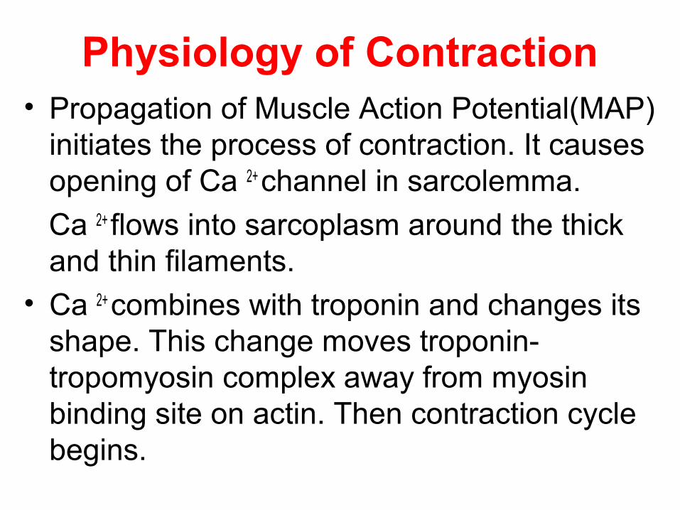
Physiology of Contraction• Propagation of Muscle Action Potential(MAP)
initiates the process of contraction. It causes opening of Ca 2+ channel in sarcolemma.
Ca 2+ flows into sarcoplasm around the thick and thin filaments.
• Ca 2+ combines with troponin and changes its shape. This change moves troponin-tropomyosin complex away from myosin binding site on actin. Then contraction cycle begins.

Contraction CycleFour Steps:
1.ATP Hydrolysis -The myosin head includes ATP-binding site and ATPase. Myosin heads hydrolyze ATP and become reoriented and energized.
ADPp

Contraction Cycle2.Formation of Crossbridges- The
energised myosin head attaches to myosin binding site on actin and releases phosphate group. This attachment is referred as Crossbridge.
ADP
p
Contraction Cycle
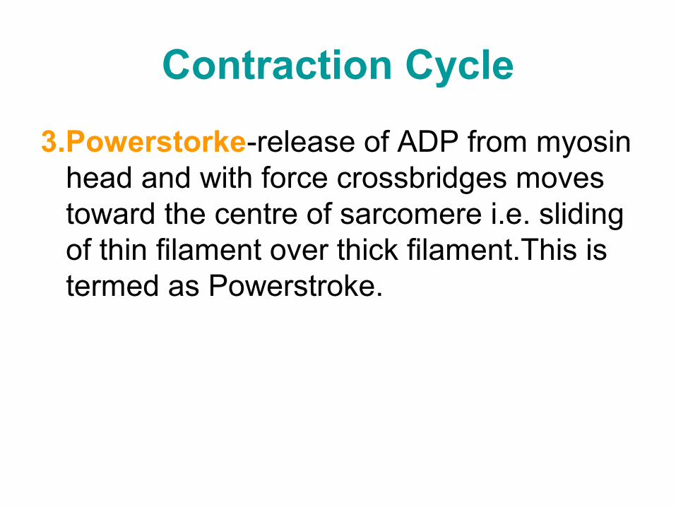
3.Powerstorke-release of ADP from myosin head and with force crossbridges moves toward the centre of sarcomere i.e. sliding of thin filament over thick filament.This is termed as Powerstroke.

Contraction Cycle
4.Detachment of myosin from actin- At the end of powerstroke, the crossbridges remains firmly attached to actin until another molecule of ATP binds to myosin. Then myosin head detaches from actin.
The contraction cycle continues as long as ATP is available and Ca2+ level near the filament is sufficiently high.

Neuromuscular Junction
• Anatomy

Neuromuscular Junction• MNJ is the synapse between a somatic
neuron and a skeletal muscle fiber.• Synaptic end bulbs- at MNJ the end of
motor neuron, called axon terminal divides into a cluster of synaptic end bulbs.
• Each bulb contains hundreds of membrane enclosed sacs called synaptic vesicles.
• Inside each synaptic vesicle are thousands of molecules of neurotransmitters- Acetylcholine (Ach)

Neuromuscular Junction• Motor end plate- It is region of
sarcolemma opposite the synaptic end bulbs. Each motor end plate has 30 -40 million Ach receptors.
• Physiology
• A nerve impulse (Nerve Action Potential) elicits Muscle Action Potential in the following way:-
Neuromuscular Junction
1. Release of Ach- Arrival of nerve impulse at the synaptic end bulbs causes many synaptic vesicles o undergo exocytosis.
2. During exocytosis, the synaptic vesicles fuse with neuron’s plasma membrane.
3. The Ach the diffuses across the synaptic cleft between neuron and motor end plate.

Neuromuscular Junction4. Activation of Ach receptors: Binding of
two molecules of Ach to the receptor on motor end plate open an ion channel. Cations most importantly Na+ flow across the membrane (sarcolemma)
5. Production of Muscle Action potential: The influx of Na+ makes muscle fiber more positively charged.This change triggers muscle action potential

Neuromuscular Junction
6. Termination of Ach activity- The effect of Ach binding is for moment, because Ach is rapidly broken down by enzyme acetyl cholinesterase. This cannot activate Ach receptors.
7. This ends production of Muscle Action Potential and muscle gains its original form.

Neuromuscular Junction


