Date post: | 10-Dec-2015 |
Category: |
Documents |
Upload: | cesaryvane |
View: | 212 times |
Download: | 0 times |
Electrochemical biosensors
Niina J. Ronkainen,*aH. Brian Halsall
band William R. Heineman
b
Received 3rd November 2008
First published as an Advance Article on the web 1st February 2010
DOI: 10.1039/b714449k
Electrochemical biosensors combine the sensitivity of electroanalytical methods with the inherent
bioselectivity of the biological component. The biological component in the sensor recognizes its
analyte resulting in a catalytic or binding event that ultimately produces an electrical signal
monitored by a transducer that is proportional to analyte concentration. Some of these sensor
devices have reached the commercial stage and are routinely used in clinical, environmental,
industrial, and agricultural applications. The two classes of electrochemical biosensors,
biocatalytic devices and affinity sensors, will be discussed in this critical review to provide an
accessible introduction to electrochemical biosensors for any scientist (110 references).
1. Introduction
1.1 Background
Sensors are devices that register a physical, chemical, or bio-
logical change and convert that into a measurable signal.1 The
sensor contains a recognition element that enables the selective
response to a particular analyte or a group of analytes, thus
minimizing interferences from other sample components
(Fig. 1). Another main component of a sensor is the transducer
or the detector device that produces a signal. A signal processor
collects, amplifies, and displays the signal.
Electrochemical biosensors, a subclass of chemical sensors,
combine the sensitivity, as indicated by low detection limits, of
electrochemical transducers with the high specificity of bio-
logical recognition processes. These devices contain a biological
recognition element (enzymes, proteins, antibodies, nucleic
acids, cells, tissues or receptors) that selectively reacts with
the target analyte and produces an electrical signal that is
related to the concentration of the analyte being studied.
Electrochemical biosensors can be divided into two main
categories based on the nature of the biological recognition
process i.e. biocatalytic devices and affinity sensors.2 Bio-
catalytic devices incorporate enzymes, whole cells or tissue
slices that recognize the target analyte and produce electro-
active species. Special emphasis will be placed on enzyme
electrodes for the detection of glucose, lactose, and xanthine.
Affinity sensors rely on a selective binding interaction between
the analyte and a biological component such as an antibody,
Fig. 1 A schematic of a biosensor with electrochemical transducer.
aDepartment of Chemistry, Benedictine University, 5700 College Road,Lisle, IL 60532-0900, USA. E-mail: [email protected];Fax: +1 630 829 6547; Tel: +1 630 829 6549
bDepartment of Chemistry, University of Cincinnati,P.O. Box 210172, Cincinnati, OH 45221-0172, USA.E-mail: [email protected], [email protected];Fax: +1 513 556 9239; Tel: +1 513 556 9274, +1 513 556 9210
Niina J. Ronkainen
Niina J. Ronkainen receivedher BS in chemistry andbiology at Butler University(Indianapolis, USA) in 1997and her PhD at the Universityof Cincinnati (USA) in 2003where she specialized in bio-analytical chemistry. From2003–2004 she taught chemis-try as a visiting assistantprofessor at Tulane University(New Orleans, USA). In 2004she joined Benedictine Univer-sity as an assistant professorof chemistry. She currentlydoes basic research in bio-
sensors and electrochemistry. She is an active member of theChemical Education division of the American Chemical Society.
H. Brian Halsall
H. Brian Halsall is a professorof chemistry, and a member ofthe Sensors & Biosensors Groupin the Department of Chemistryat the University of Cincinnati.He received a BSc (Hons) andPhD in chemistry at the Uni-versity of Birmingham, UK.This was followed by post-doctoral work at UCLA, afterwhich he joined the staff of theMAN Program at Oak RidgeNational Laboratory beforesettling in Cincinnati. Hisprincipal research interestsinclude biosensors, electro-chemical immunoassay, andglycoprotein biochemistry.
This journal is �c The Royal Society of Chemistry 2010 Chem. Soc. Rev., 2010, 39, 1747–1763 | 1747
CRITICAL REVIEW www.rsc.org/csr | Chemical Society Reviews
Dow
nloa
ded
by M
cGill
Uni
vers
ity o
n 24
Nov
embe
r 20
12Pu
blis
hed
on 0
1 Fe
brua
ry 2
010
on h
ttp://
pubs
.rsc
.org
| do
i:10.
1039
/B71
4449
KView Article Online / Journal Homepage / Table of Contents for this issue
nucleic acid, or a receptor. Immunosensors and DNA hybridi-
zation biosensors with electrochemical detection will be
discussed as examples of affinity sensors.
Biosensors constitute an interdisciplinary field that is
currently one of the most active areas of research in analytical
chemistry. Using biosensors typically eliminates the need for
sample preparation. The biosensor’s performance is usually
experimentally evaluated based on its sensitivity, limit of
detection (LOD), linear and dynamic ranges, reproducibility
or precision of the response, selectivity and its response to
interferences.1 Other parameters that are often compared
include the sensor’s response time (i.e. the time after adding
the analyte for the sensor response to reach 95% of its final
value), operational and storage stability, ease of use and
portability. Ideally, the sensing surface should be regenerable
in order for several consecutive measurements to be made. For
many clinical, food, environmental, and national defense
applications, the sensor should be capable of continuously
monitoring the analyte on-line. However, disposable, single-use
biosensors are satisfactory for some important applications
such as personal blood glucose monitoring by diabetics.
1.1.1 Biocatalytic sensors. Although many types of bio-
recognition elements have been used in biosensing devices,
electrochemical biosensors primarily use enzymes due to their
high biocatalytic activity and specificity.3 Biocatalytic sensors
using enzymes as the recognition element often have relatively
simple designs and do not require expensive instrumentation.
Such sensors are typically easy to use, compact, and inexpensive
devices. Different detection configurations can be used such as
stationary sample solution vs. flow conditions or bulk sample
solution vs. a microdrop detected using a microelectrode.
Biocatalytic sensors can also be easily adapted to automatic
clinical lab and/or industrial analysis. Personal blood glucose
monitoring devices are the most successful commercial applica-
tion of biocatalytic sensors.
Biocatalytic sensors incorporate biological components
such as enzymes, whole cells or tissue slices that recognize
the target analyte and produce electroactive species or some
other detectable outcome.2 Enzymes, globular proteins com-
posed mainly of the 20 naturally occurring amino acids that
catalyze biochemical reactions, are the oldest and still most
commonly used biorecognition element in biosensors.2,4
Enzymes can increase the rate of a reaction significantly
relative to an uncatalyzed reaction. The enzyme–substrate
interactions can be characterized by kinetic studies. Para-
meters such as origin and availability of the biological com-
ponent, its operational and storage stability as well as
immobilization procedure should be considered when preparing
a biocatalytic sensor.5 Also, sensitivity of the biorecognition
element to experimental conditions such as pH, temperature,
and stirring should be minimal and variation between measure-
ments should be as low as possible.3 Because of their complex
molecular structures, enzymes often have exquisite specificity
for their substrate molecule and can detect individual sub-
stances in a complex mixture, such as urine or blood, very
selectively. This removes the need for time-consuming, labor-
intensive, and interference-prone sample pretreatment and
separation steps used in composite methods. The arrangement
of amino acids at the active site of the enzyme, often found at
the centroid of the protein, bind with the specific sub-
strate making the enzyme selective for one type of substrate
molecule.4 Many enzymes also incorporate small nonprotein
chemical groups, such as cofactors or prosthetic groups, into
the structures of their active site that help determine substrate
specificity.4 The inherent selectivity of enzymes often circumvents
the signals produced by interfering species that are sometimes
found in complex samples. However, enzyme activity is often
further modulated by other components such as activators and
inhibitors.4 Researchers also had to find ways to manage the
enzyme adsorption that could lead to electrode fouling as well
as denaturation and loss of enzyme’s catalytic activity on the
electrode surface.5 Biocatalytic biosensors will be described in
more detail in Section 2.
Many biochemical analytes of interest are not amenable to
detection by enzyme electrodes due to the lack of sufficiently
selective enzymes being available for the analyte or the analyte
not being commonly found in living systems.1,5 That is when
affinity biosensors are considered as an alternative method.
1.1.2 Affinity biosensors. Affinity sensors use the selective
and strong binding of biomolecules such as antibodies (Ab),
membrane receptors, or oligonucleotides, with a target analyte
to produce a measurable electrical signal.2 The molecular
recognition in affinity biosensors is mainly determined by
the complementary size and shape of the binding site to the
analyte of interest.2 The high affinity and specificity of the
biomolecule for its ligand make these sensors very sensitive
and selective.1 The binding process such as DNA hybridization
or antibody–antigen (Ab–Ag) complexation is governed by
thermodynamic considerations.2
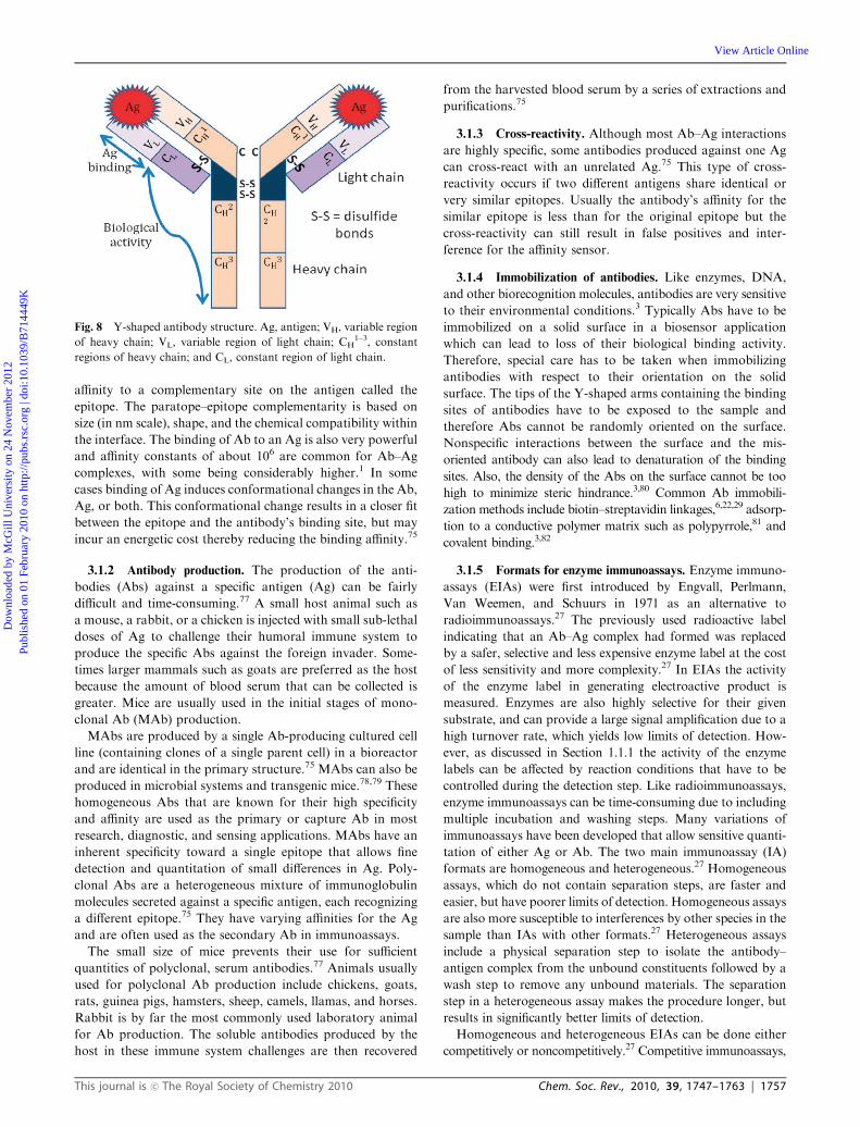
Immunosensors are Ab-based affinity biosensors where the
detection of an analyte, an antigen or hapten, is brought about
by its binding to a region of an Ab.6 The electrochemical
transducer responds to the binding event and converts the
electrical response to an output that can be amplified, stored,
and displayed. Complementary regions of the Ab bind to an
Ag that was used to produce the antibodies in a host organism
William R. Heineman
William R. Heineman is a Dis-tinguished Research Professorin the Department of Chemis-try at the University ofCincinnati. He received a BSin chemistry at Texas TechUniversity and a PhD at theUniversity of North Carolinain Chapel Hill and was a post-doctoral associate at CaseWestern Reserve Universityand The Ohio State Univer-sity. His research interestsinclude spectroelectrochemistry,electrochemical immunoassay,sensors, and bioanalytical
chemistry. He is a recipient of the Charles N. Reilley Awardin Electroanalytical Chemistry and the Torbern Bergman Medalfrom the Analytical Section of the Swedish Chemical Society.
1748 | Chem. Soc. Rev., 2010, 39, 1747–1763 This journal is �c The Royal Society of Chemistry 2010
Dow
nloa
ded
by M
cGill
Uni
vers
ity o
n 24
Nov
embe
r 20
12Pu
blis
hed
on 0
1 Fe
brua
ry 2
010
on h
ttp://
pubs
.rsc
.org
| do
i:10.
1039
/B71
4449
K
View Article Online
such as a rabbit or a mouse with high specificity and affinity.4
Such polyclonal Abs are heterogeneous with respect to their
binding domain, and may be refined by a selection process to
yield monoclonal Abs—MAbs—all of whose members of a
particular MAb clone are identical. Abs and MAbs can be
developed for a wide range of substances. Theoretically, if an
Ab can be raised against a particular analyte, an immuno-
sensor could be developed to detect for that substance. Immuno-
sensors are well known among analytical methods for their
extremely low detection limits.6 Immunoassays and immuno-
sensors have been developed for both quantitative and quali-
tative applications.1,2 Immunosensors can be used to detect
trace levels (ppb, ppt) of bacteria, viruses, drugs, hormones,
pesticides, and numerous other chemicals.1,2 Examples of
immunosensor applications include monitoring food safety
related to severe allergies (such as peanuts), detecting environ-
mental pollutants such as herbicides and pesticides in water
and soil, detecting biomedical substances such as warfarin,
and monitoring for biowarfare agents such as toxins, bacteria,
viruses, and spores.1,2 Relatively inexpensive kits such as for
home pregnancy and fertility tests can be produced once the
assay is fully developed. In the past, the limited availability of
Ab varieties mainly produced by university and small bio-
technology companies has slowed down the affinity biosensor
development.6 However, antibodies are now sold by many
sources including large manufacturers of laboratory reagents
such as Sigma Aldrich.
Nucleic acids have been less commonly used as the bio-
recognition element in affinity sensors compared to antibodies.
Biorecognition using DNA or RNA nucleic acid fragments
relies on either complementary base-pairing between the sensor’s
nucleic acid sequence and the analyte of interest, or generating
nucleic acid structures, known as aptamers, that recognize and
bind to three-dimensional surfaces, such as those of proteins.
Nucleic acids are now becoming of greater importance as the
biorecognition agent in sensors since a recent rapid expansion
in knowledge of their structure and how to manipulate them.1
DNA affinity probes are typically used in medical diagnostics
to detect cancers, viral infections, and genetic diseases.1 Affinity
biosensors will be described in more detail in Section 3.
1.2 Electrochemical detection
Most biosensors use electrochemical detection for the trans-
ducer because of the low cost, ease of use, portability, and
simplicity of construction.1,2 The reaction being monitored
electrochemically typically generates a measurable current
(amperometry), a measurable charge accumulation or poten-
tial (potentiometry) or alters the conductive properties of the
medium between electrodes (conductometry).3 Use of electro-
chemical impedance spectroscopy by monitoring both resis-
tance and reactance in the biosensor is also becoming more
common.3
Electrochemistry is a surface technique and offers certain
advantages for detection in biosensors. It does not depend
strongly on the reaction volume, and very small sample volumes
can be used for measurement.6 Electrochemical detection can
be used to achieve low detection limits in immunoassays with
little or no sample preparation, and atto- and zeptomole
detecting electrochemical immunoassays have been constructed.7,8
In homogeneous immunoassays, which have no separation
step to isolate the antibody–antigen complex from the unbound
assay constituents, electrochemical detection is not affected by
sample components such as chromophores, fluorophores, and
particles that often interfere with spectrophotometric detec-
tion. Therefore electrochemical measurements can be made on
colored or turbid samples such as whole blood, without
interference from fat globules, red blood cells, hemoglobin,
and bilirubin.9,10
Electrochemical techniques are generally organized into
three main categories of measurement: current, potential and
impedance. This article focuses primarily on those techniques
that measure current since they are the most commonly used in
biosensors.
1.2.1 Voltammetry/amperometry. Voltammetric and ampero-
metric techniques are characterized by applying a potential to a
working (or indicator) electrode versus a reference electrode and
measuring the current.11 The current is a result of electrolysis
by means of an electrochemical reduction or oxidation at the
working electrode. The electrolysis current is limited by the mass
transport rate of molecules to the electrode.11
The term voltammetry is used for those techniques in which
the potential is scanned over a set potential range. The current
response is usually a peak or a plateau that is proportional to
the concentration of analyte. Voltammetric methods include
linear sweep voltammetry, cyclic voltammetry, hydrodynamic
voltammetry, differential pulse voltammetry, square-wave
voltammetry, ac voltammetry, polarography, and stripping
voltammetry.11 These methods have a wide dynamic range,
and are useful for low level quantitation.
In amperometry, changes in current generated by the
electrochemical oxidation or reduction are monitored directly
with time while a constant potential is maintained at the
working electrode with respect to a reference electrode.5 It is
the absence of a scanning potential that distinguishes ampero-
metry from voltammetry. The technique is implemented by
stepping the potential directly to the desired value and then
measuring the current, or holding the potential at the desired
value and flowing samples across the electrode as in flow
injection analysis. Current is proportional to the concentration
of the electroactive species in the sample. Amperometric
biosensors have additional selectivity in that the oxidation or
reduction potential used for detection is characteristic of the
analyte species.1
Amperometric detection is commonly used with biocatalytic
and affinity sensors because of its simplicity and low LOD.12
Advantageously, the fixed potential during amperometric
detection results in a negligible charging current (the current
needed to apply the potential to the system), which minimizes
the background signal that adversely affects the limit of
detection. In addition, hydrodynamic amperometric techniques
can provide significantly enhanced mass transport to the
electrode surface,11,13 for example when the working electrode
moves with respect to the solution by rotating or vibrating,14,15
or in flow conditions where the sample solution passes over
the stationary electrodes.13,16,17 Electrochemical detection in
flow systems can be used in environmental monitoring and
This journal is �c The Royal Society of Chemistry 2010 Chem. Soc. Rev., 2010, 39, 1747–1763 | 1749
Dow
nloa
ded
by M
cGill
Uni
vers
ity o
n 24
Nov
embe
r 20
12Pu
blis
hed
on 0
1 Fe
brua
ry 2
010
on h
ttp://
pubs
.rsc
.org
| do
i:10.
1039
/B71
4449
K
View Article Online
industrial processes more easily than steady-state batch
systems, since the flow conditions allow the solution to be
changed more easily in multistep assay procedures, and are
ideal for on-line monitoring.
Electrochemical sensors are part of an electrochemical cell
that consists of either three electrodes or two electrodes.
A typical three electrode electrochemical cell consists of a
working (or indicator) electrode of a chemically stable solid,
conductive material, such as platinum, gold, or carbon
(e.g. graphite); a reference electrode, usually consisting of
silver metal coated with a layer of silver chloride (Ag/AgCl);
and a platinum wire auxiliary electrode. The reference
electrode is usually further removed from the site of the redox
reaction in order to maintain a known and stable reference
potential.3 One advantage of this system is that the charge
from electrolysis passes through the auxiliary electrode instead
of the reference electrode, which protects the reference
electrode from changing its half-cell potential. A two electrode
system has only the working and reference electrodes. If the
current density is low enough (omA cm�2) then the reference
electrode can carry the charge with no adverse effect.5 Both
three electrode systems and two electrode systems are used for
sensors. However, two electrodes are generally preferred for
disposable sensors because long-term stability of the reference
is not needed and the cost is lower.
These electrodes can be easily miniaturized, so dimensions
on the order of micrometres are common, while nanometre
sizes have been demonstrated.18–20 Nanowires, nanoparticles,
and carbon nanotubes are now being incorporated into bio-
sensors. Shrinking electrode dimensions may lead to higher
sensitivity.3 Very small sample volumes (on the order of
microlitres and less) are required to detect with such small
electrodes due to their small surface areas, and this is a signifi-
cant advantage when the sample sizes are limited.21,22 Further-
more, electrochemical detectors and their required control
instrumentation can be easily miniaturized at a relatively low
cost by micromachining, making possible the manufacture of
field-portable instruments for biosensing. Since the limiting
current in voltammetry is temperature-dependent, the detec-
tion cell should be maintained at a constant temperature for
running calibrants and samples in order to obtain accurate
and precise results.23
Screen-printed electrodes (SPEs), patterned minielectrode
systems with working, reference and auxiliary electrodes, have
gained popularity in electrochemical biosensors due to their
low cost and ease and speed of mass production using thick
film technology.6 An SPE for detecting oxygen is shown in
Fig. 2. SPEs can also be miniaturized easily making them an
attractive transducer choice for microfluidic systems and
portable meters. The patterned working electrode is typically
made of conductive carbon ink that results in a rough surface
that makes difficult the exact determination of electrode
area.24 Gold coated and gold-based SPE sensors have been
used in stripping voltammetry to determine trace levels of lead,
copper, cadmium, and mercury in water samples.25 Nafion
coated SPE biosensors with immobilized butyrylcholinesterase
have also been developed to detect low levels of pesticides.26
Disposable SPEs have also been used in immunochemical
sensors and to measure blood glucose.27
Interdigitated array (IDA) electrodes are good amperometric
electrochemical transducers in biosensors (Fig. 3). IDAs are
made of two pairs of working electrodes consisting of parallel
strips of metal fingers that are interdigitated and separated by
insulating material.6,28 One electrode array serves as an anode
for oxidation and the other as a cathode for reduction as shown
in Fig. 3 for one anode finger and the adjacent cathode fingers.
The main advantage of using an IDA is the redox cycling of the
electroactive enzyme product or mediator that occurs when
different potentials are applied to the two electrodes causing
oxidation–reduction cycling when the electrode reaction is
reversible. The redox cycling provides lower limits of detection
because the current due to oxidation of each redox active
molecule contributes multiple times to the detection current.6,28
As a result, the signal-to-noise ratio is improved significantly
and a lower detection limit is obtained. Signal enhancement
increases as the spacing and width of the metal fingers decrease
because the diffusion distances for the redox species are shorter.
Typical signal enhancements provided by the IDA are about
3–10� and can be up to 1000� depending on the dimensions of
the IDA.28 IDA electrodes have been used as detectors in
electrochemical immunoassays.29 An IDA with dimensions on
the nanoscale was used for immunoassay detection of a virus.30
Fig. 2 Diagram of a screen printed electrode (SPE). Ref., reference
electrode; Aux., auxiliary electrode; and Work., working electrode.
Fig. 3 Cycling of a redox active species at the interdigitated array
electrode (IDA). Alkaline phosphatase (ALP) hydrolyzes o-phosphate
from a p-aminophenyl phosphate under alkaline conditions. R is the
reduced p-aminophenol (PAP). O is the oxidized p-quinone imine
(PQI).
1750 | Chem. Soc. Rev., 2010, 39, 1747–1763 This journal is �c The Royal Society of Chemistry 2010
Dow
nloa
ded
by M
cGill
Uni
vers
ity o
n 24
Nov
embe
r 20
12Pu
blis
hed
on 0
1 Fe
brua
ry 2
010
on h
ttp://
pubs
.rsc
.org
| do
i:10.
1039
/B71
4449
K
View Article Online
1.2.2 Impedance. Electrochemical impedance spectroscopy
(EIS), described by Lorenz and Schulze in 1975,31 measures the
resistive and capacitive properties of materials upon perturba-
tion of a system by a small amplitude sinusoidal ac excitation
signal typically of 2–10 mV.5,32 The frequency is varied over a
wide range to obtain the impedance spectrum. The in-phase and
out-of-phase current responses are then determined to obtain
the resistive and capacitive components of impedance, respec-
tively. Impedance methods are powerful because they are
capable of sampling electron transfer at high frequency and
mass transfer at low frequency. Impedimetric detection is
primarily used for affinity biosensors.27 It can be used to
monitor immunological binding events such as antibody
(Ab)–antigen (Ag) binding on an electrode surface, for example,
where the small changes in impedance are proportional to the
concentration of the measured species, the Ag.
The surface of the electrode can be modified by a highly
specific biological recognition element. In one approach the
recognition elements are incorporated in a conductive polymer
film formed on the surface of a working electrode by electro-
chemical deposition (Fig. 4). During the detection step, a
known voltage is applied to the electrode and the resulting
current is measured. The electron transfer resistance at the
interface between the electrode and the solution changes
slightly when analyte binds. Directly monitoring the formation
of an antibody–antigen conjugated layer provides a label-
free detection system with many potential advantages such
as higher signal-to-noise ratio, ease of detection, lower assay
cost, faster assays and shorter detector response times. How-
ever, regenerating the sensing surface for a subsequent measure-
ment in an impedance biosensor is typically very time-consuming
and not reproducible.27 This continues to be the biggest
limitation of immunosensors involving Ab–Ag complexes with
high affinity constants. The regeneration conditions can also
damage and release the immunoreagent bound to the surface
of the transducer.27
Electrochemical biosensors using impedance spectroscopy
to detect analytes have recently gained popularity among
the biosensor community.3 EIS has some advantages over
the widely used amperometric detection. The active site
participating in the biologically mediated redox reaction must
be easily accessible to the analyte solution and in close
proximity to the electrode surface. As discussed before, redox
mediators have been used to help overcome the accessibility
and proximity limitations but cause the detection to be limited
by the mediator’s mass transfer rate. Furthermore, some
additional redox active species such as urate and ascorbate
that are often present in the sample matrix can contribute to
the amperometric signal if the detection potential is not care-
fully chosen. Being directly able to impedimetrically monitor
the Ab–Ag binding helps by-pass the aforementioned limita-
tions. EIS is also insensitive to most environmental distur-
bances. However, biosensors using impedance detection
have to be carefully designed to minimize nonspecific binding
of the analyte. Nonspecific binding in affinity sensors will be
discussed further in Section 3.1.7. Using nanomaterials
such as gold nanoparticles and carbon nanotubes in electro-
chemical impedance sensors is advantageous due to the increased
electrode surface area, improved electrical conductivity of
the sensing interface, chemical accessibility to the analyte, and
electrocatalysis.32 Recent applications of impedance spectro-
scopy in affinity sensors will be described in Section 3.1.8.
1.2.3 Conductometry. Conductometric detection monitors
changes in the electrical conductivity of the sample solution, or
a medium such as nanowires, as the composition of the
solution/medium changes in the course of the chemical reac-
tion. Conductometric biosensors often include enzymes whose
charged products result in ionic strength changes, and thus
increased conductivity. Conductometry has been used as the
detection mode in biosensors for environmental monitoring
and clinical analysis. A conductometric tyrosinase biosensor
was developed to measure ppb amounts of pollutants such as
diuron, and atrazine and its metabolites.33 Conductometric
immunosensors have also been developed to detect foodborne
pathogens such as enterohemorrhagic Escherichia coli O157:H7
and Salmonella spp., which are of concern to biosecurity.34
The sensitive, low volume biosensor consists of an immuno-
sensor that is based on an electrochemical sandwich immuno-
assay, and a reader device for measuring the signal.34 Drug
detection of methamphetamine in human urine has also been
done using conductometry.35
1.2.4 Potentiometry. Potentiometric sensors are based on
measuring the potential of an electrochemical cell while drawing
negligible current. Common examples are the glass pH elec-
trode and ion selective electrodes for ions such as K+, Ca2+,
Na+, Cl�.1,2 The sensors use an electrochemical cell with
two reference electrodes to measure the potential across a
membrane that selectively reacts with the charged ion of
interest. These chemical sensors can be turned into biosensors
by coating them with a biological element such as an enzyme
that catalyzes a reaction that forms the ion that the underlying
electrode is designed to sense. For example, a sensor for
penicillin can be made by coating a pH electrode with
penicillinase, which catalyzes a reaction of penicillin that also
generates H+.36 The pH electrode senses the change in pH at
its surface, which is an indirect measure of penicillin.Fig. 4 A diagram of an Ab–Ag affinity sensor with impedimetric
detection.
This journal is �c The Royal Society of Chemistry 2010 Chem. Soc. Rev., 2010, 39, 1747–1763 | 1751
Dow
nloa
ded
by M
cGill
Uni
vers
ity o
n 24
Nov
embe
r 20
12Pu
blis
hed
on 0
1 Fe
brua
ry 2
010
on h
ttp://
pubs
.rsc
.org
| do
i:10.
1039
/B71
4449
K
View Article Online
Field effect transistors have been adapted to chemical
sensors (ChemFETs) by incorporation into an electrochemical
cell.37,38 They can also be made into biosensors by coating the
sensing surface with a biological agent such as described above
for penicillin.39 The light addressable potentiometric sensor
(LAPS) determines the surface potential optically by means of
the photovoltaic effect.40 The LAPS can also be used as a
biosensor by adding a biological element to its surface, such as
an oligonucleotide.41
1.2.5 Miniaturized electrochemical transducers. Miniaturi-
zation is a growing trend in analytical chemistry. In order to
design and manufacture small biosensors, the transducer or
the electrode needs to be small and portable. The manufacturing
capabilities for depositing microelectrodes on surfaces are
good and microelectrodes can easily be deposited on a micro-
fluidic chip or other solid surface using vapor deposition.6
Usually the electrode is part of a bigger device such as a
handheld meter or a microfluidic system.
Microelectrodes are defined as electrodes with a diameter in
the micrometre scale, and are made as disks or cylinders from
carbon fibers or metal microwires.18,19 The first measurements
using microelectrodes to measure the concentration of oxygen
in biological tissues were made in early 1940s,42 and they have
since been used to measure electroactive species in critical
places such as inside a mammalian brain.18 Measurements
with voltammetric microelectrodes have been made even inside
a very small, live biological cell.43 This is because the important
reactions occur at the microelectrode surface instead of bulk
solution, and the very small sensing surface area of a micro-
electrode can be easily inserted into very small drops or spaces
without causing much disturbance or damage. Carbon fiber
microelectrodes have been used to detect 190 zmol of catechol-
amine release from a single, stimulated rat nerve cell,44 to
directly monitor catecholamines released from adrenal cells in
culture,45 and to measure the release of serotonin from neuronal
vesicles achieving a 4.8 zmol detection limit.46 Microelectrodes
have also been used as detectors in microvolume electro-
chemical immunoassays.22 The nanoamp to picoamp currents
generated at microelectrodes are so small that they are virtually
nondestructive,18 and amplification of the small currents produced
is typically required in order to observe the signals.6
2. Biocatalytic sensors
2.1 Introduction to enzyme-based electrodes
Enzyme electrodes are electrochemical probes with a thin
layer of immobilized enzyme on the surface of the working
electrode.47,48 The enzyme is the most critical component of
the enzyme electrode since it provides the selectivity for the
sensor and catalyzes the formation of the electroactive product
for detection.49 The electroactive product can be monitored
directly using amperometry, in which the produced current is
measured in response to an applied, constant voltage. Alter-
natively, the disappearance of the redox active reactant in an
enzyme-catalyzed reaction can be monitored by the electrode.
The activity of the immobilized enzyme depends on solution
parameters and electrode design. The rapid enzymatic
catalysis can also sometimes provide significant signal
amplification in a biosensor.5 The shelf life and stability of
an enzyme generally determine the lifespan of the biosensor.
The use of enzyme electrodes as biosensors will continue to
increase because they are simple and inexpensive to construct,
they provide rapid analysis, they easily regenerate, and they
are reusable.2,5 However, the number of available enzyme-
based biosensors is still smaller than the number of potential
analytes. Another disadvantage of enzyme electrodes is that
the enzyme layer in the biosensor has to be replaced periodi-
cally since it gradually loses activity. Also, clever electro-
chemical detection strategies or membranes are sometimes
required to prevent interference from other redox active
species at certain detection potentials.
Development of biocatalytic sensors for medical appli-
cations, primarily blood glucose monitoring starting in the
late 1960s, was the main driving force for this research area.5
Enzyme-based biosensors can be historically divided into three
generations. First-generation biosensors were oxygen-based
whereas second-generation are mediator-based. Third-generation
biosensors are so-called directly coupled enzyme electrodes.
Electrodes coated with glucose oxidase (GOx) have been
widely used in detection of glucose since the pioneering work
of Clark and Lyons in the 1950s and 1960s (Fig. 5).50 These
amperometric sensors became known as the first-generation
biosensors or Clark oxygen electrodes and were soon imple-
mented by Updike and Hicks, who constructed the first func-
tional biocatalytic sensor for glucose.51 In the first-generation
biosensors, an oxidase enzyme is immobilized behind a semi-
permeable membrane at the surface of a Pt electrode.
GOx is a readily available, inexpensive, and stable enzyme
from Aspergillis niger that is among the most important
enzymes in biosensor applications and industrial processes.
GOx is highly specific for b-D-glucose, which can be detected
via the following reactions.2,5,52
b-D-Glucose + GOx–FAD - GOx–FADH2
+ d-D-gluconolactone (1)
GOx–FADH2 + O2 - GOx–FAD + H2O2 (2)
H2O2 - 2e� + O2 + 2H+ (3)
Fig. 5 Oxygen-dependent first-generation biosensor with ampero-
metric detection.
1752 | Chem. Soc. Rev., 2010, 39, 1747–1763 This journal is �c The Royal Society of Chemistry 2010
Dow
nloa
ded
by M
cGill
Uni
vers
ity o
n 24
Nov
embe
r 20
12Pu
blis
hed
on 0
1 Fe
brua
ry 2
010
on h
ttp://
pubs
.rsc
.org
| do
i:10.
1039
/B71
4449
K
View Article Online
In eqn (1) the prosthetic group of the enzyme, FAD, is reduced
and glucose is oxidized to d-D-gluconolactone. Molecular
oxygen acts as the oxidizing agent to produce hydrogen
peroxide (eqn (2)). During the oxidation of H2O2 at a working
electrode two electrons are transferred directly to the electrode
(eqn (3)), resulting in the current response of the enzyme
electrode. These first-generation sensors required the ample
and constant presence of ambient oxygen as a co-substrate for
the enzyme to function optimally. However, oxygen is not very
soluble in aqueous solutions and can therefore limit the
currents produced in the presence of the analyte.
Direct redox reactions between enzymes and electrodes are
very rare because most proteins tend to denature at the
electrode surface and many direct electron transfer reactions
are slow and irreversible.1 However, a limited number of
enzymes such as horseradish peroxidase have proven capable
of direct electron transfer between the enzyme active site’s
prosthetic group and the electrode.53 The active site of an
enzyme that allows the selective targeting of an analyte is
usually buried within the enzyme’s tertiary protein structure,
near the centroid of the protein.4 Therefore, the electrons
produced in the enzyme-catalyzed reaction cannot always be
easily and rapidly transferred to the electrode surface thereby
limiting the electrical communication between the enzyme and
the transducer. The widely accepted Marcus theory of electron
transfer states that electron transfer decays exponentially with
distance.54,55 Therefore enzymes often require some assistance
with electron transfer to the transducer surface.
Artificial redox mediators are small, soluble molecules
capable of undergoing rapid and reversible redox reactions,
which shuttle electrons between the redox center at the active
site of the enzyme and the electrode surface. Mediators have
replaced O2 molecules as the electron shuttle (eqn (4)) in
glucose sensors. Mediators are re-oxidized at relatively low
potentials and generate a current when they come in contact
with the working electrode (eqn (5)).
GOx–FADH2 + 2MediatorOx - GOx–FAD
+ 2MediatorRed + 2H+ (4)
2MediatorRed - 2MediatorOx + 2e� (5)
Mediators should ideally be nontoxic, independent of the
pH, stable in both the oxidized and reduced forms, and
unreactive with oxygen.1 Although many organic compounds
are capable of acting as enzyme mediators, organometallic
redox compounds are the most common.1,2 Examples of
previously used mediators include quinones, organic conducting
salts, dyes, ruthenium complexes, ferrocene, and ferricyanide
derivatives. Mediated enzyme electrodes had a much better
sensor performance than the first-generation biosensors
mainly due to eliminating the O2 dependence and being able
to control the concentration of the oxidizing agent in the
biosensor.1 Hand selecting the oxidizing agent for the sensor
also allowed more suitable oxidation potentials to be used for
the amperometric sensors. These mediated enzyme electrodes
were named second-generation biosensors.
By carefully selecting a mediator and a suitable redox
potential, the transduction event at the second-generation
biosensor could be measured in a potential range where other
possible sample components such as ascorbate, urate, and
paracetamol are not oxidized or reduced thereby minimizing
interferences.5 Incorporating redox mediators also allowed
other oxidoreductase enzymes such as peroxidases and dehydro-
genases to be used as the biorecognition element in the sensor
thereby expanding the list of possible target analytes.
Third-generation biosensors have the biorecognition com-
ponent coupled with the electrode by co-immobilizing the
enzyme and the mediator at an electrode surface. This can
be achieved by direct electrical contact between the enzyme
and the electrode, immobilizing the enzyme and mediator in a
conducting polymer, or ‘wiring’ the enzyme to the electrode by
immobilizing it in a redox polymer (Fig. 6) as first described by
Heller et al.56,57 The co-immobilization prevents the mediators
from diffusing out of the biosensor film. The co-immobilized
mediators, or the flexible surrounding redox polymer, help to
transport electrons between the enzyme’s active site and the
working electrode surface in an array of rapid electron relays
and hence generate high current densities.2 The enzymes
immobilized in flexible redox polymers that are covalently
attached to the electrode have been called ‘wired enzymes’.
The 3rd-generation sensors are ideal for repeated measure-
ments since neither mediator nor enzyme need to be added.
This self-contained nature also lowers the cost per measure-
ment and opens up possibilities for continuously monitoring
the analytes.
2.2 Preparing enzyme electrodes
2.2.1 Methods for immobilizing enzymes to electrode surfaces.
Enzyme electrodes have been studied extensively and various
physical and chemical schemes have been used to immobilize
enzymes on the electrochemical transducer. The objective is to
have an intimate contact between the enzyme and the trans-
ducer’s sensing surface without blocking the active site of the
enzyme or drastically altering the enzyme geometry.2 Immobili-
zation methods are considered successful if the biosensors pre-
pared are stable, reusable, and maintain the selectivity of the
enzyme. Although immobilization may alter the conformation of
the enzyme, thereby reducing its activity, many methods have
been successful. Some immobilization methods even improve
enzyme stability by minimizing enzyme unfolding. The enzyme
should have high Vmax and low Km values when immobilized on
Fig. 6 Third-generation catalytic biosensor containing enzymes
wired to the electrode through a conducting redox polymer.
This journal is �c The Royal Society of Chemistry 2010 Chem. Soc. Rev., 2010, 39, 1747–1763 | 1753
Dow
nloa
ded
by M
cGill
Uni
vers
ity o
n 24
Nov
embe
r 20
12Pu
blis
hed
on 0
1 Fe
brua
ry 2
010
on h
ttp://
pubs
.rsc
.org
| do
i:10.
1039
/B71
4449
K
View Article Online
the transducer.2 Vmax is the maximal velocity of a reaction that
occurs at high substrate concentrations when the enzymes are
saturated. By having an immobilized enzyme with a high Vmax
the electrochemical transducer responding to a reaction catalyzed
by the enzyme has a broader range where the signal is propor-
tional to substrate concentration for reliable quantitation of the
analyte. Km, the Michaelis constant, is the substrate concen-
tration at which the reaction velocity is half-maximal. Enzymes
with low Km reach maximal catalytic efficiency at low substrate
concentrations. The immediate environment around the immobi-
lized enzyme can be carefully designed to enhance the enzyme
activity and the overall biosensor performance.
The easiest approach is to physically entrap a solution of
the enzyme between preformed membranes on the electrode
surface.2 The inner membrane protects the electrode surface
from interfering substances and electrode fouling due to
adsorption. The outer membrane also provides some selecti-
vity based on the pore size or chemical nature of the polymer,
stabilizes the sensor response by moderating the substrate
diffusion to the enzyme layer, and provides a biocompatible
outer surface for the sensor.5 In physical immobilization
methods the native composition of the enzyme is preserved
since the methods do not involve the formation of covalent
bonds.34 Chemical methods involve the formation of covalent
bonds between the functional groups of the enzyme and the
electrode material.5 Common enzyme immobilization methods
include enzyme entrapment against the electrode using a
preformed membrane; encapsulation; inclusion in a gel or
electropolymerized film; incorporation in a carbon paste;
and using biospecific interactions such as biotin–avidin binding,
adsorption, cross-linking, and covalent attachment (Fig. 7).58,59
Covalent bonding provides the most stable immobilization of
proteins followed by cross-linking and encapsulation.1 Covalent
bonding to the transducer links functional groups on the
enzyme such as NH2, COOH, OH, and SH that are not
necessary for the catalytic activity of the enzyme. The coupling
reactions need to be done under mild conditions (low ionic
strengths, low temperatures, and near physiological pHs) and
often in the presence of the enzyme–substrate in order to protect
the catalytic activity of the enzyme.1 Adsorption is the least
stable of the common immobilization methods.1 The forces
linking the biorecognition element to the transducer in adsorp-
tion are primarily very weak van der Waals forces with occa-
sional hydrogen bonds that are not very stable or permanent.1
Therefore the lifetime of a sensor prepared using adsorption is
rather limited. However, adsorption is very easy because it does
not require any reagents or clean-up and is less disruptive to the
enzymes.1 The formation of intermolecular interactions with
the surface may compete with similar interactions stabilizing the
enzyme, and is often a prelude to denaturation. This is probably
why adsorption usually works best in the short term, because
the protein deformation increases with time. Adsorption is
often sufficient for short-term studies. The stability of immobi-
lized enzymes with respect to time, temperature, and pH is
typically greater making enzyme electrodes preferable to soluble
enzyme assays.5,52 Covering the immobilized enzyme layer with
a membrane or a polymer coating also helps to minimize
interferences by physically blocking some interfering species
from approaching the electrode surface.52
2.2.2 Optimizing enzyme electrodes. Although many enzyme
electrodes have been fabricated and some sensors have reached
the commercial stage, some factors that prevent their wider
adaptation and successful routine use still remain. Research
continues in trying to overcome the dependence of enzyme
activity on the solution conditions such as temperature, pH,
ionic strength, and buffer composition.60 Ideally the solution
conditions should remain constant between samples and during
the measurements. The enzyme electrode should also have a
wide linear range. Enzymes become saturated with their sub-
strate at high concentrations due to their active sites becoming
the limiting reagent, thus causing the signal response to no
longer be proportional to the analyte concentration. The
amount of enzyme incorporated into the sensor can however
be adjusted based on the expected sensor application. The
catalytic biosensor should also be biocompatible since blood
and other biological fluids are the most common sample
matrices for enzyme electrodes. Many blood components foul
the electrode in a matter of minutes unless special precautions
are taken in designing the sensor’s outermost surface properties
and permeability to prevent the adsorption of sample com-
ponents on the electrode surface.60 Product design requirements
also include optimization of sample introduction, sample size,
the sensor’s reproducibility, selectivity, sensitivity, stability, cost,
and ease of use.5 The storage stability of enzymes immobilized
on electrode surface varies from hours to months depending on
the sensor preparation and design, and the storage environment.
2.3 Examples of biocatalytic sensors
2.3.1 Glucose sensors. Enzyme electrodes are produced
commercially and are routinely used in biomedical appli-
cations such as glucose testing in clinical laboratories and
personal monitoring by diabetic patients.2,5,48 Low cost blood
glucose home monitoring kits consisting of handheld battery
operated meters and disposable glucose test strips based on
glucose oxidase (GOx) or glucose dehydrogenase enzyme
electrodes are sold off the shelf worldwide. Biosensors for this
application must be easy to use, reliable, and inexpensive.1 In a
typical sensor, a single drop of blood is placed on a disposable
PVC sample strip on which the dry reagents have been
deposited using a method similar to ink-jet printing techno-
logy. The test strip also contains two electrodes, one holding
the enzyme and a mediator for the amperometric detectionFig. 7 Common methods of immobilizing enzymes onto an electrode
surface.
1754 | Chem. Soc. Rev., 2010, 39, 1747–1763 This journal is �c The Royal Society of Chemistry 2010
Dow
nloa
ded
by M
cGill
Uni
vers
ity o
n 24
Nov
embe
r 20
12Pu
blis
hed
on 0
1 Fe
brua
ry 2
010
on h
ttp://
pubs
.rsc
.org
| do
i:10.
1039
/B71
4449
K
View Article Online
of glucose, and the other serving as a reference electrode.
The current produced when a potential is applied gives a read-
out to a liquid crystal display on the glucose meter. The
commercially sold glucose test strips are second- or third-
generation biosensors and no longer rely on oxygen as the
oxidizing agent. Ferricyanide is a commonly used mediator for
the second-generation sensors. Eqn (1), (4), and (5) describe
the sequence of reactions for such a sensor when GOx is used
as the enzyme. The commercially sold blood glucose meters
typically have a range of 1.1–33.3 mM glucose with a precision
of �3–8% and test time of about 30 seconds or less.1
Some invasive and minimally invasive implantable glucose
sensors that have an intimate contact between the biological
fluids or tissues and the biocatalytic sensor have been developed.5,61
The minimally invasive blood glucose sensors are inserted
subcutaneously into the arm or belly of a patient. More
invasive, intravascular sensors that measure glucose levels in
hospitalized diabetes patients are also being developed. Some
problems such as pain, skin irritation, limited lifetime of the
sensor, and accuracy of the data continue to slow down their
wider use.62
2.3.2 Xanthine sensors. Xanthine oxidase (XO) catalyzes the
oxidation of xanthine to uric acid (eqn (6)). Amperometric
biosensors using immobilized XO are highly specific for xanthine,
which can be measured by the following redox reaction:
Xanthine + O2 + XO - uric acid + H2O2 + XO (6)
Xanthine is an intermediate of purine metabolism and is
produced after adenosine triphosphate (ATP) decomposition.
The physiological conversion of xanthine by xanthine oxidase
is of increasing medical interest.63 Moreover, xanthine sensors
are frequently used in food industries to determine the freshness
of fish. The need for maintaining an acceptable quality of
fish sold to consumers requires rapid and reliable analytical
methods that detect the products formed in their degradation
processes. After the death of a fish, nucleotides such as ATP
are most affected by degradation and give rise to the formation
of inosine, which is transferred to hypoxanthine by action of
the enzyme nucleoside phosphorylase.64 Hypoxanthine causes
a bitter taste in the degrading meat.65 XO catalyzed oxidation
of hypoxanthine to xanthine and conversion of xanthine to
uric acid occurs in two steps.64 The quantitation of xanthine or
hypoxanthine can therefore be used to determine the freshness
of fish.64,65 Other existing methods for detecting xanthine or
hypoxanthine such as anion-exchange chromatography, thin
layer chromatography, precipitation and capillary electro-
phoresis are complicated and very time-consuming. Therefore
biocatalytic sensors with amperometric detection continue to
be developed to monitor the freshness of fish meat.66–68
2.3.3 Lactate sensors. Lactate, an ester of lactic acid, is a
product of fermentation and is produced during cellular
respiration as glucose is broken down. Its concentration in
blood rises from the normal value of 0.9 mM to about 12 mM
due to strenuous exercise such as running, which results in
anaerobic metabolism.69 Small handheld electrochemical
lactate meters for use in sports medicine capable of inter-
mittent ‘‘spot’’ lactate monitoring are being manufactured by
Senslab (Germany) and Arkray (Japan).70 These sensors
require only 0.5 mL and 5 mL blood samples, respectively.
The concentration of lactate in blood is also a sensitive
measure of oxygen deprivation from ischemia, trauma, and
hemorrhage, which can lead to life-threatening shock, and its
measurement has therefore become a vital component in
medical monitoring.70 Blood lactate levels are used as indi-
cators of conditions such as acidosis or bacterial meningitis.71
Conventional photometric assays for lactate are slow and not
suited for continuous lactate monitoring systems that are
being developed for medical applications. Bench top lactate
biosensors are also routinely used to measure lactic acid in
milk and other foods.
Four different enzymes have been used as the biorecognition
component in lactate biosensors: lactate dehydrogenase, lactate
oxidase, lactate monooxidase, and cytochrome b2.1 Some of the
electrochemical lactate sensors include mediators such as
NAD+/NADH and ferricyanide.1 All of these enzymes help
ultimately to produce a current at the working electrode that is
measured amperometrically.
2.4 Interference-based enzyme electrodes
Interference-based enzyme electrodes are probes used for quanti-
tative analysis based on the changes in the rate of catalytic
reactions when enzyme effectors bind, such as inhibitors or
activators.72 Usually, the binding of an inhibitor to the enzyme’s
active site or another site that causes the conformation of the
enzyme to be altered, results in a decreased electrochemical
response because substrate cannot freely access the catalytic site.
Sensors using this operating principle have been developed for
pesticides such as organophosphate and carbamate, respiratory
poisons such as cyanide and azide, as well as toxic heavy metals.2
Enzymes that have been used in these enzyme electrodes include
tyrosinase, horseradish peroxidase, and acetylcholinesterase. Due
to the operating mechanism and the uses these sensors have, they
have also been called enzyme inhibition biosensors or toxin
biosensors.2,72
2.5 Biosensors based on tissue and bacteria
Some biocatalytic sensors incorporate cellular materials such
as plant tissues as the recognition component.2 These bio-
catalytic electrodes function in a manner similar to that for
conventional enzyme electrodes (i.e., enzymes present in the
tissue or cell produce or consume electrochemically detectable
species). Whole cells and tissue slices are sometimes a better
source of enzymatic activity compared to isolated enzymes as
some enzymes are expensive or not commercially available in
the pure state.2 Also, many isolated enzymes have limited
stability and lifetime compared to enzymes in their native
environment. However, the sensor response may be slower for
these sensors because there is more tissue material for the
substrate to diffuse through.1
Bananatrode, a banana tissue containing electrode, was one
of the early uses of tissue in a biosensor.63 The banana tissue,
which is rich with polyphenol oxidase (PPO), can be mixed in a
carbon paste matrix to yield a fast responding and sensitive
dopamine sensor. The amperometric probe has high biocatalytic
activity, good time stability, and favorable selectivity.73
This journal is �c The Royal Society of Chemistry 2010 Chem. Soc. Rev., 2010, 39, 1747–1763 | 1755
Dow
nloa
ded
by M
cGill
Uni
vers
ity o
n 24
Nov
embe
r 20
12Pu
blis
hed
on 0
1 Fe
brua
ry 2
010
on h
ttp://
pubs
.rsc
.org
| do
i:10.
1039
/B71
4449
K
View Article Online
Live microorganisms have also been coupled with electro-
chemical transducers (i.e. electrodes) to monitor biotechno-
logical processes such as brewing, food manufacturing,
waste-water treatment, energy production, and pharma-
ceutical synthesis.1,2,74 Bacteria are often immobilized on
transducers by microencapsulation where an inert membrane
is used to trap the microbe on the electrode surface.1 Changes
in the respiration activity of the microorganism, induced by
the target analyte, results in a lower surface concentration of
electroactive metabolites (e.g., oxygen), which can be detected
by the electrochemical transducer.2,74 Some microorganisms
also produce electroactive metabolites that can be monitored
directly.1 Using microbes in biosensors gained popularity
because they are typically cheaper to obtain than isolated
enzymes, are less sensitive to inhibition by other sample com-
ponents, and are more tolerant of slight temperature and pH
variations than enzymes.1,5 Some of their disadvantages include
longer recovery times after exposure to the analyte of interest,
longer response times, hysteresis effects, and possible loss in
selectivity due to containing many types of enzymes.1,5,74
3. Affinity biosensors
3.1 Immunoassays and immunosensors
Immunoassays gained popularity for biomedical applications
in the 1970s because of the impressively low detection limits
and high selectivities for analyzing complex samples that could
be achieved with relatively simple procedures and instru-
mentation. The availability of highly selective antibodies for
an increasingly wide variety of important analytes was also an
important factor in the growth of the method over the
following decades. The development of more sensitive labels
and detection devices also improved the sensitivity of the
assays even further. Once immunoassays became more com-
mon, the development of more convenient immunosensors
that are easier and faster to use gained momentum.
Most applications of immunoassays (IA) and immuno-
sensors with electrochemical detection were initially developed
at research laboratories due to the level of expertise required,
time, and the high initial cost of developing and optimizing a
new immunoassay.27 However, the cost of immunological
reagents continues to decrease with recent developments in
molecular biology techniques. Many of the early radioimmuno-
assays were developed for biomedical applications such as
detecting hormones and disease related proteins, but appli-
cations in environmental, agricultural, processed food and
beverage areas, and to detect harmful chemical and biological
agents in national defense, have become more common.6,27
The advantages of IAs such as exceptionally high specificity
of Ab for Ag, small sample volumes, low detection limits, little
or no sample preparation, reduced use of chemicals, little
waste, and ease of automation, far outweigh their limitations,
thus making the IAs an attractive alternative to the more
conventional quantitative analytical methods like chromato-
graphy and mass spectrometry.6 Many IA formats also allow
the simultaneous analysis of multiple samples, which improves
efficiency and makes the assays relatively fast and cost effec-
tive. The immunoassays and affinity biosensors are relatively
easy to use once fully developed and optimized, and the
reductions in chemicals used, waste disposal, expensive instru-
ments and maintenance also help lower the overall cost per
analysis. The four key factors involved in the design of a
sensitive immunoassay are its format, the type of label, the
method of detection, and being able to minimize nonspecific
binding (NSB).27 These factors will be discussed further in
Sections 3.1.4–3.1.7.
3.1.1 Biorecognition and immunochemical reactions. IgG
antibodies (Ab), large Y-shaped glycoproteins of MW E150 kDa, are produced by a host in response to the presence
of a foreign molecule called antigen (Ag).5 Antigens are any-
thing that the body recognizes as foreign such as chemical
compounds, proteins, and particulate matter (dust, pollen,
etc.).75 Abs are produced by specialized B lymphocyte cells
of the immune system and can usually be found in blood
serum, tissue fluids, and membranes of vertebrates.75 Antigens
commonly have relatively high molecular weights, are recog-
nized as nonself or foreign by the immune system and have a
certain level of chemical complexity.75 For example, synthetic
homopolymers composed of a single sugar or amino acid tend
to lack immunogenicity regardless of their large size due to a
lack of structural complexity.75 The production of Abs against
low molecular weight analytes (MW o 1000 g mol�1) called
haptens is more challenging and often requires coupling the
hapten to a carrier protein with a spacer molecule before an
immune response can be provoked in the host animal.75,76
IgGs have four polypeptide chains (two identical heavy
chains with MW of 50 000 or higher and two identical and
smaller light chains with MW of about 25 000) that are held
together by disulfide bonds and noncovalent interactions such
as hydrogen bonds as seen in Fig. 8.75 Each chain has several
different domains. Each Ab molecule has two identical binding
sites and is therefore called bivalent. The highly selective
antigen-binding site is formed at the tips of each of the Y
arms where a heavy-chain variable domain (VH) and a light-
chain variable domain (VL) come close together.75 These
complementarity-determining regions (CDRs) are the domains
that differ most in their sequence and structure between
different antibodies. Parts of VL and VH contribute to the
finger like loops that interact with the antigens.75 The non-
covalent interaction between the Ab and Ag is highly specific,
which makes antibodies an excellent biorecognition element
for affinity biosensors. However, unlike in enzyme–substrate
interactions, Ab–Ag binding does not lead to an irreversible
chemical alteration in either the Ab or the Ag.75 The non-
covalent interactions that are cumulative and form the basis
for the binding interaction include hydrogen bonding, ionic
bonds, hydrophobic interactions, and van der Waals forces. A
very close fit resulting from a high degree of complementarity
between the Ab and the Ag is required for the noncovalent
interactions to form since they operate over very short dis-
tances. Sometimes the exceptional selectivity can be a dis-
advantage when an Ab is selective only for one isomer of the
Ag when a sensor should ideally measure the total amount of
the Ag type.1
The unique antibody-binding region of the CDR is also
called the paratope, and recognizes and binds with high
1756 | Chem. Soc. Rev., 2010, 39, 1747–1763 This journal is �c The Royal Society of Chemistry 2010
Dow
nloa
ded
by M
cGill
Uni
vers
ity o
n 24
Nov
embe
r 20
12Pu
blis
hed
on 0
1 Fe
brua
ry 2
010
on h
ttp://
pubs
.rsc
.org
| do
i:10.
1039
/B71
4449
K
View Article Online
affinity to a complementary site on the antigen called the
epitope. The paratope–epitope complementarity is based on
size (in nm scale), shape, and the chemical compatibility within
the interface. The binding of Ab to an Ag is also very powerful
and affinity constants of about 106 are common for Ab–Ag
complexes, with some being considerably higher.1 In some
cases binding of Ag induces conformational changes in the Ab,
Ag, or both. This conformational change results in a closer fit
between the epitope and the antibody’s binding site, but may
incur an energetic cost thereby reducing the binding affinity.75
3.1.2 Antibody production. The production of the anti-
bodies (Abs) against a specific antigen (Ag) can be fairly
difficult and time-consuming.77 A small host animal such as
a mouse, a rabbit, or a chicken is injected with small sub-lethal
doses of Ag to challenge their humoral immune system to
produce the specific Abs against the foreign invader. Some-
times larger mammals such as goats are preferred as the host
because the amount of blood serum that can be collected is
greater. Mice are usually used in the initial stages of mono-
clonal Ab (MAb) production.
MAbs are produced by a single Ab-producing cultured cell
line (containing clones of a single parent cell) in a bioreactor
and are identical in the primary structure.75 MAbs can also be
produced in microbial systems and transgenic mice.78,79 These
homogeneous Abs that are known for their high specificity
and affinity are used as the primary or capture Ab in most
research, diagnostic, and sensing applications. MAbs have an
inherent specificity toward a single epitope that allows fine
detection and quantitation of small differences in Ag. Poly-
clonal Abs are a heterogeneous mixture of immunoglobulin
molecules secreted against a specific antigen, each recognizing
a different epitope.75 They have varying affinities for the Ag
and are often used as the secondary Ab in immunoassays.
The small size of mice prevents their use for sufficient
quantities of polyclonal, serum antibodies.77 Animals usually
used for polyclonal Ab production include chickens, goats,
rats, guinea pigs, hamsters, sheep, camels, llamas, and horses.
Rabbit is by far the most commonly used laboratory animal
for Ab production. The soluble antibodies produced by the
host in these immune system challenges are then recovered
from the harvested blood serum by a series of extractions and
purifications.75
3.1.3 Cross-reactivity. Although most Ab–Ag interactions
are highly specific, some antibodies produced against one Ag
can cross-react with an unrelated Ag.75 This type of cross-
reactivity occurs if two different antigens share identical or
very similar epitopes. Usually the antibody’s affinity for the
similar epitope is less than for the original epitope but the
cross-reactivity can still result in false positives and inter-
ference for the affinity sensor.
3.1.4 Immobilization of antibodies. Like enzymes, DNA,
and other biorecognition molecules, antibodies are very sensitive
to their environmental conditions.3 Typically Abs have to be
immobilized on a solid surface in a biosensor application
which can lead to loss of their biological binding activity.
Therefore, special care has to be taken when immobilizing
antibodies with respect to their orientation on the solid
surface. The tips of the Y-shaped arms containing the binding
sites of antibodies have to be exposed to the sample and
therefore Abs cannot be randomly oriented on the surface.
Nonspecific interactions between the surface and the mis-
oriented antibody can also lead to denaturation of the binding
sites. Also, the density of the Abs on the surface cannot be too
high to minimize steric hindrance.3,80 Common Ab immobili-
zation methods include biotin–streptavidin linkages,6,22,29 adsorp-
tion to a conductive polymer matrix such as polypyrrole,81 and
covalent binding.3,82
3.1.5 Formats for enzyme immunoassays. Enzyme immuno-
assays (EIAs) were first introduced by Engvall, Perlmann,
Van Weemen, and Schuurs in 1971 as an alternative to
radioimmunoassays.27 The previously used radioactive label
indicating that an Ab–Ag complex had formed was replaced
by a safer, selective and less expensive enzyme label at the cost
of less sensitivity and more complexity.27 In EIAs the activity
of the enzyme label in generating electroactive product is
measured. Enzymes are also highly selective for their given
substrate, and can provide a large signal amplification due to a
high turnover rate, which yields low limits of detection. How-
ever, as discussed in Section 1.1.1 the activity of the enzyme
labels can be affected by reaction conditions that have to be
controlled during the detection step. Like radioimmunoassays,
enzyme immunoassays can be time-consuming due to including
multiple incubation and washing steps. Many variations of
immunoassays have been developed that allow sensitive quanti-
tation of either Ag or Ab. The two main immunoassay (IA)
formats are homogeneous and heterogeneous.27 Homogeneous
assays, which do not contain separation steps, are faster and
easier, but have poorer limits of detection. Homogeneous assays
are also more susceptible to interferences by other species in the
sample than IAs with other formats.27 Heterogeneous assays
include a physical separation step to isolate the antibody–
antigen complex from the unbound constituents followed by a
wash step to remove any unbound materials. The separation
step in a heterogeneous assay makes the procedure longer, but
results in significantly better limits of detection.
Homogeneous and heterogeneous EIAs can be done either
competitively or noncompetitively.27 Competitive immunoassays,
Fig. 8 Y-shaped antibody structure. Ag, antigen; VH, variable region
of heavy chain; VL, variable region of light chain; CH1–3, constant
regions of heavy chain; and CL, constant region of light chain.
This journal is �c The Royal Society of Chemistry 2010 Chem. Soc. Rev., 2010, 39, 1747–1763 | 1757
Dow
nloa
ded
by M
cGill
Uni
vers
ity o
n 24
Nov
embe
r 20
12Pu
blis
hed
on 0
1 Fe
brua
ry 2
010
on h
ttp://
pubs
.rsc
.org
| do
i:10.
1039
/B71
4449
K
View Article Online
also known as limited reagent assays, are often used when the
antigen is small and has only one epitope.75 In a competitive
assay a limited amount of Ab is used, which is insufficient to
bind with all the Ag molecules in the sample. A fixed, known
amount of labeled Ag is mixed with the unknown sample
and allowed to incubate. Unlabeled Ag and the labeled Ag
compete for binding to the limited number of capture Ab sites.
Rinses are required to separate the unbound Ag from the
bound prior to the detection step. A decrease in signal
response indicates the presence of the Ag in the sample
being analyzed. The ratio of limited Ab reagent to the added
labeled Ag must remain constant between samples to obtain
quantitative results.
Noncompetitive assays are also called excess reagent assays
and are better suited for large analyte molecules with several
epitopes.75 The Ag sample is incubated with an excess of Ab
reagent. All the Ag molecules form a complex with antibodies,
but not all of the Ab-binding sites are occupied. To detect the
amount of Ag attached to an Ab, a labeled secondary Ab
is added which binds to another, available epitope on the
bound Ag. This leads to the formation of a sandwich complex
(Ab:Ag:Ab*). Unbound excess reagent is washed away after
each incubation step. The electrochemical signal produced
during the detection step is directly proportional to the
amount of Ag in the unknown sample.
Sandwich IAs are often referred to as enzyme-linked immuno-
sorbent assays (ELISA) because the antibody or the antigen is
immobilized on a solid surface such as a bead, membrane, a
polystyrene well, or an electrode surface. Fig. 9 shows the main
steps in a sandwich enzyme immunoassay. Having the immuno-
reactants of the ELISA immobilized makes it easy to separate
bound from unbound material during the assay washing steps.27
3.1.6 Enzyme labels and substrates. The enzyme label
chosen for the IA with electrochemical detection should have
a high catalytic activity for the corresponding substrate and be
fairly stable in the sample matrix. It should also be readily
available in a purified and soluble form at a reasonable cost.
The enzyme label should contain surface functional groups
that can be used to form conjugates with other molecules as
needed without impairing its catalytic activity or compromising
the biorecognition events. The redox active product that is
formed by the enzyme catalysis should have a low redox
potential to minimize interference from other components in
the sample, while the substrate should be electroinactive at the
measuring potential to keep the background signal low.27 It is
usually not necessary to remove oxygen from the sample if the
observed reaction is an oxidation occurring between +200
and +900 mV. The lower end of the range is more desirable
because the more positive values may result in electrolysis of
the solvent. Several enzymes satisfy the above requirements
and are used in electrochemical IAs and immunosensors. The
most commonly used enzyme labels are alkaline phosphatase
(ALP), b-galactosidase (b-Gal), horseradish peroxidase
(HRP), and glucose oxidase (GOx).1,2,27 GOx has a lower
activity than the other enzyme labels and is typically used in
amperometric immunoassays where the product is detected
directly.
3.1.7 Nonspecific binding. Nonspecific binding (NSB)
involves the adsorption of conjugated enzyme or other labels
used for immunoassay to materials other than the analyte.27
This phenomenon, which increases the background signal, is
the major determinant of the detection limit of the IA and
therefore including procedures that minimize NSB in immuno-
assays is critical. NSB can be reduced with blockers such as a
nonionic surfactant, Tween 20, protein blockers such as
bovine serum albumin (BSA), polyethylene glycol,83 gelatin,84
casein,85 and proprietary blended commercial products. Self-
assembling monolayers of oligo(ethylene glycol)86–88 and
dextran layers89 have also been used successfully to prevent
NSB on affinity biosensor surfaces. These NSB blocker re-
agents are commercially available and widely used in affinity
biosensors.
With plastic surfaces, such as polystyrene used to make
beads and microtiter wells, hydrophobic interactions usually
dominate the adsorption process.8 The adsorption is entropi-
cally driven and can usually be minimized by physically coating
the exposed areas of the reaction vessel by surface treatments
such as a mixture of bovine serum albumin and a detergent
such as Tween 20.90 Sulfonate ion-pairing reagents have been
found to reduce NSB on positively charged surfaces.8 Deter-
gents and proteins can be added to the buffer to block NSB
with bead-based immunoassays.14,15 A 13-fold reduction
in detection limit has been seen in blocked electrochemical
immunoassays compared to the unblocked assays.90 Contact
between NSB blocking agents and the electrode transducer
should be avoided because the blockers may adsorb on the
electrode surface, fouling it.27
3.1.8. Applications of immunoassays. Immunoassays and
enzyme sensors have been incorporated into portable instru-
ments capable of quickly measuring multiple analytes. A good
example is the i-STATt, which is able to make measurements
on small volumes (17–95 mL) of whole blood.91 The i-STATt
analyzer is based on single-use disposable cartridges con-
taining a microfabricated biosensor array. The system auto-
matically calibrates the sensors and analyzes the sample.
Ion-selective electrodes are used to determine Na+, K+,
Cl�, Ca2+, pH and pCO2. Amperometric enzyme biosensors
are used to determine glucose, lactate and creatinine using the
principles described above. Recently, cartridges capable of
sandwich immunoassay with electrochemical detection using
Fig. 9 Sandwich enzyme immunoassay steps. Ab, antibody; Ag,
antigen; Ab*, enzyme-labeled secondary antibody; S, substrate; P,
product; and shaded oval, nonspecific binding blocker.
1758 | Chem. Soc. Rev., 2010, 39, 1747–1763 This journal is �c The Royal Society of Chemistry 2010
Dow
nloa
ded
by M
cGill
Uni
vers
ity o
n 24
Nov
embe
r 20
12Pu
blis
hed
on 0
1 Fe
brua
ry 2
010
on h
ttp://
pubs
.rsc
.org
| do
i:10.
1039
/B71
4449
K
View Article Online
the principles described above have been commercialized.92
Single cartridges for cardiac markers creatine kinase MB
(CK-MB), cardiac troponin I (cTnI) and B-type natriuretic
peptide (BNP) use alkaline phosphatase as the enzyme
label.
Affinity biosensors using impedance spectroscopy with gold
(Au) nanoparticles as the solid support for the biorecognition
element have been developed for the IgE antibody to a protein
allergen from dust mites,93,94 human immunoglobulin (hIgG),95
and carcinoembryonic antigen (CEA),96 a glycoprotein that is
produced only during fetal development. Au nanoparticles of
several different sizes are now commercially available and their
use in biosensors has become very popular.32 These Au
nanoparticles are also biocompatible. Biomolecules immobi-
lized on Au nanoparticles are usually stable and able to retain
their biological activity. Au nanoparticles are typically used to
form a single layer or a three-dimensional network on a
conductive electrode surface or are incorporated into a ceramic
sol–gel or polymer film.32
Impedance sensors using carbon nanotubes (CNTs) as the
sensor interface on which the capture Ab is immobilized have
also been reported.97–99 CNTs contain allotropes of carbon
arranged in sheets that have been rolled up into highly
conductive, hollow tubes of various nanometre dimensions.
CNTs have been incorporated in the sensing layer of
impedance biosensors due to their exceptionally high con-
ductivity and increased active surface area. CNT towers have
been used in impedance detection of mouse IgG and prostate
cancer cells.98,99
3.2 DNA hybridization biosensors
3.2.1 Background. Nucleic acid layers can also be used as
the biorecognition element coupled with electrochemical trans-
ducers in affinity biosensors. Electrochemical DNA hybridi-
zation biosensors are useful in the diagnosis of genetic or
infectious diseases, in environmental monitoring, to detect
microorganism contaminants in food and beverages, and for
national defense applications, among others.5
3.2.2 Detection mechanism. Complementary DNA base-
pairing is the basis for the biorecognition process in hybridi-
zation biosensors. Short, 20–40 basepair single-stranded DNA
segments with the ability to selectively bind with target analyte
are immobilized on the electrode surface.5 The DNA frag-
ments have to be immobilized in a way that retains their
stability, reactivity, accessibility to target analyte and optimal
orientation.5 Sensor surface coverage by DNA probes is also
important in minimizing nonspecific binding.5,100 An electrical
signal is produced when target DNA binds to the comple-
mentary sequence of the capture or probe DNA in a process
called hybridization. An electrochemical signal can result from
an electroactive indicator that binds preferentially to the DNA
duplexes instead of single-stranded DNA probes such as
ferrocenyl naphthalene diimide (FND).100 Electrochemical
measurement of a catalytic product from a captured enzyme
label such as horseradish peroxidase or alkaline phosphatase
can also be used as a measure of hybridization.101,102 The
enzymatic amplification of the binding event allows
measurements down to 3000 copies of target DNA or
zmols.103 Nanoparticle labels such as colloidal gold have also
been used to quantitate binding.2 Label-free electrochemical
measurement of hybridization induced changes in capacitance
or conductivity at the transducer surface have been used.5 The
nucleotide base guanine can be oxidized at the electrode and
the signal amplified by a redox mediator such as Ru(bpy)32+.
Like other biological macromolecules with complex
structures, the experimental conditions, such as tempera-
ture, ionic strength, and time allowed for hybridization, have
to be controlled in order to achieve high selectivity and
sensitivity.
3.2.3 Aptamer production. Single-stranded, 15–40 bases
long DNA or RNA oligonucleotide sequences that are used
as the biorecognition component called aptamers in biosensors
are rapidly screened in the SELEX (systematic evolution of
ligands by exponential enrichment) process for their ability to
selectively bind low molecular weight organic, inorganic or
protein targets.104,105 In solution, the synthetic nucleotide
chains form intramolecular interactions that fold the aptamer
molecules into a complex three-dimensional shape. The unique
shape of the aptamer allows it to bind tightly and selectively
with its target molecule. Aptamers can either bind to small
sections of macromolecules, such as proteins, or they can
engulf a small molecular target.
The selection process for aptamers has been around since
1990.104,105 An aptamer for a desired target molecule is chosen
from a large pool of random DNA and RNA sequences
generated using automated oligonucleotide synthesis methods
by successive cycles of binding to the immobilized target
molecule, followed by removing unbound material, and repli-
cating the bound nucleic acid strands for another round of
SELEX using the polymerase chain reaction (PCR). Chosen
aptamers after several cycles of SELEX can also be chemically
modified to increase their stability and affinity for a target
molecule. Once the sequence of nucleic acids in an aptamer for
a specific target is known, the aptamer can be synthesized in
large quantities. Like other biological molecules, aptamers
are sensitive to their environment and have to be protected
from high temperatures and DNAase enzymes. A variety of
strategies for developing aptamer-based electrochemical bio-
sensors are possible.106
3.2.4 Applications of DNA sensors. Osmetech has commer-
cialized an electrochemical sensor (eSensors
) based on the
selective reaction between a DNA capture probe immobilized
on the electrode surface and target DNA in the sample.107,108
The biosensor uses a sandwich type assay as shown in
Fig. 10A. Self-assembled monolayer (SAM) technology is used
to create the chemical layer attached to the gold electrode. The
monolayers are mixed SAMs, each comprised of a sequence-
specific capture probe (or probes) and an insulator com-
ponent. The DNA capture probe is immobilized on the gold
using an alkane thiol linker that projects it beyond a layer of
shorter alkane thiols. The shorter layer covers the surface
between the DNA capture probes and thereby minimizes
interference from redox active materials in the sample and
nonspecific adsorption, by blocking their access. Exposing the
This journal is �c The Royal Society of Chemistry 2010 Chem. Soc. Rev., 2010, 39, 1747–1763 | 1759
Dow
nloa
ded
by M
cGill
Uni
vers
ity o
n 24
Nov
embe
r 20
12Pu
blis
hed
on 0
1 Fe
brua
ry 2
010
on h
ttp://
pubs
.rsc
.org
| do
i:10.
1039
/B71
4449
K
View Article Online
Fig. 10 Commercially available electrochemical DNA sensor (eSensors) by Osmetech: (a) detection principle, (b) assay genotyping principle, (c)
disposable biosensor printed circuit. (Published with permission of copyright holder, Clinical Micro Sensors, Inc. dba Osmetech Molecular
Diagnostics.)
1760 | Chem. Soc. Rev., 2010, 39, 1747–1763 This journal is �c The Royal Society of Chemistry 2010
Dow
nloa
ded
by M
cGill
Uni
vers
ity o
n 24
Nov
embe
r 20
12Pu
blis
hed
on 0
1 Fe
brua
ry 2
010
on h
ttp://
pubs
.rsc
.org
| do
i:10.
1039
/B71
4449
K
View Article Online
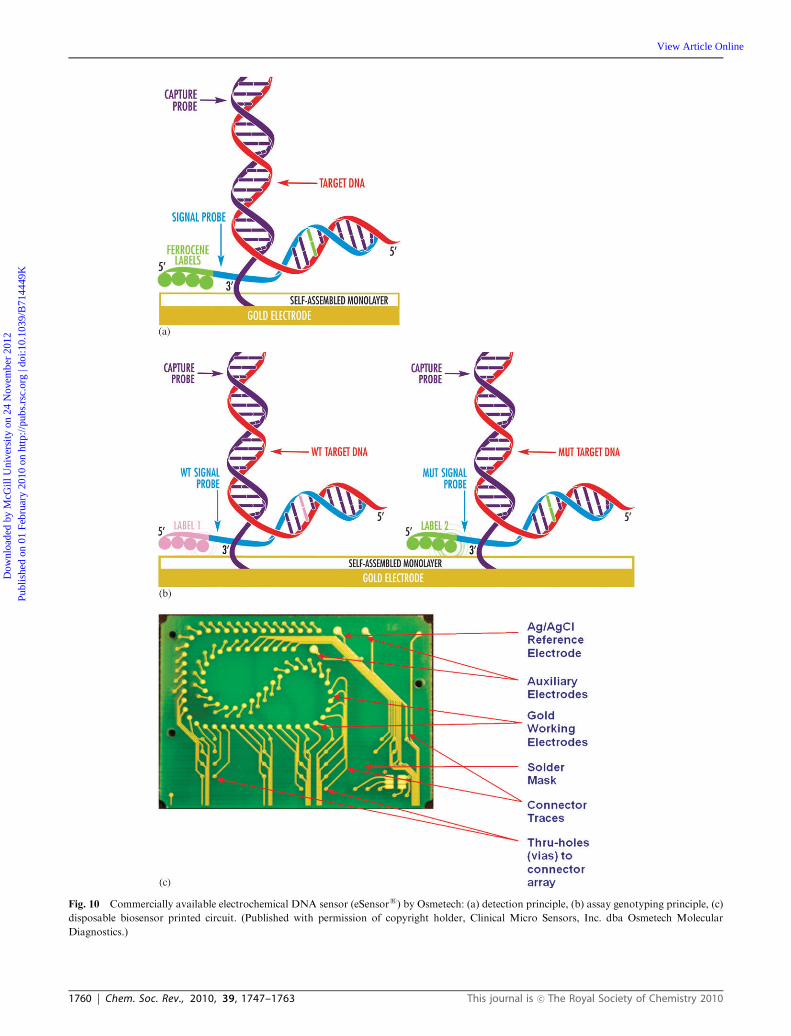
electrode to the sample results in hybridization between the
capture probe and the complementary strand of the target
DNA. The capture probe is designed to be shorter than the
complementary target strand, leaving a segment on the target
DNA where a signal probe containing an electroactive label
can bind. The label, ferrocene, is detected by measuring the
peak current for its oxidation by a positive potential scan in ac
voltammetry. The layer of alkane thiols is sufficiently thin as to
not interfere with the electrochemistry. The current is propor-
tional to the target DNA concentration in the sample. As
shown in Fig. 10B, genotyping can be done using different
ferrocene labels with distinguishable electrochemical poten-
tials for each label. The biochip consists of a microarray of
72 working electrodes, a Ag/AgCl reference electrode and two
auxiliary electrodes (Fig. 10C). Each working electrode of the
array can be interrogated independently which allows multiple
measurements to be made on the same chip. The chip is used
with a cartridge that features an auto-fill sample chamber,
microfluidic circulation to accelerate hybridization, and con-
tact with a resistive heating element. Osmetech has received
FDA clearance for eSensors
assays for detecting cystic fibrosis
carriers, and for identifying single-nucleotide polymorphisms
(SNPs) which result in increased sensitivity to warfarin, a
commonly prescribed blood anticoagulant.
3.3 Biosensors based on receptors
Receptors are proteins embedded in the cellular membrane
that specifically bind to their target analytes resulting in
physiological changes. The physiological response can be
opening ion-channels, producing second messenger systems,
or activating enzymes.1 A binding event at the receptor usually
causes the conformation of the receptor to change, which is
translated into an amplified electrochemical potential change.5
Unlike Abs that bind tightly with their complementary Ag,
receptors are like messengers that transmit signals upon ligand
binding between different parts of a biological system.1 Most
receptors are difficult to isolate and tend to bind to classes of
compounds having common chemical properties rather than
being highly specific for a given analyte like Abs.1,2 Therefore,
receptor-based biosensors are usually class-specific affinity
sensors that may not be a good feature for some biosensor
applications. Examples of receptor-based sensors include ion-
channel sensors where receptors in a lipid bilayer open or close
in response to a binding event with a ligand resulting in a rapid
ion flux through the membrane protein that causes a change in
the transmembrane conduction.109 The ion-channel membrane
proteins contain pores that allow ions such as Na+, K+ or
Ca2+ to flow through the channel until the potential difference
reaches equilibrium or the channel closes in response to a
stimulus. Also, nerve fibers from crayfish have been used to
monitor for local anesthetics and other drugs at low levels
(down to 10�15 M) with fast response times.110 Unfortunately,
these systems relying on axons from crayfish have a lifetime of
only 4 to 8 hours.
4. Conclusions
Catalytic and affinity biosensors with electrochemical detec-
tion continue to play an important role in many clinical,
environmental, industrial, pharmaceutical, defense, and
security applications due to their superior sensitivity and
selectivity. Although many electrochemical sensors are still
in the development and testing phases, some have reached
the consumer market as handheld devices, portable units used
for field measurements or are routinely used in a laboratory
setting. Recent developments in nanotechnology and mate-
rial science as well as being able to custom engineer the
biorecognition component will further push the develop-
ment of useful and reliable biosensor devices. The sometimes
limited shelf life and stability of the biorecognition component
as well as nonspecific binding continue to be the biggest
limitations of biosensors. However, many strategies have
helped with overcoming or minimizing these problems.
Notes and references
1 B. R. Eggins, Chemical Sensors and Biosensors, John Wiley &Sons, West Sussex, England, 2002.
2 J. Wang, Analytical Electrochemistry, John Wiley & Sons VCH,Hoboken, New Jersey, USA, 2006.
3 D. Grieshaber, R. MacKenzie, J. Voros and E. Reimhult,Sensors, 2008, 8, 1400.
4 R. A. Copeland, Enzymes, John Wiley & Sons VCH, New York,NY, USA, 2000.
5 Bioelectrochemistry Fundamentals, Experimental Techniques andApplications, ed. P. N. Bartlett, John Wiley & Sons, West Sussex,England, 2008.
6 N. J. Ronkainen-Matsuno, J. H. Thomas, H. B. Halsall andW. R. Heineman, TrAC, Trends Anal. Chem., 2002, 21(4),213.
7 C. G. Bauer, A. V. Eremenko, E. Ehrentreich-Forster, F. F. Bier,A. Makower, H. B. Halsall, W. R. Heineman and F. W. Scheller,Anal. Chem., 1996, 68(15), 2453.
8 S. H. Jenkins, W. R. Heineman and H. B. Halsall,Anal. Biochem.,1988, 168(2), 292.
9 C. A. Wijayawardhana, H. B. Halsall and W. R. Heineman,Electroanalytical Methods of Biological Materials, ed. A. Brajter-Toth, J. Q. Chambers, Marcel Dekker, New York, 2002,pp. 329–365.
10 H. Yao, S. H. Jenkins, A. J. Pesce, H. B. Halsall andW. R. Heineman, Clin. Chem. (Washington, D. C.), 1993, 39,1432.
11 Laboratory Techniques in Electroanalytical Chemistry, ed.P. T. Kissinger and W. R. Heineman, Marcel Dekker Inc, NewYork, NY, USA, 1996.
12 H. B. Halsall and W. R. Heineman, J. Int. Fed. Clin. Chem., 1990,2, 179–187.
13 M. Trojanowicz, M. Szewczynska and M. Wcislo, Electroanalysis,2003, 15(5–6), 347.
14 C. A. Wijayawardhana, S. Purushothama, M. A. Cousino,H. B. Halsall and W. R. Heineman, J. Electroanal. Chem.,1999, 468, 2.
15 C. A. Wijayawardhana, H. B. Halsall and W. R. Heineman, Anal.Chim. Acta, 1999, 399, 3.
16 R. Puchades and A. Maquieira, Crit. Rev. Anal. Chem., 1996,26(4), 195.
17 T. Jiang, H. B. Halsall and W. R. Heineman, J. Agric. FoodChem., 1995, 43, 1098.
18 R. M. Wightman and D. O. Wipf, Electroanalytical Chemistry,ed. A. J. Bard, Marcel Dekker, New York, USA, 1989,p. 267.
19 R. M. Wightman, Anal. Chem., 1981, 53(9), 1125A.20 J. Heinze, Angew. Chem., Int. Ed. Engl., 1993, 32, 1268.21 J. T. Thomas, N. J. Ronkainen-Matsuno, S. Farrell, H. B. Halsall
and W. R. Heineman, Microchem. J., 2003, 74, 267.22 S. Farrell, N. J. Ronkainen-Matsuno, H. B. Halsall and
W. R. Heineman, Anal. Bioanal. Chem., 2004, 379, 358.23 P. T. Kissinger and W. R. Heineman, J. Chem. Educ., 1983, 60(9),
702.
This journal is �c The Royal Society of Chemistry 2010 Chem. Soc. Rev., 2010, 39, 1747–1763 | 1761
Dow
nloa
ded
by M
cGill
Uni
vers
ity o
n 24
Nov
embe
r 20
12Pu
blis
hed
on 0
1 Fe
brua
ry 2
010
on h
ttp://
pubs
.rsc
.org
| do
i:10.
1039
/B71
4449
K
View Article Online
24 J. P. Hart, A. Crew, E. Crouch, K. C. Honeychurch andR. M. Pemberton, Anal. Lett., 2005, 37, 789.
25 S. Laschi, I. Palchetti and M. Mascini, Sens. Actuators, B, 2006,114, 460.
26 E. V. Gogol, G. A. Evtugyn, J. L. Marty, H. C. Budnikov andV. G. Winter, Talanta, 2000, 53, 379.
27 Immunoassay and Other Bioanalytical Methods, ed. J. M. vanEmon, CRC Press, Boca Raton, FL, USA, 2007.
28 O. Niwa, M. Morita and H. Tabei, Anal. Chem., 1990, 62,447.
29 J. H. Thomas, S. K. Kim, P. J. Hesketh, H. B. Halsall andW. R. Heineman, Anal. Chem., 2004, 76, 2700.
30 A. Bange, J. Tu, X. Zhu, C. Ahn, H. B. Halsall andW. R. Heineman, Electroanalysis, 2007, 19(21), 2202.
31 W. Lorenz and K. D. Schulze, J. Electroanal. Chem., 1975, 65(1),141.
32 I. I. Suni, TrAC, Trends Anal. Chem., 2008, 27(7), 604.33 T. M. Anh, S. V. Dzyadevych, M. C. Van, N. J. Renault,
C. N. Duc and J.-M. Chovelon, Talanta, 2004, 63, 365.34 Z. Muhammad-Tahir and E. C. Alocilja, Biosens. Bioelectron.,
2003, 18, 813.35 K. Yagiuda, A. Hemmi, S. Ito, Y. Asano, Y. Fushinuki,
C. Y. Chen and I. Karube, Biosens. Bioelectron., 1996, 11(8),703.
36 G. J. Papariello, A. K. Mukherji and C. M. Shearer, Anal. Chem.,1973, 45, 790.
37 C. Colapicchioni, A. Barbaro, F. Porcelli and I. Giannini, Sens.Actuators, B, 1991, 4, 245.
38 A. Barbaro, C. Colapicchioni, E. Davini, G. Mazzamurro,A. Piotto and F. Porcelli, Adv. Mater., 1992, 4(6), 402.
39 S. Caras and J. Janata, Anal. Chem., 1980, 52, 1935.40 D. G. Hafeman, J. W. Parce and H. M. McConnell, Science,
1988, 240, 1182.41 T. V. Shishkanova, R. Volf, M. Krondak and V. Kral, Biosens.
Bioelectron., 2007, 22, 2712.42 P. W. Davies and F. Brink, Rev. Sci. Instrum., 1942, 13,
524.43 J. B. Chien, R. A. Wallingford and A. G. Ewing, J. Neurochem.,
1990, 54, 633.44 T. K. Chen, G. Luo and A. G. Ewing, Anal. Chem., 1994, 66,
3031.45 J. A. Jankowski, T. J. Schroeder, R. W. Holz and
R. M. Wightman, J. Biol. Chem., 1992, 267, 18329.46 D. Bruns and R. Jahn, Nature, 1995, 377, 62.47 L. X. Tang and P. Vadgama, Med. Biol. Eng. Comput., 1990, 28,
B18.48 J. Wang, Anal. Chem., 1995, 67, 487.49 J. Wang, Electroanalysis, 2001, 13, 983.50 L. C. Clark and C. Lyons, Ann. N. Y. Acad. Sci., 1962, 102,
29.51 S. J. Updike and G. P. Hicks, Nature, 1967, 214, 986.52 S. R. Mikkelsen and E. Corton, Bioanalytical Chemistry, John
Wiley & Sons, Hoboken, New Jersey, USA, 2004.53 L. Gorton, A. Lindgren, T. Larsson, F. Muteanu, T. Ruzgas and
I. Gazaryan, Anal. Chim. Acta, 1999, 400, 91.54 A. J. Bard and L. R. Faulkner, Electrochemical Methods Funda-
mentals and Applications, John Wiley & Sons, Hoboken, NewJersey, USA, 2001.
55 Electron Transfer from Isolated Molecules to Biomolecules,Part Two, Advances in Chemical Physics, ed. J. Jortner andM. Bixon, John Wiley & Sons, New York, NY, USA, 1999,vol. 107.
56 B. A. Gregg and A. Heller, Anal. Chem., 1990, 62, 258.57 T. J. Ohara, R. Rajagopalan and A. Heller, Anal. Chem., 1993,
65, 3512.58 E. Lojou and P. Bianco, J. Electroceram., 2006, 16, 79.59 S. Cosnier, Electroanalysis, 2005, 19, 1715.60 L. X. Tang and P. Vadgama, Med. Biol. Eng. Comput., 1990, 28,
B18.61 D. C. Klonoff, Diabetes Care, 2005, 28, 1231.62 D. C. Klonoff, Diabetes Care, 2004, 27, 834.63 F. Arslan, A. Yaar and E. Kilic, Artif. Cells, Blood Substitutes,
Biotechnol., 2006, 34, 113.64 L. Agui, J. Manso, P. Yanez-Sedeno and J. M. Pingarron, Sens.
Actuators, B, 2006, 113, 272.
65 H. Shizuko Nakatani, L. Vieira dos Santos, C. Peralta Pelegrine,S. Terezinha Marques Gomes, M. Matsushita, N. Evelazio deSouza and J. Vergilio Visentainer, Am. J. Biochem. Biotechnol.,2005, 1, 85.
66 S. H. Hu and C. C. Liu, Electroanalysis, 1997, 9, 1229.67 J. Niu and J. Y. Lee, Sens. Actuators, B, 2000, 62, 190.68 H. Okuma and E. Watanabe, Biosens. Bioelectron., 2002, 17,
367.69 G. Palleschi, M. Mascini, L. Bernardi and P. Zeppilli, Med. Biol.
Eng. Comput., 1990, 28, B25.70 J.-W. Mo and W. Smart, Front. Biosci., 2004, 9, 3384.71 J. Boldt, Crit. Care, 2002, 6(1), 52.72 P. W. Alexander and G. A. Rechnitz, Electroanalysis, 2000, 12(5),
343.73 J. S. Sidwell and G. A. Rechnitz, Biotechnol. Lett., 1985, 7(6),
419.74 I. Karube and M. Suzuki, Microbial Biosensors in Biosensors: A
Practical Approach A, ed. E. G. Cass, The Practical ApproachSeries 7, Oxford University Press, Oxford, England, 1990.
75 R. A. Goldsby, T. J. Kindtm, B. A. Osborne and J. Kuby,Immunology, W.H. Freeman and Company, New York, NY,USA, 2003.
76 K. A. Fahnrich, M. Pravda and G. G. Guilbault, Anal. Lett.,2002, 35(8), 1269.
77 Antibodies: Volume 1: Production and Purification, ed.G. Subramanian, Springer, New York, NY, USA, 2004.
78 K. Graumann and A. Premstaller, Biotechnol. J., 2006, 1(2), 111.79 S. A. Kellermann and L. L. Green, Curr. Opin. Biotechnol., 2002,
13(6), 593.80 B. Lu, M. R. Smyth and R. Kennedy, Analyst, 1996, 121(3),
29R.81 W. J. Sung and Y. H. Bae, Sens. Actuators, B, 2006, 114(1),
164.82 R. Danczyk, B. Krieder, A. North, T. Webster, H. HogenEsch
and A. Rundell, Biotechnol. Bioeng., 2003, 84(2), 215.83 A. Kumari, Use of (poly)ethylene glycol modified solid supports in
electrochemical immunoassay, MS thesis, University of Cincinna-ti, Cincinnati, 1991.
84 K. Kato, Y. Umedo, F. Suzuki and A. Kosaka, Clin. Chim. Acta,1980, 102(2–3), 261.
85 R. F. Vogt Jr, D. L. Phillips, L. O. Henderson, W. Whitfield andF. W. Spierto, J. Immunol. Methods, 1987, 101(1), 43.
86 C. D. Hodneland, Y. S. Lee, D. H. Min and M. Mrksich, Proc.Natl. Acad. Sci. U. S. A., 2002, 99, 5048.
87 C. S. Chen, M. Mrksich, S. Huang, G. M. Whitesides andD. E. Ingber, Biotechnol. Prog., 1998, 14, 356.
88 M. Mrksich and G. M. Whitesides, Annu. Rev. Biophys. Biomol.Struct., 1996, 25, 55.
89 B. Johnsson, S. Lofas and G. Lindquist, Anal. Biochem., 1991,198, 268.
90 N. Kaneki, Y. Xu, A. Kumari, H. B. Halsall, W. R. Heinemanand P. T. Kissinger, Anal. Chim. Acta, 1994, 287, 253.
91 D. Cunningham, Anal. Chim. Acta, 2001, 429, 1.92 D. D. Cunningham and J. Russell, Abstract 1650–5, Pittcon,
Orlando, Florida, 2006.93 H. Huang, Z. Liu and X. Yang, Anal. Biochem., 2006, 356,
208.94 H. Huang, P. Ran and Z. Liu, Bioelectrochemistry, 2007, 70,
257.95 M. Wang, L. Wang, H. Yuan, X. Ji, C. Sun, L. Ma, Y. Bai, T. Li
and J. Li, Electroanalysis, 2004, 16, 757.96 H. Tang, J. Chen, L. Nie, Y. Kuang and S. Yao, Biosens.
Bioelectron., 2007, 22, 1061.97 Y. H. Yun, V. N. Shanov, M. J. Shulz, Z. Dong, A. Jazieh,
W. R. Heineman, H. B. Halsall, D. K. Y. Wong, A. Bange, Y. Tuand S. Subramanian, Sens. Actuators, B, 2006, 120, 298.
98 Y. H. Yun, A. Bange, W. R. Heineman, H. B. Halsall,V. N. Shanov, Z. Dong, S. Pixley, M. Behbehani, A. Jazieh, D.K. Y. Wong, A. Bhattacharya and M. J. Shulz, Sens. Actuators,B, 2007, 123, 177.
99 Y. H. Yun, Z. Dong, V. N. Shanov andM. J. Shulz,Nanotechnology,2007, 18, 465.
100 S. Takenaka, K. Yamashita, M. Takagi, Y. Uto and H. Kondo,Anal. Chem., 2000, 72, 1334.
101 Y. Zhang, H. Kim and A. Heller, Anal. Chem., 2003, 75, 3267.
1762 | Chem. Soc. Rev., 2010, 39, 1747–1763 This journal is �c The Royal Society of Chemistry 2010
Dow
nloa
ded
by M
cGill
Uni
vers
ity o
n 24
Nov
embe
r 20
12Pu
blis
hed
on 0
1 Fe
brua
ry 2
010
on h
ttp://
pubs
.rsc
.org
| do
i:10.
1039
/B71
4449
K
View Article Online
102 J. Wang, D. Xu, R. Polsky and E. Arzum, Talanta, 2002, 56,931.
103 Y. Zhang, H. Kim and A. Heller, Anal. Chem., 2003, 75,3267.
104 A. D. Ellington and J. W. Szostak, Nature, 1990, 346, 818.105 C. Tuerk and L. Gold, Science, 1990, 249, 505.106 I. Willner and M. Zayats, Angew. Chem., Int. Ed., 2007, 46,
6408–6418.
107 R. M. Umek, S. W. Lin, J. Vielmetter, R. H. Terbrueggen,B. Irvine, C. J. Yu, J. F. Kayyem, H. Yowanto, G. F. Blackburn,D. H. Farkas and Y.-P. Chen, J. Mol. Diagn., 2001, 3(2), 74.
108 R. H. Liu, W. A. Coty, M. Reed and G. Gust, IVD Technol.,2008, 14(4), 31.
109 N. Matsuno, M. Murawsky, J. Ridgeway and J. Cuppoletti,Biochim. Biophys. Acta, Biomembr., 2004, 1665, 184.
110 R. M. Buch and G. A. Rechnitz, Anal. Chem., 1989, 61, 533A.
This journal is �c The Royal Society of Chemistry 2010 Chem. Soc. Rev., 2010, 39, 1747–1763 | 1763
Dow
nloa
ded
by M
cGill
Uni
vers
ity o
n 24
Nov
embe
r 20
12Pu
blis
hed
on 0
1 Fe
brua
ry 2
010
on h
ttp://
pubs
.rsc
.org
| do
i:10.
1039
/B71
4449
K
View Article Online