184 11 Cell Signaling by Chemical Messengers Within a complex organism such as the human, different organs, tissues, and indi- vidual cell types have developed specialized functions. Yet each cell must contribute in an integrated way as the body grows, differentiates, and adapts to changing con- ditions. Such integration requires communication that is carried out by chemical messengers traveling from one cell to another or by direct contact of cells with the extracellular matrix or with each other. The eventual goal of such signals is to change actions carried out in target cells by intracellular proteins (metabolic enzymes, gene regulatory proteins, ion channels, or cytoskeletal proteins). In this chapter, we present an overview of signaling by chemical messengers. Chemical messengers. Chemical messengers (also called signaling molecules) transmit messages between cells. They are secreted from one cell in response to a specific stimulus and travel to a target cell, where they bind to a specific receptor and elicit a response (Fig. 11.1). In the nervous system, these chemical messen- gers are called neurotransmitters; in the endocrine system, they are hormones, and in the immune system, they are called cytokines. Additional chemical mes- sengers include retinoids, eicosanoids, and growth factors. Depending on the dis- tance between the secreting and target cells, chemical messengers can be classi- fied as endocrine (travel in the blood), paracrine (travel between nearby cells), or autocrine (act on the same cell or on nearby cells of the same type). Receptors and Signal Transduction. Receptors are proteins containing a bind- ing site specific for a single chemical messenger and another binding site involved in transmitting the message (see Fig. 11.1). The second binding site may interact with another protein or with DNA. They may be either plasma membrane receptors (which span the plasma membrane and contain an extracellular binding domain for the messenger) or intracellular binding proteins (for messengers able to diffuse into the cell) (see Fig. 11.1). Most plasma membrane receptors fall into the categories of ion channel receptors, tyrosine kinase receptors, tyrosine-kinase associated recep- tors (JAK-STAT receptors), serine-threonine kinase receptors, or heptahelical receptors (proteins with seven -helices spanning the membrane). When a chemical messenger binds to a receptor, the signal it is carrying must be converted into an intracellular response. This conversion is called signal transduction. Signal Transduction for Intracellular Receptors. Most intracellular receptors are gene-specific transcription factors, proteins that bind to DNA and regulate the transcription of certain genes (Gene transcription is the process of copying the genetic code from DNA to RNA.). Signal Transduction for Plasma Membrane Receptors. Mechanisms of signal transduction that follow the binding of signaling molecules to plasma membrane receptors include phosphorylation of receptors at tyrosine residues (receptor tyro- sine kinase activity), conformational changes in signal transducer proteins (e.g., proteins with SH2 domains, the monomeric G protein Ras, heterotrimeric G pro- teins) or increases in the levels of intracellular second messengers. Second mes- sengers are nonprotein molecules generated inside the cell in response to Fig. 11.1. General features of chemical mes- sengers. SECRETION RECEPTOR BINDING SIGNAL TRANSDUCTION RESPONSE Stimulus Chemical messengers Plasma membrane receptor Target cell Intracellular receptor Secretory cell 1 2
Transcript
184
11 Cell Signaling by ChemicalMessengers
Within a complex organism such as the human, different organs, tissues, and indi-vidual cell types have developed specialized functions. Yet each cell must contributein an integrated way as the body grows, differentiates, and adapts to changing con-ditions. Such integration requires communication that is carried out by chemicalmessengers traveling from one cell to another or by direct contact of cells with theextracellular matrix or with each other. The eventual goal of such signals is tochange actions carried out in target cells by intracellular proteins (metabolicenzymes, gene regulatory proteins, ion channels, or cytoskeletal proteins). In thischapter, we present an overview of signaling by chemical messengers.
Chemical messengers. Chemical messengers (also called signaling molecules)transmit messages between cells. They are secreted from one cell in response to aspecific stimulus and travel to a target cell, where they bind to a specific receptorand elicit a response (Fig. 11.1). In the nervous system, these chemical messen-gers are called neurotransmitters; in the endocrine system, they are hormones,and in the immune system, they are called cytokines. Additional chemical mes-sengers include retinoids, eicosanoids, and growth factors. Depending on the dis-tance between the secreting and target cells, chemical messengers can be classi-fied as endocrine (travel in the blood), paracrine (travel between nearby cells), orautocrine (act on the same cell or on nearby cells of the same type).
Receptors and Signal Transduction. Receptors are proteins containing a bind-ing site specific for a single chemical messenger and another binding site involvedin transmitting the message (see Fig. 11.1). The second binding site may interactwith another protein or with DNA. They may be either plasma membrane receptors(which span the plasma membrane and contain an extracellular binding domain forthe messenger) or intracellular binding proteins (for messengers able to diffuse intothe cell) (see Fig. 11.1). Most plasma membrane receptors fall into the categories ofion channel receptors, tyrosine kinase receptors, tyrosine-kinase associated recep-tors (JAK-STAT receptors), serine-threonine kinase receptors, or heptahelicalreceptors (proteins with seven �-helices spanning the membrane). When a chemicalmessenger binds to a receptor, the signal it is carrying must be converted into anintracellular response. This conversion is called signal transduction.
Signal Transduction for Intracellular Receptors. Most intracellular receptorsare gene-specific transcription factors, proteins that bind to DNA and regulatethe transcription of certain genes (Gene transcription is the process of copyingthe genetic code from DNA to RNA.).
Signal Transduction for Plasma Membrane Receptors. Mechanisms of signaltransduction that follow the binding of signaling molecules to plasma membranereceptors include phosphorylation of receptors at tyrosine residues (receptor tyro-sine kinase activity), conformational changes in signal transducer proteins (e.g.,proteins with SH2 domains, the monomeric G protein Ras, heterotrimeric G pro-teins) or increases in the levels of intracellular second messengers. Second mes-sengers are nonprotein molecules generated inside the cell in response to
Fig. 11.1. General features of chemical mes-sengers.
SECRETION
RECEPTOR BINDING
SIGNALTRANSDUCTION RESPONSE
Stimulus
Chemicalmessengers
Plasma membranereceptor
Targetcell
Intracellularreceptor
Secretorycell
1
2
185CHAPTER 11 / CELL SIGNALING BY CHEMICAL MESSENGERS
T H E W A I T I N G R O O M
Mya Sthenia is a 37-year-old woman who complains of increasing mus-cle fatigue in her lower extremities with walking. If she rests for 5 to 10minutes, her leg strength returns to normal. She also notes that if she talks
on the phone, her ability to form words gradually decreases. By evening, her uppereyelids droop to the point that she has to pull her upper lids back in order to see nor-mally. These symptoms are becoming increasingly severe. When Mya is asked tosustain an upward gaze, her upper eyelids eventually drift downward involuntarily.When she is asked to hold both arms straight out in front of her for as long as sheis able, both arms begin to drift downward within minutes. Her physician suspectsthat Mya Sthenia has myasthenia gravis and orders a test to determine whether shehas antibodies in her blood directed against the acetylcholine receptor.
Ann O’Rexia, who suffers from anorexia nervosa, has increased herweight to 102 lb from a low of 85 lb (see Chapter 9). On the advice of herphysician, she has been eating more to prevent fatigue during her daily
jogging regimen. She runs about 10 miles before breakfast every second day andforces herself to drink a high-energy supplement immediately afterward.
Dennis Veere was hospitalized for dehydration resulting from cholera toxin(see Chapter 10). In his intestinal mucosal cells, cholera A toxin indirectlyactivated the CFTR channel, resulting in secretion of chloride ion and Na�
ion into the intestinal lumen. Ion secretion was followed by loss of water, resulting invomiting and watery diarrhea. Dennis is being treated for hypovolemic shock.
I. GENERAL FEATURES OF CHEMICAL MESSENGERS
Certain universal characteristics to chemical messenger systems are illustrated inFigure 11.1. Signaling generally follows the sequence: (1) the chemical messengeris secreted from a specific cell in response to a stimulus; (2) the messenger diffusesor is transported through blood or other extracellular fluid to the target cell; (3) areceptor in the target cell (a plasma membrane receptor or intracellular receptor)specifically binds the messenger; (4) binding of the messenger to the receptor elic-its a response; (5) the signal ceases and is terminated. Chemical messengers elicittheir response in the target cell without being metabolized by the cell.
Another general feature of chemical messenger systems is that the specificity ofthe response is dictated by the type of receptor and its location. Generally, eachreceptor binds only one specific chemical messenger, and each receptor initiates acharacteristic signal transduction pathway that will ultimately activate or inhibitcertain processes in the cell. Only certain cells, the target cells, carry receptors forthat messenger and are capable of responding to its message.
The means of signal termination is an exceedingly important aspect of cell signaling,and failure to terminate a message contributes to a number of diseases, such as cancer.
hormone binding that continue transmission of the message. Examples include3�,5�-cyclic AMP (cAMP), inositol trisphosphate (IP3), and diacylglycerol (DAG).
Signaling often requires a rapid response and rapid termination of the mes-sage, which may be achieved by degradation of the messenger or second messen-ger, the automatic G protein clock, deactivation of signal transduction kinases byphosphatases, or other means.
Acetylcholine is released by neu-rons and acts on acetylcholinereceptors at neuromuscular junc-
tions to stimulate muscular contraction.Myasthenia gravis is an acquired autoim-mune disease in which the patient has devel-oped pathogenic antibodies against thesereceptors. Mya Sthenia’s decreasing ability toform words and her other symptoms of mus-cle weakness are being caused by the inabil-ity of acetylcholine to stimulate repeatedmuscle contraction when the numbers ofeffective acetylcholine receptors at neuro-muscular junctions are greatly reduced.
Endocrine hormones enable Ann
O’Rexia to mobilize fuels from heradipose tissue during her periods of
fasting and during jogging. While she fastsovernight, �-cells of her pancreas increasesecretion of the polypeptide hormoneglucagon. The stress of prolonged fasting andchronic exercise stimulates release of corti-sol, a steroid hormone, from her adrenal cor-tex. The exercise of jogging also increasessecretion of the hormones epinephrine andnorepinephrine from the adrenal medulla.Each of these hormones is being released inresponse to a specific signal and causes acharacteristic response in a target tissue,enabling her to exercise. However, each ofthese hormones binds to a different type ofreceptor and works in a different way.
Ann O’Rexia’s fasting is accompa-nied by high levels of theendocrine hormone glucagon,
which is secreted in response to low bloodglucose levels. It enters the blood and actson the liver to stimulate a number of path-ways, including the release of glucose fromglycogen stores (glycogenolysis) (see Chap-ter 3). The specificity of its action is deter-mined by the location of receptors. Althoughliver parenchymal cells have glucagonreceptors, skeletal muscle and many othertissues do not. Therefore, glucagon cannotstimulate glycogenolysis in these tissues.
186 SECTION TWO / CHEMICAL AND BIOLOGICAL FOUNDATIONS OF BIOCHEMISTRY
Most chemical messengers (includ-ing neurotransmitters, cytokines,and endocrine hormones) are con-
tained in vesicles that fuse with a region ofthe cell membrane when the cell receives astimulus to release the messenger. Mostsecretory cells use a similar set of proteins toenable vesicle fusion, and fusion is usuallytriggered by Ca2� influx, as seen with therelease of acetylcholine.
Myasthenia gravis is a disease ofautoimmunity caused by the pro-duction of an antibody directed
against the acetylcholine receptor in skeletalmuscle. In this disease, B and T lympho-cytes cooperate in producing a variety ofantibodies against the nicotinic acetyl-choline receptor. The antibodies then bindto various locations in the receptor andcross-link the receptors, forming a multi-receptor antibody complex. The complex isendocytosed and incorporated into lyso-somes, where it is degraded. Mya Sthenia,therefore, has fewer functional receptors foracetylcholine to activate.
A. General Features of Chemical Messenger Systems
Applied to the Nicotinic Acetylcholine Receptor
The individual steps involved in cell signaling by chemical messengers are illus-trated with acetylcholine, a neurotransmitter that acts on nicotinic acetylcholinereceptors on the plasma membrane of certain muscle cells. This system exhibits theclassic features of chemical messenger release and specificity of response.
Neurotransmitters are secreted from neurons in response to an electrical stimu-lus called the action potential (a voltage difference across the plasma membrane,caused by changes in Na� and K� gradients, that is propagated along a nerve). Theneurotransmitters diffuse across a synapse to another excitable cell, where theyelicit a response (Fig. 11.2). Acetylcholine is the neurotransmitter at neuromuscularjunctions, where it transmits a signal from a motor nerve to a muscle fiber that elic-its contraction of the fiber. Before release, acetylcholine is sequestered in vesiclesclustered near an active zone in the presynaptic membrane. This membrane also hasvoltage-gated Ca2� channels that open when the action potential reaches them,resulting in an influx of Ca2�. Ca2� triggers fusion of the vesicles with the plasmamembrane, and acetylcholine is released into the synaptic cleft. Thus, the chemicalmessenger is released from a specific cell in response to a specific stimulus.
Acetylcholine diffuses across the synaptic cleft to bind to plasma membranereceptors on the muscle cells called nicotinic acetylcholine receptors (Fig. 11.3).The subunits are assembled around a channel, which has a funnel-shaped openingin the center. As acetylcholine binds to the receptor, a conformational change opensthe narrow portion of the channel (the gate), allowing Na� to diffuse in and K� todiffuse out (A uniform property of all receptors is that signal transduction beginswith conformational changes in the receptor.). The change in ion concentration
Synapticvesicle (ACh)
Presynapticnerve terminal
Presynapticmembrane
Synaptic cleft
Postsynapticmembrane
Ca2+ channel
Junctional fold
Voltage-gatedNa+ channel
AChreceptors
ACh synapticvesicles
Muscle cell
Fig. 11.2. Acetylcholine receptors at the neuromuscular junction. A motor nerve terminatesin several branches; each branch terminates in a bulb-shaped structure called the presynapticbouton. Each bouton synapses with a region of the muscle fiber containing junctional folds.At the crest of each fold, there is a high concentration of acetylcholine receptors, which aregated ion channels.
γ
K+
α
γ
α
AChNa+
Fig. 11.3. The nicotinic acetylcholine recep-tor. Each receptor is composed of five sub-units, and each subunit has four membrane-spanning helical regions. The two � subunitsare identical and contain binding sites foracetylcholine. When two acetylcholine mole-cules are bound, the subunits change their con-formation so that the channel in the center ofthe receptor is open, allowing K� ions to dif-fuse out and Na� ions to diffuse in.
187CHAPTER 11 / CELL SIGNALING BY CHEMICAL MESSENGERS
Mya Sthenia was tested with aninhibitor of acetylcholinesterase,edrophonium chloride, adminis-
tered intravenously (see Chapter 8, Fig.8.18). After this drug inactivates acetyl-cholinesterase, acetylcholine that is releasedfrom the nerve terminal accumulates in thesynaptic cleft. Even though Mya expressesfewer acetylcholine receptors on her musclecells (due to the auto-antibody–induceddegradation of receptors), by increasing thelocal concentration of acetylcholine, thesereceptors have a higher probability of beingoccupied and activated. Therefore, acuteintravenous administration of this short-acting drug briefly improves muscular weak-ness in patients with myasthenia gravis.
activates a sequence of events that eventually triggers the cellular response—contraction of the fiber.
Once acetylcholine secretion stops, the message is rapidly terminated by acetyl-cholinesterase, an enzyme located on the postsynaptic membrane that cleavesacetylcholine. It is also terminated by diffusion of acetylcholine away from thesynapse. Rapid termination of message is a characteristic of systems requiring arapid response from the target cell.
B. Endocrine, Paracrine, and Autocrine
The actions of chemical messengers are often classified as endocrine, paracrine, orautocrine (Fig. 11.4). Each endocrine hormone is secreted by a specific cell type
Fig. 11.4. Endocrine, autocrine, and paracrine actions of hormones and other chemical mes-sengers.
188 SECTION TWO / CHEMICAL AND BIOLOGICAL FOUNDATIONS OF BIOCHEMISTRY
Fig. 11.5. Small molecule neurotransmitters.
(generally in an endocrine gland), enters the blood, and exerts its actions on specifictarget cells, which may be some distance away. In contrast to endocrine hormones,paracrine actions are those performed on nearby cells, and the location of the cellsplays a role in specificity of the response. Synaptic transmission by acetylcholineand other neurotransmitters (sometimes called neurocrine signaling) is an exampleof paracrine signaling. Acetylcholine activates only those acetylcholine receptorslocated across the synaptic cleft from the signaling nerve and not all muscles withacetylcholine receptors. Paracrine actions are also very important in limiting theimmune response to a specific location in the body, a feature that helps prevent thedevelopment of autoimmune disease. Autocrine actions involve a messenger actingon the cell from which it is secreted, or on nearby cells that are the same type as thesecreting cells.
C. Types of Chemical Messengers
Three major signaling systems in the body employ chemical messengers: the nerv-ous system, the endocrine system, and the immune system. Some messengers aredifficult to place in just one such category.
1. THE NERVOUS SYSTEM
The nervous system secretes two types of messengers: small molecule neurotrans-mitters, often called biogenic amines, and neuropeptides. Small molecule neuro-transmitters are nitrogen-containing molecules, which can be amino acids or arederivatives of amino acids (e.g., acetylcholine and �-aminobutyrate, Fig. 11.5).Neuropeptides are usually small peptides (between 4 and 35 amino acids), secretedby neurons, that act as neurotransmitters at synaptic junctions or are secreted intothe blood to act as neurohormones.
2. THE ENDOCRINE SYSTEM
Endocrine hormones are defined as compounds, secreted from specific endocrinecells in endocrine glands, that reach their target cells by transport through theblood. Insulin, for example, is an endocrine hormone secreted from the � cellsof the pancreas. Classic hormones are generally divided into the structural cate-gories of polypeptide hormones (e.g., insulin –see Chapter 6, Fig. 6.15 for thestructure of insulin ), catecholamines such as epinephrine (which is also a neu-rotransmitter), steroid hormones (which are derived from cholesterol), and thy-roid hormone (which is derived from tyrosine). Many of these endocrine hor-mones also exert paracrine or autocrine actions. The hormones that regulatemetabolism are discussed throughout this chapter and in subsequent chapters ofthis text.
Some compounds normally considered hormones are more difficult to catego-rize. For example, retinoids, which are derivatives of vitamin A (also called retinol)and vitamin D (which is also derived from cholesterol) are usually classified as hor-mones, although they are not synthesized in endocrine cells.
3. THE IMMUNE SYSTEM
The messengers of the immune system, called cytokines, are small proteins with amolecular weight of approximately 20,000 daltons. Cytokines regulate a network ofresponses designed to kill invading microorganisms. The different classes ofcytokines (interleukins, tumor necrosis factors, interferons, and colony-stimulatingfactors) are secreted by cells of the immune system and usually alter the behaviorof other cells in the immune system by activating the transcription of genes for pro-teins involved in the immune response.
γ-aminobutyrate (GABA)
H3C CH2 CH2 (CH3)3NC O
O+
–OOC CH2 CH2 H3NCH2+
Acetylcholine
The catecholamine hormone epi-nephrine (also called adrenaline) isthe fight, fright, and flight hor-
mone. Epinephrine and the structurally sim-ilar hormone norepinephrine are releasedfrom the adrenal medulla in response to avariety of immediate stresses, includingpain, hemorrhage, exercise, hypoglycemia,and hypoxia. Thus, as Ann O’Rexia begins tojog, there is a rapid release of epinephrineand norepinephrine into the blood.
Epinephrine
CH3
C C
H H
OH
H
NHHO
HO
Norepinephrine
C C
H H
OH
H
NH2HO
HO
189CHAPTER 11 / CELL SIGNALING BY CHEMICAL MESSENGERS
Lotta Topaigne suffered enormously painful gout attacks affecting her great toe(see Clinical Comments, Chapter 8). The extreme pain was caused by the releaseof a leukotriene that stimulated pain receptors. The precipitated urate crystals in
her big toe stimulated recruited inflammatory cells to release the leukotriene.
4. THE EICOSANOIDS
The eicosanoids (including prostaglandins [PG], thromboxanes, and leukotrienes)control cellular function in response to injury (Fig.11.6) These compounds are allderived from arachidonic acid, a 20-carbon polyunsaturated fatty acid that is usu-ally present in cells as part of the membrane lipid phosphatidylcholine (see Chap-ter 5, Fig. 5.21). Although almost every cell in the body produces an eicosanoid inresponse to tissue injury, different cells produce different eicosanoids. Theeicosanoids act principally in paracrine and autocrine functions, affecting the cellsthat produce them or their neighboring cells. For example, vascular endothelial cells(cells lining the vessel wall) secrete the prostaglandin PGI2 (prostacyclin), whichacts on nearby smooth muscle cells to cause vasodilation (expansion of the bloodvessel).
5. GROWTH FACTORS
Growth factors are polypeptides that function through stimulation of cellular prolifer-ation. For example, platelets aggregating at the site of injury to a blood vessel secrete
Selection and Proliferation of B Cells Producing the Desired Antibody. Interleukins, a class of cytokine, illustrate some of the sig-naling involved in the immune response. Interleukins are polypeptide factors with molecular weights ranging from 15,000 to25,000 Daltons. They participate in a part of the immune response called humoral immunity, which is carried out by a popula-
tion of lymphoid B cells producing just one antibody against one particular antigen. The proliferation of cells producing that particularantibody is mediated by receptors and by certain interleukins.
Bacteria phagocytized by macrophages are digested by lysosomes. (1) A partially digested fragment of the bacterial protein (the blueantigen) is presented on the extracellular surface of the macrophage by a membrane protein called an MHC (major histocompatibility com-plex). (2) Certain lymphoid cells called T-helper cells contain receptors that can bind to the displayed antigen–MHC complex, a processthat activates the T-cell (direct cell-to-cell signaling, requiring recognition molecules). (3) The activated T-helper cell then finds and bindsto a B cell whose antigen receptor binds a soluble fragment of that same bacterially derived antigen molecule (again, direct cell-to-cell sig-naling). The bound T cell secretes interleukins, which act on the B cell (a paracrine signal). The interleukins thus stimulate proliferation ofonly those B cells capable of synthesizing and secreting the desirable antibody. Furthermore, the interleukins determine which class ofantibody is produced.
Antigen
Bacteria
Macrophage
MHCT-cell receptor
B cellProliferatingB cells
ActivatedT-helper cell
T-helpercell
Cytokinese.g., IL-4, IL-5
and IL-6
Antigen receptor(membrane-bound
antibody)
4
1 2
3
ProstacyclinPGI2
COOH
OH
O
OH
COO–
Arachidonic acid(C20:4,∆5,8,11,14)
Fig. 11.6. Eicosanoids are derived fromarachidonic acid and retain its original 20 car-bons (thus the name eicosanoids). Allprostaglandins, such as prostacyclin, also havean internal ring.
190 SECTION TWO / CHEMICAL AND BIOLOGICAL FOUNDATIONS OF BIOCHEMISTRY
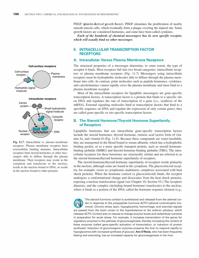
Fig. 11.7. Intracellular vs. plasma membranereceptors. Plasma membrane receptors haveextracellular binding domains. Intracellularreceptors bind steroid hormones or other mes-sengers able to diffuse through the plasmamembrane. Their receptors may reside in thecytoplasm and translocate to the nucleus,reside in the nucleus bound to DNA, or residein the nucleus bound to other proteins.
PDGF (platelet-derived growth factor). PDGF stimulates the proliferation of nearbysmooth muscle cells, which eventually form a plaque covering the injured site. Somegrowth factors are considered hormones, and some have been called cytokines.
Each of the hundreds of chemical messengers has its own specific receptor,which will usually bind no other messenger.
II. INTRACELLULAR TRANSCRIPTION FACTOR
RECEPTORS
A. Intracellular Versus Plasma Membrane Receptors
The structural properties of a messenger determine, to some extent, the type ofreceptor it binds. Most receptors fall into two broad categories: intracellular recep-tors or plasma membrane receptors (Fig. 11.7) Messengers using intracellularreceptors must be hydrophobic molecules able to diffuse through the plasma mem-brane into cells. In contrast, polar molecules such as peptide hormones, cytokines,and catecholamines cannot rapidly cross the plasma membrane and must bind to aplasma membrane receptor.
Most of the intracellular receptors for lipophilic messengers are gene-specifictranscription factors. A transcription factor is a protein that binds to a specific siteon DNA and regulates the rate of transcription of a gene (i.e., synthesis of themRNA). External signaling molecules bind to transcription factors that bind to aspecific sequence on DNA and regulate the expression of only certain genes; theyare called gene-specific or site-specific transcription factors.
B. The Steroid Hormone/Thyroid Hormone Superfamily
of Receptors
Lipophilic hormones that use intracellular gene-specific transcription factorsinclude the steroid hormones, thyroid hormone, retinoic acid (active form of vita-min A), and vitamin D (Fig. 11.8). Because these compounds are water-insoluble,they are transported in the blood bound to serum albumin, which has a hydrophobicbinding pocket, or to a more specific transport protein, such as steroid hormone-binding globulin (SHBG) and thyroid hormone-binding globulin (TBG). The intra-cellular receptors for these hormones are structurally similar and are referred to asthe steroid hormone/thyroid hormone superfamily of receptors.
The steroid hormone/thyroid hormone superfamily of receptors reside primarilyin the nucleus, although some are found in the cytoplasm. The glucocorticoid recep-tor, for example, exists as cytoplasmic multimeric complexes associated with heatshock proteins. When the hormone cortisol (a glucocorticoid) binds, the receptorundergoes a conformational change and dissociates from the heat shock proteins,exposing a nuclear translocation signal (see Chapter 10, Section VI.) The receptorsdimerize, and the complex (including bound hormone) translocates to the nucleus,where it binds to a portion of the DNA called the hormone response element (e.g.,
Cell-surface receptors
Intracellular receptors
Cell-surfacereceptor
Hydrophilic signalmolecule
Carrierprotein
Small hydrophobicsignal molecule
Cytosolicreceptor
Nuclearreceptor
DNA
Plasmamembrane
The steroid hormone cortisol is synthesized and released from the adrenal cor-tex in response to the polypeptide hormone ACTH (adrenal corticotrophic hor-mone). Chronic stress (pain, hypoglycemia, hemorrhage, and exercise) signals
are passed from the brain cortex to the hypothalamus to the anterior pituitary, whichreleases ACTH. Cortisol acts on tissues to change enzyme levels and redistribute nutrientsin preparation for acute stress. For example, it increases transcription of the genes forregulatory enzymes in the pathway of gluconeogenesis, thereby increasing the content ofthese enzymes (called gene-specific activation of transcription, or induction of proteinsynthesis). Induction of gluconeogenic enzymes prepares the liver to respond rapidly tohypoglycemia with increased synthesis of glucose. Ann O’Rexia, who has been frequentlyfasting and exercising, has an increased capacity for gluconeogenesis in her liver.
191CHAPTER 11 / CELL SIGNALING BY CHEMICAL MESSENGERS
the glucocorticoid receptor binds to the glucocorticoid response element, GRE).Most of the intracellular receptors reside principally in the nucleus, and some ofthese are constitutively bound, as dimers, to their response element in DNA (e.g.,the thyroid hormone receptor). Binding of the hormone changes its activity and itsability to associate with, or disassociate from, DNA. Regulation of gene transcrip-tion by these receptors is described in Chapter 16.
3, 5, 3'–Triiodothyronine (T3)
CH2 CH
NH2
COOHHO O
I I
I
1,25–Dihydroxycholecalciferol(1,25–(OH)2D3)
HO OH
C CH2
CH2
CH3
CH2 CH2 C OH
CH3
CH3
HH3C
25
1
A. Cortisol B. Aldosterone
C. Thyroid hormone (T3) D. Vitamin D3
E. Retinoids
All-trans retinoic acid 9-Cis retinoic acid
C
CH2OH
HO
OHC
O
O O
C
CH2OH
OHO OH
COOH
COOH
Fig. 11.8. Steroid hormone/thyroid hormone superfamily. A. Cortisol (a glucocorticoid). B. Aldosterone (an androgen). C. Thyroid hormone D.Vitamin D3. E. Retinoids.
Recently several nuclear receptors have been identified that play importantroles in intermediary metabolism, and they have become the target of lipid-lowering drugs. These include the peroxisome proliferator activated receptors
(PPAR �, � and �), the liver X-activated receptor (LXR), the farnesoid X-activated recep-tors (FXR), and the pregnane X receptor (PXR). These receptors form heterodimers withthe 9-cis retinoic acid receptor (RXR) and bind to their appropriate response elements inDNA in an inactive state. When the activating ligand binds to the receptor (oxysterols forLXR, bile salts for FXR, secondary bile salts for PXR, and fatty acids and their derivativesfor the PPARs), the complex is activated, and gene expression is altered. Unlike the cor-tisol receptor, these receptors reside in the nucleus and are activated once their ligandsenter the nucleus and bind to them.
192 SECTION TWO / CHEMICAL AND BIOLOGICAL FOUNDATIONS OF BIOCHEMISTRY
Signal transduction pathways, likea river, run in one direction. From agiven point in a signal transduction
pathway, events closer to the receptor arereferred to as “upstream,” and events closerto the response are referred to as “down-stream.”
III. PLASMA MEMBRANE RECEPTORS AND SIGNAL
TRANSDUCTION
All plasma membrane receptors are proteins with certain features in common: anextracellular domain that binds the chemical messenger, one or more membrane-spanning domains that are �-helices, and an intracellular domain that initiates sig-nal transduction. As the ligand binds to the extracellular domain of its receptor, itcauses a conformational change that is communicated to the intracellular domainthrough the rigid �-helix of the transmembrane domain. The activated intracellulardomain initiates a characteristic signal transduction pathway that usually involvesthe binding of a specific intracellular signal transduction protein.
The pathways of signal transduction for plasma membrane receptors have twomajor types of effects on the cell: (1) rapid and immediate effects on cellular ionlevels or activation/inhibition of enzymes and/or (2) slower changes in the rate ofgene expression for a specific set of proteins. Often, a signal transduction pathwaywill diverge to produce both kinds of effects.
A. Major Classes of Plasma Membrane Receptors
Individual plasma membrane receptors are grouped into the categories of ion chan-nel receptors, receptors that are kinases or bind kinases, and receptors that workthrough second messengers. This classification is based on the receptor’s generalstructure and means of signal transduction.
1. ION CHANNEL RECEPTORS
The ion channel receptors are similar in structure to the nicotinic acetylcholinereceptor (see Fig. 11.3). Signal transduction consists of the conformational changewhen ligand binds. Most small molecule neurotransmitters and some neuropeptidesuse ion channel receptors.
2. RECEPTORS THAT ARE KINASES OR BIND KINASES
Several types of receptors that are kinases or bind kinases are illustrated in Figure11.9. Their common feature is that the intracellular domain of the receptor (or anassociated protein) is a kinase that is activated when the messenger binds to theextracellular domain. The receptor kinase phosphorylates an amino acid residue onthe receptor (autophosphorylation) or an associated protein. The message is propa-gated through signal transducer proteins that bind to the activated messenger–-receptor complex (e.g., Grb2, STAT, or Smad).
Homodimer
A. Tyrosine kinase receptor
P
Growth factor
Tyrosine kinasedomain
P
Heterodimer
B. Jak-Stat receptors
Cytokine
JAKJAK
STAT
P
Heterodimer
C. Serine/threonine kinase receptors
Cytokine dimer
Phosphorylation
Smad Signaltransducerprotein
Signaltransducerprotein
Signaltransducerprotein
Serine kinasedomain
P
Tyrosine kinasedomain
P
PSH2
domain
P
P
Fig. 11.9. Receptors that are kinases or bind kinases. The kinase domains are shown in blue, and the phosphorylation sites are indicated withblue arrows. A. Tyrosine kinase receptors. B. JAK-STAT receptors. C. Serine/threonine kinase receptors.
Protein kinases transfer a phos-phate group from ATP to thehydroxyl group of a specific
amino acid residue in the protein. Tyrosinekinases transfer the phosphate group tothe hydroxyl group of a specific tyrosineresidue and serine/threonine proteinkinases to the hydroxyl of a specific serineor threonine residue (serine is more oftenphosphorylated than threonine in targetproteins). Different protein kinases havespecificity for distinct amino acidsequences (containing a tyrosine, serine,or threonine). Thus, two different proteinkinases target distinct sequences (andusually different proteins) for phosphory-lation. A protein containing both targetsequences could be a substrate for bothprotein kinases.
193CHAPTER 11 / CELL SIGNALING BY CHEMICAL MESSENGERS
3. HEPTAHELICAL RECEPTORS
Heptahelical receptors (which contain 7-membrane spanning �-helices) are themost common type of plasma membrane receptor. They work through second mes-sengers, which are small nonprotein compounds, such as cAMP, generated insidethe cell in response to messenger binding to the receptor (Fig. 11.10). They continueintracellular transmission of the message from the hormone/cytokine/neurotrans-mitter, which is the “first” messenger. Second messengers are present in low con-centrations so that their concentration, and hence the message, can be rapidlyinitiated and terminated.
B. Signal Transduction through Tyrosine Kinase Receptors
The tyrosine kinase receptors are summarized in Figure 11.9A. They generally existin the membrane as monomers with a single membrane-spanning helix. One mole-cule of the growth factor generally binds two molecules of the receptor and pro-motes their dimerization (Fig. 11.11). Once the receptor dimer has formed, theintracellular tyrosine kinase domains of the receptor phosphorylate each other oncertain tyrosine residues (autophosphorylation). The phosphotyrosine residues formspecific binding sites for signal transducer proteins.
1. RAS AND THE MAP KINASE PATHWAY
One of the domains of the receptor containing a phosphotyrosine residue forms abinding site for intracellular proteins with a specific three-dimensional structureknown as the SH2 domain (the Src homology 2 domain, named for the first proteinin which it was found, the src protein of the Rous sarcoma virus). The adaptor
cAMP or DAG, IP3second messenger
HeterotrimericG protein
α β γ
Hormone first messenger
Heptahelical receptors
Membraneassociatedenzyme
Cellular response
GDP
Fig. 11.10. Heptahelical Receptors and SecondMessengers. The secreted chemical messenger(hormone, cytokine, or neurotransmitter) is thefirst messenger, which binds to a plasma mem-brane receptor such as the heptahelical recep-tors. The activated hormone–receptor complexactivates a heterotrimeric G protein and viastimulation of membrane-bound enzymes, dif-ferent G-proteins lead to generation of one ormore intracellular second messengers, such ascAMP, diacylglycerol (DAG), or inositoltrisphosphate (IP3).
Although many different signal transducer proteins have SH2 domains, andmany receptors have phosphotyrosine residues, each signal transducer proteinis specific for one type of receptor. This specificity of binding results from the
fact that each phosphotyrosine residue has a different amino acid sequence around itthat forms the binding domain. Likewise, the SH2 domain of the transducer protein isonly part of its binding domain.
Fig. 11.11. Signal transduction by tyrosine kinase receptors. (1) Binding and dimerizaion. (2) Autophosphorylation. (3) Binding of Grb2 and SOS.(4) SOS is a GEF (guanine nucleotide exchange protein) that binds Ras, a monomeric G protein anchored to the plasma membrane. (5) GEF acti-vates the exchange of GTP for bound GDP on Ras. (6) Activated Ras containing GTP binds the target enzyme Raf, thereby activating it.
Growth factor bindingand dimerization
1.
Auto-crossphosphorylation2. Binding of adaptor proteinssuch as Grb2
3.
Complex assembly4. Guanine nucleotide exchangeand activation of Ras
5.
Ras binds raf and initiatesMAP kinase pathway
6.
Growth factor
Tyrosinekinasedomain
P P
Growth factor
P
P
P
P
P
PGDP
GDP GTP
Ras GTP
Raf
Ras
SOS(GEF)
Grb2
194 SECTION TWO / CHEMICAL AND BIOLOGICAL FOUNDATIONS OF BIOCHEMISTRY
protein Grb2, which is bound to a membrane phosphoinositide, is one of theproteins with an SH2 domain that binds to phosphotyrosine residues on growth fac-tor receptors. Binding to the receptor causes a conformational change in Grb2 thatactivates another binding site called an SH3 domain. These activated SH3 domainsbind the protein SOS (SOS is an acronym for “son of sevenless,” a name unrelatedto the function or structure of the compound). SOS is a guanine nucleotideexchange factor (GEF) for Ras, a monomeric G protein located in the plasma mem-brane (see Chapter 9, Section III.C.2.) SOS activates exchange of guanosinetriphosphate (GTP) for guanosine diphosphate (GDP) on Ras, causing a conforma-tional change in Ras that promotes binding of the protein Raf. Raf is a serine pro-tein kinase that is also called MAPKKK (mitogen activated protein kinase kinasekinase.) Raf begins a sequence of successive phosphorylation steps called a phos-phorylation cascade (When a kinase in a cascade is phosphorylated, it binds andphosphorylates the next enzyme in the cascade.). The MAP kinase cascade termi-nates at a gene transcription factor, thereby regulating transcription of certain genesinvolved in cell survival and proliferation.
Many tyrosine kinase receptors (as well as heptahelical receptors) also haveadditional signaling pathways involving phosphatidylinositol phosphates.
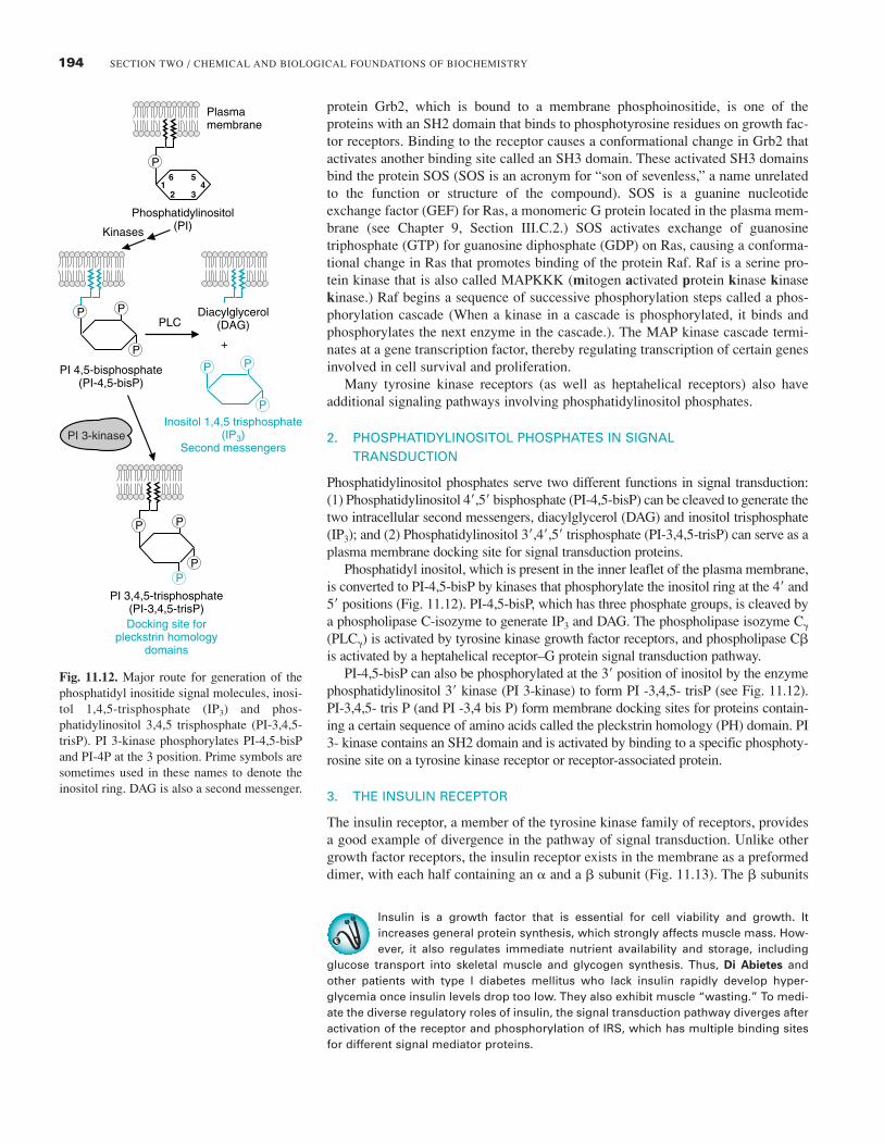
2. PHOSPHATIDYLINOSITOL PHOSPHATES IN SIGNAL TRANSDUCTION
Phosphatidylinositol phosphates serve two different functions in signal transduction:(1) Phosphatidylinositol 4�,5� bisphosphate (PI-4,5-bisP) can be cleaved to generate thetwo intracellular second messengers, diacylglycerol (DAG) and inositol trisphosphate(IP3); and (2) Phosphatidylinositol 3�,4�,5� trisphosphate (PI-3,4,5-trisP) can serve as aplasma membrane docking site for signal transduction proteins.
Phosphatidyl inositol, which is present in the inner leaflet of the plasma membrane,is converted to PI-4,5-bisP by kinases that phosphorylate the inositol ring at the 4� and5� positions (Fig. 11.12). PI-4,5-bisP, which has three phosphate groups, is cleaved bya phospholipase C-isozyme to generate IP3 and DAG. The phospholipase isozyme C�
(PLC�) is activated by tyrosine kinase growth factor receptors, and phospholipase C�is activated by a heptahelical receptor–G protein signal transduction pathway.
PI-4,5-bisP can also be phosphorylated at the 3� position of inositol by the enzymephosphatidylinositol 3� kinase (PI 3-kinase) to form PI -3,4,5- trisP (see Fig. 11.12).PI-3,4,5- tris P (and PI -3,4 bis P) form membrane docking sites for proteins contain-ing a certain sequence of amino acids called the pleckstrin homology (PH) domain. PI3- kinase contains an SH2 domain and is activated by binding to a specific phosphoty-rosine site on a tyrosine kinase receptor or receptor-associated protein.
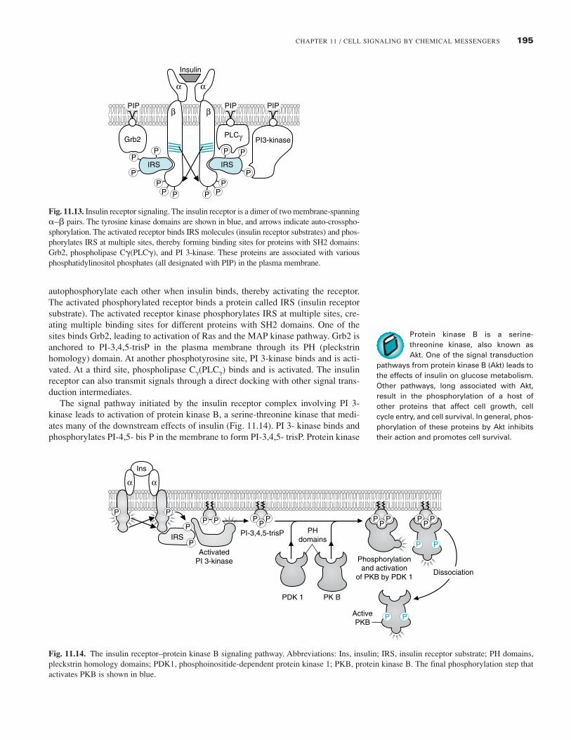
3. THE INSULIN RECEPTOR
The insulin receptor, a member of the tyrosine kinase family of receptors, providesa good example of divergence in the pathway of signal transduction. Unlike othergrowth factor receptors, the insulin receptor exists in the membrane as a preformeddimer, with each half containing an � and a � subunit (Fig. 11.13). The � subunits
Insulin is a growth factor that is essential for cell viability and growth. Itincreases general protein synthesis, which strongly affects muscle mass. How-ever, it also regulates immediate nutrient availability and storage, including
glucose transport into skeletal muscle and glycogen synthesis. Thus, Di Abietes andother patients with type I diabetes mellitus who lack insulin rapidly develop hyper-glycemia once insulin levels drop too low. They also exhibit muscle “wasting.” To medi-ate the diverse regulatory roles of insulin, the signal transduction pathway diverges afteractivation of the receptor and phosphorylation of IRS, which has multiple binding sitesfor different signal mediator proteins.
Fig. 11.12. Major route for generation of thephosphatidyl inositide signal molecules, inosi-tol 1,4,5-trisphosphate (IP3) and phos-phatidylinositol 3,4,5 trisphosphate (PI-3,4,5-trisP). PI 3-kinase phosphorylates PI-4,5-bisPand PI-4P at the 3 position. Prime symbols aresometimes used in these names to denote theinositol ring. DAG is also a second messenger.
PI 3-kinase
Plasmamembrane
Kinases
Phosphatidylinositol(PI)
6 5
2 31 4
P
PLC
+
PI 4,5-bisphosphate(PI-4,5-bisP)
PI 3,4,5-trisphosphate(PI-3,4,5-trisP)Docking site for
pleckstrin homologydomains
Diacylglycerol(DAG)
Inositol 1,4,5 trisphosphate(IP3)
Second messengers
P P
P
P P
P
P P
P
P
195CHAPTER 11 / CELL SIGNALING BY CHEMICAL MESSENGERS
autophosphorylate each other when insulin binds, thereby activating the receptor.The activated phosphorylated receptor binds a protein called IRS (insulin receptorsubstrate). The activated receptor kinase phosphorylates IRS at multiple sites, cre-ating multiple binding sites for different proteins with SH2 domains. One of thesites binds Grb2, leading to activation of Ras and the MAP kinase pathway. Grb2 isanchored to PI-3,4,5-trisP in the plasma membrane through its PH (pleckstrinhomology) domain. At another phosphotyrosine site, PI 3-kinase binds and is acti-vated. At a third site, phospholipase C�(PLC�) binds and is activated. The insulinreceptor can also transmit signals through a direct docking with other signal trans-duction intermediates.
The signal pathway initiated by the insulin receptor complex involving PI 3-kinase leads to activation of protein kinase B, a serine-threonine kinase that medi-ates many of the downstream effects of insulin (Fig. 11.14). PI 3- kinase binds andphosphorylates PI-4,5- bis P in the membrane to form PI-3,4,5- trisP. Protein kinase
Insulin
IRS IRS
α α
β
PP P
β
PPP
P P PP
P P
PIP PIP PIP
Grb2PLCγ PI3-kinase
Fig. 11.13. Insulin receptor signaling. The insulin receptor is a dimer of two membrane-spanning�–� pairs. The tyrosine kinase domains are shown in blue, and arrows indicate auto-crosspho-sphorylation. The activated receptor binds IRS molecules (insulin receptor substrates) and phos-phorylates IRS at multiple sites, thereby forming binding sites for proteins with SH2 domains:Grb2, phospholipase C�(PLC�), and PI 3-kinase. These proteins are associated with variousphosphatidylinositol phosphates (all designated with PIP) in the plasma membrane.
Ins
αα
P P P PP
P P
P P
P PP
P PP
P
P
P
P
ActivatedPI 3-kinase
PI-3,4,5-trisP PHdomains
PDK 1 PK B
ActivePKB
Dissociation
Phosphorylationand activation
of PKB by PDK 1
IRS
Fig. 11.14. The insulin receptor–protein kinase B signaling pathway. Abbreviations: Ins, insulin; IRS, insulin receptor substrate; PH domains,pleckstrin homology domains; PDK1, phosphoinositide-dependent protein kinase 1; PKB, protein kinase B. The final phosphorylation step thatactivates PKB is shown in blue.
Protein kinase B is a serine-threonine kinase, also known asAkt. One of the signal transduction
pathways from protein kinase B (Akt) leads tothe effects of insulin on glucose metabolism.Other pathways, long associated with Akt,result in the phosphorylation of a host ofother proteins that affect cell growth, cellcycle entry, and cell survival. In general, phos-phorylation of these proteins by Akt inhibitstheir action and promotes cell survival.
196 SECTION TWO / CHEMICAL AND BIOLOGICAL FOUNDATIONS OF BIOCHEMISTRY
Although Jak is an acronym forjanus kinase, it has been sug-gested that it stands for “just
another kinase”. It was named for Janus, atwo-headed god of the Romans.
B and PDK1 (phosphoinositide-dependent kinase-1) are recruited to the membraneby their PH domains, where PDK1 phosphorylates and activates protein kinase B.Many other signal transducer proteins have PH domains and are docked at the mem-brane, where they can find and bind each other. Thus, the insulin signal divergesagain and again. Insulin is covered in more detail in Chapters 26, 36 and 43.
C. Signal Transduction by JAK-STAT Receptors
Tyrosine kinase-associated receptors called Jak-STAT receptors are often used bycytokines to regulate the proliferation of certain cells involved in the immuneresponse (see Fig. 11.9B). The receptor itself has no intrinsic kinase activity butbinds (associates with) the tyrosine kinase Jak (janus kinase). Their signal trans-ducer proteins, called STATs (signal transducer and activator of transcription), arethemselves gene-specific transcription factors. Thus, Jak-STAT receptors have amore direct route for propagation of the signal to the nucleus than tyrosine kinasereceptors.
Each receptor monomer has an extracellular domain, a membrane-spanningregion, and an intracellular domain. As the cytokine binds to these receptors, theyform dimers (either homodimers or heterodimers, between two distinct receptor mol-ecules) and may cluster (Fig. 11.15). The activated Jaks phosphorylate each otherand intracellular tyrosine residues on the receptor, forming phosphotyrosine-bindingsites for the SH2 domain of a STAT. STATs are inactive in the cytoplasm until theybind to the receptor complex, where they are also phosphorylated by the bound JAK.Phosphorylation changes the conformation of the STAT, causing it to dissociate fromthe receptor and dimerize with another phosphorylated STAT, thereby forming anactivated transcription factor. The STAT dimer translocates to the nucleus and bindsto a response element on DNA, thereby regulating gene transcription.
There are many different STAT proteins, each with a slightly different aminoacid sequence. Receptors for different cytokines bind different STATs, which thenform heterodimers in various combinations. This microheterogeneity allows differ-ent cytokines to target different genes.
D. Receptor Serine/Threonine Kinases
Proteins in the transforming growth factor superfamily use receptors that have ser-ine/threonine kinase activity and associate with proteins from the Smad family,which are gene-specific transcription factors (see Fig. 11.9C). This superfamilyincludes transforming growth factor � (TGF-�), a cytokine/hormone involved intissue repair, immune regulation, and cell proliferation, and bone morphogeneticproteins (BMPs), which control proliferation, differentiation, and cell death duringdevelopment.
P P P
P P
P P
P
P PP
PP
P
Receptor binds andphosphorylates STATs
3.
STATs dissociate from receptor, dimerize
4.
Receptors bindcytokines, dimerize,and bind Jaks
1. Jaks phosphorylateeach other and thereceptor
2.
JakJakCytosol
STAT
Fig. 11.15. Steps in Jak-STAT receptor signaling.
197CHAPTER 11 / CELL SIGNALING BY CHEMICAL MESSENGERS
A simplified version of TGF-�1 binding to its receptor complex and activatingSmads is illustrated in Fig. 11.16 The TGF-� receptor complex is composed of twodifferent single membrane-spanning receptor subunits (type I and type II), whichhave different functions even though they both have serine kinase domains. TGF-�binds to a type II receptor. The activated type II receptor recruits a type I receptor,which it phosphorylates at a serine residue, forming an activated receptor complex.The type I receptor then binds a receptor-specific Smad protein (called R-Smads),which it phosphorylates at serine residues. The phosphorylated R-Smad undergoesa conformational change and dissociates from the receptor. It then forms a complexwith another member of the Smad family, Smad 4 (Smad 4 is known as the com-mon Smad, Co-Smad, and is not phosphorylated). The Smad complex, which maycontain several Smads, translocates to the nucleus, where it activates or inhibits thetranscription of target genes. Receptors for different ligands bind different Smads,which bind to different sites on DNA and regulate the transcription of differentgenes.
E. Signal Transduction through Heptahelical Receptors
The heptahelical receptors are named for their 7-membrane spanning domains,which are �-helices (see Fig. 11.10; see also Chapter 7, Fig. 7.10). Although hun-dreds of hormones and neurotransmitters work through heptahelical receptors, theextracellular binding domain of each receptor is specific for just one polypeptidehormone, catecholamine, or neurotransmitter (or its close structural analog).Heptahelical receptors have no intrinsic kinase activity but initiate signal transduc-tion through heterotrimeric G proteins composed of �, � and � subunits. However,different types of heptahelical receptors bind different G proteins, and different Gproteins exert different effects on their target proteins.
1. HETEROTRIMERIC G PROTEINS
The function of heterotrimeric G proteins is illustrated in Figure 11.17 using a hor-mone that activates adenylyl cyclase (e.g., glucagon or epinephrine). While the �subunit contains bound GDP, it remains associated with the � and � subunits, eitherfree in the membrane or bound to an unoccupied receptor (see Fig. 11.17, part 1).When the hormone binds, it causes a conformational change in the receptor that acti-vates GDP dissociation and GTP binding. The exchange of GTP for bound GDPcauses dissociation of the � subunit from the receptor and from the �� subunits (seeFig. 11.17, part 2). The � and � subunits are tethered to the intracellular side of the
TGF-β bindsto Type II receptor
1.
TGF-β
Type II
Type II receptorphosphorylatesType I receptor
2.
P
Activated Type I receptorphosphorylates R-Smad
3. R-Smad complexeswith Co-Smad andmigrates to nucleus
4.
P
PP
S S
SR-Smad
S
PPS
R-SmadS
Co-Smad
Fig. 11.16. Serine/threonine receptors and Smad proteins. TGF-� (transforming growth factor �), which is composed of two identical subunits,communicates through a receptor dimer of type I and type II subunits that have serine kinase domains. The type I receptor phosphorylates anR-Smad (receptor-specific Smad), which binds a Co-Smad (common Smad, also called Smad 4).
198 SECTION TWO / CHEMICAL AND BIOLOGICAL FOUNDATIONS OF BIOCHEMISTRY
The importance of signal termina-tion is illustrated by the “internalclock” of G proteins, which is the
rate of spontaneous hydrolysis of GTP to GDP.Mutations in ras (the gene encoding Ras) thatdecrease the rate of GTP hydrolysis are foundin about 20 to 30% of all human cancers,including approximately 25% of lung cancers,50% of colon cancers, and more than 90% ofpancreatic cancers. In these mutations of Ras,GTP hydrolysis is decreased and Ras remainslocked in the active GTP-bound form, ratherthan alternating normally between inactiveand active state in response to extracellularsignals. Consequently, MAP kinase pathwaysare continuously stimulated and drive cell pro-liferation, even in the absence of growth fac-tors that would be required for ras activationin normal cells.
plasma membrane through lipid anchors, but can still move around on the membranesurface. The GTP-� subunit binds its target enzyme in the membrane, thereby chang-ing its activity. In this example, the �-subunit binds and activates adenylyl cyclase,thereby increasing synthesis of cAMP (see Fig. 11.17, part 3).
With time, the G� subunit inactivates itself by hydrolyzing its own bound GTPto GDP and Pi. This action is unrelated to the number of cAMP molecules formed.Like the monomeric G proteins, the GDP-� subunit then dissociates from its targetprotein, adenylyl cyclase (see Fig. 11.16, part 4). It reforms the trimeric G proteincomplex, which may return to bind the empty hormone receptor. As a result of thisGTPase “internal clock,” sustained elevations of hormone levels are necessary forcontinued signal transduction and elevation of cAMP.
A large number of different heterotrimeric G protein complexes are generallycategorized according to the activity of the � subunit (Table 11.1). The 20 or more dif-ferent isoforms of G� fall into four broad categories: G�s, G�i/0, G�q/11, andG�12/1313. G�s refers to � subunits, which, like the one in Figure 11.17, stimulateadenylyl cyclase (hence the s). G� subunits that inhibit adenylyl cyclase are called G�i.
The �� subunits likewise exist as different isoforms, which also transmit messages.
Receptor bindshormone
αβ γ
1.
Target protein bindsGTP-Gαs
3. GTP is hydrolyzedand Gαs dissociates
4. Gαs reassociates withβγ subunits and receptor
5.
GDP
αβGDP
G protein exchanges GTP for GDP and dissociates
α
α
αβ γ β γ
2.
GDP
Adenylylcyclase ATP
cAMP
Pi
GDP GTPGTP
GTP
αGDP
Fig. 11.17. Heptahelical receptors and heterotrimeric G proteins. (1) The intracellular domains of the receptor form a binding site for a G pro-tein containing GDP bound to the �-subunit. (2) Hormone binding to the receptor promotes the exchange of GTP for GDP. As a result, the com-plex disassembles, releasing the G protein �-subunit from the �� complex. (3) The Gs �-subunit binds to a target enzyme, thereby changing itsactivity. The �� complex may simultaneously target another protein and change its activity. (4) Over time, bound GTP is hydrolysed to GDP,causing dissociation of the �-subunit from adenylyl cyclase. The GDP-�-subunit reassociates with the �� subunit and the receptor.
Acetylcholine has two types of receptors: nicotinic ion channel receptors, the receptors inhibited by antibodies in myastheniagravis, and muscarinic receptors, which exist as a variety of subtypes. The M2 muscarinic receptors activate a G�i/o het-erotrimeric G protein in which release of the �� subunit controls K� channels and pacemaker activity in the heart. Epinephrinehas several types and subtypes of heptahelical receptors: � receptors work through a G�s and stimulate adenylyl cyclase; �2
receptors in other cells work through a G�i protein and inhibit adenylyl cyclase; �1 receptors work through G�q subunits and activate phos-pholipase C�. This variety in receptor types allows a messenger to have different actions in different cells.
199CHAPTER 11 / CELL SIGNALING BY CHEMICAL MESSENGERS
2. ADENYLYL CYCLASE AND CAMP PHOSPHODIESTERASE
cAMP is referred to as a second messenger because changes in its concentrationreflect changes in the concentration of the hormone (the first messenger). Whena hormone binds and adenylyl cyclase is activated, it synthesizes cAMP fromadenosine triphosphate (ATP). cAMP is hydrolyzed to AMP by cAMP phospho-diesterase, which also resides in the plasma membrane (Fig. 11.18). Theconcentration of cAMP and other second messengers is kept at very low levelsin cells by balancing the activity of these two enzymes so that cAMP levels canchange rapidly when hormone levels change. Some hormones change the con-centration of cAMP by targeting the phosphodiesterase enzyme rather thanadenylyl cyclase. For example, insulin lowers cAMP levels by causing phospho-diesterase activation.
cAMP exerts diverse effects in cells. It is an allosteric activator of protein kinaseA (see Chapter 9, section III.B.3), which is a serine/threonine protein kinase thatphosphorylates a large number of metabolic enzymes, thereby providing a rapidresponse to hormones such as glucagon and epinephrine. The catalytic subunits ofprotein kinase A also enter the nucleus and phosphorylate a gene-specific tran-scription factor called CREB (cyclic AMP response element-binding protein). Thus,cAMP also activates a slower response pathway, gene transcription. In other celltypes, cAMP directly activates ligand-gated channels.
3. PHOSPHATIDYLINOSITOL SIGNALING BY HEPTAHELICAL RECEPTORS
Certain heptahelical receptors bind the q isoform of the G� subunit (G�q), whichactivates the target enzyme phospholipase C� (see Fig.11.12). When activated,phospholipase C� hydrolyzes the membrane lipid phosphatidyl inositol bis phos-phate (PI-4,5-bisP) into two second messengers, diacylglycerol (DAG) and1,4,5-inositol trisphosphate (IP3). IP3 has a binding site in the sarcoplasmicreticulum and the endoplasmic reticulum that stimulates the release of Ca2� (Fig.11.19). Ca2� activates enzymes containing the calcium–calmodulin subunit,including a protein kinase. Diacylglycerol, which remains in the membrane, acti-vates protein kinase C, which then propagates the response by phosphorylatingtarget proteins.
F. Changes in Response to Signals
Tissues vary in their ability to respond to a message through changes in receptoractivity or number. Many receptors contain intracellular phosphorylation sitesthat alter their ability to transmit signals. Receptor number is also varied through
Table 11.1 Subunits of Heterotrimeric G Proteins
G� subunit Action Some Physiologic Uses
�s; G� (s) * stimulates adenyl cyclase Glucagon and epinephrine to regulate metabolic enzymes, regulatory polypeptide hormones to control steroid hor-mone and thyroid hormone synthesis, and by some neurotransmitters (e.g., dopamine)to control ion channels
�i/o; G� (i/o) inhibits adenylyl cyclase Epinephrine, many neurotransmitters including acetylcholine, dopamine, serotonin. (signal also flows Has a role in the transducin pathway, which mediates detection of light through �� subunits) in the eye.
�12/13; G� (12/13) Physiologic connections are Thromboxane A2, lysophosphatidic acidnot yet well established
*There is a growing tendency to designate the heterotrimeric G protein subunits without using subscripts so that they are actually visible to the naked eye.
Dennis Veere was hospitalized fordehydration resulting from choleratoxin (see Chapter 10). Cholera A
toxin was absorbed into the intestinalmucosal cells, where it was processed andcomplexed with Arf (ADP-ribosylation fac-tor), a small G protein normally involved invesicular transport. Cholera A toxin is anNAD-glycohydrolase, which cleaves NADand transfers the ADP ribose portion to otherproteins. It ADP-ribosylates the G�s subunitof heterotrimeric G proteins, thereby inhibit-ing their GTPase activity. As a consequence,they remain actively bound to adenylylcyclase, resulting in increased production ofcAMP. The CFTR channel is activated, result-ing in secretion of chloride ion and Na� ioninto the intestinal lumen. The ion secretion isfollowed by loss of water, resulting in vomit-ing and watery diarrhea.
Some signaling pathways crossfrom the MAP kinase pathway tophosphorylate CREB, and all het-
erotrimeric G protein pathways diverge toinclude a route to the MAP kinase pathway.These types of complex interconnections insignaling pathways are sometimes calledhormone cross-talk.
200 SECTION TWO / CHEMICAL AND BIOLOGICAL FOUNDATIONS OF BIOCHEMISTRY
down-regulation. After a hormone binds to the receptor, the hormone–receptorcomplex may be taken into the cell by the process of endocytosis in clathrin-coated pits (see Chapter 10, Section III.B.1.) The receptors may be degraded orrecycled back to the cell surface. This internalization of receptors decreases thenumber available on the surface under conditions of constant high hormone lev-els when more of the receptors are occupied by hormones and results in decreasedsynthesis of new receptors. Hence, it is called down-regulation.
IV. SIGNAL TERMINATION
Some signals, such as those that modify the metabolic responses of cells or trans-mit neural impulses, need to turn off rapidly when the hormone is no longer beingproduced. Other signals, such as those that stimulate proliferation, turn off moreslowly. In contrast, signals regulating differentiation may persist throughout ourlifetime. Many chronic diseases are caused by failure to terminate a response at theappropriate time.
The first level of termination is the chemical messenger itself (Fig.11.20). Whenthe stimulus is no longer applied to the secreting cell, the messenger is no longersecreted, and existing messenger is catabolized. For example, many polypeptidehormones such as insulin are taken up into the liver and degraded. Termination ofthe acetylcholine signal by acetylcholinesterase has already been mentioned.
Within each pathway of signal transduction, the signal may be turned off at spe-cific steps. The receptor might be desensitized to the messenger by phosphorylation.G proteins, both monomeric and heterotrimeric, automatically terminate messagesas they hydrolyze GTP. Termination also can be achieved through degradation of thesecond messenger (e.g., phosphodiesterase cleavage of cAMP). Each of these ter-minating processes is also highly regulated.
Another important pathway for reversing the message is through protein phos-phatases, enzymes that reverse the action of kinases by removing phosphate groups
O
–O
CH2
5'
3'
NH2
PPi
O
OPO OH
HHHH
N
NN
NCHCH
C
CC
OO P
O
O P
O–O
O– O– O–
P
O
CH2
NH2
ATP cAMP(3',5'-cyclic AMP)
AMP
O
OH OH
HHHH
N
NN
NCHCH
C
CC
O–O P
O
O–
CH2
NH2
O
OH OH
HHHH
N
NN
NCHCH
C
CC
Gαs
Adenylylcyclase
GTP
cAMPphosphodiesterase
Fig. 11.18. Formation and cleavage of the cyclic phosphodiester bond in cAMP. When activated by G�s, adenyl cyclase converts ATP to 3�,5�-cyclic AMP � PPi . cAMP phosphodiesterase hydrolyzes cAMP to AMP.
IP3 receptor
IP3
Smooth ER
Ca2+
P P
P
In myasthenia gravis, increasedendocytosis and degradation ofacetylcholine receptors lead to a
signal transduction pathway that decreasessynthesis of new receptors. Thus, downreg-ulation of acetylcholine receptors is part ofthis disease.
201CHAPTER 11 / CELL SIGNALING BY CHEMICAL MESSENGERS
Fig. 11.20. Sites of signal termination.Processes that terminate signals are shown inblue.
from proteins. Specific tyrosine or serine/threonine phosphatases (enzymes thatremove the phosphate group from specific proteins) exist for all of the sites phos-phorylated by signal transduction kinases. Some receptors are even protein phos-phatases.
CLINICAL COMMENTS
Mya Sthenia. Mya Sthenia has myasthenia gravis, an autoimmune dis-ease caused by the production of antibodies directed against the nicotinicacetylcholine receptor in skeletal muscles. The diagnosis is made by his-
tory (presence of typical muscular symptoms), physical examination (presence ofinability to do specific repetitive muscular activity over time), and tests such as theinhibition of acetylcholinesterase activity. The diagnosis can be further confirmedwith an electromyogram (EMG) showing a partial blockade of ion flux across mus-cular membranes and a diagnostic procedure involving repetitive electrical nervestimulation.
Ann O’Rexia. Anorexia nervosa presents as a distorted visual self-image often associated with compulsive exercise. Although Ann has beengaining weight, she is still relatively low on stored fuels needed to sustain
the metabolic requirements of exercise. Her prolonged starvation has resulted inrelease of the steroid hormone cortisol and the polypeptide hormone glucagon,whereas levels of the polypeptide hormone insulin have decreased. Cortisol acti-vates transcription of genes for some of the enzymes of gluconeogenesis (the syn-thesis of glucose from amino acids and other precursors; see Chapter 3.) Glucagonbinds to heptahelical receptors in liver and adipose tissue and, working throughcAMP and protein kinase A, activates many enzymes involved in fasting fuelmetabolism. Insulin, which is released when she drinks her high-energy supple-ment, works through a specialized tyrosine kinase receptor to promote fuel stor-age. Epinephrine, a catecholamine released when she exercises, promotes fuelmobilization.
Dennis Veere. In the emergency room, Dennis received intravenousrehydration therapy (normal saline [0.9% NaCl]) and oral hydration ther-apy with a glucose-electrolyte solution to increase his glucose-dependent
Na� uptake from the intestinal lumen (see Chapter 10). Dennis quickly recoveredfrom his bout of cholera. Cholera is self-limiting, possibly because the bacteriaremain in the intestine, where they are washed out of the system by the diffusewatery diarrhea. Over the past three years, Percy Veere has persevered through thedeath of his wife and the subsequent calamities of his grandson Dennis “theMenace” Veere, including salicylate poisoning, suspected malathion poisoning, andnow cholera. Mr. Veere decided to send his grandson home for the remainder of thesummer.
BIOCHEMICAL COMMENTS
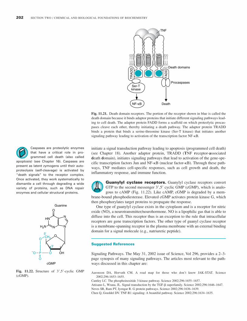
Death domain receptors. The cytokine TNF (tumor necrosis fac-tor) uses a type of receptor called the death domain receptor (Fig. 11.21).These receptors function as a trimer when they bind TNF (which is also a
trimer). On TNF binding, an inhibitory protein called the “silencer of death” isreleased from the receptor. The receptor then binds and activates several adaptorproteins. One adaptor protein, FADD (fas-associated death domain), recruits andactivates the zymogen form of a proteolytic enzyme called caspase. Caspases
Stimulus
Response
Release
ReceptorSignaltransduction
Messenger
Secondmessenger
TERMINATORS
DiffusionDegradation
Desensitizationdown-regulation
Protein phosphatases
GTPases
Phosphodiesterases
202 SECTION TWO / CHEMICAL AND BIOLOGICAL FOUNDATIONS OF BIOCHEMISTRY
Fig. 11.21. Death domain receptors. The portion of the receptor shown in blue is called thedeath domain because it binds adaptor proteins that initiate different signaling pathways lead-ing to cell death. The adaptor protein FADD forms a scaffold on which proteolytic procas-pases cleave each other, thereby initiating a death pathway. The adaptor protein TRADDbinds a protein that binds a serine-threonine kinase (Ser-T kinase) that initiates anothersignaling pathway leading to activation of the transcription factor NF-�B.
initiate a signal transduction pathway leading to apoptosis (programmed cell death)(see Chapter 18). Another adaptor protein, TRADD (TNF receptor-associateddeath domain), initiates signaling pathways that lead to activation of the gene-spe-cific transcription factors Jun and NF-�B (nuclear factor-�B). Through these path-ways, TNF mediates cell-specific responses, such as cell growth and death, theinflammatory response, and immune function.
Guanylyl cyclase receptors. Guanylyl cyclase receptors convertGTP to the second messenger 3�,5� cyclic GMP (cGMP), which is analo-gous to cAMP (Fig. 11.22). Like cAMP, cGMP is degraded by a mem-
brane-bound phosphodiesterase. Elevated cGMP activates protein kinase G, whichthen phosphorylates target proteins to propagate the response.
One type of guanylyl cyclase exists in the cytoplasm and is a receptor for nitricoxide (NO), a neurotransmitter/neurohormone. NO is a lipophilic gas that is able todiffuse into the cell. This receptor thus is an exception to the rule that intracellularreceptors are gene transcription factors. The other type of guanyl cyclase receptoris a membrane-spanning receptor in the plasma membrane with an external bindingdomain for a signal molecule (e.g., natriuretic peptide).
Suggested References
Signaling Pathways. The May 31, 2002 issue of Science, Vol 296, provides a 2–3-page synopsis of many signaling pathways. The articles most relevant to the path-ways discussed in this chapter are:
Aaronson DA, Horvath CM. A road map for those who don’t know JAK-STAT. Science2002;296:1653–1655.
Cantley LC. The phosphoinositide 3-kinase pathway. Science 2002;296:1655–1657.Attisano L, Wrana, JL. Signal transduction by the TGF-� superfamily. Science 2002;296:1646–1647.Neves SR, Ram PT, Iyengar R. G protein pathways. Science 2002;296:1636–1639.Chen Q, Goeddel DV. TNF-R1 signaling: A beautiful pathway. Science 2002;296:1634–1635.
Death domains
DeathNF-κB
TR
AD
D
FA
DD
ProcaspasesSer-Tkinase
TNF
O
O
CH2
O
Guanine
cGMP
O
OP–O OH
3'
5'
N
NN
NH2N
H
Fig. 11.22. Structure of 3�,5�-cyclic GMP(cGMP).
Caspases are proteolytic enzymesthat have a critical role in pro-grammed cell death (also called
apoptosis) (see Chapter 16). Caspases arepresent as latent zymogens until their auto-proteolysis (self-cleavage) is activated by“death signals” to the receptor complex.Once activated, they work systematically todismantle a cell through degrading a widevariety of proteins, such as DNA repairenzymes and cellular structural proteins.
203CHAPTER 11 / CELL SIGNALING BY CHEMICAL MESSENGERS
REVIEW QUESTIONS—CHAPTER 11
1. Which of the following is a general characteristic of all chemical messengers?
(A) They are secreted by one cell, enter the blood, and act on a distant target cell. (B) To achieve a coordinated response, each messenger is secreted by several types of cells. (C) Each messenger binds to a specific protein receptor in a target cell. (D) Chemical messengers must enter cells to transmit their message.(E) Chemical messengers are metabolized to intracellular second messengers to transmit their message.
2. Which of the following is a characteristic of chemical messengers that bind to intracellular transcription factor receptors?
(A) They are usually cytokines or polypeptide hormones.(B) They are usually small molecule neurotransmitters.(C) They exert rapid actions in cells.(D) They are transported through the blood bound to proteins.(E) They are always present in high concentrations in the blood.
Use the following case history for questions 3 and 4. To answer this question, you do not need to know more about parathy-roid hormone or pseudophypoparathyroidism than the information given.
Pseudohypoparathyroidism is a heritable disorder caused by target organ unresponsiveness to parathyroid hormone (apolypeptide hormone secreted by the parathyroid gland). One of the mutations causing this disease occurs in the gene encod-ing Gs� in certain cells.
3. The receptor for parathyroid hormone is most likely
(A) an intracellular transcription factor.(B) a cytoplasmic guanylyl cyclase.(C) a receptor that must be endocytosed in clathrin-coated pits to transmit its signal.(D) a heptahelical receptor.(E) a tyrosine kinase receptor.
4. This mutation most likely
(A) is a gain-of-function mutation.(B) decreases the GTPase activity of the G�s subunit.(C) decreases synthesis of cAMP in response to parathyroid hormone.(D) decreases generation of IP3 in response to parathyroid hormone.(E) decreases synthesis of phosphatidylinositol 3,4,5-trisphosphate in response to parathyroid hormone.
5. SH2 domains on proteins are specific for which of the following sites?
(A) Certain sequences of amino acids containing a phosphotyrosine residue(B) PI-3,4,5 trisphosphate in the membrane(C) GTP-activated Ras(D) Ca2�-calmodulin(E) Receptor domains containing phosphoserine residues