Environmental Health Perspectives • VOLUME 111 | NUMBER 3 | March 2003 273 Histopathologic and Histochemical Biomarker Responses of Baltic Clam, Macoma balthica, to Contaminated Sydney Harbour Sediment, Nova Scotia, Canada Kok-Leng Tay, 1 Swee Joo Teh, 2 Ken Doe, 3 Ken Lee, 4 and Paula Jackman 3 1 Environmental Protection Branch, Environment Canada, Dartmouth, Nova Scotia, Canada; 2 University of California, Davis, California, USA; 3 Environmental Conservation Branch, Environment Canada, Moncton, New Brunswick, Canada; 4 Department of Fisheries and Oceans, Bedford Institute of Oceanography, Dartmouth, Nova Scotia, Canada Sydney Harbour is located on the northeast coast of Cape Breton Island, Nova Scotia, Canada. Since the early 1900s it has received contamination from various industrial and domestic sources including marine transport, ship repair, fish processing, coal mining and processing facilities, steel mills, and untreated sewage. Concerns about the environmental qual- ity of Sydney Harbour were raised in the 1980s when high levels of a carcinogen, benzo[a]pyrene, were detected in lobsters, leading to closure of the lobster fishery in the south arm of the harbor ( 1,2). At the same time, other studies revealed that harbor sediments were highly contaminated with polynuclear aromatic hydrocarbons (PAHs), polychlorinated biphenyls (PCBs), and heavy metals (3–7). The major sources of these conta- minants, especially PAHs and PCBs, are believed to originate from the adjacent Muggah Creek, which received effluents from the Sysco steel operation and runoff from the tar ponds associated with a coking facility. Sediment PAH concentrations at the mouth of Muggah Creek are among the highest reported for cont- aminated marine sites in the world (5,8,9). Recently, with the support of the munici- pal, provincial, and federal governments, a cleanup program was initiated for Muggah Creek and its vicinities. At the same time, Canada’s Toxic Substance Research Initiative (TSRI) has funded a 3-year study to investigate the ecologic and health risk of contaminated sediments in Sydney Harbour. The aim of this study is to assess the cumulative ecosys- tem and human health effects resulting from exposure to mixtures of toxic substances in the harbor. In addition, the study will assist in monitoring the effectiveness of remedia- tion efforts under way at Muggah Creek. The present study was part of the TSRI ecologic and health risk assessment conducted in 1999. It was designed to assess the chronic biologic effects of Sydney Harbour sediments on marine organisms. The study used a stan- dard bioaccumulation test method developed by the U.S. Environmental Protection Agency (EPA) in 1989 (10) to expose the Baltic clam ( Macoma balthica) to sediments collected from various parts of Sydney Harbour. The exposed clams were then subject to extensive histopathologic and histochemical observations to look for early indication of cytologic and physiologic alterations in the tissue and organs. The goal was to evaluate use of these biomark- ers, which may link to death of cells and organs, reproductive failure, and potential tumor formation in the clams, to assess the harbor sediments. The other goal was to develop a suite of histopathologic and histo- chemical biomarkers to complement the U.S. EPA bioaccumulation test method and other standard sediment bioassays such as the amphipod lethality, sea urchin fertilization, Microtox solid-phase and bacterial exoenzyme tests, which are currently recommended by Environment Canada for sediment assessment. Most of the existing standard sediment bioassays use short-term lethal and sublethal end points, whereas end points for histopatho- logic and histochemical biomarkers are chronic measurements based on changes or alterations of cells, tissues, and organs. These biomarkers maintain in situ cellular, tissue, and organ-system relationships, allowing the investigator to observe biologic effects associ- ated with toxicity in localized portions of an organ and the subsequent alterations in fluids, tissues, or cells at other locations. In addition, they can be used to reveal target organs for the toxic effects of a contaminant; reveal the routes of exposure and uptake of contami- nants in test organisms; reflect bioavailability of toxic substances; and serve as early warning of population and community stress (11,12). Materials and Methods Sediment sampling. Sediment sampling was conducted in Sydney Harbour 20–22 October 1999. We used a 0.1-m 2 Van Veen grab to col- lect samples at 10 preselected sampling stations and one reference station from the mouth of the harbor (Figure 1). At each station, whole sediments from two to three grabs were pooled in a 20-L bucket lined with a clean polyethyl- ene bag and were homogenized by stirring with a stainless-steel potato masher or spoon. A subsample of 4 L was collected for this study and another 4 L for other toxicity tests includ- ing the marine amphipod 10-day whole sedi- ment lethality test and two sediment sublethal toxicity tests, the Microtox solid-phase and Address correspondence to K.-L. Tay, Environmental Protection Branch, Environment Canada, 45 Alderney Drive, Dartmouth, Nova Scotia, Canada B2Y 2N6. Telephone: (902) 426-8304. Fax: (902) 426-3897. E-mail: [email protected]We thank F.-C. Teh of the University of California- Davis for her excellent histologic work and H.C. Lim of the Agricultural College, Truro, Nova Scotia, for her assistance in preparation of tissue samples for enzyme and antibody study. We also thank P. Stewart of Envirosphere Consultants Limited for statistical analyses; S. Hall of Environment Canada for map preparation; T. King and J. Dalziel of Fisheries and Oceans Canada for chemical analyses; and A.W. Smith and D. Taylor-Prime of Environment Canada for providing literature support. This research was supported by Health Canada under the Toxic Substances Research Initiative. Received 26 December 2001; accepted 24 June 2002. Sediments in Sydney Harbour, Nova Scotia, are highly contaminated by polynuclear aromatic hydrocarbons (PAHs), polychlorinated biphenyls (PCBs), and heavy metals. Histopathologic and histochemical evaluations were made on the Baltic clam, Macoma balthica, exposed to 11 Sydney Harbour sediment samples. Histologic lesions in digestive gland (tubular dilation or atrophy, macrophage aggregates, tubular cell necrosis, and tissue inflammation) and gonads (macrophage aggregates, supporting cell, germ cell, and ovarian cell necroses) were frequently detected in clams exposed to the most contaminated sediments from the harbor. Clams exposed to these contami- nated sediments also had the highest acid phosphatase activity. The average scores of tubular dila- tion or atrophy, ovarian cell necrosis, and the sums of mean digestive gland lesions correlated significantly with sediment PCBs, and the activities of acid phosphatase correlated significantly with sediment heavy metals, PAHs, and PCBs. Among the lesions, digestive gland tubular dilation or atrophy, tubular cell, germ cell, and ovarian cell necroses, and the activity of acid phosphatase are the best sublethal effect indicators in Macoma exposed to Sydney Harbour sediments. Key words: biomarkers, chronic biologic effects, clams, histology, histochemistry, Macoma balthica, marine sediment, polynuclear aromatic hydrocarbons, polychlorinated biphenyls. Environ Health Perspect 111:273–280 (2003). doi:10.1289/ehp.5437 available at http://dx.doi.org/ [Online 25 October 2002] Research | Articles
Transcript
Environmental Health Perspectives • VOLUME 111 | NUMBER 3 | March 2003 273
Histopathologic and Histochemical Biomarker Responses of Baltic Clam,Macoma balthica, to Contaminated Sydney Harbour Sediment, Nova Scotia,Canada
Kok-Leng Tay,1 Swee Joo Teh,2 Ken Doe,3 Ken Lee,4 and Paula Jackman3
1Environmental Protection Branch, Environment Canada, Dartmouth, Nova Scotia, Canada; 2University of California, Davis,California, USA; 3Environmental Conservation Branch, Environment Canada, Moncton, New Brunswick, Canada; 4Department of Fisheries and Oceans, Bedford Institute of Oceanography, Dartmouth, Nova Scotia, Canada
Sydney Harbour is located on the northeastcoast of Cape Breton Island, Nova Scotia,Canada. Since the early 1900s it has receivedcontamination from various industrial anddomestic sources including marine transport,ship repair, fish processing, coal mining andprocessing facilities, steel mills, and untreatedsewage.
Concerns about the environmental qual-ity of Sydney Harbour were raised in the1980s when high levels of a carcinogen,benzo[a]pyrene, were detected in lobsters,leading to closure of the lobster fishery inthe south arm of the harbor (1,2). At thesame time, other studies revealed that harborsediments were highly contaminated withpolynuclear aromatic hydrocarbons (PAHs),polychlorinated biphenyls (PCBs), and heavymetals (3–7). The major sources of these conta-minants, especially PAHs and PCBs, arebelieved to originate from the adjacent MuggahCreek, which received effluents from the Syscosteel operation and runoff from the tar pondsassociated with a coking facility. SedimentPAH concentrations at the mouth of MuggahCreek are among the highest reported for cont-aminated marine sites in the world (5,8,9).
Recently, with the support of the munici-pal, provincial, and federal governments, acleanup program was initiated for MuggahCreek and its vicinities. At the same time,Canada’s Toxic Substance Research Initiative(TSRI) has funded a 3-year study to investigatethe ecologic and health risk of contaminated
sediments in Sydney Harbour. The aim ofthis study is to assess the cumulative ecosys-tem and human health effects resulting fromexposure to mixtures of toxic substances inthe harbor. In addition, the study will assistin monitoring the effectiveness of remedia-tion efforts under way at Muggah Creek.
The present study was part of the TSRIecologic and health risk assessment conductedin 1999. It was designed to assess the chronicbiologic effects of Sydney Harbour sedimentson marine organisms. The study used a stan-dard bioaccumulation test method developedby the U.S. Environmental Protection Agency(EPA) in 1989 (10) to expose the Baltic clam(Macoma balthica) to sediments collectedfrom various parts of Sydney Harbour. Theexposed clams were then subject to extensivehistopathologic and histochemical observationsto look for early indication of cytologic andphysiologic alterations in the tissue and organs.The goal was to evaluate use of these biomark-ers, which may link to death of cells andorgans, reproductive failure, and potentialtumor formation in the clams, to assess theharbor sediments. The other goal was todevelop a suite of histopathologic and histo-chemical biomarkers to complement the U.S.EPA bioaccumulation test method and otherstandard sediment bioassays such as theamphipod lethality, sea urchin fertilization,Microtox solid-phase and bacterial exoenzymetests, which are currently recommended byEnvironment Canada for sediment assessment.
Most of the existing standard sedimentbioassays use short-term lethal and sublethalend points, whereas end points for histopatho-logic and histochemical biomarkers arechronic measurements based on changes oralterations of cells, tissues, and organs. Thesebiomarkers maintain in situ cellular, tissue,and organ-system relationships, allowing theinvestigator to observe biologic effects associ-ated with toxicity in localized portions of anorgan and the subsequent alterations in fluids,tissues, or cells at other locations. In addition,they can be used to reveal target organs for thetoxic effects of a contaminant; reveal theroutes of exposure and uptake of contami-nants in test organisms; reflect bioavailabilityof toxic substances; and serve as early warningof population and community stress (11,12).
Materials and Methods
Sediment sampling. Sediment sampling wasconducted in Sydney Harbour 20–22 October1999. We used a 0.1-m2 Van Veen grab to col-lect samples at 10 preselected sampling stationsand one reference station from the mouth ofthe harbor (Figure 1). At each station, wholesediments from two to three grabs were pooledin a 20-L bucket lined with a clean polyethyl-ene bag and were homogenized by stirringwith a stainless-steel potato masher or spoon.A subsample of 4 L was collected for this studyand another 4 L for other toxicity tests includ-ing the marine amphipod 10-day whole sedi-ment lethality test and two sediment sublethaltoxicity tests, the Microtox solid-phase and
We thank F.-C. Teh of the University of California-Davis for her excellent histologic work and H.C. Limof the Agricultural College, Truro, Nova Scotia, forher assistance in preparation of tissue samples forenzyme and antibody study. We also thank P. Stewartof Envirosphere Consultants Limited for statisticalanalyses; S. Hall of Environment Canada for mappreparation; T. King and J. Dalziel of Fisheries andOceans Canada for chemical analyses; and A.W.Smith and D. Taylor-Prime of Environment Canadafor providing literature support.
This research was supported by Health Canadaunder the Toxic Substances Research Initiative.
Received 26 December 2001; accepted 24 June2002.
Sediments in Sydney Harbour, Nova Scotia, are highly contaminated by polynuclear aromatichydrocarbons (PAHs), polychlorinated biphenyls (PCBs), and heavy metals. Histopathologic andhistochemical evaluations were made on the Baltic clam, Macoma balthica, exposed to 11 SydneyHarbour sediment samples. Histologic lesions in digestive gland (tubular dilation or atrophy,macrophage aggregates, tubular cell necrosis, and tissue inflammation) and gonads (macrophageaggregates, supporting cell, germ cell, and ovarian cell necroses) were frequently detected in clamsexposed to the most contaminated sediments from the harbor. Clams exposed to these contami-nated sediments also had the highest acid phosphatase activity. The average scores of tubular dila-tion or atrophy, ovarian cell necrosis, and the sums of mean digestive gland lesions correlatedsignificantly with sediment PCBs, and the activities of acid phosphatase correlated significantlywith sediment heavy metals, PAHs, and PCBs. Among the lesions, digestive gland tubular dilationor atrophy, tubular cell, germ cell, and ovarian cell necroses, and the activity of acid phosphataseare the best sublethal effect indicators in Macoma exposed to Sydney Harbour sediments. Keywords: biomarkers, chronic biologic effects, clams, histology, histochemistry, Macoma balthica,marine sediment, polynuclear aromatic hydrocarbons, polychlorinated biphenyls. Environ HealthPerspect 111:273–280 (2003). doi:10.1289/ehp.5437 available at http://dx.doi.org/ [Online 25October 2002]
Research | Articles
echinoderm fertilization tests. We collectedadditional subsamples for chemical and grainsize analyses.
The 4-L samples were placed in clean poly-ethylene buckets maintained at 6–11°C andshipped on the same day to the EnvironmentCanada Environmental Quality Laboratory inMoncton, New Brunswick. Upon arrival tothe laboratory, samples were checked, labeled,and stored at 4°C before use in biologic tests.
Macoma balthica exposure test. We col-lected the lamellibranch M. balthica (L.)(Baltic clam), mean (± SD) shell length of 1.5cm ± 0.2 (0.9–1.5 cm shell length; 0.17–0.7 gwet weight), at Walton Beach, Nova Scotia,Canada, on 15 November 1999 and shippedthem to the Moncton Environment CanadaLaboratory on the same day. Animals were col-lected for similar size ranges to minimize dif-ferences in age and maturity. Clams wereacclimated to 10 ± 2°C and 28 ± 2 ppt salinityand maintained in this environment until usedfor testing on 22 November. The sedimentsamples collected from Sydney Harbour plus acontrol sample from Walton Beach were usedin the test following the U.S. EPA sedimentbioaccumulation test method (10), which iscurrently used by the Environment CanadaDisposal at Sea Program to assess uptake ofcontaminants in marine organisms (13).
Three days before starting the test, wehomogenized test sediments and added 2 Lportions to 4-L test chambers (29 cm internaldiameter and 23 cm high). The depth of thetest sediments in the chambers was approxi-mately 10 cm. The test chambers were thenfilled with approximately 2 L of clean seawater (30 ± 2 ppt) and aerated with oil-freecompressed air at a rate of approximately 150mL/min.
Three days later, M. balthica were removedfrom their holding sediment by sieving thecontents through a 0.5-cm sieve. Animals weredouble-counted, and animals were added toeach of the test chambers. We replaced anyanimals not burrowed within the first 24 hrto avoid using unhealthy animals and emptyshells at the start of the test.
We used 12 test chambers including thecontrol and reference with no replicates forthe test. Testing was performed at 15 ± 1°Cwith a 16-hr light and 8-hr dark photoperiodwith lighting provided by overhead fluores-cent fixtures at an intensity of 400–600 lux.Tests were checked daily for observations, aer-ation, and temperature. Twice a week, eachchamber was monitored for temperature, pH,salinity, and dissolved oxygen. Approximately75% of the overlying water was renewed twotimes a week with clean seawater. A sample ofoverlying water was taken at the start and endof the test for ammonia analysis.
At the end of 28 days, the contents of eachtest chamber were sieved through a 0.5-cm
sieve. We recorded the number of dead clams.The live clams were shucked and tissues wereremoved and processed for histopathologicand histochemical evaluations.
Histopathologic and histochemical studies.Fifteen of the 37 exposed clams (except sta-tion 23, which had 33 clams) from each treat-ment including the control and referencewere shucked. The tissue samples were fixedin 10% neutral buffered formalin forhistopathologic and immunohistochemicalanalysis. Another 15 unexposed clams fromthe original group collected from the fieldwere processed in the same manner to serve asadditional control. The fixed samples were
dehydrated, cleared, and embedded in paraf-fin. Sections (5–7 µm) were cut in series fromeach sample and mounted onto two glassslides (two sections per slide). We stained thefirst slide in Harris hematoxylin and eosin forhistopathologic analysis and processed thesecond slide for immunolocalization ofCYP1A (P450) antibodies (14). We used 191clams for the histopathologic survey and 52clams for P450 observation.
We shucked and processed another fourclams from each treatment including the con-trol and reference for freeze-drying and glyco-methacrylate embedment as described by Tehand Hinton (14). Four unexposed clams were
Articles | Tay et al.
274 VOLUME 111 | NUMBER 3 | March 2003 • Environmental Health Perspectives
Figure 1. PAHs (A) and PCBs (B) in sediment samples from Sydney Harbour.
Walton BeachSydney Harbour Sydney Sydney
NorthSydney
NorthSydney
2 0 2km
PAH (mg/kg)2–6768–134135–201≥ 202
5–924925–1,8431,844–2,763≥ 2,764
PCB (µg/kg)
A B
Table 1. Particle size, total organic carbon, PAHs and PCBs in the control, reference, and treatment sedimentsamples.
also processed to serve as additional control.Two sections (5 µm) were serially sectionedand reacted at room temperature for localiza-tion of glycogen with periodic acid-Schiffreagent (PAS), acid phosphatase (ACP),adenosine triphosphatase (ATP), and γ-glu-tamyl transpeptidase (GGT). Details of eachenzyme protocol were described by Teh andHinton (14). We used 52 clams for thisobservation.
Before ranking the histopathologic andhistochemical alterations, we established a setof measurements for the degree of cytologicalterations and staining intensity of eachenzyme and antibody to ensure consistency ofscoring. The scoring for histopathologic alter-ations considered the number and severity ofeach type of lesion detected in the clams. Forexample, specimens with no observable lesionsin the tissues were rated as 0. Those withsmall number and slight tissue lesions wereconsidered to be in mild condition and weregiven a score of 1. Those with moderate num-ber and moderate lesions were scored as 2, andthose specimens with a high number of exten-sive lesions were considered as severe and werescored as 3. We used the same process toscore the histochemical alterations. Eachenzyme and antibody was semiquantitativelyranked on a scale of –1 to 2 (–1 = decreasedreaction; 0 = normal reaction; 1 = increased;and 2 = enhanced) based on the stainingintensity seen in control clams.
Statistical methods. We used SYSTATcomputer software to compare the number ofcellular alterations in individual lesions andgroups of lesions among the unexposed, con-trol, reference, and treated clams. We usedanalysis of variance (ANOVA) to estimate thelevel of significance. A nonparametric test(Kruskal-Wallace one-way ANOVA of Mann-Whitney U-test) was run on the same com-parisons and generally was in close agreementwith the ANOVA. We calculated Pearson cor-relations between lesions and environmentalvariables (sediment content of organic conta-minants and heavy metals) and determined
significance. Occurrence of apicomplexan dis-eases (parasite) in kidney, heart, and intestineand unidentified ovarian parasite in ovary inthe unexposed, control, and exposed clamswere counted. We tested the potential causesof tissue lesions and histochemical alterationsby the observed parasites to ensure that theiroccurrence was not a factor when makingassumption in the cause-and-effect measure-ment of the cellular alternations and histo-chemical studies.
Results
Concentrations of contaminants in SydneyHarbour sediments. Results of the sedimentchemical analyses indicated that SydneyHarbour sediments are contaminated withPAHs, PCBs, and heavy metals (arsenic, mer-cury, cadmium, chromium, copper, lead,
molybdenum, nickel, silver, vanadium, andzinc) (Tables 1 and 2). The concentrations ofthese contaminants decreased with increasingdistance from the mouth of Muggah Creek.The concentrations of organic contaminants,such as the PAHs and PCBs compounds, inthe sediments collected from the stations nearthe mouth of Muggah Creek are many timeshigher than those reported for other NorthAmerican major harbors such as Halifax,Vancouver, and Boston harbors (15). In con-trast to the organic contaminants, heavy met-als in Sydney Harbour are much lower thanin the other industrial harbors.
Macoma balthica exposure test. All testclams (12 treatments of 37 clams) survived the28-day exposure, except the station 23 clams,which had a 89.2% survival. Survival of clamswas considered acceptable in all treatments.
Articles | Histopathologic and histochemical biomarkers in clams
Environmental Health Perspectives • VOLUME 111 | NUMBER 3 | March 2003 275
Figure 2. Ovary (A), testis (B), and digestive gland (C) of M. balthica exposed to control sediments. Abbreviations: DD, digestive diverticulum; G, germinal vesicle;MO, mature oocytes; N, nucleus; PDD, primary digestive duct; PSc, primary spermatocyte; SDD, secondary digestive duct; SSc, secondary spermatocyte; SZ,spermatozoon.
Figure 3. Digestive gland lesions in M. balthica exposed to Sydney Harbour sediments. (A) TDA, station 7.(B) TBN, station 1. (C) DMA, station 3. (D) DINF, station 3. UOP, unidentified ovarian parasite.
Digestive gland lesions. Digestive glands ofcontrol clams are shown in Figure 2. Fourlesions were identified in the digestive gland ofthe exposed clams: tubular dilation or atrophy(TDA), macrophage aggregates (DMA), tubu-lar cell necrosis (TBN), and inflammation(DINF) (Figure 3, Table 3). Only 1 of the 7female clams in the control group had a mildcase of TDA. Mild and moderate TDA wasobserved in 40% of the clams exposed to thereference sample, whereas only 1 of the 15 ref-erence clams had a severe case of TBN. NoDMA and DINF were found in the referenceclams.
Forty-five percent of digestive glandlesions observed in all of the 162 reference
and treated clams were found in the 50 clamsexposed to sediment samples from stations 1,2, and 3 (Figure 4). These stations located atthe mouth of Muggah Creek had the highestsediment PAHs, PCBs, and heavy metals(Tables 1 and 2). One exception is the station13 clams. This station, which is also close tothe mouth of the creek, had high concentra-tions of PAHs in the sediment samples.However, only TDA was detected in theseclams. Also, the number of TDA observed inthese clams was lower than the stations withlow sediment contaminants such as station 7at mid-harbor and station 23 at outer harbor.
Fifty-two of the 162 clams exposed to thereference and treatment sediments developed
TDA in the digestive gland. The highest TDAscore occurred in clams exposed to station 3sediments, which had the highest concentra-tions of PCBs and the second highest concen-tration of PAHs. Clams in this station also hadhigh scores of TBN and DINF in the digestiveglands. The number of TDA (r = 0.67, p =0.018) and the totals of mean digestive glandlesions (r = 0.69, p = 0.012) were significantlycorrelated with the total sediment PCBs(Figure 5). Both DMA and TBN were foundonly in station 1, 2, 3, and 26 clams, whileDINF was detected only in station 2 and 3clams. Statistical analysis showed a significantcorrelation between the number of DMA andsediment Pb concentrations (r = 0.7, p = 0.02).
The number of digestive gland lesionsobserved was not significantly differentbetween male and female clams. There was noindication that Apicomplexan diseasesobserved in intestine were responsible for theincrease number of lesions in the digestivegland. Lesions in the reference site clams weresignificantly more than in control clams, indi-cating that the reference sediments may not beproperly selected for this study. Comparingwith the control clams, lesions increased sig-nificantly in clams exposed to the three mostcontaminated stations (stations 1, 2, and 3),station 5, which was another station closer toMuggah Creek, and station 7, which waslocated in a slight depression at the middle ofthe south arm of the harbor.
Ovarian lesions. Ovaries of control clamsare shown in Figure 2. Four lesions wereobserved in the gonad of exposed femaleclams: macrophage aggregates (GMA), sup-porting cell necrosis (SCN), primary and sec-ondary germ cell necroses (GCN), and ovacell necrosis (OCN) (Figure 6, Table 4). Nolesions were observed in the unexposed andreference female clams, with the exception of
Articles | Tay et al.
276 VOLUME 111 | NUMBER 3 | March 2003 • Environmental Health Perspectives
Figure 5. Relationship between PCBs and TDA (r =0.67, p = 0.018); sums of mean digestive glandlesions (Digsum; r = 0.69, p = 0.012); and OCN (r =0.71, p = 0.01).
TDA
/Dig
sum
/OCN
1,000 2,000 3,000 4,000
PCBs (µg/kg)
TDADigsumOCN
1.5
1.0
0.5
0.00
Table 3. Means of digestive gland lesions in unexposed, control, reference, and treated clams.
Figure 4. Totals of mean digestive gland lesions (“digestive”) in M. balthica exposed to Sydney Harboursediments by location.
Sydney
NorthSydney
2 0 2km
Digestive
0–0.33
0.33–0.66
0.66–0.99
0.99–1.33
four clams (two each in the unexposed andreference samples) that had mild to moderateGCN. No lesions were observed in controlfemale clams.
Forty-four percent of female gonad lesionswere observed in clams exposed to sedimentscollected from stations 1, 2, and 3, which hadthe highest sediment concentrations of PAHs,PCBs, and heavy metals. One exception is thestation 13 clams. Though the sediment sam-ple from this station had very high concentra-tions of PAHs, clams exposed to this samplehad the lowest number of female gonadlesions than all other harbor sediments exceptthe reference site. The highest scores of GMA,SCN, and OCN (Figure 7) were found,respectively, in clams from stations 1, 2, and3. The exception is GCN, which had thehighest scores in station 7 clams.
GCN was the most frequently observedovarian lesion in the treatment clams. Thenumber of GCN in all the treatment clams
including the reference site was significantlydifferent from the control. Only clams in sta-tions 1, 6, and 7 had significantly more lesionsthan the reference. Statistical analyses showedthat only the number of OCN had a signifi-cant correlation with the sediment concentra-tions of PCBs (r = 0.71, p = 0.01; Figure 5).There was a weak, but insignificant, correla-tion between the total heavy metal concentra-tions in sediments and the sums of meanfemale gonad lesions (r = 0.55, p = 0.08).OCN, GMA, and SCN had zero incidencesin the unexposed, control, and reference clamsand showed increase in the treatment clams.No correlation was observed between ovarianlesion and the unidentified ovarian parasitethat was detected in a significant number offemale clams (Figure 6C).
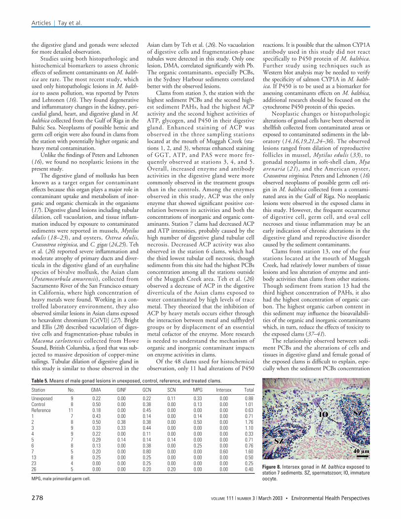
Testicular lesions. Testes of control clamsare shown in Figure 2. GMA, gonadal inflam-mation (GINF), GCN, SCN, and male pri-mordial germ cells were observed in testes ofmale clams used in this study (Table 5). Therewere no significant differences in the scores ofthese lesions among the unexposed, control,reference, and treated clams. In 1 of the 15clams exposed to the station 7 sediments, ahermaphroditic (intersex) gonad was detected(Figure 8).
Histochemical study. Three enzymes (ACP,ATP, and GGT), one antibody (CYP1A;P450), and a cytoplasmic glycogen (PAS) were
localized, and their staining intensities werecompared between the control and treatmentclams (Figure 9, Table 6). The control clamshave normal activities of ACP, GGT, andP450, and variable activities of ATP and PAS.Decreased activities of ACP (stations 6 and 7)(Figure 7) and ATP (station 7) only occurredin clams exposed to sediment samples from themiddle section of the south arm of the harbor.Increased and enhanced ACP activitiesoccurred more frequently in clams exposed tosediments collected from the mouth ofMuggah Creek, with the exception of station13. ACP was the only enzyme correlated sig-nificantly with sediment PAHs (r = 0.83, p =0.003), PCBs (r = 0.77, p = 0.01; Figure 10),and heavy metals (Ag, As, Cd, Cu, Hg, Pb andZn; r = 0.63–0.72; p = 0.01–0.04). Only 11 ofthe 48 clams used for histochemical observa-tion have increased activities of P450.
Discussion
In this study, we used a suite of histopatho-logic and histochemical biomarkers and asemiquantitative scoring system to assess thechronic effects of contaminated sediments onM. balthica in a controlled laboratory envi-ronment. A screening evaluation was con-ducted on major organs and tissues ofrandomly selected clams to identify lesionsthat can be used as indicators of biologiceffects. Based on the results of this evaluation,
Articles | Histopathologic and histochemical biomarkers in clams
Environmental Health Perspectives • VOLUME 111 | NUMBER 3 | March 2003 277
Figure 6. Female gonad lesions in M. balthicaexposed to Sydney Harbour sediments. (A) DINFand GCN in station 6. (B) GCN and SCN in station 1.(C) GCN and OCN in station 7. UOP, unidentifiedovarian parasite.
Table 4. Means of female gonad lesions in unexposed, control, reference, and treated clams.
Figure 7. Means of OCN (A) and ACP (B) in M. balthica exposed to Sydney Harbour sediments by location.
2 0 2km
0–0.300.30–0.600.60–0.900.90–1.17
OCN ACP
–0.25–00–0.250.25–0.500.50–0.750.75–1.00
A B
the digestive gland and gonads were selectedfor more detailed observation.
Studies using both histopathologic andhistochemical biomarkers to assess chroniceffects of sediment contaminants on M. balth-ica are rare. The most recent study, whichused only histopathologic lesions in M. balth-ica to assess pollution, was reported by Petersand Lehtonen (16). They found degenerativeand inflammatory changes in the kidney, peri-cardial gland, heart, and digestive gland in M.balthica collected from the Gulf of Riga in theBaltic Sea. Neoplasms of possible hemic andgerm cell origin were also found in clams fromthe station with potentially higher organic andheavy metal contamination.
Unlike the findings of Peters and Lehtonen(16), we found no neoplastic lesions in thepresent study.
The digestive gland of mollusks has beenknown as a target organ for contaminanteffects because this organ plays a major role incontaminant uptake and metabolism of inor-ganic and organic chemicals in the organisms(17). Digestive gland lesions including tubulardilation, cell vacuolation, and tissue inflam-mation induced by exposure to contaminatedsediments were reported in mussels, Mytilusedulis (18–23), and oysters, Ostrea edulis,Crassostrea virginica, and C. gigas (24,25). Tehet al. (26) reported severe inflammation andmoderate atrophy of primary ducts and diver-ticula in the digestive gland of an euryhalinespecies of bivalve mollusk, the Asian clam(Potamocorbula amurensis), collected fromSacramento River of the San Francisco estuaryin California, where high concentration ofheavy metals were found. Working in a con-trolled laboratory environment, they alsoobserved similar lesions in Asian clams exposedto hexavalent chromium [Cr(VI)] (27). Brightand Ellis (28) described vacuolation of diges-tive cells and fragmentation-phase tubules inMacoma carlottensis collected from HoweSound, British Columbia, a fjord that was sub-jected to massive deposition of copper-minetailings. Tubular dilation of digestive gland inthis study is similar to those observed in the
Asian clam by Teh et al. (26). No vacuolationof digestive cells and fragmentation-phasetubules were detected in this study. Only onelesion, DMA, correlated significantly with Pb.The organic contaminants, especially PCBs,in the Sydney Harbour sediments correlatedbetter with the observed lesions.
Clams from station 3, the station with thehighest sediment PCBs and the second high-est sediment PAHs, had the highest ACPactivity and the second highest activities ofATP, glycogen, and P450 in their digestivegland. Enhanced staining of ACP wasobserved in the three sampling stationslocated at the mouth of Muggah Creek (sta-tions 1, 2, and 3), whereas enhanced stainingof GGT, ATP, and PAS were more fre-quently observed at stations 3, 4, and 5.Overall, increased enzyme and antibodyactivities in the digestive gland were morecommonly observed in the treatment groupsthan in the controls. Among the enzymesobserved in this study, ACP was the onlyenzyme that showed significant positive cor-relation between its activities and both theconcentrations of inorganic and organic cont-aminants. Station 7 clams had decreased ACPand ATP intensities, probably caused by thehigh number of digestive gland tubular cellnecrosis. Decreased ACP activity was alsoobserved in the station 6 clams, which hadthe third lowest tubular cell necrosis, thoughsediments from this site had the highest PCBsconcentration among all the stations outsideof the Muggah Creek area. Teh et al. (26)observed a decrease of ACP in the digestivediverticula of the Asian clams exposed towater contaminated by high levels of tracemetal. They theorized that the inhibition ofACP by heavy metals occurs either throughthe interaction between metal and sulfhydrylgroups or by displacement of an essentialmetal cofactor of the enzyme. More researchis needed to understand the mechanism oforganic and inorganic contaminant impactson enzyme activities in clams.
Of the 48 clams used for histochemicalobservation, only 11 had alterations of P450
reactions. It is possible that the salmon CYP1Aantibody used in this study did not reactspecifically to P450 protein of M. balthica.Further study using techniques such asWestern blot analysis may be needed to verifythe specificity of salmon CYP1A in M. balth-ica. If P450 is to be used as a biomarker forassessing contaminants effects on M. balthica,additional research should be focused on thecytochrome P450 protein of this species.
Neoplastic changes or histopathologicalterations of gonad cells have been observed inshellfish collected from contaminated areas orexposed to contaminated sediments in the lab-oratory (14,16,19,21,24–36). The observedlesions ranged from dilation of reproductivefollicles in mussel, Mytilus edulis (33), togonadal neoplasms in soft-shell clam, Myaarenaria (21), and the American oyster,Crassostrea virginica. Peters and Lehtonen (16)observed neoplasms of possible germ cell ori-gin in M. balthica collected from a contami-nated area in the Gulf of Riga. No neoplasticlesions were observed in the exposed clams inthis study. However, the frequent occurrenceof digestive cell, germ cell, and oval cellnecroses and tissue inflammation may be anearly indication of chronic alterations in thedigestive gland and reproductive disordercaused by the sediment contaminants.
Clams from station 13, one of the fourstations located at the mouth of MuggahCreek, had relatively lower numbers of tissuelesions and less alteration of enzyme and anti-body activities than clams from other stations.Though sediment from station 13 had thethird highest concentration of PAHs, it alsohad the highest concentration of organic car-bon. The highest organic carbon content inthis sediment may influence the bioavailabili-ties of the organic and inorganic contaminantswhich, in turn, reduce the effects of toxicity tothe exposed clams (37–41).
The relationship observed between sedi-ment PCBs and the alterations of cells andtissues in digestive gland and female gonad ofthe exposed clams is difficult to explain, espe-cially when the sediment PCBs concentration
Articles | Tay et al.
278 VOLUME 111 | NUMBER 3 | March 2003 • Environmental Health Perspectives
Table 5. Means of male gonad lesions in unexposed, control, reference, and treated clams.
Figure 8. Intersex gonad in M. balthica exposed tostation 7 sediments. SZ, spermatozoon; IO, immatureoocyte.
was < 2 mg/kg. In fact, as demonstrated inFigure 5, there was no clear relationshipbetween these two variables in sediments with< 2 mg/kg PCBs. It is possible that the rela-tionship may be masked by the integratedeffects of the mixtures of inorganic andorganic contaminants in the Sydney Harboursediments. Figure 5 indicates that, when theconcentration of sediment PCBs increases to3 mg/kg, this relationship becomes muchmore apparent. Furthermore, there werelikely individual variations in sensitivity anduptake of contaminants within treatment. Itis, therefore, important that future body bur-den analysis focus on individual organismsinstead of pooled samples.
Most of the histopathologic studies onshellfish focused on female gonads. Recentstudies by Teh and associates (26,27) showedmild or moderate necrosis of spermatogoniaand moderate to severe granulomatous
inflammation in testis of the Asian clam P.amurensis. SCN and GCN, tissue inflamma-tion (GINF), and macrophage aggregation(GMA) were observed in the exposed malesin this study. However, because some of theselesions, especially GCN, SCN, and GMA,were also detected in the unexposed and con-trol males, it is difficult to associate the occur-rence of these lesions in the exposed clamswith sediment contaminants.
Overall, the results of this study showedthat digestive cell and germ cell necroses andinflammations in both digestive gland andgonad, especially the female gonad, were themost common histopathologic lesionsdetected in M. balthica exposed to SydneyHarbour sediments. At least one digestivegland lesion (TDA) and one female gonadallesion (OCN) correlated significantly withthe organic sediment contaminants. A lesserdegree of correlation existed between these
lesions and the inorganic sediment contami-nants. Among the histochemical biomarkersused in this study, only the activity of acidphosphatase was correlated with the concen-tration of sediment heavy metals, PAHs, andPCBs. Although no significant differences forATP, GGT, PAS, and P450 were observed inthis study, in general, they were more active inthe clams exposed to the test sediments thanthe control. The observed responses of thesehistopathologic and histochemical biomarkerswere a likely indication of chronic biologiceffects induced by exposure to a mixture ofinorganic and organic contaminants in thesediments. In addition to showing early indi-cation of environmental stress on the health ofthe test organisms, they can also be used tolocalize the target tissues and organs of sedi-ment contaminants in the affected individuals.When both biomarkers are used together, theyare effective tools for detecting chronic bio-logic effects of environmental contaminants.
The exposure conditions used in ourhistopathologic and histochemical evaluationwere identical to that used in the U.S. EPAbioaccumulation test method to quantify
Articles | Histopathologic and histochemical biomarkers in clams
Environmental Health Perspectives • VOLUME 111 | NUMBER 3 | March 2003 279
Figure 9. Alterations of the activities of enzyme and antibody in M. balthica in the control and SydneyHarbour sediments. (A) Normal ACP in digestive gland of control clams. (B) Enhanced ACP reaction indigestive gland of station 3 clams. (C) Decreased ATP reaction in digestive gland of station 7 clams. (D)Enhanced ATP reaction in digestive gland of station 4 clams. (E) Increased P450 staining in digestive glandof station 6 clams. (F) Enhanced GGT reaction in digestive gland of station 4 clams. The arrows indicatestaining of enzyme and antibody reactions.
Table 6. Means of enzyme and antibody scores(n = 4) in control, reference, and treated clams.
Figure 10. Relationship between means of ACPscore in M. balthica exposed to Sydney Harboursediments and the concentrations of PAHs (mg/kg;r = 0.83, p = 0.003) and PCBs (µg/kg ÷ 10; r = 0.77,p = 0.01) in the sediments.
1.2
0.8
0.4
0.0
–0.4
ACP
sco
re
0 100 200 300 400
PAHs (mg/kg)
PAHsPCBs
PCBs (µg/kg ÷ 10)
contaminant uptake in the tissue of M. balth-ica. The U.S. EPA method was designed topredict bioaccumulation of contaminants inindividuals or uptake of contaminants throughthe food chain; it cannot be used for predictingthe health of the test organisms. The suite ofhistopathologic and histochemical biomarkersidentified in this study can be used to comple-ment the U.S. EPA test method by expandingthe potential of the test results to includeidentification and localization of chroniceffects of the bioaccumulated contaminants inthe test organisms. Although preliminaryobservations in this study suggest correlationbetween some of the selected biomarker assaysand contaminant levels, regulatory acceptanceof their general use has not been provided bythis study. Logistical constraints of our fieldand laboratory programs limited the numberof samples, particularly the histochemical dataset, and thus constrained the power of our sta-tistical analysis. Additional studies are cur-rently being conducted to validate thesebiomarker responses against a suite ofapproved regulatory lethal and sublethal testsused by Environment Canada for the assess-ment of sediment contaminants.
REFERENCES
1. Sirota GR, Uthe JF, Sreedharan A, Matheson R, Musial CJ,Hamilton KG. Polynuclear aromatic hydrocarbons in lob-ster (Homarus americanus) and sediments in the vicinity ofa coking facility. In: Proceedings of the 5th InternationalSymposium on Polynuclear Aromatic Hydrocarbons:Chemical Analysis and Biological Effects (Cooke M, DennisAJ, eds). Columbus, OH:Battelle Press, 1983;1123–1136.
2. Sirota GR, Uthe JF, Robinson DG, Musial CJ. PolycyclicAromatic Hydrocarbons in American Lobster (Homarusamericanus) and Blue Mussels (Mytilus edulis) Collectedin the Area of Sydney Harbour, Nova Scotia. CanadianManuscript Report of Fisheries and Aquatic Sciences,No.1758. Dartmouth, Nova Scotia, Canada:Fisheries andOceans, 1984.
3. Wendland T. 1978 - 79 Sydney Harbour Survey: BenthicFauna Distribution and Related Studies. EnvironmentalTechnology Project. Sydney, Nova Scotia, Canada:Collegeof Cape Breton, 1979.
4. Hildebrand LP. Environmental Quality in Sydney andNortheast Industrial Cape Breton, Nova Scotia.Environmental Protection Service Surveillance ReportEPS-5-AR-82-3. Dartmouth, Nova Scotia, Canada:AtlanticRegion, Environment Canada, 1982.
5. Matheson RAF, Trider GL, Ernst WR, Hamilton KG, HennigarPA. Investigation of Polynuclear Aromatic HydrocarbonContamination of Sydney Harbour, Nova Scotia.Environmental Protection Service Surveillance Report EPS-5-AR-83-6. Dartmouth, Nova Scotia, Canada:AtlanticRegion, Environment Canada, 1983.
6. Kieley KM, Hennigar PA, Matheson RAF, Ernst WR.Polynuclear Aromatic Hydrocarbons and HeterocyclicAromatic Compounds in Sydney Harbour, Nova Scotia: A1986 survey. Environmental Protection ServiceSurveillance Report EPS-5-AR-88-7. Dartmouth, NovaScotia, Canada:Atlantic Region, Environment Canada, 1988.
7. Vandermeulen JH. PAH and Heavy Metal Pollution of theSydney Estuary: Summary and Review of Studies to 1987.Canadian Technical Report, Hydrography and OceanSciences, No. 108. Dartmouth, Nova Scotia, Canada:Fisheriesand Oceans, 1989.
8. Gearing JN, Buckley DE, Smith JN. Hydrocarbon andmetal contents in a sediment core from Halifax Harbour: achronology of contamination. Can J Fish Aquat Sci48:2344–2354 (1991).
9. Stewart PL, White L. A Review of Contaminants on theScotian Shelf and in Adjacent Coastal Waters: 1970-1995.Canadian Technical Report of Fisheries and AquaticSciences, No. 2351. Dartmouth, Nova Scotia, Canada:Fisheries and Oceans, 2001.
10. U.S. EPA. Guidance Manual - Bedded SedimentBioaccumulation Tests. EPA/600/R-93/183. Washington,DC:U.S. Environmental Protection Agency, Office ofResearch and Development, 1993.
11. Hinton DE. Histopathologic biomarkers. In: Biomarkers –Biochemical, Physiological, and Histological Markers ofAnthropogenic Stress (Huggett FJ, Kimerle RA, MehrlePMJr, Bergman HL, eds). SETAC Special PublicationSeries. Boca Raton, FL:Lewis Publishers,1992;155–209.
12. Gardner GR. Chemically induced histopathology in aquaticinvertebrates. In: Pathobiology of Marine and EstuarineOrganisms. Advances in Fisheries Science (Couch JA,Fournie JW, eds). Boca Raton, FL:CRC Press, 1993;359–391.
13. Environment Canada. Users Guide to the Application Formfor Ocean Disposal. Environmental Protection Series,Report EPS 1/MA/1. Ottawa, Ontario, Canada:EnvironmentCanada, 1995.
14. Teh SJ, Hinton DE. Detection of enzyme histochemicalmarkers of hepatic preneoplasia and neoplasia in medaka(Oryzias latipes). Aquat Toxicol 24:163–182 (1993).
15. Tay KL, Doe KG, Wade SJ, Vaughan DA, Berrigan RE,Moore MJ. Sediment bioassessment in Halifax Harbour.Environ Toxicol Chem 11:1567–1581 (1992).
16. Peters EC, Lehtonen KK. Chemical and other stressors inthe Gulf of Riga: interpreting multiple lesions in the clamMacoma balthica . In: Abstracts of the NationalShellfisheries Association Annual Meeting, 20–24 April1997, Fort Walton Beach, Florida. Hanover, PA:NationalShellfisheries Association, 1997;351.
17. Rainbow PS. The significance of trace metal concentra-tions in marine invertebrates. In: Ecotoxicology of Metalsin Invertebrates (Dallinger R, Rainbow PS, eds). SETACSpecial Publications Series. Boca Raton, FL:LewisPublishers, 1993:3–24.
18. Auffret M. Histopathological changes related to chemicalcontamination in Mytilus edulis from field and experimen-tal conditions. Mar Ecol Prog Ser 46:101–107 (1988).
19. Yevich PP, Yevich C, Pesch G. Effects of Black RockHarbour Dredged Material on the Histopathology of theBlue Mussel Mytilus edulis and Polychaete Worm NephtysIncisa after Laboratory and Field Exposures. TechnicalReport D-87-8. Vicksburg, MS:U.S. Army EngineerWaterways Experiment Stations, 1987.
20. Lowe DM. Alterations in cellular structure of Mytilusedulis resulting from exposure to environmental contami-nants under field and experimental conditions. Mar BiolProg Ser 46:91–100 (1988).
21. Yevich PP, Barszcz CA. Histopathology as monitor formarine pollution. Results of the histopathologic examina-tion of the animals collected for the US 1976 MusselWatch Program. Rapp P-V Réun Cons Int Explor Mer182:96–102 (1983).
22. Yevich PP, Yevich CA. Use of histopathology in biomoni-toring marine invertebrates. In: Biomonitoring of CoastalWaters and Estuaries (Kramer KJM, ed). Boca Raton,FL:CRC Press, 1998;179–204.
23. Nott JA, Moore MN. Effects of polycyclic aromatic hydro-carbons on molluscan lysosomes and endoplasmic reticu-lum. Histochem J 19:357–368 (1987).
24. Berthou F, Balouet G, Bodennec G, Marchand M. The
occurrence of hydrocarbons and histopathological abnor-malities in oysters for seven years following the wreck ofthe Amoco Cadiz in Brittany (France). Mar Environ Res23:103–133 (1987).
25. Gardner GR, Yevich PP, Hareshbarger JC, Malcolm AR.Carcinogenicity of Black Rock Harbor sediment to theeastern oyster and trophic transfer of Black Rock Harborcarcinogens from the blue mussel to the winter flounder.Environ Health Perspect 90:53–66 (1990).
26. Teh SJ, Clark SL, Brown CL, Luoma SN, Hinton DE.Enzymatic and histopathologic biomarkers as indicators ofcontaminant exposure and effect in Asian clam(Potamocorbula amurensis). Biomarkers 4(6):497–509 (1999).
27. Teh SJ, Werner I, Hinton DE. Sublethal effects ofchromium-VI in the Asian clam (Potamocorbula amuren-sis). Mar Environ Res 50:295–300 (2000).
28. Bright DA, Ellis DV. Aspects of histology in Macoma car-lottensis (Bivalvia: Tellinidae) and in situ histopathologyrelated to mine-tailings discharge. Mar Biol Assoc U.K69:447–464 (1989).
29. Yevich PP, Barry MM. Ovarian tumors in the quahogMercenaria mercenaria. J Invertebr Pathol 14:266–267(1969).
30. Barry MM, Yevich PP. Incidence of gonadal cancer in thequahog Mercenaria mercenaria. Oncology 26:87–96 (1972).
31. Walker HA, Lorda E, Saila SB. A comparison of the inci-dence of five pathological conditions in soft-shell clams,Mya arenaria, from environments with various pollutionhistories. Mar Environ Res 5:109–123 (1981).
32. Gardner GR, Pruell RJ. Quincy Bay Study, Boston Harbor: AHistopathological and Chemical Assessment of WinterFlounder, Lobster and Soft-Shelled Clam Indigenous toQuincy Bay, Boston Harbor and an in Situ Evaluation ofOysters Including Sediment (Surface and Cores) Chemistry.Boston, MA:U.S. Environmental Protection Agency,Region 1, 1988.
33. Sunila I. Histopathology of mussels (Mytilus edulis L.) fromthe Tvarminne area, the Gulf of Finland (Baltic Sea). AnnZool Fenn 24:55–69 (1987).
36. LeBlanc GA, Bain LJ. Chronic toxicity of environmentalcontaminants: sentinels and biomarkers. Environ HealthPerspect 105(suppl 1):65–80 (1997).
37. Landrum PF, Nihart SR, Eadie FJ, Herche LR. Reduction inbioavailability of organic contaminants to the amphipodPontoporeia hoyi by dissolved organic matter of sedimentinterstitial waters. Environ Toxicol Chem 6:11–20 (1987).
38. Day KE. Effects of dissolved organic carbon on accumula-tion and acute toxicity of fenvalerate, deltamethrin andcyhalothrin to Daphnia magna (Straus). Environ ToxicolChem 10:91–101 (1991).
39. DeWitt TH, Ozretich RJ, Swartz RC, Lamberson JO,Schults DW, Ditsworth GR, Jones JKP, Hoselton L, SmithLM. The influence of organic matter quality on the toxicityand partitioning of sediment-associated fluoranthene.Environ Toxicol Chem 11:197–208 (1992).
40. Weston DP, Mayer LM. In vitro digestive fluid extractionas a measure of the bioavailability of sediment-associatedpolycyclic aromatic hydrocarbons: sources of variationand implications for partitioning models. Environ ToxicolChem 17(5):820–829 (1998).
41. Gensemer RW, Dixon DG, Greenberg BM. Amelioration ofthe photo-induced toxicity of polycyclic aromatic hydro-carbons by a commercial humic acid. Ecotoxicol EnvironSaf 39:57–64 (1998).
Articles | Tay et al.
280 VOLUME 111 | NUMBER 3 | March 2003 • Environmental Health Perspectives