lhe management 01 pain in dentistry requires a thorough knowledge of the fifth cranial nerve (Fig. 12-1). The right and left trigcminal nerves provide, among other func- tions, the overwhelming majority of sensory innervation from ilie teeth, bone, and soft tissues of the oral cavity. The trigeminal nerve is also the largest cranial nerve. It is composed of a small motor rool and a considerably larg- er (tripartite) sensory root. The motor root supplies the muscles of mastication and other muscles in the region. The three branches of the sensory root supply the skin of the entire face and the mucous membrane of the cranial viscera and oral cavity, except for the pharynx and the base of the tongue. Table 12-1 summarizes the functions of the trigeminal and the 11 other cranial nerves. Motor Root The motor root of the trigeminal nerve arises separately from the sensory root, originating in the motor nucleus within the pons and medulla oblongata (Fig. 12-2). Its libers, forming a small nerve root, travel anteriorly along with, but entirely separate from, the larger sensory root to the region of the semilunar (or gasserian) ganglion. At the semilunar ganglion the motor root passes in a later- al and inferior direction under the ganglion toward the foramen ovale, through which it leaves the middle cra- nial fossa along with the third division of the sensory root, the mandibular nerve (Figs. 12-3 and 12-4). Just after leaving the skull, the motor root unites with the sensory root of the mandibular division to form a single nerve trunk. Motor fibers of the trigeminal nerve supply the fol- lowing muscles: 1. Masticatory a. Massetcr b. Temporalis e. Pterygoideus medialis d. Pterygoideus lateralis 2. Mylohyoid 3. Anterior belly of the digastric 4. Tensor tympani 5. Tensor veli palatini Sensory Root Sensory root fibers of the trigeminal nerve comprise the central processes of ganglion cells located in the trigem- inal (semilunar or gasserian) ganglion. There are two gan- glia, one innervating each side of the face. They are locat- ed in Meckels cavity, on the anterior surface of the petrous portion of die temporal bone (Fig. 12-3). The ganglia are flat and crescent shaped, their convexities fac- ing anteriorly and downward, and they measure approx- imately 1.0 X 2.0 cm. Sensory root fibers enler the con- cave portion of each crescent, and the three sensory divi- sions of the trigeminal nerve exit from the convexity: 1. The ophthalmic division (V } ) travels anteriorly in the lateral wall of the cavernous sinus to the medial part of the superior orbital fissure, through which it exits the skull into the orbit. 2. The maxillary division (V 2 ) travels anteriorly and downward to exit the cranium through the foramen 143
Transcript
lhe management 01 pain in dentistry requires a thoroughknowledge of the fifth cranial nerve (Fig. 12-1). The rightand left trigcminal nerves provide, among other func-tions, the overwhelming majority of sensory innervationfrom ilie teeth, bone, and soft tissues of the oral cavity.The trigeminal nerve is also the largest cranial nerve. It iscomposed of a small motor rool and a considerably larg-er (tripartite) sensory root. The motor root supplies themuscles of mastication and other muscles in the region.The three branches of the sensory root supply the skin ofthe entire face and the mucous membrane of the cranialviscera and oral cavity, except for the pharynx and thebase of the tongue. Table 12-1 summarizes the functionsof the trigeminal and the 11 other cranial nerves.
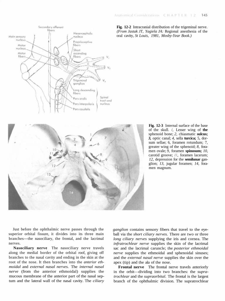
Motor RootThe motor root of the trigeminal nerve arises separatelyfrom the sensory root, originating in the motor nucleuswithin the pons and medulla oblongata (Fig. 12-2). Itslibers, forming a small nerve root, travel anteriorly alongwith, but entirely separate from, the larger sensory rootto the region of the semilunar (or gasserian) ganglion. Atthe semilunar ganglion the motor root passes in a later-al and inferior direction under the ganglion toward theforamen ovale, through which it leaves the middle cra-nial fossa along with the third division of the sensoryroot, the mandibular nerve (Figs. 12-3 and 12-4). Justafter leaving the skull, the motor root unites with thesensory root of the mandibular division to form a singlenerve trunk.
Motor fibers of the trigeminal nerve supply the fol-lowing muscles:
2. Mylohyoid3. Anterior belly of the digastric4. Tensor tympani5. Tensor veli palatini
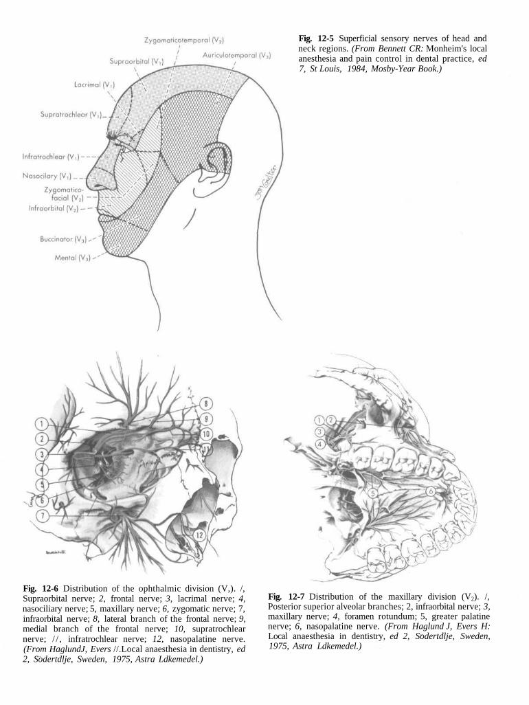
Sensory RootSensory root fibers of the trigeminal nerve comprise thecentral processes of ganglion cells located in the trigem-inal (semilunar or gasserian) ganglion. There are two gan-glia, one innervating each side of the face. They are locat-ed in Meckels cavity, on the anterior surface of thepetrous portion of die temporal bone (Fig. 12-3). Theganglia are flat and crescent shaped, their convexities fac-ing anteriorly and downward, and they measure approx-imately 1.0 X 2.0 cm. Sensory root fibers enler the con-cave portion of each crescent, and the three sensory divi-sions of the trigeminal nerve exit from the convexity:
1. The ophthalmic division (V}) travels anteriorly inthe lateral wall of the cavernous sinus to the medialpart of the superior orbital fissure, through which itexits the skull into the orbit.
2. The maxillary division (V2) travels anteriorly anddownward to exit the cranium through the foramen
143
Fig. 12-1 Distribution of the trigeminal nerve./, The branches are as follows: 2, gasserianganglion; 3, mandibular nerve and foramenovale; 4, maxillary nerve and foramen rotun-dum;5, ophthalmic nerve and superior orbitalfissure; 6, nasociliary nerve; 7, frontal nerve; 8,lacrimal nerve; 9, supraorbital nerve; 10,supratrochlear nerve; / /, zygomatic nerve; 12,anterior superior alveolar branches; 13, poste-rior superior alveolar branches; 14, buccalnerve; 15, posterior nasal branches; 16,greater palatine nerve; 17, infraorbital nerve;18, nasopalatine nerve; 19, auriculotcmporalnerve; 20, lingual nerve; 21, inferior alveolarnerve; 22, mental nerve. (From Haglund J,Evers H: Local anaesthesia in dentistry, ed 2,Sodertdlje, Sweden, 1975, Astra Lakemedel.)
On exiting the cranium through their respectiveforamina,* the three divisions of the trigeminal nervedivide into a multitude of sensory branches.
• • •Each of the three divisions of the trigeminal nerve is
described, but more attention will be devoted to themaxillary and mandibular divisions because of theirgreater importance in pain control in dentistry. Figure12-5 illustrates the sensory distribution of the trigeminalnerve.
Ophthalmic Division (V7)The ophthalmic division is the first branch of the trigem-inal nerve. It is exclusively sensory and is the smallest ofthe three divisions. It leaves the cranium and enters theorbit through the superior orbital fissure (Fig. 12-6). Thenerve trunk is approximately 2.5 cm long. It supplies the
rotundum into the upper portion of the pterygopala-tine fossa.
3. The mandibular division(V$) travels almost directlydownward to exit the skull, along with the motorroot, through the foramen ovale. These two rootsthen intermingle, forming one nerve trunk thatenters the infratemporal fossa.
eyeball, conjunctiva, lacrimal gland, parts of the mucousmembrane of the nose and paranasal sinuses, and theskin of the forehead, eyelids, and nose. When the oph-thalmic nerve (Vx) is paralyzed, the ocular conjunctivabecomes insensitive to touch.
*To be somewhat more precise, since the three branches of the trigem-inal nerve are almost exclusively sensory, they do not exit from the cra-nium but rather enter into the cranium, carrying nerve impulses fromthe periphery toward the brain.
Fig. 12-2 Intracranial distribution of the trigeminal nerve.(From Jastak JT, Yagiela JA: Regional anesthesia of theoral cavity, St Louis, 1981, Mosby-Year Book.)
Fig. 12-3 Internal surface of the baseof the skull. /, Lesser wing of thesphenoid bone; 2, chiasmatic sulcus;3, optic canal; 4, sella turcica; 5, dor-sum sellae; 6, foramen rotundum; 7,greater wing of the sphenoid; 8, fora-men ovale; 9, foramen spinosum; 10,carotid groove; / / , foramen lacerum;12, depression for the semilunar gan-glion; 13, jugular foramen; 14, fora-men magnum.
Just before the ophthalmic nerve passes through thesuperior orbital fissure, it divides into its three mainbranches—the nasociliary, the frontal, and the lacrimalnerves.
Nasociliary nerve The nasociliary nerve travelsalong the medial border of the orbital roof, giving offbranches to the nasal cavity and ending in the skin at theroot of the nose. It then branches into the anterior eth-moidal and external nasal nerves. The internal nasalnerve (from the anterior ethmoidal) supplies themucous membrane of the anterior part of the nasal sep-tum and the lateral wall of the nasal cavity. The ciliary
ganglion contains sensory fibers that travel to the eye-ball via the short ciliary nerves. There are two or threelong ciliary nerves supplying the iris and cornea. Theinfratrochlear nerve supplies the skin of the lacrimalsac and the lacrimal caruncle; the posterior ethmoidalnerve supplies the ethmoidal and sphenoidal sinuses;and the external nasal nerve supplies the skin over theapex (tip) and the ala of the nose.
Frontal nerve The frontal nerve travels anteriorlyin the orbit—dividing into two branches: the supra-trochlear and the supraorbital. The frontal is the largestbranch of the ophthalmic division. The supratrochlear
Fpge8
naanb
bp
MTfis
fd
greater wing of the sphenoid bone. Once outside the cra-nium, the maxillary nerve crosses the uppermost part ofthe pterygopalatine fossa, between the pterygoid platesof the sphenoid bone and the palatine bone. As it cross-es the pterygopalatine fossa, it gives off branches to thesphenopalatine ganglion, the posterior superior alveolarnerve, and the zygomatic branches. It then angles lateral-ly in a groove on the posterior surface of the maxilla,entering the orbit through the inferior orbital fissure.Within the orbit it occupies the infraorbital groove andbecomes the infraorbital nerve, which courses anteriorlyinto the infraorbital canal.
The maxillary division emerges on the anterior sur-face of the face through the infraorbital foramen,where it divides into its terminal branches, supplyingthe skin of the face, nose, lower eyelid, and upper lip(Fig. 12-7). Following is a breakdown of maxillary divi-sion innervation;
1. Skin ofa. Middle portion of the faceb. Lower eyelidc. Side of the nosed. Upper lip
ig. 12-4 Inferior surface of the base of the skull. 1, Palatalrocess of the maxilla; 2, horizontal process of the palate; 3,reater palatine foramen; 4, lesser palatine foramina; 5, lat-ral pterygoid plate; 6, foramen ovale; 7, foramen spinosum;, foramen lacerum;9, carotid canal; 10, foramen magnum.
erve supplies the conjunctiva and skin of the medialspect of the upper eyelid and the skin over the lowernd mesial aspects of the forehead. The supraorbitalerve is sensory to the upper eyelid, to the scalp as farack as the parietal bone, and to the lambdoidal suture.
Lacrimal nerve The lacrimal nerve is the smallestranch of the ophthalmic division. It supplies the lateralart of the upper eyelid and a small adjacent area of skin.
axillary Division (V2)he maxillary division of the trigeminal nerve arises
rom the middle of the trigeminal ganglion. Intermediaten size between the ophthalmic and mandibular divi-ions, it is purely sensory in function.
Origins The maxillary nerve passes horizontallyorward, leaving the cranium through the foramen rotun-um (Fig. 12-3). The foramen rotundum is located in the
e. Hard palate3- Maxillary teeth and periodontal tissues
Branches The maxillary division gives off branchesin four regions—within the cranium, in the pterygopala-tine fossa, in the infraorbital canal, and on the face.
Branch within the cranium Immediately afterseparating from the trigeminal ganglion, the maxillarydivision gives off a small branch, the middle meningealnerve, that travels with the middle meningeal artery toprovide sensory innervation to the dura mater.
Branches in the pterygopalatine fossa Afterexiting the cranium through the foramen rotundum, themaxillary division crosses the pterygopalatine fossa. Inthis fossa several branches are given off (Fig. 12-8)—thezygomatic nerve, the pterygopalatine nerves, and theposterior superior alveolar nerve.
The zygomatic nerve comes off the maxillary divisionin the pterygopalatine fossa and travels anteriorly, enter-ing the orbit through the inferior orbital fissure, where itdivides into the zygomaticotemporal and zygomaticofa-cial nerves—the zygomaticotemporal supplying senso-ry innervation to the skin on the side of the forehead,and the zygomaticofacial supplying the skin on theprominence of the cheek. Just before leaving the orbitthe zygomatic nerve sends a branch that communicateswith the lacrimal nerve of the ophthalmic division. This
Fig. 12-5 Superficial sensory nerves of head andneck regions. (From Bennett CR: Monheim's localanesthesia and pain control in dental practice, ed7, St Louis, 1984, Mosby-Year Book.)
Sn
mn
2
F
i
(
ig. 12-6 Distribution of the ophthalmic division (V,). /,upraorbital nerve; 2, frontal nerve; 3, lacrimal nerve; 4,asociliary nerve; 5, maxillary nerve; 6, zygomatic nerve; 7,nfraorbital nerve; 8, lateral branch of the frontal nerve; 9,edial branch of the frontal nerve; 10, supratrochlear
12-7 Distribution of the maxillary division (V2). /,erior superior alveolar branches; 2, infraorbital nerve; 3,illary nerve; 4, foramen rotundum; 5, greater palatinee; 6, nasopalatine nerve. (From Haglund J, Evers H:l anaesthesia in dentistry, ed 2, Sodertdlje, Sweden,, Astra Ldkemedel.)
Fig. 12-8 Branches of V2 in the pterygopalatinefossa. /, Maxillary nerve; 2, posterior superior alve-olar branches. (From Haglund J, Evers H: Localanaesthesia in dentistry, ed 2, Sodertdlje, Sweden,1975, Astra Ldkemedel.)
branch carries secretory fibers from the sphenopalatineganglion to the lacrimal gland.
The pterygopalatine nerves are two short trunks thatunite in the pterygopalatine ganglion and are then redis-tributed into several branches. They also serve as a com-munication between the pterygopalatine ganglion andthe maxillary nerve (V2)- Postganglionic secretomotorfibers from the pterygopalatine ganglion pass throughthese nerves and back along V2 to the zygomatic nerve,through which they are routed to the lacrimal nerve andlacrimal gland.
Branches of the pterygopalatine nerves include thosethat supply four areas—the orbit, the nose, the palate,and the pharynx.
1. The orbital branches supply the periosteum of theorbit.
2. The nasal branches supply the mucous membranesof the superior and middle conchae, the lining of theposterior ethmoidal sinuses, and the posterior por-tion of the nasal septum. One branch is significant indentistry, the nasopalatine nerve, which passesacross the roof of the nasal cavity downward andforward, where it lies between the mucous mem-brane and the periosteum of the nasal septum. Thenasopalatine nerve continues downward, reachingthe floor of the nasal cavity and giving branches tothe anterior part of the nasal septum and the floor ofthe nose. It then enters the incisive canal, throughwhich it passes into the oral cavity via the incisiveforamen, located in the midline of the palate about 1cm posterior to the maxillary central incisors. The
right and left nasopalatine nerves emerge togetherthrough this foramen and provide sensation to thepalatal mucosa in the region of the premaxilla(canines through central incisors) (Fig. 12-9).The palatine branches are the greater (or anterior)palatine nerve and the lesser (middle and posterior)palatine nerves (Fig. 12-10). The greater (or anteri-or) palatine nerve descends through the ptery-gopalatine canal, emerging on the hard palatethrough the greater palatine foramen (which is usu-ally located about 1 cm toward the palatal midline,just distal to the second molar). Sicher and DuBrulhave stated that the greater palatine foramen maybe located 3 to 4 mm in front of the posterior bor-der of the hard palate.1 The nerve courses anterior-ly between mucoperiosteum and the osseous hardpalate, supplying sensory innervation to the palatalsoft tissues and bone anterior to the first premolar,where it communicates with terminal fibers of thenasopalatine nerve (Fig. 12-10). It also provides sen-sory innervation to some parts of the soft palate.The middle palatine nerve emerges from the lesserpalatine foramen, along with the posterior palatinenerve. The middle palatine nerve provides sensoryinnervation to the mucous membrane of the softpalate; the tonsillar region is innervated, in part, bythe posterior palatine nerve.The pharyngeal branch is a small nerve that leaves theposterior part of the pterygopalatine ganglion, passesthrough the pharyngeal canal, and is distributed to themucous membrane of the nasal part of the pharynx,posterior to the auditory (eustachian) tube.
Fig. 12-9 Nerves of the nasal sep-tum. (From Bennett CR: Monheim'slocal anesthesia and pain control indental practice, ed 7, St Louis, 1984,Mosby- Year Book.)
The posterior descends from thein the pterygopaldivision enters Commonly there a
a single trunk will arise. Passing downward through thepterygopalatine fossa they reach the inferior temporal(posterior) surface of the maxilla. When two trunks arepresent, one remains external to the bone, continuingdownward on the posterior surface of the maxilla to pro-vide sensory innervation to the buccal gingiva in themaxillary molar region and adjacent facial mucosal sur-faces, while the other branch enters into the maxilla(along with a branch of the internal maxillary artery)through the posterior superior alveolar canal to traveldown the posterior or posterolateral wall of the maxil-lary sinus and provide sensory innervation to themucous membrane of the sinus. Continuing downward,this second branch of the PSA provides sensory innerva-tion to the alveoli, periodontal ligaments, and pulpal tis-sues of the maxillary third, second, and first molars (withthe exception [in 28% of patients2] of the mesiobuccalroot of the first molar).
Branches in the infraorbital canal Within theinfraorbital canal the maxillary division (V2) gives offtwo branches of significance in dentistry, the middlesuperior and anterior superior alveolar nerves. While inthe infraorbital groove and canal, the maxillary divisionis known as the infraorbital nerve.
superior alveolar (PSA) nerve main trunk of the maxillary divisionatine fossa just before the maxillarythe infraorbital canal (Fig. 12-11).re two PSA branches, but on occasion
The middle superior alveolar (MSA) nerve branch-es off the main nerve trunk (V2 ) within the infraorbitalcanal to form a part of the superior dental plexus,1
composed of the posterior, middle, and anterior superi-or alveolar nerves. The site of origin of the MSA nervevaries, from the posterior portion of the infraorbitalcanal to the anterior portion, near the infraorbital fora-men. The MSA nerve provides sensory innervation tothe two maxillary premolars and, perhaps, to themesiobuccal root of the first molar as well as the peri-odontal tissues, buccal soft tissue, and bone in the pre-
Fig. 12-11 Posterolateral view of the max-illa illustrating the posterior superior alve-olar (PSA) nerves on the posterior aspectof the maxillary tuberosity (/). Injectingthe PSA nerves provides pulpal anesthesiato the first, second, and third molars(except the mesiobuccal root of the firstmolar).
FovsuanL1
mMmfoim
are provided by either the PSA or the ASA nerves, mostfrequently the latter.'
The anterior superior alveolar (ASA) nerve, a rela-tively large branch, is given off the infraorbital nerve (V2 )approximately 6 to 10 mm before the latter's exit fromthe infraorbital foramen. Descending within the anteriorwall of the maxillary sinus, it provides pulpal innervationto the central and lateral incisors and the canine as wellas sensory innervation to the periodontal tissues, buccalbone, and mucous membranes of these teeth (Fig. 12-12).
The ASA nerve communicates with the MSA and givesoff a small nasal branch that innervates the anterior partof the nasal cavity, along with branches of the ptery-gopalatine nerves. In persons without an MSA nerve theASA frequently provides sensory innervation to the pre-molars and occasionally the mesiobuccal root of the firstmolar.
The actual innervation of individual roots of all teeth,bone, and periodontal structures in both the maxilla andthe mandible derives from terminal branches of largernerves in the region. These nerve networks are termed
ig. 12-12 Anterior superior alveolar (ASA) nerve (boneer the nerves removed). /, Branches of the ASA nerve; 2,perior dental plexus; 3, dental branches; 4, interdentald interradicular branches. (From Haglund J, Evers H:
ocal anaesthesia in dentistry, ed 2, Sddertdlje, Sweden,975, Astra Ldkemedel.)
olar region. Traditionally it has been stated that theSA nerve is absent in 3O%3 to 54%4 of individuals. In aore recent dissection study Loetscher and Walton2
und the MSA nerve to be present in 72% of the spec-ens examined. In its absence its usual innervations
the dental plexus.The superior dental plexus is composed of smaller
nerve fibers from the three superior alveolar nerves (andin the mandible, from the inferior alveolar nerve). Threetypes of nerves emerge from these plexuses—dentalnerves, interdental branches, and interradicular branch-es—and each is accompanied along its pathway by a cor-responding artery.
The dental nerves are those that enter a tooththrough the apical foramen, dividing into many smallbranches within the pulp. Pulpal innervation of all teethis derived from dental nerves. Although in mostinstances one easily identifiable nerve is responsible, in
some cases (usually the maxillary first molar) more thanone nerve will be.
The interdental branches (also termed perforatingbranches) travel through the entire height of the inter-radicular septum, providing sensory innervation to theperiodontal ligaments of adjacent teeth through the alve-olar bone. They emerge at the height of the crest of theinteralveolar septum and enter the gingiva to innervatethe interdental papillae and buccal gingiva.
The interradicular branches traverse the entireheight of the interradicular or interalveolar septum, pro-viding sensory innervation to the periodontal ligamentsof adjacent roots. They terminate in the periodontal liga-ment (PDL) at the root furcations.
Branches on the face Through the infraorbitalforamen the infraorbital nerve emerges onto the face todivide into its terminal branches—the inferior palpebral,the external nasal, and the superior labial. The inferiorpalpebral branches supply the skin of the lower eyelidwith sensory innervation, the external nasal branchesprovide sensory innervation to the skin on the lateralaspect of the nose, and the superior labial branches pro-vide sensory innervation to the skin and mucous mem-branes of the upper lip.
Although anesthesia of these nerves is not necessaryfor adequate pain control during dental treatment, theyare frequently blocked in the process of carrying outother anesthetic procedures.
Summary Following is a summary of the branchesof the maxillary division (italicized nerves denote thoseof special significance in dental pain control):
1. Branches within the craniuma. Middle meningeal nerve
2. Branches within the pterygopalatine fossaa. Zygomatic nerve
b. Pterygopalatine nerves(1) Orbital branches(2) Nasal branches
(a) Nasopalatine nerve(3) Palatine branches
(a) Greater (anterior palatine nerve)(b) Lesser (middle and posterior) palatine
nerves(4) Pharyngeal branch
c. Posterior superior alveolar nerve3. Branches within the infraorbital canal
a. Middle superior alveolar nerveb. Anterior superior alveolar nerve
4. Branches on the facea. Inferior palpebral branchesb. External nasal branchesC. Superior labial branches
Mandibular Division (V3)
The mandibular division is the largest branch of thetrigeminal nerve. It is a mixed nerve with two roots—alarge sensory root and a smaller motor root (the latterrepresenting the entire motor component of the trigem-inal nerve). The sensory root of the mandibular divisionoriginates at the inferior angle of the trigeminal ganglion,whereas the motor root arises in motor cells located inthe pons and medulla oblongata. The two roots emergefrom the cranium separately through the foramen ovale,the motor root lying medial to the sensory. They unitejust outside the skull and form the main trunk of thethird division. This trunk remains undivided for only 2 to3 mm before it splits into a small anterior and a large pos-terior division (Fig. 12-13).
Fig. 12-13 Distribution of the mandibular division (V3). /,Trigeminal nerve; 2, gasserian ganglion; 3. mandibularnerve; 4, buccal nerve; 5, maxillary nerve; 6, ophthalmic-nerve; 7, auriculotemporal nerve; 8, inferior alveolar nerve;9, lingual nerve; 10, mental nerve. (From HaglundJ, Evers//.Local anaesthesia in dentistry, ed 2, Sodertdlje, Sweden,1975, Astra Ldkemedel.)
1. Sensory roota. Skin of
(1) Temporal region(2) Auricula(3) External auditory meatus(4) Cheek(5) Lower lip(6) Lower part of the face (chin region)
b. Mucous membrane of(1) Cheek(2) Tongue (anterior two thirds)(3) Mastoid cells
c. Mandibular teeth and periodontal tissuesd. Bone of the mandiblee. Temnoromandibular jointf. Parotid gland
b. Mylohyoidc. Anterior belly of the digastricd. Tensor tympanie. Tensor veli palatini
Branches The third division of the trigeminal nervegives off branches in three areas—from the undividednerve, and from the anterior and posterior divisions.
Branches from the undivided nerve On leavingthe foramen ovale the main undivided nerve trunk givesoff two branches during its 2 to 3 mm course. These arethe nervus spinosus (meningeal branch of the mandibu-lar nerve) and the medial pterygoid nerve. The nervusspinosus reenters the cranium through the foramenspinosum along with the middle meningeal artery to sup-ply the dura mater and mastoid air cells. The medialpterygoid nerve is a motor nerve to the medial (internal)pterygoid muscle. It gives off small branches that aremotor to the tensor veli palatini and tensor tympani.
Branches from the anterior division Branchesfrom the anterior division of V3 provide motor innerva-tion to the muscles of mastication and sensory innerva-tion to the mucous membrane of the cheek and buccalmucous membrane of the mandibular molars.
The anterior division is significantly smaller than theposterior. It runs forward under the lateral (external)pterygoid muscle for a short distance and then reachesthe external surface of that muscle by either passingbetween its two heads or, less frequently, winding overits upper border. From this point it is known as the buc-cal nerve. While under the lateral pterygoid muscle, the
buccal nerve gives off several branches—the deep tem-poral nerves (to the temporal muscle) and the masseterand lateral pterygoid nerves (providing motor innerva-tion to the respective muscles).
The buccal nerve, also known as the buccinator nerveand the long buccal nerve, usually passes between the twoheads of the lateral pterygoid to reach the external surfaceof that muscle (Fig. 12-13). It then follows the inferior partof the temporal muscle and emerges under the anteriorborder of the masseter muscle, continuing in an anterolat-eral direction. At the level of the occlusal plane of themandibular third or second molar it crosses in front of theanterior border of the ramus and enters the cheekthrough the buccinator muscle. Sensory fibers are distrib-uted to the skin of the cheek. Other fibers pass into theretromolar triangle, providing sensory innervation to thebuccal gingiva of the mandibular molars and themucobuccal fold in that region.The buccal nerve does notinnervate the buccinator muscle; the facial nerve does.Nor does it provide sensory innervation to the lower lipor corner of the mouth. This is significant, because somedoctors do not administer the "long" buccal injection fol-lowing inferior alveolar nerve block until the lower lip hasbecome numb. Their thinking is that the long buccalnerve block will provide anesthesia of the lower lip andtherefore might falsely lead them to believe that their infe-rior alveolar nerve block has been successful, when in factit has been missed. Such concern is unwarranted. The longbuccal nerve block should be administered immediate-ly following inferior alveolar nerve block.
Anesthesia of the buccal nerve is important for dentalprocedures requiring soft tissue manipulation on thebuccal surface of the mandibular molars.
Branches of the posterior division The posteri-or division of V3 is primarily sensory, with a small motorcomponent. It descends for a short distance, downwardand medially to the lateral pterygoid muscle, at whichpoint it branches into the auriculotemporal, lingual, andinferior alveolar nerves.
The auriculotemporal nerve is not profoundly sig-nificant in dentistry. It traverses the upper part of theparotid gland and then crosses the posterior portion ofthe zygomatic arch. It gives off a number of branches, allof which are sensory. These include (1) a communica-tion with the facial nerve, providing sensory fibers tothe skin over the areas of innervation of the followingmotor branches of the facial nerve: the zygomatic, thebuccal, and the mandibular; (2) a communication withthe otic ganglion, providing sensory, secretory, and vaso-motor fibers to the parotid gland; (3) the anterior auric-ular branches, supplying the skin over the helix and tra-gus of the ear; (4) branches to the external auditorymeatus, innervating the skin over the meatus and thetympanic membrane; (5) articular branches to the pos-terior portion of the temporomandibular joint; and (6)
the superficial temporal branches, supplying the skinover the temporal region.
The lingual nerve is the second branch of the poste-rior division of Vv It passes downward medial to the lat-eral pterygoid muscle and, as it descends, lies betweenthe ramus and the medial pterygoid muscle in the ptery-gomandibular space. It runs anterior and medial to theinferior alveolar nerve, whose path it parallels. It thencontinues downward and forward, deep to the pterygo-mandibular raphe and below the attachment of the supe-rior constrictor of the pharynx, to reach the side of thebase of the tongue slightly below and behind themandibular third molar (Figs. 12-13 and 12-14). Here itlies just below the mucous membrane in the lateral lin-gual sulcus, where it is so superficial in some personsthat it may be seen just below the mucous membrane. Itthen proceeds anteriorly across the muscles of thetongue, looping downward and medial to the sub-mandibular (Wharton's) duct to the deep surface of thesublingual gland, where it breaks up into its terminalbranches.
The lingual nerve is the sensory tract to the anteriortwo thirds of the tongue. It provides both general sensa-tion and gustation (taste) for this region. It is the nervethat supplies fibers for general sensation, whereas thechorda tympani (a branch of the facial nerve) suppliesfibers for taste. In addition, the lingual nerve provides sen-sory innervation to the mucous membranes of the floor ofthe mouth and the gingiva on the lingual of the mandible.
The inferior alveolar nerve is the largest branch ofthe mandibular division (Fig. 12-14). It descends, medialto the lateral pterygoid muscle and lateroposterior to thelingual nerve, to the region between the spheno-mandibular ligament and the medial surface of themandibular ramus, where it enters the mandibular canalat the level of the mandibular foramen. Throughout itspath it is accompanied by the inferior alveolar artery (abranch of the internal maxillary artery) and the inferioralveolar vein. The artery lies just anterior to the nerve.The nerve, the artery, and the vein travel anteriorly in themandibular canal as far forward as the mental foramen,where the nerve divides into terminal branches: the inci-sive nerve and the mental nerve.
Bifid (from the Latin meaning "cleft into two parts")inferior alveolar nerves and mandibular canals have beenobserved radiographically and categorized by Langlais etal.5 In 6000 panoramic radiographs studied, bifidmandibular canals were evident in 0.95%. The bifidmandibular canal is clinically significant in that it increas-es the difficulty of achieving adequate anesthesia in themandible with conventional techniques. This is especial-ly so in the Type 4 variation (Fig. 12-15), in which twoseparate mandibular foramina are present on each sideof the mouth.
The mylohyoid nerve branches from the inferior alve-olar nerve prior to the latter's entry into the mandibularcanal (Figs. 12-14 and 12-16). It runs downward and for-ward in the mylohyoid groove on the medial surface ofthe ramus and along the body of the mandible to reachthe mylohyoid muscle. The mylohyoid is a mixed nerve,being motor to the mylohyoid muscle and the anteriorbelly of the digastric. It is thought to contain sensoryfibers that supply the skin on the inferior and anteriorsurfaces of the mental protuberance. It may also providesensory innervation to the mandibular incisors. There isevidence6 that the mylohyoid may also in some personsbe involved in supplying pulpal innervation to portionsof the mandibular molars, usually the mesial root of themandibular first molar.
Once the inferior alveolar nerve enters the mandibu-lar canal, it travels anteriorly along with the inferior alve-olar artery and vein. The dental plexus serves themandibular posterior teeth, entering through theirapices and providing pulpal innervation. Other fiberssupply sensory innervation to the buccal periodontal tis-sues of these same teeth.
Fig. 12-15 A, Variations of bifid mandibular canals. B and C, Radiographs of a Type 4 bifidmandibular canal (on the patient's right, B, outlined in C). (From Langlais RP Broadus R,Glass BJ: Bifid tnandibular canals in panoramic radiographs, J Am Dent Assoc 110:923-926, 1985. Copyright the American Dental Association. Reprinted by permission.)
Fig. 12-16 Lingual aspect of the mandible illus-trates the mylohyoid nerve and position of the nee-dle for anesthesia of that nerve when partial anes-thesia exists in the mandibular molars. /, Lingula; 2,mandibular foramen; 3, mylohyoid groove; 4, injec-
tion site (below second molar); 5, artifact.
At the mental foramen the inferior alveolar nervedivides into its two terminal branches, the incisive nerveand the mental nerve (Fig. 12-17). The incisive nerveremains within the mandibular canal and forms a nerveplexus that innervates the pulpal tissues of the mandibu-
lar first premolar, canine, and incisors via the dentalbranches. The mental nerve exits the canal through themental foramen and divides into three branches thatinnervate the skin of the chin and the skin and mucousmembrane of the lower lip.
Fig. 12-17 Branches of V3 within themandibular canal (lateral plate of boneremoved). /, Inferior alveolar nerve; 2,mental foramen; 3, mental nerve; 4, den-tal branches.
Summary The following outline summarizes thebranches of the mandibular division (italicized nervesdenote those especially significant in dental pain control):
1. Undivided nervea. Nervus spinosusb. Nerve to the medial pterygoid muscle
2. Divided nervea. Anterior division
(1) Nerve to the lateral pterygoid muscle(2) Nerve to the masseter muscle(3) Nerve to the temporal muscle(4) Buccal nerve
In addition to the neuroanatomy of pain control in den-tistry, it is important to be aware of the relationship ofthese nerves to the osseous and soft tissues throughwhich they course.
The maxilla (more properly, the right and left maxil-lae) is the largest bone of the face, excluding themandible. Its anterior (or facial) surface (Fig. 12-18) isdirected both forward and laterally. At its inferior bordersare a series of eminences that correspond to the roots ofthe maxillary teeth. The most prominent is usually found
Fig. 12-18 Skull with the mandible removed. Notice in par-ticular the root eminences of the maxillary teeth. 1, Anteriornasal spine; 2, infraorbital foramen;3, maxilla; 4, nasal bone;5, zygomatic bone.
over the canine tooth, often referred to as the canineeminence. Superior to the canine fossa (located just dis-tal to the canine eminence) is the infraorbital foramen,through which blood vessels and terminal branches ofthe infraorbital nerve emerge. Bone in the region of themaxillary teeth is quite commonly of the more porouscancellous variety, leading to a significantly greater inci-dence of clinically adequate anesthesia than in areaswhere more dense cortical bone is present, such as inthe mandible. In many areas bone over the apices of the
maxillary teeth either is paper thin or shows evidence ofdehiscence (Fig. 12-19).
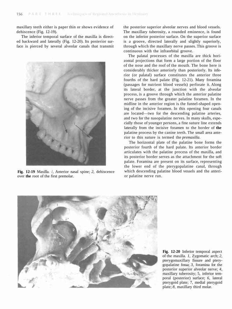
The inferior temporal surface of the maxilla is direct-ed backward and laterally (Fig. 12-20). Its posterior sur-face is pierced by several alveolar canals that transmit
Fig. over
the posterior superior alveolar nerves and blood vessels.The maxillary tuberosity, a rounded eminence, is foundon the inferior posterior surface. On the superior surfaceis a groove, directed laterally and slightly superiorly,through which the maxillary nerve passes. This groove iscontinuous with the infraorbital groove.
The palatal processes of the maxilla are thick hori-zontal projections that form a large portion of the floorof the nose and the roof of the mouth. The bone here isconsiderably thicker anteriorly than posteriorly. Its infe-rior (or palatal) surface constitutes the anterior threefourths of the hard palate (Fig. 12-21). Many foramina(passages for nutrient blood vessels) perforate it. Alongits lateral border, at the junction with the alveolarprocess, is a groove through which the anterior palatinenerve passes from the greater palatine foramen. In themidline in the anterior region is the funnel-shaped open-ing of the incisive foramen. In this opening four canalsare located—two for the descending palatine arteries,and two for the nasopalatine nerves. In many skulls, espe-cially those of younger persons, a fine suture line extendslaterally from the incisive foramen to the border of thepalatine process by the canine teeth. The small area ante-rior to this suture is termed the premaxilla.
The horizontal plate of the palatine bone forms theposterior fourth of the hard palate. Its anterior borderarticulates with the palatine process of the maxilla, andits posterior border serves as the attachment for the softpalate. Foramina are present on its surface, representing
12-19 Maxilla. /, Anterior nasal spine; 2, dehiscencethe root of the first premolar.
the lower end of the pterygopalatine canal, throughwhich descending palatine blood vessels and the anteri-or palatine nerve run.
Fig. 12-20 Inferior temporal aspectof the maxilla. 1, Zygomatic arch; 2,pterygomaxillary fissure and ptery-gopalatine fossa; 3, foramina for theposterior superior alveolar nerve; 4,maxillary tuberosity; 5, inferior tem-poral (posterior) surface; 6, lateralpterygoid plate; 7, medial pterygoidplate; 8, maxillary third molar.
Fthgrte
The mandible is the largest and strongest bone of theface. It consists of a curved horizontal portion (the body)and two perpendicular portions (the rami).
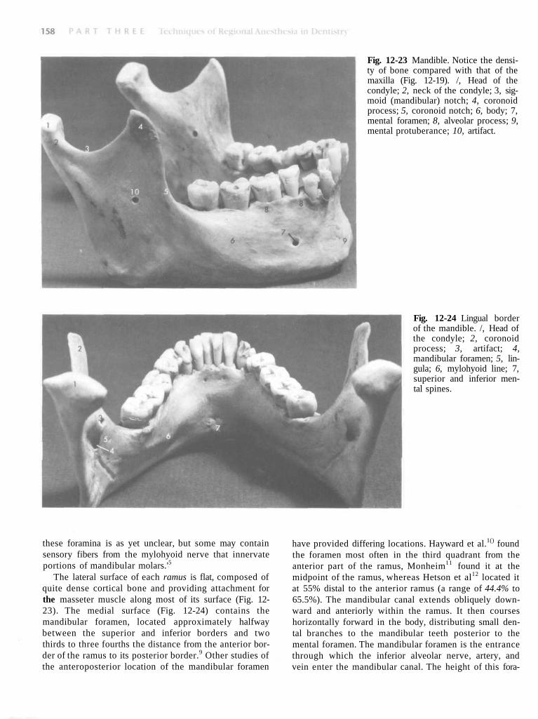
The external (lateral) surface of the body of themandible is marked in the midline by a faint ridge, anindication of the symphysis of the two pieces of bonefrom which the mandible is created (Figs. 12-22 and 12-23). The bone forming the buccal alveolar processes inthe anterior region (incisors) is usually less dense thanthat over the posterior teeth, permitting infiltration(supraperiosteal) anesthesia to be employed with someexpectation of success (in adults usually in the area ofthe lateral incisor only). In the region of the second pre-molar on each side, midway between the upper andlower borders of the body, lies the mental foramen.Phillips et al, in an evaluation of 75 dry, adult humanmandibles, determined that the usual position of themental foramen is below the crown of the second pre-molar.7 The mental nerve, artery, and vein exit themandibular canal here. Bone along this external surfaceof the mandible is commonly quite thick cortical bone.
The lingual border of the body of the mandible is con-cave from side to side (Fig. 12-24). Extending upward and
ig. 12-21 Palate. /, Incisive foramen; 2, palatal process ofe maxilla; 3, horizontal process of the palatal bone; 4,eater palatine foramen; 5, lesser palatine foramen; 6, pos-rior nasal spine.
backward is the mylohyoid line, giving origin to themylohyoid muscle. Bone along the lingual of themandible is usually quite thick; however, in approxi-mately 68% of mandibles there are lingual foramina locat-ed in the posterior (molar) region.8 The function of
Fig. 12-23 Mandible. Notice the densi-ty of bone compared with that of themaxilla (Fig. 12-19). /, Head of thecondyle; 2, neck of the condyle; 3, sig-moid (mandibular) notch; 4, coronoidprocess; 5, coronoid notch; 6, body; 7,mental foramen; 8, alveolar process; 9,mental protuberance; 10, artifact.
Fig. 12-24 Lingual borderof the mandible. /, Head ofthe condyle; 2, coronoidprocess; 3, artifact; 4,mandibular foramen; 5, lin-gula; 6, mylohyoid line; 7,superior and inferior men-tal spines.
tsp
qt2mbtdt
hese foramina is as yet unclear, but some may containensory fibers from the mylohyoid nerve that innervateortions of mandibular molars.'5
The lateral surface of each ramus is flat, composed ofuite dense cortical bone and providing attachment forhe masseter muscle along most of its surface (Fig. 12-3). The medial surface (Fig. 12-24) contains theandibular foramen, located approximately halfway
etween the superior and inferior borders and twohirds to three fourths the distance from the anterior bor-er of the ramus to its posterior border.9 Other studies ofhe anteroposterior location of the mandibular foramen
have provided differing locations. Hayward et al.1() foundthe foramen most often in the third quadrant from theanterior part of the ramus, Monheim11 found it at themidpoint of the ramus, whereas Hetson et al12 located itat 55% distal to the anterior ramus (a range of 44.4% to65.5%). The mandibular canal extends obliquely down-ward and anteriorly within the ramus. It then courseshorizontally forward in the body, distributing small den-tal branches to the mandibular teeth posterior to themental foramen. The mandibular foramen is the entrancethrough which the inferior alveolar nerve, artery, andvein enter the mandibular canal. The height of this fora-
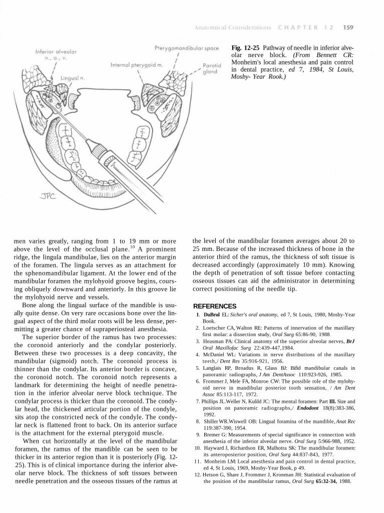
Fig. 12-25 Pathway of needle in inferior alve-olar nerve block. (From Bennett CR:Monheim's local anesthesia and pain controlin dental practice, ed 7, 1984, St Louis,Mosby- Year Rook.)
men varies greatly, ranging from 1 to 19 mm or moreabove the level of the occlusal plane.10 A prominentridge, the lingula mandibulae, lies on the anterior marginof the foramen. The lingula serves as an attachment forthe sphenomandibular ligament. At the lower end of themandibular foramen the mylohyoid groove begins, cours-ing obliquely downward and anteriorly. In this groove liethe mylohyoid nerve and vessels.
Bone along the lingual surface of the mandible is usu-ally quite dense. On very rare occasions bone over the lin-gual aspect of the third molar roots will be less dense, per-mitting a greater chance of supraperiosteal anesthesia.
The superior border of the ramus has two processes:the coronoid anteriorly and the condylar posteriorly.Between these two processes is a deep concavity, themandibular (sigmoid) notch. The coronoid process isthinner than the condylar. Its anterior border is concave,the coronoid notch. The coronoid notch represents alandmark for determining the height of needle penetra-tion in the inferior alveolar nerve block technique. Thecondylar process is thicker than the coronoid. The condy-lar head, the thickened articular portion of the condyle,sits atop the constricted neck of the condyle. The condy-lar neck is flattened front to back. On its anterior surfaceis the attachment for the external pterygoid muscle.
When cut horizontally at the level of the mandibularforamen, the ramus of the mandible can be seen to bethicker in its anterior region than it is posteriorly (Fig. 12-25). This is of clinical importance during the inferior alve-olar nerve block. The thickness of soft tissues betweenneedle penetration and the osseous tissues of the ramus at
the level of the mandibular foramen averages about 20 to25 mm. Because of the increased thickness of bone in theanterior third of the ramus, the thickness of soft tissue isdecreased accordingly (approximately 10 mm). Knowingthe depth of penetration of soft tissue before contactingosseous tissues can aid the administrator in determiningcorrect positioning of the needle tip.
REFERENCES1. DuBrul EL: Sicher's oral anatomy, ed 7, St Louis, 1980, Mosby-Year
Book.2. Loetscher CA, Walton RE: Patterns of innervation of the maxillary
first molar: a dissection study, Oral Surg 65:86-90, 19883. Heasman PA: Clinical anatomy of the superior alveolar nerves, BrJ
Oral Maxillofac Surg 22:439-447,1984.4. McDaniel WL: Variations in nerve distributions of the maxillary
teeth,/ Dent Res 35:916-921, 1956.5. Langlais RP, Broadus R, Glass BJ: Bifid mandibular canals in
panoramic radiographs, J Am DentAssoc 110:923-926, 1985.6. Frommer J, Mele FA, Monroe CW: The possible role of the mylohy-
oid nerve in mandibular posterior tooth sensation, / Am DentAssoc 85:113-117, 1972.
7. Phillips JL.Weller N, Kulild JC: The mental foramen: Part III. Size andposition on panoramic radiographs,/ Endodont 18(8):383-386,1992.
8. Shiller WR.Wiswell OB: Lingual foramina of the mandible, Anat Rec119:387-390, 1954.
9. Bremer G: Measurements of special significance in connection withanesthesia of the inferior alveolar nerve. Oral Surg 5:966-988, 1952.
10. Hayward J, Richardson ER, Malhotra SK: The mandibular foramen:its anteroposterior position, Oral Surg 44:837-843, 1977.
11. Monheim LM: Local anesthesia and pain control in dental practice,ed 4, St Louis, 1969, Mosby-Year Book, p 49.
12. Hetson G, Share J, Frommer J, Kronman JH: Statistical evaluation ofthe position of the mandibular ramus, Oral Surg 65:32-34, 1988.