12 The Biology of Bone Grafts Carlos Roberto Galia and Luis Fernando Moreira Rio Grande do Sul Federal University, Porto Alegre, RS, Brazil School of Medicine, Post-Graduate Programme of Surgery and Hospital de Clinicas University Hospital Department of Surgery 1Division of Orthopaedics and 2Division of Surgical Oncology Brazil 1. Introduction The use of bone transplants in orthopaedic procedures has become crucial to treat a great number of bone diseases including bone tumour operations, knee or total hip revision arthroplasty and even beyond the orthopaedic scope such as in craniomaxillofacial surgery. Approximately 10% to 15% of the orthopaedic procedures carried out every year in the U.S.A. employ some kind of musculoskeletal transplant. Annually, about 650 thousand bone-based grafts are distributed by the American Tissue Banks, which clearly shows the importance of processing, controlling and storage of this type of material. As opposed to other organs such as heart, liver or kidneys and most of the soft tissues, the bone can be processed by many ways, can be stored longer and has been implanted till recently without prior testing compatibility. Moreover, the grafts can be obtained from oneself, living or cadaver donors, or derived from other species or even from non organic biomaterials. However, the offer for grafts is far behind the demand. Despite the success rate of about 85% with the use of bone grafts in orthopaedic surgery, patient waiting list for these grafts keep growing day by day either in the public or private health service. The homologous deep-frozen grafts have been frequently used, although availability is very limited and a certain risk of transmitting contagious diseases cannot be thoroughly ruled out. On the other hand, though homologous freeze-dried grafts have virtually eliminated the risk of transmissible diseases is still lacking availability and therefore, alternative biomaterials from synthetic or natural source have been considered and carefully studied. Among them, the use of freeze-dried grafts from bovine sources is a suitable alternative with many advantages over autologous or homologous deep-frozen ones due to their great physicochemical and structural resemblance to human bone and their practically endless availability. As a non specific xenograft however, it may possibly present adverse immunological and inflammatory reactions. Nevertheless, our physicochemical processing protocols have been developed in order to significantly minimise these problems, decreasing antigenicity and thus, turning the bovine freeze-dried bone an important biomaterial for large use in reconstructive surgery. Up to date, it is necessary, an increased knowledge about some kinds of available grafts including biological and mechanical aspects, sources and a careful analysis of outcomes, since transplant of grafts in general is responsible for a remarkable improvement in people’s quality of life and their social reinsertion. Moreover, bone implants are far from being a www.intechopen.com
Transcript
12
The Biology of Bone Grafts
Carlos Roberto Galia and Luis Fernando Moreira Rio Grande do Sul Federal University, Porto Alegre, RS, Brazil School of Medicine,
Post-Graduate Programme of Surgery and Hospital de Clinicas University Hospital Department of Surgery 1Division of Orthopaedics and 2Division of Surgical Oncology
Brazil
1. Introduction
The use of bone transplants in orthopaedic procedures has become crucial to treat a great
number of bone diseases including bone tumour operations, knee or total hip revision
arthroplasty and even beyond the orthopaedic scope such as in craniomaxillofacial surgery.
Approximately 10% to 15% of the orthopaedic procedures carried out every year in the U.S.A. employ some kind of musculoskeletal transplant. Annually, about 650 thousand bone-based grafts are distributed by the American Tissue Banks, which clearly shows the importance of processing, controlling and storage of this type of material. As opposed to other organs such as heart, liver or kidneys and most of the soft tissues, the bone can be processed by many ways, can be stored longer and has been implanted till recently without prior testing compatibility. Moreover, the grafts can be obtained from oneself, living or cadaver donors, or derived from other species or even from non organic biomaterials. However, the offer for grafts is far behind the demand. Despite the success rate of about 85% with the use of bone grafts in orthopaedic surgery, patient waiting list for these grafts keep growing day by day either in the public or private health service. The homologous deep-frozen grafts have been frequently used, although availability is very
limited and a certain risk of transmitting contagious diseases cannot be thoroughly ruled
out. On the other hand, though homologous freeze-dried grafts have virtually eliminated
the risk of transmissible diseases is still lacking availability and therefore, alternative
biomaterials from synthetic or natural source have been considered and carefully studied.
Among them, the use of freeze-dried grafts from bovine sources is a suitable alternative
with many advantages over autologous or homologous deep-frozen ones due to their great
physicochemical and structural resemblance to human bone and their practically endless
availability. As a non specific xenograft however, it may possibly present adverse
immunological and inflammatory reactions. Nevertheless, our physicochemical processing
protocols have been developed in order to significantly minimise these problems,
decreasing antigenicity and thus, turning the bovine freeze-dried bone an important
biomaterial for large use in reconstructive surgery.
Up to date, it is necessary, an increased knowledge about some kinds of available grafts including biological and mechanical aspects, sources and a careful analysis of outcomes, since transplant of grafts in general is responsible for a remarkable improvement in people’s quality of life and their social reinsertion. Moreover, bone implants are far from being a
www.intechopen.com
Recent Advances in Arthroplasty
236
novel issue and it is crucial that already in the XXI century, we have to know the reasons for its use and, over all matters, to clearly determine our results. This chapter is intended to present a brief review of the history of bone grafts, indications for use, storage details and differences of processing techniques as well as to present the experience of our department with the use of freeze-dried bone from bovine origin in a thousand orthopaedic procedures.
2. History
The idea of transplanting tissues between individuals of the human species and even between individuals of distinct species is very ancient, dating to the early Christian era. Empirically, Cosmas and Damian, in the second century, were regarded as the pioneers of bone transplantation by having removed a leg of an individual due to an advanced tumour and soon deployed a new leg taken from a freshly killed Moro. However, it was Sir William MacEwan in 1880 who successfully reconstructed the humerus of a boy with the tibia obtained from another child. On the other hand, the first transplant between different species was conducted by the Dutch surgeon Job van Meekren in 1668. According to Godwin, he successfully transplanted part of the skull of a dog in a traumatic defect of the skull of a soldier. Deeply concerned of being excommunicated by the church for having undergone a non-Christian method of treatment, the soldier asked the surgeon to remove the graft, but due to time elapsed, this could not be completely removed since it was already fully integrated. Is not well known when the first studies on bone grafts started. However, Ollier and Barth in the late 19th century, were the first to intensively investigate and report the biological properties of the tissue and concluded, though partially correct, that bone and periosteum remain viable when transplanted, contributing to new bone formation. Assuming that the cells survive in the graft, even when removed from the donor, Albee in 1915, concluded in a classical work, the most suitable tissues for transplant are those originated from the connective tissue such as bone, fat and fascia. Ten years later, in 1925, Lexer published his experience with transplants and total joint replacement called "arthroplasty" to all surgical procedures performed in an attempt to restore joint movement.
3. Terminology
It is important to know the terminology frequently used when dealing with this kind of procedure in relation to (a) different types of graft; (b) distinction between grafts and transplants; (c) the process of new bone formation, and; (d) the use of consolidation and osteointegration, since these terms vary greatly in the literature and an attempt to standardized them has not been effective. The nomenclature most commonly used is as follows:
3.1 Types of grafts Grafts received from oneself and reimplanted in oneself should be called autogenic, autogenous, autologous or autograft. The graft obtained from other individuals, but intended to a host of the same species, should be called allogeneic, allogenous, homologous, allograft or homograft, while grafts from donors to hosts of different species should be called xenogenic, xenogenous, xenologous, xenografts or interspecific.
www.intechopen.com
The Biology of Bone Grafts
237
a. Grafting and transplantation
A Brazilian Board Resolution on Biomaterials - BBRB No. 220 of December 27th, 2006 considers the term transplantation to describe the placement of bone graft during a surgical procedure, so the graft is the product and the use of this tissue is called a transplant.
b. Bone formation and osteointegration
In the process of bone formation and the biological events responsible for osteointegration the following terms are used: osteogenesis when bone growth is derived from osteoblasts transferred along with the graft; osteoinduction when the newly formed bone derives from the recruitment of mesenchymal cell receptors that differentiate into osteoblasts; osteoconduction when the ability of a material (usually inorganic) allows the new bone tissue from pre-existing bone, requiring the presence of osteoprogenitor (preosteoblast) cells or bone tissue.
c. consolidation and osteointegration
On the biological process occurring after transplantation it should be clear that the graft can consolidate and/or embedded (incorporate) into the host bone. Therefore, consolidation and incorporation processes are distinct and should be well understood respectively, as the union that occurs between the graft and host bone or reabsorption and cell recolonisation between graft and host bone resulting in a progressive replacement of one by another. Thus, the imaging methods for assessment of these features can only provide an idea of the evolution of this process, but precise assessment on the consolidation and incorporation can be only determined by histopathology.
4. Biological characteristics
Bone tissue basically consists of an organic matrix of collagen type I, containing low molecular weight proteoglycans and non-collagen proteins, a mineral part (mainly hydroxyapatite) and water, corresponding to 25%, 65% and 10% of the bone weight, respectively. Despite the great power for repair, the bone does not always respond appropriately when affected by extensive osteolysis. Because of these situations, extremely frequent in orthopaedic surgery, especially in RTHAs, there is eagerness to search for high quality bone grafts and other biomaterials that can fill these gaps and restore availabilty. Bone grafts can be of cortical, cancellous or cortico-cancellous types, depending on the site of origin and can be used in blocks, segments or morselised. These different types of bone grafts will provide distinct mechanical and biological responses. For instance, the cortical bone is less osteogenic than the spongy bone; showing however, a higher structural quality, for long periods and even in the absence of adequate integration. The greater osteogenic features of cancellous bone (Figures 1a, 1b and 1c) has been recognised for more than 40 years, which has stimulated its use in many situations, being crucial for use in RTHAs. After the transplant of a spongy bone there is a sequence of histological events that starts
with inflammatory response, macrophage invasion, neovascularization and differentiation
of mesenchymal cells into osteoblasts that places an osteoid layer in a necrotic trabecular
bone remainings. Viable nuclei are reabsorbed by osteoblasts and the matrix is eventually
replaced by necrotic trabeculae of newly formed bone. All these events are probably
mediated by inducing protein factors.
www.intechopen.com
Recent Advances in Arthroplasty
238
Fig. 1a. Revision as of right femoral component with bone graft and frozen morcelised impacted. 70 months of evolution.
www.intechopen.com
The Biology of Bone Grafts
239
Fig. 1b. Revision of femoral component left with bone graft and bovine freeze-dried, morcelised impacted. 60 months of evolution.
www.intechopen.com
Recent Advances in Arthroplasty
240
Fig. 1c. Revision of the acetabular component with lyophilized bovine bone graft and graft block frozen morcelised impacted. 82 months of evolution.
Histologic assessment was performed and described by Buma et al. in eight patients who underwent revision of the acetabular component after previous operation with impacted bone grafting technique. All but one, that has not even shown revascularization, samples revealed different stages of integration depending on the time elapsed following implantation. At 4 months, there was an established revascularization, osteoblasts replacing parts of the implant and presence of a small graft and new bone formation. Samples with a longer evolution showed the graft replaced with new bone. Another sample with 28 months in contact with the cement layer revealed viable bone presenting, however, a predominantly fibrous tissue interface5. Similar results on the femoral component were demonstrated in another study published by Ullmark and Obrant in 2002 and by Galia in acetabular biopsies in patients with traumatic dislocation of RTHA (Figures 2a and 2b) 3 years after the first revision procedure.
www.intechopen.com
The Biology of Bone Grafts
241
Fig. 2a. Biopsy bone grafting with bovinefreeze-dried three years after surgery. White arrow
= graft; black arrow = new bone
In a classic study, Urist described ectopic bone formation after intramuscular
implantation of demineralised bovine bone matrix in rabbits and rats. This discovery was
crucial in the biomaterial and graft fields and supported the search for substances capable
of inducing cell differentiation present in bone matrix. Subsequent investigations, led by
Urist demonstrated that low molecular weight proteins could be extracted from
demineralised bone matrix, having great osteogenic activity and being called bone
morphogenetic proteins (BMPs), and belonging to a superfamily of proteins called
transforming growth factors beta (TGF-ß) responsible for inducing growth. The
superfamily of TGF-ß proteins regulates many biological processes including cell growth,
differentiation and embryo formation. BMPs have been shown to be important regulators
in the development and regeneration of skeletal tissue having the BMP2 and BMP7 the
highest osteoinductive potential. Another important factor is the reaction triggered by
antigenic grafts. The antigens present into the graft cells probably play a very important
role on the outcomes of bone transplants. It seems clear that these reactions are not
mediated by T or B lymphocytes, but by other cells in the bone marrow. In vitro evidences
suggest that the granulocyte lineages in the bone marrow are responsible for triggering
immune responses and removal of bone marrow cells therefore and can be directly related
to a decrease in graft immune response.
Fresh, frozen and lyophilized bone grafts were experimentally compared in rabbits from the
immunological point of view. The first two grafts caused serologically detectable immune
response, while the third one, highly purified (free of fat and marrow cells) was not able to
immunologically sensitize animals.
www.intechopen.com
Recent Advances in Arthroplasty
242
Fig. 2b. Biopsy of frozen human bone graft. 36 months of evolution. White arrow = graft; black arrow = new bone.
Although the freeze-dried grafts, whether human or bovine, are available in many medical
centres worldwide, most work on RTHAs refers to the use of frozen bone grafts in blocks or
cortical, but especially, most recently the spongy morcelised and impacted.
Tagil, in his PhD thesis published in 2000, attempted to explain the reasons for succeeding
using the technique of frozen morcelised and impacted bone, once theoretically, the large
volume of necrotic bone exposed to great mechanical stress tend to collapse, as in the
vascular necrosis of the femoral head or knee. After detailed study, Tagil found out the
following possibilities: (a) morcelised bone, as in a comminuted fracture, would produce
extensive surface contact allowing access and release of biologically active substances; (b)
impaction may improve osteoconductive properties of the graft leading to the release of
BMPs and this way favouring osteointegration and; (c) the high elasticity may allow small
deformations that would stimulate new bone formation. The importance of this study lays
in the fact that it is essential, from a scientific standpoint, to know the pros and cons of a due
technique and why results may be good or bad, to better use and indicate such a procedure
more securely and confidentially.
5. Mechanical characteristics
In addition to the biological characteristics, the mechanical aspects are very relevant and can
be decisive in the choice and obtained results using a due graft. Itoman and Nakamura, in
www.intechopen.com
The Biology of Bone Grafts
243
1991, studied the histological and biomechanical properties of different ways of bone graft
processing in rats and noted that an increased stiffness of the bone after freezing at -80°C
and lyophilising it. The freeze-dried demineralised bone has initially decreased its
mechanical strength. However, after 16 weeks, a progressive increase in resistance of the
grafts was observed and was likely related to its biological interaction, which might be an
indicator of the osteoinductive properties of the graft. They also concluded that on the
features of integration, as expected, the autologous bone showed the best results.
The physical properties of human and bovine trabecular bone are documented and their results are available, however, the range of dispersion is very wide. The Young's module, for example, in one study ranged from 70 to 673 MPa and compressive strength varied between 2.44 and 6.24 MPa. This dispersion occurred for both human and bovine bone and may be related to several factors such as donor age, bone density and methodology used in the study. Cornu et al. in 2001, has demonstrated in vitro that the lyophilised morcelised and impacted bone is mechanically superior to the morcelised and impacted deep-frozen bone since, at least, has the same resistance after impaction, that is achieved however, more quickly and with fewer impacts, and the authors assumed as to the fact that lyophilised material was devoid of fat and bone marrow. Moreover, Macedo et al. in 1999, using an automated compression machine compared in vitro, the compressive strength of frozen and freeze-dried bovine bone rehydrated for an hour and found out that deep-frozen bovine bone grafts after defrosting, has similar compressive loads and deformation rate of the rehydrated lyophilised bovine bone.
6. Methods sterilization, infectious diseases and biosafety
Another extremely important issue is the study of methods of sterilization of frozen and freeze-dried grafts, since there still remains controversy and need for further studies. The currently available techniques have advantages and disadvantages regarding efficacy and maintenance of mechanical and biological properties. Significant deleterious effects on the use of cobalt 60 in the sterilisation of freeze-dried grafts have been reported, demonstrating that, even at low dosage, radiation is capable of destroying the morphogenetic properties, mainly in non-demineralised bone. The effects of radiation on the biomechanics of the grafts are dose-dependent. Fidele et al. in 1995, studied the damage caused by the application of different doses of gamma radiation exposure on seven biomechanical parameters of frozen allogenic patellar bone-grafting. The sterilization dose accepted for inactivation of HIV, for example, is about 25 kGy. However, four out of the seven parameters measured were reduced after 20 kGy and after 30 kGy all parameters have shown significant reductions. Also, Zhang, Cornu and Delloye in 1997, in an experimental study in rats, compared the ability of graft osteoinduction after gamma radiation sterilisation (25 kGy), ethylene oxide (EO) at 55°C and 40°C or preservation in ethanol showing that the OE at 40°C and ethanol have not negatively affected the osteoinductive capacity, gamma radiation has decreased 40% and OE to 55°C had an almost completely loss of this potential. Some authors indicated that other factors in sterilisation need to be observed are the toxic residues from the OE that may remain in the graft and are released when in contact with liquids. Reference is also considered to the toxicity of gamma radiation when in contact with fat present in the graft, in addition to the mechanical change that the radiation may cause.
www.intechopen.com
Recent Advances in Arthroplasty
244
Another way to sterilise is to autoclave the bone, although it has also limitations. According to the recommendation of the European Community, aimed at inactivating prions (infectious proteins), tissues from bovine origin should be sterilised in an autoclave at 132°C for 1h, but the biomechanical effects of this method of sterilisation has proven to reduce in approximately 70%the mechanical resistance to compression of the graft. Moreover, many authors support the safety of non-sterilised bone grafts provided by banks that adopt appropriate methodology for donor selection and quality control and such control is so important that when applying these criteria, 20% to 30% of the harvested material will be discarded due to bacteriological or serological inadequate results. Despite this severe control, to name a few, in November 2001 there were two reported cases in the U.S., by Clostridium sordellii infection in surgical patients who received bone transplants. In March 2002, the Centre for Disease Control and Prevention (CDC, Atlanta) has received 26 reports from different locations of secondary bacterial infections in transplanted musculoskeletal tissues. In these events the CDC investigated donors and confirmed the presence of those bacteria in their tissues. Several other notifications have been done and are reported every year, which provides the need for even a more strict control, revision of adopted routines and, perhaps, searching for methods of sterilisation that may minimally interfere with quality of the graft. Taylor in 2002, stated that prions, responsible for Creutzfeldt-Jakob disease in humans and bovine spongiform encephalitis, unlike bacteria and viruses, are resistant to procedures such as autoclaving or exposure to sodium hydroxide, but, the association of alkaline substances and heat even at 100°C for 1 minute appears to be effective to inactivate prions. In addition, when isolated used, sodium hypochlorite also demonstrated ability to deactivate prions. Alike prions, potentially transmissible viral diseases such as hepatitis A, B or C, HIV and HTLV, and other more recently isolated viruses and certain types of tumour cells, should also be of concern, even with current knowledge and care from the point of view of serological or other diagnostic methods, which clearly demonstrates the importance of a wide discussion about sterilisation processes. As published by Sugihara et al. in 1999, about 1% of tumour cells in 137 femoral heads subjected to histological examination in patients undergoing THA due to primary arthrosis and Palmer et al. also found high rates of histological abnormalities in 1146 patients with hip joint arthrosis, even suggesting that pathological examinations should be included as part of screening for donor tissues.
7. Processing and storage
Many ways of processing and storage bone tissue for clinical application have been proposed and used in Tissue Banks around the world. Among them, we highlight the deep-frozen (-80oC) and lyophilisation, which are the most widely used and accepted methods. The ultralow freezing temperature is reached in freezers that go as low as -80oC with graphical systems constantly monitoring the temperature, having their own power generators and emergency alarms alerting when temperature increases. So, after rigorous screening of donors (living or dead), culture and serological tests for bacteria and fungi, and processing in a surgical environment the graft is subjected to freezing at-80C that allows its storage for up to 5 years. However, it is worth of note that low temperatures do not play any role in the sterilisation of the material, but decreases antigenicity indeed, allowing more useful handling of the graft.
www.intechopen.com
The Biology of Bone Grafts
245
Lyophilisation, as proposed and spread in 1951 by the USA Navy Tissue Bank24, is a
technique by which dehydration of the water contained in a product is removed by
sublimation, i.e., starting from a previously frozen material, placed under vacuum, the
water goes directly from a solid state into mist30. Before that, however, the bone graft is
washed, centrifuged, decellularised, chemically degreased and subjected to physical
processes of cleaning and sterilisation preserving only the protein-mineral matrix, thus
decreasing antigenicity23 (Figure 3).
Fig. 3. Histological section of bone sample degreased, decellularized and freeze-dried. Note that the only remaining protein-mineral matrix.
Moreover, lyophilisation is a method of processing and storage of musculoskeletal tissues
that allows not only the use of human bones, tendons and fascias (allogeneic), but also and
mainly the bovine (xenogenic) ones, since by reducing antigenicity, provides
biocompatibility, sterilisation and can be stored at room temperature.
Currently, in major health centres different types of bone grafts are available for
reconstructive orthopaedic surgery: frozen and freeze-dried autologous, allogeneic and
lyophilised xenogenic (bovine). The autologous graft, though not requiring processing, has
been considered from the integration viewpoint, the preferred tissue for bone replacement.
There is evidence that survival of osteoblasts and osteocytes from autologous graft are
dependent on the quality of the receptor bed. Moreover, the amount of graft obtained is
limited, surgical time is increased and complications, either systemic or local, of the
intervention required to obtain the graft ranges from 21% to 49%.
www.intechopen.com
Recent Advances in Arthroplasty
246
The frozen allogeneic graft is the most widely used and accepted nowadays, but due to the
reduced number of Tissue Banks in our country and the small number of donations is not
always available to a wider use. Moreover, despite of all care and biosafety standards
adopted by the banks there is still some risk of transmission of infectious diseases and
tumours.
The lyophilised allogeneic graft, produced by the majority of U.S. tissue banks, is still not
widespread among us, and few studies have reported the use of this graft in orthopaedic
surgery and with a small number of cases and limited methods for assessing
osteointegration, further studies are needed to better define its use (Figures 4a and 4b).
Fig. 4a. Sample of lyophilized bovine graft.
The xenogenic grafts, commonly used in dentistry, although controversial, are beginning to
be used in orthopaedic surgeries, especially those of bovine origin, due to its easy
preparation, availability and similarity. The bovine bone has chemical composition,
porosity, size, shape and biological behaviour similar to its human counterpart as it can be
clearly observed in Table 1. Besides those features, bovine graft provides structural support,
osteoconduction and a high content of calcium and phosphorus, all of them essential factors
for the newly formed bone tissue.
The most important issue, regardless of the type of graft chosen by the surgeon, is the
guarantee that the graft has been processed and stored according to the standards of graft
management as established by the Associations of Tissue Banks and the national and
international health authorities.
www.intechopen.com
The Biology of Bone Grafts
247
Fig. 4b. Sample graft ½ lyophilized human femoral head.
Table 1. Physical and Chemical analysis of lyophilised bones HCPATB
www.intechopen.com
Recent Advances in Arthroplasty
248
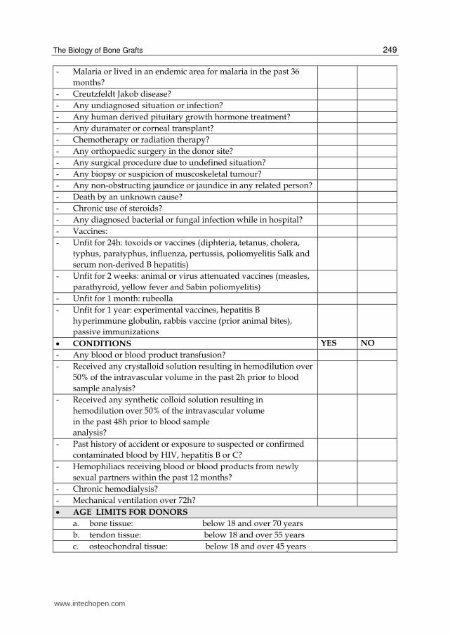
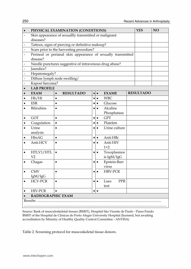
8. Musculoskeletal tissue banks
Like in the USA, where the American Association of Tissue Banks (AATB) regulates and supervises the functioning of all in-country tissue banks since 1976, in Brazil the Ministry of Health, with the Norm No. 220 of December 27th, 2006, provides the technical regulations for the functioning of Musculoskeletal and Skin Tissue Banks of human origin. This regulation relates to the facility characteristics, screening of living donors or cadavers, equipment and human resources. These rules, although quite rigid, greatly increased the quality of processed tissues and the safety on its use. In relation to screening, the decision to accept or reject a donor is carried out by the Medical Director of the Bank of Musculoskeletal Tissues (BMST) after rigorous tests and following a established protocol. As the protocols used by the Tissue Banks, one must consider the history, physical examination and laboratory tests of the donor.
Name: Medical record No.:
Date of Birth: Age (y): Sex: Race:
Checking date: Collecting date:
DONOR PAST HISTORY
CONDITIONS YES NO
Toxic substances or illegal drug abuses
HAS THE DONOR HAD ANY BEHAVIOUR OR
OTHER CONDITION BELOW IN THE PAST
12 MONTHS?
- Professional sex, illegal drugs or has any related person with this
done so?
- Multiple sexual partners?
- Homosexual intercourse or sex with homosexual related
persons?
- Any unprotected sexual intercourse with known HIV,
hepatitis B, C or other blood transmitted infection
person?
- Arrested or has been in a prison?
- Submitted to acupuncture, tattooing, and permanent makeup or
piercing?
- A blood transfused or hemodialysed sexual partner?
- Any malignant disease?
- AIDS/HIV infection?
- Any autoimmune disease?
- Any neurological, degenerative or impairing disease?
- Chronic kidney failure?
- Past or active tuberculosis?
- Sexually transmitted diseases?
- Any osteometabolic diseases?
- Any skin burns?
www.intechopen.com
The Biology of Bone Grafts
249
- Malaria or lived in an endemic area for malaria in the past 36
months?
- Creutzfeldt Jakob disease?
- Any undiagnosed situation or infection?
- Any human derived pituitary growth hormone treatment?
- Any duramater or corneal transplant?
- Chemotherapy or radiation therapy?
- Any orthopaedic surgery in the donor site?
- Any surgical procedure due to undefined situation?
- Any biopsy or suspicion of muscoskeletal tumour?
- Any non-obstructing jaundice or jaundice in any related person?
- Death by an unknown cause?
- Chronic use of steroids?
- Any diagnosed bacterial or fungal infection while in hospital?
- Vaccines:
- Unfit for 24h: toxoids or vaccines (diphteria, tetanus, cholera,
typhus, paratyphus, influenza, pertussis, poliomyelitis Salk and
serum non-derived B hepatitis)
- Unfit for 2 weeks: animal or virus attenuated vaccines (measles,
parathyroid, yellow fever and Sabin poliomyelitis)
- Unfit for 1 month: rubeolla
- Unfit for 1 year: experimental vaccines, hepatitis B
Source: Bank of musculoskeletal tissues (BMST), Hospital São Vicente de Paulo - Passo Fundo BMST of the Hospital de Clinicas de Porto Alegre University Hospital (licensed, but awaiting accreditation by Ministry of Healthy Quality Control Committee - ANVISA)
Table 2. Screening protocol for muscoskeletal tissue donors.
www.intechopen.com
The Biology of Bone Grafts
251
9. Conclusion
There are many differences when comparing the types of graft, with variations as for the source, production methods, processing, uses (block or shredded) sterilisation, storage and cost of the process. Thus, it is essential the implementation of protocols for processing and quality control of all types of bone grafts. This measure will facilitate the monitoring and analysis of the results obtained in distinct surgical procedures, and shall provide grafting material of better quality, thoroughly tested and ready available. Although there are reports of the use of bone grafts and transplants for many years, many mechanical and biological issues such as physical and chemical composition, incorporation, bone remodelling and immune responses are still incompletely assessed. These issues are still requiring more knowledge and further clinical and laboratory investigations to allow a more scientifically based choice of the graft and appropriate indication to surgical use on any particular situation. There is no doubt on the importance of bone transplants in orthopaedic surgery, especially in hip procedures. Their results are well-known and, to some extent predictable when its use is carried out by experienced surgeons. It is also indubitable that bone tissue is a biologically privileged material, since several alternative bone grafts can be used quite well. It is undeniable, however, that we still knowing little about many aspects of the host-graft interaction and sometimes seems we are resigned with this situation, when in fact, many studies have yet to be carried out in several ways to attempt to answer questions that sometimes are underestimated, since "most of the time the graft works". For instance, why do 85% of the grafts theoretically integrate? Though it seems a good percentage, how about the other “not so lucky” 15% of the cases? Is it a matter of technique, immune response, mechanical effect or biological feature? Which is the best substitute for autologous grafts? The frozen allogeneic sterile or non-sterile or the freeze-dried irradiated or autoclaved graft? Should a method of definitive sterilisation be employed by the Tissue Banks? These are just some of important yet unanswered issues that should be carefully investigated and analysed if best clinical responses, increased biosafety and lower complication rates as well as higher scientific basis to precisely and reliably analyse the grafts and surgical outcomes are to be sought.
10. References
American Association of Tissue Banks - AATB's. http://www.aatb.org [accessed Mar 29th, 2011]
Autograft, allograft, and xenograft [Lecture 17]. http://www.pharmacy.wisc.edu/courses/718-430/handouts/ tisgraft.pdf [accessed Feb 27th, 2011] Barth H. Histologische untersuchungen uber knochen-transplantation. Beitr. Path. Anat.
Allg. Path.;17: 65-142; 1895 (in German). Buck BE, Malinin TI. Human bone and tissue allografts. Preparation and safety. Clin
Orthop. Jun; (303):8-17; 1994. Buma P, Lamerigts N, Schreurs BW, Gardeniers J, Versleyen D, Slooff TJ. Impacted graft
incorporation after cemented acetabular revision. Histological evaluation in 8 patients. Acta Orthop Scand. Dec;67(6):536-40; 1996.
www.intechopen.com
Recent Advances in Arthroplasty
252
Cornu O, Bavadekar A, Godts B, Delloye C, Vantomme J, Banse X. Processed freeze-dried bone is more efficient than fresh frozen for impaction bone grafting. In: 47th Annual Meeting, Orthopaedic Research Society; Feb 25-28; San Francisco, California. 1081-81; 2001.
Czitrom AA, Axelrod T, Fernandes B. Antigen presenting cells and bone allotransplantation. Clin Orthop. Jul-Aug; (197):27-31; 1985.
de Roeck NJ, Drabu KJ. Impaction bone grafting using freeze-dried allograft in revision hip arthroplasty. J Arthroplasty. 2001 Feb; 16(2):201-6.
Fideler BM, Vangsness CT Jr, Lu B, Orlando C, Moore T. Gamma irradiation: effects on biomechanical properties of human bone-patellar tendon-bone allografts. Am J Sports Med. Sep-Oct; 23(5):643-6; 1995.
Finkemeier CG. Bone-grafting and bone-graft substitutes. J Bone Joint Surg Am. Mar; 84-A(3):454-64; 2002.
Fred II A. The fundamental principles involved in the use of the bone graft in surgery. Am J Med Sci. Mar; 149(3): 313-25; 1915.
Friedlaender GE, Strong DM, Sell KW. Studies on the antigenicity of bone. I. Freeze-dried and deep-frozen bone allografts in rabbits. J Bone Joint Surg Am. Sep; 58(6):854-8; 1976.
Galia, CR. Liophilised impacted bone grafts from human and bovine origin in TRHAs. Porto Alegre, 2004, 127p. Doctorate Thesis – Faculty of Medicine, Post-graduate Programme in Surgery. Rio Grande do Sul Federal University, 2004.
Garcia, VD. Tissue and organ transplants. 2 ed. São Paulo, SP, Brazil, 2006. Gie GA, Linder L, Ling RS, Simon JP, Slooff TJ, Timperley AJ. Impacted cancellous allografts
and cement for revision total hip arthroplasty. J Bone Joint Surg Br. Jan; 75(1):14-21; 1993.
Godwin L. Tissue banking and allograft transplantation Jun 2000. http://www.iscpubs.com/articles/abl/b0006god.pdf [accessed Nov 22nd, 2003] Goldberg VM. Selection of bone grafts for revision total hip arthroplasty. Clin Orthop. Dec;
(381):68-76; 2000. Gonçalves HR. Methods of acetabular bone graft incorporation in THA with bone loss
[dissertation]. São Paulo, SP, Brazil. Santa Casa de São Paulo Faculty of Medical Sciences 2003 (In Portuguese).
Heliotis M, Tsiridis EE. Fresh frozen bone in femoral impaction grafting: can developments in bone regeneration improve on this? Med Hypotheses. Dec; 57(6):675-8; 2001.
National Press. Brazil’s official Press nº 185, 1, Section Sept 24th, 2002. Norm nº 1.686, Sept 20th, 2002. https://www.in.gov.br/imprimir.asp?id=1081142100&tela=imp.
[accessed Apr 1st, 2011] Invasive Streptococcus pyogenes after allograft implantation - Colorado, 2003. http://www.cdc.gov/mmwr/preview/mmwrhtml/mm5248a1.htm [accessed Jan
23rd, 2011] Itoman M, Nakamura S. Experimental study on allogenic bone grafts. Int Orthop.;15(2):161-
5; 1991. Kakiuchi M, Ono K, Nishimura A, Shiokawa H. Preparation of bank bone using defatting,
freeze-drying and sterilisation with ethylene oxide gas. Part 1. Experimental evaluation of its efficacy and safety. Int Orthop.;20(3):142-6; 1996.
www.intechopen.com
The Biology of Bone Grafts
253
Kreuz FP, Hyatt GW, Turner TC. et al: The preservation and clinical use of freeze-dried bone. J Bone Joint Surg (Am) 33:863-873, 1951.
Lexer E. Joint transplantations and arthoplasty. Surg Gynecol Obstet.;40: 782-809; 1925. Li Zi-zuang, Lu Shi-bi, Wag Ji-fang. The study of repairing ability of freeze-dried bone
allograft. Zhonghua Wai Ke Za Zhi Dec;32(12):765-7; 1994. (English abstract). Lind M, Krarup N, Mikkelsen S, Horlyck E. Exchange impaction allografting for femoral
revision hip arthroplasty: results in 87 cases after 3.6 years' follow-up. J Arthroplasty. Feb; 17(2):158-64; 2002.
Liu W. Reconstitution of osteoinductive bone xenograft: bioassay in mice. Zhonghua Yi Xue Za Zhi. Jul;71(7):378-80, 28, 1991(English abstract).
Lubboc. Maitrise orthopédique [accessed Jan 29th, 2011]:http://www.maitrise-orthop.com/ gesto/lubboc.shtml (in French).
Luchese AC, Dec hechi ED. Lyophilisation process. In: XI Research and Bioethics Meeting 2003 (Oct 20th-22nd); Porto Alegre, RS, Brazil.
www2.pucpr.br/educacao/pibic//evento/files/CE08.html. [accessed Mar 13th, 2011].
Macedo CAS, Galia CR, Silva ALB, César PC, Sanches PRS, Duarte LS et al. Compression resistance of deep-frozen and freeze-dried bone of bovine origin. Rev Bras Ortop. 34(9/10): 529- 33; 1999 (in Portuguese).
Moreau MF, Gallois Y, Basle MF, Chappard D. Gamma irradiation of human bone allografts alters medullary lipids and releases toxic compounds for osteoblast-like cells. Biomaterials. Feb; 21(4):369-76; 2000.
Nogami H, Urist MR. Explants, transplants and implants of a cartilage and bone morphogenetic matrix. Clin Orthop. (103): 235-51; 1974.
Oliveira RC, Sicca CM, Silva TL, Cestari TM, Oliveira OT, Buzalaf MAR, et al. Temperature effect on the denaturation of microgranular cortical bone of bovine origin. Microscopic and biochemistry assessment of cell response in rats. Revista FOB. Jul/Dez; 7(3/4): 85-93; 1999 (in Portuguese).
Palmer SH, Gibbons CL, Athanasou NA. The pathology of bone allograft [abstract]. J Bone Joint Surg Br. Mar; 81(2):333-5; 1999.
Poumarat G, Squire P. Comparison of mechanical properties of human, bovine bone and a new processed bone xenograft. Biomaterials. Apr; 14(5):337-40; 1993.
Reddi AH, Cunningham NS. Initiation and promotion of bone differentiation by bone morphogenetic proteins [abstract]. J Bone Miner Res. Dec; 8 Suppl 2:S499-502; 1993.
Seiler 3rd JG, Johnson J, Hand G, Microsurgery Clinic. Iliac crest autogenous bone grafting: donor site complications. J Southern Orthopedic Association [online].
Internet:http://www.medscape.com/viewarticle/410431 [Accessed Sept. 7th, 2011]. Slooff TJ, Buma P, Schreurs BW, Schimmel JW, Huiskes R, Gardeniers J. Acetabular and
femoral reconstruction with impacted graft and cement. Clin Orthop. Mar; (324):108-15; 1996.
Slooff TJ, Huiskes R, van Horn J, Lemmens AJ. Bone grafting in total hip replacement for acetabular protrusion [abstract]. Acta Orthop Scand. Dec; 55(6):593-96; 1984.
Sommerville SM, Johnson N, Bryce SL, Journeaux SF, Morgan DA. Contamination of banked femoral head allograft: incidence, bacteriology and donor follow up [abstract]. Aust N Z J Surg. Jul; 70(7):480-4; 2000.
www.intechopen.com
Recent Advances in Arthroplasty
254
Springfield DS. Massive autogenous bone grafts. Orthop Clin North Am. Apr; 18(2):249-56; 1987.
Sugihara S, van Ginkel AD, Jiya TU, van Royen BJ, van Diest PJ, Wuisman PI. Histopathology of retrieved allografts of the femoral head. J Bone Joint Surg Br. Mar; 81(2):336-41; 1999.
Tägil M. The morselized and impacted bone graft. Animal experiments on proteins, impaction and load [Thesis]. Lund (Sweden): Lund University Hospital; 2000.
Taylor D. Inactivation of the BSE agent. C R Acad Sci III. Jan; 325(1):75-6; 2002. Thien TM, Welten ML, Verdonschot N, Buma P, Yong P, Schreurs BW. Acetabular revision
with impacted freeze-dried cancellous bone chips and a cemented cup: a report of 7 cases at 5 to 9 years' follow-up. J Arthroplasty. Aug; 16(5):666-70; 2001.
Turek SL. Orthopaedics: Principles and applications. 4th ed. Editora Manole Ltda, Rio de Janeiro, RJ, Brazil. p.756; 1991 (in Portuguese).
Urist MR, Hernandez A. Excitation transfer in bone. Deleterious effects of cobalt 60 radiation-sterilization of bank bone. Arch Surg. Oct; 109(4):486-93; 1974.
Vajaradul Y. Bone banking in Thailand. A 10-year experience (1984-1994).Clin Orthop. Feb; (323):173-80; 1996.
Viceconti M, Toni A, Brizio L, Rubbini L, Borrelli A. The effect of autoclaving on the mechanical properties of bank bovine bone [abstract]. Chir Organi Mov. Jan-Mar; 81(1):63-8; 1996.
Zhang Q, Cornu O, Delloye C. Ethylene oxide does not extinguish the osteoinductive capacity of demineralized bone. A reappraisal in rats. Acta Orthop Scand. Apr; 68(2):104-8; 1997.
www.intechopen.com
Recent Advances in ArthroplastyEdited by Dr. Samo Fokter
ISBN 978-953-307-990-5Hard cover, 614 pagesPublisher InTechPublished online 27, January, 2012Published in print edition January, 2012
InTech ChinaUnit 405, Office Block, Hotel Equatorial Shanghai No.65, Yan An Road (West), Shanghai, 200040, China
Phone: +86-21-62489820 Fax: +86-21-62489821
The purpose of this book was to offer an overview of recent insights into the current state of arthroplasty. Thetremendous long term success of Sir Charnley's total hip arthroplasty has encouraged many researchers totreat pain, improve function and create solutions for higher quality of life. Indeed and as described in a specialchapter of this book, arthroplasty is an emerging field in the joints of upper extremity and spine. However,there are inborn complications in any foreign design brought to the human body. First, in the chapter oninfections we endeavor to provide a comprehensive, up-to-date analysis and description of the management ofthis difficult problem. Second, the immune system is faced with a strange material coming in huge amounts ofmicro-particles from the tribology code. Therefore, great attention to the problem of aseptic loosening hasbeen addressed in special chapters on loosening and on materials currently available for arthroplasty.
How to referenceIn order to correctly reference this scholarly work, feel free to copy and paste the following:
Carlos Roberto Galia and Luis Fernando Moreira (2012). The Biology of Bone Grafts, Recent Advances inArthroplasty, Dr. Samo Fokter (Ed.), ISBN: 978-953-307-990-5, InTech, Available from:http://www.intechopen.com/books/recent-advances-in-arthroplasty/the-biology-of-bone-grafts