66
12 Digestive system 4 Liver & Pancreas
12Digestive system 4
Liver &
Pancreas
12-001Liver
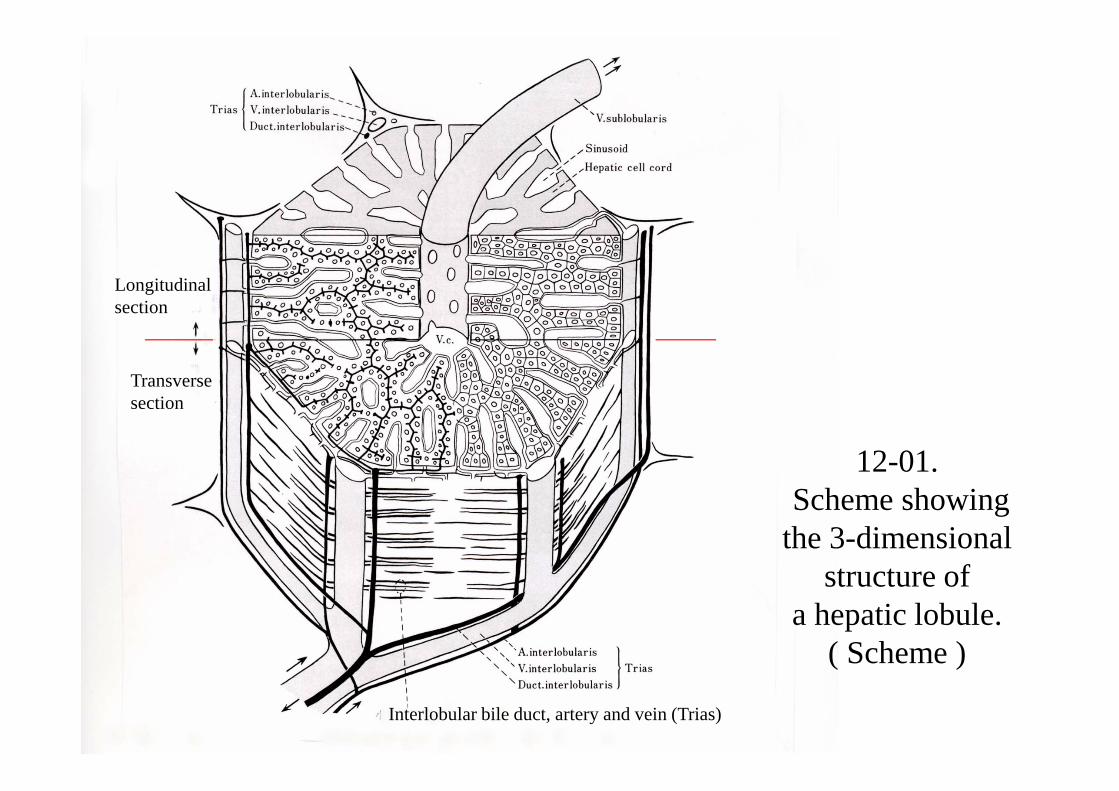
12-01.Scheme showing
the 3-dimensional structure of
a hepatic lobule.( Scheme )
Longitudinal section
Transverse section
Interlobular bile duct, artery and vein (Trias)

12-02. Liver, general view 1. Human, H-E stain, x 13.

12-03. Liver, general view 2. Human, H-E stain, x 25.
Capsula fibrosa
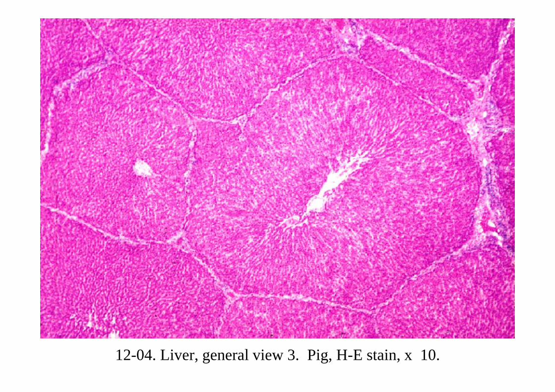
12-04. Liver, general view 3. Pig, H-E stain, x 10.
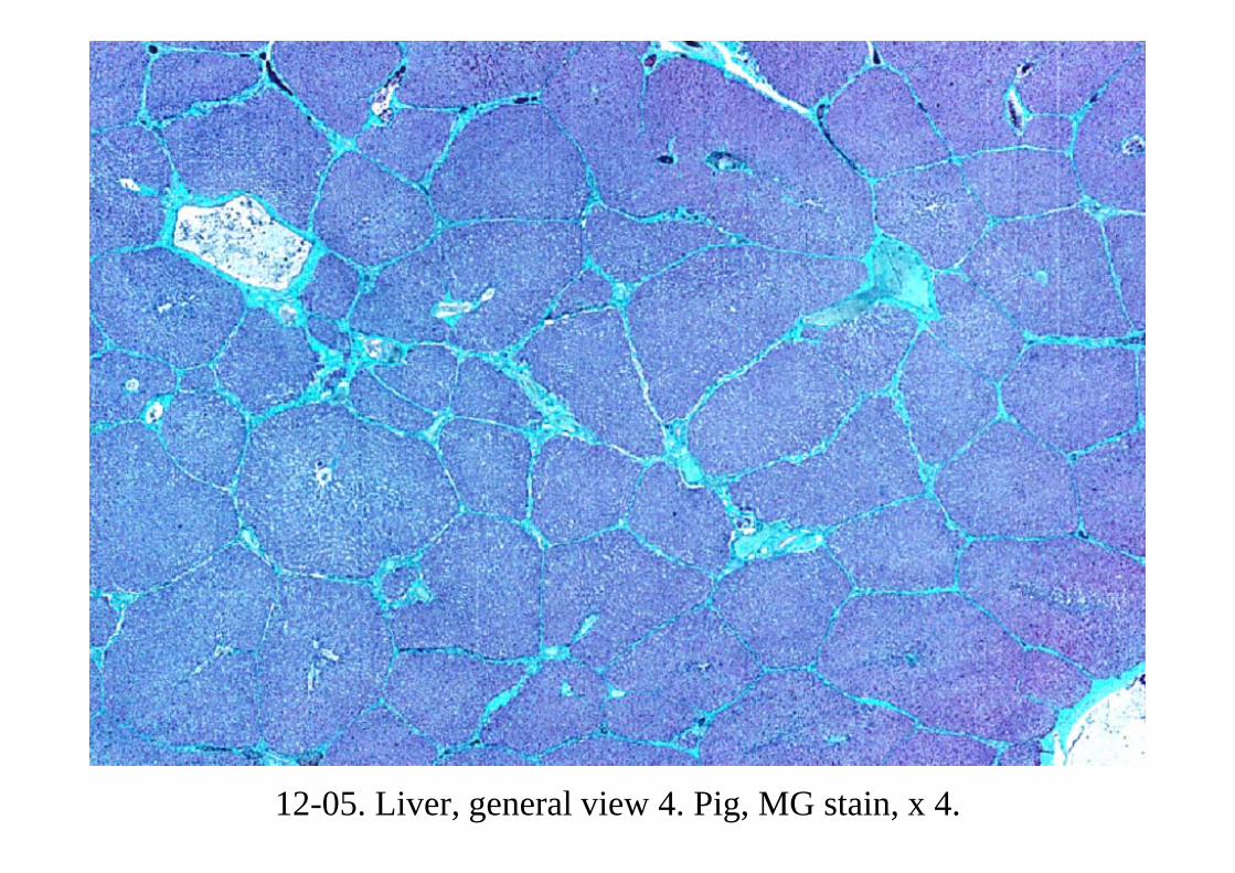
12-05. Liver, general view 4. Pig, MG stain, x 4.

12-06. Hepatic lobule, general view 1. Pig, MG stain, x 25.

12-07. Hepatic lobule, general view 2. Rabbit, H-E stain, x 25.
Vc
Vi
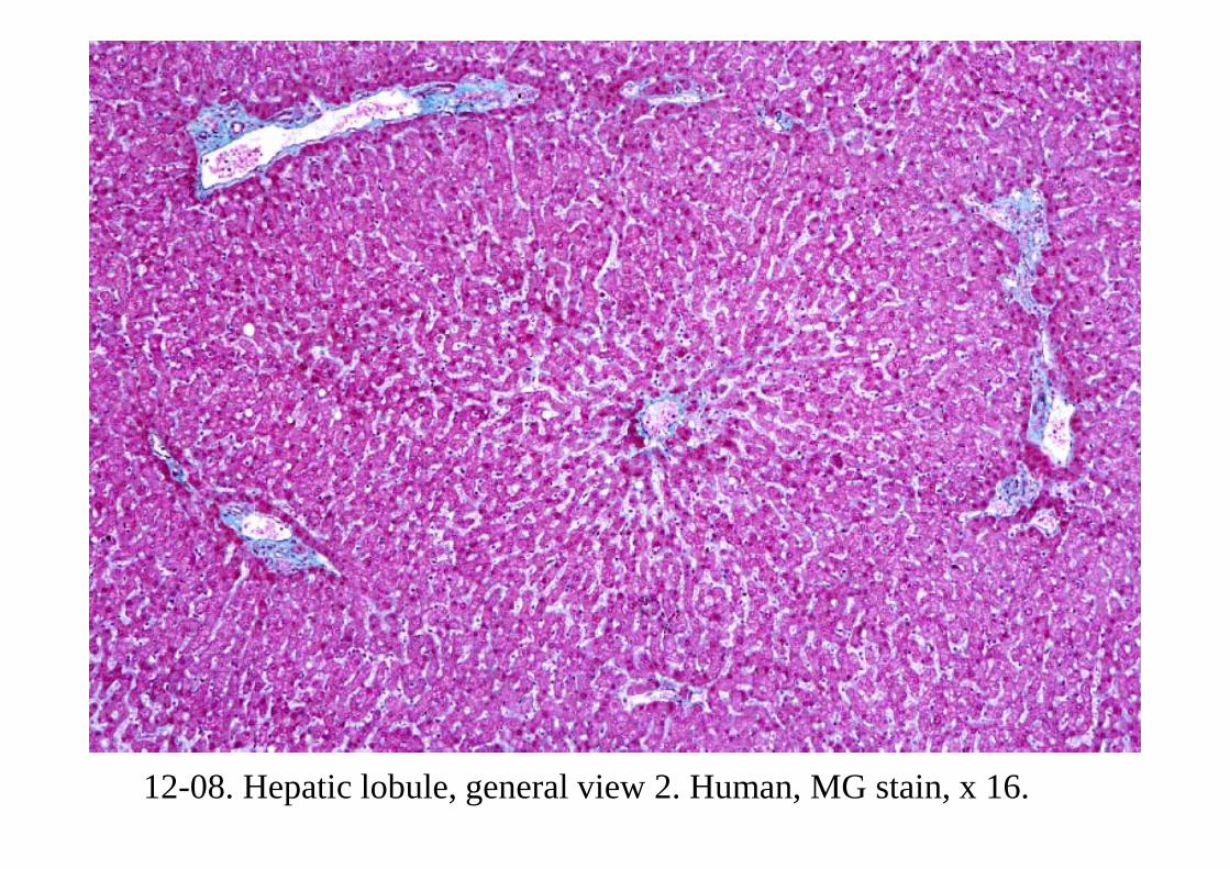
12-08. Hepatic lobule, general view 2. Human, MG stain, x 16.
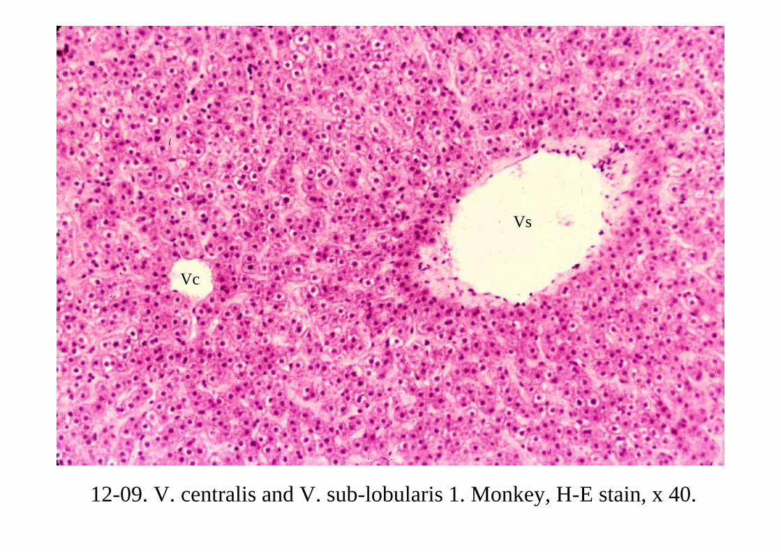
12-09. V. centralis and V. sub-lobularis 1. Monkey, H-E stain, x 40.
Vc
Vs
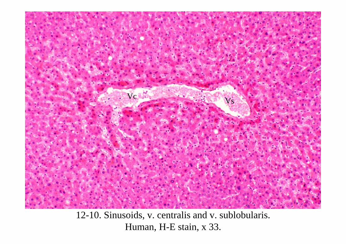
12-10. Sinusoids, v. centralis and v. sublobularis. Human, H-E stain, x 33.
Vc Vs

12-11. Hepatic lobule, general view 3. Human, H-E stain, x 25.
Vs
ViVi
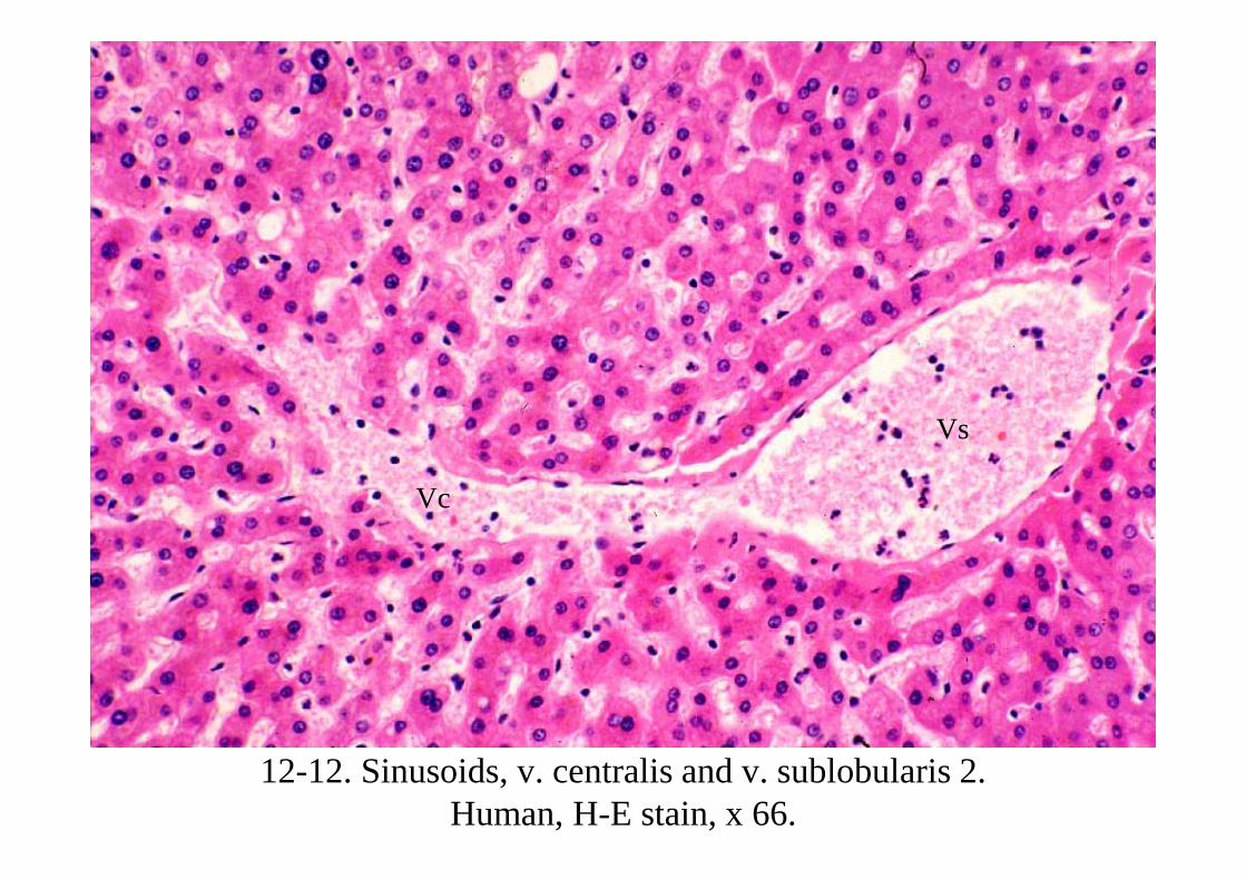
12-12. Sinusoids, v. centralis and v. sublobularis 2. Human, H-E stain, x 66.
Vs
Vc
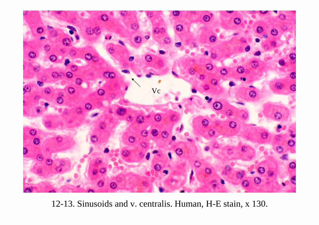
12-13. Sinusoids and v. centralis. Human, H-E stain, x 130.
Vc

12-14. Interlobular connective tissue 1. Human, H-E stain, x 66.
Vi 1
Vi 2
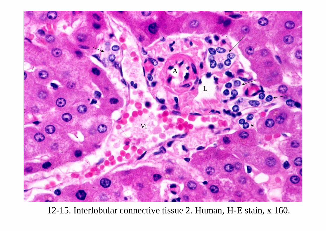
12-15. Interlobular connective tissue 2. Human, H-E stain, x 160.
Vi
L
A

12-16. Interlobular connective tissue 3. Human, H-E stain, x 160.

12-17. Interlobular connective tissue 4. Human, MG stain, x 64.
Vi

12-18. Hepatic cell cords and sinusoids 1. Human, MG stain, x 160.
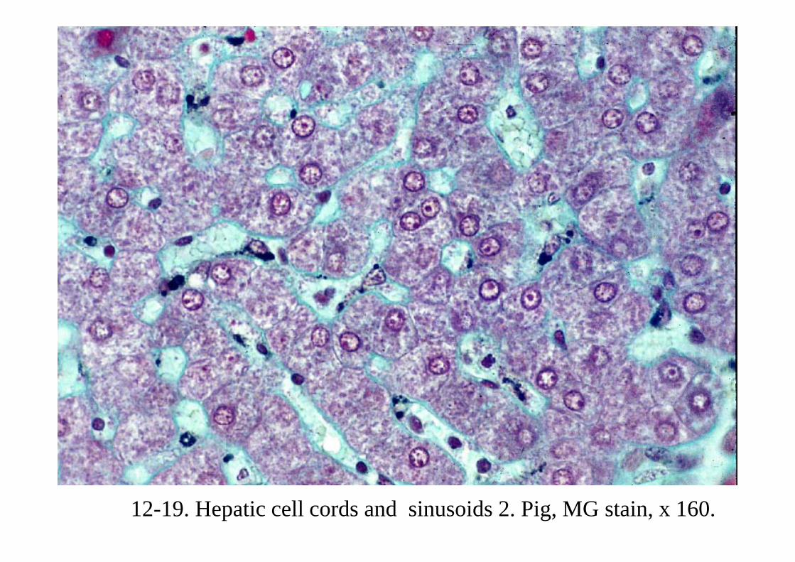
12-19. Hepatic cell cords and sinusoids 2. Pig, MG stain, x 160.
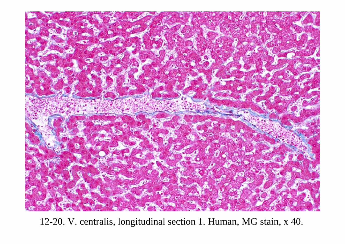
12-20. V. centralis, longitudinal section 1. Human, MG stain, x 40.
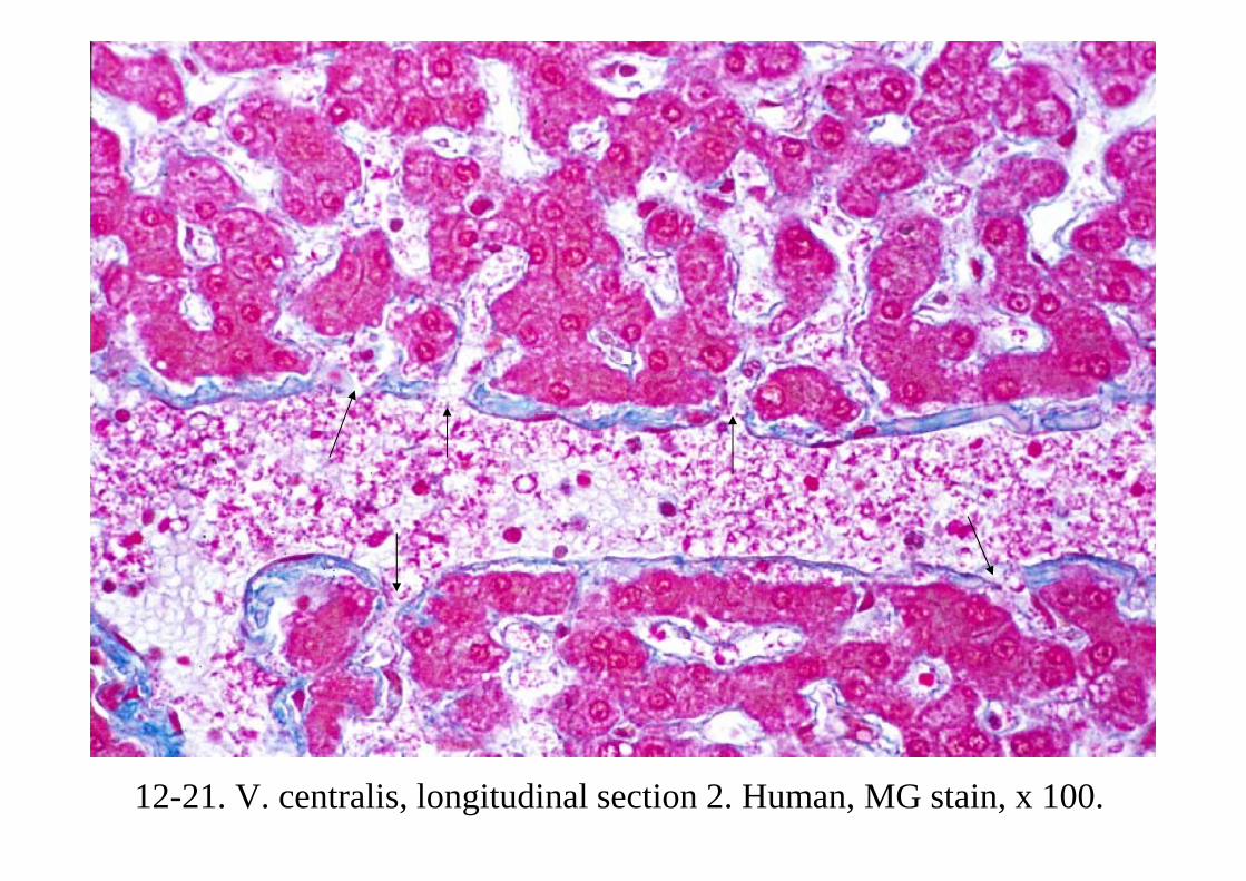
12-21. V. centralis, longitudinal section 2. Human, MG stain, x 100.

12-22. V. centralis and V. sub-lobularis. Longitudinal section. Human, MG stain, x 25.
Vs
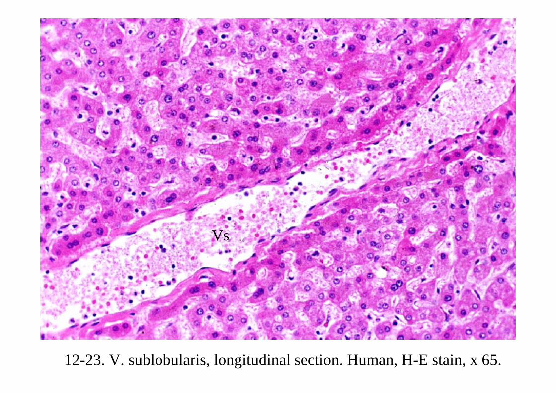
12-23. V. sublobularis, longitudinal section. Human, H-E stain, x 65.
VsVs

12-24. Hepatic cell cords, bile canaliculi and sinusoids. (Scheme).
Canal of Hering
Interlobular artery
Interlobular connective tissue
Interlobular bile duct
Vit. A storing cell of Ito
Interlobular bile duct
Canal of Hering
Interlobular vein
Stellate cells of Kupffer
EndotheliumReticularfibers
Space of Disse
Bile canaliculus
Lumen of sinusoid
Microvilli
Collagenous fibers

12-25. Hepatic cell cord and bile canaliculi 1. Human, MG stain, x 400.

12-26. Hepatic cell cords and bile canaliculi 2. Human, MG stain, x 400.
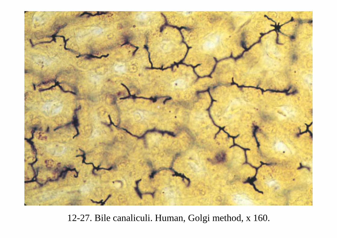
12-27. Bile canaliculi. Human, Golgi method, x 160.

12-28. Bile canaliculi and canal of Hering 1. Human, MG stain, x 330.
Bile canaliculus

12-29. Bile canaliculi and canal of Hering 2. Human, MG stain, x 400.

12-30. V. centralis and reticular fibers. Monkey, Suzuki’s silver impregnation and Kern-echtrot stain, x 64.
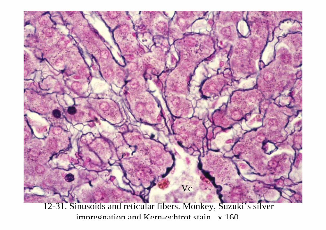
12-31. Sinusoids and reticular fibers. Monkey, Suzuki’s silver impregnation and Kern-echtrot stain, x 160.
Vc
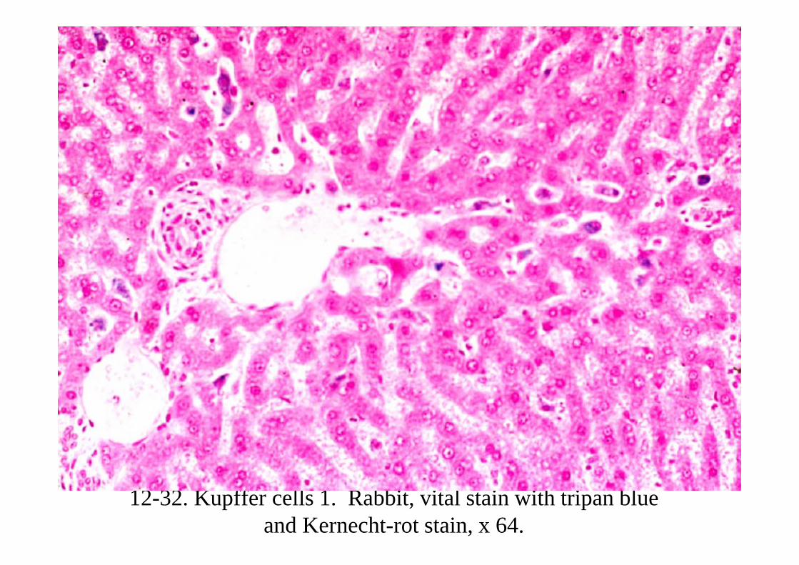
12-32. Kupffer cells 1. Rabbit, vital stain with tripan blue and Kernecht-rot stain, x 64.

12-33. Kupffer cells 2. Rabbit, vital stain with tripan blue and Kernecht-rot stain, x 250.
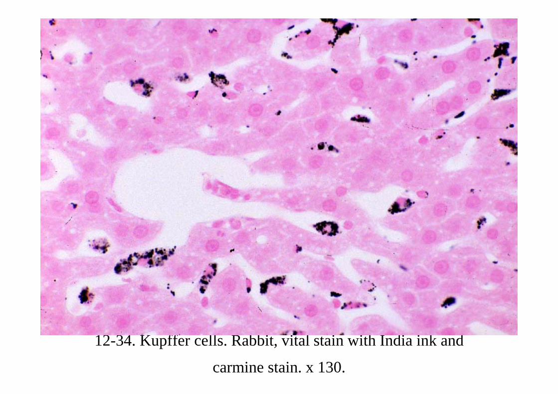
12-34. Kupffer cells. Rabbit, vital stain with India ink and
carmine stain. x 130.
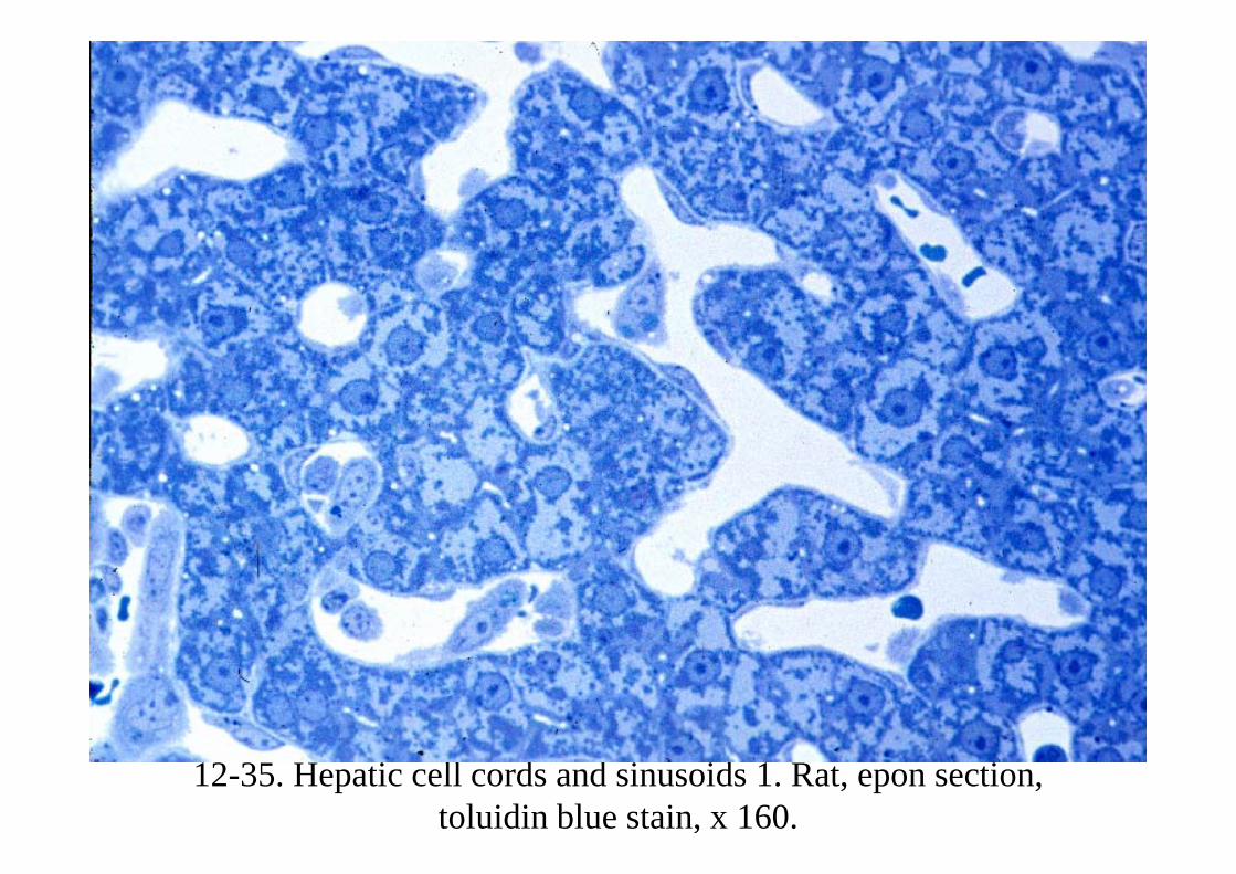
12-35. Hepatic cell cords and sinusoids 1. Rat, epon section, toluidin blue stain, x 160.

12-36. Hepatic cell cords and sinusoids 2. Rat, epon section, toluidin blue stain, x 400.
Space of Disse

12-37. Glycogen granules in hepatic cells. x 160.
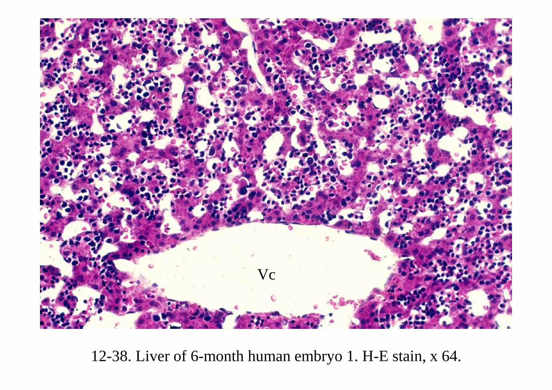
12-38. Liver of 6-month human embryo 1. H-E stain, x 64.
Vc

12-39. Liver of 6-month human embryo 2. H-E stain, x 160.
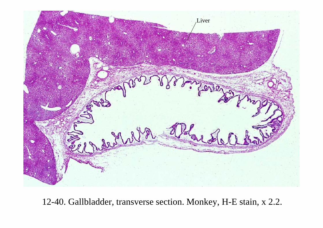
12-40. Gallbladder, transverse section. Monkey, H-E stain, x 2.2.
Liver
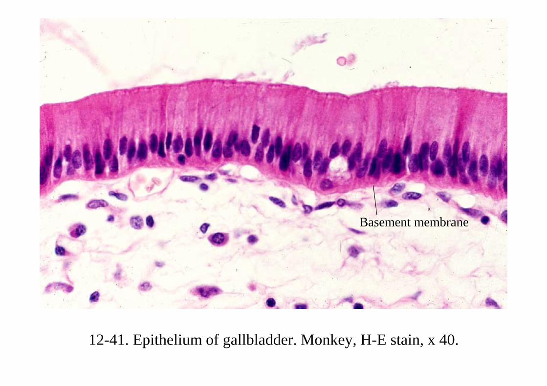
12-41. Epithelium of gallbladder. Monkey, H-E stain, x 40.
Basement membrane
12-002 Pancreas
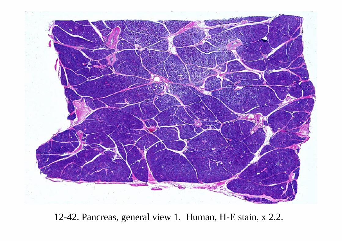
12-42. Pancreas, general view 1. Human, H-E stain, x 2.2.

12-43. Pancreas, general view 2. Human, H-E stain, x 25.

12-44. Pancreatic islet and exocrine acini. Human, H-E stain, x 64.

12-45. Exocrine acini and zymogen granules. Rabbit, toluidin blue and eosin stain, x 225.
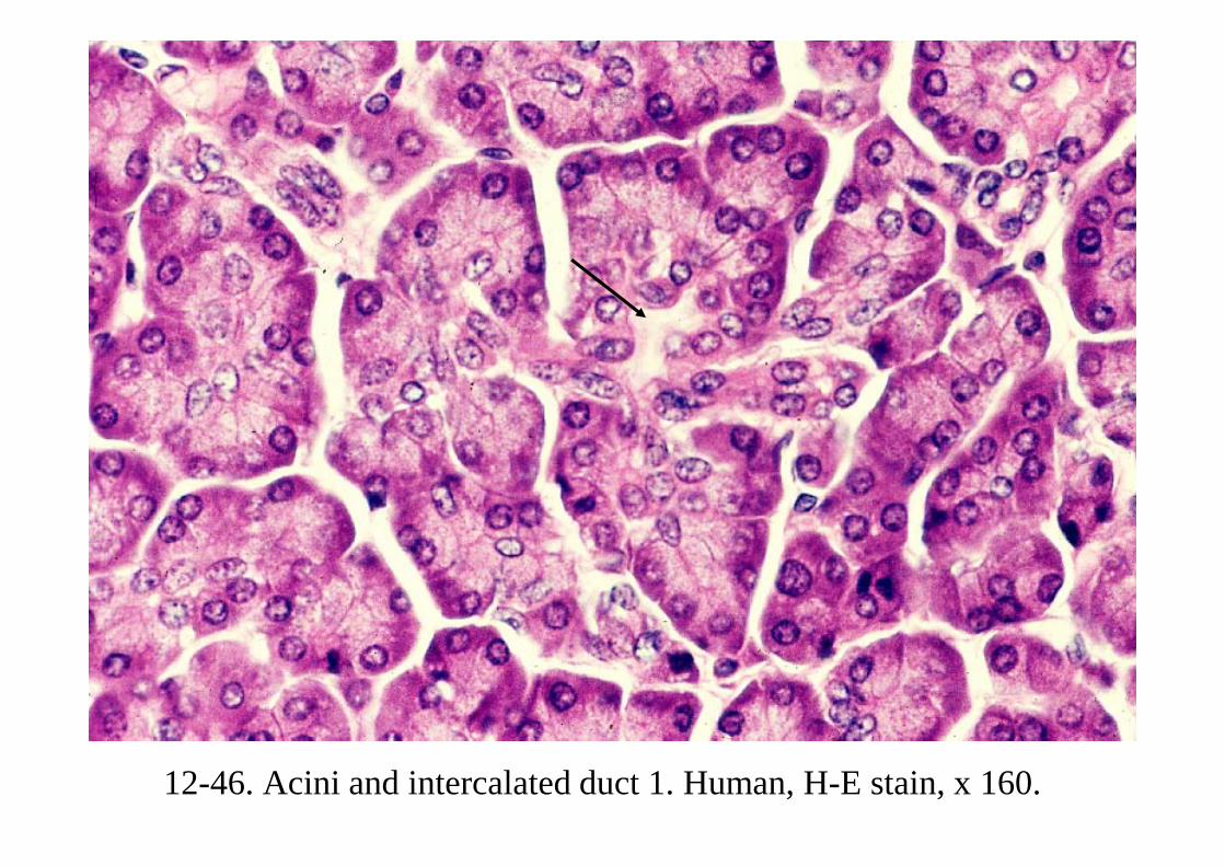
12-46. Acini and intercalated duct 1. Human, H-E stain, x 160.
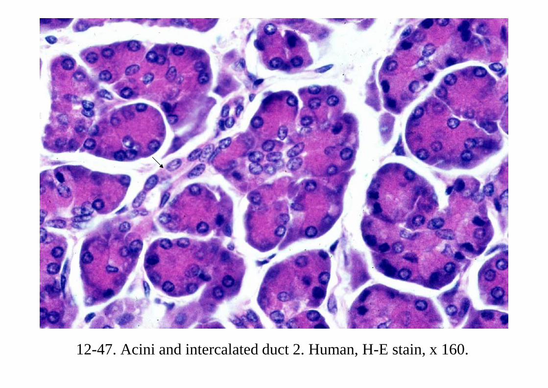
12-47. Acini and intercalated duct 2. Human, H-E stain, x 160.
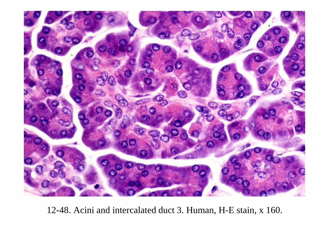
12-48. Acini and intercalated duct 3. Human, H-E stain, x 160.
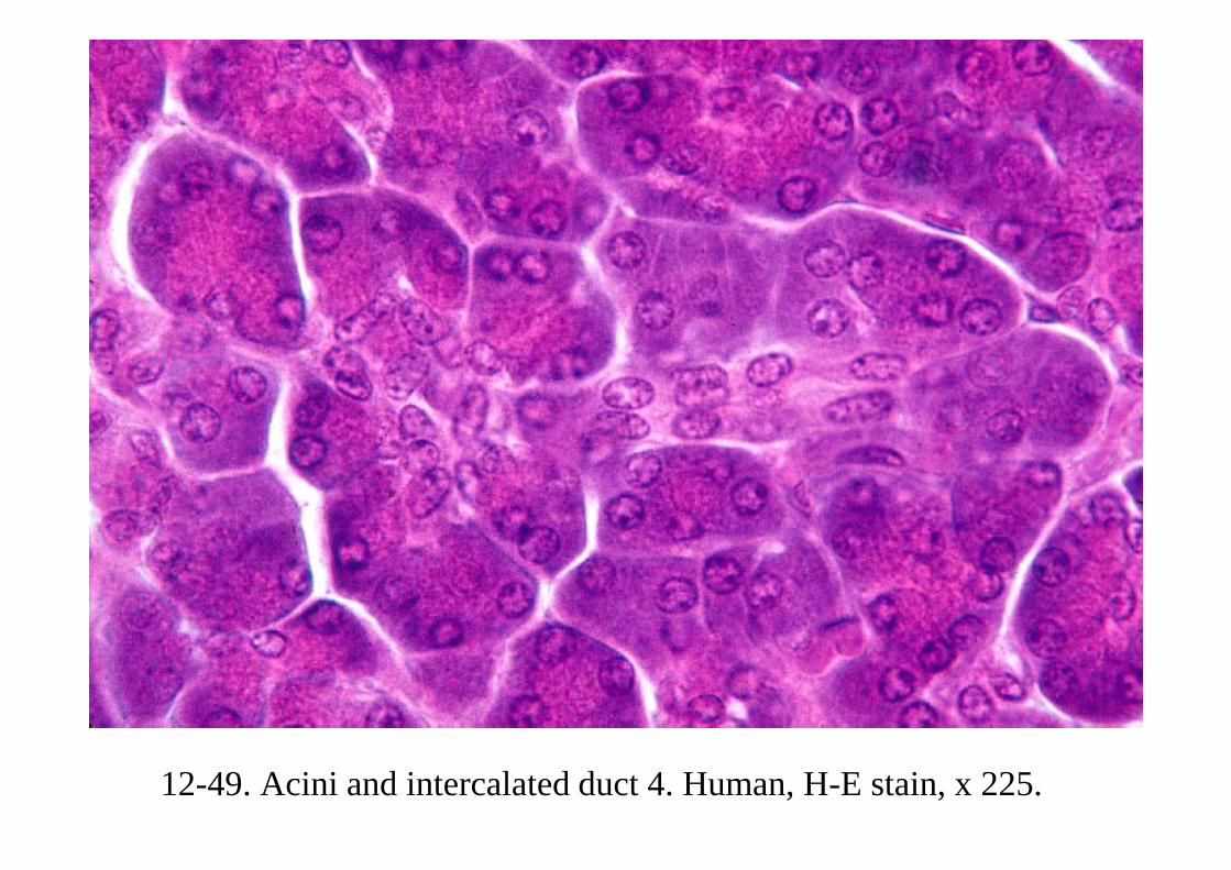
12-49. Acini and intercalated duct 4. Human, H-E stain, x 225.

12-50. Pancreatic islet 1. Human, H-E stain, x 100.
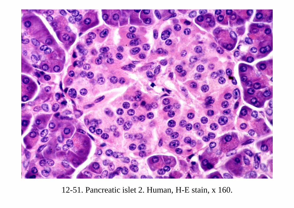
12-51. Pancreatic islet 2. Human, H-E stain, x 160.

12-52. Pancreatic islet 3. Human, MG stain, x 160.

12-53. Pancreatic islet 4. Human, victoria blue and Kernechtrot stain, x 160.
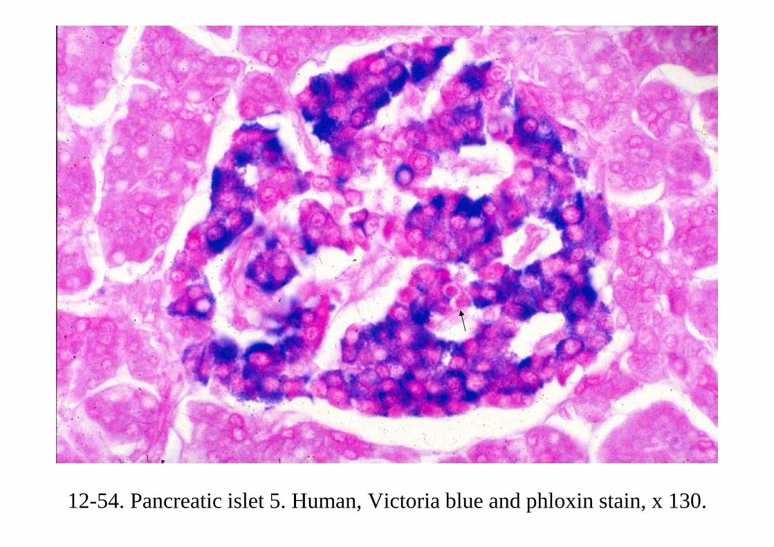
12-54. Pancreatic islet 5. Human, Victoria blue and phloxin stain, x 130.
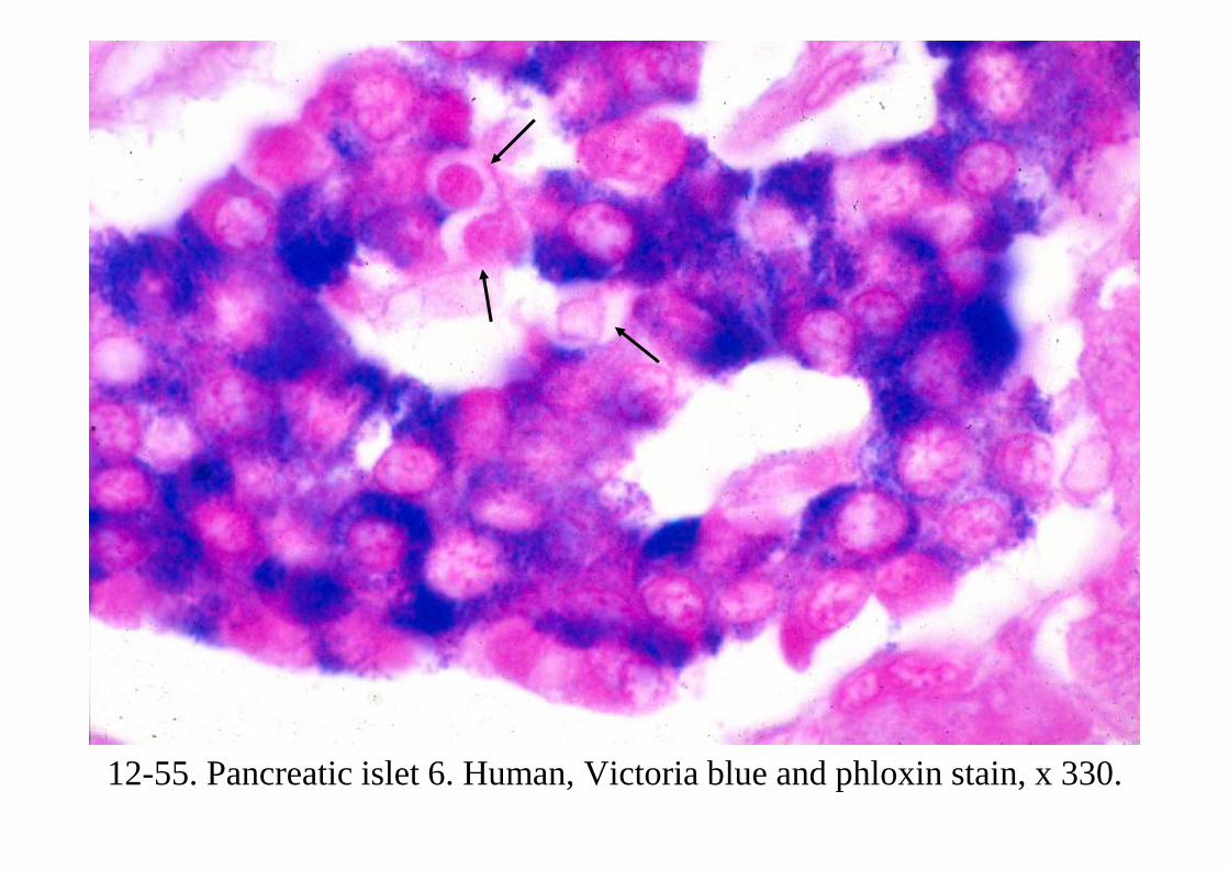
12-55. Pancreatic islet 6. Human, Victoria blue and phloxin stain, x 330.

12-56. Pancreatic islet 7. Human, Victoria blue, phloxin and light greenstain x 100

12-57. Pancreatic islet 8. Human, Victoria blue, phloxin and light green stain, x 160.
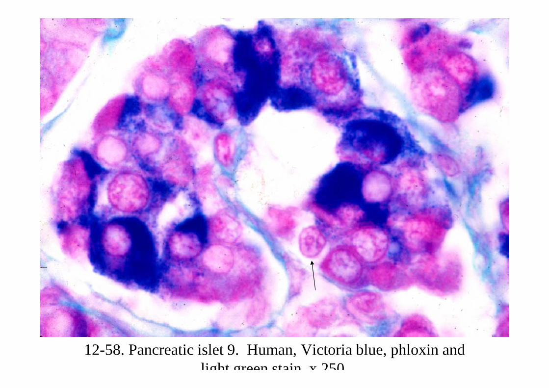
12-58. Pancreatic islet 9. Human, Victoria blue, phloxin and light green stain x 250
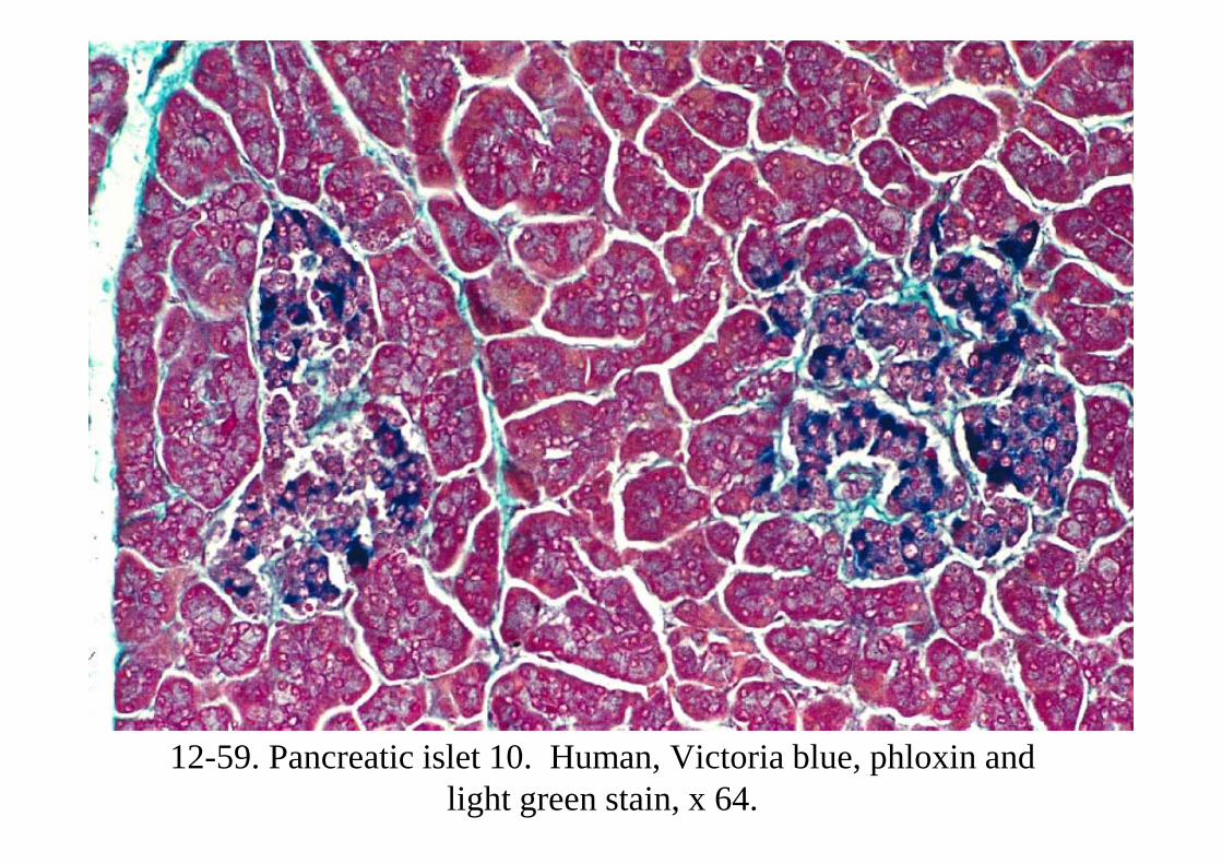
12-59. Pancreatic islet 10. Human, Victoria blue, phloxin and light green stain, x 64.
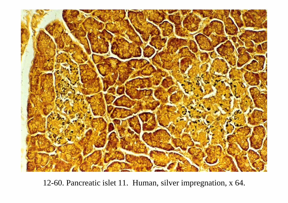
12-60. Pancreatic islet 11. Human, silver impregnation, x 64.
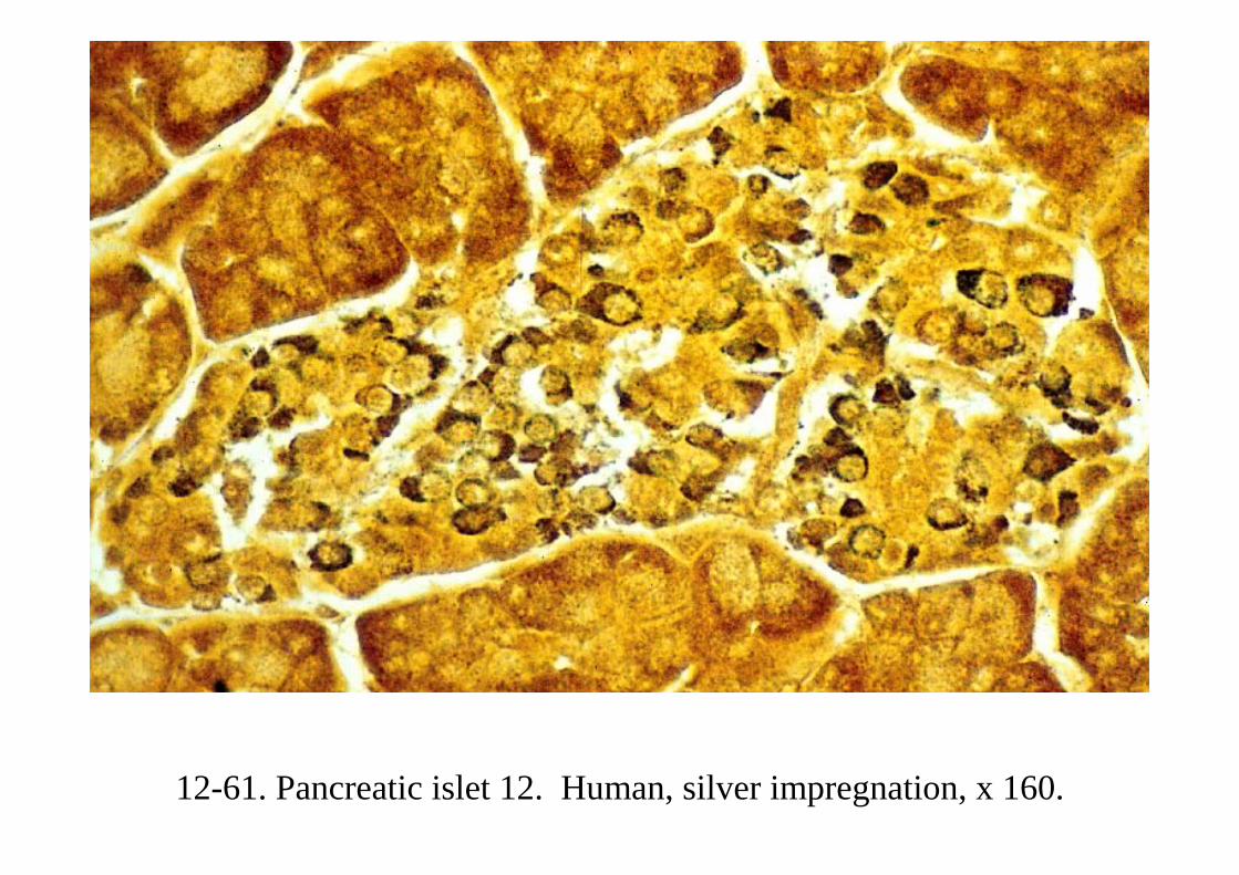
12-61. Pancreatic islet 12. Human, silver impregnation, x 160.

12-62. Pancreatic islet. Antiglucagon reaction, x 160.
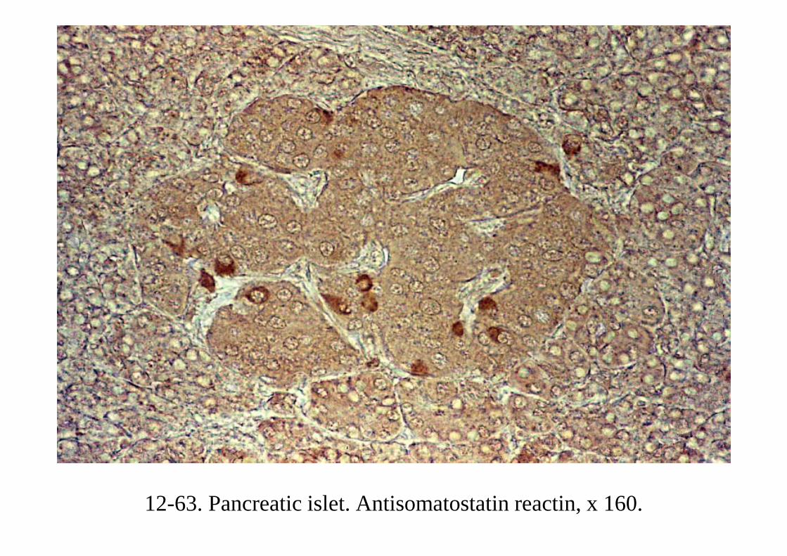
12-63. Pancreatic islet. Antisomatostatin reactin, x 160.


