Send Orders for Reprints to [email protected]12 The Open Dermatology Journal, 2015, 9, 12-20 1874-3722/15 2015 Bentham Open Open Access Trichoscopy Simplified Ebtisam Elghblawi * Dermatology OPD, STJTL, Tripoli, Libya Abstract: It has been a long while since skin surfaces and skin lesions have been examined by dermoscopy. However examining the hair and the scalp was done again recently and gained attention and slight popularity by the practical tool, namely trichoscopy, which can be called in a simplified way as a dermoscopy of the hair and the scalp. Trichoscopy is a great tool to examine and asses an active scalp disease and hair and other signs can be specific for some scalp and hair diseases. These signs include yellow dots, dystrophic hairs, cadaverized (black dots), white dots and exclamation mark hairs. Trichoscopy magnifies hair shafts at higher resolution to enable detailed examinations with measurements that a naked eye cannot distinguish nor see. Trichoscope is considered recently the newest frontier for the diagnosis of hair and scalp disease. Aim of this paper. The aim of this paper is to simplify and sum up the main trichoscopic readings and findings of hair and scalp disorders that are commonly encountered at clinic dermatology settings. Keywords: Dermoscopy, diagnosis, hair, hair loss, scalp dermoscopy, trichoscopy. INTRODUCTION Any dermatology clinic will be quite busy and in many instances faced with many patients mostly women complaining of hair loss, which can have significant effects on their self-esteem and quality of life. A normal terminal hair is identical in thickness and colour right through its length (Fig. 1). The width of normal hairs is usually more than 55 mm. Trichoscopy of normal scalp illustrates follicular units composing of 2-4 terminal hairs and 1 or 2 vellus hairs (less than 0.03 mm in width). Trichoscopy can be either performed with a handheld dermoscope or a videodermoscope (with software equipped). Trichoscopy works similar to the skin surface and at right angles to the histological plane; like the histopathology [1]. It should examine hair and scalp in all locations namely frontal, occipital, and temporal areas. The choice of fluid immersion is individual choice. Water, ultrasound gels, aqueous gels, liquid paraffin, alcohol, and oil can be used to enhance clarity and visualization. However air bubbles or paraffin can impede or blurs vision and also gels [2]. Trichoscopy aids in visualizing hair at a working magnifications of 20-fold to 70- fold, and some higher up to 160 [3, 4]. Trichoscopy is a quick, easy, priceless, novel, efficient, and a non-invasive device, which can save time and money to achieve a clear-cut diagnosis and enable treatment to start [4]. It assists visualization of the surface and beneath constitutions and colour outlines of scalp and hair [5]. It offers rapid appreciation of scalp and hair disorders, and advances diagnostic accuracy. It predicts the course of the *Address correspondence to this author at the Dermatology OPD, STJTL, Tripoli, Libya; E-mail: [email protected]Fig. (1). Normal scalp and hair (uniform shaft and colour). disease and lessens the needless need for biopsies. It can be generally alienated into hair signs, vascular patterns, pigment patterns and interfollicular patterns, all of which can denote specific disease and aid in making the proper diagnosis. Likewise recently, its function extended to aid in diagnosing some inflammatory scalp conditions like lichen planopilaris (LPP), scalp psoriasis, and discoid lupus erythematosus (DLE). TRICHOSCOPIC MAIN AND SPECIAL FINDINGS Starting off with the main clinical appreciations and then the main trichoscopic main findings and recognitions are shown (Table 1).
Abstract: It has been a long while since skin surfaces and skin lesions have been examined by dermoscopy. However examining the hair and the scalp was done again recently and gained attention and slight popularity by the practical tool, namely trichoscopy, which can be called in a simplified way as a dermoscopy of the hair and the scalp. Trichoscopy is a great tool to examine and asses an active scalp disease and hair and other signs can be specific for some scalp and hair diseases. These signs include yellow dots, dystrophic hairs, cadaverized (black dots), white dots and exclamation mark hairs.
Trichoscopy magnifies hair shafts at higher resolution to enable detailed examinations with measurements that a naked eye cannot distinguish nor see.
Trichoscope is considered recently the newest frontier for the diagnosis of hair and scalp disease.
Aim of this paper. The aim of this paper is to simplify and sum up the main trichoscopic readings and findings of hair and scalp disorders that are commonly encountered at clinic dermatology settings.
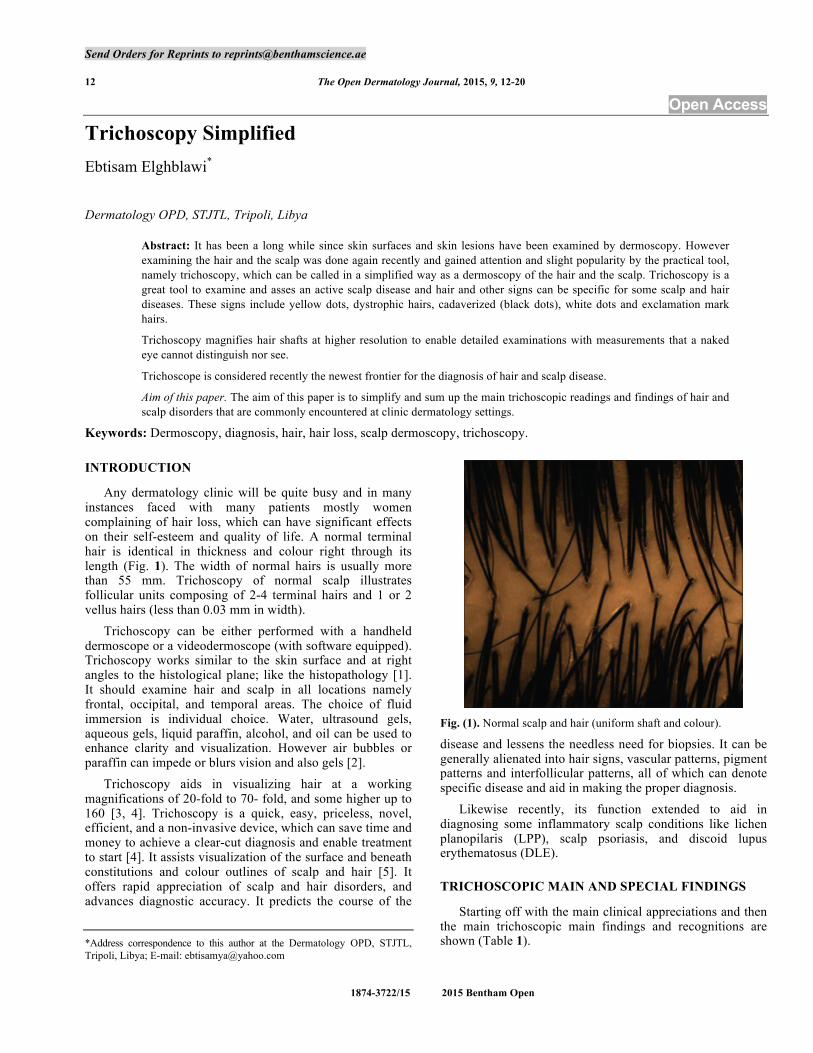
Any dermatology clinic will be quite busy and in many instances faced with many patients mostly women complaining of hair loss, which can have significant effects on their self-esteem and quality of life. A normal terminal hair is identical in thickness and colour right through its length (Fig. 1). The width of normal hairs is usually more than 55 mm. Trichoscopy of normal scalp illustrates follicular units composing of 2-4 terminal hairs and 1 or 2 vellus hairs (less than 0.03 mm in width). Trichoscopy can be either performed with a handheld dermoscope or a videodermoscope (with software equipped). Trichoscopy works similar to the skin surface and at right angles to the histological plane; like the histopathology [1]. It should examine hair and scalp in all locations namely frontal, occipital, and temporal areas. The choice of fluid immersion is individual choice. Water, ultrasound gels, aqueous gels, liquid paraffin, alcohol, and oil can be used to enhance clarity and visualization. However air bubbles or paraffin can impede or blurs vision and also gels [2]. Trichoscopy aids in visualizing hair at a working magnifications of 20-fold to 70- fold, and some higher up to 160 [3, 4]. Trichoscopy is a quick, easy, priceless, novel, efficient, and a non-invasive device, which can save time and money to achieve a clear-cut diagnosis and enable treatment to start [4]. It assists visualization of the surface and beneath constitutions and colour outlines of scalp and hair [5]. It offers rapid appreciation of scalp and hair disorders, and advances diagnostic accuracy. It predicts the course of the
*Address correspondence to this author at the Dermatology OPD, STJTL, Tripoli, Libya; E-mail: [email protected]
Fig. (1). Normal scalp and hair (uniform shaft and colour).
disease and lessens the needless need for biopsies. It can be generally alienated into hair signs, vascular patterns, pigment patterns and interfollicular patterns, all of which can denote specific disease and aid in making the proper diagnosis. Likewise recently, its function extended to aid in diagnosing some inflammatory scalp conditions like lichen planopilaris (LPP), scalp psoriasis, and discoid lupus erythematosus (DLE).
TRICHOSCOPIC MAIN AND SPECIAL FINDINGS
Starting off with the main clinical appreciations and then the main trichoscopic main findings and recognitions are shown (Table 1).
Trichoscopy Simplified The Open Dermatology Journal, 2015, Volume 9 13
Table 1. Types of alopecia.
Non-Cicatricial Alopecias Cicatricial Alopecias
Female pattern hair loss (FPHL) Telogen effluvium (TE)
Androgenetic alopecia (AGA) Alopecia areata (AA)
Alopecia areata incognita (AAI) Scalp psoriasis and seborrheic
(DLE) Frontal fibrosing alopecia (FFA) Folliculitis decalvans and tufted
folliculitis. Dissecting cellulitis (DC)
Pseudopelade Brocq
NON-CICATRICIAL ALOPECIAS
Female Pattern Hair Loss (FPHL): FPHL typically presents with noticeable patterns of hair loss, thus making a bedside diagnosis possible [6, 7].
Trichoscopic Findings
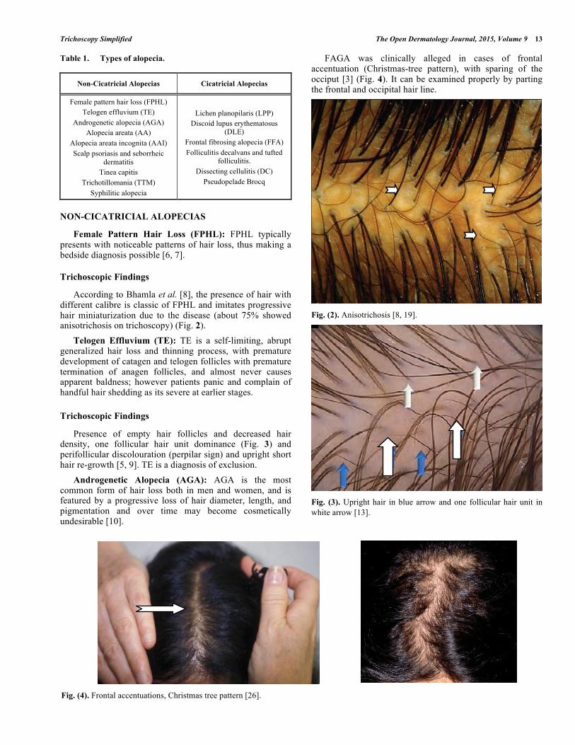
According to Bhamla et al. [8], the presence of hair with different calibre is classic of FPHL and imitates progressive hair miniaturization due to the disease (about 75% showed anisotrichosis on trichoscopy) (Fig. 2). Telogen Effluvium (TE): TE is a self-limiting, abrupt generalized hair loss and thinning process, with premature development of catagen and telogen follicles with premature termination of anagen follicles, and almost never causes apparent baldness; however patients panic and complain of handful hair shedding as its severe at earlier stages.
Trichoscopic Findings
Presence of empty hair follicles and decreased hair density, one follicular hair unit dominance (Fig. 3) and perifollicular discolouration (perpilar sign) and upright short hair re-growth [5, 9]. TE is a diagnosis of exclusion. Androgenetic Alopecia (AGA): AGA is the most common form of hair loss both in men and women, and is featured by a progressive loss of hair diameter, length, and pigmentation and over time may become cosmetically undesirable [10].
FAGA was clinically alleged in cases of frontal accentuation (Christmas-tree pattern), with sparing of the occiput [3] (Fig. 4). It can be examined properly by parting the frontal and occipital hair line.
Fig. (2). Anisotrichosis [8, 19].
Fig. (3). Upright hair in blue arrow and one follicular hair unit in white arrow [13].
Fig. (4). Frontal accentuations, Christmas tree pattern [26].
frontal and occipital hair line.
!
frontal and occipital hair line.
!
14 The Open Dermatology Journal, 2015, Volume 9 Ebtisam Elghblawi
Trichoscopic Findings
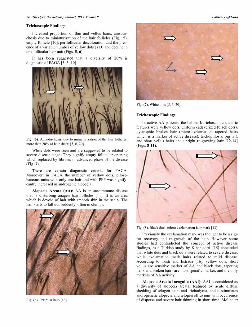
Increased proportion of thin and vellus hairs, anisotri-chosis due to miniaturization of the hair follicles (Fig. 5), empty follicle [10], perifollicular discoloration and the pres-ence of a variable number of yellow dots (YD) and decline in one follicular hair unit (Figs. 5, 6). It has been suggested that a diversity of 20% is diagnostic of FAGA [3, 5, 10].
Fig. (5). Anisotrichosis, due to miniaturization of the hair follicles; more than 20% of hair shafts [5, 6, 20].
White dots were seen and are suggested to be related to severe disease stage. They signify empty follicular opening which replaced by fibrosis in advanced phase of the disease (Fig. 7). There are certain diagnostic criteria for FAGA. Moreover, in FAGA the number of yellow dots, pilose-baceous units with only one hair and with PFP was signify-cantly increased in androgenic alopecia. Alopecia Areata (AA): AA is an autoimmune disease that is disturbing anagen hair follicles [11]. It is an area which is devoid of hair with smooth skin in the scalp. The hair starts to fall out suddenly, often in clumps.
Fig. (6). Peripilar halo [13].
Fig. (7). White dots [5, 6, 20].
Trichoscopic Findings
In active AA patients, the hallmark trichoscopic specific features were yellow dots, uniform cadaverized (black dots), dystrophic broken hair (micro-exclamation, tapered hairs which is a marker of active disease), trichoptilosis, pig tail, and short vellus hairs and upright re-growing hair [12-14] (Figs. 8-11).
Fig. (8). Black dots, micro exclamation hair mark [13].
Previously the exclamation mark was thought to be a sign for recovery and re-growth of the hair. However some studies had contradicted the concept of active disease findings, as a Turkish study by Kibar et al. [15] concluded that white dots and black dots were related to severe disease, while exclamation mark hairs related to mild disease. According to Tosti and Estrada [16], yellow dots, short vellus are sensitive marker of AA and black dots, tapering hairs and broken hairs are most specific marker, and the only markers of AA activity. Alopecia Areata Incognita (AAI): AAI is considered as a diversity of alopecia areata, featured by acute diffuse shedding of telogen hairs and trichodynia, and it stimulates androgenetic alopecia and telogen effluvium with occurrence of disperse and severe hair thinning in short time. Molina et
Trichoscopy Simplified The Open Dermatology Journal, 2015, Volume 9 15
al. [17] stated AAI affects mostly women below forty however there is some disagreement.
Fig. (9). Yellow dots [13].
Fig. (10). Exclamation mark hair [13].
Fig. (11). Pig tails [14].
Trichoscopic Findings
According to Tosti and Estrada [16], there are numerous, diffuse yellow dots of different size and uniform colours within the follicular orifices of both empty and hair-bearing with a large number of re-growing of tapered terminal hairs in the entire scalp (Fig. 12).
Fig. (12). YD in whole scalp; hairy and none hairy areas.
Scalp Psoriasis and Seborrheic Dermatitis: Psoriasis and seborrheic dermatitis are equally chronic erythemato-squamous dermatoses that can involve the scalp. It may be hard to distinguish between both clinically when it affects only the scalp [18] and thus it poses a diagnostic challenge, however involvement of frontal hair lines is distinctive for scalp psoriasis.
Trichoscopic Findings
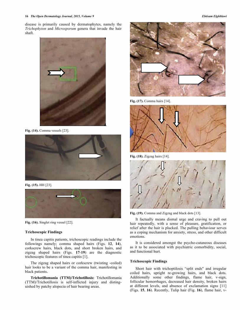
Atypical red vessels (ARV), red dots and globules (RDG), signet ring vessels (SRV), structureless red areas (SRA), glomerular vessels (GV), twisted red loops (TRL), perifollicular pigmentation (PP) and hidden hairs (HH) were seen mostly in favour of psoriasis while twisted red loops (TRL) and comma vessels (CV) were specific for seborrheic dermatitis [18, 19] (Figs. 13-16).
Fig. (13). RDG top and arborsing vessels bottom [23].
Tinea Capitis: A common condition, of superficial fungal infection of the scalp, which is seen in children. The
16 The Open Dermatology Journal, 2015, Volume 9 Ebtisam Elghblawi
disease is primarily caused by dermatophytes, namely the Trichophyton and Microsporum genera that invade the hair shaft.
Fig. (14). Comma vessels [23].
Fig. (15). HH [23].
Fig. (16). Singlet ring vessel [22].
Trichoscopic Findings
In tinea capitis patients, trichoscopic readings include the followings namely; comma shaped hairs (Figs. 12, 14), corkscrew hairs, black dots, and short broken hairs, and zigzag shaped hairs (Figs. 17-19) are the diagnostic trichoscopic features of tinea capitis [1]. The zigzag shaped hairs or corkscrew (twisting -coiled) hair looks to be a variant of the comma hair, manifesting in black patients. Trichotillomania (TTM)/Trichotillosis: Trichotillomania (TTM)/Trichotillosis is self-inflicted injury and disting-uished by patchy alopecia of hair bearing areas.
Fig. (17). Comma hairs [14].
Fig. (18). Zigzag hairs [14].
Fig. (19). Comma and Zigzag and black dots [13].
It factually means dismal urge and craving to pull out hair repeatedly, with a sense of pleasure, gratification, or relief after the hair is plucked. The pulling behaviour serves as a coping mechanism for anxiety, stress, and other difficult emotions. It is considered amongst the psycho-cutaneous diseases as it to be associated with psychiatric comorbidity, social, and functional hurt.
Trichoscopic Findings
Short hair with trichoptilosis “split ends" and irregular coiled hairs, upright re-growing hairs, and black dots. Additionally some other findings, flame hair, v-sign, follicular hemorrhages, decreased hair density, broken hairs at different levels, and absence of exclamation signs [11] (Figs. 15, 16). Recently, Tulip hair (Fig. 16), flame hair, v-
Trichoscopy Simplified The Open Dermatology Journal, 2015, Volume 9 17
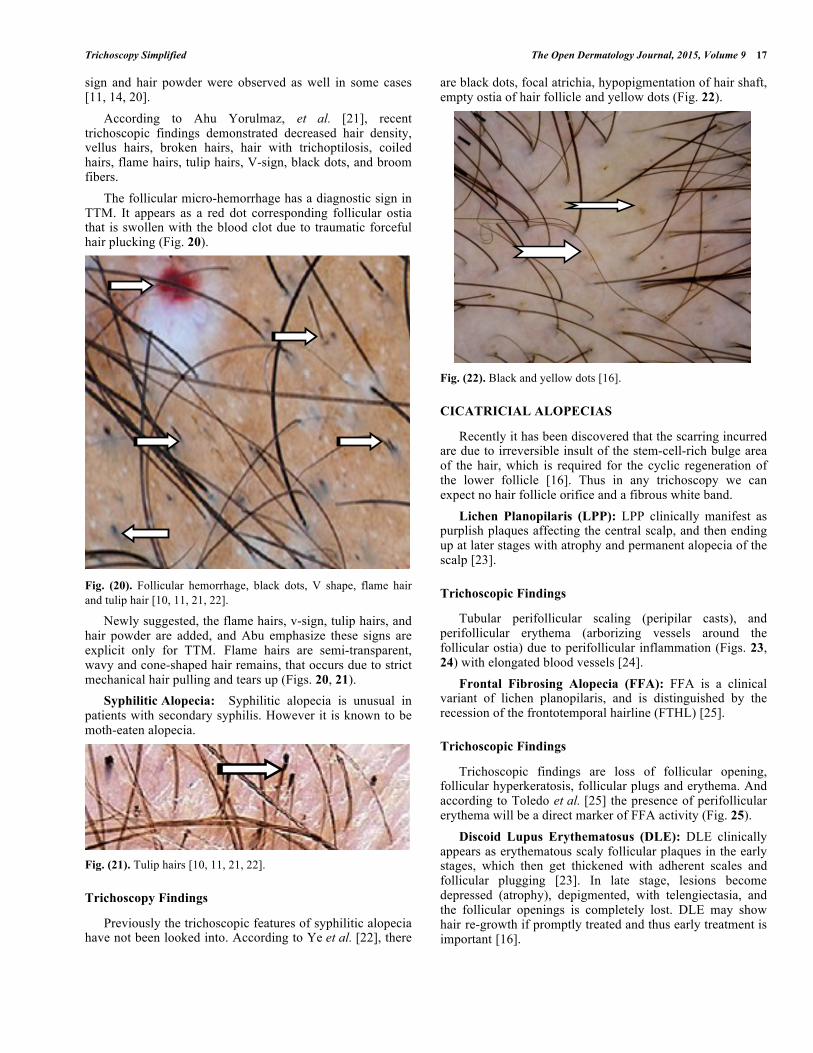
sign and hair powder were observed as well in some cases [11, 14, 20]. According to Ahu Yorulmaz, et al. [21], recent trichoscopic findings demonstrated decreased hair density, vellus hairs, broken hairs, hair with trichoptilosis, coiled hairs, flame hairs, tulip hairs, V-sign, black dots, and broom fibers. The follicular micro-hemorrhage has a diagnostic sign in TTM. It appears as a red dot corresponding follicular ostia that is swollen with the blood clot due to traumatic forceful hair plucking (Fig. 20).
Fig. (20). Follicular hemorrhage, black dots, V shape, flame hair and tulip hair [10, 11, 21, 22].
Newly suggested, the flame hairs, v-sign, tulip hairs, and hair powder are added, and Abu emphasize these signs are explicit only for TTM. Flame hairs are semi-transparent, wavy and cone-shaped hair remains, that occurs due to strict mechanical hair pulling and tears up (Figs. 20, 21). Syphilitic Alopecia: Syphilitic alopecia is unusual in patients with secondary syphilis. However it is known to be moth-eaten alopecia.
Fig. (21). Tulip hairs [10, 11, 21, 22].
Trichoscopy Findings
Previously the trichoscopic features of syphilitic alopecia have not been looked into. According to Ye et al. [22], there
are black dots, focal atrichia, hypopigmentation of hair shaft, empty ostia of hair follicle and yellow dots (Fig. 22).
Fig. (22). Black and yellow dots [16].
CICATRICIAL ALOPECIAS
Recently it has been discovered that the scarring incurred are due to irreversible insult of the stem-cell-rich bulge area of the hair, which is required for the cyclic regeneration of the lower follicle [16]. Thus in any trichoscopy we can expect no hair follicle orifice and a fibrous white band. Lichen Planopilaris (LPP): LPP clinically manifest as purplish plaques affecting the central scalp, and then ending up at later stages with atrophy and permanent alopecia of the scalp [23].
Trichoscopic Findings
Tubular perifollicular scaling (peripilar casts), and perifollicular erythema (arborizing vessels around the follicular ostia) due to perifollicular inflammation (Figs. 23, 24) with elongated blood vessels [24]. Frontal Fibrosing Alopecia (FFA): FFA is a clinical variant of lichen planopilaris, and is distinguished by the recession of the frontotemporal hairline (FTHL) [25].
Trichoscopic Findings
Trichoscopic findings are loss of follicular opening, follicular hyperkeratosis, follicular plugs and erythema. And according to Toledo et al. [25] the presence of perifollicular erythema will be a direct marker of FFA activity (Fig. 25). Discoid Lupus Erythematosus (DLE): DLE clinically appears as erythematous scaly follicular plaques in the early stages, which then get thickened with adherent scales and follicular plugging [23]. In late stage, lesions become depressed (atrophy), depigmented, with telengiectasia, and the follicular openings is completely lost. DLE may show hair re-growth if promptly treated and thus early treatment is important [16].
18 The Open Dermatology Journal, 2015, Volume 9 Ebtisam Elghblawi
Fig. (23). White dots of LPP [1].
Trichoscopic Findings
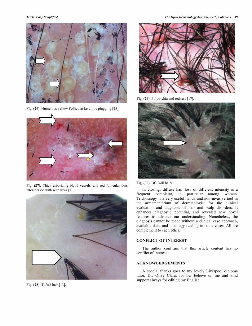
Scattered dark-brown discoloration of the skin, large yellow dots and thick arborizing vessels [24] (Figs. 26, 27). According to Tosti and Estrada [16] scalp atrophy is appreciated by a diffuse white colour of the scalp. Arborizing and tortuous vessels are seen inside DLE plaques. Red to pinkish-red, round and polycyclic dots of uniform size are often distributed around follicular openings and may be a peculiar finding as well. Folliculitis Decalvans (FD) and Tufted Folliculitis (TF): Folliculitis decalvans (baldheadedness with scarring) is a variety of alopecia that associated with scarring. It is distinguished by redness, swelling and pustules oozing pus around the hair follicle which leads to inflammation of the hair follicle (folliculitis) with damage of the hair follicle and thus permanent hair loss with scaring. It is also called tufted folliculitis (TF) [26].
Trichoscopic Findings
Multiple upright tufted hairs of 5-20 hair shafts per follicle ostia (polytrichia) with starburst pattern perifollicular hyperplasia in folliculitis decalvans [23] (Figs. 28, 29).
Fig. (24). Perifollicular scales/ casts [1].
Dissecting Cellulitis (DC): Is an uncommon condition that affects the scalp vertex and posterior neck, commonly seen in black males (skin type 5&6) aged 20-40 years, and called doll hair (Fig. 30). It starts clinically as simple folliculitis or perifolliculitis then rapidly erupts as multiple painful nodules with purulent discharge that coalesce to form interconnecting abscesses and sinuses.
Trichoscopic Findings
It is seen "3D" yellow dots imposed over dystrophic hairs in dissecting cellulitis [24]. Pseudopelade Brocq: Pseudopelade of Brocq is an unusual form of enduring alopecia of the scalp and mostly affects middle aged and older women. The cause of which is unknown. Its diagnosis by exclusion of LPP and DLE, and described as ‘foot print in snow’.
Trichoscopy Features
According to Rudnicka et al. [27], the trichoscopy features of classic pseudopelade of Brocq are nonspecific. It appears as white areas with no follicular openings. Also some solitary dystrophic hairs can be seen at the periphery of the lesion.
Fig. (27). Thick arborizing blood vessels, and red follicular dots interspersed with scar areas [1].
Fig. (28). Tufted hair [13].
Fig. (29). Polytrichia and redness [17].
Fig. (30). DC Doll hairs.
In closing, diffuse hair loss of different intensity is a frequent complaint, in particular among women. Trichoscopy is a very useful handy and non-invasive tool in the armamentarium of dermatologist for the clinical evaluation and diagnosis of hair and scalp disorders. It enhances diagnostic potential, and revealed new novel features to advance our understanding. Nonetheless, the diagnosis cannot be made without a clinical case approach, available data, and histology reading in some cases. All are complement to each other.
CONFLICT OF INTEREST
The author confirms that this article content has no conflict of interest.
ACKNOWLEDGEMENTS
A special thanks goes to my lovely Liverpool diploma tutor, Dr. Olive Clara, for her believe on me and kind support always for editing my English.
20 The Open Dermatology Journal, 2015, Volume 9 Ebtisam Elghblawi
REFERENCES
[1] El-Taweel AE, El-Esawy F, Abdel-Salam O. Different trichoscopic features of tinea capitis and alopecia areata in pediatric patients. Dermatol Res Pract 2014; 2014: 848763.
[2] Rudnicka L, Olszewska M, Rakowska A, Oledzka E, Slowinsja M. Trichoscopy: a new method for diagnosing hair loss. J Drugs Dermtol 2008; 7(7): 651-4.
[3] Rakowska A. Trichoscopy (hair and scalp videodermoscopy) in the healthy female. Method standardization and norms for measurable parameters. J Dermatol Case Rep 2009; 3(1): 14-9.
[4] Rakowska A, Slowinska M, Kowalska-Oledzka E, Olszewska M, Rudnicka L. Dermoscopy in female androgenic alopecia: method standardization and diagnostic criteria. Int J Trichol 2009; 1(2): 123-30.
[5] Pedrosa AF, Morais P, Lisboa C, Azevedo F. The importance of trichoscopy in clinical practice. Dermatol Res Pract 2013; 2013: 986970.
[6] Zhang X, Caulloo S, Zhao Y, Zhang B, Cai Z, Yang J. Female pattern hair loss: clinico-laboratory findings and trichoscopy depending on disease severity. Int J Trichol 2012; 4(1): 23-8.
[7] Herskovitz I, de Sousa IC, Tosti A. Vellus hairs in the frontal scalp in early female pattern hair loss. Int J Trichol 2013; 5(3): 118-20.
[8] Bhamla SA, Dhurat RS, Saraogi PP. Is trichoscopy a reliable tool to diagnose early female pattern hair loss? Int J Trichol 2013; 5(3): 121-5.
[9] Jain N, Doshi B, Khopkar U. Trichoscopy in alopecias: diagnosis simplified. Int J Trichol 2013; 5(4): 170-8.
[10] Galliker NA, Trüeb RM. Value of trichoscopy versus trichogram for diagnosis of female androgenetic alopecia. Int J Trichol 2012; 4(1): 19-22.
[11] Peralta L, Morais P. Photoletter to the editor: the friar tuck sign in trichotillomania. J Dermatol Case Rep 2012; 6(2): 63-4.
[12] de Moura LH, Duque-Estrada B, Abraham LS, Barcaui CB, Sodre CT. Dermoscopy findings of alopecia areata in an African-American patient. J Dermatol Case Rep 2008; 2(4): 52-4.
[13] Jain N, Doshi B, Khopkar U. Trichoscopy in alopecias: Diagnosis simplified. Int J Trichol 2013; 5(4): 170-8.
[14] Thakur BK, Verma S, Raphael V, Khonglah Y. Extensive tonsure pattern trichotillomania-trichoscopy and histopathology aid to the diagnosis. Int J Trichol 2013; 5(4): 196-8.
[15] Kibar M, Aktan S, Lebe B, Bilgin M. Trichoscopic findings in alopecia areata and their relation to disease activity, severity and clinical subtype in Turkish patients. Australas J Dermatol 2015; 56(1): e1-6.
[16] Tosti A, Duque-Estrada B. Dermoscopy in hair disorders. J Egypt Women Dermatol Soc 2009; 7(1): 1-4.
[17] Molina L, Donati A, Valente NS, Romiti R. Alopecia areata incognita. Clinics (Sao Paulo) 2011; 66(3): 513-5.
[18] Kibar M, Aktan S, Bilgin M. Dermoscopic findings in scalp psoriasis and seborrheic dermatitis; two new signs; signet ring vessel and hidden hair. Indian J Dermatol 2015; 60: 41-5.
[19] Kim G, Jung H, Kim B, et al. Dermoscopy can be useful in differentiating scalp psoriasis from seborrhoeic dermatitis. Br J Dermatol 2011; 164(3): 652-6.
[20] Ankad BS, Naidu MV, Beergouder SL, Sujana L. Trichoscopy in trichotillomania: A useful diagnostic tool. Int J Trichol 2014; 6(4): 160-3.
[21] Yorulmaz A, Artuz F, Erden O. A case of trichotillomania with recently defined trichoscopic findings. Int J Trichol 2014; 6(2): 77-9.
[22] Ye Y, Zhang X, Zhao Y, et al. The clinical and trichoscopic features of syphilitic alopecia. J Dermatol Case Rep 2014; 8(3): 78-80.
[23] Ankad BS, Beergouder SL, Moodalgiri VM. Lichen planopilaris versus discoid lupus erythematosus: a trichoscopic perspective. Int J Trichol 2013; 5(4): 204-7.
[24] Rakowska A, Slowinska M, Kowalska-Oledzka E, et al. Trichoscopy of cicatricial alopecia. J Drugs Dermatol 2012; 11(6): 753-8.
[25] Toledo-Pastrana T, Hernández MJ, Camacho Martínez FM. Perifollicular erythema as a trichoscopy sign of progression in frontal fibrosing alopecia. Int J Trichol 2013; 5(3): 151-3.
[26] Fabris MR, Melo CP, Melo DF. Folliculitis decalvans: the use of dermatoscopy as an auxiliary tool in clinical diagnosis. An Bras Dermatol 2013; 88(5): 814-6.
[27] Rudnicka L, Rakowska A, Kerzeja M, Olszewska M. Hair shafts in trichoscopy: clues for diagnosis of hair and scalp diseases. Dermatol Clin 2013; 31(4): 695-708.
This is an open access article licensed under the terms of the Creative Commons Attribution Non-Commercial License (http://creativecommons.org/licenses/by-nc/ 3.0/) which permits unrestricted, non-commercial use, distribution and reproduction in any medium, provided the work is properly cited.