Page 1
tj. -rt
T \ \
ptii-ffi t V ■>' ', >>:',' ,V ’f H» J ‘ \ *u » *<u J .,- ^>;» ’* fj
•>'t"' /• -* *' - . w
' 1 Xi\ 0 « i £ ** , < «V*,».*> i t" * ,"•r'V 5
1 «* * »
WSW
# p |
M B
*>■.
¥; v -
A, r„ - ^ *
13
Rabson, 1987; Kalish, Radin, Phair, at al^, 1983; Stroebel,
Daniel, Lau , et |1,, 1982) have recently been developed for
the diagnosis of both mycobacterial antigens, and antibodies
to mycobacteria, in serum, pleural, synovial, and
cerebrospinal fluid. These techniques are reliable and may
differentiate active tuberculosis from & variety of other
di s e a s e s ,
With very few e x c e p t i o n s , however, studies on the serology of
tuberculosis have shown that sera from a p r o p o r t i o n of
patients with n o n - m y c a b a c t e r ial diseases, and healthy
i n d i v i d u a l s , c o n t a i n a n t i b o d i e s which r e a c t with
M y c o b a c t e r i a l antigens. There a r e several reasons for this
(Grange, 1980);
i. antibodies produced in response to contact with
environmental mycobacteria)
ii. antibodies produced to antigens common to mycobacteria
and other bacteria as a result of contact with the
latter;
iii. cry]|»+ i3 Infection by environmental mycobacteria in other
diseases)
iv. unproven mycobacterial aetiology of certain other
diseases;
V. an Increase in the background level of antibodies due
to polyclonal stimulation of antibody production;
vi. a "non-immune" reaction from serum factors.
m .
f:
.
A
Page 2
Persons who develop tuberculous disease or become iniected
with tubercle bacilli demonstrate delayed hypersensitivity to
certain myeobactarial antigens (Youmans, 1986), Such
hypersensitivity can be detected by introducing small amounts
of tuberculoprotein (tuberculin; PPD) into the skin (Drutz
and Graybi 11 , 1987). In the face of competent c e l ],-mediated
immunity, however, the tuberculin skin test provides no
diagnostic information relative to acute illness unless it
can be established that the skin t e s t has converted from
negative to positive in temporal relation be the illness. A
positive skin test therefore only indicates that the patient
has experienced tuberculosis or a closely related
mycobacterial infection at some time in the past (Drutz and
Grayblll, 1987).
Positive and negative tuberculin reactions must however be
interpreted with caution, A false positive reaction may occur
as a result of conversion to tuberculin hypersensitivity by
persona* vaccinated with BCG, and due to possible
cross-reaction PPD, the oxact antigenic composition of
which is unknown, with the proteins of other mycobacteria
(Youmans, 1986), A negative skin test can signify no
tuberculosis, or an anergic immune state (Daniel) Oxtoby,
Pinto, at £ii, 1981; Ellner, 1986), caused by advanced and
disseminated tuberculosis, or immune suppression due to viral
Page 3
^ Vi; . ' ft
tAwrVV’,,'vi;'“l- \ 'j -
* «
“■ v h ' ' $ *•. A •>. .*/■
*
15
infections, lymphoreticular malignancies, Hodgkin's diseasa,
s a r c o i d o s i s , and immunosuppressive therapy (Drutz and
Graybill, 198?; Youmans, 1986).
Characterisation of the ^tuberculosis, antigens which are
responsible for eliciting antibody responses and delayed
hypersensitivity In infected persons is therefore necessary
in order to reduce the low sensitivity and non-specificity of
currently available diagnostic tests, Ideally, mycobacterial
antigens which can be characterised to ihe level of protein
or po l y p e p t i d e , and which are unique to Mi.tubercul.osi,s, could
act as specific tools for the diagnosis of mycobacterial
i nfection.
id> PREVEHTI0N„AND_VACC1NATX0N- PRDGRAMMES
The basic principle involved in the prevention of
tuberculosis focuses on procedures that prevent contact
between susceptible persons and the diseased carrier
(Youmans, 1986). Intensive efforts have been made to control
tuberculosis by these means. Mass chest roenterogram
screening programmes have been conducted in order to detect
persons with pathologic pulmonary findin"'*/ (Youmans* 1986).
Diseased persons diagnosed in this way have then been
intensively treated with chemotherapeutic agents to render
them non-infectious (Youmans, 1986). In many co m m u n i t i e s ,
tuberculin testing surveys of populations are conducted
Page 4
regularly. Those found to be tuberculin positive.are then
further4 bested and treated if found to be infected with
&ut1Ufe&,££Ei.2§i.S (Druta. and Craybill, 1987).
The application cf these procedures for the detection,
isolation and treatment of tuberculosis has resulted in a
steady drop in the Incidence and prevalence of, ana mortality
from, tuberculosis in the Western world (Youmans, 1986).
However, most cases of tuberculosis have been found in areas
where ther*} is a low Icjvel of education, lack of, sanitation,
overpopulation, and o v e r c r o w d ! n g . The preventive programmes
described above are enormously expensive, present problems
with tho full cooperation of everyone in the targeted
population groupsi and require facilities for the detection
of tuberculosis and the treatment of known cases. These
programmes are therefore of limited benefit to those
populations most at risk. Other means of control in such
populations must therefore be found.
An important approach to the prevention of tuberculosis is
vaccination against tho disease. A vaccine is available in
the form of BCG (Bacillus Calmette-Guerin), BCG is an
attenuated mutant of a virulent strain of Mi.bovi_s, developed
in the early years of the twentieth century (Calmette, 1931).
BCG vaccination against tuberculosis has been used for many
years in practically all national programmes (Youmans, 1986).
A high level of immunity can be induced, which persists for
Page 5
* ' - .**l 9- n , * ' **>*' ’ /. ‘ .> ' >■• ^
' rt £ ** i sf *> •
‘SI*
■ ■•'•: .■:.■■• ■ W-i.; . "■" ■■ ■«*' -LlL -11 *A» ' " '\*•« . ^ ■ t. - *K' - * 's. ..
^ * ;4* : % ' \ X ■
% v - ' v . '' °i ’A C /*V ^ ’W / ’
17
as long as 10 years after a single vaccination with BCG
(Youmans, 1986). Although it has been instrumental in
controlling tuberculosis in the Western woi'ld* BCG has,
however*, proved completely inefficient in providing
protection against M^tubercul_osi_s infections in a large-scale
clinical trial in rural Southern India (Ten Dam, 1984). In
addition, vaccination with SCG induces tuberculin
hypersensitivity in the recipient, and as such a valuable
diagnostic tool may be lost.
It is thus of considerable importance that a standardised,
specific and effective vaccine be found against
N. tuberculosis.
Page 6
i•? m v c o b a c t e r i a l _ a n t i g e n s
An important feature in the pathogenesis pf tuberculosis is
the nature and specificity of the response of the immune
system tb antigens of tubercle bacilli. These antigens are
nuraerous, but as yet have been poorly defined. A variety of
polysaccharide and protein antigens from mycobacteria have
been described (Young, 1988; Chaparas, 1982; Daniel and
J a n i c k i , 1978). There is currently no standard nomenclature
for mycobacterial antigens; different investigators use
different codes based on various reference systems. The
methods used to isolate mycobacterial antigens to date have
largely employed physiochemical fractionation, including
ion-exchange chromatography, molecular exclusion
chromatography, density gradient ultracentrifugation,
isoelectric focussing, and zonal electrophoresis (Daniel and
Janickij 1978). Salt or solvent solubility has often been
used'in combination with these techniques (Daniel and
J a n i c k i , 1978).
It was not until the work of Janicki and his col 1aborators
(1971) that a widely and readily applied system of
identification and nomenclature for individual mycobacterial
antigens became available. Their work was based on a
relatively simple Immunoelectrophoresis technique, offering
the advantage of ready applicability with easily obtainable
materials (Daniel and Janicki, 1978). Such
Page 7
■■urtgy-
' :. ’ .. 2.:.* ', V'v"** ,*.1 ”«{>■ •*» ‘V'f "/’’ •’,
•* m , r -
, M 4%' - >-.,
BSSyS';. -• *,* ' *' • assy**. 3^ <(■-j-i* J s « ' t,
xjf ■'■ •
J*y < *» ^|, V*"* <■' ’t^ ■ • '».-■"
6; # , ’• , aa , ►» , ‘ -I fA, * r _ * A , 5
.'„.V „
19
m mm m i
J W M , .„, w l f . 1
W i f e*' ' V
I m m u n o e l e c t r o p h o r e s i s techniques have been used to identify
11 major mycobacterial antigens (Daniel and Janicki, 1978). A
second reference system based on crossed immuno
electrophoresis (CIE) was more recently introduced (Gloss,
Harboe, Axelsen, et a l y 1980) It has certain advantages over
other characterisation systems in that it covers a large
range of antigen molecules; it has been particularly useful
for identifying antigens present in mycobacterial culture
filtrates. Significant progress has been made in the
purification and analysis of several of the antigens
originally defined by CIE (antigens MPB64, MPB70 , and MPB80
of M.boifis BCG) , and partial or complete sequence data has
been obtained in some cases (Wiker, Harboe, Bennedsen, et
tii.i 1988; Harboe, N a g a i , Patarroyo, et al._, 1986).
Disadvantages of the CIE system however, include low
reproducibility of results, and the identification of single
precipitin lines corresponding to multi-molecular structures
Cfot* example, lin e 7 of the £Lle£jrae CIE system has been
s h o w n to represent not one, but 3 antigens subsequently
identified using monoclonal antibodies) (Engers, Abe, Bloom,
et al 1985) .
Cell wall polysaccharides, proteins and peptides have all
been shown to be antigenic under different experimental
conditions (Daniel and Janicki, 1978). With respect to
polysaccharide antigens (Table 1), however, the ability to
eli'iit delayed hypersensitivity reactions remains doubtful
J i ..“ i'Sb'-V' -A > -id*. - > *. : ■ •' • '■ •: ■*2L \ ■ , ,t "! 3 ‘ ‘ ’ ’ * » •' ^ 'J " v ’ . '■ ;Vrtfj# . ' 4 I , s
V
•i1
■■V'•‘*i '■*
i* - - \ ,
t :
Page 8
TABLE 1 M^e6bactefUI_carboh^drate-an^i.gens (Young, 1988)
Antigen class Distribution Location
Glyeopeptidclipids IjKaviumR^TnUracGl l_ar e
u l.aoeu m
M^^ar atubercu I_os i.s, Tspecies specific)
capsule
Phenolic glycolipids H^lefir.aaM. kansasii
capsule
M^bovis M.tuberculosis (canetti) Tspecies specific)
Lipooligosaccharides Mi.!i§Q.S§Sii
M^szulgal,
Hi.S£!lS£i Tspecies specific)
capsule
Phosphatidyl inositol all mycobacteria
mannosides
membrane
Arabinogaiactan all mycobacteria cell wall
Lipoarabinomannan all mycobacteria cell wall, capsule
Page 9
Vfci- /w*?« * VT"J f, “. |f .' '*' ■ ■ "’ ■ * ■» ’■~'1;j£''' •'- '» f;-'-'- '".»' - •':*• - ■ b "'■'- ’
-< .•.<> ■ *. - i : >•>’■-. ■ - ’ 5 1 -ic.'"* *. • ,J» ■* i * '* M*" “:-V ' ‘ ‘i ■
- ■ ■*«. /Y-V , ■' . •
S»W'A ' %S'\ ■ r
L>’- ‘ - *..,>'7 ’*V’ :='.r . ' .>•
J r 4 •••-,*•;*« *" *> * v -t v- *«
•;T A?’1 jtf'r 1 $„.?**JW {ft-* p F "
■' '-.,a.‘!
#} \
i ■-.
21
arid may be limited to guinea pig models only (Daniel and
J a n i c k i , 1978). In addition, these antigens have been shown
to be non-specific, being shared by all species of
mycobacteria, and by nacardia and eorynebacteria as well
(Young, 1988). Cell wall arabinogalactan and arabinomannan
have been found to be excellent antigens in serological
systems, and the antigenic determinant of arabinogalactan has
been identified to be a major arabinose side chain (Daniel
and J a n i c k i , 1978).
Some mycobacterial antigens have been shown to be
species-specific (Daniel and Janicki, 1978; Young, 1988).
Antigens I and 2 (arabinogalactan 2) have been identified to
be polysaccharide antigens and have been found to be widely
distributed among the mycobacteria (Daniel and Janicki,
1978). Antigens 6, 7, and 8 have also found to be present in
culture filtrates from the majority of mycobacterial species
studied (Daniel and Janicki, 1978). Antigen 5, a cytoplasmic
protein antigen, has been demonstrated to be limited to
H. tuber>ju 1 osi_s and M_>.bovi.s (Daniel and Janicki, 1978).
Antigen 6 has been shown to contain at least 2 antigenic
determinants, one of which appears to be specific for
M. tubercul_qsi.s, and the other of which is present on several
other mycobacteria (Daniel and Janicki, 1978).
It is unreasonable to expect real success in antigen
purification using physiochemical techniques, as the a n t i g e n
In*
tt, •
v'-f
$
V ,
„w
My
I \
!>. C-
ii /..“V
,'Ti. | *r r
''t '1; iv %«v
Page 10
’ :5~ 'r-‘' '’' ’/ -F* • ,1 , r, '.*• >; **'• . .-
■ • M * V - ■- • ' \ : *> ' > it - ;■ •fSfe t- .- :,v , c . -
V«" ! >\ * ‘St ’ 't' ‘k * V>- v V ,*T * *'* ' li> ‘ .» "4 L* ,
...
o o
purified by such means have a greater or lesser extent of
molecular heterogeneity. All antigenic materials .ire derived
from physically disrupted or* lysed cells, and there is no
reason to expect that their release is a uniform process;
many antigens are probably derived from the cell wall, where
repeating units abound, and individual antigenic determinants
may be expected to be present on various-raised fragments in
varying combinations and numbers (Daniel and Janickl, 1978).
Purified, well characterised, and standardisable antigens are
necessary for the accurate diagnosis of tuberculosis, and
also for the assessment of cell-mediated immunological
responses to tuberculous Infection, A means to survey all of
the protean antigens of {^tuber cu 1 os is,, without carrying out
a rigorous biochemical isolation of each component, could
facilitate the identification of specific diagnostic tools,
And would be a useful prerequisite for the selection of a
polypeptide vaccine candidate for further study,
T .
Page 11
I* '>t. • ' v t ' v • , , *A,",• -V. . ** f) ' * , ‘ i . ■'* - ’I ,* s & / ,•' i, . % * t • \ ,, x .
«ir ■•:,?> i*:>
ii*,\
7 !
23
11.8 MYCOBAGfERXAL_ANTIGENS^IDENTXFIED_WnH_!lQMgCL
ANT IBODX„M!2-S1£QM1IMANT^DNA_.TECHNOLOGY
Recombinant DNA technology offers an effective strategy to
thoroughly and systematically examine the antigens encoded in
linear segments of the M^tubercul_gsi_s genome, and the
development of mottoclonal antibody technology has provided a
means for producing monospecific antibody reagents which can
be used to dissect mycobacterial antigens (Kolk, Ho, Klatser,
et a L 9 8 4 ) . Application of these techniques for the
analysis of mycobacterial antigens has resulted in the
identification and detailed structural characterisation of a
variety of protein antigens from !3i.t ubercu l.os is , M^isErae,
and H._bgvi.s, in addition to the previously described
carbohydrate and glycolipid components. DNA from
M. tubercu 1 os i_s , M.i.l.eg.rae , and M,i.k9!ii.=L was first cloned into
IS£herlchi>a i_cal.i (E^coli) using standard plasmid, cosmid,
or IN bacteriophage vectors (Jacobs, Docherty, Curtiss, et
ali., 1986; Clark-Curtiss, Jacobs, Docherty, et_al._, 1985;
Bhattacharya and Bhattachdrya, 1984; Thole, Dauwerse, Das, et
ali., 1985). These studies showed that mycobacterial promoters
were (mostly) recognised weakly, if at all, by the E^co^i
transcriptional machinery, but that mycobacterial
translational signals, however, were expressed
satisfactorily. More recent experiments, however, have
Indicated that although moat of the recombinant mycobacterial
proteins identified to date have been fusion proteins, some
\ X W :■ '
V ' /W'* < ,
I 1
m >' ~'">y '■' '’X. *<>•
<*n "*‘
«f;,s
4'V. '.!
\
\
*i
%
I
Page 12
24
r e c o m b i n a n t , proteins were capable of being expressed from
their own signals (Shinniek, 1987; Cohen, Mayer, Rumsohlag,
e t _a lti,, 1987 J Lamb, Kingston, Estrada-Garcia , §.t_alx , 1988),
These latter studies suggest that some mycobacterial
promoters are functional in E«.eali.
The combination of monoclonal antibodies and the *)s gt 11
expression system has been a powerful influence on the
progress made in the identification of novel mycobacterial
antigens over the past few years, and recombinant clones
expressing antigenic determinants recognised by monoclonal
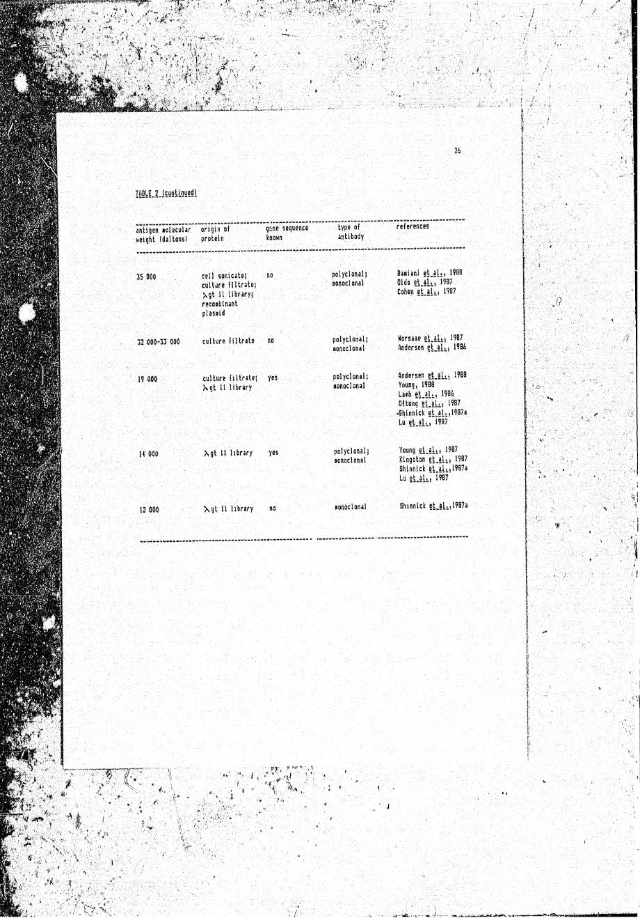
antibodies have been isolated (Young, 1988) (Table 2). The
presence of shared, epitopes among mycobacterial antigens has
boon demonstrated using monoclonal antibodies (Daniel and
Olds, 1985; Ivanyi. , Sinha, Aston, et_aj,._, 1983; Hudson and
Young, 1987; Anderson, Barry, and Buchanan, 1988). A panel of
monoclonal antibodies now exists which are able to recognise
antigens unique to (Engera, Abe, Bloom, et al^,
I9SS). Less success has been achieved in the development at
species-specific probes for M t ube re u 1. os i s . Although some
antigenic diversity has been demonstrated, (Coates, Allen,
Hewitt et alj., 1981), the majority of the monoclonal
antibodies used failed to distinguish between M^-tube^culosis
Hi.k21i.LS. BCG (Young, 1988).
Page 13
*•*. waj.pw* -r y'<*** jr 5’*t"'T. psij?jps»{«pjp®pp 3jp S5!W!W!y !! ^*, / s < ■ J-rr,t,’' > ' .'
25
ISIIU !jitub|CEHl9Sli.E2lSiB5.iiii!UiisL!!5i!!!1.3!lUk9iiiS
antigen nolecular origin of gene sequence type of references
Haight (ialions! protein iinoHn antibody
n m culture filtrates yes
Xgt 11 library
48 m culture filtrate no
45 000 ieltsanUatss yes Xgt 1! library
55 COO moaMnint nophsjid
19 000 culture filtrate no
38 600 cell sonicate) yes culture filtrate)
Ngt 11 library
polyclonal)aonoclonal
Young et_aL, 1987 Shirtnick et.ah, 1907
Mersen et al., 19B&
Lu iLUti'iw
polyclonal!
Mnoclonal
Collins et al., 1988
polyclonals
nonodonal
polyclonal
polyclonal;
tonodonal
polyclonal!oonoc1onal
Qftung e t j l u 1987 S M n n i c f g t . i l j 1987a Uflh e t . a l !t) i ? 8 6S h i n n i c k i 1 9 0 7 S h i n n i c k t U L i I W Andersen t L l l i i IW®Shinnick et.lL) 1?88Young t t . l U i 1W7 Oaalani e t a i M 1988 Lu etjlii” ?
Colien gt.aL, 19S7
H orsaae g t _ a l i ( 1987
Young e t j l t t 19 8 4 Andersen § L § l i | 19B8 young e t » a l i t 19B7 Andersen e t j i i i 1^8 6
.ucontinuetl on next page
Page 14
26
TABLE 2 (continued)^
a n l i q e r « o i e c u U r " “origin'of. . . . . . . . gtne sequence type of references
■weisht (daltons) protein known antibody
35 000 cell sonicate! culture filtratej
X g t 11 library! recoabinant piasaid
no polyclonaljnonoclonal
Daaiani et.al., 198B Olds e t J L , 1907 Cahen e t . a h , 1907
19 000
culture filtrate no
culture filtrate!
>x gt 11 library
yes
polyclonal;aonoclonal
polyclonaljnonoclonal
Horsaae e t j h , 1907 Andersen e t . a h , 190&
Andersen et_al(, 1908
Young, 1900 Lanb e t j L t W 8 6 Qftung e t . a L , 1987 >Shinnicfc et.ai!i(1907a l.u e t . a h , 1987
H 000 gt 11 library yes polyclonaljnonoclonal
Young e t . a h j 1907 Kingston et.allt 1987 Shinnick e t j L , 1 9 8 7 a
Lu S L i U i 1907
12 000 X g t U library no monoclonal Shinnick e t . a h , 1987a
Page 15
In addition to screening antigens with monoclonal antibodies,
mycobSu-:ter I al antigens have also been identified using
p o i y c U n a i antisera (Thole, Dauwerse, Das, et al.., 1985;
Young, Kent, and Young, 1987; Young and Davis, 1983), The
majority of the antigens identified with polyclonal
antibodies have been shown to overlap with the proteins
identified using monoclonal antibodies, The mycobacterial
antigens identified to date using antibody probes are shown
in Table 2.
It is important to note that the use. of antibodies as probes
way result in a focus of attention on antigens which are
important in the humoral response, but which may play only a
minor rol« in the T cell response, The human T cell response
has heon assessed using purified mycobacterial antigens
t Y o u n g , K e n t , Rees, £t | L , 1986; Ottenhof, Klatser, Ivanyi ,
ft a 1 . i 1986) . or antigens expressed in recombinant DNA
.•iion^s (Emmrich, Thole, van E m b d e n , at al*.* 1986; Mustafa,
Gill, Ner I and , fit a K , 19 S 6; Oftung, Mustafa, Husson, et a.U,
1937; Thole, van S c h o o t e n , Keulen, et al^, 1988), T cell
recognition, as judged by lymphocyte proliferation, has been
demonstrated for thus* antigens (Table 3), Limiting dilution
analysis has shown that T cells which recognise the 65 kDa
protein make up a major portion (20*/.) of the total
antimyeofaacterial T cell response following immunisation of
mice with M. fcufeia.ftfulo‘vl.-a (Kaulfmann, Vath, Thole, et, al.^,
1987),
Page 16
* * t * * * t'P’ * • *-.....’ l . ■ »• • •■ • ■
•'*,, i',! » |v ^ » ' '
■mi'-A **,v. ' -
r*v,. -v':. ■.-:; .-'tk-- 5iP.\
4*& " ,* * ? v .j * ^
* -r f . 1 J V£*W *..,. • * -',■ *■ »t ■' ’• "■ ' '■>"'■
~ 20
IfilUj 0ilyl§C£!lIS§lLfiC9i§lOL!iM£b„5Ul!!iiiE.tbLECSliiECitL2!!-Si.LLi!SE!!2M5l
sf)tig**fi aolecular T Tyapiiocyte antiqun source references
weight (daltons) populations
£9 009 polyclonal purified antigen Coil ins e t j l . , 1938
45 000 polyclonal( SD5 blot; D a d a n i e t _ a h , 1900clone recQabinant clone. laab et a l . , 1904
0 ft u n g "§ tj L i 1907 Kaufiann e t j K , 1907
5 2 030*55 000 ; clone SDS blot LaaB and Younij, 1907
3 9 4 0 8 ■. polyclonal' purified antigen Horsaae j t . a U , 1907l-aib e t j L , 1904
38 flOO polyclonal purified antigen Young e ^ a U , 1906
IS 000 . polyclonal SDS blot Daiiani et.al., 1900
12 000^33 000 polyclonal purified antigen Kingston et ajL, ;)07
Young i L i l l i ’i w
28 000 polyclonal SOS dlot Daiiani et.al,,,, 1900
(9 000 polyclonal) recdabinant d o n e Laib e t j h , 1904clone Oftung et.al,,, 1987
Horsaae et.al,,,, 1907
, continued on next page
Page 17
antigen 'oo lecu la r ~ f.ya phdcy te * ! gen source references
Height (daltons) populations
U 000-18 000 clone recoabinant d o n s Mustafa e t a h , I9B6Laab and Young, 1937
14 000 polyclonal; purified antigen; Oftung e t . a K , 1987clone recoabinant clone Kaufaann e t _ a L , 19B7
Kingston e t _ a h , 19B7
' ?’•
' , , ' ' 7 ,. “a. yi4-' t 1 t • 1 •* ‘ •• 1 ^ 'tf r ,
■ ' ■ -< '■ " H \ ; ■' v
' , •_ i ■ ■;. • ; / . v- > ■v ■ ’• *"•’ • • ■*; • ■ ' t
Page 18
Techniques have also been developed to allow the
identification of antigens by direct screening with T cells.
Antigens separated by SDS*-polyacrylamide gel electrophoresis
(SDS— PAGE) and transferred to nitrocellulose membranes have
been assessed for recognition by uloned or polyclonal T cell
populations (Young and Lamb, 1986; Abou-Zeid, Filley, Steele
et a L , 198?) (Table 3). Analysis of T cell clones in this
way is analogous to the identification of antigens using a
panel of monoclonal antibodies, and has resulted in the
identification of several novel antigenic specificities
(Young, 1988). ■
Screening of SDS-PAGE fractionated antigens u s i n g polyclonal
T cell populations has provided a unique important approach
to the identifleation of i mmunodominant, biologically active
antigens of important biological function (Young, 1988). .
Comparison of recognition patterns from patients, BCG
vaccineas, and healthy individuals may provide a means for
assessment of the relative contribution of individual
polypeptides \l,o the overall cellular immune response (Young,
1988).
Recombinant DNA technology, monoclonal and polyclonal
antibody, and T cell-based assays provide a novel source of
mycobacterial antigens. Thess can be subsequently purified
using the appropriate antibodies, and can be analysed by T
cell recogrition assays. In addition, sequence analysis,
Page 19
31
epitope mapping, and analysis of protein function (Young,
1988) for the antigens are now possible with use of such
technology. The applications of these antigens include;
— taxonomic.' assignment of-.mycobacteria-, based on antigen
and DNA characteristics;
- identification and isolation of antigens with diagnostic
and/or protective potential;
large-scale production of such antigens, and the
production of synthetic p e p f d e s produced on the basis
of identified nucleotide and amino acid sequences.
Page 20
1 • 2. REC 0M31N ANT^DN AZTECH NOLOG Y
Recombinant DNA technology has been defined as "the formation
q£ new combinations of heritable material by the insertion of
n u c l e i c acid molecules, produced by whatever means outside
the cell, into any virus, bacterial plasmid, or other vector
s ystem so as to allow their incorporation into a host
organism In which t h e y do n o t naturally occur, bu'c in which
they are: capable of continued p ropagation” (Seckl , 1985).
Clcning involves the in_vitro joining of passenger DNA to
vector DNA, and the propagation of the resulting hybrid
molecules into suitable host cells to obtain a clone of
identical cells harbouring recombinant DNA, a n d derived from
a single parental cell.
1.9.1 yector-host_s^stems„f or_molecul.ar_.cloning
Three types of vectors have generally been used to clone
fragments of foreign DNA and to propagate them in the host
cell of choice, which has usually been Ei.co.li. (Maniatis,
Fritsch, and Samb r o o k , 1982). These are plasmids,
bacteriophage , and cosmids. These vectors are different in
siae and structure, but share the following properties:
i. they can replicate autonomously in Ej_co.Li , even when
in recombinant form}
H . they Can be easily separated from bacterial nucleic
ii llf'tHl
Page 21
acid;
ii. they contain regions of DNA which are not essential for
their propagation in host bacteria. Foreign DNA inserted
into these regions is replicated and propagated as. if it
were vector DNA*
The various types of available cloning vectors - plasmids*
bacteriophage X , cosmids, and single-stranded bacteriophages
*“ have particular biological and physiological properties
that make each vector suitable for different cloning purposes
(Maniatis, Fritseh, and Sambrook, 1982).
l*9-2. S^ng^e-stranded_DNA_vectors
Single-stranded DNA eloning vectors have been used chiefly as
sources of templates for sequencing) and as sources of
strand-specific probes for nucleic acid hybridisation
(Maniatis, Fritseh, and Sambrook, 1982). The relative
instability of DNA inserts that are larger than about 1
kilobase CkL) effectively eliminates their usefulness for
most other- cloning purposes,
ElSs.tdds
Plasmids have been described- as extrachromosomal genetic
elements present in a variety of bacterial species (Perbal,
1984; Maniatis, Fritseh, and Sambrook, 1982). They are
r r;
Page 22
double-strandedi closed circular DNA molecules that range in
size from 1 kb to greater than 200 kb. In most cases, plasmid
c l o n i n g vectors have been derived from naturally occurring
plasmids that have a "relaxed'’ kind of replication control,
implying' that they are found in multiple (10-200) copy number
inside the bacterial cell (Maniatis, Fritsch, and S a m b r o o k ,
1982), Plasmids with a "stringent” replication control have
been found to be present in a single copy per bacterial cell
(Maniatis, Fritsch, and Sambrook, 1982).
The demonstration of single sites for several restriction
endonucleases, and of d r u g resistance markers in plasmid DNA
has ifled the introduction of foreign DNA into plasmids,
and the s e i s e ion of the resultant recombinant plasmids
(Perk a , 1984). Plasmid vectors are well adapted for cloning
DNA fragments whose siaes range, from a few hundred base pairs
Ibp) up to approximately 9 000 bp (Perbal, 1984). The choice
of a particular plasmid depends essentially on the
availability a £ restriction Sites compatible with those
present in the fragment to be cloned, and of the drug
resistance mavk-ers for the selection of recombinants. The
ability of plasmids to accept moderately sized fragments of
DNA has made them the vectors of choice for subcloning from
genomic DNA libraries constructed in cosmids or
b&cteriophage . They have also been described as being the
only vectors suitable for the cloning of complimentary DNA
(cDNA) (Perbal, 1984).
/
• • * ' ' • i- : ' V - ,"'7f'■ - ' ' ■ . ' " ■
Page 23
§|.£ter i.o£ha£e_X (Maniatis, Fritsch, S a m b r o o k , 1982)
Bacteriophage X has been described as a double stranded DNA
virus with a genome siae of approximately 50 kb. X DNA is in
the form of a linear duplex molecule with single-stranded
complementary ends 12 nucleotides in length (cohesive e n d s ) .
After entering the host cell, the DNA circularises through
pairing of the cohesive ends. X became a basic vector for
molecular cloning because of an interesting feature of its
molecular organization; X can not only replicate using lytic
functions, but can also exist in an integrated form in the
bacterial genome (Maniatis, Fritsch, and Sambrook, 1982).
During lytie growth., the circular DNA is replicated manyfold
in the cell, phage r^ene products are synthesised, progeny
phage particles are formed and mature, and the cell
eventually lyses, releasing many new .infectious particles.
Al t e m a t i v o l y , during lysogenic growth, the X genome is
integrated into the bacterial host DNA and is subsequently0
replicated and transmitted to progeny bacteria as any oiher
chromosomal gene.
Many of the X genes involved in recombination and
lysogenisation are not essential for phage multiplication,
and can therefore be deleted and replaced by foreign DNA
without impairment of the replicative functions, This
provides the basis for construction of A - d e r i v e d cloning
vectors. There is no single X vector suitable for cloning
Page 24
36
all DNA fragments, and the choice of the cloning vector Is
influenced by the V'estrlotion enzyme (s) that is to be
e m p l o y e d , and the size of the fragment of foreign DNA that is
to ba inserted. Only about 60'/, of the viral genome (the left
arm) approximately 20 kb in length) including the head and
tail genes, and the right arm) is necessary for lytic
propagation of the phage, The centre one-third jf the phage
OKA, termed the "stuffer" region, can be replaced by foreign
DNA. It has been well documented that the viability of X
decreases dramatically when DNA longer than 105/. (53 kb) or
shorter than 787. (38 kb) is packaged (Maniatis, Fritsch, and
Sambrook, 1982). It is therefore important to choose a
combination of vector and foreign DNA such that the size of
the recombinant phage falls within acceptable limits.
Cloning of the DNA into X vectors involves the following 3
steps*
i, elimination of the stuffer region DNA after digestion
with restriction endonucleases;
ii, ligation of the foreign DNA to the X aims;
ill. packaging and multiplication of the recombinant DNA
molecules to give rise to infectious recombinant pha g 1?.
Page 25
The limited size capacity of X vectors is a disadvantage in
some situations, particularly for the cloning of eukaryotic
DNA fragments * Cosmid vectors were thus specifically designed
for cloning large fragments of DNA. The essential components
of cosmid vectors are:
i. a drug-resistance marker and a plasmid origin of
replication;
LI, a small sine, so that DNA fragments up to 45 kb in
length can be accommodated;
H i . a DNA fragment that carries the ligated cohesive er.ds
(cos sites) of bacteriophage X.
Cosmids can circularise like phage DNA and replicate as
normal plasmids without the expression of the X phage
functions. The use of cosmid vectors, however, present.?
several problems, including
i« vector to vector ligation whereby intramolecular
recombination between 2 or more vector molecules can
lead to a faster-replicating cosmid vector and
eventually to loss of the cosmid by segregation;
11, "scjrambl 1 n g” caused by insertion into the same vector
molecule of 2 or more foreign fragments that were
not adjacent to one another in the original genome;
Cosmids (Perbal , 1984; Maniatis, F'ritseh, and
Sambrook, 1982) .
Page 26
i i.9 • 6 gac tor i oGhasa-^-at-ii
Th. b . c U r l o p h . j . N St 11 >'»* ” v e r“1 * r°*'r U ’‘
which it lh. v o l o r of oho i c o for th. g o n . r a t l o n of
U r g e r a c o m h l n n n t g . no-le H b r . - i . s . T h « » . i n clude (Young «nd
I
D a v i s , 1983):
I , ,A gt 11 is an expression Vector, and can therefore
express foreign genetic material in addition to
containing the foreign genetic material in an
integrated form;
II. the recombinant DNA can be propagated in the X gt 11
host aa a single copy genomic insert, thus enhancing
its stability and facilitating r e - e x p r e s s i o n ;
iii. X gt It can respond to induction with a rapid increase
in copy number, and high level transcription of the
foreign DNA;
iv. X gt 11 and its bacterial hosts Include features that
minimise degradation of foreign proteins expressed
in the cells*
In addition, this vector has Li sen demonstrated to be
efficient in generating many clones from small amounts of DNA
(Davis, Dibner, and Battey, 1986), and to have an advantage
over plasmid vectors for library formation in that the
efficiency and reproducibility of in_.vl.fero packaging of A DNA
iii. d i f f i c u l t y in s c r e e n i n g large n u m b e r s of hos t colonies.
Page 27
1. high (Huynh, Young, and Davis, 1985). Furthermore,
screening and manipulating phage libraries has been shown to
offer many technical advantages over screening bacterial
colonies (Davis, Dibner, and Battey, 1986).
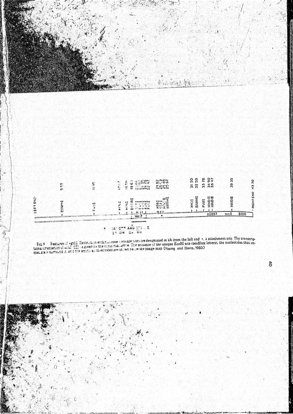
The structure of X gt U is shown in Figure 1. The site of
insertion of foreign DNA has been described as a unique Eco
R'l cleavage site located within the LacZ gene, 53 base pairs
upstream from the £-galactoaidase translation termination
codon (Young and Davis, 1983). ' 11 was constructed to
accommodate up to 8.2 kb of in,,.. DNA, assuming a maximum
wild type packageable DNA length of 1055C (Young and Davis,
1983). The vector has bean shown to produce a
temperature-sensitive repressor which is inactive at 42 C,
and to contain an amber mutation which renders it
lysis-defective in host bacteria that lack the amber
suppressor (Huynh, Young, and Davis, 1985). The site of
insertion of foreign DNA ift X gt U was chosen to be within
the structural gene for |S-galactosidase, and thus foreign DNA
sequences in the vector have the potential to be expressed as
fusion proteins with jS-galactosidase (Huynh, Young, and
D a v i s , 1985).
proper expression of foreign DNA in recombinant lysogens has
been shown to depend on the orientation and reading frame of
the insert DNA with respect to those of lacZ. Thus, it has
been suggested that one-sixth of N gt ii reeombinants
Page 28
%a«>
, i Hit*. ^ **4"*_o r a» c#t r.*CVlV
h w **'.,*•? <**>
SS?«'£— «
to *<* ro tfj
m m3 8 92 £§ v ! »
CIB57
mK)w10
ao
sK1
QZU)
£
m>i5 SIOO
•h ' S’; * m
A At* 'i' *
*-» ft-
r,;i »:■«» „ _I {::■ .* *.» « * T.fc. !!!A, Jtr * .<* W M t t iS t l
5’ * Sft.'V. Sii "il *'* i‘«- wf'1
, v „ , , in Kb irom the left f nd v A attachment site The transcnp.
“ V-m m «.: uw* dmipiB & « K 1 Mie ibo.dface .ettersi. the nucicotmes thai im-
i.*;? p a k r * (Young and Davis, 1903)
Page 29
containing a specific foreign insert would be expected to
produce £ -gaiactosidaa* fused to the protein of interest
'Young -and Davis, 1983 i.
Sin:e X g' 11 w a s d«si gr»ed as an expression vector,
*s.t.r«afit C?JA l i b ra ri es »: .r.strucUvd in X gt 11 :an b«
screened with antibody pn'faes fcr antigens p» o-iw.&d by
ro: ;-mk*nani „■ ivne*; Vi-.ng and Davis, 1 9 8 3 5 , Individual
ro; .'ss;I fidRt a are -i:c e e m - d m ‘He t o r ® wi p hage p l aq ue s in a
lawn p r o t o a & o - tot’ i d o a t c ells < Young and Davis,
Si'. Fr iAeiiss released ay the lysis of »ells within the
p l a t e s are ia»-3bi I i sed -<r, nitrocellulose filters, and the
^ p j- i, * €> £ f; £J (jfC* t t*0 *i VI * fo t. 1 DCii i & £ £v u <3 c it * c £
*ho «w’,*;gc*r. -;£ iK'.e»'0'3>. sY.jfig and Davia, Y-sung ar» i
lavs 3 , i ->S S f.-. Ar«* •« t* rending is reveal od f. y S'/bae->,t'.j«r, t * ••
}*j‘' fc.iKi5 * , tilt&f'i •« , * :, ; |..i; % * v o * >'**afce• 1 ed :3€*,, y
ac;t.&id;o3 e Y:ir.g an;: iiJtvt a , i .*y Ji Vvjjvg ar.J Davi s, 1 <sj »* »
%t 1 »*?J r.' , - i I V a fit * bod i t?S I *•*£ > *5». JCstf1. ,
1r v s s k ; r.ak y , si • , I > -S > *
Several problems have fceen sh-^wn to be associated with the
production o f f o r e i g n pt ->t eins in E ..c c 1 i, (Young and Davis,
19s* 3“ ; the X gt 11 ve..t<-.r was designed specifically t )
min;miise these diff i iulties «Young and Davis, 1983> . The
first problem is the achievement cl an adequate level o£
•axpreasi-sn of the foreign DNA sequence. It was iound that
fusing these DNA sequences to /3-galactosidase gene sequences
Page 30
f l .
V £
42
ensured that the foreign DNA would be expressed efficiently
io EL.co.li,, which has a strong l_a.cZ promoter (Young and Davis,
1983). The second problem is the ability to control the
production of foreign proteins, which have often been shown
to be toxic to the host cell and kill it before sufficient
amounts of antigens are produced (Young and Davis, 1983). It
was found that this could be minimised by using host cells
which produced large amounts of the l.ac operon repressor to
prevent l.acZ-di rected expression of the fusion protein until
the number of Infected cells was sufficiently large (Young
and Davis, 1983), LacZ-'-directed expression could then be
induced by inactivating the repressor with isopropyl- ft)
' t .niogalactopyranoside (IPTG). The third problem associated
with the production of fusion proteins in IL-SSli Is the
instability of the fusion proteins themselves. The position
chosen for fusion with the (8-gaiactosidase gene,
correspond!ng to a region close to the carboxyl terminus of
the [&~gaiactosidase protein, appeared to aid the stability of
the fusion protein and hence overcome this problem to a
certain extent (Young and Davis, 1983). The use of l.on
protease deficient host cells for the screening procedure
was also found to aid in maintaining the stability of fusion
proteins (Huynh, Young, and Davis, 1985).
\ gt 11 recombinant libraries can therefore be used to
produce preparative amounts of recombinant proteins in the
form of fusion polypeptides (Hyunh, Young, and Davis, 1985).
Page 31
Hybrid proteins can be produced by A gt li clones as
lysogens in E..col_i.. Lysogens can be £."own to high cell
density, lacZ-directed fusion protein production induced, and
the cells are harvested and ] ysed (Huynh, Young, and Davis,
1985!. Recombinant proteins can then be resolved from other
proteins in the cell lysates by polyacrylamide gel
electrophoresis, and can be Further purified by classical
. ,li)nn chromatography.
The ability to clone and express mycobacterial genes in
E , e o U has provided an approach towards major progress in the
characterisation of mycobacterial antigens. As discussed in
-Section 1.8, the combination of the A gt 11 expression
‘system, monoclonal antibodies, and the establishment of T
cell clones reactive to M^tubercul_osis, has enabled the
idea t i f i cat, i en of several novel antigens of potential
clinical importance. In order to obtain the maximum benefit
from the advances described in the latter two sections, it is
Important that the investigation of antigen structure
Should be closely integrated with study of the microbiology
and immunology of tuberculous disease (Young, 1988).
Page 32
Cloning of the antigens encoded by the M^tuberculosis genome
provides an initial starting point whereby large quantities
o£ recombinant antigens can be produced easily, using
standardisable techniques. Once this has been achieved,
detailed characterisation of these antigens can then be made,
the aim of the present study, therefore, is to generate
recombinant jj. tuberculosis antigens utilising the X gt 11
expression systom, and to characterise these antigens by
polyacrylamide gel electrophoresis and Western blotting.
Page 33
C H A P T ER 2 M A T E R I A L S AND M E T H ODS
Page 34
n . i PREE4S*U9H-2E.H.-.tstet£alasU-S2!JI£4IE
organisms were collected by scraping col
^ r o 7 p o s i t i v . sp,tu„ cul turas) off I.owensteln-Jensen slop..
,.to Physiologic,1 s a l i n e . The suspensions ».r. c.ntrif,S .d
5 minutes to r . « u . any conta.lr.atinSat noo g
i- =r! -anrf centrifuged at 2 000 g for T h e s u p e r n a t a n t w a s c o l l e c t e d a n d t e n s
30 -lo„ , « at 4»C, followed by 3 successive washings of the
f i l e t s in saline. M y c o b a c t e r i a were then heat-killed by
a u - d U v i r t g , washed 3 more times as before, counted, and made
% f.o 1C X 1 0 6 -'ml in saline. The organisms were sonicated
sE Soniprep 150 sonicator.
h e s c r . i c a t e d m a t e r i a l was c e n t r i f u g e d a t 2 0 0 0 g f o r 15
■3 ,jia peak to peak) at 4°C in a M~
; centrifi
t t o s u p e r n a t a n t c a r e f u l l y c o l l e c t e d , a n d t h e p r o t . i n
r a t i o n o f t h e s o n i c a t e d e t e r m i n e d u s i n g t h e B l o r a d
p r o t e i n e s t i n a t i o n k i t b l o r a d L a b o r a t o r i e s , R i c h m o n d , CA,
a c c o . d i n * t h e m a n u f a c t u r e r ' s i n s t r u c t i o n s . The ^
„ y , . b a c t e r i a l s o n i c a t e w a s a l i q u o t . d a n d s t o r e d a t - = 0 C
an t 1 1 r e q u i r e d .
Rabbits were i m m u n i s e d i n t r a m u s c u l a r l y with I ml ea
M . tuberculosis, sonicate, 0.5 mg/ml in saline. I m m u n i s a t i o n s
war. r e p e a t e d at 7 ~ d a y intervals for 4 c o n s e c u t i v e w e e k s .
Blood was obtained from the r a b b i t s by bleuding from
Page 35
veins. The serum was separated and tested for the presence of
antibodies to Mi.tuber£ulidsi_s u&ing the ELISA assay of W a d e e ,
Cohen, and Rabson <1987). Serum from 3 healthy, saline-
immmunised rabbits was used as a negative c o n t r o l . All sera
were aliquoted and stewed at ~20®C until required.
The ELISA was performed in flat-bottomed 96-well plates
(Nunc, Denmark), the wells of which were coated with 100 ul
of 10 ug/ml M , tuberculosis sonicate in sodium carbonate
buffer, pH 9 , 6 (Appendix 8), for 1 hour at room temperature.
The plates were washed 4 times with PBS-0,05% Tween, pH 7.2
(appendix 8) and the unbound sites then blocked with 100 ul
per well of 0,5'/, bovine serum albumin (BSAj Seravac, SA) in
sodium carbonate buffer for 1 hour at room temperature.
Plates wars washed as before, 100 ul of a 1/40 dilution of
anti^Mitubereuloaia serum in PBS-“Tween were added to each
w a l l , and the plates wore incubated at room temperature for 2
hours. After washing as before, 100 ul of peroxidase-
conjugated swine anti-rabbit IgG (Dako Immunoglobulins,
Denmark), diluted 1/2 000 with PBS-Tween, were added to each
well, and the plates were incubated at room temperature fo«> 1
hour. The plates were washed again, 100 ul of peroxidase
substrate (appendix 5) ware added to each well, and the
colour reaction terminated after 10 minutes by the addition
of 2.5 M HjSO* (50 ul per wall). The absorbance of each well
was read at 492 nm in a Titartek Nultiskan MC plate reader
(Flow Laboratories, Sweden).
Page 36
In order to ascertain that the antibodies to M J_tubercul_osi_a
were able to recognise specific M tuberculigsi,3 antigens
immobilised on n i t r o c e l l u l o s e , Western blots of the
sonicate were screened with the
anti~M^tubgrculssis antibodies. Proteins and polypeptides in
the mycobacterial sonicate were resolved by gel
electrophoresis on 107, polyacrylamide-SOS Laemmli gels
i Laemml i , 1 7 0 ; appendix 7). The mycobacterial sonicate was
solubilised in 1/3 volume splitting solution (see section
■J > 3 i , boiled for 5 minutes, and applied to the gels at a
concentration of 100 ug per well, Conditions for
electrophoraisIs and immunoblotting were the same as those
J&siribed in Section 2.9 and Section 2,10.
2s,d AHTIB0DIE5_T;}J-GALACT03IDASE
fion.i :1 snal antibodies to ^S-galactosidase, in the form of
A B C i ii* fluid, were obtained as a gift from Professor
E.Dawdle, Department <ji Clinical Science and Immunology,
University of Cape T c w n , South Africa. They were stored in
0 . IK sodium aside at 4°C,
JUl ISOLATION QF M.Ufaeraulasis DNA
Mycobacteria from positive sputum cultures were harvested
from Lowenst.ain-Jensen d o p e s , and washed extensively with
saline. To 2 ml of pelleted mycobacterial cells, 4 ml of
Page 37
CSoehringei' Mannheim, W,Germany) and EDTA, pH 3.0, to final
ccneentratl-sns of 2 mg/ml and 40 mM respectively. The
mya«slja>;tw»'ia were incubated on a rotating wheel for 2 hours
at 37°C. Sodium dadoyyl sulphate iSDS; BDH, Poole, England),
pH 7. 2, was then added to a final concentration of IV,, and
the intubation was continued for a further 30 m.nuirs at
3?°C.
Art o^ual volume >sf a 1st <volume/volume) mixture of
ihli/fciirm tchlorc-form/iisoajnylalcchol 24; 1) (Merck,
D a r m s t a d t » W» Germany* and Tris-saturated phenol (Merck,
Barrai.tadt, W.Germany) was than added, and the aqueous phase
was oxtrasSte„i with gentle agitation for 10 minutes. The
phases were separated by centrifugation at 1 500 g for
I-} t,an»*taii. Chi*rofor»/phenoi extraction of the aqueous phase
was repeatod twice uore. The aqueous phase was then extracted
.sn:o with an equal volume of 24:1 (volume/volume)
ihl^rofirm/1soamylalcohol with gent 1a agitation for S
m i n u t e s , followed by centrifugation at I BOO g for 5 minutes.
Sodium acetate itJnlvar, S. A.) was added to 0,3 M to the
aqueous phase, and DNA was precipitated with 2.5 volumes of
iaa-coId absolute ethanol (Merck, Darmstadt, W.Germany). The
DNA wais <;ol lectod by cent rifugation at 5 000 g for 30 minutes
at Q°C, and roauspended in 2 ml TE buffer (appendix S).
DNase » free RNasa (Boehri ngep Mannheim, W .Germany) was added
Page 38
to the DNA solution to a concentration of 50 ug/ml, and the
solution was incubated at 37°C for SO minutes, Proteinase K
(Boehringer M a n n h e l m ( V/, Germany) was then added to 100 ug/ml,
and tho .solution incubated for a further 30 minutes at 37°C .
The DNA was then extracted once with chloroform/isoamyl-
alcohol, precipitated with 2.5 volumes of ice-cold absolute
ethanol) and collected by centrifugation as before. The DNA
was dried at room temperature, resuspended in 1 ml TE buffer,
pH 8 .j iappendix 8>, and quantified spectrophotometrically
fappandix 3).
2 S g S Q y i I M H I » B M A „ L IBRARY..CONSTRUCTIQN
£ .„S», I, .lotgeduetion
Libraries of genomic DNA may be prepared in 2 ways (Maniatis,
Fritfiish, and S a m b r o o k , 19S2) , The First approach involves the
digestion of genomic DNA to completion with a restriction
oRsymfl, and insertion of the resulting fragments in an
appropriate A bacteriophage v e c t o r . This method suffers from
2 drawbacks: first, if the sequence of interest contains
recognition aits (a) for the particular restriction enayme
c h o s e n , it will be elonod in 2 or more pieces, In addition,
the- sequence may not be cloned at all, if, for example, it is
contained in a larger DNA fragment than the vector can
accept, Second, if very largo genomic DNA is used Initially -
the genome of M ^ t u b e r c u i o M s is 2,1 X 10⁹ bp in sice (Imaeda,
Page 39
», •”>s *rr
' 'V ' *
* 1.
£' S* +' a-V.*
V
50
IA
Barksdale, and K i r e h heimer, 1982), large compared to that ofg
§i.££Lii which is 4 X 4 0 bp in aJ.se, and the human genome,
which ia approximately 3.9 X 10⁹ bp in sice (Stryer, 1938) -
the average also of the fragments generated by cleavage of
ths DMA with many restriction enaymes ts relatively small
(approximately 4 kb), and an entire library therefore
contains a vary large number of recombinant b a cteriophages,
and screening thus becomes laborious and expensive.
Problems in library sonstruction can be avoided by cloning
fragments that are generated by random shearing of genomic
DNA tManlafcis, Fritseh, and Sambrook, 1982), This method
fenfiures that there is no systematic exclusion of sequences
Cv-.m *h© cloned library merely because of an unfortunate
distribution of restriction sites. In addition, knowledge of
tho iistribut ion of restriction sites in or around the
ae^uenco s£ interest ia not required before cloning Is
attempted,
The basi'* stops for the construction of a M. tuberculosis
genomic library in the /\ gt 11 vector (Young and Davis, 19S 5>
ara shown in Figure Z, and are summarised as follows!
i. The DNA of X gt 11, which accepts inserts up to
3,2 kb in length, ia digested with Eeo R 1 to
to produce left and right arms, which carry all the
genetic information for lytic growth of the virus,
Page 40
, P ’ ’ * •>‘\ '* *■'&"
t *.■*■ h ■' ••'• :; ■ •*
* " I t \ w„r
recombinant phage
Infect E. Cel with recombinant phage
Screen pfcKjues and omplfy recombinants
Phage stock conutetJng of a library of rccomblnani donoa
Page 41
Author Chezzi CName of thesis Cloning of the DNA of Mycobacterium tuberculosis and analysis of the expressed protein antigens 1989
PUBLISHER:University of the Witwatersrand, Johannesburg ©2013
LEGAL NOTICES:
Copyright Notice: All materials on the U n i v e r s i t y o f t he W i t w a t e r s r a n d , J o h a n n e s b u r g L i b r a r y website are protected by South African copyright law and may not be distributed, transmitted, displayed, or otherwise published in any format, without the prior written permission of the copyright owner.
Disclaimer and Terms of Use: Provided that you maintain all copyright and other notices contained therein, you may download material (one machine readable copy and one print copy per page) for your personal and/or educational non-commercial use only.
The University o f the W itw atersrand, Johannesburg , is not responsible for any errors or omissions and excludes any and all liability for any errors in or omissions from the information on the Library website.