Date post: | 11-Apr-2017 |
Category: |
Health & Medicine |
Upload: | shape-society |
View: | 39 times |
Download: | 0 times |
The “Evolution” of Coronary Atherosclerosis
(Libby. Circulation 2001;104: 365)
NormalArtery
LesionInitiation
Fibro-fattyStage
VulnerablePlaque
PlaqueRupture
Fibrous,CalcifiedPlaque
EndothelialErosion
Progression over time (yrs):
Can the Trajectories of the Natural Histories of Coronary Atherosclerosis Be Identified
Prior to Adverse Coronary Events?Opportunities for Intervention
Snapshotat time ofangina or MI
Snapshotto identifyvulnerability
Snapshotto identify likelihood
to develop vulnerabilityor progression
Quiescent,Stable plaque no symptoms
Fibrotic/Scarred plaque angina
Vulnerable,Ruptured Plaque MI, sudden death
?
The Effect of Physiologic Shear Stress on Endothelial Structure and Function
Physiologic shear stress (~15-50 dynes/cm2) isvasculoprotective:
(Malek, et al. JAMA 1999; 282:2035)
• Enhances endothelial quiescence - decreases proliferation
• Enhances vasodilation
• Enhances anti-oxidant status
• Enhances anti-coagulant and anti-thrombotic status
The Detrimental Effect of Low Shear Stress on Endothelial Structure and Function
Low shear stresses and disturbedlocal flow (< ~ 6 dynes/cm2)are atherogenic:
(Malek, et al. JAMA 1999; 282:2035)
• Cell proliferation, migration• Expression of vascular adhesion molecules, cytokines, mitogens
• Monocyte recruitment and activation
• Procoagulant and prothrombotic state• Local oxidation
Promotes:
Original angiogram ofa portion of an artery
studied
Composite reconstruction of portion of the arterial segment,consisting of outer arterial wall, plaque, and lumen:
Isolated view of reconstructed outer arterial wall:
Isolated view of reconstructed lumen:
Isolated view of reconstructed atherosclerotic plaque:
Example of 3-D Reconstruction of Arterial Segment
(Stone, et al. Circulation 2003;108:438)
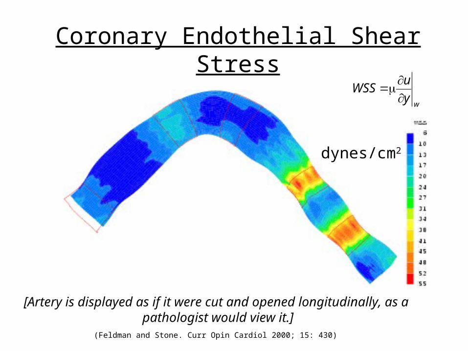
Coronary Endothelial Shear Stress
wyuWSS
dynes/cm2
[Artery is displayed as if it were cut and opened longitudinally, as a pathologist would view it.]
(Feldman and Stone. Curr Opin Cardiol 2000; 15: 430)
Changes in Native Arteries
Change in Plaque Thickness (mm) Change in EEM Radius (mm)
Change in Lumen Radius (mm) Change in ESS (dynes/cm2)
Regions of baseline low ESS::• increase in plaque thickness• enlargement of EEM (outward remodeling)
Regions of baselinephysiologic ESS:• little change in any variableRegions of baselineincreased ESS:• increase in lumen radius• increase in EEM radius• decrease in ESS(outward remodeling)
ESS at Baseline andVascular Outcomes 6 mo later:
p<0.001
p<0.001p=0.03
(Stone, et al. Circulation 2003;108:438)
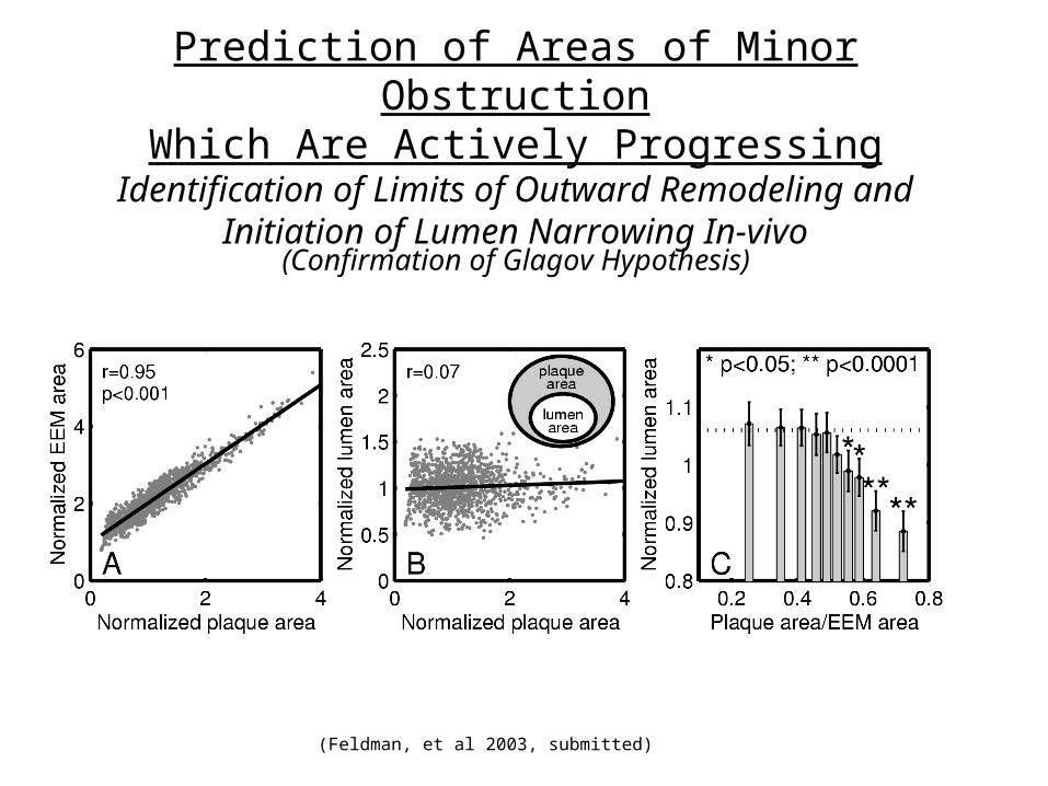
Prediction of Areas of Minor ObstructionWhich Are Actively Progressing
Identification of Limits of Outward Remodeling and Initiation of Lumen Narrowing In-vivo
(Feldman, et al 2003, submitted)
(Confirmation of Glagov Hypothesis)
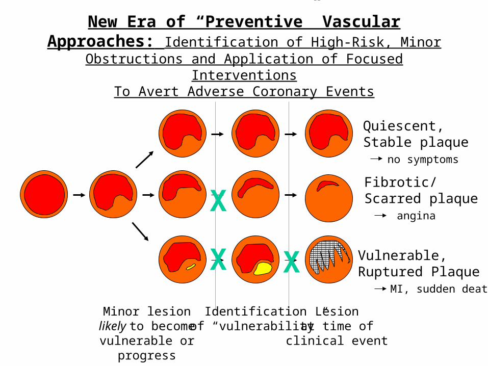
New Era of “Preventive” Vascular Approaches: Identification of High-Risk, Minor Obstructions and Application of
Focused InterventionsTo Avert Adverse Coronary Events
Lesionat time of
clinical event
Identificationof “vulnerability”
Minor lesion likely to become vulnerable
or progress
Quiescent,Stable plaque no symptoms
Fibrotic/Scarred plaque angina
Vulnerable,Ruptured Plaque MI, sudden death
X X
X