B io F actsheet 1 Number 139 www.curriculum-press.co.uk Answering exam questions on the heart When answering most exam questions on the heart it is essential that you know the basic anatomy of the heart, the cardiac cycle and how the heart beat is controlled. You will be able to interpret and analyse more complicated data interpretation questions only when you have this basic knowledge and understand it. Study the diagram below. Fig 1. Section through human heart 3 There are four valves in the heart whose job is to prevent backflow of blood in the heart. * The tricuspid valve is between the right atrium and right ventricle. It will be open when the heart relaxes and fills (diastole) and shut when the ventricle is contracting (systole). * The bicuspid (mitral) valve is between the left atrium and the left ventricle. It will be open when the heart relaxes and fills (diastole) and shut when the ventricle is contracting (systole). * The pulmonary semilunar valve, at the base of the pulmonary arch, is shut when the heart relaxes and fills, but opens when the right ventricle contracts to pump blood through the arch to the lungs. * The aortic semilunar valve, at the base of the aortic arch, is shut when the heart relaxes and fills, but opens when the left ventricle contracts to pump blood through the arch to the body. Learn that:- 1 There are four chambers, right atrium, left atrium, right ventricle and left ventricle. A very common error is that candidates confuse the left and right sides of the heart. 2 There is a specific set of blood vessels associated with each heart chamber. * The right atrium receives deoxygenated blood from the body, (the venous return), via two veins called venae cavae. The superior/ anterior vena cava returns blood from the head and neck and the inferior/posterior vena cava returns blood from the rest of the body. * The left atrium receives oxygenated blood from the lungs via four pulmonary veins. * The right ventricle receives deoxygenated blood from the right atrium and pumps it to the lungs, for oxygenation, via the pulmonary arch. This divides into two pulmonary arteries, one to each lung. * The left ventricle receives oxygenated blood from the left atrium and pumps it to the body via the aortic arch. The aortic arch leads to the dorsal aorta which is the main artery to the body. Exam Hint: - Candidates frequently err by associating the wrong vessels with specific heart chambers, or by reversing the blood flow in particular vessels or chambers, or by reversing the state of blood oxygenation in the left and right sides of the heart. Remember, the right side contains deoxygenated blood, the left side contains oxygenated blood. Exam Hint:- Students frequently mix the atrio-ventricular valves up by incorrectly placing the tricuspid valves on the left side and the bicuspid valves on the right side. Another common error is to have the valves open or shut at the wrong times in the cardiac cycle (heart beat cycle). Right Left Left Right Septum Pulmonary semilunar valve Tricuspid valve Venae cavae from head, neck & body Bicuspid/mitral valve Aortic semilunar valve Aortic arch to body Pulmonary arch to lungs Pulmonary veins from lungs
Transcript
Bio Factsheet
1
Number 139www.curriculum-press.co.uk
Answering exam questions on the heartWhen answering most exam questions on the heart it is essential that you know the basic anatomy of the heart, the cardiac cycle and howthe heart beat is controlled. You will be able to interpret and analyse more complicated data interpretation questions only when you havethis basic knowledge and understand it. Study the diagram below.
Fig 1. Section through human heart
3 There are four valves in the heart whose job is to prevent backflow ofblood in the heart.
* The tricuspid valve is between the right atrium and right ventricle.It will be open when the heart relaxes and fills (diastole) and shutwhen the ventricle is contracting (systole).
* The bicuspid (mitral) valve is between the left atrium and the leftventricle. It will be open when the heart relaxes and fills (diastole)and shut when the ventricle is contracting (systole).
* The pulmonary semilunar valve, at the base of the pulmonaryarch, is shut when the heart relaxes and fills, but opens when theright ventricle contracts to pump blood through the arch to thelungs.
* The aortic semilunar valve, at the base of the aortic arch, is shutwhen the heart relaxes and fills, but opens when the left ventriclecontracts to pump blood through the arch to the body.
Learn that:-1 There are four chambers, right atrium, left atrium, right ventricle
and left ventricle. A very common error is that candidates confuse theleft and right sides of the heart.
2 There is a specific set of blood vessels associated with each heartchamber.* The right atrium receives deoxygenated blood from the body, (the
venous return), via two veins called venae cavae. The superior/anterior vena cava returns blood from the head and neck and theinferior/posterior vena cava returns blood from the rest of thebody.
* The left atrium receives oxygenated blood from the lungs via fourpulmonary veins.
* The right ventricle receives deoxygenated blood from the rightatrium and pumps it to the lungs, for oxygenation, via thepulmonary arch. This divides into two pulmonary arteries, oneto each lung.
* The left ventricle receives oxygenated blood from the left atriumand pumps it to the body via the aortic arch. The aortic arch leadsto the dorsal aorta which is the main artery to the body.
Exam Hint: - Candidates frequently err by associating the wrong vesselswith specific heart chambers, or by reversing the blood flow in particularvessels or chambers, or by reversing the state of blood oxygenation inthe left and right sides of the heart. Remember, the right side containsdeoxygenated blood, the left side contains oxygenated blood.
Exam Hint:- Students frequently mix the atrio-ventricular valves upby incorrectly placing the tricuspid valves on the left side and thebicuspid valves on the right side. Another common error is to havethe valves open or shut at the wrong times in the cardiac cycle (heartbeat cycle).
Right
Left
LeftRight
SeptumPulmonary semilunar valve
Tricuspid valve
Venae cavae fromhead, neck & body
Bicuspid/mitral valve
Aortic semilunar valve
Aortic arch to body
Pulmonary arch to lungs
Pulmonary veins from
lungs
Bio Factsheet
2
139. Answering exam questions on the heartwww.curriculum-press.co.uk
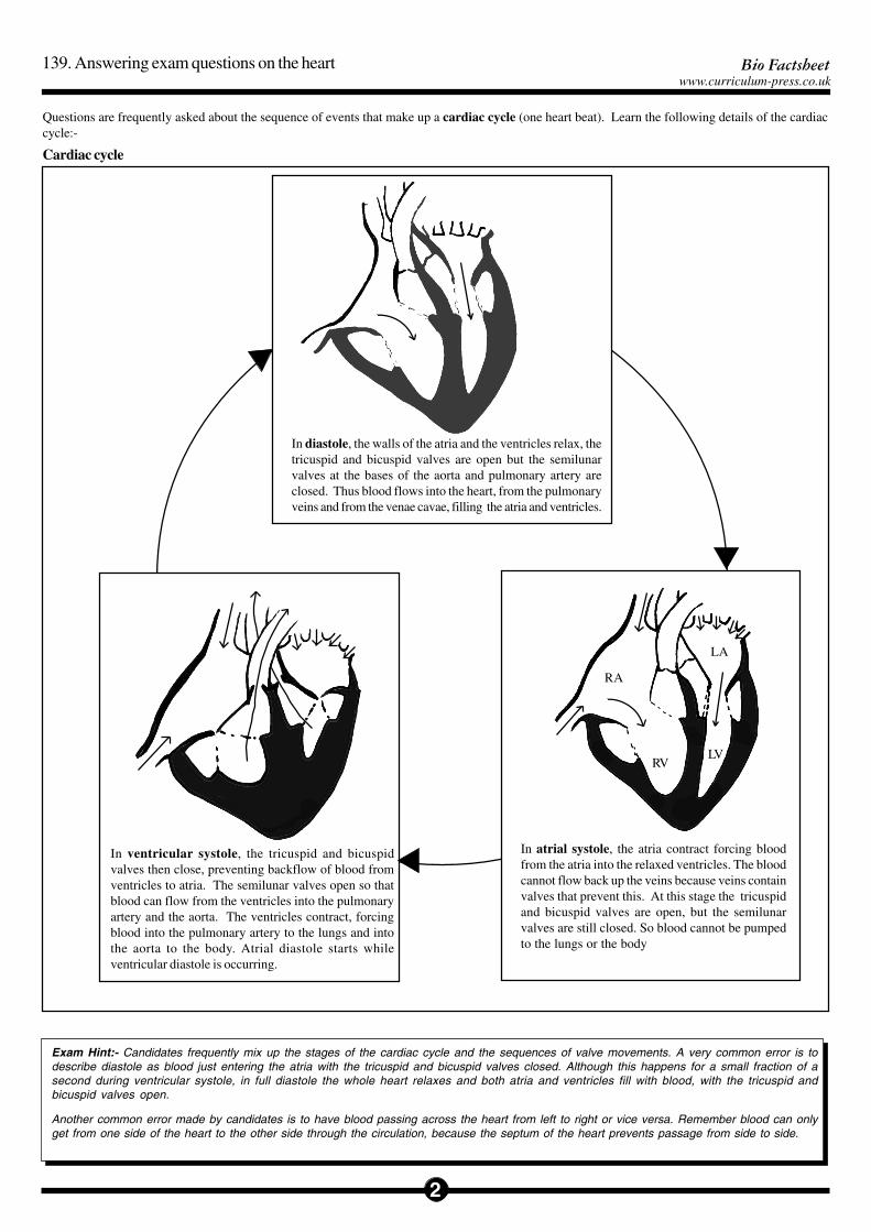
In diastole, the walls of the atria and the ventricles relax, thetricuspid and bicuspid valves are open but the semilunarvalves at the bases of the aorta and pulmonary artery areclosed. Thus blood flows into the heart, from the pulmonaryveins and from the venae cavae, filling the atria and ventricles.
RVLV
RA
LA
In atrial systole, the atria contract forcing bloodfrom the atria into the relaxed ventricles. The bloodcannot flow back up the veins because veins containvalves that prevent this. At this stage the tricuspidand bicuspid valves are open, but the semilunarvalves are still closed. So blood cannot be pumpedto the lungs or the body
In ventricular systole, the tricuspid and bicuspidvalves then close, preventing backflow of blood fromventricles to atria. The semilunar valves open so thatblood can flow from the ventricles into the pulmonaryartery and the aorta. The ventricles contract, forcingblood into the pulmonary artery to the lungs and intothe aorta to the body. Atrial diastole starts whileventricular diastole is occurring.
Exam Hint:- Candidates frequently mix up the stages of the cardiac cycle and the sequences of valve movements. A very common error is todescribe diastole as blood just entering the atria with the tricuspid and bicuspid valves closed. Although this happens for a small fraction of asecond during ventricular systole, in full diastole the whole heart relaxes and both atria and ventricles fill with blood, with the tricuspid andbicuspid valves open.
Another common error made by candidates is to have blood passing across the heart from left to right or vice versa. Remember blood can onlyget from one side of the heart to the other side through the circulation, because the septum of the heart prevents passage from side to side.
Cardiac cycle
Questions are frequently asked about the sequence of events that make up a cardiac cycle (one heart beat). Learn the following details of the cardiaccycle:-
Bio Factsheet
3
139. Answering exam questions on the heartwww.curriculum-press.co.uk
The diagram below gives details that you need to know to answer questions on the control of the heart beat. Remember that the heart co-ordinates its owncardiac cycle.
Fig 2. Section of heart showing its conducting system
Sino-atrial node (SAN/pacemaker)in wall of right atrium
Atrio-ventricular node (AVN)in top of venrticular septum
• After the cardiac muscle fibres have depolarized and contracted theythen repolarise and relax.
Exam Hint:- Candidates often forget to mention this, or make thepoint that the repolarisation sequence of the parts of the heart willfollow the depolarization sequence, - right atrium, left atrium,ventricles from apex.
Though the heart co-ordinates its own beat, at a mean frequency of 72beats minute-1, (the inherent frequency of ventricular cardiac muscleand also the mean impulse generation rate of the SAN), the frequencyand force of the heart beat is modified by the autonomic nervoussystem.
• The sympathetic nerves to the heart go directly to the SAN/AVN andto cardiac muscle. Increased stimulation of the sympathetic nerves(and stimulation by adrenaline/nor-adrenaline) accelerates the heart – itincreases the frequency and force of the heart beat. Decreasedstimulation has the reverse effects.
• The parasympathetic nerves to the heart (branches of the vagusnerve) go to the SAN and AVN. Increase in parasympathetic stimulationdecreases the impulse frequency of the SAN and increases the delaytime at the AVN thus slowing the heart beat. A decrease inparasympathetic stimulation increases the impulse frequency of theSAN and decreases the delay time at the AVN, thus complementing theeffects of sympathetic stimulation.
Exam Hint:- When describing the control of the heart beatcandidates often forget to include the role of the nervous system, orif they do they often get the sympathetic and parasympatheticsystems confused. Few candidates make the point that the heartrate is a result of balancebetween sympathetic andparasympathetic stimulation.
• Cardiac (heart) muscle is myogenic. This means it will contract andrelax rhythmically of its own accord. The mean inherent rhythm rateof ventricular muscle is 72 contractions minute-1. This gives the meanheart beat rate.
Exam Hint: - When answering questions about the control of theheart beat these facts should start the account. However, manycandidates forget to include these facts but start their answers bydescribing the SAN/pacemaker.
• The SAN generates impulses which spread through and depolarize thecardiac muscle causing contraction. Thus the right atrium receivesimpulses before the left atrium and so the right atrium contracts beforethe left atrium.
Exam Hint: - Candidates often mix up the sides of the heart hereby putting the SAN in the left atrium.
Where the atria and ventricles join is a thick ring of fibrous connectivetissue which is the ‘skeleton’ to which the cardiac muscle is attached.The impulse from the SAN cannot pass this and can only pass to theventricles via the AVN.
Exam Hint:- Few candidates remember this and most neglect toexplain why the impulses can only be routed via the AVN.
• The AVN has fairly high electrical resistance and delays the impulsepassage by about 0.1 second. The impulses are then very rapidlyshunted through the bundles of His to the apex of the heart and aredispersed through the ventricular muscle, causing depolarization andcontraction.
Exam Hint: - Many candidates forget to refer to the AVN-delaywhich is responsible for delaying ventricular contraction until theatria are starting relaxation. Candidates frequently forget to pointout that the ventricles contract from the apex upwards – a factlinked to the rapid shunt of the bundles of His and the branchingout of the Purkyne fibres from the apex upwards.
Bio Factsheet
4
139. Answering exam questions on the heartwww.curriculum-press.co.uk
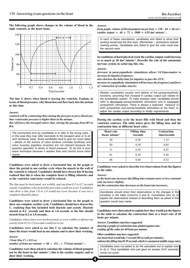
The following graph shows changes in the volume of blood in theright ventricle as the heart beats.
0 0.2 0.4 0.6 0.8 1.0 1.2 1.440
60
80
100
120
140
Vol
ume/
cm
3
B
C
A
Time/seconds
The line A shows when blood is leaving the ventricle. Explain, interms of blood pressure, why blood does not flow back into the atriumat this time.
Answer:ventricle will be contracting thus raising the pressure to force blood out;thus ventricular pressure is higher than in the atrium;this will force the tricuspid valves shut, closing the passage from RV toRA;
The commonest error by candidates is to refer to the wrong valve –in this case they may refer incorrectly to the bicuspid valve or to anarch semilunar valve. Some candidates tend to give too much detail– details of the actions of valve tendons (chordae tendinae) andvalve muscles (papillary muscles) are not relevant because thequestion specified ‘in terms of blood pressure’. To do this is poorexam technique because it wastes time and cannot score extramarks.
Candidates were asked to draw a horizontal line on the graph toshow the period in one cardiac cycle when the muscle in the wall ofthe ventricle is relaxed. Candidates should have drawn line B havingrealized that this is when the complete heart is filling (diastole) andso the ventricles (and atria) would be relaxed.
The line must be horizontal, not wobbly, and run from 0.2 to 0.7 secondsexactly. Candidates who lacked this precision would not score. Candidateswho drew a line from 1.0 to 1.4 would not score because it was not acompletes diastolic phase.
Candidates were asked to draw a horizontal line on the graph toshow one complete cardiac cycle. Candidates should have drawn lineC, realizing that this included both diastole and systole. Diastolestarted at 0.2 seconds and then at 1.0 seconds so the line shouldstretch from 0.2 to 1.0 seconds.
Candidates whose lines were not horizontal, or were wobbly or did not runexactly from 0,2 to 1.0 seconds would not score.
Candidates were asked to use line C to calculate the number oftimes the heart would beat in one minute and to show their working.
Answer1 beat (line C) = 0.8 secnumber of beats per minute = 60 ÷ 0.8 ; = 75 beats minute-1 ;
Candidates were then asked to calculate the volume of blood pumpedout by the heart in dm3 minute-1, (this is the cardiac output), and toshow their working.
Answer:from graph, volume of blood pumped out per beat = 130 - 64 = 66 cm3 ;cardiac output = 66 x 75 ÷ 1000 = 4.95 dm3 minute-1 ;
In each of these calculations, candidates who failed to show theirworking would lose the first mark. (Remember – semicolons indicatemarking points). Candidates who failed to give the units could losethe second mark.
In conditions of hard physical work the cardiac output could increaseto as much as 20 dm3 minute-1. Describe the role of the autonomicnervous system in achieving this rise.
Answer:decrease in parasympathetic stimulation allows SAN/pacemaker toincrease its impulse frequency;also shortens the delay time for impulses to pass the AVN;increase in sympathetic stimulation will increase the frequency and forceof contraction of cardiac muscle;
Weaker candidates usually omit details of the parasympatheticfunctions, assuming that increase in cardiac output just relates tothe sympathetic system. To make answers clear, candidates shouldrefer to decreased parasympathetic stimulation and to increasedsympathetic stimulation. There is always a balanced measure ofboth sympathetic and parasympathetic stimulation to the heart, toadjust the cardiac output according to bodily requirements.
During the cardiac cycle the heart fills with blood and then theventricles contract. The table below gives the filling time and thecontraction time at different heart rates.
Heart rate Filling time Contraction/beats minute-1 /seconds time/seconds
40 0.38 1.15
50 0.39 0.85
71 0.39 0.47
81 0.39 0.36
88 0.40 0.32
Candidates were asked to describe two observations from the figuresin the table.
Answer:as the heart rate increases the filling time remains more or less constant/only increases slightly;but the contraction time decreases as the heart rate increases;
Candidates should direct their observations to the changes in thevariables in the table (columns 2 and 3). Candidates who try toexplain the observations instead of describing them as asked in thequestion would lose marks.
Candidates were then asked to explain how they would use the figuresin the table to calculate the contraction time at a heart rate of 60beats per minute.
Answer: Candidates may have suggested:drawing a graph of contraction time plotted against rate;reading off the value for 60 beats per minute;
Other candidates may have suggested:one heart beat would take 1 second (60 seconds per minute);subtract the filling time/0.39 seconds which is constant at middle range rates;
Candidates were not asked to do the calculation but to explain howto do it. Thus candidates who just gave an answer (0.61 seconds)would not score.
5
Bio Factsheetwww.curriculum-press.co.uk
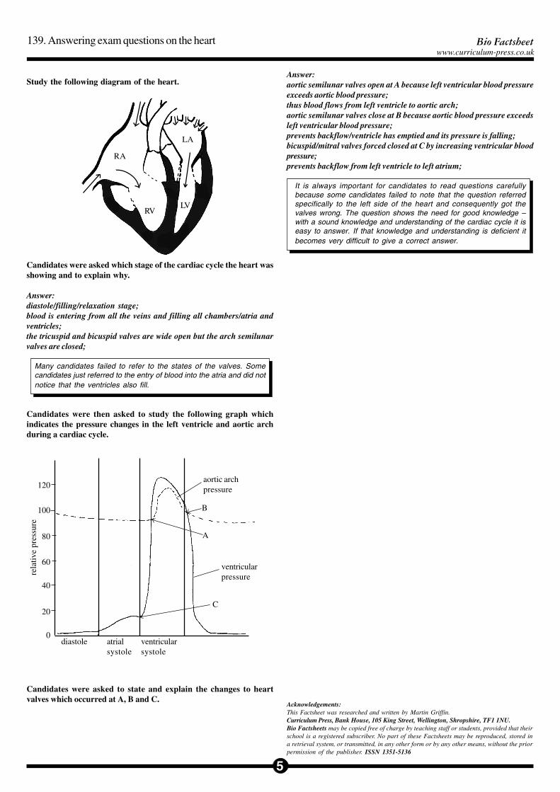
Study the following diagram of the heart.
RVLV
RA
LA
Candidates were asked which stage of the cardiac cycle the heart wasshowing and to explain why.
Answer:diastole/filling/relaxation stage;blood is entering from all the veins and filling all chambers/atria andventricles;the tricuspid and bicuspid valves are wide open but the arch semilunarvalves are closed;
Many candidates failed to refer to the states of the valves. Somecandidates just referred to the entry of blood into the atria and did notnotice that the ventricles also fill.
Candidates were then asked to study the following graph whichindicates the pressure changes in the left ventricle and aortic archduring a cardiac cycle.
diastole atrialsystole
ventricularsystole
ventricularpressure
aortic archpressure
B
A
C
Candidates were asked to state and explain the changes to heartvalves which occurred at A, B and C.
Answer:aortic semilunar valves open at A because left ventricular blood pressureexceeds aortic blood pressure;thus blood flows from left ventricle to aortic arch;aortic semilunar valves close at B because aortic blood pressure exceedsleft ventricular blood pressure;prevents backflow/ventricle has emptied and its pressure is falling;bicuspid/mitral valves forced closed at C by increasing ventricular bloodpressure;prevents backflow from left ventricle to left atrium;
It is always important for candidates to read questions carefullybecause some candidates failed to note that the question referredspecifically to the left side of the heart and consequently got thevalves wrong. The question shows the need for good knowledge –with a sound knowledge and understanding of the cardiac cycle it iseasy to answer. If that knowledge and understanding is deficient itbecomes very difficult to give a correct answer.
Acknowledgements:This Factsheet was researched and written by Martin Griffin.Curriculum Press, Bank House, 105 King Street, Wellington, Shropshire, TF1 1NU.Bio Factsheets may be copied free of charge by teaching staff or students, provided that theirschool is a registered subscriber. No part of these Factsheets may be reproduced, stored ina retrieval system, or transmitted, in any other form or by any other means, without the priorpermission of the publisher. ISSN 1351-5136