39
HISTOLOGY CARDIOVASCULAR SYSTEM II

HISTOLOGY
CARDIOVASCULAR SYSTEM II

Post-capillary venules
•Capillaries -- postcapillary venules – veins
•High endothelial venules (cuboidal cells)
•“Loosest” ZO
•Inflammation: leukocyte exudation
Infection/inflammation

Post-Capillary Venule
Note the endothelial cells (arrow heads). They are high cuboidal in nature, therefore these vessels are called HEV (high endothelial vessels). These vessels possess the loosest junctional complexes, facilitating the entry of immune cells (arrow) from the lumen into the connective tissue, during inflammation or as a normal phenomenon in locations such as lymph nodes.
PCV becomes small vein
Lymph node

HEVs play an important role in “homing effect” in lymphoid organs like lymph node. This is the site where lymphocytes enter a lymph node from circulation.
lymphocyte
EC
Post capillary venule

Small artery and vein
Small Artery Small vein
T. intima: endothelium, subendothelial connective tissue T. media: very few smooth muscle cells, arranged in circular fashionT. adventitia: more developed than media
Small Vein

Medium-sized Veins Muscular artery
T. intima: endothelium, sparse sub endothelial connective tissue T. media: few smooth muscle cells, arranged in circular fashionT. adventitia: well developed than media
Vein
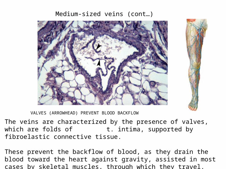
VALVES (ARROWHEAD) PREVENT BLOOD BACKFLOW
The veins are characterized by the presence of valves, which are folds of t. intima, supported by fibroelastic connective tissue.
These prevent the backflow of blood, as they drain the blood toward the heart against gravity, assisted in most cases by skeletal muscles, through which they travel.
Medium-sized veins (cont…)
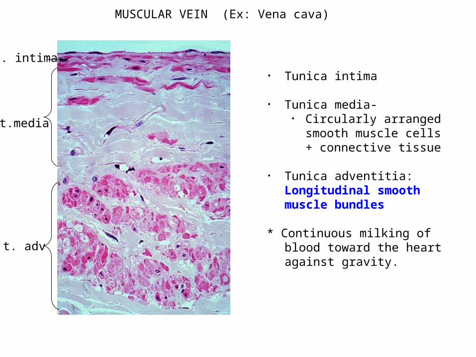
MUSCULAR VEIN (Ex: Vena cava)
• Tunica intima
• Tunica media-• Circularly arranged smooth
muscle cells + connective tissue
• Tunica adventitia: Longitudinal smooth muscle bundles
* Continuous milking of blood toward the heart against gravity.
t. intima
t.media
t. adv
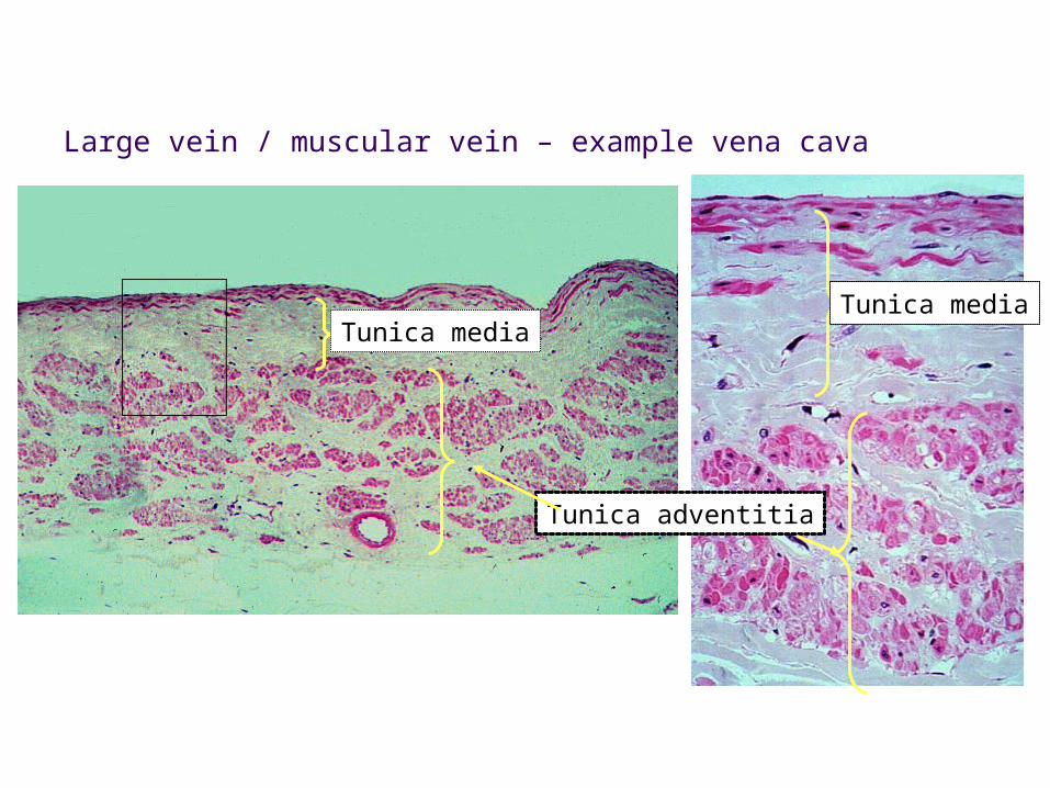
Large vein / muscular vein – example vena cava
Tunica mediaTunica media
Tunica adventitia

Large Vein (H&E)
T. adventitia
T. media
T. intima
Example : Vena cava
IVC?

LYMPHATIC SYSTEM
Returns excess tissue fluid to circulation
Starts off as blind-ended lymphatic capillaries and empty into the circulation; unidirectional valves
Flow is sluggish and aided by contraction of skeletal muscles
Bundles of filament anchor vessels to surrounding CT
No clear cut separation into tunics
Lack tight junctions: proteins and large molecules return to vascular compartment.
Not present in nervous tissue, bone marrow and cartilage
Impaired function - edema

Lymphatic
Lymphatic capillary
arteriole
venule
Lymphatic

Afferents
Efferent

All vessels, except arterioles, capillaries and post capillary venules,
have 3 layers:
Tunica intima
Tunica media
Tunica adventitia
Lecture Recap
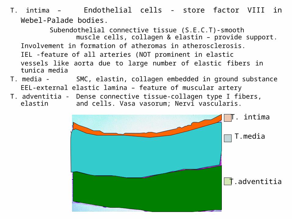
T. intima – Endothelial cells - store factor VIII in Webel-Palade bodies. Subendothelial connective tissue (S.E.C.T)-smooth
muscle cells, collagen & elastin – provide support.Involvement in formation of atheromas in atherosclerosis.IEL -feature of all arteries (NOT prominent in elastic vessels like aorta due to large number of elastic fibers in
tunica mediaT. media - SMC, elastin, collagen embedded in ground substance
EEL-external elastic lamina – feature of muscular arteryT. adventitia - Dense connective tissue-collagen type I fibers, elastin
and cells. Vasa vasorum; Nervi vascularis.
T. intima
T.media
T.adventitia

Elastic artery
Continuous capillary
Muscular Artery
Arteriole

Sinusoidal capillary
Fenestrated capillary
Muscular vein
T. Adv
Continuous capillary

Clinical Considerations
Atherosclerosis Marfan’s Syndrome Aortic Aneurysm Cerebral Aneurysm Peripheral Vascular Disease Varicose veins Lymphedema

Cardiovascular Diseases
Affects heart and circulatory system Predominant damage to blood vessel occurs due to atherosclerosis
and hypertension
Atherosclerosis- Formation of atheroma in the wall, which can decrease blood flow the region/can rupture /or perforate and are prone for clot formation (thrombus), which can travel as emboli.
INFLAMMATORY DISEASE
Hypertension- Can cause damage to smaller blood vessels by scarring, hardening, narrowing of blood vessels and eventually become less elastic. It can both predispose and accelerate development of atherosclerosis.
Arterioles - Radius is equal to the thickness of the wall. Resistance is inversely proportional to the diameter.

Major clinical manifestations of cardiovascular diseases
Coronary Heart Disease
Cerebrovascular Disease
Peripheral Vascular Disease
Angina Stroke Gangrene
Heart attack Transient ischemic attack
Intermittent
Claudication
Sudden death
Dementia
Heart failure
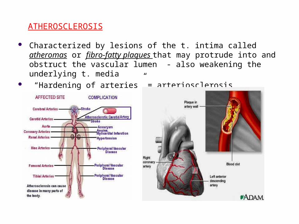
ATHEROSCLEROSIS
Characterized by lesions of the t. intima called atheromas or fibro-fatty plaques that may protrude into and obstruct the vascular lumen - also weakening the underlying t. media
“Hardening of arteries” = arteriosclerosis

OOooh…aww…Chest pains!!
Shortness of breath..
Back pain……..
Pain radiating to the jaw…..
Symptoms:

Heart disease begins when cholesterol, fatty material, and calcium build up in the arteries, a process known as atherosclerosis.

Risk factors:
SmokingHTNDMHypercholesterolemia
Lack of exerciseUnhealthy dietStressType ‘A’ personality

HistopathogenesisLDL in the blood contains triglycerides and lipids. These are insoluble in water medium of blood.
When there is excessive LDL, the endothelial cells produce free radicals which oxidize this LDL.
This oxidized product now initiates migration of monocytes into the tunica intima, which now become macrophages.
Smooth muscle cells (SMC ) also migrate from the t. media to the t. intima (subendothelial CT).

SMC and macrophages engulf oxidized LDL to form – foam cells
SMC proliferate and secrete collagen and other ECM – thickens t. intima forming fatty streaks
Cytokines from the SMC converts these fatty streaks into fibrofatty plaques - which bulges into the lumen and also compresses the t. media
• Results in luminal obstruction & weakened vascular walls (aneurysm)

Electrocardiogram (ECG or EKG). Stress tests
Echocardiography Computerized tomography (CT) scans

Coronary angiography via cardiac catheterization isconsidered the "gold standard" of heart disease tests

1 2
3 4
Progression of atherosclerosis

Luminal obstruction of the coronary artery will lead to ischemia of the
myocardium
N
1
2
Normal myocardium
Ischemic changes
Phagocytocytosis of the myocytes

SEQUELAE OF CORONARY VASCULAR OCCLUSION
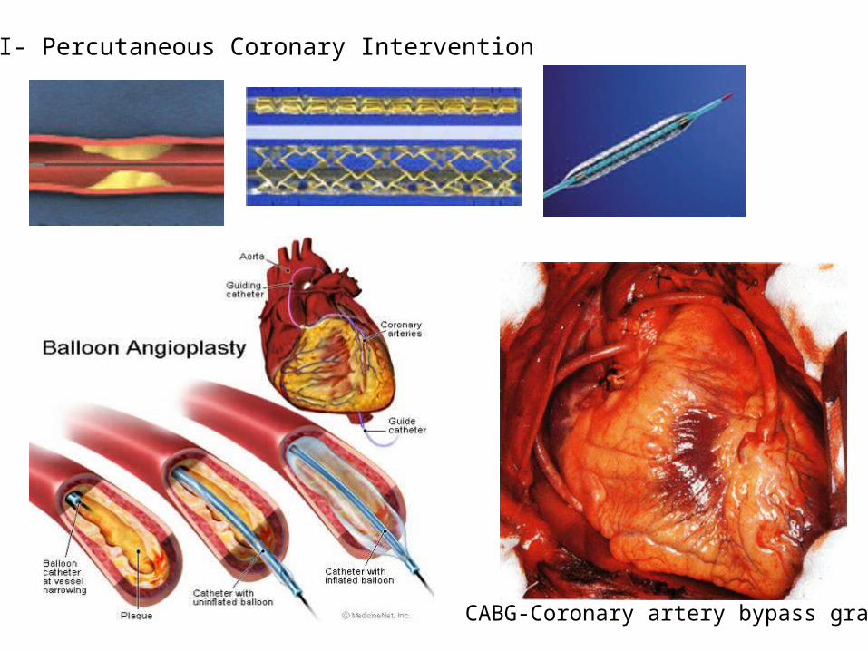
PCI- Percutaneous Coronary Intervention
CABG-Coronary artery bypass graft

Marfan’s Syndrome
Autosomal dominant connective tissue disorder with characteristic skeletal, cardiovascular and ocular manifestations.
Defect in the Fibrillin-1 gene -fibrillin is a glycoprotein that forms a scaffold on which elastin is deposited- elastic fibers.
Abnormal production and fragmentation of elastic fibers of t. media = weakened wall > aortic aneurysm and dissection and death.

AORTIC ANEURYSM

Cerebral Aneurysm

Endovascular coiling treatment with platinum coils.
Another treatment is surgical clipping of aneurysm.
Angiograms of Aneurysms Pre and Post Treatment

Peripheral Artery Disease
6-7 times prone to stroke or heart disease Major cause is atherosclerosis Another is diabetes
Predisposing condition Dx: doppler ultrasound Arteriogram can pinpoint the location of block
S & S: Pain and numbness Claudication Decreased wound healing Tissue death-gangrene Leading cause of amputations
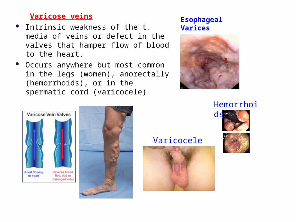
Varicose veins Intrinsic weakness of the t. media of veins
or defect in the valves that hamper flow of blood to the heart.
Occurs anywhere but most common in the legs (women), anorectally (hemorrhoids), or in the spermatic cord (varicocele)
Esophageal Varices
Varicocele
Hemorrhoids

Lymphedema
Penoscrotal lymphedema Obstruction to lymphatics
Causes:TraumaPost surgicalPost radiationInflammationParasitic obstructionObstruction due to metastasis
filarial


