Page 1
ORIGINAL PAPER
177Lu–DO3A–HSA–ZEGFR:1907: characterization as a potentialradiopharmaceutical for radionuclide therapyof EGFR-expressing head and neck carcinomas
Susan Hoppmann • Shibo Qi • Zheng Miao •
Hongguang Liu • Han Jiang • Cathy S. Cutler •
Ande Bao • Zhen Cheng
Received: 18 December 2011 / Accepted: 7 March 2012 / Published online: 16 March 2012
� SBIC 2012
Abstract Epidermal growth factor receptor 1 (EGFR) is an
attractive target for radionuclide therapy of head and neck
carcinomas. Affibody molecules against EGFR (ZEGFR)
show excellent tumor localizations in imaging studies.
However, one major drawback is that radiometal-labeled
Affibody molecules display extremely high uptakes in the
radiosensitive kidneys which may impact their use as radi-
otherapeutic agents. The purpose of this study is to further
explore whether radiometal-labeled human serum albumin
(HSA)–ZEFGR bioconjugates display desirable profiles for
the use in radionuclide therapy of EGFR-positive head and
neck carcinomas. The ZEFGR analog, Ac–Cys–ZEGFR:1907,
was site-specifically conjugated with HSA. The resulting
bioconjugate 1,4,7,10-tetraazacyclododecane-1,4,7-triace-
tic acid (DO3A)–HSA–ZEGFR:1907 was then radiolabeled
with either 64Cu or 177Lu and subjected to in vitro cell uptake
and internalization studies using the human oral squamous
carcinoma cell line SAS. Positron emission tomography
(PET), single photon emission computed tomography
(SPECT), and biodistribution studies were conducted using
SAS-tumor-bearing mice. Cell studies revealed a high
(8.43 ± 0.55 % at 4 h) and specific (0.95 ± 0.09 % at 4 h)
uptake of 177Lu–DO3A–HSA–ZEGFR:1907 as determined by
blocking with nonradioactive ZEGFR:1907. The internaliza-
tion of 177Lu–DO3A–HSA–ZEGFR:1907 was verified in vitro
and found to be significantly higher than that of 177Lu-
labeled ZEFGR at 2–24 h of incubation. PET and SPECT
studies showed good tumor imaging contrasts. The biodis-
tribution of 177Lu–DO3A–HSA–ZEGFR:1907 in SAS-tumor-
bearing mice displayed high tumor uptake (5.1 ±
0.44 % ID/g) and liver uptake (31.5 ± 7.66 % ID/g) and
moderate kidney uptake (8.5 ± 1.08 % ID/g) at 72 h after
injection. 177Lu–DO3A–HSA–ZEGFR:1907 shows promising
in vivo profiles and may be a potential radiopharmaceutical
for radionuclide therapy of EGFR-expressing head and neck
carcinomas.
Keywords 177Lu � Affibody � Epidermal growth factor
receptor � Human serum albumin � Radionuclide therapy
Introduction
Epidermal growth factor receptor 1 (EGFR) is an approx-
imately 180-kDa transmembrane glycoprotein consisting of
an extracellular-ligand-binding domain, a transmembrane
domain, and an intracellular domain with tyrosine kinase
activity for signal transduction. As a member of the erbB
S. Hoppmann � S. Qi � Z. Miao � H. Liu � H. Jiang �Z. Cheng (&)
Molecular Imaging Program at Stanford (MIPS),
Department of Radiology and Bio-X Program,
Canary Center at Stanford for Cancer Early Detection,
Stanford University,
1201 Welch Road,
Lucas Expansion, P095,
Stanford, CA 94305, USA
e-mail: [email protected]
C. S. Cutler
Research Reactor Center (MURR),
Radiopharmaceutical Sciences Institute,
Nuclear Engineering and Sciences Institute,
Nuclear Engineering,
University of Missouri,
Columbia, MO 65211, USA
A. Bao
Departments of Radiology and Otolaryngology—Head
and Neck Surgery,
University of Texas Health Science Center at San Antonio,
San Antonio, TX 78229, USA
123
J Biol Inorg Chem (2012) 17:709–718
DOI 10.1007/s00775-012-0890-3
Page 2
receptor family, EGFR is widely expressed in most epi-
thelial cells from several normal tissues, including skin,
liver, and basal cells of the prostate epithelium [1–3].
Dysregulation of EGFR disrupts normal cellular patterns
by increasing cell proliferation and angiogenic potential
and inhibiting apoptosis [4]. Thus, overexpression of
EGFR has been frequently detected in a wide range of
human tumors, including small cell carcinoma of the head
and neck [1–3, 5–8]. Moreover, recent studies have dem-
onstrated a correlation between EGFR overexpression and
metastasis formation, therapy resistance, poor prognosis,
and short survival for some cancer types [1, 3, 5–9].
Head and neck squamous cell carcinoma (HNSCC),
including cancers of the oral cavity, oropharynx, hypo-
pharynx, and larynx, is one of the most commonly diag-
nosed cancers worldwide, with over 600,000 new cases
every year [10]. Although there have been slightly
decreased incidence and mortality rates during recent
years, 260 new cases and 11,480 deaths of HNSCC
occurred in the USA alone in 2010 [11]. HNSCC has been
characterized by high levels of EGFR expression in
approximately 90 % of tumors [12], correlating with poor
clinical outcome [13], decreased response to radiotherapy,
and increased locoregional recurrence following definitive
radiotherapy [14].
EGFR has become an attractive target for treatment of
HNSCC. Inhibition of tumor growth can be obtained by
either blocking the extracellular-ligand-binding domain of
EGFR or by inhibiting intracellular tyrosine kinase activity
[15]. Owing to the intrinsic radiosensitivity of this tumor
type, targeted radionuclide therapy offers a realistic
approach to improve the treatment of HNSCC [16].
Affibody proteins have been shown to be a promising
platform for the development of imaging or therapeutic
agents for different molecular targets [17, 18]. Affibody
molecules are engineered small proteins with 58 amino acid
residues (6–7 kDa) and a three-a-helical bundle structure,
as derived from one of the IgG-binding domains of staph-
ylococcal protein A [19]. Several anti-EGFR Affibody
proteins (ZEFGR) with high affinities in the nanomolar range
have been reported recently [20, 21]. Particularly
ZEGFR:1907 was demonstrated to show excellent EGFR-
tumor-targeting abilities, which was confirmed in our
research using either 64Cu or Cy5.5 fluorescent dye labeled
ZEGFR:1907 for small-animal positron emission tomography
(PET) and optical imaging studies [22, 23]. However,
besides the high and specific tumor imaging contrast, che-
lator modified and radiometal-labeled Affibody proteins
typically exhibit extremely high renal uptake (more than
100 % of the injected dose per gram of tissue, ID/g). This
could result in very high radiation doses to the radiation-
sensitive kidneys and thus represents a critical concern
when using Affibody molecules for radionuclide therapy.
We have recently demonstrated that conjugation of human
serum albumin (HSA) with an anti-human epidermal growth
factor receptor 2 (HER2) Affibody (ZHER2) can produce
HSA–ZHER2 bioconjugates which exhibit improved phar-
macokinetics in terms of high tumor uptake, but dramatically
reduced kidney uptake [24]. These promising results have
encouraged us to further explore whether radiometal-labeled
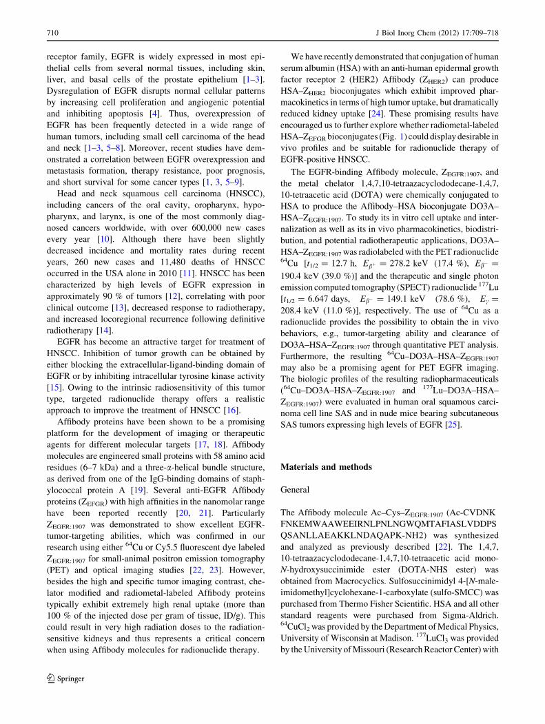
HSA–ZEFGR bioconjugates (Fig. 1) could display desirable in
vivo profiles and be suitable for radionuclide therapy of
EGFR-positive HNSCC.
The EGFR-binding Affibody molecule, ZEGFR:1907, and
the metal chelator 1,4,7,10-tetraazacyclododecane-1,4,7,
10-tetraacetic acid (DOTA) were chemically conjugated to
HSA to produce the Affibody–HSA bioconjugate DO3A–
HSA–ZEGFR:1907. To study its in vitro cell uptake and inter-
nalization as well as its in vivo pharmacokinetics, biodistri-
bution, and potential radiotherapeutic applications, DO3A–
HSA–ZEGFR:1907 was radiolabeled with the PET radionuclide64Cu [t1/2 = 12.7 h, Ebþ = 278.2 keV (17.4 %), Eb� =
190.4 keV (39.0 %)] and the therapeutic and single photon
emission computed tomography (SPECT) radionuclide 177Lu
[t1/2 = 6.647 days, Eb� = 149.1 keV (78.6 %), Ec =
208.4 keV (11.0 %)], respectively. The use of 64Cu as a
radionuclide provides the possibility to obtain the in vivo
behaviors, e.g., tumor-targeting ability and clearance of
DO3A–HSA–ZEGFR:1907 through quantitative PET analysis.
Furthermore, the resulting 64Cu–DO3A–HSA–ZEGFR:1907
may also be a promising agent for PET EGFR imaging.
The biologic profiles of the resulting radiopharmaceuticals
(64Cu–DO3A–HSA–ZEGFR:1907 and 177Lu–DO3A–HSA–
ZEGFR:1907) were evaluated in human oral squamous carci-
noma cell line SAS and in nude mice bearing subcutaneous
SAS tumors expressing high levels of EGFR [25].
Materials and methods
General
The Affibody molecule Ac–Cys–ZEGFR:1907 (Ac-CVDNK
FNKEMWAAWEEIRNLPNLNGWQMTAFIASLVDDPS
QSANLLAEAKKLNDAQAPK-NH2) was synthesized
and analyzed as previously described [22]. The 1,4,7,
10-tetraazacyclododecane-1,4,7,10-tetraacetic acid mono-
N-hydroxysuccinimide ester (DOTA-NHS ester) was
obtained from Macrocyclics. Sulfosuccinimidyl 4-[N-male-
imidomethyl]cyclohexane-1-carboxylate (sulfo-SMCC) was
purchased from Thermo Fisher Scientific. HSA and all other
standard reagents were purchased from Sigma-Aldrich.64CuCl2 was provided by the Department of Medical Physics,
University of Wisconsin at Madison. 177LuCl3 was provided
by the University of Missouri (Research Reactor Center) with
710 J Biol Inorg Chem (2012) 17:709–718
123
Page 3
a specific activity of 25 Ci/mg. Human oral squamous car-
cinoma cell line SAS was obtained from the American Type
Culture Collection. The cells were maintained in high-glu-
cose Dulbecco’s modified Eagle’s medium (DMEM) sup-
plemented with 10 % fetal bovine serum, 100 U penicillin
per milliliter, and 100 lg streptomycin per milliliter (Invit-
rogen). Female athymic nude mice (nu/nu) were purchased
from Charles River Laboratories. All other instruments,
including the radioactive dose calibrator, were the same as
those previously reported [26].
Bioconjugation of HSA with DOTA
and Ac–Cys–ZEGFR:1907
The conjugation of DOTA-NHS ester and Ac–Cys–
ZEGFR:1907 was performed in the same fashion as described
previously for preparation of HSA–ZHER2 bioconjugates
[24]. Briefly, the HSA was conjugated to DOTA-NHS ester
in 200 lL borate buffer (50 mM, pH 8.5) in a molar ratio
of 1:100. The resulting product is referred to as DO3A–
HSA to indicate it is no longer a full-fledged DOTA che-
lator but is rather a derivative of DO3A. The HSA–DO3A
conjugate was then reacted with 1 mg sulfo-SMCC in a
molar ratio of 1:5. Finally, Ac–Cys–ZEGFR:1907 was site-
specifically conjugated to the DO3A- and Sulfo-SMCC-
modified HSA via the cysteine residue. The reaction was
performed in a molar ratio of 1:5 using 200 lg DO3A–
HSA–sulfo-SMCC and 150 lg Ac–Cys–ZEGFR:1907. The
final bioconjugate, DO3A–HSA–ZEGFR:1907, was then
concentrated through a microcentrifuge tube (Millipore,
30 kDa, 0.5 mL) to a final volume of 20 lL. After each
step, the protein concentration was measured by the bicinch-
oninic acid assay (Pierce), and the samples were analyzed via
matrix-assisted laser desorption/ionization time of flight mass
spectrometry. The 1,4,7,10-tetraazacyclododecane-1,4,7-tria-
cetic acid-10-maleimidoethylacetamide-conjugated Ac–Cys–
ZEGFR:1907, DO3A–ZEGFR:1907, which was reported by us
before, was also used in our studies for comparison [22].
Radiolabeling of DO3A–HSA–ZEGFR:1907
Approximately 200 lg DO3A–HSA–ZEGFR:1907 was radi-
olabeled with either 64Cu or 177Lu by addition of
37–74 MBq (1–2 mCi) of 64CuCl2 or 177LuCl3 in 0.1 N
sodium acetate buffer of pH 6.0 or pH 5.0, respectively.
After an incubation time of 1 h at 39 �C, the radiolabeled
complexes were purified by a PD-10 column (GE Health-
care), eluted with phosphate-buffered saline (PBS; pH 7.4),
and passed through a 0.22-lm Millipore filter for both
in vitro cell uptake studies and animal experiments. To
prevent radiolysis, 1 % ascorbic acid was added to the177Lu-labeled bioconjugate. For comparison, radiolabeling
of DO3A–ZEGFR:1907 with 177Lu was performed in the
same way as for DO3A–HSA–ZEGFR:1907.
In vitro stability
The stability of 177Lu–DO3A–HSA–ZEGFR:1907 was asses-
sed in vitro by adding 50 lL tracer to 1 mL mouse serum
(final activity concentration 2.59 MBq/mL; 70 lCi/mL).
The samples were incubated at 37 �C. The stability of the
tracer was assessed by size-exclusion chromatography on
Fig. 1 A 1,4,7,10-
tetraazacyclododecane-1,4,7-
triacetic acid (DO3A)–human
serum albumin (HSA)–anti-
epidermal growth factor
receptor 1 (EGFR) Affibody
(ZEGFR:1907) bioconjugate. The
red regions indicate the lysine
residues of HSA suitable for
conjugation with 1,4,7,10-
tetraazacyclododecane-1,4,7,10-
tetraacetic acid (DOTA) and
ZEFGR molecules (bluestructures). The radiolabeling
occurs via complexation of
either 177Lu or 64Cu with the
DO3A chelator of the
bioconjugates
J Biol Inorg Chem (2012) 17:709–718 711
123
Page 4
NAP-5 columns (GE Healthcare) at incubation times of 4,
24, 48, 72, and 168 h. The column was equilibrated with
PBS and the contents were eluted at room temperature;
0.35-mL fractions were collected. The radioactivity of all
fractions was counted using a PerkinElmer 1470 automatic
c-counter, and the stability was expressed as the percentage
of the radioactivity in the original tube.
In vitro cell uptake and internalization assays
In vitro cell uptake assays of both 64Cu–DO3A–HSA–
ZEGFR:1907 and 177Lu–DO3A–HSA–ZEGFR:1907 were per-
formed as previously described [24]. Briefly, SAS cells
(2 9 105 per well) were seeded in 12-well tissue culture
plates and allowed to attach overnight. The cells were
washed twice with serum-free DMEM and incubated with
the probes [1.5 lCi per well, final concentration approxi-
mately 6 nM (0.2 lg)] in 400 lL serum-free DMEM at 4
and 37 �C. The nonspecific binding of the probes with SAS
cells was determined by co-incubation with nonradioactive
Ac–Cys–ZEGFR:1907 (6 lg per well, final concentration
2.14 lM). After 0.5, 1, 2, and 4 h (177Lu–DO3A–HSA–
ZEGFR:1907) and 2 h (64Cu–DO3A–HSA–ZEGFR:1907) the
cells were washed three times with cold PBS and lysed
with the addition of 200 lL of 0.2 M NaOH. The radio-
activity of all fractions was counted using a PerkinElmer
1470 automatic c-counter. The uptake (counts per minute)
was expressed as the percentage of added radioactivity.
For the cell internalization assay, SAS cells (5 9 105 per
well) were seeded in six-well tissue culture plates and
allowed to attach overnight. The cells were washed twice
with serum-free DMEM medium and incubated with 177Lu–
DO3A–HSA–ZEGFR:1907 and 177Lu–DO3A–ZEGFR:1907 [each
5 lCi per well, final concentrations approximately 10 nM
(0.66 lg) and approximately 10 nM (0.1 lg), respectively]
in 800 lL serum-free DMEM at 37 �C. After 1 min, 30 min,
1 h, 2 h, 4 h, 6 h, 12 h, and 24 h the medium was collected
and the cells were washed twice with cold PBS. Then, the
cells were treated with an acid washing buffer (0.2 M glycine/
HCl buffer with 4 M urea, pH 2.0) for 5 min at 4 �C and
additionally rinsed with the same buffer. The solution col-
lected was considered as the membrane-bound fraction. The
cells were lysed in 500 lL of 0.2 M NaOH and the solution
collected was considered as the internalized fraction. The
radioactivity of all fractions was counted using a PerkinElmer
1470 automatic c-counter.
Small-animal PET
All animal studies were performed in compliance with
federal and local institutional rules for animal experimen-
tation. Protocols were approved by the Stanford Adminis-
trative Panel on Laboratory Animal Care (APLAC 18086).
Approximately 3 9 106 SAS cells suspended in PBS were
subcutaneously implanted in the right upper shoulder of
female athymic nu/nu mice. Tumors were allowed to grow
to 0.5–0.7 cm in diameter (3–4 weeks). The tumor-bearing
mice were subjected to in vivo biodistribution and imaging
studies. Small-animal PET of tumor-bearing mice (n = 4
for each group) was performed using a micro-PET R4
rodent-model scanner (Siemens Medical Solutions USA).
A 180–210-lCi dose (6.7–7.8 MBq, 36–42 lg) of 64Cu–
DO3A–HSA–ZEGFR:1907 was injected via the tail vein into
mice bearing SAS tumors . At various times after injection
(1, 4, 24, 48, and 72 h) the mice were anesthetized with
2 % isoflurane and placed in the prone position near the
center of the field of view of the scanner. Static scans (for
1, 4, and 24 h, 3-min scans; for 48 and 72 h, 5-min scans)
were obtained, and the images were reconstructed by a
two-dimensional ordered subsets expectation maximum
algorithm. The method for quantification analysis of small-
animal PET images was the same as previously reported
[26].
Small-animal SPECT/X-ray computed tomography
For small-animal SPECT and X-ray computed tomography
(CT), a 200–300-lCi dose (7.4–11.1 MBq, 26.7–40.5 lg)
of 177Lu–DO3A–HSA–ZEGFR:1907 was injected via the tail
vein into SAS-tumor-bearing mice (n = 3). After 4, 24, 72,
and 96 h, the mice were anesthetized with 2 % isoflurane
and placed in the prone position near the center of the field
of view of the scanner. Nuclear imaging and CT were
performed with a combined SPECT/CT scanner for small
animals (X-SPECT; Gamma Medica).
For micro-CT image acquisition, the 512 images (170-lm
slice thickness) were acquired in 5 min at 0.4 mA and
80 kVp. SPECT was performed using a 1-mm multipinhole
collimator (single head, 360� of rotation, 64 projections, 30 s
per projection, and a 5-cm field of view). The SPECT and
CT images were then reconstructed as described previously
[24]. All data were imported into Amira (Mercury Com-
puting Systems, Chelmsford, UK) for processing and
visualization.
Biodistribution studies
For biodistribution studies, a 20–25-lCi dose (740–925 kBq;
2.7–3.3 lg) of 177Lu–DO3A–HSA–ZEGFR:1907 was injected
through the tail vein into mice bearing SAS xenografts (n = 4
for each group), and they were killed at different times after
injection (1, 4, 24, 48, 72, 120, and 240 h). Tumor and normal
tissues were excised and weighed, and their radioactivity
measured using a c-counter. The radioactivity uptake in the
tumor and normal tissues was expressed as the percentage of
injected radioactivity per gram of tissue.
712 J Biol Inorg Chem (2012) 17:709–718
123
Page 5
Statistical analysis
Statistical analysis was performed using Student’s two-
tailed t test for unpaired data. A 95 % confidence level was
chosen to determine the significance between groups, with
P \ 0.05 being significantly different.
Results
Synthesis, conjugation, radiochemistry, and stability
Ac–Cys–ZEGFR:1907 with an N-terminal cysteine residue
and DOTA were successfully conjugated with HSA. Three
DO3A molecules were conjugated on average to one HSA
molecule as determined by matrix-assisted laser desorp-
tion/ionization time of flight mass spectrometry. The
average molecular masses of DO3A–HSA–ZEGFR:1907
bioconjugates were 75.3, 81.8, 89.6, 95.0 and 101.9 kDa,
representing different numbers of Affibody molecules
conjugated per HSA protein (one to five Affibody mole-
cules on each HSA molecule). Owing to the more complex
nature of the final agent and the conjugation method,
several bioconjugates (e.g., bioconjugates with a different
number of DO3A molecules) may be hidden under one
average molecular mass signal. DO3A–HSA–ZEGFR:1907
was then successfully radiolabeled with either 64Cu or177Lu. Purification of the radioactive reaction mixtures
using a PD-10 column resulted in 64Cu–DO3A–HSA-
ZEGFR:1907 and 177Lu–DO3A–HSA–ZEGFR:1907 with decay-
corrected yields of more than 70 %. High specific activities
of 64Cu–DO3A–HSA–ZEGFR:1907 or 177Lu–DO3A–HSA–
ZEGFR:1907 (14–36 MBq/nmol, 5.0–12.9 mCi/mg) were
obtained at the end of the synthesis.
The serum stability showed that 177Lu–DO3A–HSA–
ZEGFR:1907 has good resistance to dissociation of 177Lu
from the tracer, with more than 95 % of the probe intact at
an incubation time of 4 h and more than 80 % of the probe
intact at incubation times of 24, 48, 72, and 168 h in mouse
serum.
In vitro assay and small-animal PET of 64Cu–DO3A–
HSA–ZEGFR:1907
The uptake of 64Cu–DO3A–HSA–ZEGFR:1907 in SAS cells
over an incubation period of 2 h at 37 �C is shown in
Fig. 2a. The probe showed a high and specific uptake
(26.84 ± 2.5 vs. 9.64 ± 1.51 % for the control and the
blocking group, respectively). Decay-corrected coronal and
transaxial microPET images of a mouse bearing SAS
tumor at 1, 4, 24, and 48 h after tail vein injection of 64Cu–
DO3A–HSA–ZEGFR:1907 are shown in Fig. 2b. The SAS
tumor was visible but with a low tumor-to-background
contrast at 1 and 4 h after injection, and excellent tumor-to-
background contrasts were observed at 24, 48, and 72 h
after injection. Quantification analysis revealed that the
SAS tumor uptake values increased with time, and they
were 3.34 ± 0.21, 5.19 ± 0.35, 7.01 ± 1.13, 7.72 ± 1.14,
and 7.48 ± 0.96 % ID/g at 1, 4, 24, 48 and 72 h, respec-
tively (Fig. 2c). In addition to the tumor, moderate activity
accumulation was observed in the liver (e.g., 11.34 ±
1.08 % ID/g at 24 h), but much lower activity accumula-
tion was found in the kidneys (e.g., 2.45 ± 1.72 % ID/g at
24 h). Relatively low radioactivity concentrations were
found in the blood (region of interest over the heart):
4.73 ± 0.49 and 3.51 ± 0.31 % ID/g at 1 and 4 h after
injection., respectively (Fig. 2c). Although the stability of
this conjugate was not tested in this study, previous studies
using a 64Cu–DOTA–Affibody conjugate showed that the
complex is stable in mouse plasma in vitro. A low (below
5 %) decomposition was found at an incubation time of 4 h
in mouse serum in vitro [22].
In vitro assays of 177Lu–DO3A–HSA–ZEGFR:1907
Cell uptake of 177Lu–DO3A–HSA–ZEGFR:1907 at 37 and 4 �C
over an incubation period of 0.5–4 h is shown in Fig. 3a.177Lu–DO3A–HSA–ZEGFR:1907 accumulated slowly in the
SAS cells and reached 1.97 ± 0.08 % of the applied activity
at 0.5 h at 37 �C. The uptake increased to 8.43 ± 0.55 % at
4 h at 37 �C. An approximate fourfold to fivefold lower
accumulation of 177Lu–DO3A–HSA–ZEGFR:1907 in the cell
lysates was observed at all time points at 4 �C compared with
37 �C, indicating internalization that occurs at physiologic
temperature. Finally, cell surface receptor binding and
internalization of the radiopharmaceutical were shown to be
inhibited by the presence of a large molar excess of unlabeled
ZEGFR:1907 (P \ 0.05) at all incubation time points (e.g.,
0.95 ± 0.09 % at 4 h). Cell association of 177Lu–DO3A–
HSA–ZEGFR:1907 at 4 h was inhibited by 80 and 65 % at 37
and 4 �C, respectively, indicating that the probe was specif-
ically targeting EFGR.
The internalization data for 177Lu–DO3A–HSA–
ZEGFR:1907 and 177Lu–DO3A–ZEGFR:1907 are shown in
Fig. 3b and c, respectively. For both radiopharmaceuticals,
the percentage of the internalized tracers was much higher
than that of the cell-surface-bound fraction. 177Lu–DO3A–
HSA–ZEGFR:1907 showed a relatively slow internalization,
with 1.45 ± 0.1 and 7.9 ± 0.5 % internalization of the
receptor-bound tracer at incubation times 1 and 30 min,
respectively, whereas 38.74 ± 1.4 % of the tracer was inter-
nalized at 24 h. In comparison, 177Lu–DO3A–ZEGFR:1907
internalized faster, with 2.27 ± 0.2 and 9.6 ± 0.8 % at 2 and
30 min, respectively, whereas 28.9 ± 2.2 % of the tracer was
internalized at 24 h. A significantly higher percentage of
added 177Lu–DO3A–HSA–ZEGFR:1907 compared with
J Biol Inorg Chem (2012) 17:709–718 713
123
Page 6
177Lu–DO3A–ZEGFR:1907 was internalized into SAS cells at
24 h (P \ 0.05). This suggests that the presence of multiple
Affibody molecules on HSA may help to promote internali-
zation of the tracer.
In vivo studies of 177Lu–DO3A–HSA–ZEGFR:1907
The in vivo biodistribution of 177Lu–DO3A–HSA–
ZEGFR:1907 was examined in an SAS-tumor-bearing mouse
model. The biodistribution of 177Lu–DO3A–HSA–
ZEGFR:1907 at 1, 4, 24, 48, 72, 120, and 240 h is shown in
Fig. 4a. Slow but relatively high levels of radioactivity
accumulation in the SAS tumors were observed. The tumor
uptake in SAS tumors was 2.00 ± 0.80 % ID/g at 1 h and
continually increased to 3.13 ± 0.95 % ID/g at 4 h and
4.57 ± 0.23 % ID/g at 24 h. The tumor uptake reached a
plateau of 5.24 ± 0.67 and 5.31 ± 0.44 % ID/g at 48 and
72 h, respectively. Finally, the tumor uptake of 177Lu–
DO3A–HSA–ZEGFR:1907 decreased to 3.72 ± 0.64 and
0.66 ± 0.2 % ID/g at 120 and 240 h after injection, respec-
tively. 177Lu–DO3A–HSA–ZEGFR:1907 also displayed a rel-
atively slow blood clearance. Blood values of 31.87 ± 0.95
and 19.07 ± 1.05 % ID/g were observed at 1 and 4 h after
injection, respectively. The blood clearance of 177Lu–
DO3A–HSA–ZEGFR:1907 fits the second-order exponential
decay model (R2 = 0.9987). With the administered dose used
for this study, 177Lu–DO3A–HSA–ZEGFR:1907 exhibited a
two-phase clearance pattern, in which 64.3 % of the injected
dose cleared fast with1.99-h half clearance time and 35.7 %
cleared more slowly with 25.50-h half clearance time. The
tracer showed high liver uptake at all time points (e.g., 4 h
after injection; 54.93 ± 4.05 % ID/g). The kidneys, how-
ever, showed a moderate radioactivity accumulation (e.g.,
11.14 ± 1.0 and 7.65 ± 1.68 % ID/g at 4 and 48 h after
injection, respectively). These data indicate the tracer was
cleared predominantly through the hepatobiliary system and
to a minor extent through the renal system. Figure 4b shows
the tumor-to-organ ratios of 177Lu–DO3A–HSA–ZEGFR:1907.
The tumor-to-muscle ratio increased slowly from 3.3 at 1 h
and reached a plateau of approximately 7.9 at 72–240 h.
However, the tumor-to-blood ratio increased tenfold from 1.7
at 48 h to 16.7 at 240 h.
The SPECT images correlate with the results obtained
from the biodistribution studies. Images acquired at 4, 24,
48, and 72 h after injection of 177Lu–DO3A–HSA–
ZEGFR:1907 demonstrated significant liver accumulation and
good tumor localization, and high tumor-to-background
contrasts were observed at the later times after injection
(Fig. 5).
Discussion
Affibody molecules have shown excellent tumor-targeting
properties in molecular imaging studies of tumors
expressing EGFR [22, 27–30]. However, the use of
radiometal-labeled, chelator-modified Affibody molecules
for radiotherapeutic applications is questionable, mainly
Fig. 2 a Cell uptake of 64Cu–
DO3A–HSA–ZEGFR:1907 in SAS
cells in the presence or absence
of nonradioactive Ac–Cys–
ZEGFR:1907 after 120 min
incubation. Data are shown as
the mean ± the standard
deviation (SD) percentage of
applied radioactivity (n = 4).
b Representative decay-
corrected coronal (top) and
transaxial (bottom) positron
emission tomography (PET)
images at 1, 4, 24, 48, and 72 h
after tail vein injection of 64Cu–
DO3A–HSA–ZEGFR:1907.
Arrows indicate the location of
the tumors. c Uptake levels
(percentage of injected dose per
gram of tissue) of tumor,
muscle, liver, and kidney
derived from multiple time
point small-animal PET images
after tail vein injection of 64Cu–
DO3A–HSA–ZEGFR:1907. Data
are shown as the mean ± SD of
four measurements
714 J Biol Inorg Chem (2012) 17:709–718
123
Page 7
because of their extremely high kidney uptakes (generally
more than 100 % ID/g for radiometal-labeled Affibody
proteins) [31–33]. The high kidney retention is caused by
reabsorption of radiolabeled Affibody molecules in the
proximal tubules via luminal endocytosis after glomerular
filtration. Furthermore, it is known that peptides
conjugated with residualizing radiolabels are trapped in
the tubular cell lysosomes and therefore deliver high
radiation doses to the radiation-sensitive kidneys [34]. As
shown in earlier studies, the fusion of an Affibody protein
to a small albumin-binding domain (ABD) peptide results
in reduced kidney uptake [35, 36]. In our previous study
we developed an anti-HER2 HSA–Affibody conjugate, a
multimeric ligand with a size much smaller than that of
conventional antibodies (below 100 kDa), which exhibits
high tumor targeting and reduced kidney uptake in
SKOV3 xenograft mouse models [24]. That work also
revealed that Affibody–albumin conjugates display several
distinct advantages for imaging or therapy applications.
For example, (a) a quick and efficient covalent conjuga-
tion and labeling technique, (b) improved pharmacoki-
netics in terms of a lower kidney accumulation, and more
importantly, (c) a possible multiple and simultaneous
binding to HER2 owing to the attachment of several Af-
fibody molecules.
In the present study, HSA was further used to modify a
novel EGFR binder, Ac–Cys–ZEGFR:1907, and the radiola-
beled bioconjugate was evaluated for its potential in
radionuclide therapy of head and neck cancer. Targeted
radionuclide therapy has great potential for treatment of
HNSCC, because this type of cancer has a decreased
response to radiotherapy and increased locoregional
recurrence following definitive radiotherapy [14]. More-
over, the radiopharmaceutical developed could also be used
for radionuclide therapy for many other types of EGFR-
positive cancer. This fact leads to a broader application
range for using HSA–ZEFGR than for HSA–ZHER2. The
series of experiments performed confirmed our hypothesis
that anti-EGFR Affibody–HSA conjugates display good
tumor-targeting abilities and preferred pharmacokinetics
for potential radiotherapeutic applications in treatment of
HNSCC.
The human oral squamous carcinoma cell line SAS used
in this study exhibits high expression of EGFR in vitro and
in xenotransplants in vivo [25]. In vitro cell uptake studies
demonstrate the specificity and ability of the radiolabeled
conjugates to target EGFR. Furthermore 177Lu–DO3A–
HSA–ZEGFR:1907 can be internalized into SAS cells after
binding EGFR at the membrane. Interestingly, in vitro cell
internalization studies of both 177Lu–DO3A–HSA–
ZEGFR:1907 and 177Lu–DO3A–ZEGFR:1907 demonstrate that
the radiolabeled HSA–Affibody bioconjugate displays
significantly higher internalization rates than the radiola-
beled Affibody molecule alone at later time points. This is
another important advantage for HSA–Affibody conjugates
for potential use in radionuclide therapy. The higher
internalization rate might be caused by albumin-mediated
endocytosis of the radiopharmaceutical in addition to the
EGFR-mediated internalization. The supportive effect of
Fig. 3 In vitro cell studies of 177Lu–DO3A–HSA–ZEGFR:1907. a In
vitro cell uptake of 177Lu–DO3A–HSA–ZEGFR:1907 in SAS cells over
time at 4 and 37 �C in the presence or absence of nonradioactive
Ac–Cys–ZEGFR:1907. b, c Cell-associated radioactivity as a function of
time after incubation of SAS cells with 177Lu–DO3A–HSA–
ZEGFR:1907 (b) or 177Lu–DO3A–ZEGFR:1907 (c). The radioactivity that
was removed from the cells by treatment with acidic washing buffer
(0.2 M glycine buffer containing 4 M urea, pH 2.0) was considered as
the membrane-bound fraction. The radioactivity measured in the cell
lysates was considered as the internalized fraction. All data are
presented as means ± SD (n = 4)
J Biol Inorg Chem (2012) 17:709–718 715
123
Page 8
albumin contributing to the internalization of drugs in
tumor cells was also shown by others [37].
Quantitative PET, SPECT, and biodistribution data con-
firm the good tumor targeting properties of both 64Cu- and177Lu-labeled DO3A–HSA–ZEGFR:1907. Importantly, the
retention of the radioactivity measured in the tumor is very
long. The retention of radiopharmaceuticals in the tumor is a
critical factor in radionuclide therapy applications. In contrast,
the short peptide EGFR is rapidly degraded in lysosomes after
binding with and internalization in EGFR-positive tumors
[38]. This disadvantageous property leads to a short intra-
cellular retention of the radionuclide and decreases the effi-
ciency of the therapy and increases unwanted side effects,
such as uptake in normal tissues. Compared with the uptake
of 64Cu–DO3A–ZEGFR:1907 in A431 tumors determined by
PET quantification [22], the uptake of 64Cu–DO3A–HSA–
ZEGFR:1907 in SAS tumors was found to be half as much at 4 h
after injection. In contrast, the tumor uptake of 111In–DOTA–
ZEGFR:1907 in A431-tumor-bearing mice is only half as much
as for 64Cu–DO3A–HSA–ZEGFR:1907 (2.4 ± 0.3 % ID/g
Fig. 4 Biodistribution results
(a) and tumor-to-organ ratios
(b) for 177Lu–DO3A–HSA–
ZEGFR:1907 in nude mice bearing
subcutaneously xenografted
SAS human head and neck
cancer. The biodistribution data
are expressed as the percentage
of the injected dose per gram of
tissue after intravenous injection
of the probe at 1, 4, 24, 48, 120
,and 240 h (n = 4)
Fig. 5 Small-animal single
photon emission computed
tomography/X-ray computed
tomography of a mouse bearing
SAS tumor xenograft at 4, 24,
48, and 72 h after
administration of 177Lu–DO3A–
HSA–ZEGFR:1907. Arrowsindicate the location of tumors.
White indicates the highest
radioactivity accumulation
716 J Biol Inorg Chem (2012) 17:709–718
123
Page 9
vs. 5.19 ± 0.35 at 4 h after injection) [39]. These differences
can be explained by the size and different pharmacokinetic
properties of the tracers, by the different radiometals used,
and also by the lower expression of EGFR in SAS tumors
compared with A431 tumors.
The differences in tumor uptake between 64Cu–DO3A–
HSA–ZEGFR:1907 calculated from PET studies and 177Lu–
DO3A–HSA–ZEGFR:1907 obtained from biodistribution
studies, e.g., 7.72 ± 1.14 versus 5.24 ± 0.67 % ID/g at
72 h, could be caused by the different amounts of injected
radiotracer (approximately 40 lg for the 64Cu tracer and
approximately 3 lg for the 177Lu tracer). Several authors
have shown an influence of the injected dose of EGFR-
targeting proteins on the organ distribution [22, 28, 39–40].
In previous studies, we showed that the tumor uptake of64Cu–DO3A–ZEGFR:1907 is higher when there is blocking
with a small amount (50 lg) of cold tracer [22]. Further-
more, Tolmachev et al. [28] identified a maximum uptake
of 111In–DOTA–ZEGFR:2377 in A431 tumors between 30
and 50 lg of injected protein. The liver uptake measured
from the PET studies was shown to be one third as high as
that determined via biodistribution experiments. It was
observed that an increase of the injected dose of the
radiotracer resulted in a partial blocking with unlabeled
compound and thus a decrease in the uptake in EGFR-
expressing organs, e.g., the liver but also in the blood and
muscle. On the other hand, the differences between the64Cu- and the 177Lu-labeled bioconjugates could also be
caused by the different stabilities of the DO3A–metal
complex.
Compared with many radiometal-labeled Affibody pro-
teins (64Cu, 111In), the 64Cu- and 177Lu-labeled DO3A–
HSA–ZEGFR:1907 conjugates show much lower uptake in
the kidneys as determined in PET, SPECT, and biodistri-
bution studies. In comparison with the biodistribution
studies we performed recently using 64Cu–DO3A–
ZEGFR:1907, the kidney uptake for 177Lu–DO3A–HSA–
ZEGFR:1907 is eight times lower at 4 h after injection
(11.14 ± 1.0 vs. 88.45 ± 14.42 % ID/g). However, the
liver uptake of 177Lu–DO3A–HSA–ZEGFR:1907 was three
times higher than that of 64Cu–DO3A–ZEGFR:1907 at 4 h
after injection (54.93 ± 4.05 vs. 18.99 ± 2.88 % ID/g).
This indicates clearly that the conjugation of HSA signif-
icantly reduces the kidney uptake of the protein but
increases the liver uptake. However, increased liver uptake
does not make the agent questionable for therapeutic
use. The predicted dose of radiation absorbed by liver
for 177Lu–DO3A–HSA–ZEGFR:1907 was calculated be
2.6 mGy/MBq. If a radiation-treatment absorbed dose of
30–50 Gy is assumed for the tumor, the corresponding
dose for the liver will be 8.1–13.5 Gy, which is far below
the dose limit for liver in humans [41]. The high liver
uptake of the conjugate can be explained by several facts,
e.g., the high EGFR expression in the liver tissue [22], a
nonspecific clearance of the probe through the hepatobil-
iary system, and a presumable transchelation of 64Cu in the
liver [42]. Furthermore, the high accumulation in liver
tissues can be caused by the metabolic fate of the HSA
component of the radiotracer.
In conclusion, HSA–ZEFGR conjugates were prepared
and labeled successfully with both 64Cu and 177Lu. 177Lu–
DO3A–HSA–ZEGFR:1907 provides high specificity, high
sensitivity, and displays good tumor contrasts as shown in
SPECT studies. The in vivo properties and pharmacoki-
netics of the 177Lu–DO3A–HSA–ZEGFR:1907 conjugate
make it a potential radiopharmaceutical for treatment of
head and neck cancer.
Acknowledgments This work was supported, in part, by National
Cancer Institute (NCI) grant 5R01 CA119053 and NCI In Vivo
Cellular Molecular Imaging Center grant P50 CA114747. The pro-
duction of 177Lu was supported by Department of Energy grant
84900-001-10.
Conflict of interest The authors declare that they have no conflict
of interest.
References
1. Bellezza I, Bracarda S, Caserta C, Minelli A (2006) Targeting of
EGFR tyrosine kinase by ZD1839 (‘‘Iressa’’) in androgen-
responsive prostate cancer in vitro. Mol Genet Metab 88:114–122
2. Hynes NE, Lane HA (2005) ERBB receptors and cancer: the
complexity of targeted inhibitors. Nat Rev Cancer 5:341–354
3. Schlomm T, Kirstein P, Iwers L, Daniel B, Steuber T et al (2007)
Clinical significance of epidermal growth factor receptor protein
overexpression and gene copy number gains in prostate cancer.
Clin Cancer Res 13:6579–6584
4. Normanno N, De Luca A, Bianco C, Strizzi L, Mancino M et al
(2006) Epidermal growth factor receptor (EGFR) signaling in
cancer. Gene 366:2–16
5. Dei Tos AP, Ellis I (2005) Assessing epidermal growth factor
receptor expression in tumours: what is the value of current test
methods? Eur J Cancer 41:1383–1392
6. Harari PM (2004) Epidermal growth factor receptor inhibition
strategies in oncology. Endocr Relat Cancer 11:689–708
7. Marks RA, Zhang S, Montironi R, McCarthy RP, MacLennan GT
et al (2008) Epidermal growth factor receptor (EGFR) expression
in prostatic adenocarcinoma after hormonal therapy: a fluores-
cence in situ hybridization and immunohistochemical analysis.
Prostate 68:919–923
8. Rocha-Lima CM, Soares HP, Raez LE, Singal R (2007) EGFR
targeting of solid tumors. Cancer Control 14:295–304
9. Shariat SF, Bensalah K, Karam JA, Roehrborn CG, Gallina A
et al (2007) Preoperative plasma HER2 and epidermal growth
factor receptor for staging and prognostication in patients with
clinically localized prostate cancer. Clin Cancer Res 13:
5377–5384
10. Parkin DM, Bray F, Ferlay J, Pisani P (2005) Global cancer
statistics, 2002. CA Cancer J Clin 55:74–108
11. Jemal A, Siegel R, Xu J, Ward E (2010) Cancer statistics, 2010.
CA Cancer J Clin 60:277–300
J Biol Inorg Chem (2012) 17:709–718 717
123
Page 10
12. Bonner JA, Raisch KP, Trummell HQ, Robert F, Meredith RF
et al (2000) Enhanced apoptosis with combination C225/radiation
treatment serves as the impetus for clinical investigation in head
and neck cancers. J Clin Oncol 18:47S–53S
13. Rubin Grandis J, Melhem MF, Gooding WE, Day R, Holst VA
et al (1989) Levels of TGF-alpha and EGFR protein in head and
neck squamous cell carcinoma and patient survival. J Natl Cancer
Inst 90:824–832
14. Ang KK, Berkey BA, Tu X, Zhang HZ, Katz R et al (2002)
Impact of epidermal growth factor receptor expression on sur-
vival and pattern of relapse in patients with advanced head and
neck carcinoma. Cancer Res 62:7350–7356
15. Castillo L, Etienne-Grimaldi MC, Fischel JL, Formento P, Magne
N et al (2004) Pharmacological background of EGFR targeting.
Ann Oncol 15:1007–1012
16. Potamianos S, Varvarigou AD, Archimandritis SC (2000)
Radioimmunoscintigraphy and radioimmunotherapy in cancer:
principles and application. Anticancer Res 20:925–948
17. Nord K, Gunneriusson E, Ringdahl J, Stahl S, Uhlen M, Nygren
PA (1997) Binding proteins selected from combinatorial libraries
of an alpha-helical bacterial receptor domain. Nat Biotechnol
15:772–777
18. Wikman M, Steffen AC, Gunneriusson E, Tolmachev V, Adams
GP et al (2004) Selection and characterization of HER2/neu-
binding affibody ligands. Protein Eng Des Sel 17:455–462
19. Nilsson FY, Tolmachev V (2007) Affibody molecules: new
protein domains for molecular imaging and targeted tumor ther-
apy. Curr Opin Drug Discov Devel 10:167–175
20. Friedman M, Nordberg E, Hoiden-Guthenberg I, Brismar H,
Adams GP et al (2007) Phage display selection of affibody
molecules with specific binding to the extracellular domain of the
epidermal growth factor receptor. Protein Eng Des Sel
20:189–199
21. Friedman M, Orlova A, Johansson E, Eriksson TL, Hoiden-
Guthenberg I, Tolmachev V et al (2008) Directed evolution to low
nanomolar affinity of a tumor-targeting epidermal growth factor
receptor-binding affibody molecule. J Mol Biol 376:1388–1402
22. Miao Z, Ren G, Liu H, Jiang L, Cheng Z (2010) Small-animal
PET imaging of human epidermal growth factor receptor positive
tumor with a 64Cu labeled affibody protein. Bioconjug Chem
21:947–954
23. Miao Z, Ren G, Liu H, Jiang L, Cheng Z (2010) Cy5.5-labeled
affibody molecule for near-infrared fluorescent optical imaging of
epidermal growth factor receptor positive tumors. J Biomed Opt
15:036007
24. Hoppmann S, Miao Z, Liu S, Liu H, Ren G et al (2011) Radio-
labeled affibody–albumin bioconjugates for HER2-positive can-
cer targeting. Bioconjug Chem 38(4):613–622
25. Niu G, Li Z, Xie J, Le QT, Chen X (2009) PET of EGFR anti-
body distribution in head and neck squamous cell carcinoma
models. J Nucl Med 50:1116–1123
26. Cheng Z, De Jesus OP, Namavari M, De A, Levi J et al (2008)
Small-animal PET imaging of human epidermal growth factor
receptor type 2 expression with site-specific 18F-labeled protein
scaffold molecules. J Nucl Med 49:804–813
27. Malmberg J, Tolmachev V, Orlova A (2011) Imaging agents for
in vivo molecular profiling of disseminated prostate cancer—
targeting EGFR receptors in prostate cancer: comparison of
cellular processing of [111In]-labeled affibody molecule
Z(EGFR:2377) and cetuximab. Int J Oncol 38:1137–1143
28. Tolmachev V, Rosik D, Wallberg H, Sjoberg A, Sandstrom M
et al (2010) Imaging of EGFR expression in murine xenografts
using site-specifically labelled anti-EGFR 111In–DOTA–Z
EGFR:2377 affibody molecule: aspect of the injected tracer
amount. Eur J Nucl Med Mol Imaging 37:613–622
29. Nordberg E, Orlova A, Friedman M, Tolmachev V, Stahl S et al
(2008) In vivo and in vitro uptake of 111In, delivered with the
affibody molecule (ZEGFR:955)2, in EGFR expressing tumour
cells. Oncol Rep 19:853–857
30. Jokerst JV, Miao Z, Zavaleta C, Cheng Z, Gambhir SS (2011)
Affibody-functionalized gold-silica nanoparticles for Raman
molecular imaging of the epidermal growth factor receptor. Small
7:625–633
31. Cheng Z, De Jesus OP, Kramer DJ, De A, Webster JM et al
(2010) 64Cu-labeled affibody molecules for imaging of HER2
expressing tumors. Mol Imaging Biol 12:316–324
32. Tolmachev V, Nilsson FY, Widstrom C, Andersson K, Rosik D,
Gedda L et al (2006) 111In-benzyl-DTPA-ZHER2:342, an affi-
body-based conjugate for in vivo imaging of HER2 expression in
malignant tumors. J Nucl Med 47:846–853
33. Ren G, Zhang R, Liu Z, Webster JM, Miao Z, Gambhir SS et al
(2009) A 2-helix small protein labeled with 68 Ga for PET
imaging of HER2 expression. J Nucl Med 50:1492–1499
34. Behr TM, Goldenberg DM, Becker W (1998) Reducing the renal
uptake of radiolabeled antibody fragments and peptides for
diagnosis and therapy: present status, future prospects and limi-
tations. Eur J Nucl Med 25:201–212
35. Tolmachev V, Orlova A, Pehrson R, Galli J, Baastrup B,
Andersson K et al (2007) Radionuclide therapy of HER2-positive
microxenografts using a 177Lu-labeled HER2-specific affibody
molecule. Cancer Res 67:2773–2782
36. Tolmachev V, Wallberg H, Andersson K, Wennborg A, Lundq-
vist H, Orlova A (2009) The influence of Bz-DOTA and CHX-
A’’-DTPA on the biodistribution of ABD-fused anti-HER2 affi-
body molecules: implications for (114 m)In-mediated targeting
therapy. Eur J Nucl Med Mol Imaging 36:1460–1468
37. Wosikowski K, Biedermann E, Rattel B, Breiter N, Jank P et al
(2003) In vitro and in vivo antitumor activity of methotrexate
conjugated to human serum albumin in human cancer cells. Clin
Cancer Res 9:1917–1926
38. Todderud G, Carpenter G (1989) Epidermal growth factor: the
receptor and its function. Biofactors 2:11–15
39. Tolmachev V, Friedman M, Sandstrom M, Eriksson TL, Rosik D
et al (2009) Affibody molecules for epidermal growth factor
receptor targeting in vivo: aspects of dimerization and labeling
chemistry. J Nucl Med 50:274–283
40. Divgi CR, Welt S, Kris M, Real FX, Yeh SD et al (1991) Phase I
and imaging trial of indium 111-labeled anti-epidermal growth
factor receptor monoclonal antibody 225 in patients with squa-
mous cell lung carcinoma. J Natl Cancer Inst 83:97–104
41. Emami B, Lyman J, Brown A, Coia L, Goitein M et al (1991)
Tolerance of normal tissue to therapeutic radiation. Int J Radiat
Oncol Biol Phys 21:109–122
42. Boswell CA, Sun X, Niu W et al (2004) Comparative in vivo
stability of copper-64-labeled cross-bridged and conventional
tetraazamacrocyclic complexes. J Med Chem 47:1465–1474
718 J Biol Inorg Chem (2012) 17:709–718
123