Page 1
18F- FDG PET/CT in the Diagnosis of Tumor Thrombosis
T Davidson1, E Konen2 , O Goitein2, A Avigdor3, S T Zwas1 , E Goshen1
Departments of Nuclear Medicine1,Radiology2 and Hemato-Oncology3
Sheba Medical Center, Tel Aviv University, Israel
Page 2
Background
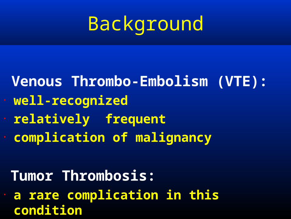
Venous Thrombo-Embolism (VTE):• well-recognized • relatively frequent • complication of malignancy
Tumor Thrombosis:• a rare complication in this condition
Page 3
Largest series described six
cases of Tumor Thrombosis diagnosed by PET with simultaneous CT
Lai P, et al.
Detection of tumour thrombus by 18F-FDG-PET/CT
imaging.
Eur J Cancer Prev. 2007 Feb;16(1):90-4.
Background
Page 4
• The role of PET/CT in the diagnosis
of Tumor Thrombosis • The Differential diagnosis of
Tumor Thrombosis from Venous Thrombo-Embolism
Aims
Page 5
Materials and Methods
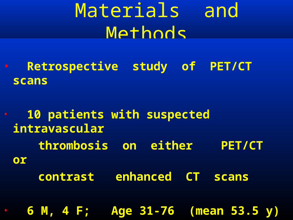
• Retrospective study of PET/CT scans
• 10 patients with suspected intravascular
thrombosis on either PET/CT or contrast enhanced CT scans
• 6 M, 4 F; Age 31-76 (mean 53.5 y)
Page 6
Materials and Methods
• In 8/10 pts the intravascular lesion was
an incidental finding on PET-CT scan during investigation of patients with known malignancy
• 2/10 pts were referred to PET-CT for
further evaluation of a known intravascular lesion diagnosed on CT /MRI
Page 7
Materials and Methods
Criteria for a positive PET :
• Increased focal or linear uptake of
18F-FDG in the involved vessel• Standard uptake value (SUV)
above 2.5
Page 8
Materials and Methods
• Findings were categorized: PET positive (+) or PET negative
(-)
• Compared to contrast enhanced CT ultrasound doppler, pathology when available, clinical follow-up
Page 9
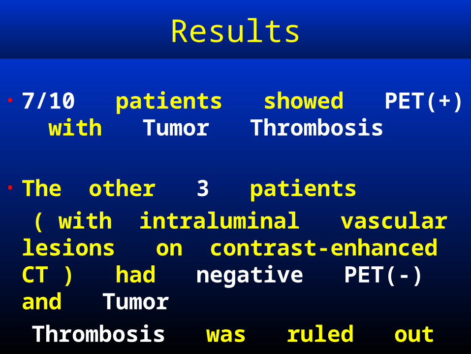
• 7/10 patients showed PET(+) with Tumor Thrombosis
• The other 3 patients ( with intraluminal vascular
lesions on contrast-enhanced CT ) had negative PET(-) and Tumor
Thrombosis was ruled out
Results
Page 10
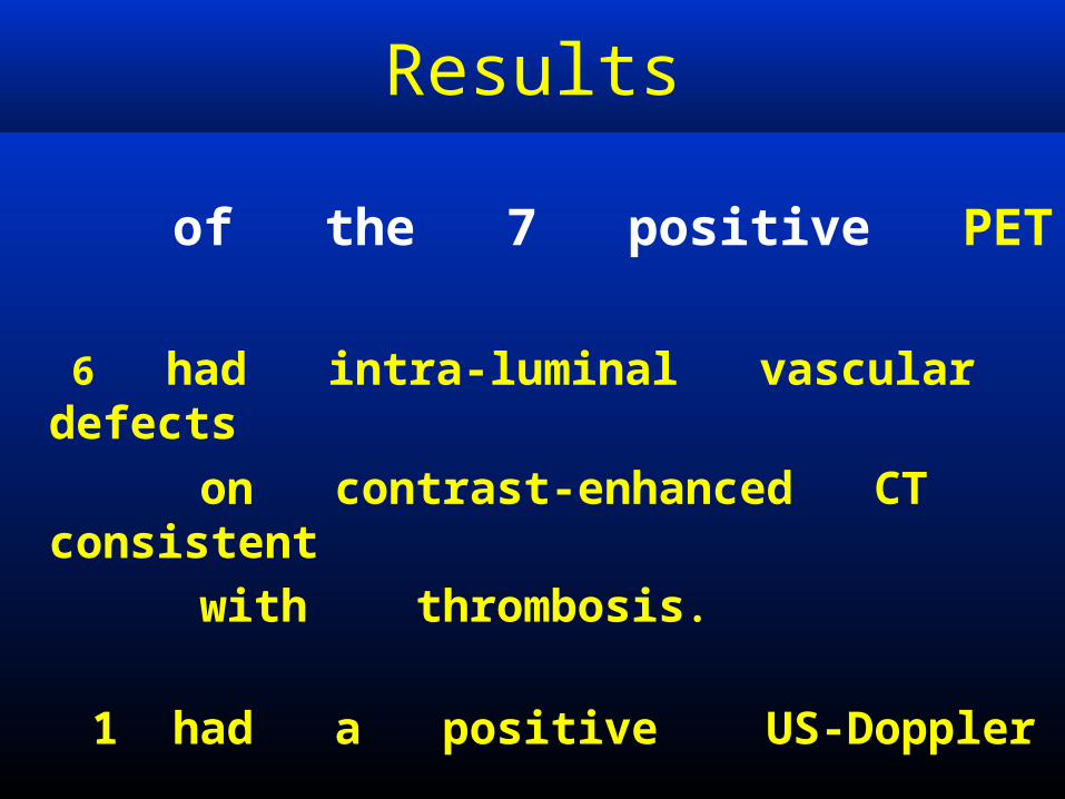
of the 7 positive PET
6 had intra-luminal vascular defects
on contrast-enhanced CT consistent
with thrombosis.
1 had a positive US-Doppler
Results
Page 11
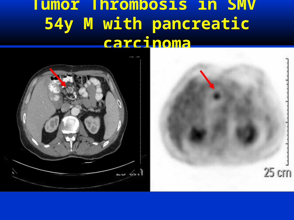
Tumor Thrombosis in SMV54y M with pancreatic
carcinoma
Page 12
Tumor Thrombosis in SMV54y M with pancreatic
carcinoma
Page 13
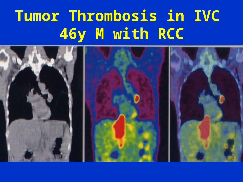
Tumor Thrombosis in IVC 46y M with RCC
Page 15
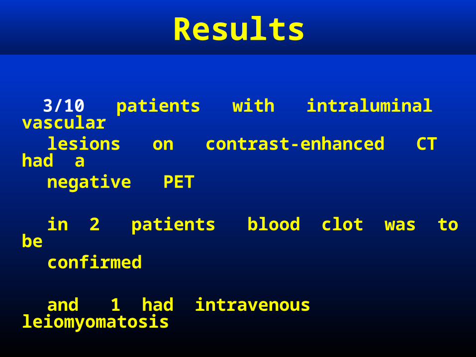
Results
3/10 patients with intraluminal vascular
lesions on contrast-enhanced CT had a
negative PET in 2 patients blood clot was to be confirmed
and 1 had intravenous leiomyomatosis
Page 16
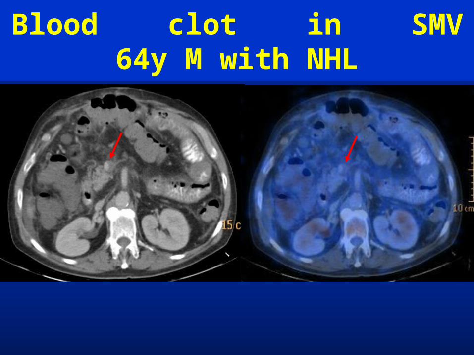
Blood clot in SMV64y M with NHL
Page 17
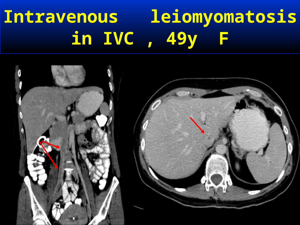
Intravenous leiomyomatosisin IVC , 49y F
Page 18
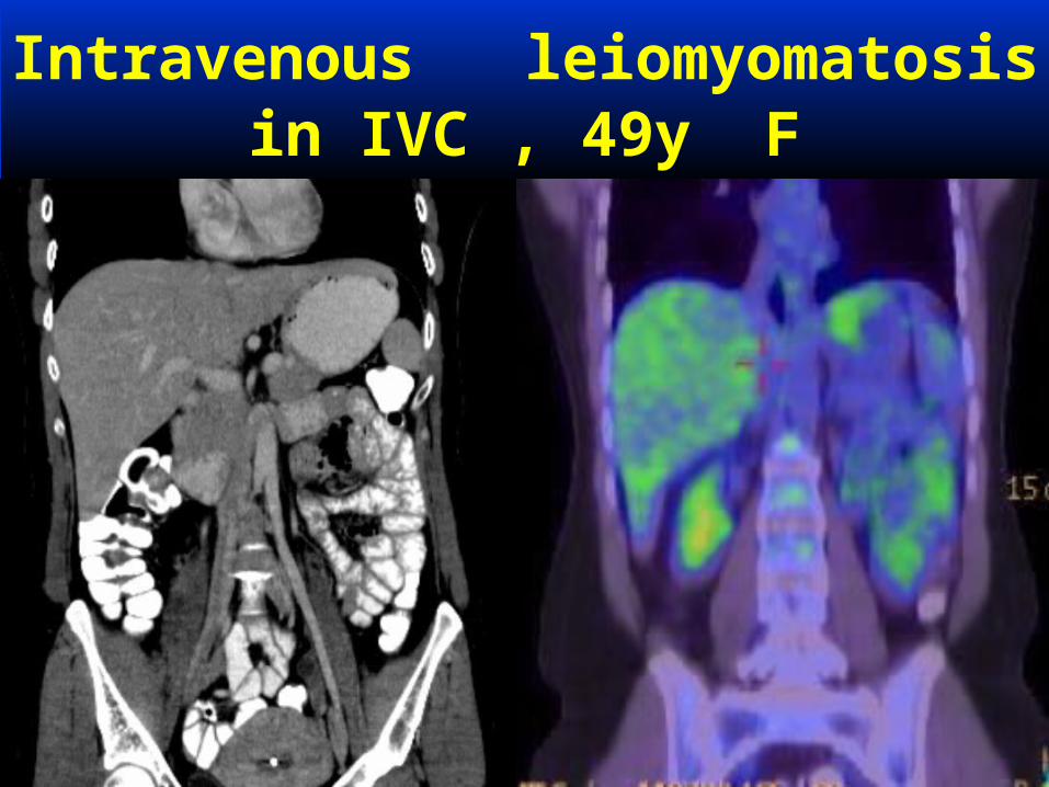
Intravenous leiomyomatosisin IVC , 49y F
Page 19
Results
PET/CT correctly differentiated
between Tumor Thrombosis and benign Venous Thrombosis in all our patients
Page 21
Results
Underlying pathology in 7 patients with Tumor Thrombosis
4 lymphoma 1 pancreatic ca 1 renal cell ca 1 head - neck squamous cell ca
Page 22
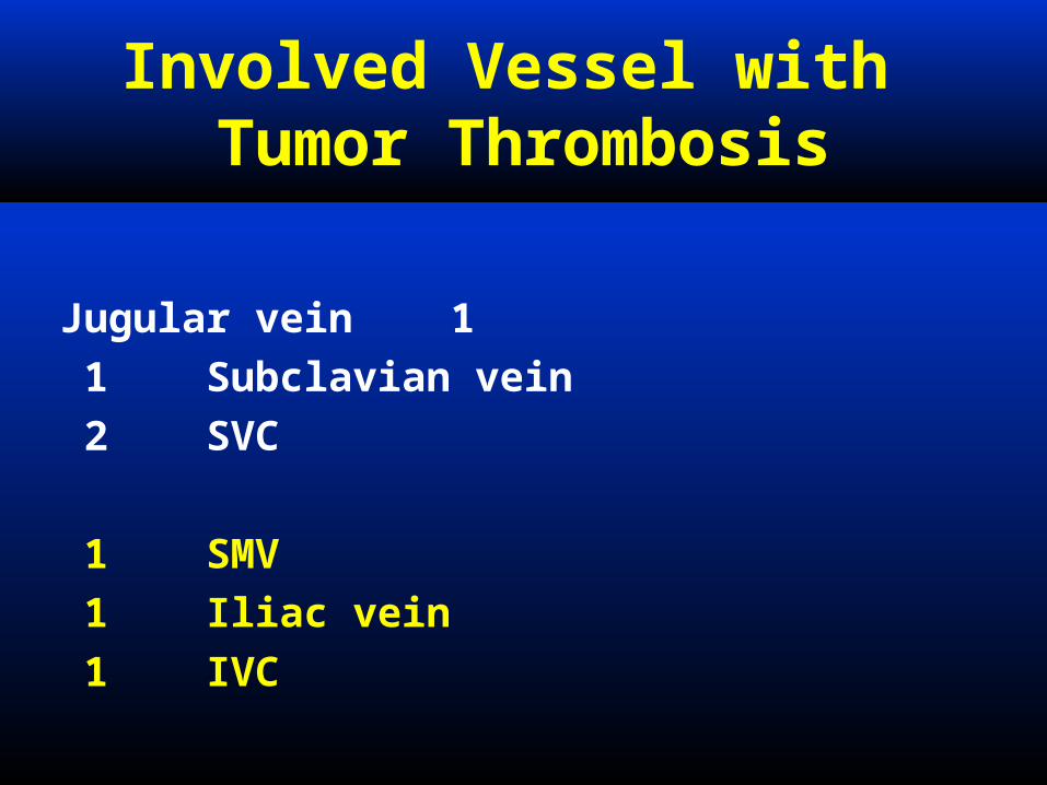
Involved Vessel with Tumor Thrombosis
1 Jugular vein
1 Subclavian vein 2 SVC
1 SMV 1 Iliac vein 1 IVC
Page 23
Potential Pitfalls
False positive PET findings
may be due to • inflammatory lesions ,
including infected catheters
in the venous vasculature
Page 24
Potential Pitfalls
In contrast, missed diagnoses may relate:• to the size of the lesion, • the avidity of the underlying pathological process to FDG
Page 25
Conclusion
Contrast-enhanced CT defines extent of thrombotic lesions, while PET contributes the functional information of these lesions


![Time Series Encodings with Temporal Convolutional Networks · Time Series Encodings with Temporal Convolutional Networks Markus Thill 1[0000 00026429 180X], Wolfgang Konen 1343 4209],](https://static.documents.pub/doc/80x56/6086b4ddea791645c50a7881/time-series-encodings-with-temporal-convolutional-time-series-encodings-with-temporal.jpg)


