109
1931-6 Preparatory School to the Winter College on Micro and Nano Photonics for Life Sciences Dan COJOC 4 - 8 February 2008 Laboratorio TASC INFM, Italy Introduction to biophotonics
1931-6
Preparatory School to the Winter College on Micro and NanoPhotonics for Life Sciences
Dan COJOC
4 - 8 February 2008
Laboratorio TASCINFM, Italy
Introduction to biophotonics

Preparatory School to the ICTP Winter College Micro and Nano Photonics for Life Sciences, Trieste, 2008 February 4-8 1/108
Introduction to Biophotonics
Dan Cojoc
CNR-Instituto Nazionale per la Fisica della Materia
Laboratorio Nazionale TASC Trieste, Italy

Preparatory School to the ICTP Winter College Micro and Nano Photonics for Life Sciences, Trieste, 2008 February 4-8 2/108
Preparing this course I considered the following:
� The audience is non-homogeneous (from physics and/or biology)
� You have basic understanding of optics and biology (Preparatory lectures on cell biology, diffraction theory, optical microscopy were provided)
� Advanced lectures on Biophotonics topics will be presented at Winter College next week
� Lot of information can be found in internet
� Not all the information can be included in a 3 h lecture
The goal of the course is to provide you a basic introduction to Biophotonics with some history and examples and to open your appetite for this fascinating field of science.
Motto:
“We should know and we will know”, David Hilbert, 1930
(expressing his disagreement with the
“ignoramus et ignorabimus / we do not know and will not know”
conclusion of Emil du Bois-Reymond, in his
“On the limits of our understanding of nature” of 1872
Preparatory School to the ICTP Winter College Micro and Nano Photonics for Life Sciences, Trieste, 2008 February 4-8 3/108
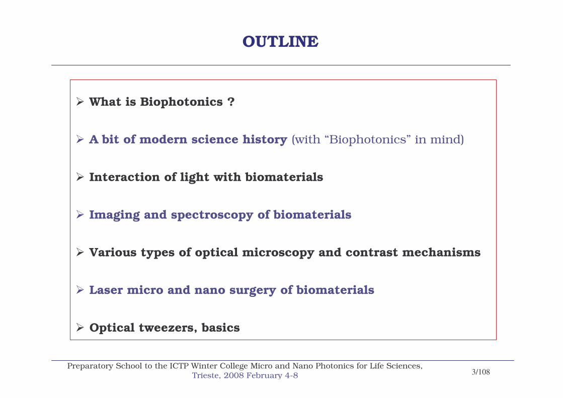
OUTLINE
� What is Biophotonics ?
� A bit of modern science history (with “Biophotonics” in mind)
� Interaction of light with biomaterials
� Imaging and spectroscopy of biomaterials
� Various types of optical microscopy and contrast mechanisms
� Laser micro and nano surgery of biomaterials
� Optical tweezers, basics

Preparatory School to the ICTP Winter College Micro and Nano Photonics for Life Sciences, Trieste, 2008 February 4-8 4/108
What is Biophotonics?
The application of light and other forms of radiant energy to the life sciences.
Interdisciplinary science studying the interaction of light with biological material – where “light” includes all forms of radiant energy whose quantum
unit is the photon.
Three definitions of Biophotonics among of many other:
1
2
3
D. Matthews, Optik & Photonik June 2007 No. 2
Biophotonics is the science of generating and harnessing light (photons) toimage, detect and manipulate biological materials.
Biophotonics is used in BIOLOGY to probe for molecular mechanisms, functionand structure. It is used in MEDICINE to study tissue and blood at the macro (large-scale) and micro (very small scale) organism level to detect, diagnose and treat diseases in a way that are non-invasive to the body.
T. Husser IEEE-LEOS 2004

Preparatory School to the ICTP Winter College Micro and Nano Photonics for Life Sciences, Trieste, 2008 February 4-8 5/108
Photonics
Life Sciences
Bio
Photo
nics
Subject areas:Biochemistry, Cell Biology, Developmental Biology, Ecology, Evolution and Diversity of Life, Functional and Comparative, Morphology, Genetics and Disease, Genetics and Molecular Biology Immunology, Microbiology, Neuroscience, Plant Science, Science and Society, Structural Biology, Virology

Preparatory School to the ICTP Winter College Micro and Nano Photonics for Life Sciences, Trieste, 2008 February 4-8 6/108
http://enews.lbl.gov/MicroWorlds/teachers/alsposter.pdf
The E-M Spectrum

Preparatory School to the ICTP Winter College Micro and Nano Photonics for Life Sciences, Trieste, 2008 February 4-8 7/108
“Light” Spectrum

Preparatory School to the ICTP Winter College Micro and Nano Photonics for Life Sciences, Trieste, 2008 February 4-8 8/108
• 1590 – Hans and Zacharias Janssen make the first microscope.
• 1675 – Anton van Leeuwenhoek uses a simple microscope with only one lens to look at blood, insects and many other objects. He was first to describe cells and bacteria through microscope.
• 18th century – Several technical innovations make microscopes better and easier to handle, which leads to microscopy becoming more and more popular among scientists (e.g. Lens doublet reduces the chromatic abberations).
• 1878 – Ernst Abbe formulates a mathematical theory correlating resolution to the wavelength of light. Abbes formula make calculations of maximum resolution in microscopes possible.
• 1903 – Richard Zsigmondy develops the ultramicroscope and is able to study objects below the wavelength of light.
• 1932 – Frits Zernike invents the phase-contrast microscope that allows the study of colorlessand transparent biological materials.
• 1938 – Ernst Ruska develops the electron microscope. The ability to use electrons in microscopy greatly improves the resolution and greatly expands the borders of exploration.
• 1981 – Gerd Binnig and Heinrich Rohrer invent the scanning tunneling microscope that gives three-dimensional images of objects down to the atomic level.
Some important discoveries in Microscopy
A bit of modern science history(with “Biophotonics” in mind)
http://nobelprize.org/

Preparatory School to the ICTP Winter College Micro and Nano Photonics for Life Sciences, Trieste, 2008 February 4-8 9/108
Ultramicroscopy an example of instrumentation development, starting in 1900 …
1903 – Zsigmondy and Siedentopf develop the first ultramicroscope,being able to study objects werll below the wavelength of light.
The sun beams fell upon a mirror S, were reflected from thisinto the lens L, which brought them to the focus at 6, over which was arranged a low power microscope.(total magnification about 100 diameters)
N.B.: With ordinary illumination, even with the best objectives, they were not perceptible.
Sample: colloidal gold solution
NO

Preparatory School to the ICTP Winter College Micro and Nano Photonics for Life Sciences, Trieste, 2008 February 4-8 10/108
Working principle:The sample is illuminated from two sides by a blue laser forming a thin sheetof light. Fluorescent light is thus emitted only from a thin optical section and collected by the objective lens. Stray light is blocked by a GFP filter and the image is projected through the tube lens onto the camera target.
Ultramicroscopy: three-dimensional visualization ofneuronal networks in the whole mouse brain
H.U. Dodt et al, Nature Methods 4 331 (2007)
… 2007 �

Preparatory School to the ICTP Winter College Micro and Nano Photonics for Life Sciences, Trieste, 2008 February 4-8 11/108
(a) Surface of a whole mouse brain reconstructed from 550 optical sections. Both the GFP and autofluorescence signal are imaged.
(b) Excised whole hippocampus reconstructed from 410 optical sections. Single cell bodies are visible.
(c) 3D reconstruction of part of a whole hippocampus using 132 optical sections.
(d) 3D reconstruction of dendritic spines of CA1 pyramidal neurons obtained with a higherresolution objective (20; NA, 0.4) in a whole hippocampus (430 optical sections, deconvolved).
Ultramicroscopy 2007 Results: Brain imaging
Scale bar 500 umScale bar 1 mm Scale bar 200 um Scale bar 5 um
H.U. Dodt et al, Nature Methods 4 331 (2007)

Preparatory School to the ICTP Winter College Micro and Nano Photonics for Life Sciences, Trieste, 2008 February 4-8 12/108
http://www.biology.arizona.edu/
Major events in Cell Biology & Imaging

Preparatory School to the ICTP Winter College Micro and Nano Photonics for Life Sciences, Trieste, 2008 February 4-8 13/108
Old (1838) Scleiden & Schwann
1. The cell is the unit of structure, physiology, and organization in living things. OK
2. The cell retains a dual existence as a distinct entity and a building block in the construction of organisms. OK
3. Cells form by free-cell formation, similar to the formation of crystals(spontaneous generation). WRONG
Modern Cell Theory
1. All known living things are made up of cells.
2. The cell is structural & functional unit of all living things.
3. All cells come from pre-existing cells by division. 4. (Spontaneous Generation does not occur). 5. Cells contains hereditary information which is passed from cell to cell during cell
division. 6. All cells are basically the same in chemical composition.7. All energy flow (metabolism & biochemistry) of life occurs within cells.
Cell Theory

Preparatory School to the ICTP Winter College Micro and Nano Photonics for Life Sciences, Trieste, 2008 February 4-8 14/108
http://nobelprize.org/
Discoveries in the field of X rays(Nobel Prizes – incomplete list)
1901 - W. C. Röntgen: the discovery of X-rays.
1914 - M. von Laue: the discovery of X-rays by crystals.
1915 - W. H. Bragg and W. L. Bragg: the determination of crystal structures using X-rays.
1927 - A. H. Compton: revealing the particle nature of X-rays in scattering experiments on electrons.
1936 - P. Debye: for determining molecular structures by X-ray diffraction in gases.
1962 - M. F. Perutz and J.C. Kendrew: determining the structure of hemoglobin and myoglobin.
1962 - F. Crick, J. Watson and M. Wilkins: their discoveries concerning the molecular structure of nucleic acids and its significance for information transfer in living material.
1964 - D. Crowfoot Hodgkin: the determination of the structure of penicillin and other important biochemical substances.
1979 - A. M. Cormack and G. N. Hounsfield: the development of computerized tomography.
1985 - H. A. Hauptman and J. Karle: for the development of direct methods for X-ray crystallographic structure determination.
1988 - J. Deisenhofer, R. Huber and H. Michel: the determination of protein structures crucial to photosynthesis.

Preparatory School to the ICTP Winter College Micro and Nano Photonics for Life Sciences, Trieste, 2008 February 4-8 15/108
1869 Johann Friedrich Miescher identifies a weakly acidic substance of unknown function in thenuclei of human white blood cells. This substance will later be called deoxyribonucleic acid, or DNA.
1912 Physicist Sir William Henry Bragg, and his son, Sir William Lawrence Bragg, discover that they can deduce the atomic structure of crystals from their X-ray diffraction patterns. This scientiFic tool will be key in helping Watson and Crick determine DNA's structure.
1924 Microscope studies using stains for DNA and protein show that both substances are present in chromosomes.
1928 Franklin Griffith, a British medical officer, discovers that genetic information can be transferred from heat-killed bacteria cells to live ones. This phenomenon, called transformation, provides the first evidence that the genetic material is a heat-stable chemical.
1944 Oswald Avery, and his colleagues Maclyn McCarty and Colin MacLeod, identify Griffith's transforming agent as DNA. However, their discovery is greeted with skepticism, in part because many scientists still believe that DNA is too simple a molecule to be the genetic material.
1949 Erwin Chargaff, a biochemist, reports that DNA composition is speciesspecific; that is, that the amount of DNA and its nitrogenous bases varies from one species to another. In addition, Chargaff finds that the amount of adenine equals the amount of thymine, and the amount of guanine equals the amount of cytosine in DNA from every species.
1953 James Watson and Francis Crick discover the molecular structure of DNA.
Milestones in DNA History
http://nobelprize.org/

Preparatory School to the ICTP Winter College Micro and Nano Photonics for Life Sciences, Trieste, 2008 February 4-8 16/108
1953 James Watson and Francis Crick discover the molecular structure of DNA
Biophotonics has already played a central role in the most famous paperin modern biology: J.D. Watson and F.H.C. Crick, Nature, 25 April 1953

Preparatory School to the ICTP Winter College Micro and Nano Photonics for Life Sciences, Trieste, 2008 February 4-8 17/108

Preparatory School to the ICTP Winter College Micro and Nano Photonics for Life Sciences, Trieste, 2008 February 4-8 18/108
See/Read/Think-on their Nobel Lectures:http://nobelprize.org/nobel_prizes/medicine/laureates/1962/
The molecular configurationof nucleic acids
The involvement of RNA in the synthesis of proteins
On the Genetic Code

Preparatory School to the ICTP Winter College Micro and Nano Photonics for Life Sciences, Trieste, 2008 February 4-8 19/108
The two ribbons symbolize the two phosphate - sugar chains, and the horizonal rods the pairs of bases holding the chains together.
The vertical line marks the fibre axis.
“We wish to suggest a structure for the salt of deoxyribose nucleic acid (DNA). This structure has novel features which are of considerable biological interest …
We wish to put forward a radically different structure for the salt of DNA.
This strucure has two helical chains each coiled round the same axis.”

Preparatory School to the ICTP Winter College Micro and Nano Photonics for Life Sciences, Trieste, 2008 February 4-8 20/108
“The purpose of this communication is to describe, in a preliminary way, some of the experimental evidence for the ploy-nucleotide chain configuration being helical, and existing in this form when in the natural state.”
Fibre diagram of DNA from B. coli. Fibre axis vertical.
Diffraction pattern of system of helicescorresponding to structure of DNA.
The squares of Bessel functions are plotted about 0 on the equator and on the first, second third and fifth layer lines for half of the nucleotide mass at 20 A. A diameter and remainder distributed along a radius, the mass at a given radius being proportional to the radius. About C on the tenth layer the similar functions are plotted for an outer diameter of 12 A.

Preparatory School to the ICTP Winter College Micro and Nano Photonics for Life Sciences, Trieste, 2008 February 4-8 21/108
Light sourceInvestigated
Subject DetectorAnalysis
Instrumentation
Photonics
Life Sciences
Bio
Photo
nics
Subject areas:Biochemistry, Cell Biology, Developmental Biology, Ecology, Evolution and Diversity of Life, Functional and Comparative, Morphology, Genetics and Disease, Genetics and Molecular Biology Immunology, Microbiology, Neuroscience, Plant Science, Science and Society, Structural Biology, Virology

Preparatory School to the ICTP Winter College Micro and Nano Photonics for Life Sciences, Trieste, 2008 February 4-8 22/108
Interaction of light with biomaterials
Light source
InvestigatedBiomaterial - Subject
In complex materials, any combination of interactions are possible. The exact nature of each process depends on the
physical and chemical structure of the biomaterial
• Transmission
• Refraction
• Scattering
Detector
• Reflection
• Absorption

Preparatory School to the ICTP Winter College Micro and Nano Photonics for Life Sciences, Trieste, 2008 February 4-8 23/108
Light-Induced Processes
Prasad, Introduction to Biophotonics, John Wiley & Sons © 2003
Tissue light interaction
RadiativeTissue autofluorescence
Fluorescence from variousconstituents of the tissue
Nonradiative
Photochemical•Excited state reaction•Occurs even at low opticalpower density
.
Photoablation•Direct breaking of cellular structure
•Performed by high energy UV radiation
Photodisruption•Shockwave generation at high pulse intensity•Fragmentation and cutting of the tissue by mechanical force of shockwave
.
Thermal•Light absorption converted to heat•Can produce coagulation, vaporization,carbonization and melting
Plasma – induced ablation•Induced by high intensity short pulse•Dielectric breakdown creats ionizedplasma that interacts with light yoproduce ablation

Preparatory School to the ICTP Winter College Micro and Nano Photonics for Life Sciences, Trieste, 2008 February 4-8 24/108
Light Scattering Processes
Introduction to Biophotonics – Prasad - John Wiley & Sons © 2003
Light Scattering
Elastic Scattering
Incident and scattered photonsare of the same frequency
Inelastic ScatteringIncident and scattered photons
are of different frequencies
Rayleigh scattering
•Scattering by particles of sizesmaller than the wavelength �of light. •Scattering depens on the �-4
hence significantly more forblue than for red light. •Forward and backwardscattering are the same
Mie scattering Brillouin Scattering Raman Scattering
•Scattering by particles of size comparable with �.•Weaker wavelengthdependence: �-X where0.4 < X < 0.5. •Forward scattering preffered.
The difference in energygenerates acousticphonons
The difference in energygenerates a vibrationalexcitation in the molecule

Preparatory School to the ICTP Winter College Micro and Nano Photonics for Life Sciences, Trieste, 2008 February 4-8 25/108
Imaging and spectroscopy of bio-materials
Scattering and absorptionFluorescenceOptical Microscopy
Transmission and AbsorptionPhase contrastConfocalFluorescence Correlation Spectroscopy
Fluorescence Lifetime ImagingMultiphoton ExcitationSpontaneous Raman SpectroscopyCoherent Anti-Stokes Raman Scattering Mcroscopy (CARS)Second Harmonic Generation MicroscopyStimulated Emission Depletion, Image Deconvolution MicroscopyNearfiled Optical MicroscopySingle Molecule Detection
Courtesy Prof. T. Huser (slides: 25-30, 32-35,4, 5152,55, 63,64,69, 85-88)NSF Center for Biophotonics Science and Technology, University of California, Davis, USA
http://cbst.ucdavis.edu/education/short-courses/osabiophotonicscourse.pdf/download

Preparatory School to the ICTP Winter College Micro and Nano Photonics for Life Sciences, Trieste, 2008 February 4-8 26/108
Main sources for light loss in biological materials: absorption and light scattering

Preparatory School to the ICTP Winter College Micro and Nano Photonics for Life Sciences, Trieste, 2008 February 4-8 27/108
Scattering in tissue

Preparatory School to the ICTP Winter College Micro and Nano Photonics for Life Sciences, Trieste, 2008 February 4-8 28/108
Absorption spectroscopy
routinely used technique in the biosciences

Preparatory School to the ICTP Winter College Micro and Nano Photonics for Life Sciences, Trieste, 2008 February 4-8 29/108
Application for absorption spectroscopy

Preparatory School to the ICTP Winter College Micro and Nano Photonics for Life Sciences, Trieste, 2008 February 4-8 30/108
Light microscopy enables live imaging at the cellular level

Preparatory School to the ICTP Winter College Micro and Nano Photonics for Life Sciences, Trieste, 2008 February 4-8 31/108
Some relevant length scales

Preparatory School to the ICTP Winter College Micro and Nano Photonics for Life Sciences, Trieste, 2008 February 4-8 32/108
Various types of optical microscopy and contrast mechanisms

Preparatory School to the ICTP Winter College Micro and Nano Photonics for Life Sciences, Trieste, 2008 February 4-8 33/108
Transmission/absorption microscopy of biological samples requires preparation and staining

Preparatory School to the ICTP Winter College Micro and Nano Photonics for Life Sciences, Trieste, 2008 February 4-8 34/108
The most important light microscopy imaging technique: Phase contrast microscopy avoids staining

Preparatory School to the ICTP Winter College Micro and Nano Photonics for Life Sciences, Trieste, 2008 February 4-8 35/108
Phase contrast microscopy

Preparatory School to the ICTP Winter College Micro and Nano Photonics for Life Sciences, Trieste, 2008 February 4-8 36/108
http://www.microscopyu.com/tutorials/java/dic/wavefrontcomparison/index.html
In DIC microscopy, the spatial relationship and phase difference between ordinary and extraordinary wavefronts is governed either by the position of the objective prism (Nomarski DIC) or the relationship between the polarizer and a thin quartz retardation plate in a de Sénarmont design.
Differential Interference Contrast (DIC) Microscopy
Nomarski DIC Sénarmont

Preparatory School to the ICTP Winter College Micro and Nano Photonics for Life Sciences, Trieste, 2008 February 4-8 37/108
Phase contrast - DIC microscopy
HeLa cells have been cultured continuously for scientific use since they were first taken from the tumor of a woman suffering from cervical cancer in the 1950s. They have been utilized for many purposes, including the development of a polio vaccine, the pursuit of a cure for diseases such as leukemia and cancer, and the study of the cellular effects of drugs and radiation.
http://www.microscopyu.com/galleries/dicphasecontrast/index.html
Phase Contrast Image DIC ImageHeLa Cell Culture

Preparatory School to the ICTP Winter College Micro and Nano Photonics for Life Sciences, Trieste, 2008 February 4-8 38/108
Polarization microscopy
http://www.microscopyu.com/articles/polarized/polarizedintro.html

Preparatory School to the ICTP Winter College Micro and Nano Photonics for Life Sciences, Trieste, 2008 February 4-8 39/108
Asbestos is a generic name for a group of naturally occurring mineral fibers, which have been widely used, for example, in insulating materials, brake pads and to reinforce concrete. They can be harmful to health when inhaled and it is important that their presence in the environment be easily identified. Samples are commonly screened using scanning electron microscopy and x-ray microanalysis, but polarizing microscopy provides a quicker and easier alternative that can be utilized to distinguish between asbestos and other fibers and between the major types asbestos – chrysotile, crocidolite and amosite. With the use of crossed polarsit is possible to deduce the permitted vibration direction of the light as it passes through the specimen, and with the whole wave plate, a determination of the slow and fast vibration directions. Under crossed polars, chrysotile shows pale interference colors - low order whites (a). When a full wave plate is added (530-560 nanometers), the colors are transformed. Aligned Northeast-Southwest, the wave plate is additive and gives blue and yellow in the fiber (b). When aligned Northwest-Southeast (c) the plate is subtracting to give a paler yellow fiber with no blue. From this it is possible to deduce that the slow vibration direction is parallel with the long axis of the fiber.
http://www.microscopyu.com/articles/polarized/polarizedintro.html
Example: Identification of Asbestos Fibers

Preparatory School to the ICTP Winter College Micro and Nano Photonics for Life Sciences, Trieste, 2008 February 4-8 40/108
Fluorescenceis an electronic process

Preparatory School to the ICTP Winter College Micro and Nano Photonics for Life Sciences, Trieste, 2008 February 4-8 41/108
Excitation / Emission spectroscopy of native fluorophoresin tissue (autofluorescence)

Preparatory School to the ICTP Winter College Micro and Nano Photonics for Life Sciences, Trieste, 2008 February 4-8 42/108
Fluorescence microscopy
More specific information requiresoptical labeling: Fluorescent dyes spreadthroughout the entire optical spectrum
Fluorescent probes, fluorochromes:Molecules capable of undergoing electronictransitions that result in fluorescence.Fluorophore:The structural domain or specific regionof a molecule that is capable ofexhibiting fluorescence.

Preparatory School to the ICTP Winter College Micro and Nano Photonics for Life Sciences, Trieste, 2008 February 4-8 43/108
Fluorescence microscopy

Preparatory School to the ICTP Winter College Micro and Nano Photonics for Life Sciences, Trieste, 2008 February 4-8 44/108
E. Di Fabrizio et al J. Electron Spectrosc. Relat. Phenom., 144-147, 957, 2005.
Topography
Chlorine
Potassium
Phosphorus
Scanning x-ray microscope picture of human liver cell. (a) topographic image. By varying the incoming photon energy according to the absorption edge of selected elements, and by inserting a fluorescence detector, we were able to map the distribution of some elements inside the cell.
DIC and fluorescence X-ray microscopy
See also the lecture on X-ray diffractive elements by D. Cojoc next week

Preparatory School to the ICTP Winter College Micro and Nano Photonics for Life Sciences, Trieste, 2008 February 4-8 45/108
Fluorescence resonance energy transfer (FRET) enables measurements of molecular association/dissociation and lengths

Preparatory School to the ICTP Winter College Micro and Nano Photonics for Life Sciences, Trieste, 2008 February 4-8 46/108
Fluorescent dyes can be linked to biomolecules

Preparatory School to the ICTP Winter College Micro and Nano Photonics for Life Sciences, Trieste, 2008 February 4-8 47/108
Fluorescent proteins can be “fused” to other proteins and co-expressed to intrinsically label proteins in cells

Preparatory School to the ICTP Winter College Micro and Nano Photonics for Life Sciences, Trieste, 2008 February 4-8 48/108
Fluorescence microscopy
Some drawbacks:
See the lectures next week for pros and cons

Preparatory School to the ICTP Winter College Micro and Nano Photonics for Life Sciences, Trieste, 2008 February 4-8 49/108
Total intern reflection fluorescence (TIRF) microscopy excites only a very narrow region right above the glass interface

Preparatory School to the ICTP Winter College Micro and Nano Photonics for Life Sciences, Trieste, 2008 February 4-8 50/108
Confocal fluorescence microscopy efficiently suppresses background signals and enables three-dimensional sectioning
Pinhole ���� High axial resolution

Preparatory School to the ICTP Winter College Micro and Nano Photonics for Life Sciences, Trieste, 2008 February 4-8 51/108
Examples of the value of confocal fluorescence microscopy

Preparatory School to the ICTP Winter College Micro and Nano Photonics for Life Sciences, Trieste, 2008 February 4-8 52/108
Y
Scanning confocal fluorescence microscopy

Preparatory School to the ICTP Winter College Micro and Nano Photonics for Life Sciences, Trieste, 2008 February 4-8 53/108
Fluorescence correlation spectroscopy (FCS) measures molecular diffusion times and association/dissociation rate

Preparatory School to the ICTP Winter College Micro and Nano Photonics for Life Sciences, Trieste, 2008 February 4-8 54/108
Time-correlated single photon counting (TCSPC) enables fast fluorescence lifetime measurements

Preparatory School to the ICTP Winter College Micro and Nano Photonics for Life Sciences, Trieste, 2008 February 4-8 55/108
Pulsed laser sources combined with TCSPC enable lifetime analysis per pixel in confocal images: fluorescence lifetime imaging

Preparatory School to the ICTP Winter College Micro and Nano Photonics for Life Sciences, Trieste, 2008 February 4-8 55+1/108
2-photon fluorescence microscopy

Preparatory School to the ICTP Winter College Micro and Nano Photonics for Life Sciences, Trieste, 2008 February 4-8 55+2/108
2-photon absorption

Preparatory School to the ICTP Winter College Micro and Nano Photonics for Life Sciences, Trieste, 2008 February 4-8 55+3/108
Multi-photon fluorescence microscopy

Preparatory School to the ICTP Winter College Micro and Nano Photonics for Life Sciences, Trieste, 2008 February 4-8 55+4/108
Multi-photon fluorescence microscopy

Preparatory School to the ICTP Winter College Micro and Nano Photonics for Life Sciences, Trieste, 2008 February 4-8 55+5/108
Multi-photon fluorescence microscopy
Living neurons

Preparatory School to the ICTP Winter College Micro and Nano Photonics for Life Sciences, Trieste, 2008 February 4-8 55+6/108
Raman spectroscopy
Drawback of Fluorescence: is limited by the need to label and photobleaching-> Raman provides molecular information

Preparatory School to the ICTP Winter College Micro and Nano Photonics for Life Sciences, Trieste, 2008 February 4-8 55+7/108
Raman scattering is the interaction of photons and intrinsic molecular bonds

Preparatory School to the ICTP Winter College Micro and Nano Photonics for Life Sciences, Trieste, 2008 February 4-8 55+8/108
Confocal Raman microscopy

Preparatory School to the ICTP Winter College Micro and Nano Photonics for Life Sciences, Trieste, 2008 February 4-8 55+9/108
Example: Raman spectroscopy can distinguidhbetween ds DNA and protein-DNA compexes

Preparatory School to the ICTP Winter College Micro and Nano Photonics for Life Sciences, Trieste, 2008 February 4-8 55+10/108
An example for micro-Raman analysis of single cellsoptically trapped cells
Real-time detection of hyperosmotic stress response in a single Saccharomyces yeast cell
• the cell stress response is the reaction of a living cell to ambient changes which are potentially harmful: for example, an increase in temperature, pH, saline concentration, the presence of toxins
• the basic reaction at the fermentation process is the response of the Saccharomyces yeast cells on the hyperosmotic stress
+ glucose =
ethanolC2H5OH
glycerolC3H8O3

Preparatory School to the ICTP Winter College Micro and Nano Photonics for Life Sciences, Trieste, 2008 February 4-8 55+11/108
30 min after the experiment starts
Detection area 1 femtoLDetectable concentration 100 atto_molDetectable number of molecules 108
G. P. Singh et al. Analyt. Chemistry, 2005, 77, 2564
difference spectrum of a single yeast cell under the hyperosmoticpressure
10% pure ethanol and glycerol in water
Real-time detection of hyperosmotic stress response in a single Saccharomyces yeast cell

Preparatory School to the ICTP Winter College Micro and Nano Photonics for Life Sciences, Trieste, 2008 February 4-8 55+12/108
Singh G P, Creely C M, Volpe G, Grotsch H and Petrov D, Anal. Chem. 77 2564–8, (2005)
An example for micro-Raman analysis of single cells:
The biochemical changes taking place duringthe lag phase and G1 phase of a single S. cerevisiae yeast cell
(A) The growth curve shows the phases of yeast cellpopulation.
(B) The yeast cell cycle diagram shows the phases of the
cell cycle. The abbreviations used for yeast cell cyclestand for the standard terms: 1 M, mitosis; S, synthesis; G1 and G2, gap phases. For our experiments cells thatdid not have a bud were chosen.
(C) Upper row shows the budding of a yeast cell and itssubsequent growth while in the optical trap. Lower rowshows the same cell at a moment when it is released fromthe optical trap

Preparatory School to the ICTP Winter College Micro and Nano Photonics for Life Sciences, Trieste, 2008 February 4-8 55+13/108
Huang et. al., Biochemistry, V44, 10009-10019 (2005)
An example of combined Raman and fluorescence microscopy

Preparatory School to the ICTP Winter College Micro and Nano Photonics for Life Sciences, Trieste, 2008 February 4-8 55+14/108
Advantages and limitations of spontaneous Raman imaging

Preparatory School to the ICTP Winter College Micro and Nano Photonics for Life Sciences, Trieste, 2008 February 4-8 55+15/108
Coherent Anti-Stokes Raman Scattering (CARS) microscopyThe newest member to the optical microscopy family
Why develop CARS ?• Contrast signal based on vibrational characteristics, no need for fluorescent tagging
• CARS signal is at high frequency (lower wavelength) – no fluorescence interference
• Higher resolution
• More sensitive (stronger signals) than spontaneous Raman
• Microscopy – faster, more efficient imaging for real-time analysis
Two laser frequencies interact resonantlywith a specific molecular vibration:
CARS signals are generated at wavelengthsshorter than the excitation wavelengths
(anti-Stokes)

Preparatory School to the ICTP Winter College Micro and Nano Photonics for Life Sciences, Trieste, 2008 February 4-8 55+16/108
CARS microscopy setup
X.S. Xie et al., LNNL and Harvard
Key components in a CARS setupwith two tunable synchronizedLasers (5 psec, 80 MHz rep rate)
requires two synchronized ultrashort pulsed laser sources

Preparatory School to the ICTP Winter College Micro and Nano Photonics for Life Sciences, Trieste, 2008 February 4-8 55+17/108
Lasers @ 853 nm (100 �W) and 1135 nm (100 �W)
tuned to Raman shift of 2913 cm-1 C-H vibration
Unstained live bacterial cells.Signal due to cell membranes.
Unstained live HeLa cells.Bright spots due to mitochondria.
Zumbusch et. al., Phys. Rev. Lett. V82, 4142 (1999)
CARS microscopy: application to live cell imagingExample 1

Preparatory School to the ICTP Winter College Micro and Nano Photonics for Life Sciences, Trieste, 2008 February 4-8 55+18/108
Tracking trajectories of organelles inside single living cellsExample 2
X. L. Nan, E. O. Potma, and X. S. Xie, "Nonperturbative chemical imaging of organelletransport in living cells with coherent anti-stokes Raman scattering microscopy," Biophys. J. 91, 728-735 (2006).

Preparatory School to the ICTP Winter College Micro and Nano Photonics for Life Sciences, Trieste, 2008 February 4-8 55+19/108
Raman spectroscopy for single cell cancer detection
cbst.ucdavis.edu/education/courses/winter-2007-ead-bim-289/chan_cars-lecture.pdf
Spontaneous Raman spectra takes
2 minutes per cell !!!
Chan et. al., Biophys. Journal. V90, 648 (2006)

Preparatory School to the ICTP Winter College Micro and Nano Photonics for Life Sciences, Trieste, 2008 February 4-8 55+20/108
Future applications : CARS cytometry for rapid, labellesscancer cell detection and sorting
Optical trapping combined withCARS for faster spectral analysis
Trapped polystyrene beadusing two CARS beams
Potential solution for fasterchemical analysis of cells
Chan et. al., IEEE J. Sel. Topics. Quant. Elec. V11 858 (2005)

Preparatory School to the ICTP Winter College Micro and Nano Photonics for Life Sciences, Trieste, 2008 February 4-8 55+21/108

Preparatory School to the ICTP Winter College Micro and Nano Photonics for Life Sciences, Trieste, 2008 February 4-8 55+22/108
Other future directions for CARS
– Fiber based CARS for endoscopy
– CARS optical coherence tomography
See the CARS lectures by H. Rigneault next week

Preparatory School to the ICTP Winter College Micro and Nano Photonics for Life Sciences, Trieste, 2008 February 4-8 55+23/108
Second Harmonic Generation (SHG) Another nonlinear optical imaging technique
SHG: Nonlinear process in which incident light at a given frequency (�1) is converted into light at twicethe frequency (� SHG = 2 �1)
It occurs:
• only at beam focus (intense field), eliminating out-of-plane signal and enabling sub-micron spatial resolution
• when intense light interacts with matter organized on the scale of the wavelength with no inversion symmetry
To break centrosymmetry, SHG is allowed in:• interfaces or surfaces (weak)[1]• membranes with potentials (due to directional electrical field) [2,3]• biophotonic crystal structures [4]
1. Y. R. Shen, Nature 337, 519 (1989)2. L. Moreaux, O. Sandre, M. Blanchard-Desce, and J. Mertz, Opt. Lett. 25, 320 (2000).3. G. Peleg, A. Lewis, M. Linial, and L. M. Loew, Proc. Natl. Acad. Sci . 96, 6700 (1999).4. S. W. Chu, C. K. Sun, and B. L. Lin et. al., to be published in J. Microscopy (2002)

Preparatory School to the ICTP Winter College Micro and Nano Photonics for Life Sciences, Trieste, 2008 February 4-8 55+24/108
The triple helical structure of collagen, the most abundant structuralprotein in the cornea and the entire body, satisfies this requirement
SHG signal is uniquely sensitive to collagen structure
Example: SHG microscopy in collagen
P. Stoller et all Appl. Opt. 42 5209 (2003)
Optical Setup

Preparatory School to the ICTP Winter College Micro and Nano Photonics for Life Sciences, Trieste, 2008 February 4-8 55+25/108
ApplicationsMany pathological conditions are
characterized by abnormal collagenstructure
• Cornea: keratoconus, bullouskeratopathy, trauma
• Skin: melanoma, Ehlers-Danlos, scarring, burns
• Cartilage: osteoarthritis, post-traumaticdegeneration
• Spinal column: intervertebral disk disease
• Liver: fibrosis/cirrhosis
• Bone: osteogenesis imperfecta
Polarization microscope imageof a section of rat tail tendon.
Second-harmonic signal as a function of transverse position in a section of rat tailtendon obtained with a 1um scanresolution and the 40 objective with the 100-mm collimating lens
P. Stoller et all Appl. Opt. 42 5209 (2003)
Example: SHG microscopy in collagen

Preparatory School to the ICTP Winter College Micro and Nano Photonics for Life Sciences, Trieste, 2008 February 4-8 55+26/108
Stimulated Emission Depletion (STED) Fluorescence MicroscopyOptical super-resolution � 20 nanometers
The million dollarmicroscope
http://www.nanowerk.com/news/newsid=1568.php
Working principle: Excite with a short pulse (10^-9s) Quickly follow with a secondsynchronised pulse (shaped so that it illuminates a ring around the sample rather than a spot) that stimulates emission.
Those molecules hit by the body of the ring are forced to dump their energy, while those that sit in the hole in the middle of the ring are allowed to fluoresce as normal. Careful tuning of the second pulse's intensity can narrow the size of the central hole to (in theory) the size of a molecule. This ring-shaped pulse effectively provides a tiny aperture for studying samples.
Observe the residual spontaneous emission by the detector.
S.W. Hell, and J. Wichmann"Breaking the diffraction resolution limit by stimulated emission.“
Opt. Lett. 19 780 (1994).

Preparatory School to the ICTP Winter College Micro and Nano Photonics for Life Sciences, Trieste, 2008 February 4-8 55+27/108
Neurofilaments in human neuroblastoma recorded in the confocal mode and withSTED after nonlinear deconvolution displaying a focal plane resolution of 20 to 30 nm
G. Donnert et al., Proc. Natl. Acad. Sci. U.S.A. 103, 11440 (2006).
Stefan W. Hell, Far-Field Optical Nanoscopy, Review Science, 316 1153 (2007)
STED
See the “Super-resolution” lectures by Volker Westphalnext week at the Winter College

Preparatory School to the ICTP Winter College Micro and Nano Photonics for Life Sciences, Trieste, 2008 February 4-8 55+28/108
Mats G. L. Gustafsson, PNAS 102 13081 2005
Principle: Resolution extension through the moire´ effect. If an unknownsample structure (a) is multiplied by a known regular illumination pattern (b), a beat pattern (moire´ fringes) will appear (c). The moire´ fringes occur at the spatial difference frequencies between the pattern frequency and eachspatial frequency component of the sample structure and can be coarseenough to observe through the microscope even if the original unknownpattern is unresolvable. Otherwise-unobservable sample information can bededuced from the fringes and computationally restored.
Nonlinear structured-illumination microscopyWide-field fluorescence imaging with theoretically unlimited resolution

Preparatory School to the ICTP Winter College Micro and Nano Photonics for Life Sciences, Trieste, 2008 February 4-8 55+29/108
(a)The region of frequency space that is observable by conventional microscopy(b)An example of a sinusoidal illumination pattern. (c)The illumination pattern has three frequency components: one at the origin (black), representing
the average intensity, and two at k1, representing the modulation (dark gray). These are also the frequency components of the effective excitation under linear (i.e., nonsaturating) structuredillumination. Under conditions of saturation, or other nonlinear effects, a theoretically infinite number of additional components appear in the effective excitation; the three lowest harmonicsare shown here (light gray).
(d)Observable regions for conventional microscopy (black), linear structured illumination (dark gray), and nonlinear structured-illumination microscopy (light gray) based on those three lowestharmonics.
(e)Corresponding observable regions if the procedure is repeated with other pattern orientations.
Resolution extension by nonlinear structured illumination
M.G.L. Gustafsson, PNAS 102 13081 2005

Preparatory School to the ICTP Winter College Micro and Nano Photonics for Life Sciences, Trieste, 2008 February 4-8 55+30/108
Near-field scanning optical microscopy
Near-field scanning optical microscopy….… conceived in 1928 - realized in 1984

Preparatory School to the ICTP Winter College Micro and Nano Photonics for Life Sciences, Trieste, 2008 February 4-8 55+31/108
Near-field optics relies on the production of sub-wavelength lightsources: fiber tips

Preparatory School to the ICTP Winter College Micro and Nano Photonics for Life Sciences, Trieste, 2008 February 4-8 55+32/108
The strong confinement of light by near-field optics hasenabled single molecule fluorescence excitation

Preparatory School to the ICTP Winter College Micro and Nano Photonics for Life Sciences, Trieste, 2008 February 4-8 55+33/108
Logical consequence: Ultrasensitive confocal fluorescencemicro-spectroscopy enables single molecule detection
Single Molecule Detection provides
• Fluorescence lifetime
• Triplet state lifetime
• Spectral shifts (jumps)
• Intensity fluctuations
• Orientation
• Diffusion
• Molecular distances (FRET)
• Conformational changes
• Reaction kinetics
Single Molecule Detection is limited by:
• no information on molecular identity
• photobleaching
• long-lived excited states limit # photons
• specific labeling necessary

Preparatory School to the ICTP Winter College Micro and Nano Photonics for Life Sciences, Trieste, 2008 February 4-8 55+34/108
Prasad, Introduction to Biophotonics, John Wiley & Sons © 2003
Tissue light interaction
RadiativeTissue autofluorescence
Fluorescence from variousconstituents of the tissue
Nonradiative
Photochemical•Excited state reaction•Occurs even at low opticalpower density .
Photoablation•Direct breaking of cellular structure
•Performed by high energy UV radiation
Photodisruption•Shockwave generation at high pulse intensity•Fragmentation and cutting of the tissue by mechanical force of shockwave
.
Thermal•Light absorption converted to heat•Can produce coagulation, vaporization,carbonization and melting
Plasma – induced ablation•Induced by high intensity short pulse•Dielectric breakdown creats ionizedplasma that interacts with light yoproduce ablation
1 ms pulses10-106 Watt/cm2
sec-min expLow intensity
10 - 100 ns pulses
100 fs - 10 ns pulses107-106 Watt/cm2
Laser micro and nano surgery of biomaterials

Preparatory School to the ICTP Winter College Micro and Nano Photonics for Life Sciences, Trieste, 2008 February 4-8 55+35/108
Plasma formation: laser induced ionization
J. Colombelli et al, Medicine/Scineces 22 651 (2006)
J. Colombelli et al, Rev Sci Instr 75 472 (2004)
A. Vogel et al. Appl Phys B 81 1015 (2005)
La réaction induisant l’ionisation d’un milieu exposé à une impulsion laser se divise en deux phases:
- l’absorption simultanée de plusieurs photons incidents provoque la libération d’un électron, ou électron quasi libre.
- l’ionisation en cascade. L’ionization d’impact produit deux electrons libres qui pourront être de nouveau accélérés par absorption de nouveaux photons. Le phénomène d’impact peut ainsi se reproduire en cascade pendant toute la durée de l’impulsion laser, provoquant une avalanche d’électrons libres qui forment le plasma.

Preparatory School to the ICTP Winter College Micro and Nano Photonics for Life Sciences, Trieste, 2008 February 4-8 55+36/108
Dans un milieu aqueux, l’intensité de seuil augmentede 3 ordres de grandeur lorsque la durée d’impulsiondiminue de 10 ns à 100 fs. Cependant, l’énergiedéposée n’est autre que la puissance multipliée par la durée d’impulsion et, par conséquent, il faut 100 foismoins d’énergie pour produire un plasma en utilisantdes impulsions ultra-courtes. C’est donc autantd’énergie qui ne sera pas simultanément absorbée par le plasma.
A.Vogel et al, Chem Rev 103 577 (2003)
t1 : Ionisation du milieu, formation du plasmat2 : Expansion spatiale du plasma t3 : Expansion maximale
La formation d’un plasma survientau delà d’une énergie de seuil.
l’évolution spatiale d’un plasma en trois temps
J. Colombelli et al, Medicine/Scineces 22 651 (2006)
Plasma formation: laser induced ionization

Preparatory School to the ICTP Winter College Micro and Nano Photonics for Life Sciences, Trieste, 2008 February 4-8 55+37/108
Laser micro and nano surgery: cell biology applications
En biologie cellulaire et du développement, il y a deux manièresd’appliquer la chirurgie au laser:
1. tire avantage des effets secondaires induits par l’expansion d’un plasma sur une échelle de quelques micromètres afin de détruire une cellule ou un groupe de cellules dans un organisme vivant en développement. Cette ablation est déjà possible en utilisant desimpulsions longues de l’ordre de quelques nanosecondes et on parlealors encore de microchirurgie.
2. nanochirurgie s’adresse plus particulièrement à l’étude d’élémentssubcellulaires. La destruction ciblée de structures subcellulaires oud’organelles est ainsi possible sans affecter les parties superficielles(membranes) seulement si le procédé ablatif est parfaitementcontrôlé, c’est-à-dire sans perturbation mécanique ou thermale.
J. Colombelli et al, Medicine/Scineces 22 651 (2006)

Preparatory School to the ICTP Winter College Micro and Nano Photonics for Life Sciences, Trieste, 2008 February 4-8 55+38/108
Laser nano surgery: cell biology applications
Nous considérons trois modes d’utilisation de la nanochirurgie en biologie cellulaire, en fonction de la cible à étudier :
L’ ablation a première est la simple miniaturisation de la chirurgie classique qui consiste à éliminer une partie de la cellule tout en préservant l’imperméabilité de sa membrane et d’observer, comme en biologie du développement, les mécanismes par lesquels la cellule réagit localement ou glo-balement. Ainsi, il est possible de détruirespécifiquement des mitochondries, un centriole ou une neurite.
La dissection permet d’avoir accès à des structures cellulaires normalementmasquées. On peut ainsi accéder à la membrane plasmique des cellules de plantespour effectuer des mesures par patch-clamp] ou y insérer des bactéries]. La dissectionà l’échelle intracellulaire permet également de disséquer des sous régionschromosomiques.
L’induction concerne plus spécifiquement l’étude de phénomènes dynamiques. L’utilisation du laser permet d’induire des modifications structurales et de changerl’équilibre dynamique de façon ciblée et non invasive, permettant l’observation ducomportement hors équilibre ou du retour vers l’équilibre. Cela concerne les différentséléments du cytosquelette, les flux cytoplasmiques, la motilité cellulaire et la morphogenèse.
J. Colombelli et al, Medicine/Scineces 22 651 (2006)

Preparatory School to the ICTP Winter College Micro and Nano Photonics for Life Sciences, Trieste, 2008 February 4-8 55+39/108
Example: Nanosurgery Cytoskeleton in Interphase Cells
A. cellule Ptk-2 transfectée de façon stable avec une construction �-tubuline-YFP. Lesmicrotubules peuvent êtres sectionnés par un laser UV à impulsions (470 ps). L’irradiation àbasse énergie (minimum 50 nJ) le long d’une ligne (pointillés bleus) forme un front de catastrophes artificielles, créant ainsi de nouvelles pointes plus (+) et moins (-) desmicrotubules présents dans le volume de dissection. Chaque microtubule réagit de manière différente.
La dépolymérisation (flèches rouges en B, C, D) de la pointe (+) est le phénomène le plus fréquent après la catastrophe et le sauvetage alors que la pointe (-) reste relativement stable.
5 �m
J. Colombelli et al, Medicine/Scineces 22 651 (2006)J. Colombelli et al, Traffic 6 1093 (2005)

Preparatory School to the ICTP Winter College Micro and Nano Photonics for Life Sciences, Trieste, 2008 February 4-8 55+40/108
What is an optical tweezers ?
A single-beam gradient force trap obtained by tightly focusing a
cw laser beam through a high NA objective - 3D trap
www.bell-labs.com/user/feature/archives/ashkin/A. Ashkin, et al Optics Letters 11 288 1986
Founder:Arthur Ashkin,Bell Labs, USA
cWn
QF m=
F – trapping forceQ – dimensionless efficiency coefficientW – power of the laser beamnm – refractive index of the mediumc – light speed

Preparatory School to the ICTP Winter College Micro and Nano Photonics for Life Sciences, Trieste, 2008 February 4-8 55+41/108
input ray
output ray
change ofoptical momentum
input ray
output rayforce exertedon the sphere
Sir Isac Newton: “For every action force, there is a corresponding reaction force which is equalin magnitude and opposite in direction”.
bead pushes lightto left
light pushes beadto right
Ray optics explanation of optical trapping

Preparatory School to the ICTP Winter College Micro and Nano Photonics for Life Sciences, Trieste, 2008 February 4-8 55+42/108
Trapping with Gaussian beams
A. Ashkin et al, Opt. Lett. 11 288 1986
High NA (NA > 1) Short WD (WD < 1 mm)High resolution: r < �
A. Ashkin et al, PRL 24 156 1970
Low Numerical aperture (NA < 0.8) Long Working Distance (WD > 1 mm)Limited resolution: r=1.2�/NA> 1.5 �
2D trapping with single Gaussian beam
3D trapping with counter propagating beams
Particle size: d > �
High index:n=np/nm>1
3D trapping with single Gaussian beam

Preparatory School to the ICTP Winter College Micro and Nano Photonics for Life Sciences, Trieste, 2008 February 4-8 55+43/108
Types of particles:
• Material:Dielectric (polystyrene, silica); Metallic (gold, silver, copper), Biological (cells, macro-molecules, intracellular structures,DNA filaments), Low index (ultrasound agent contrast)• Size: 5 nm – 20 µm• Shape: spherical, cylindrical, arbitrary
Types of laser beams:
• Gaussian
• Laguerre-Gaussian
• Bessel
z axis propagationnon diffracted beam
x-y intensity profile
x-y intensity profile
LG carries also orbital angular momentum that can be transferred to the trapped particles
Characteristics of optical traps

Preparatory School to the ICTP Winter College Micro and Nano Photonics for Life Sciences, Trieste, 2008 February 4-8 55+44/108
Micrometer sized glass or polystyrene beads are commonly used as attachment handles of the materials under investigation.
The advantage of this approach is the clear and uniform interaction between the beads and the laser beam.
Typical stiffness: 100 pN/micrometer
Typical displacements: 1-500 nm
Typical forces: 0.1-100 pN
Measurable speeds: ~1 kHz
Characteristics of optical traps

Preparatory School to the ICTP Winter College Micro and Nano Photonics for Life Sciences, Trieste, 2008 February 4-8 55+45/108
Characteristics of optical traps
Comparison of forces with other techniques and biological processes:
Optical traps 0.1 - 100 pN
Electric fields (electrophoresis) 0-1 pN
AFM 10 - 10000 pN
Kinesin step 3-5 pN
RNA polymerase stalling 15-30 pN
Virus motor stalling ~50 pN
DNA conformational change ~65 pN
Biotin-streptavidin binding 300-400 pN
Courtesy Prof. D. Petrov, ICFO, Barcelona, Spainhttp://users.icfo.es/Dmitri.Petrov/Teaching/lectures.htm

Preparatory School to the ICTP Winter College Micro and Nano Photonics for Life Sciences, Trieste, 2008 February 4-8 55+46/108
The physics behind optical tweezers
Radiation pressure is the force per unit area on a object due tochange in light momentum
The light momentum of a single photon is:
The change is momentum can be calculated by the difference is momentum flux between entering and leaving a object

Preparatory School to the ICTP Winter College Micro and Nano Photonics for Life Sciences, Trieste, 2008 February 4-8 55+47/108
Some physics behind optical tweezers
A 1 mW laser beam reflecting from a mirror gives a pressure of:
The weight of a 10 um water drop is 5x10-12 kG.
Gravity pulls the drop with 5x10 -11 N.
Hence, the gravity force and the light force are comparable.
= 10 10 10 10 −−−−11111111 ΝΝΝΝ
gravitylight pressure

Preparatory School to the ICTP Winter College Micro and Nano Photonics for Life Sciences, Trieste, 2008 February 4-8 55+48/108
The weight of a 10 µm water drop is 5x10-13 kG.
10 µµµµm
Imagine being able to pick up and move a single cell without physically touching it. Moreover to measure mechanical properties of single cells, or making its spectroscopical studies.
Such a technique could give enormous advantage for better understanding of behavior of living cells, micromachines, microfluides, colloid physics.

Preparatory School to the ICTP Winter College Micro and Nano Photonics for Life Sciences, Trieste, 2008 February 4-8 55+49/108
OPTICAL TRAP
SELECT AND MOVE A GIVEN CELL
REMOTELY APPLY CONTROLLED FORCESON LIVING CELLS, INTERNAL PARTS OF CELLS,AND LARGE BIOLOGICAL MOLECULES WITHOUTOPTICAL DAMAGE
MEASURE MECHANICAL PROPERTIES (FOR INSTANCE, ELASTICITY) OF DIFFERENT PARTS OF CELLS
MEASURE THE FORCES GENERATED BY SINGLEMOTOR MOLECULES IN THE PICONEWTON RANGE
USING OPTICAL SPECTROSCOPY TO MEASURE
TEMPERATURE, DNA STRUCTURE
CELL VIABILITY, INTRACELLULAR pH
OF A GIVEN SINGLE CELL

Preparatory School to the ICTP Winter College Micro and Nano Photonics for Life Sciences, Trieste, 2008 February 4-8 55+50/108
Pro’s and Con’s optical traps for Biophysics
Pro’s:•Measurable forces and distances are well suited for enzyme dynamics and molecular motors•They work in normal buffer conditions
Con’s:Radiation damages of samples
On line (free lectures)
http://users.icfo.es/Dmitri.Petrov/Teaching/lectures.htm
See the lectures (5) on Optical Tweezers given by K. Dholakia,
E. Di Fabrizio and D. Cojoc next week at Winter college

Preparatory School to the ICTP Winter College Micro and Nano Photonics for Life Sciences, Trieste, 2008 February 4-8 55+51/108
Web sites (examples)
http://www.cbst.ucdavis.edu/http://vlib.org/Science/Cell_Biology/index.shtmlhttp://omlc.bme.ogi.edu/classroom/ece532/http://www.emc.maricopa.edu/faculty/farabee/BIOBK/BioBookTOC.html
Books
• Alberts, Johnson, Lewis, Raff, Roberts, Walter, “Molecular Biology of the Cell”,4th Edition, Garland Science, 2002• Prasad, P.N., “Introduction to Biophotonics”, John Wiley & Sons, Inc., 2003• Lakowicz, J.R., “Principles of Fluorescence Spectroscopy”, 2nd Edition, KluwerAcademic, 1999• Pawley, J.B., “Handbook of Biological Confocal Microscopy”, 2nd Edition,Plenum Press, 1995• Marriott, G., Parker, I., Methods in Enzymology, Vol. 360 (Part A) and Vol. 361(Part B), “Biophotonics”, Academic Press, 2003
Journals (examples)
Biophotonics International (free)Journal of Biomedical OpticsSingle molecules
Further reading

Preparatory School to the ICTP Winter College Micro and Nano Photonics for Life Sciences, Trieste, 2008 February 4-8 55+52/108
Acknowledgements
• Prof. J. Niemjela from ICTP for inviting me to give this lecture
• Prof. T. Huser from NSF CBST, University of California, Davis, and
Prof. D. Petrov from ICFO Barcelona, for permission to use their
lectures/slides
• To all the people not mentioned here, whose material about
Biophotonics I could find and use
• Special thanks to Prof. G. Scoles from University of Princeton and
Elettra/SISSA – Trieste, who ‘pushed’ and encouraged me
constantly toward this marvelous field of science

Preparatory School to the ICTP Winter College Micro and Nano Photonics for Life Sciences, Trieste, 2008 February 4-8 55+53/108
I hope that you agree with David Hilbert:
“We should know and we will know”
(with Biophotonics in mind �)
Thank You for Your Patience !


