www.handmicro.org 201 2020 by Korean Society for Surgery of the Hand, Korean Society for Microsurgery, and Korean Society for Surgery of the Peripheral Nerve. This is an open-access article distributed under the terms of the Creative Commons Attribution Non- Commercial License (http://creativecommons. org/ licenses/by-nc/4.0/), which permits unrestricted non-commercial use, distribution, and reproduction in any medium, provided the original work is properly cited. INTRODUCTION Xanthomas are grayish-yellow masses composed of lipid-filled foamy histio- cytes, extracellular cholesterol, giant cells, and inflammatory cells [1], and are usually caused by cholesterol accumulation in familial hypercholesterolemia or some other diseases and may be the mainstay of the clinical diagnoses of these lipid disorders [2]. Xanthomas usually involve skin and subcutaneous tissue, and sometimes involve tendon and bone deeply [3-5]. Tendinous xanthoma usually involves hand extensor tendons, but may also be found on elbow, patella, and Achilles tendons. Several tendinous xanthomas have been reported and are known to be associated with a clinical finding of hyperlipidemia [2,3]. A few case reports have been issued on xanthomas with a normal lipid profile, most concerned plane xanthoma [5] and others tendinous xanthoma of Achilles tendon [6]. Here, we report a rare case of huge multiple tendinous xanthomas in 정상적인 지질 수치를 보이는 환자의 사지에서 발생한 다발성 거대 건황색종 서두헌 1,2 , 노시균 1,2 , 신진용 1,2 , 안문영 1,2 , 김종림 1,2 , 장석주 1,2 , 이내호 1,2 1 전북대학교 의과대학 성형외과학교실 2 전북대학교 임상의학연구소-전북대학교병원 의생명연구원 Multiple Huge Tendinous Xanthomas with Normal Lipid Profiles in All Extremities Du-Heon Seo 1,2 , Si-Gyun Roh 1,2 , Jin Yong Shin 1,2 , Mun-Young An 1,2 , Jong-Lim Kim 1,2 , Suk Choo Chang 1,2 , Nae-Ho Lee 1,2 1 Department of Plastic and Reconstructive Surgery, Jeonbuk National University Medical School, Jeonju, Korea 2 Research Institute of Clinical Medicine of Jeonbuk National University-Biomedical Research Institute of Jeonbuk National University Hospital, Jeonju, Korea Xanthomas are grayish-yellow masses composed of lipid-filled foamy histiocytes and are usually accompanied by familial hypercholesterolemia or some other disease asso- ciated with dysfunctional lipid metabolism. Here, we report a case of multiple huge tendinous xanthomas with normal lipid profiles involving all extremities. These masses were large enough to cause pain, dysfunction of extremities, and cosmetic compro- mise and the condition was accompanied by cerebrotendinous xanthomatosis. Due to the presence of many masses in all extremities, a two-stage operation was planned with a time gap of several months. At 1-month follow-up visits after first and second surgeries, although extension of the left middle finger was poor as a result of sacrific- ing the 3rd extensor digitorum communis tendon, no problems such as wound dehis- cence, hematoma formation, or infection at operative sites were noted. Keywords: Cerebrotendinous xanthomatosis, Tendinous xanthoma, Hypercholesterol- emia pISSN 2586-3290 · eISSN 2586-3533 Arch Hand Microsurg 2020;25(3):201-206 https://doi.org/10.12790/ahm.20.0028 Received: May 20, 2020 Revised: June 10, 2020 Accepted: June 11, 2020 Corresponding author: Si-Gyun Roh Department of Plastic and Reconstructive Surgery, Jeonbuk National University Hospital, 20 Geonji- ro, Deokjin-gu, Jeonju 54907, Korea Tel: +82-63-250-1860 Fax: +82-63-250-1866 E-mail: [email protected]ORCID: https://orcid.org/0000-0003-2865-0075 Case Report
Transcript
www.handmicro.org 201
2020 by Korean Society for Surgery of the Hand, Korean Society for Microsurgery, and Korean Society for Surgery of the Peripheral Nerve.
This is an open-access article distributed under the terms of the Creative Commons Attribution Non- Commercial License (http://creativecommons. org/licenses/by-nc/4.0/), which permits unrestricted non-commercial use, distribution, and reproduction in any medium, provided the original work is properly cited.
INTRODUCTION
Xanthomas are grayish-yellow masses composed of lipid-filled foamy histio-cytes, extracellular cholesterol, giant cells, and inflammatory cells [1], and are usually caused by cholesterol accumulation in familial hypercholesterolemia or some other diseases and may be the mainstay of the clinical diagnoses of these lipid disorders [2]. Xanthomas usually involve skin and subcutaneous tissue, and sometimes involve tendon and bone deeply [3-5]. Tendinous xanthoma usually involves hand extensor tendons, but may also be found on elbow, patella, and Achilles tendons. Several tendinous xanthomas have been reported and are known to be associated with a clinical finding of hyperlipidemia [2,3].
A few case reports have been issued on xanthomas with a normal lipid profile, most concerned plane xanthoma [5] and others tendinous xanthoma of Achilles tendon [6]. Here, we report a rare case of huge multiple tendinous xanthomas in
Multiple Huge Tendinous Xanthomas with Normal Lipid Profiles in All Extremities
Du-Heon Seo1,2, Si-Gyun Roh1,2, Jin Yong Shin1,2, Mun-Young An1,2, Jong-Lim Kim1,2, Suk Choo Chang1,2, Nae-Ho Lee1,2 1Department of Plastic and Reconstructive Surgery, Jeonbuk National University Medical School, Jeonju, Korea
2Research Institute of Clinical Medicine of Jeonbuk National University-Biomedical Research Institute of Jeonbuk National University Hospital, Jeonju, Korea
Xanthomas are grayish-yellow masses composed of lipid-filled foamy histiocytes and are usually accompanied by familial hypercholesterolemia or some other disease asso-ciated with dysfunctional lipid metabolism. Here, we report a case of multiple huge tendinous xanthomas with normal lipid profiles involving all extremities. These masses were large enough to cause pain, dysfunction of extremities, and cosmetic compro-mise and the condition was accompanied by cerebrotendinous xanthomatosis. Due to the presence of many masses in all extremities, a two-stage operation was planned with a time gap of several months. At 1-month follow-up visits after first and second surgeries, although extension of the left middle finger was poor as a result of sacrific-ing the 3rd extensor digitorum communis tendon, no problems such as wound dehis-cence, hematoma formation, or infection at operative sites were noted.
Du-Heon Seo, et al. Multiple Huge Tendinous Xanthomas
202
all extremities with a normal lipid profile that were large enough to cause pain, motor disturbance, and poor cosmesis.
Written informed consents was received from the patient for this report.
CASE REPORT
A 68-year-old female patient visited our clinic with a 20-year history of progressively enlarging multiple masses on all ex-tremities (Fig. 1). The patient complained of pain while walk-ing due to multiple huge masses in both feet and motor dys-function of left middle finger due to huge mass arising from the 3rd extensor digitorum communis (EDC) tendon. She had no systemic symptoms.
Magnetic resonance imaging (MRI) of the left hand showed masses with central necrosis infiltrating the tendon of flexor car-pi ulnaris (FCU), flexor pollicis longus (FPL), and the 2nd~ to 4th EDC without definitive involvement of ulnar nerve or artery (Fig. 2). On her right foot, multiple large masses with central ne-crosis were scanned on the Achilles, peroneus, extensor hallucis
longus (EHL), tibialis posterior, and tibialis anterior tendons, and on the left foot, Achilles, flexor hallucis longus (FHL), and EHL tendons were infiltrated by masses. Incisional biopsy of some masses on the left hand and foot revealed they were composed of lipid, foam cells, histiocytes, and lymphocytes (Fig. 3).
Serum cholesterol and triglyceride levels were in normal ranges. Several additional tests were performed to determine the underlying cause.
Laboratory testing revealed a serum cholestanol level of 10.11 μg/mL, which confirmed a diagnosis of cerebrotendinous xan-thomatosis (CTX) (Table 1). After CTX had been diagnosed, brain MRI was performed but there was no evidence of lipid accumulation in brain parenchyma.
Fig. 1. The large, firm nontender masses with ulceration were on tendons of left hand, right elbow, both ankles and feet.
Fig. 2. In T1-weighted image, xanthoma infiltrated into the tendon of flexor carpi ulnaris was seen in left wrist. There was no definitive involvement of ulnar nerve and artery (arrowhead). The 2nd to 4th extensor tendons were involved by the xanthoma on the left dorsum of hand (arrow).
Arch Hand Microsurg 2020;25(3):201-206
https://doi.org/10.12790/ahm.20.0028 203
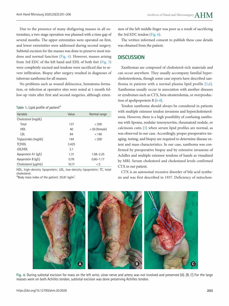
Due to the presence of many disfiguring masses in all ex-tremities, a two-stage operation was planned with a time gap of several months. The upper extremities were operated on first, and lower extremities were addressed during second surgery. Subtotal excision for the masses was done to preserve most ten-dons and normal function (Fig. 4). However, masses arising from 3rd EDC of the left hand and EHL of both feet (Fig. 5) were completely excised and tendons were sacrificed due to se-vere infiltration. Biopsy after surgery resulted in diagnoses of tuberous xanthoma for all masses.
No problems such as wound dehiscence, hematoma forma-tion, or infection at operative sites were noted at 1-month fol-low-up visits after first and second surgeries, although exten-
Table 1. Lipid profile of patienta)
Variable Value Normal rangeCholesterol (mg/dL) Total 137 <200 HDL 40 >50 (female) LDL 84 <140Triglycerides (mg/dL) 144 <200TC/HDL 3.425LDL/HDL 2.1Apoprotein A1 (g/L) 1.31 1.08–2.25Apoprotein B (g/L) 0.76 0.60–1.17Cholestanol (μg/mL) 10.11 <5
HDL, high-density lipoprotein; LDL, low-density lipoprotein; TC, total cholesterol. a)Body mass index of the patient: 30.81 kg/m2.
Fig. 4. During subtotal excision for mass on the left wrist, ulnar nerve and artery was not involved and preserved (A). (B, C) For the large masses were on both Achilles tendon, subtotal excision was done preserving Achilles tendon.
sion of the left middle finger was poor as a result of sacrificing the 3rd EDC tendon (Fig. 6).
The written informed consent to publish these case details was obtained from the patient.
DISCUSSION
Xanthomas are composed of cholesterol-rich materials and can occur anywhere. They usually accompany familial hyper-cholesterolemia, though some case reports have described xan-thoma in patients with a normal plasma lipid profile [5,6]. Xanthomas usually occur in association with another diseases or syndromes such as CTX, beta sitosterolemia, or overproduc-tion of apolipoprotein B [6-8].
Tendon xanthoma should always be considered in patients with multiple extensor tendon invasions and hypercholesterol-emia. However, there is a high possibility of confusing xantho-ma with lipoma, nodular tenosynovitis, rheumatoid nodule, or calcinosis cutis [3] when serum lipid profiles are normal, as was observed in our case. Accordingly, proper preoperative im-aging, testing, and biopsy are required to determine disease ex-tent and mass characteristics. In our case, xanthoma was con-firmed by preoperative biopsy and by extensive invasions of Achilles and multiple extensor tendons of hands as visualized by MRI. Serum cholesterol and cholestanol levels confirmed CTX in our patient.
CTX is an autosomal recessive disorder of bile acid synthe-sis and was first described in 1937. Deficiency of mitochon-
A B C
https://doi.org/10.12790/ahm.20.0028
Du-Heon Seo, et al. Multiple Huge Tendinous Xanthomas
204
drial enzyme sterol 27-hydroxylase leads to excessive choles-tanol and cholesterol production and accumulations of these sterols in several tissues. CTX is characterized by intractable diarrhea, neurologic abnormalities, and tendinous xanthom-as, especially in Achilles tendons. Sterol analysis or electroim-munoassay is required for the identification of this disease [7].
Several treatment options are available for tendon xanthoma, for example, it could be treated by tendon reconstruction with complete excision, subtotal excision, or amputation. Although Achilles tendon reconstruction or complete tendon removal or amputation have been reported for the treatment of large foot lesions [9], considerations of degree of invasion, functional outcomes, and patient willingness may favor subtotal excision with conservative treatment because total excision for masses with severe invasion on tendons may lead to motor dysfunction sequelae [3]. Of course, subtotal excision may be associated with recurrence, but appropriate medical treatment could pre-vent or delay this eventuality [10].
In conclusion, xanthoma can occur anywhere and usually ac-companies familial hypercholesterolemia. Unlike previous reports, we describe an extremely rare case involving extensive invasion by multiple huge xanthomas in all extremities that caused motion disability and poor cosmesis in a patient with a normal lipid profile that responded satisfactorily to subtotal mass excision.
Fig. 5. The masses involved in 3rd extensor digitorum communis tendon (A) and both tendons of extensor hallucis longus (B) was completely excised and then tendons were sacrificed due to severe infiltration into the tendons.
Fig. 6. Extension of the left middle finger was poor as a result of sacrificing the 3rd extensor digitorum communis tendon after surgery.
A B
Arch Hand Microsurg 2020;25(3):201-206
https://doi.org/10.12790/ahm.20.0028 205
normolipemic plane xanthoma with supraglottic involvement in a patient with hand-Schüller-Christian disease a case re-port. Am J Clin Dermatol. 2009;10:189-92.
6. Fleischmajer R, Tint GS, Bennett HD. Normolipemic tendon and tuberous xanthomas. J Am Acad Dermatol. 1981;5:290-6.
7. Verrips A, van Engelen BG, Wevers RA, et al. Presence of di-arrhea and absence of tendon xanthomas in patients with cerebrotendinous xanthomatosis. Arch Neurol. 2000;57:520-4.
8. Vega GL, Illingworth DR, Grundy SM, Lindgren FT, Connor WE. Normocholesterolemic tendon xanthomatosis with over-production of apolipoprotein B. Metabolism. 1983;32:118-25.
9. Jones FE, Soule EH, Coventry MB. Fibrous xanthoma of sy-novium (giant-cell tumor of tendon sheath, pigmented nodu-lar synovitis). A study of one hundred and eighteen cases. J Bone Joint Surg Am. 1969;51:76-86.
10. Wilkes LL. Tendon xanthoma in type IV hyperlipoprotein-emia. South Med J. 1977;70:254-5.
CONFLICTS OF INTEREST
The authors have nothing to disclose.
REFERENCES
1. Bude RO, Adler RS, Bassett DR. Diagnosis of Achilles tendon xanthoma in patients with heterozygous familial hypercholes-terolemia: MR vs sonography. AJR Am J Roentgenol. 1994; 162:913-7.
2. Kruth HS. Lipid deposition in human tendon xanthoma. Am J Pathol. 1985;121:311-5.
3. Doyle JR. Tendon xanthoma: a physical manifestation of hy-perlipidemia. J Hand Surg Am. 1988;13:238-41.
4. Wang Z, Lin ZW, Huang LL, et al. Primary non-hyperlipid-emia xanthoma of bone: a case report with review of the liter-ature. Int J Clin Exp Med. 2014;7:4503-8.
5. Lu YT, Chen TJ, Chung WH, Kuo TT, Hong HS. Cutaneous