Page 1
2017-03-12
1
2.X‐rayplanarradiographyandCT(2)
Lectures3,4
MedicalImagingSystems
JaeGwan Kim
[email protected] ,X2220
DepartmentofBioMedical ScienceandEngineering
Gwangju InstituteofSciencesandTechnologyCopyright.Mostfigures/tables/textsinthislecturearefromthetextbook“IntroductiontoMedicalImaging:Physics,EngineeringandClinicalApplicationsbyNadineBarrieSmithAndrewWebb2011”andthismaterialisonlyforthosewhotakethisclassandcannotbedistributedtoanyonewithoutthepermissionfromthelecturer.
Contents
1. Instrumentationforplanarradiography1) X‐raytube(discussedinthepreviouslecture)
2) Collimators
3) Anti‐scattergrid
4) X‐raydetectors
2. QuantitativecharacteristicsofplanarX‐rayimages1) Signaltonoiseratio
2) Spatialresolution
3) Contrasttonoiseratio
3. X‐raycontrastagents4. SpecializedX‐rayimagingtechniques5. ClinicalapplicationsofplanarX‐rayimaging
Page 2
2017-03-12
2
InstrumentationforplanarX‐ray
• X‐raytube– GeneratesX‐ray
• Collimator– ReduceX‐raydosetopatientsandComptonscatteredX‐ray
• Anti‐scattergrid– ReducefurtherthecontributionofscatteredX‐raytoimages
• Digitaldetector– ConvertsX‐rayintolight– Convertslightintovoltage– DigitizethevoltageusingADC
InstrumentationforplanarX‐ray
• Collimator(beam‐restrictor)– ThebevelangledeterminesthewidthofX‐raybeam
– X‐raybeamiswiderthanFOV,andthishas2undesirableeffects1) Patientdoseishigherthanitshouldbe2) UnnecessaryComptonscatteredX‐raysalso
increase
– Tominimizetheaboveeffects,acollimatorisplacedbetweenX‐raysourceandthepatient
– Thecollimatorconsistsofsheetsofleadandcanrestrictthebeamineitheroneortwodimensions
Page 3
2017-03-12
3
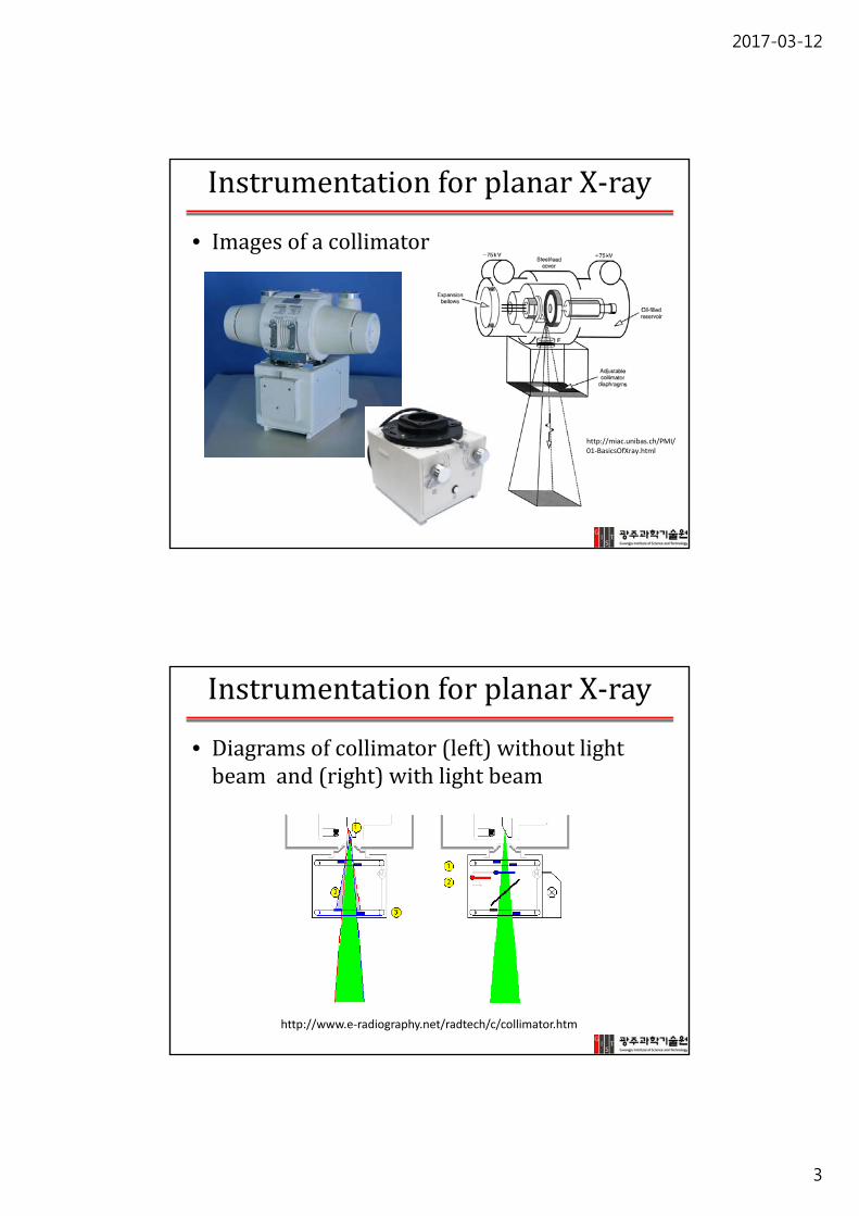
InstrumentationforplanarX‐ray
• Imagesofacollimator
http://miac.unibas.ch/PMI/01‐BasicsOfXray.html
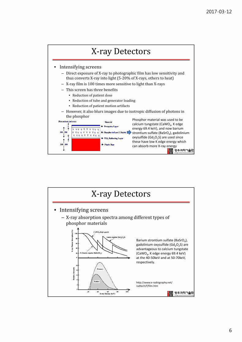
InstrumentationforplanarX‐ray
• Diagramsofcollimator(left)withoutlightbeamand(right)withlightbeam
http://www.e‐radiography.net/radtech/c/collimator.htm
Page 4
2017-03-12
4
InstrumentationforplanarX‐ray
• Anti‐scattergrid– ComptonscatteredX‐raylowersCNR– Toreducethecontributionfromsecondaryradiation,ananti‐scattergridisplacedbetweenthepatientandtheX‐raydetector
– Leadortungsten
No anti‐scatter grid With an anti‐scatter grid
InstrumentationforplanarX‐ray
• Anti‐scattergridischaracterizedbytwoproperties,thegridratioandgridfrequency– Gridratio=h/d,4:1~16:1– Gridfrequency=1/(d+t),5~7lines/mmwherehistheheight,tisthethickness,distheseparationoftheleadstrips
h
d
http://www.mikrosystems.com/applications/computed‐tomography
Page 5
2017-03-12
5
InstrumentationforplanarX‐ray
• However,usingananti‐scattergridrequirestohaveahigherradiationdose.(~10times)
• Thistrade‐offcanbeshownbyBuckyfactor(BF).• BFistheratioofmeasuredexposurewiththegridinplacetothatwithout.
• Buckyfactor=1/Gridpenetrationhttp://www.sprawls.org/ppmi2/SCATRAD/
X‐rayDetectors
• TraditionalX‐rayfilm
– Intensifyingscreen
– X‐rayfilm
• Digitaldetectortechnologies
– Computedradiography(CR):
cheaperandmorewidelyusedthandigitalradiography
– Digitalradiography(DR)
Page 6
2017-03-12
6
X‐rayDetectors
• Intensifyingscreens– DirectexposureofX‐raytophotographicfilmhaslowsensitivityandthusconvertsX‐rayintolight(5‐20%ofX‐rays,otherstoheat)
– X‐rayfilmis100timesmoresensitivetolightthanX‐rays– Thisscreenhasthreebenefits
• Reductionofpatientdose• Reductionoftubeandgeneratorloading• Reductionofpatientmotionartifacts
– However,italsoblursimagesduetoisotropicdiffusionofphotonsinthephosphor
Phosphor material was used to be calcium tungstate (CaWO4, K edge energy 69.4 keV), and now barium strontium sulfate (BaSrO4), gadolinium oxysulfide (Gd2O2S) are used since these have low K edge energy which can absorb more X‐ray energy
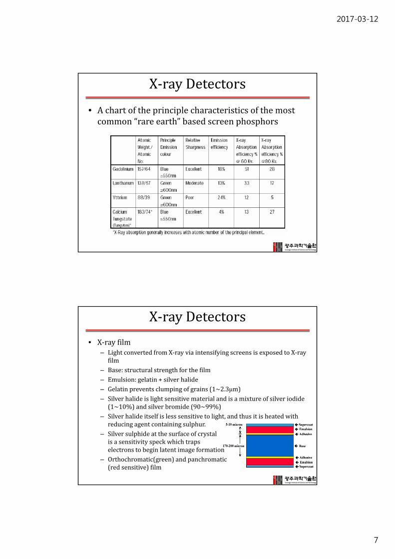
X‐rayDetectors
• Intensifyingscreens– X‐rayabsorptionspectraamongdifferenttypesofphosphormaterials
http://www.e‐radiography.net/radtech/f/film.htm
Barium strontium sulfate (BaSrO4), gadolinium oxysulfide (Gd2O2S) are advantageous to calcium tungstate (CaWO4, K edge energy 69.4 keV) at the 40‐50keV and at 50‐70keV, respectively.
Page 7
2017-03-12
7
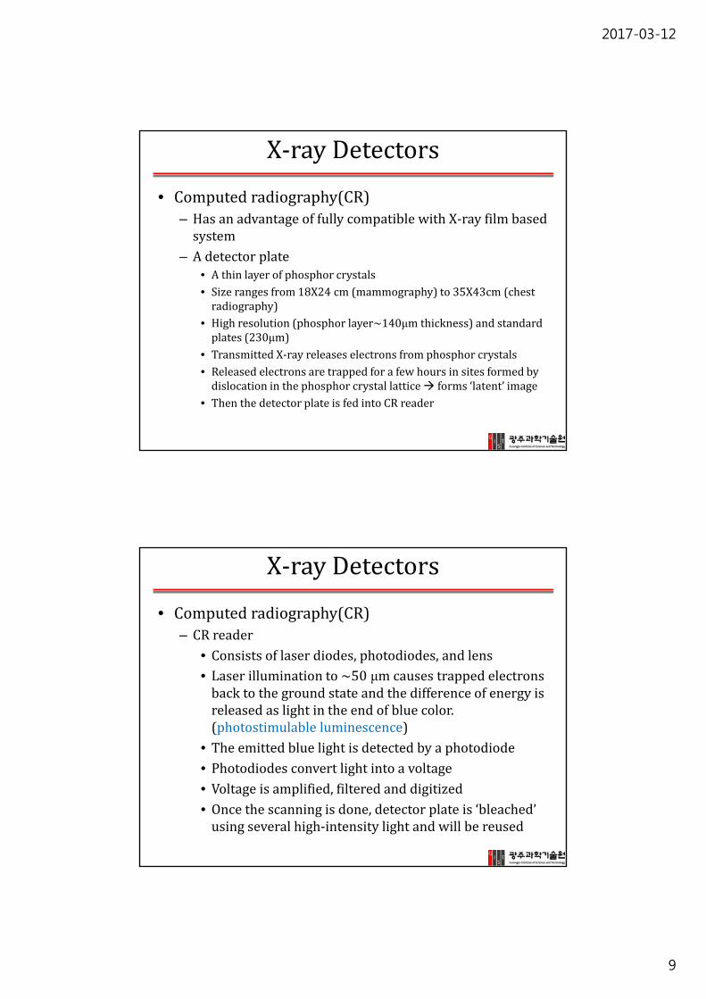
X‐rayDetectors
• Achartoftheprinciplecharacteristicsofthemostcommon“rareearth”basedscreenphosphors
X‐rayDetectors
• X‐rayfilm– LightconvertedfromX‐rayviaintensifyingscreensisexposedtoX‐rayfilm
– Base:structuralstrengthforthefilm– Emulsion:gelatin+silverhalide– Gelatinpreventsclumpingofgrains(1~2.3μm)– Silverhalideislightsensitivematerialandisamixtureofsilveriodide(1~10%)andsilverbromide(90~99%)
– Silverhalideitselfislesssensitivetolight,andthusitisheatedwithreducingagentcontainingsulphur.
– Silversulphide atthesurfaceofcrystalisasensitivityspeckwhichtrapselectronstobeginlatentimageformation
– Orthochromatic(green)andpanchromatic(redsensitive)film
Page 8
2017-03-12
8
X‐rayDetectors
• X‐rayfilmprocessing– Development:theexposedgrainsarepreferentiallyreducedtoblackmetallicsilver
– Fixing:theremainedgrainsaredissolved– Washing:removethedissolvedgrainsfromemulsion– Replenishment:ensuresthechemicalbalanceismaintained
X‐rayDetectors
• Cassetteandintensifyingscreencombination
http://www.sure‐quality.com/x‐ray‐supplies.html
http://xraymachinesss.com/category/dental‐x‐ray‐film/
http://xrayrecycling.blogspot.com/2011/05/x‐ray‐film‐recycling‐and‐its.html
Page 9
2017-03-12
9
X‐rayDetectors
• Computedradiography(CR)– HasanadvantageoffullycompatiblewithX‐rayfilmbasedsystem
– Adetectorplate• Athinlayerofphosphorcrystals• Sizerangesfrom18X24cm(mammography)to35X43cm(chestradiography)
• Highresolution(phosphorlayer~140μmthickness)andstandardplates(230μm)
• TransmittedX‐rayreleaseselectronsfromphosphorcrystals• Releasedelectronsaretrappedforafewhoursinsitesformedbydislocationinthephosphorcrystallattice forms‘latent’image
• ThenthedetectorplateisfedintoCRreader
X‐rayDetectors
• Computedradiography(CR)– CRreader
• Consistsoflaserdiodes,photodiodes,andlens• Laserilluminationto~50 μmcausestrappedelectronsbacktothegroundstateandthedifferenceofenergyisreleasedaslightintheendofbluecolor.(photostimulable luminescence)
• Theemittedbluelightisdetectedbyaphotodiode• Photodiodesconvertlightintoavoltage• Voltageisamplified,filteredanddigitized• Oncethescanningisdone,detectorplateis‘bleached’usingseveralhigh‐intensitylightandwillbereused
Page 10
2017-03-12
10
X‐rayDetectors
• Photostimulable phosphor– BaFX:Eu2+, Ba2+,F‐,XcouldbeBr‐ orI‐ oramixtureofboth
Isosurface of the electron density for the (Eu )* excited state of BaFI:Eu showing strong localization on the Eu atom (pink), Ba (blue), F(green), I (orange).
Ref: Gundiah et al., IEEE Trans Nuclear Sci, 57(3) 1702‐5, 2010
http://home.fujifilm.com/info/products/science/ip/principle.html
X‐ray
Eu2+ Eu3+
e‐
+
Trapped in Br+
empty lattice
Eu3++e‐
X‐rayDetectors
• Photostimulable luminescence:aftertheinitialexposure,excitedelectronsinthephosphormaterialremain'trapped'in'colorcenters'inthecrystallatticeuntilstimulatedbythesecondillumination.Forexample,Fuji'sphotostimulable phosphorisdepositedonaflexiblepolyesterfilmsupportwithgrainsizeabout5micrometers,andisdescribedas“bariumfluorobromide containingatraceamountofbivalenteuropiumasaluminescencecenter”.Europiumisadivalentcation thatreplacesbariumtocreateasolidsolution.WhenEu2+ ionsarestruckbyionizingradiation,theyloseanadditionalelectrontobecomeEu3+ ions.Theseelectronsentertheconductionbandofthecrystalandbecometrappedinthebromineionemptylatticeofthecrystal.Thismetastablestateishigherinenergythantheoriginalcondition,soalower‐frequencylightsourcethatisinsufficientinenergytocreatemoreEu3+ ionscanreturnthetrappedelectronstotheconductionband.AsthesemobilizedelectronsencounterEu3+ ions,theyreleaseablue‐violet400nmluminescence.Thislightisproducedinproportiontothenumberoftrappedelectrons,andthusinproportiontotheoriginalX‐raysignal(CopiedfromWikipedia)
Page 11
2017-03-12
11
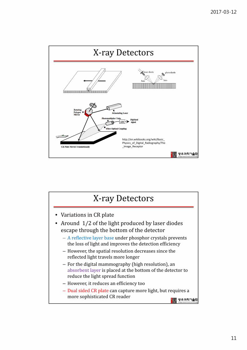
X‐rayDetectors
http://en.wikibooks.org/wiki/Basic_Physics_of_Digital_Radiography/The_Image_Receptor
X‐rayDetectors
• VariationsinCRplate• Around1/2ofthelightproducedbylaserdiodesescapethroughthebottomofthedetector– Areflectivelayerbaseunderphosphorcrystalspreventsthelossoflightandimprovesthedetectionefficiency
– However,thespatialresolutiondecreasessincethereflectedlighttravelsmorelonger
– Forthedigitalmammography(highresolution),anabsorbentlayerisplacedatthebottomofthedetectortoreducethelightspreadfunction
– However,itreducesanefficiencytoo– DualsidedCRplatecancapturemorelight,butrequiresamoresophisticatedCRreader
Page 12
2017-03-12
12
X‐rayDetectors
• NewmaterialsforCRplates– PreviouslyusedBaFX:Eu2+(bariumfluorohalide activatedwitheuropiumions,halideXisamixtureofbromineandiodine)israndomlyoriented
– CsBr:Eu2+:thincolumnarcrystalsactsasaverythinopticalfibers1) higherintrinsicspatialresolution,2) higherpackingefficiency increasesthesensitivity3) thicknessofthephosphorlayercanbeincreasedto600μm
increasethenumberofabsorbedX‐rays
– ThedynamicrangeofaCRsystemishigh,typicallytheoutputislineartoX‐rayinputfortherangeof4ordersofmagnitude.
X‐rayDetectors
• CRsystem– Thespatialresolutionislimitedby
• Laserbeamsize• Numberofsampleddatapoints• Thedegreeoflaserbeamscatteringbycrystalsinthephosphorscreen
• HRplateshavesmallerandthinnercrystals highspatialresolution
– HRisdigitizedas4096X4096andstandardis2048X2048– SNRofstandardisabouttwicehigherthanHRplateforthesameX‐rayinput
– DualsidedCRplatesandCsBr:Eu2+ increasestheSNRbyafactorof2comparedtosinglesidedandBaFX:Eu2+,respectively
Page 13
2017-03-12
13
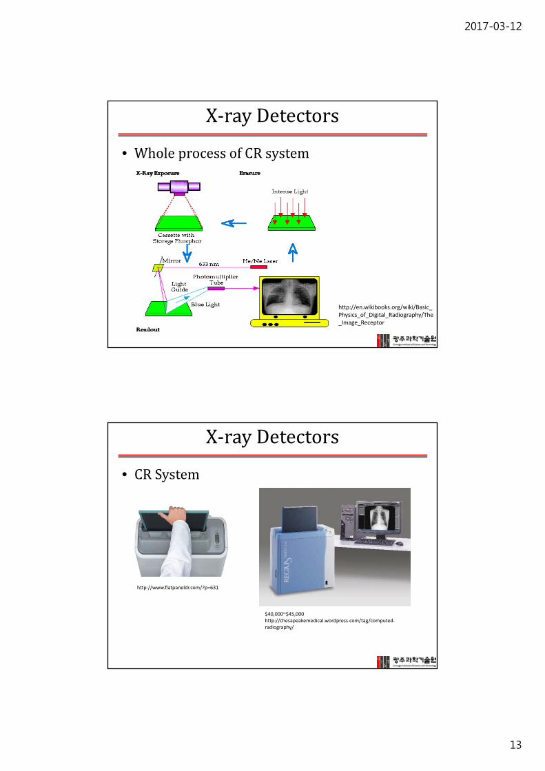
X‐rayDetectors
• WholeprocessofCRsystem
http://en.wikibooks.org/wiki/Basic_Physics_of_Digital_Radiography/The_Image_Receptor
X‐rayDetectors
• CRSystem
http://www.flatpaneldr.com/?p=631
$40,000~$45,000http://chesapeakemedical.wordpress.com/tag/computed‐radiography/
Page 14
2017-03-12
14
X‐rayDetectors
• Idealdigitalradiographysystem
X‐rayDetectors
• Digitalradiography– Indirectconversion:mostcommonlyused
• First,convertsX‐rayenergyintolightbyaCsI:Tl (ThalliumdopedCesiumIodide)orGadoliniumoxysulfide scintillator
• Secondly,convertslightintoavoltageusinga2Dphotodiodearray
• CesiumandIodinehaveK‐edgesat36and33.2keV,therefore,theX‐rayattenuationcoefficientofCsI isveryhigh highlyefficientX‐raydetector
Page 15
2017-03-12
15
X‐rayDetectors
• AschematicofindirectDRdetectorisshownbelow
• CsI:Tl isa‘needlecrystals’with5μmdiameter• Thisshapeprovidesanexcellentpackingefficiency(>80%)forbetterX‐rayabsorptionandenhancespatialresolution
• Thicknessofthislayerisaround0.6mm• ACCDcameraoralargeflatpaneldetector(FPD) consistedofthin‐filmtransistor(TFT)arraysisplacedrightunderthatCsI:Tl layer
X‐rayDetectors
• FPD:thisfilmamorphoussilicontransistorarrayislayeredontotheglass.
• AmorphoustransistorisusedinsteadofcrystallinesinceitcanbeexposedtohighdoseofX‐raywithoutdamage.
• Eachpixelofthedetectorconsistsof– Fabricatedphotodiode– Storagecapacitor– TFTswitch
• AbsorbedX‐raybyCsI rod,CsI produceslight(~green).LightgoestoTFTarrayandisconvertedtoavoltage(photodiode)andstoredincapacitors
• Multiplexerreadsthesignalline‐by‐lineandsignalsareamplifiedanddigitizedusinga14‐bitADC
Page 16
2017-03-12
16
X‐rayDetectors
• AtypicalDRsystemhas– Flatpanel43X43cm– TFTarrayof3001X3001elements pixelsamplingintervalof143μm
– Anti‐scattergrid:gridratioof~13:1andgridlinedensityof~70lines/cm(~143 μm)
– Allofthesearepackagedto~50X50cmsquare,4.5cmthicknesswithaweightof~20kg
X‐rayDetectors
• Digitalradiography– Directconversion:
• EliminatesthestepofconvertingX‐rayenergyintolightX‐raytoelectricsignal
• DirectabsorptionoftheX‐rayphotonstoproduceelectricalsignalsusingaX‐rayphotoconductorsuchasamorphousselenium(alloyedwitharsenictopreventrecrystallization)
• Selenium(atomicnumberof34,K‐edgeat13keV)islessefficientthanCsI:Tl
• Amorphousseleniummaterialislayeredonthetopofamorphoussiliconetransistorarray
• Currently,indirectconversionismorecommon,butnewmaterialsareunderinvestigationforthebetterefficiencyofdirectDR
Page 17
2017-03-12
17
X‐rayDetectors
• Photoconductors– Areasubsetofsemiconductors.– Inthedark,thesematerialsareinsulatorsbuteffectivelybecomeconductors
underillumination.– AslightorX‐rayphotonsareabsorbed,theenergyoftheincomingphoton
exciteselectronsinthephotoconductortoastateknownastheconductionbandandchargecarrierscalledelectron‐holepairsareproduced.
– Withoutanelectricfield,theexcitedelectronsreturntotheirgroundstate,thevalenceband.
– However,withanelectricfield,theelectronsintheconductionbandmovealongtheelectricfieldlines.
– ThenegativeelectronscancelthepositivesurfacechargeandthusproducevariationsinthesurfacechargethatcorrespondtotheincidentpatternoftheX‐raysoranX‐rayimage.
– Thechargecollectionwasguidedbytheelectricfield,sotheproducedchargepatternfaithfullyreproducestheX‐rayimage.TheresultinghighresolutionimageisnotstronglydependentontheSeleniumthickness.
X‐rayDetectors
• AmorphousSelenium– Anumberofphotoconductors(e.g.silicon,germanium,thalliumbromideand
mostsemiconductors)couldbeusedforX‐rayimagingdetectorsbutamorphousSelenium(a‐Se)hasmanyfeaturesthatmakeitwellsuitedforthistask.
– A‐SeiswelldevelopedtechnologicallyasithasbeenusedasaphotoconductorinphotocopiersandalsoinanX‐rayimagingtechniqueknownasxeroradiography fordecades.Itisusedinitsamorphousform,soamorphousseleniumplatescanbemadebyevaporation.Thus,incontrasttomanycrystallinephotoconductors,a‐Sebaseddetectorscanbemadelargeinarearelativelyeasilyandinexpensively.
– Theelectricproperties,namelythelowdarkorleakagecurrent,ofa‐SealsorenderitsuitableforX‐rayimaginguse.ItsotherX‐raypropertiesareasfollows:
– ~1000electron‐holepairs/50keV X‐rayatanelectricfieldof10V/um.InotherwordsW+/‐ =50eV atthisfieldstrength.
– ~50%attenuationofa50keV beamwith365umofSelenium;50%attenuationofa20keV beamwitha30umofSelenium.
Page 18
2017-03-12
18
X‐rayDetectors
• Digitalradiography– Principleofoperation:Directconversion
Ref) Introduction to digital radiography _Kodak
X‐rayDetectors
• Indirectvs Directdigitalradiography
Indirect DR
Direct DR
Page 19
2017-03-12
19
X‐rayDetectors
• Indirectvs Directdigitalradiography
Indirect DR Direct DRRef) Introduction to digital radiography _Kodak
X‐rayDetectors
• Digitalradiography
Page 20
2017-03-12
20
X‐rayDetectors
• Digitalradiography
CCD DR Flat Panel Digital X‐Ray (indirect or direct)
http://www.flatpaneldr.com/?p=631
X‐rayDetectors
• MTFcomparisonamongvariousdetectingmethods
Ref) Introduction to digital radiography _Kodak
Page 21
2017-03-12
21
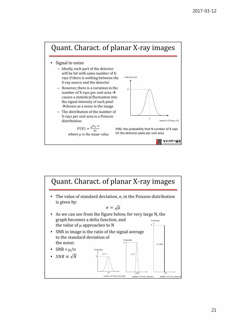
Quant.Charact.ofplanarX‐rayimages
• Signaltonoise– Ideally,eachpartofthedetectorwillbehitwithsamenumberofX‐raysifthereisnothingbetweentheX‐raysourceandthedetector
– However,thereisavariationinthenumberofX‐raysperunitareacausesastatisticalfluctuationintothesignalintensityofeachpixelshownasanoiseintheimage
– ThedistributionofthenumberofX‐raysperunitareaisaPoissondistribution:
!whereμ is the mean value
P(N): the probability that N number of X‐rays hit the detector plate per unit area
Quant.Charact.ofplanarX‐rayimages
• Thevalueofstandarddeviation,σ,inthePoissondistributionisgivenby:
• Aswecanseefromthefigurebelow,forverylargeN,thegraphbecomesadeltafunction,andthevalueofμ approachestoN
• SNRinimageistheratioofthesignalaveragetothestandarddeviationofthenoise:
• SNR=/σ
• ∝
Page 22
2017-03-12
22
Quant.Charact.ofplanarX‐rayimages
• TodoubletheSNR,therefore,numberofX‐raysdetectedneedstobe4timeshigher needtoincreaseradiationdose4times
• OperationalfactorsaffectSNRare– Thetubecurrentandexposuretime
∝
– ThetubekVp:higherkVp higherSNR,butinanon‐linearway
– Thepatientsizeandpartofthebodybeingimaged:thickerthebody lowertheSNR
– Thegeometryoftheanti‐scattergrid:thelargergridratio,thesmallerofSNR(butimprovestheCNR)
Quant.Charact.ofplanarX‐rayimages
• OperationalfactorsaffectSNRare– Theefficiencyofthedetector:thiscanbequantifiedbyaparametercalledthedetectorquantumefficiency(DQE),
out andin representtheinputSNRandoutputSNRfromthedetector.– DQEisalwayslessthan1becauseadetectoralwaysintroducesomenoisetothesystem
• StandardCRplate:~0.25• HighresolutionCRplate:~0.12• DualsidedCRplate:~0.4• CsBr:Eu2+ basedplates:~0.8
Page 23
2017-03-12
23
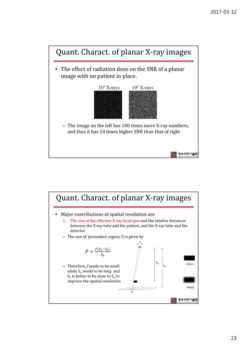
Quant.Charact.ofplanarX‐rayimages
• TheeffectofradiationdoseontheSNRofaplanarimagewithnopatientinplace.
– Theimageonthelefthas100timesmoreX‐raynumbers,andthusithas10timeshigherSNRthanthatofright
Quant.Charact.ofplanarX‐rayimages
• Majorcontributionsofspatialresolutionare1. ThesizeoftheeffectiveX‐rayfocalspotandtherelativedistances
betweentheX‐raytubeandthepatient,andtheX‐raytubeandthedetector
– Thesizeof‘penumbra’region,P,isgivenby
– Therefore,f needstobesmallwhileSo needstobelongandS1 isbettertobeclosetoSo toimprovethespatialresolution
Page 24
2017-03-12
24
Quant.Charact.ofplanarX‐rayimages
• Threedistinctpartsofashadow
• Inradiationoncology,penumbraisthespaceintheperipheryofthemaintargetofradiationtherapyandisdefinedasthevolumereceivingbetween80%and20%ofisodose
Quant.Charact.ofplanarX‐rayimages
• Majorcontributionsofspatialresolutionare2. ThepropertiesoftheX‐raydetector– ACRreaderforCRandaflatpaneldetectorofDRdeterminethe
spatialresolutiontoo.– CRreader
• Laserbeamsize• Numberofsampleddatapoints• Thedegreeoflaserbeamscatteringbycrystalsinthephosphorscreen
• HRplateshavesmallerandthinnercrystals highspatialresolution
– DigitalRadiography• PixelsizeofFPD
Page 25
2017-03-12
25
Quant.Charact.ofplanarX‐rayimages
• Spatialresolution– TheoverallMTFcanbeobtainedbyconvolutingMTFofeachimagingcomponent.
– ThemostusefulmeasureofspatialresolutionforX‐rayimagingistomeasurealinespreadfunction(LSF)byusingagrid(parallelleadsepta)
Quant.Charact.ofplanarX‐rayimages
• Threetypesoffactoraffectingcontrasttonoise1. ComptonscatteredX‐rays:theseareaffectedby
1) TheX‐rayenergyspectrumHigherX‐rayenergymoreComptonscattering
2) ThefieldofviewoftheX‐rayimageLargerFOVmoreComptonscattering
3) ThethicknessofbodypartbeingimagedThickermoreComptonscattering lowCNR
4) Thegeometryoftheanti‐scattergridHighergridratio betterCNR lowSNR
2. SNR3. Spatialresolution
Page 26
2017-03-12
26
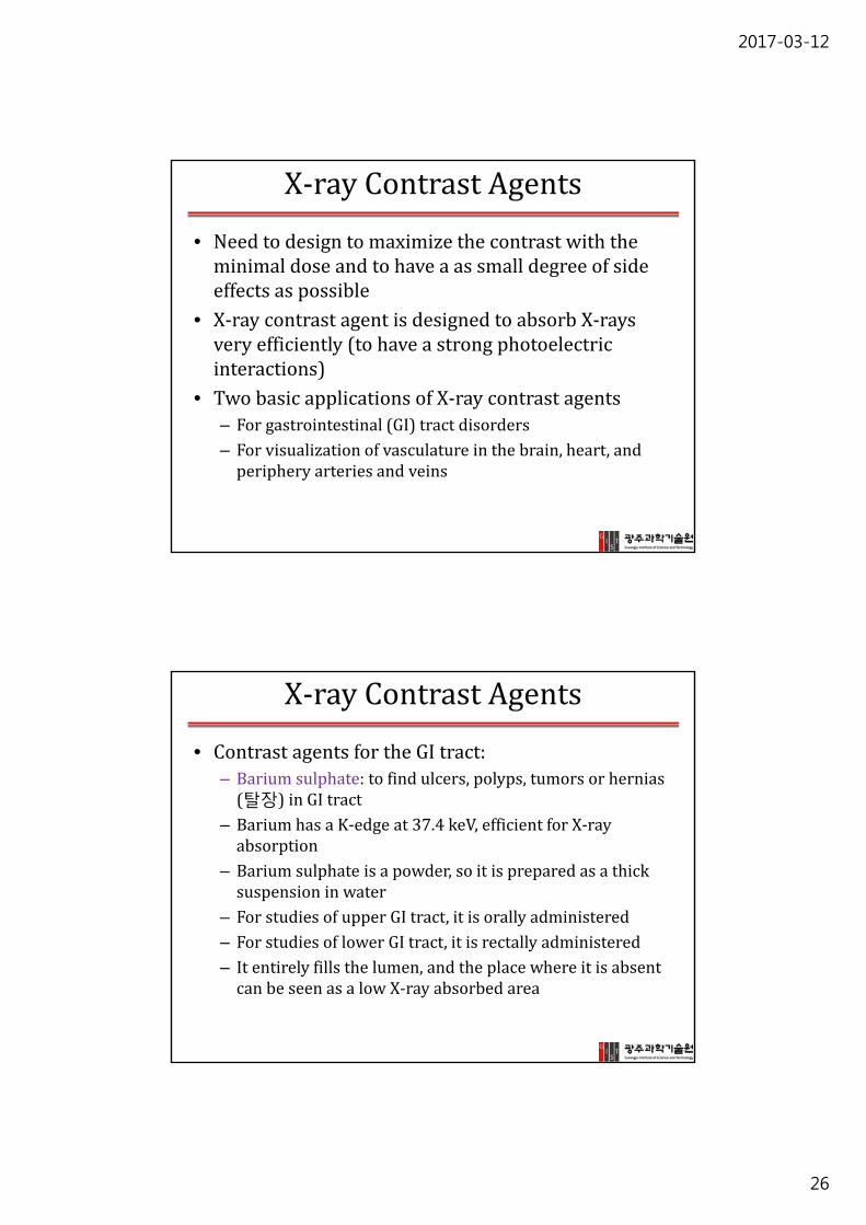
X‐rayContrastAgents
• Needtodesigntomaximizethecontrastwiththeminimaldoseandtohaveaassmalldegreeofsideeffectsaspossible
• X‐raycontrastagentisdesignedtoabsorbX‐raysveryefficiently(tohaveastrongphotoelectricinteractions)
• TwobasicapplicationsofX‐raycontrastagents– Forgastrointestinal(GI)tractdisorders– Forvisualizationofvasculatureinthebrain,heart,andperipheryarteriesandveins
X‐rayContrastAgents
• ContrastagentsfortheGItract:– Bariumsulphate:tofindulcers,polyps,tumorsorhernias(탈장)inGItract
– BariumhasaK‐edgeat37.4keV,efficientforX‐rayabsorption
– Bariumsulphate isapowder,soitispreparedasathicksuspensioninwater
– ForstudiesofupperGItract,itisorallyadministered– ForstudiesoflowerGItract,itisrectallyadministered– Itentirelyfillsthelumen,andtheplacewhereitisabsentcanbeseenasalowX‐rayabsorbedarea
Page 27
2017-03-12
27
X‐rayContrastAgents
• GIdiseases
ulcers polyps
hernias
X‐rayContrastAgents
• Bariumenematest• Bariumsulphate first,thenadministerair
• Bariumsulphate fillsthesurfaceoftheGItract
• Airdistendsthelumen• Thisisusedtocharacterizesmallpathologiesinthelargeintestine,colonandrectum
• Colonoscopyismorepopularnow
Page 28
2017-03-12
28
X‐rayContrastAgents
• Iodinebasedcontrastagents– Administeredintravenouslyintopatient– IodinehasK‐edgeat33.2keV anditabsorbsX‐raygreatlyinbloodallows~50μmdiametersmallvesselsimaging
– CurrentlyusedIodinecontrastsarebasedontri‐iodinatedbenzeneringwithdifferentsidegroups
– Toreducesideeffects,thecontrastagentneedstobenon‐ionicwithlowosmolality
– Iodixanol hasaosmolalityclosetothatofbloodandCSF(290mOsm/kg)
– Osmolarity (Osm/L)andosmolality(Osm/kg)– Hypertonicsolution:hashigherosmolality causewatercomesoutfromthecell cellshrink(isusedtotreatcerebralhemorrhage)
– Hypotonicsolution:haslowerosmolality causethecellswellbyabsorbingwatercytolysis
X‐rayContrastAgents
• Iodinebasedcontrastagents– Theseareeithermonomeric(MW650~800)ordimeric (MW1300~1600)andhaveverylowbindingtoplasmaproteinsinbloodexcretedunmetabolized intheurinewithin24hrs
– Osmolalityofdimeric agentsaremuchlowerthanmonomericagent– Majorapplicationsofiodinatedcontrastagentsaredigitalsubtractionangiography,intravenousurography(IVU),pyelography(IVP),cholangiography(imagingbileduct)
Page 29
2017-03-12
29
X‐rayContrastAgents
• Iodinebasedcontrastagents
IV urographyIV cholangiography:Bile duct imaging
SpecializedX‐rayImagingTech.
• Digitalsubtractionangiography(DSA)– Providesveryhighresolutionofbloodvessels(<100μmindiameter)
– Procedure1) Acquirearegularimage2) Injectabolusofiodinated
contrastagentsi.v.3) Acquireasecondimage4) Performanimagesubtractionof
thosetwoimages– DSAisusedtoinvestigatediseasessuchasstenosis(bloodvesselnarrowing)andclottingofarteriesandveins,irregularitiesofsystemicbloodflow
Cerebral angiogram
Page 30
2017-03-12
30
SpecializedX‐rayImagingTech.
• Digitalmammography:todetectsmalltumorsormicrocalcifications inthebreast
• Requiresaveryhighspatialresolution(<1mm)andCNR
26keV, solid line is from applying a 30μm thickness of Mo filter
SpecializedX‐rayImagingTech.
• Alowradiationdoseisnecessarytoavoidtissuedamageusemolybdenum(K‐edgesat17.9and19.6keV)asananodeforX‐raytube
• Cathodefilamentisflat(nothelical)toproduceamorefocusedelectronbeam
• Thebevelangleissmallerthanusualtoproduceaneffectivefocalsizeof0.3mmorless
• TheglasswindowofX‐raytubeisreplacedbyberylliumtopreventalossoflowX‐rayenergybeam
• Amolybdenumfilter(30mthickness)isusedtoreducetheamountofhighenergyX‐rays(>20keV)
• Sometimes,analuminumfilterisusedwhenthebreastisradio‐opaque(attenuationofX‐rayishigh)
Page 31
2017-03-12
31
SpecializedX‐rayImagingTech.
• ThedetectorcanbeeitherCRorDR‐based• Alargefocal‐spot‐to‐detectordistance(45~80cm)isusedtoreducetheeffectsofgeometricunsharpness
• Theanti‐scattergridhasa4:1or5:1gridratio,25~50linespercmseptadensity,aseptal thicknessislessthan20m,andseptal heightislessthan1mm
• Needtocompressbreastto~4cmthicknesstoimprovelowenergyX‐rayspenetrationandtoreducetheComptonscatteringeffect
SpecializedX‐rayImagingTech.
• Digitalfluoroscopy:usesacontinuousX‐rayimagingmonitorinterventionalsurgery(catheters,guide‐wires,stents,pacemakers)aswellasfordynamicstudiesofGItractandcardiovascularsystemusingacontrastagent
• UsesamodifiedDRdetectorsystem– ThicknessofCsI:Tl isincreasedto550‐650mtoincreasedetectionefficiency reducetheX‐raydose
• UsesaveryshortpulsesofX‐rays(~5‐20ms)• Typicallyacquireimagesupto30frames/sec• TheX‐raydoseperframeis~1/1000th ofthatusedduringserialimageacquisition
Page 32
2017-03-12
32
SpecializedX‐rayImagingTech.
• (left)acardiaccatheterizationlaboratory,• (right)aneurointerventional unitwithaC‐armdigitalfluoroscopy
ClinicalAppl.PlanarX‐rayImaging
• Thepresenceandseverityoffracturesorcracksinthebonestructureinthehead,chest,pelvis,arms,legs,handsandfeet
• Vascularimagingusingaiodinebasedcontrasttostudybloodflow,mainlyinthebrainandheart,andalsointheperipheryarterialandvenoussystems
• GItractdiseasesusingabariumsulphate usuallywithcontinuousmonitoringwithX‐rayfluoroscopictechniques
• Urinarytract:kidney,utreter andbladder(KUB)scansbyintravenouspyelograms(IVPs)
Page 33
2017-03-12
33
ClinicalAppl.PlanarX‐rayImaging
• DentalX‐ray– Thispreoperativephotooftooth#3,(A),
revealsnoclinicallyapparentdecayotherthanasmallspotwithinthecentralfossa.Infact,decaycouldnotbedetectedwithanexplorer.Radiographicevaluation,(B),however,revealedanextensiveregionofdemineralizationwithinthedentin(arrows)ofthemesial halfofthetooth.Whenaburrwasusedtoremovetheocclusal enameloverlyingthedecay,(C),alargehollowwasfoundwithinthecrownanditwasdiscoveredthataholeinthesideofthetoothlargeenoughtoallowthetipoftheexplorertopasswascontiguouswiththishollow.Afterallofthedecayhadbeenremoved,(D),thepulpchamber hadbeenexposedandmostofthemesialhalfofthecrownwaseithermissingorpoorlysupported.
http://en.wikipedia.org/wiki/Dental_radiography
A selection of carbide burrs
A No. 23 explorer, also known as a 'sickle probe'
ClinicalAppl.PlanarX‐rayImaging
• IVPisperformedwithiodinatedcontrastagentinjectiontovisualizethefillingandemptyingoftheurinarysystem.Anexampleisshowntotheright.
• Normalexcretionoftheagenttakesabout30minutesfromthebloodstreamviakidneys
• IVPimagesareobtainedinseriesafteragentinjectiontofindanyobstructions
Renal pelvis
Several kidney cancers occurs at here