34
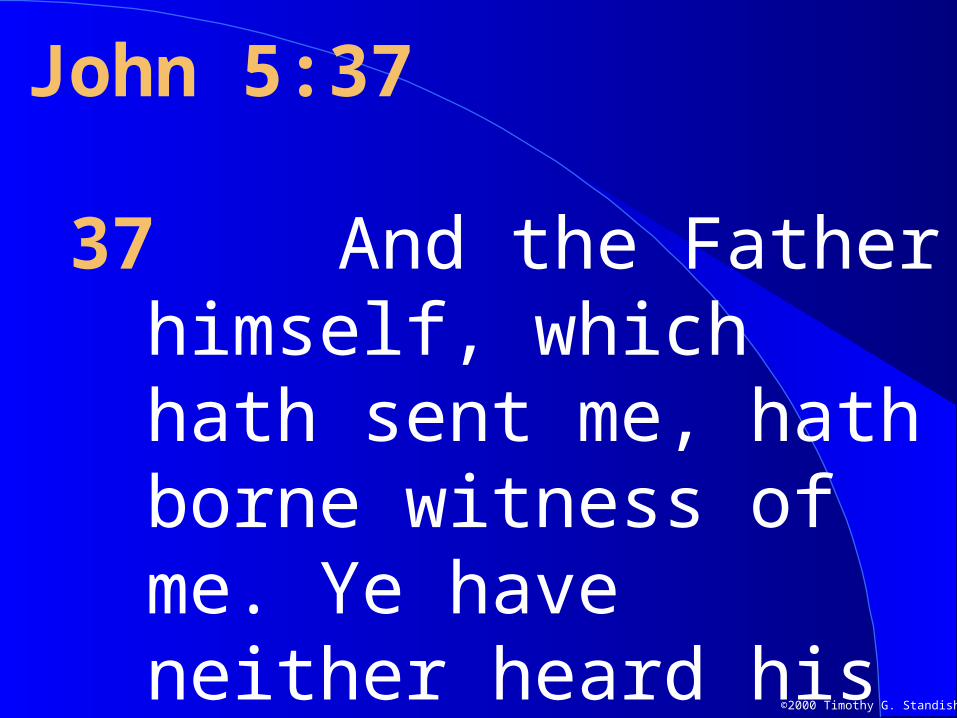
©2000 Timothy G. Standish John 5:37 37 And the Father himself, which hath sent me, hath borne witness of me. Ye have neither heard his voice at any time,
| Date post: | 02-Jan-2016 |
| Category: |
Documents |
| Upload: | percival-hood |
| View: | 215 times |
| Download: | 0 times |
©2000 Timothy G. Standish
John 5:37
37 And the Father himself, which hath sent me, hath borne witness of me. Ye have neither heard his voice at any time, nor seen his shape.
©2000 Timothy G. Standish
Structure and AnalysisStructure and Analysisofof
DNA and RNADNA and RNATimothy G. Standish, Ph. D.
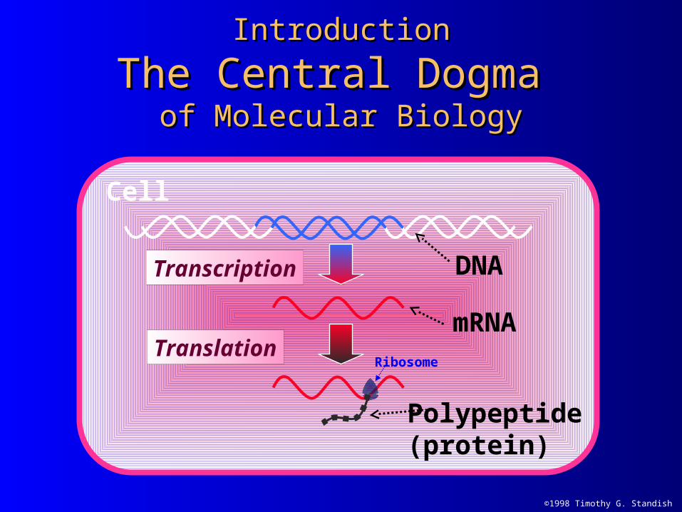
DNA
mRNA
Transcription
IntroductionIntroduction
The Central Dogma The Central Dogma of Molecular Biologyof Molecular Biology
Cell
Polypeptide(protein)
TranslationRibosome
©1998 Timothy G. Standish
©2000 Timothy G. Standish
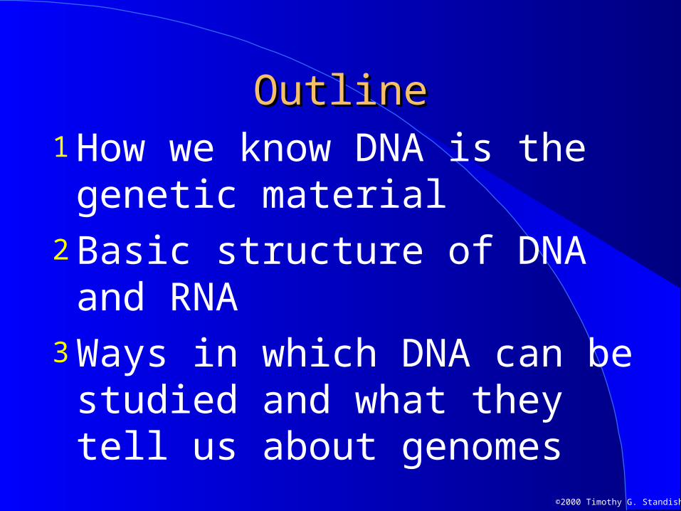
OutlineOutline1 How we know DNA is the genetic
material2 Basic structure of DNA and RNA3 Ways in which DNA can be
studied and what they tell us about genomes
©2000 Timothy G. Standish
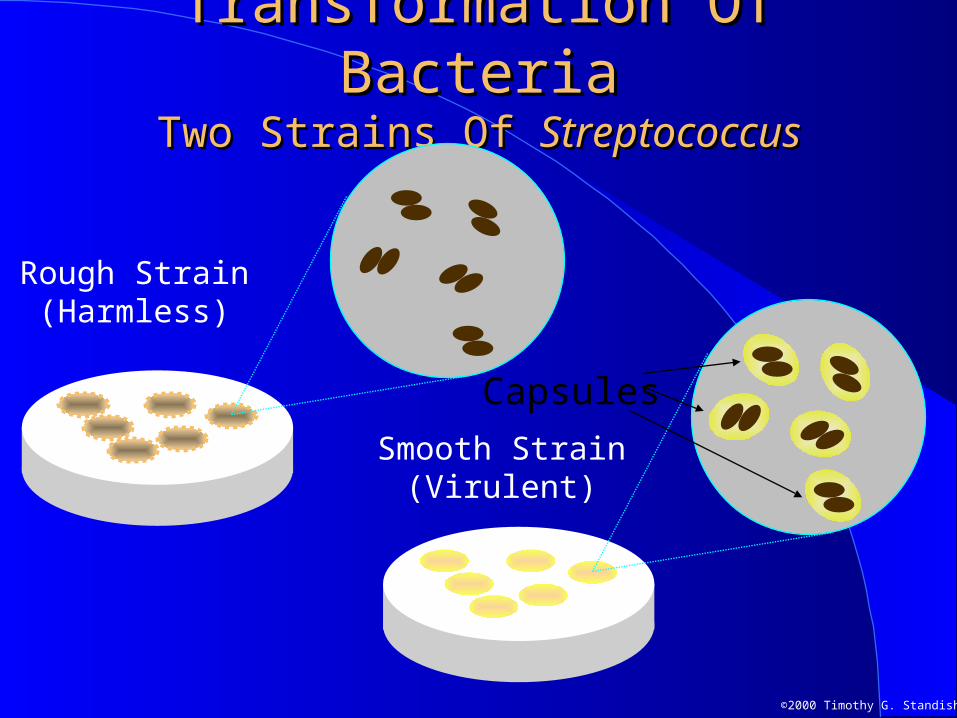
Transformation Of BacteriaTransformation Of BacteriaTwo Strains Of Two Strains Of StreptococcusStreptococcus
Capsules
Smooth Strain(Virulent)
Rough Strain(Harmless)
©2000 Timothy G. Standish
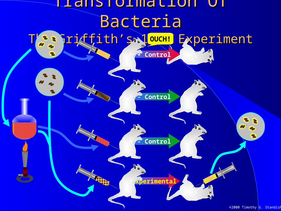
Experimental
Transformation Of BacteriaTransformation Of BacteriaThe Griffith’s 1928 ExperimentThe Griffith’s 1928 Experiment
- Control
+ Control
- Control
OUCH!
©2000 Timothy G. Standish
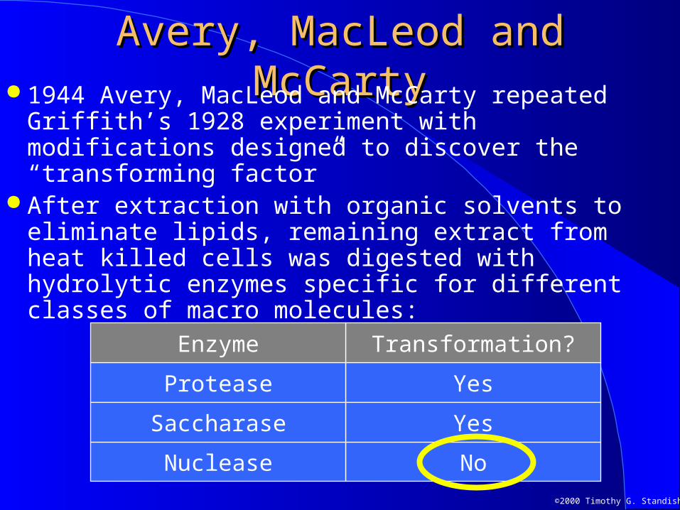
Avery, MacLeod and McCartyAvery, MacLeod and McCarty 1944 Avery, MacLeod and McCarty repeated
Griffith’s 1928 experiment with modifications designed to discover the “transforming factor”
After extraction with organic solvents to eliminate lipids, remaining extract from heat killed cells was digested with hydrolytic enzymes specific for different classes of macro molecules:
NoNuclease
YesProtease
Transformation?Enzyme
YesSaccharase
©2000 Timothy G. Standish
The Hershey-Chase The Hershey-Chase ExperiementExperiement
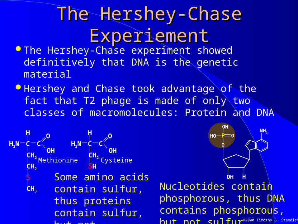
The Hershey-Chase experiment showed definitively that DNA is the genetic material
Hershey and Chase took advantage of the fact that T2 phage is made of only two classes of macromolecules: Protein and DNA
HOH
P
O
OH
HO ONH2
Nucleotides contain phosphorous, thus DNA contains phosphorous, but not sulfur.
H
OH
OH2N CC
CH2
SH
H
OH
OH2N C
CH3
C
CH2
CH2
S Some amino acids contain sulfur, thus proteins contain sulfur, but not phosphorous.
CysteineMethionine
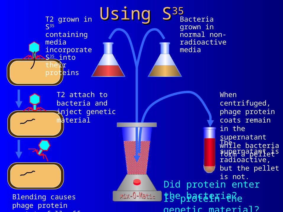
Using SUsing S3535Bacteria grown in normal non-radioactive media
T2 grown in S35 containing media incorporate S35 into their proteins
Blending causes phage protein coat to fall off
T2 attach to bacteria and inject genetic material
Is protein the genetic material?
When centrifuged, phage protein coats remain in the supernatant while bacteria form a pelletThe supernatant is radioactive, but the pellet is not.
Did protein enter the bacteria?
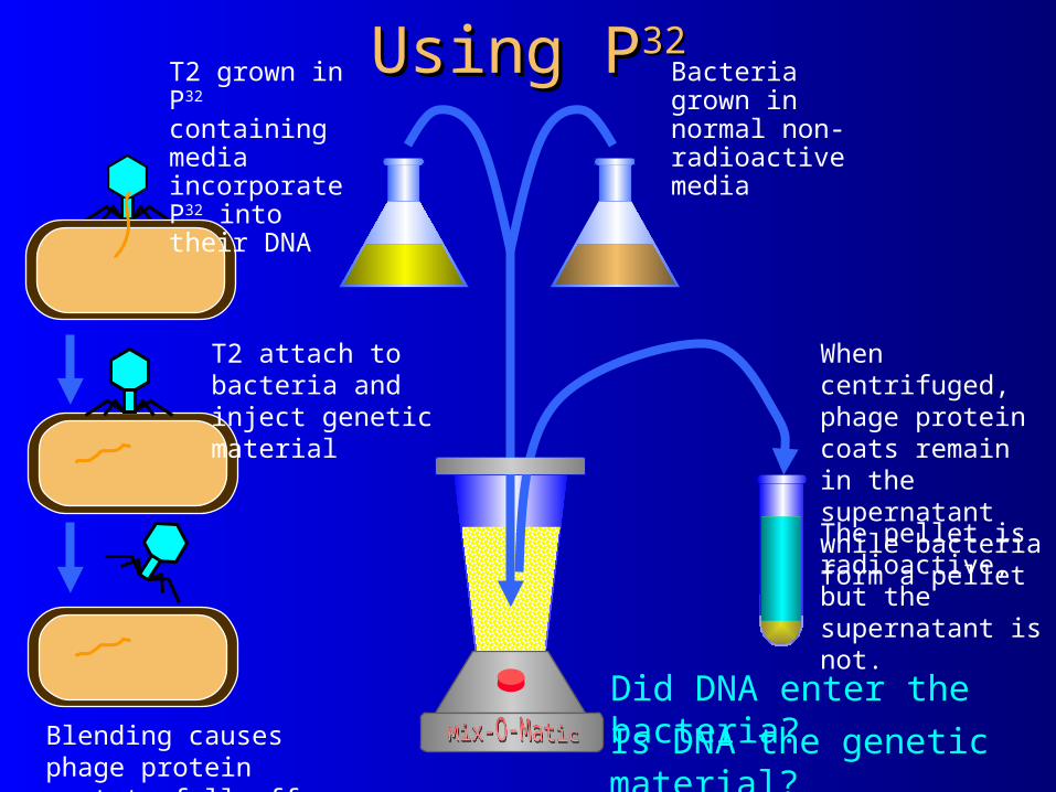
Using PUsing P3232Bacteria grown in normal non-radioactive media
T2 grown in P32 containing media incorporate P32 into their DNA
Blending causes phage protein coat to fall off
T2 attach to bacteria and inject genetic material
Is DNA the genetic material?
When centrifuged, phage protein coats remain in the supernatant while bacteria form a pelletThe pellet is radioactive, but the supernatant is not.
Did DNA enter the bacteria?
OH
OCH2
Sugar
H
HH
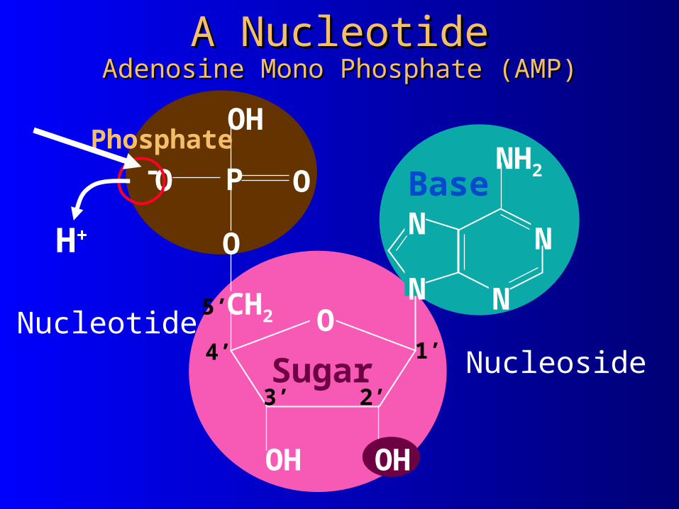
A NucleotideA NucleotideAdenosine Mono Phosphate (AMP)Adenosine Mono Phosphate (AMP)
OH
NH2
N
N N
N
BaseP
O
OH
HO O
Phosphate
2’3’
4’
5’
1’Nucleotide
Nucleoside
H+
-
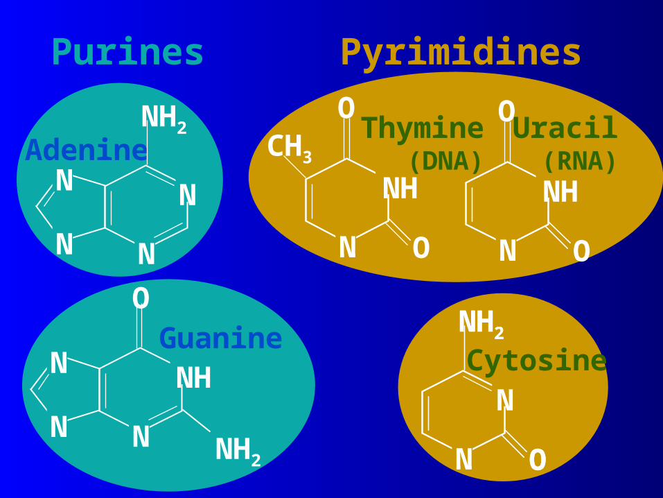
Pyrimidines
NH2
O
N
N NH
N
Guanine
N
N
Adenine
N
N
NH2
N O
NH2
N O
NH2
NCytosine
Purines
Uracil(RNA)CH3
N ON
O
NH
N ON
O
NH
Thymine(DNA)

NO
H
NO
N
NH C
ytosine
H
O
NN
N
N
N
H
H
Guanine -+
+
+
-
-
Base PairingBase PairingGuanine And CytosineGuanine And Cytosine

CH 3
N
O
N
ON
H+
- ThymineN
NN
N
HN H
-
+Adenine
Base PairingBase PairingAdenine And ThymineAdenine And Thymine
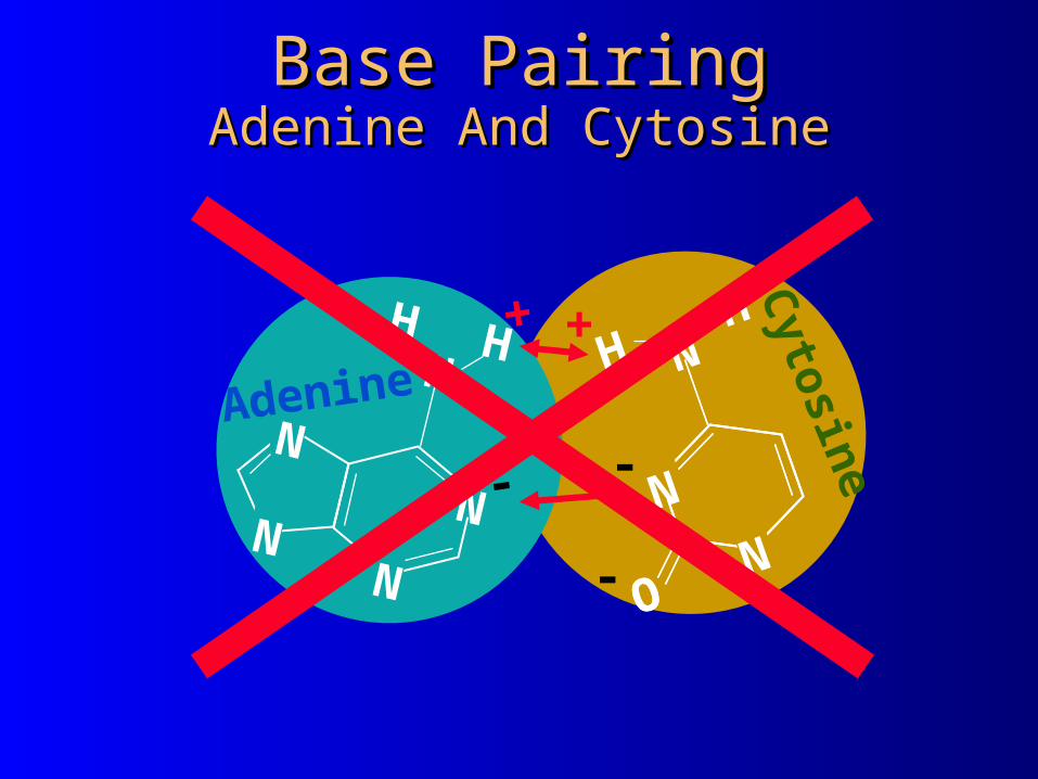
Base PairingBase PairingAdenine And CytosineAdenine And Cytosine
NO
H
NO
N
NH C
ytosine-
+
-
N
NN
N
HN
H
-
+
Adenine

Base PairingBase PairingGuanine And ThymineGuanine And Thymine
CH
3
NO
N
O
NH+
- Thymine
H
O
NN
N
N
N
H
H
Guanine
+
+
-
SU
GA
R-P
HO
SP
HA
TE
BA
CK
BO
NE
H
P
O
HO
O
O
CH2
HOH
P
O
O
HO
O
O
CH2
H
P
O
OH
HO
O
O
CH2
NH2
N
N
N
N
O
O
NH2N
NH
N
N
N O
NH2
N
B A
S E
S
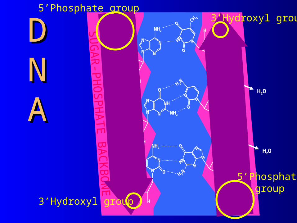
DDNNAA
OH
P
O
HO
O
O
CH2
HO
O
H 2N
NHN N
N H
H
P HO
O
O
CH2
OO
N
O
H 2N
NH
H2O
H OH
P
O
HO
O
O
CH2
CH 3
O
O
HNN
H2O
5’Phosphate group
3’Hydroxyl group
5’Phosphategroup
3’Hydroxyl group
©2000 Timothy G. Standish
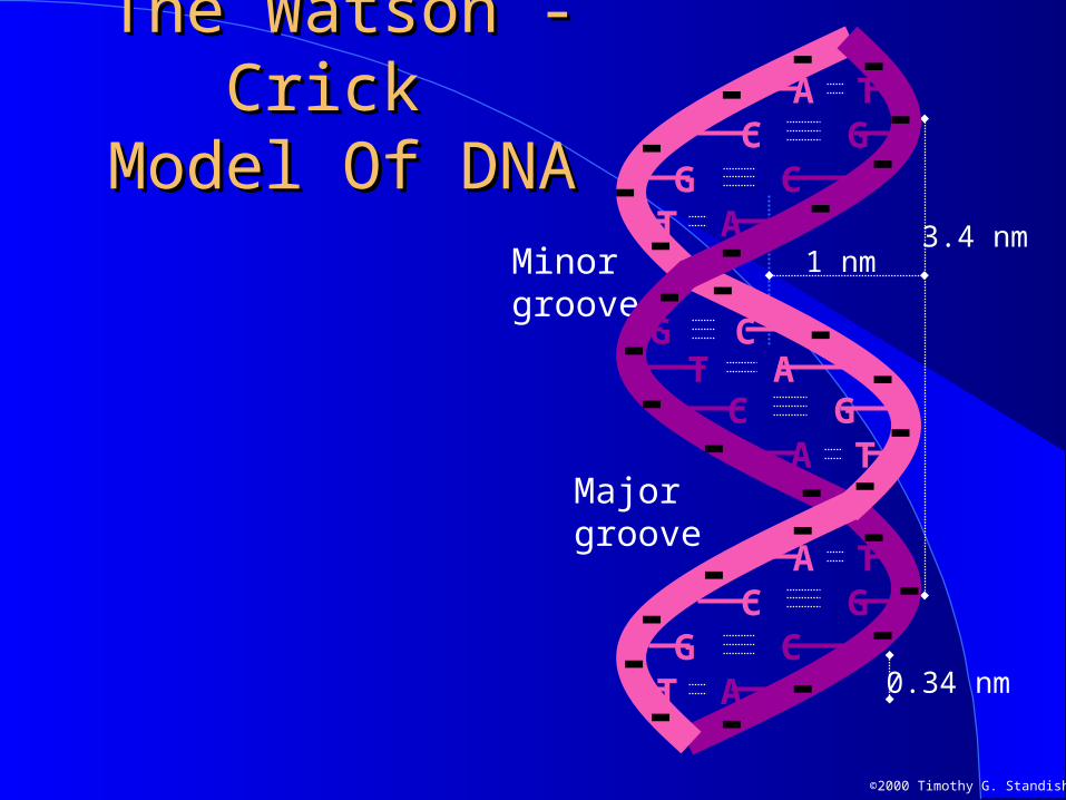
The Watson - Crick The Watson - Crick Model Of DNAModel Of DNA
3.4 nm1 nm
0.34 nm
Majorgroove
Minorgroove
A T
T AG C
C G
C GG C
T A
A T
G CT A
A TC G
--
-
-
---
--
--
--
-
--
--
-
---
--
--
--
-
-

©2000 Timothy G. Standish
Forms of the Double HelixForms of the Double Helix
0.26 nm
2.8 nmMinorgroove
Majorgroove
C GA T
T AG C
C G
G CT A
A T
G CT A
A TC G
A T
G C
1.2 nm
A DNA
1 nm
Majorgroove
Minorgroove
A T
T AG C
C G
C G
G CT A
A T
G CT A
A TC G
0.34 nm
3.9 nm
B DNA
+34.7o Rotation/Bp11 Bp/turn
-30.0o Rotation/Bp12 Bp/turn
+34.6o Rotation/Bp10.4 Bp/turn
C GG C
G CC G
C G
G CG C
G CC G
G CC G
0.57 nm
6.8 nm
0.9 nm
Z DNA
©2000 Timothy G. Standish
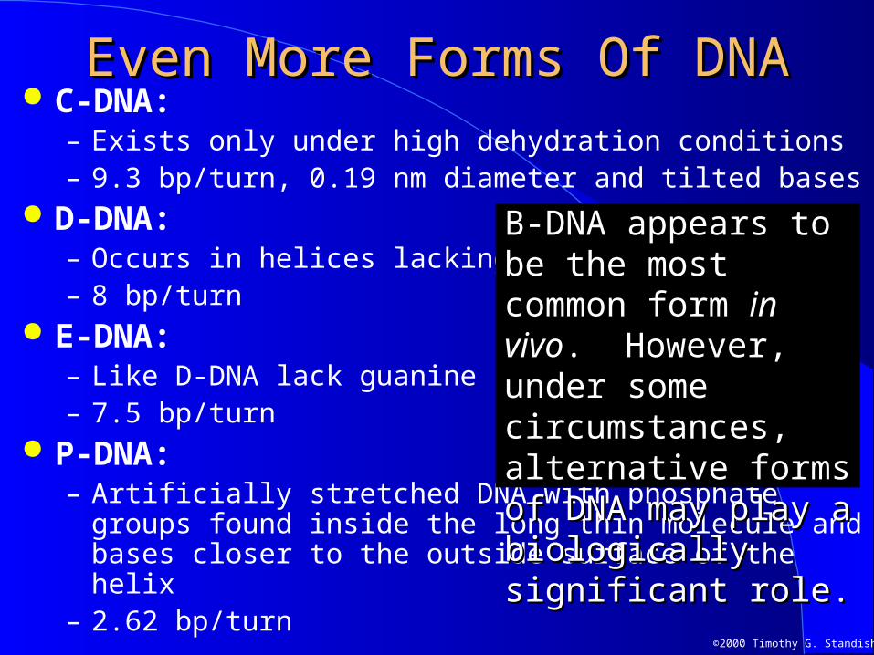
C-DNA:– Exists only under high dehydration conditions– 9.3 bp/turn, 0.19 nm diameter and tilted bases
D-DNA:– Occurs in helices lacking guanine– 8 bp/turn
E-DNA:– Like D-DNA lack guanine– 7.5 bp/turn
P-DNA:– Artificially stretched DNA with phosphate groups found inside
the long thin molecule and bases closer to the outside surface of the helix
– 2.62 bp/turn
Even More Forms Of DNAEven More Forms Of DNA
B-DNA appears to be the B-DNA appears to be the most common form most common form in in vivovivo. However, under . However, under some circumstances, some circumstances, alternative forms of DNA alternative forms of DNA may play a biologically may play a biologically significant role.significant role.

©2000 Timothy G. Standish
Denaturation and RenaturationDenaturation and Renaturation Heating double stranded DNA can overcome the
hydrogen bonds holding it together and cause the strands to separate resulting in denaturation of the DNA
When cooled relatively weak hydrogen bonds between bases can reform and the DNA renatures
TACTCGACATGCTAGCACATGAGCTGTACGATCGTG
Double stranded DNA
TACTCGACATGCTAGCACATGAGCTGTACGATCGTG
Double stranded DNA
Renaturation
TACTCGACATGCTAGCAC
ATGAGCTGTACGATCGTG
Denatured DNA
Denaturat
ion
Single stranded DNA
©2000 Timothy G. Standish
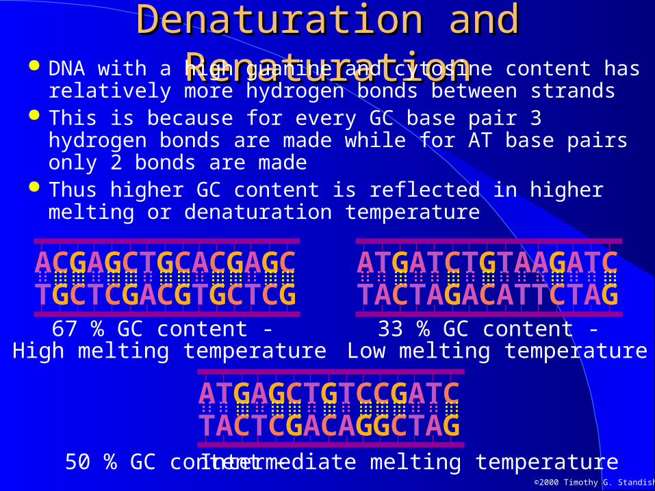
Denaturation and RenaturationDenaturation and Renaturation DNA with a high guanine and cytosine content has
relatively more hydrogen bonds between strands This is because for every GC base pair 3 hydrogen bonds
are made while for AT base pairs only 2 bonds are made Thus higher GC content is reflected in higher melting or
denaturation temperature
Intermediate melting temperature
Low melting temperature High melting temperature67 % GC content -
TGCTCGACGTGCTCGACGAGCTGCACGAGC
33 % GC content -
TACTAGACATTCTAGATGATCTGTAAGATC
TACTCGACAGGCTAGATGAGCTGTCCGATC
50 % GC content -
©2000 Timothy G. Standish
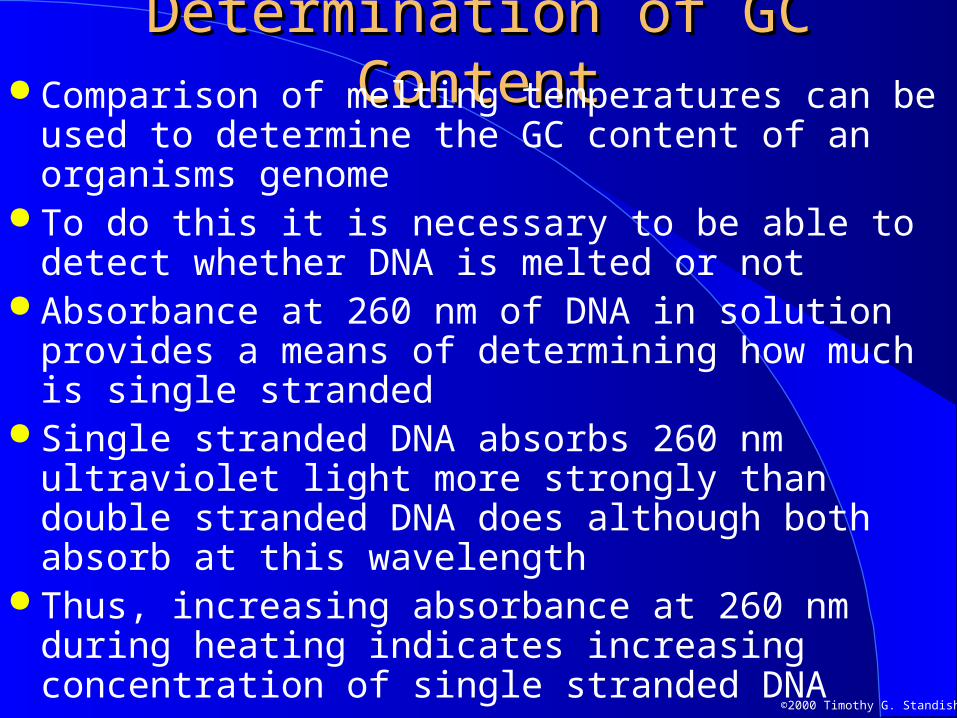
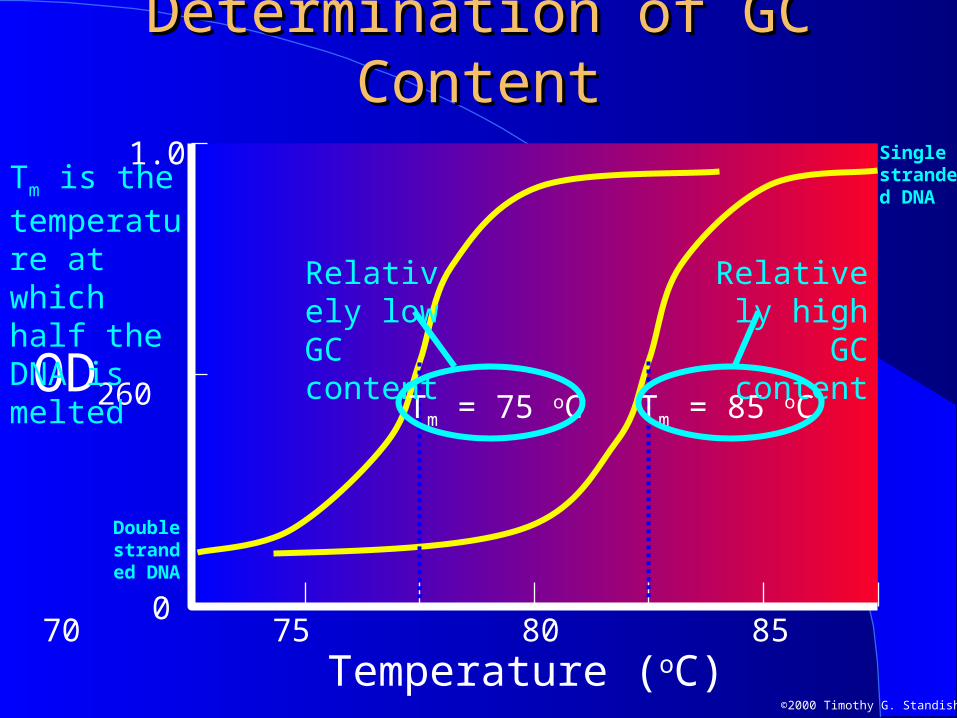
Determination of GC ContentDetermination of GC Content Comparison of melting temperatures can be used to
determine the GC content of an organisms genome To do this it is necessary to be able to detect
whether DNA is melted or not Absorbance at 260 nm of DNA in solution provides
a means of determining how much is single stranded Single stranded DNA absorbs 260 nm ultraviolet
light more strongly than double stranded DNA does although both absorb at this wavelength
Thus, increasing absorbance at 260 nm during heating indicates increasing concentration of single stranded DNA
©2000 Timothy G. Standish
Determination of GC ContentDetermination of GC Content
OD260
0
1.0
65 70 75 80 85 90 95
Temperature (oC)
Tm = 85 oCTm = 75 oC
Double stranded DNA
Single stranded DNA
Relatively low GC content
Relatively high GC content
Tm is the temperature at which half the DNA is melted
©2000 Timothy G. Standish
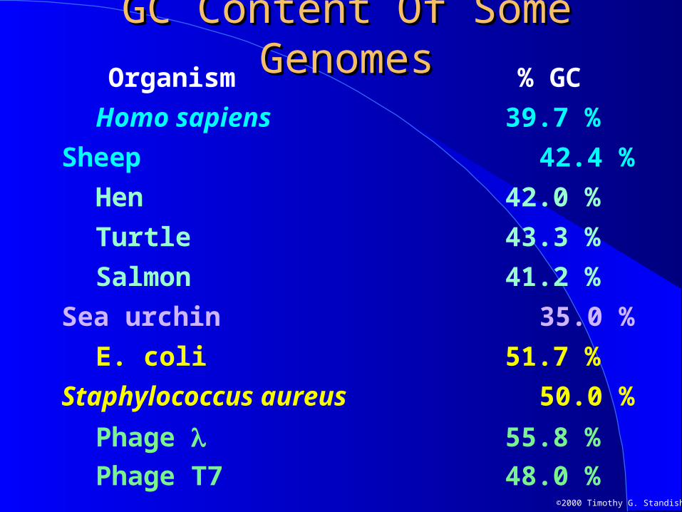
GC Content Of Some GenomesGC Content Of Some Genomes
Phage T7 48.0 %
Organism % GC
Homo sapiens 39.7 %
Sheep 42.4 %
Hen 42.0 %
Turtle 43.3 %
Salmon 41.2 %
Sea urchin 35.0 %
E. coli 51.7 %
Staphylococcus aureus 50.0 %
Phage 55.8 %
©2000 Timothy G. Standish
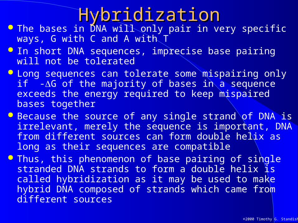
HybridizationHybridization The bases in DNA will only pair in very specific ways, G
with C and A with T In short DNA sequences, imprecise base pairing will not be
tolerated Long sequences can tolerate some mispairing only if -G of
the majority of bases in a sequence exceeds the energy required to keep mispaired bases together
Because the source of any single strand of DNA is irrelevant, merely the sequence is important, DNA from different sources can form double helix as long as their sequences are compatible
Thus, this phenomenon of base pairing of single stranded DNA strands to form a double helix is called hybridization as it may be used to make hybrid DNA composed of strands which came from different sources

©2000 Timothy G. Standish
HybridizationHybridization
DNA from source “Y”
TACTCGACAGGCTAG
CTGATGGTCATGAGCTGTCCGATCGATCAT
DNA from source “X”
TACTCGACAGGCTAG
HybridizationHybridization
©2000 Timothy G. Standish
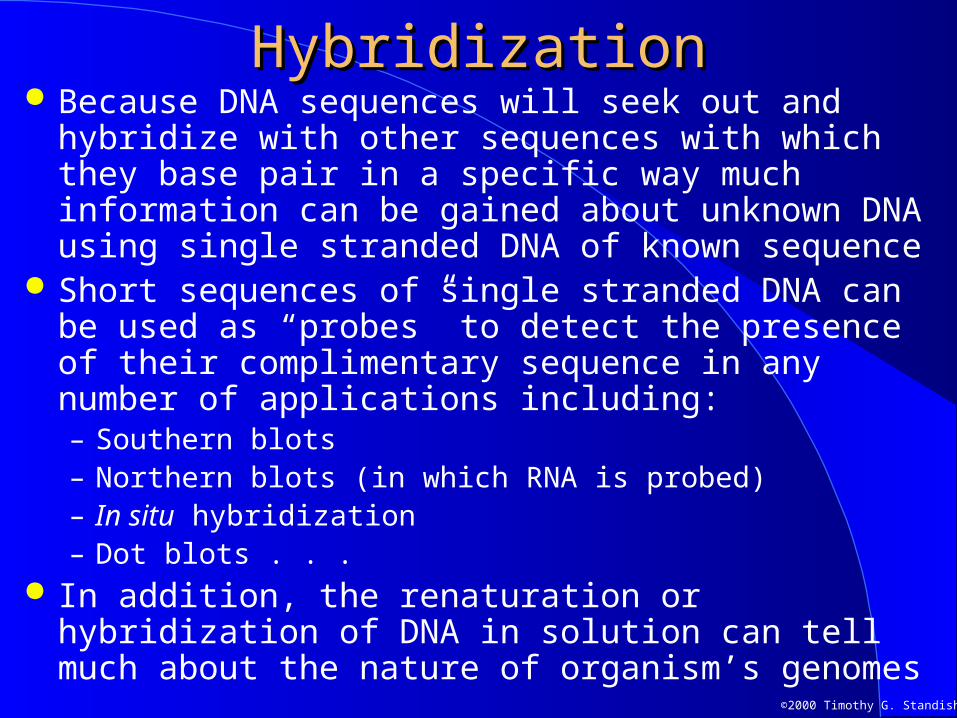
HybridizationHybridization Because DNA sequences will seek out and hybridize with
other sequences with which they base pair in a specific way much information can be gained about unknown DNA using single stranded DNA of known sequence
Short sequences of single stranded DNA can be used as “probes” to detect the presence of their complimentary sequence in any number of applications including:– Southern blots– Northern blots (in which RNA is probed)– In situ hybridization– Dot blots . . .
In addition, the renaturation or hybridization of DNA in solution can tell much about the nature of organism’s genomes
©2000 Timothy G. Standish
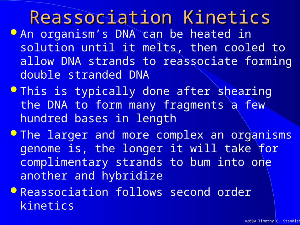
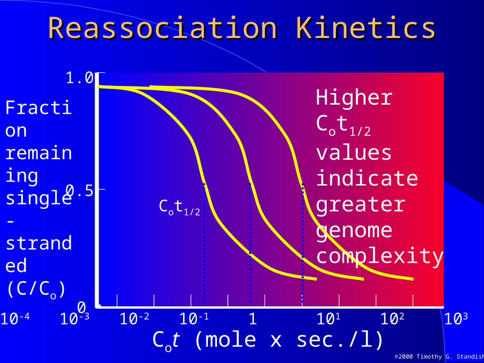
Reassociation KineticsReassociation Kinetics An organism’s DNA can be heated in solution
until it melts, then cooled to allow DNA strands to reassociate forming double stranded DNA
This is typically done after shearing the DNA to form many fragments a few hundred bases in length
The larger and more complex an organisms genome is, the longer it will take for complimentary strands to bum into one another and hybridize
Reassociation follows second order kinetics
©2000 Timothy G. Standish
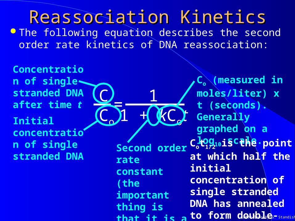
Reassociation KineticsReassociation Kinetics The following equation describes the second order
rate kinetics of DNA reassociation:
11 + kCot
=CCo
Concentration of single stranded DNA after time t
Initial concentration of single stranded DNA
Second order rate constant (the important thing is that it is a constant)
Co (measured in moles/liter) x t (seconds). Generally graphed on a log10 scale.
Cot1/2 is the point at which half the initial concentration of single stranded DNA has annealed to form double-stranded DNA
©2000 Timothy G. Standish
Reassociation KineticsReassociation Kinetics
Fraction remaining single-stranded (C/Co)
0
0.5
10-4 10-3 10-2 10-1 1 101 102 103 104
Cot (mole x sec./l)
1.0Higher Cot1/2 values indicate greater genome complexityCot1/2
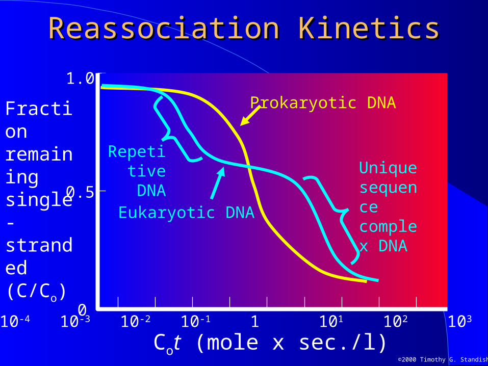
©2000 Timothy G. Standish
Reassociation KineticsReassociation Kinetics
0.5
Fraction remaining single-stranded (C/Co)
010-4 10-3 10-2 10-1 1 101 102 103 104
Cot (mole x sec./l)
1.0
Eukaryotic DNA
Prokaryotic DNA
Repetitive DNA Unique
sequence complex DNA

©2000 Timothy G. Standish
Repetitive DNARepetitive DNAOrganism % Repetitive DNA
Homo sapiens 21 %
Mouse 35 %
Calf 42 %
Drosophila 70 %
Wheat 42 %
Pea 52 %
Maize 60 %
Saccharomycetes cerevisiae 5 %
E. coli 0.3 %

©2000 Timothy G. Standish


