Nephrotic Nephrotic Syndrome Syndrome Nephrotic syndrome(NS) results from increased permeability of GBM to plasma protein. It is characterized by excessive proteinuria, hypo- proteinemia, hypercholesterolemia and edema. 1.Types of nephritic syndrome: (1) Idiopathic nephritic syndrome: Etiology of the disease is unknown, accounting for approxi- mately 90% of nephrosis in childhood. (2) Secondary nephrosis: NS resulted from systemic disease such as anaphylactoid purpura,
Transcript
NephroticNephrotic SyndromeSyndromeNephrotic syndrome(NS) results from
increased permeability of GBM to plasma protein. It is characterized by excessive proteinuria, hypo-proteinemia, hypercholesterolemia and edema.
1.Types of nephritic syndrome:(1) Idiopathic nephritic syndrome: Etiology of thedisease is unknown, accounting for approxi-
mately 90% of nephrosis in childhood.(2) Secondary nephrosis: NS resulted from systemic disease such as anaphylactoid purpura,
systemic lupus erythematosis (SLE), and so on.2.Etiology
The cause of the idiopathic NS remains unknown.Recent 10 years increasing evidence has suggestedthat the syndrome may result from an abnormalityin T-cell lymphocyte function.
3. Pathology(1) Minimal-change disease (78%), the glomeruliappear normal. The epithelial cell foot processesfused.
More than 95% of children with MCD,and better responding to corticosteroid therapy.
(2) Focal sclerosis in glomeruli (6.7%), sclerosis and hyalinosis involving a portion of glom-erular tuft, even only one of the glomeruli, accompanied tubular atrophy. IgM and C3 within sclerotic areas.
(3)Mesangial proliferation: Only mesangialproliferation. Immunoglobulin and comp-lement deposits in the mesangial area.
(4) Membrane nephrosis:GBM thicker,IC deposits.
(5) Membranoproliferative glomerulonephritis:Diffuse proliferation of mesangial cells and
mesangial matrix.Electronic density deposits and C3 deposit in mesangial and GBM.
Massive proteinuria is the most chief characteristics of nephrosis resulting from an increase in glomerular capillary wall permeability to plasma protein. The mechanism may be related to
①Molecular barrier injury :holes on GBM become larger;
②Charge barrier injury: loss of negative
charge(glycoproteins )within the GBM,protein (with negative charges)can pass through the GBM into the urine. If the damage of glomeruli is mild and the permeability is not so high, that onlylow molecular weight protein (albumin, transferrin,ect) can pass through the GBM, which is called highly selective proteinuria.,
If injuries of glomeruli is severe, both small and large proteins(a2-macroglobuline) can all pass through GBM, which is called non-selectiveproteinuria.
In NS protein loss exceeds 50mg/kg.d generally and is composed primarily of albumin.
(2) Hypoproteinemia① Plasma protein is lost by urine; ② Protein catabolism ↑, so total plasma protein concentration↓, especially albumin.
A. Hypoproteinemia stimulates generalized protein synthesis in the liver, including the lipoprotein;B. Lipid catabolism is diminished.
(4) EdemaA. Hypoalbuminemia leads to a decrease
in plasma osmotic pressure, which permits the translation of fluid from intravascularcompartment to interstitial space. B.The intravascular volume ↓ make renal
perfusion pressure ↓ activating rennin-angiotensin-aldosterone system, which stimulates distal tubular reabsorption of sodium↑.
C. Reduced intravascular volume also stimulates the release of antidiuretic hormone, which enhances the reabsorption of water in the collecting duct.
D. Because of plasma osmotic pressure ↓, the sodium and water enter interstitial space,
exacerbating edema, reduced GFR is another cause o f edema.
E. Reduced GFR is another cause o f edema.
5. Clinical ManifestationsEdema around the eyes and in lower
extremities, “pitting” in nature. With time, the edema becomes generalized and body weight increased, developing to ascitesand/or pleural effusions, and declining urine output. Anorexia, abdominal pain, and diarrhea are common.
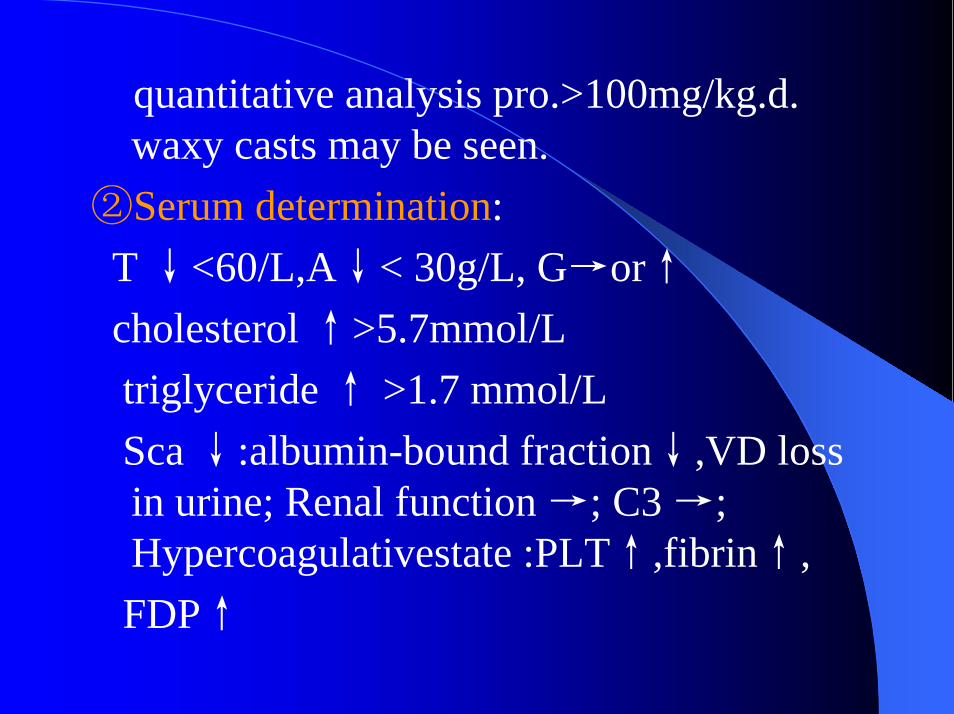
quantitative analysis pro.>100mg/kg.d. waxy casts may be seen.
②Serum determination: T ↓<60/L,A↓< 30g/L, G→or↑cholesterol ↑>5.7mmol/Ltriglyceride ↑ >1.7 mmol/LSca ↓:albumin-bound fraction↓,VD loss in urine; Renal function →; C3 →; Hypercoagulativestate :PLT↑,fibrin↑,FDP↑
7. Diagnosis① Simple nephritic syndrome(SNS): high degree edema, excessive proteinuria, hypoproteinemia, hypercholesterolemia .② Nephritic nephrosis: SNS +hematuriapresent ,hypertention may appear, and renal function and C3 may be reduced. Renal biopsy is recommended to establish a firm diagnosis prior to considering therapy.
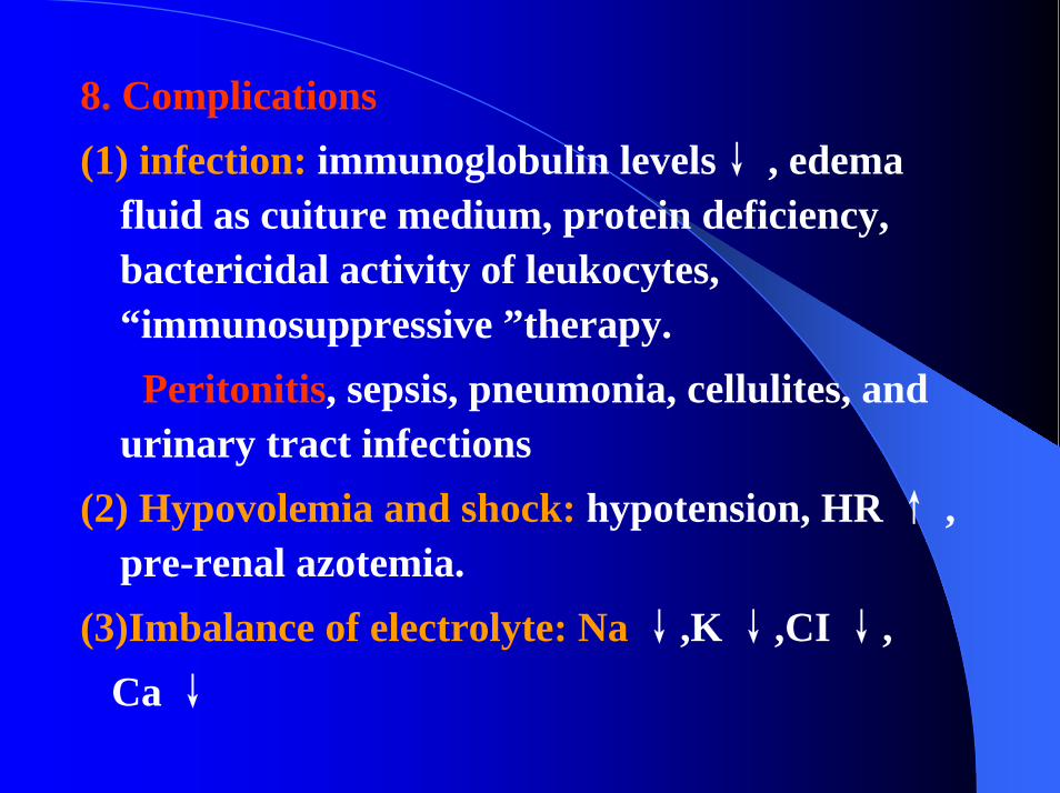
fluid as cuiture medium, protein deficiency, bactericidal activity of leukocytes, “immunosuppressive ”therapy.
Peritonitis, sepsis, pneumonia, cellulites, and urinary tract infections
(2) Hypovolemia and shock: hypotension, HR ↑ , pre-renal azotemia.
(3)Imbalance of electrolyte: Na ↓,K ↓,CI ↓,Ca ↓
(4)Hypercoagulative state and thrombosis:
Certain coagulation factors and inhibitors of fibrinolysis, antithrombin-Ⅲ ↓, platelet aggregation ↑, deficiencies of coagulation factors Ⅸ, ⅪandⅫ, and hypovolemia and hemoconcentration. anticoagulation (heparin drop and persantine ).
(5)ARF
9. Treatment(1) Rest: No restrictions are placed on the activity,but some limitations are advisablebecause of infection.
(2) Diet: Suitable for the normal child. Salt needs to be restricted only during periods of edema. The protein content of diet need not altered.(3) Diuretic therapy:A:Hydrochlorothiazide(HCT), 2 ~ 4 mg/kg.d
B. Furosemide and albumin:hypoalbuminemia patients with severe edema, salt-poor human albumin or human plasma is given first, 0.5g/kg;
If albumin is very lower, 15~ 20 g/L, 1g/kg Albumin is infused slowly over 1 hour.Furosemide given iv after 30 to 60 minutes of equilibration, 1 mg/kg.
C. Diuretics and vasodilator: refractory edema renal ,blood vessel dilator (Dopamin 1 ~ 4 μg/kg.min), and furosemide.
(4) Specific treatmentThe total course into two periods: induced remission stage and strengthened phase.
A. short-term therapy: 8 ~ 12 weeks.B. Medium-long-term therapy: predinisone
(1.5~2mg/kg.d, max 60mg/d.dose ) →Upro (-) 2 weeks,total course < 8 weeks,prednisone 1.5~2.0mg/kg in alternate-day therapy 1ms → decrement of prednison2.5~5.0mg every 2 weeks .
If period of proteinuria disappeare> 4 weeks , total course of prednisone 9 ~ 12 mos
C. Treatment of refractory patientsno responding to hormone and frequent recurrence(≥2/6 mons or 3 /year).
①prednisone with cytotoxic agents;②prednisone used for long term.D. Cytotoxic agent therapy:Side effects
sexual gland damage, bone marrow depression, hemorrhagic cystitis, etc.
E. Response to hormone therapyA. Complete response to hormone therapy (90%
MCD), prednisone 1.5~2.0 mg/kg.d given 8 weeks and proteinuria becomes negative.
B. Partial response to hormone therapy, pro. 1+or 2+ after 8weeks treatment of prednisone.
C. No response to hormone therapy, pro. 3+to 4+ after 8weeks with prednisone