7/30/2019 2009 Aplastic Anaemia British Guid http://slidepdf.com/reader/full/2009-aplastic-anaemia-british-guid 1/28 Guidelines for the diagnosis and management of aplastic anaemia Judith C. W. Marsh, 1 Sarah E. Ball, 2 Jamie Cavenagh, 3 Phil Darbyshire, 4 Inderjeet Dokal, 5 Edward C. Gordon-Smith, 6 Jane Keidan, 7 Andrew Laurie, 8 Anna Martin, 9 Jane Mercieca, 10 Sally B. Killick, 11 Rhona Stewart, 12 John A. L. Yin 13 Writing group: British Committee for Standards in Haematology 1 King’s College Hospital, 2 St Mary’s Hospital, 3 Barts and The London Hospital, London, 4 Birmingham Children’s Hospital, Birmingham, 5 Barts and The London School of Medicine and Dentistry, 6 St George’s Hospital, London, 7 Queen Elizabeth Hospital, King’s Lynn, Norfolk, 8 Ashford Hospital, Middlesex, London, 9 Patient representative, 10 St Helier Hospital, Carshalton, Surrey, 11 Royal Bournemouth Hospital, Dorset, 12 Chestereld Royal Hospital, Derbyshire, and 13 Manchester Royal Inrmary, Manchester, UK. Introduction The guideline group was selected to be representative of UK- based medical experts, experienced district general hospital haematologists and a patient representative. MEDLINE and EMBASE were searched systematically for publications in English from 2004 to 2008 using key word aplastic anaemia. The writing group produced the draft guideline which was subsequently revised by consensus by members of the General Haematology Task Force of the British Committee for Standards in Haematology. The guideline was then reviewed by 59 practising UK haematologists, the BCSH (British Committee for Standards in Haematology) and the British Society for Haematology Committee and comments incorpo- rated where appropriate. Criteria used to quote levels and grades of evidence are as outlined in appendix 3 of the Procedure for Guidelines Commissioned by the BCSH (http:// www.bcshguidelines.com/process1.asp#App3) and given at the end of this Guideline as Appendix I. The objective of this guideline is to provide healthcare professionals with clear guidance on the diagnosis and management of patients with acquired aplastic anaemia. The guidance may not be appropri- ate to patients with inherited aplastic anaemia and in all cases individual patient circumstances may dictate an alternative approach. Because aplastic anaemia is a rare disease, many of the statements and comments are based on review of the literature and expert or consensus opinion rather than on clinical studies or trials. Guidelines update A previous guideline on the diagnosis and management of aplastic anaemia was published in this journal (Marsh et al , 2003). This guideline is an update of the 2003 guideline and is to replace the 2003 guideline (Marsh et al , 2003). Summary of key recommendations • Aplastic anaemia (AA) is a rare but heterogeneous disorder. The majority (70–80%) of these cases are categorised as idiopathic because their primary aetiology is unknown. In a subset of cases, a drug or infection can be identied that precipitates the bone marrow failure/ aplastic anaemia, although it is not clear why only some individuals are susceptible. In approximately 15–20% of patients the disease is constitutional/inherited, where the disease is familial and/or presents with one or more other somatic abnormalities. • Careful history and clinical examination is important to help exclude rarer inherited forms. • A detailed drug and occupational exposure history should always be taken. Any putative drug should be discontin- ued and should not be given again to the patient. Any possible association of aplastic anaemia with drug expo- sure should be reported to the Medicines and Healthcare products Regulatory Agency (MHRA) using the Yellow card Scheme. • All patients presenting with aplastic anaemia should be carefully assessed to: (i) conrm the diagnosis and exclude other possible causes of pancytopenia with hypocellular bone marrow. (ii) classify the disease severity using standard blood and bone marrow criteria. (iii) document the presence of associated paroxysmal nocturnal haemoglobinuria (PNH) and cytogenetic clones. Small PNH clones, in the absence of haemolysis, occur in up to 50% of patients with aplastic anaemia and abnormal cytogenetic clones occur in up to 12% of patients with aplastic anaemia in the absence of myelodysplatic syndrome (MDS). Correspondence: Judith C. W. Marsh, Department of Haematological Medicine, King’s College Hospital, Denmark Hill, London SE5 9RS, UK. E-mail: [email protected]Date for guideline review May 2012 guideline First published online 10 August 2009 ª 2009 Blackwell Publishing Ltd, British Journal of Haematology , 147 , 43–70 doi:10.1111/j.1365-2141.2009.07842.x
Guidelines for the diagnosis and management of aplasticanaemia
Judith C. W. Marsh,1
Sarah E. Ball,2
Jamie Cavenagh,3
Phil Darbyshire,4
Inderjeet Dokal,5
Edward C. Gordon-Smith,6
JaneKeidan, 7 Andrew Laurie, 8 Anna Martin, 9 Jane Mercieca, 10 Sally B. Killick,11 Rhona Stewart, 12 John A. L. Yin13 Writing group:British Committee for Standards in Haematology 1 King’s College Hospital,2 St Mary’s Hospital, 3 Barts and The London Hospital, London, 4 Birmingham Children’s Hospital, Birmingham,5 Barts and The London School of Medicine and Dentistry, 6 St George’s Hospital, London,7 Queen Elizabeth Hospital, King’s Lynn, Norfolk, 8 Ashford Hospital, Middlesex, London,9 Patient representative, 10 St Helier Hospital, Carshalton, Surrey, 11 Royal BournemouthHospital, Dorset, 12 Chestereld Royal Hospital, Derbyshire, and 13 Manchester Royal Inrmary, Manchester, UK.
Introduction
The guideline group was selected to be representative of UK-based medical experts, experienced district general hospitalhaematologists and a patient representative. MEDLINE andEMBASE were searched systematically for publications inEnglish from 2004 to 2008 using key word aplastic anaemia.The writing group produced the draft guideline which wassubsequently revised by consensus by members of the GeneralHaematology Task Force of the British Committee forStandards in Haematology. The guideline was then reviewedby 59 practising UK haematologists, the BCSH (BritishCommittee for Standards in Haematology) and the BritishSociety for Haematology Committee and comments incorpo-rated where appropriate. Criteria used to quote levels and
grades of evidence are as outlined in appendix 3 of theProcedure for Guidelines Commissioned by the BCSH (http://www.bcshguidelines.com/process1.asp#App3) and given at theend of this Guideline as Appendix I. The objective of thisguideline is to provide healthcare professionals with clearguidance on the diagnosis and management of patients withacquired aplastic anaemia. The guidance may not be appropri-ate to patients with inherited aplastic anaemia and in all casesindividual patient circumstances may dictate an alternativeapproach. Because aplastic anaemia is a rare disease, many of the statements and comments are based on review of theliterature and expert or consensus opinion rather than onclinical studies or trials.
Guidelines update
A previous guideline on the diagnosis and management of aplastic anaemia was published in this journal (Marsh et al ,
2003). This guideline is an update of the 2003 guideline and isto replace the 2003 guideline (Marsh et al , 2003).
Summary of key recommendations
• Aplastic anaemia (AA) is a rare but heterogeneousdisorder. The majority (70–80%) of these cases arecategorised as idiopathic because their primary aetiology is unknown. In a subset of cases, a drug or infection canbe identied that precipitates the bone marrow failure/aplastic anaemia, although it is not clear why only someindividuals are susceptible. In approximately 15–20% of patients the disease is constitutional/inherited, where thedisease is familial and/or presents with one or more other
somatic abnormalities.• Careful history and clinical examination is important tohelp exclude rarer inherited forms.
• A detailed drug and occupational exposure history shouldalways be taken. Any putative drug should be discontin-ued and should not be given again to the patient. Any possible association of aplastic anaemia with drug expo-sure should be reported to the Medicines and Healthcareproducts Regulatory Agency (MHRA) using the Yellow card Scheme.
• All patients presenting with aplastic anaemia should becarefully assessed to:
(i) conrm the diagnosis and exclude other possible causesof pancytopenia with hypocellular bone marrow.
(ii) classify the disease severity using standard blood andbone marrow criteria.
(iii) document the presence of associated paroxysmalnocturnal haemoglobinuria (PNH) and cytogeneticclones. Small PNH clones, in the absence of haemolysis,occur in up to 50% of patients with aplastic anaemiaand abnormal cytogenetic clones occur in up to 12% of patients with aplastic anaemia in the absence of myelodysplatic syndrome (MDS).
Correspondence: Judith C. W. Marsh, Department of HaematologicalMedicine, King’s College Hospital, Denmark Hill, London SE5 9RS,UK. E-mail: [email protected] Date for guideline review May 2012
guideline
First published online 10 August 2009ª 2009 Blackwell Publishing Ltd, British Journal of Haematology , 147 , 43–70 doi:10.1111/j.1365-2141.2009.07842.x
(iv) exclude a possible late onset inherited bone marrow failure disorder.
• A multidisciplinary team (MDT) approach to the assess-ment and management of newly presenting patients isrecommended. A specialist centre with expertise inaplastic anaemia should be contacted soon after presen-tation to discuss a management plan for the patient.
• Best supportive care(i) Prophylactic platelet transfusions should be given when
the platelet count is <10 · 109 /l (or <20 · 109 /l in thepresence of fever).
(ii) There is no evidence to support the practice of giving irradiated blood components except for patients whoare undergoing bone marrow transplantation (BMT).We would recommend empirically that this practice isextended to patients receiving immunosuppressivetherapy.
(iii) Transfusion of irradiated granulocyte transfusions may be considered in patients with life-threatening
neutropenic sepsis.(iv) The routine use of recombinant human erythropoietin
(rHuEpo) in aplastic anaemia is not recommended. Ashort course of granulocyte colony-stimulating factor(G-CSF) may be considered for severe systemic infec-tion that is not responding to intravenous antibioticsand anti-fungal drugs, but should be discontinued after1 week if there is no increase in the neutrophil count.
(v) Prophylactic antibiotic and antifungal drugs should begiven to patients with neutrophil count <0 Æ5 · 109 /l.Intravenous amphotericin should be introduced intothe febrile neutropenia regimen early if fevers persistdespite broad spectrum antibiotics.
(vi) Iron chelation therapy should be considered when theserum ferritin is >1000 l g/l.
• Denitive treatment(1) Infection or uncontrolled bleeding should be treated
rst before giving immunosuppressive therapy. Thisalso applies to patients scheduled for BMT, although itmay sometimes be necessary to proceed straight toBMT in the presence of severe infection as a BMT may offer the best chance of early neutrophil recovery.
(2) Haemopoietic growth factors such as rHuEpo or G-CSFshould not be used on their own in newly diagnosedpatients in an attempt to ‘treat’ the aplastic anaemia.
(3) Prednisolone should not be used to treat patients withaplastic anaemia because it is ineffective and encour-ages bacterial and fungal infection.
(4) Allogeneic BMT from a human leucocyte antigen(HLA)-identical sibling donor is the initial treatmentof choice for newly diagnosed patients if they havesevere or very severe aplastic anaemia, are <40 yearsold and have an HLA-compatible sibling donor. Thereis no indication for using irradiation-based condition-ing regimens for patients undergoing HLA-identicalsibling BMT for aplastic anaemia. The recommended
source of stem cells for transplantation in aplasticanaemia is bone marrow.
(5) Immunosuppressive therapy is recommended for (i)patients with non-severe aplastic anaemia who aretransfusion dependent (ii) patients with severe or very severe disease who are >40 years old and (iii) youngerpatients with severe or very severe disease who do nothave an HLA-identical sibling donor. The standardimmunosuppressive regimen is a combination of antithymocyte globulin (ATG) and ciclosporin. ATGmust only be given as an in-patient. Ciclosporin shouldbe continued for at least 12 months after achieving maximal haematological response, followed by a very slow tapering, to reduce the risk of relapse. The routineuse of long term G-CSF, or other haemopoietic growthfactors, after ATG and ciclosporin, is not recommendedoutside the setting of prospective clinical trials.
(6) Matched unrelated donor (MUD) BMT may be consid-ered when a patient has severe aplastic anaemia, has no
matched sibling donor but a matched unrelated donor,is <50 years old (or 50–60 years old with good perfor-mance status), and has failed at least one course of ATGand ciclosporin. The optimal conditioning regimen forMUD BMT is uncertain, but currently a udarabine,non-irradiation-based regimen is favoured for youngerpatients.
• There is a high risk (around 33%) of relapse of aplasticanaemia in pregnancy. Supportive care is the mainstay of treatment in pregnancy and the platelet count should bemaintained >20 · 109 /l, if possible. It is safe to useciclosporin in pregnancy.
1. Denition and clinical presentation
Aplastic anaemia is dened as pancytopenia with a hypo-cellular bone marrow in the absence of an abnormal inltrateand with no increase in reticulin. For a comprehensive updateon the pathophysiology, the reader is directed to a recentreview (Young et al , 2006). These guidelines will focusspecically on idiosyncratic acquired aplastic anaemia, andwill not refer to the inevitable and predictable aplasia thatoccurs after chemotherapy and/or radiotherapy. The incidenceof acquired aplastic anaemia in Europe and North America isaround 2 per million population per year (Issaragrisil et al ,2006; Montane et al , 2008). The incidence is 2–3 times higherin East Asia. There is a biphasic age distribution with peaksfrom 10 to 25 years and >60 years. There is no signicantdifference in incidence between males and females (Heimpel,2000). Congenital aplastic anaemia is very rare, the commonesttype being Fanconi anaemia, which is inherited as anautosomal recessive disorder in most cases.
Patients with aplastic anaemia most commonly present withsymptoms of anaemia and skin or mucosal haemorrhage orvisual disturbance due to retinal haemorrhage. Infection is a
Guideline
44 ª 2009 Blackwell Publishing Ltd, British Journal of Haematology , 147 , 43–70
less common presentation. There is no lymphadenopathy orhepatosplenomegaly (in the absence of infection) and thesendings strongly suggest another diagnosis (Gordon-Smith,1991). In children and young adults, the ndings of shortstature, cafe au lait spots and skeletal anomalies should alertthe clinician to the possibility of a congenital form of aplasticanaemia, Fanconi anaemia, although Fanconi anaemia cansometimes present in the absence of overt clinical signs.Patients with Fanconi anaemia most commonly presentbetween the ages of 3 and 14 years but can occasionally present later in their 30s [up to 32 years in males and 48 yearsin females reported by Young & Alter, (1994)]. The ndings of leucoplakia, nail dystrophy and pigmentation of the skin arecharacteristic of another inherited form of aplastic anaemia,dyskeratosis congenita, with a median age at presentation of 7 years (range 6 months to 26 years) (Dokal, 2000; Walne & Dokal, 2009). Some affected patients may have none of theseclinical features and the diagnosis is made later after failure torespond to immunosuppressive therapy (Vulliamy & Dokal,
2006). A preceding history of jaundice, usually 2–3 monthsbefore, may indicate a post-hepatitic aplastic anaemia(Gordon-Smith, 1991; Young & Alter, 1994).
Many drugs and chemicals have been implicated in theaetiology of aplastic anaemia, but for only very few is therereasonable evidence for an association from case controlstudies, and even then it is usually impossible to provecausality (Baumelou et al , 1993; Young & Alter, 1994; Heim-pel, 1996; Kauffmann et al , 1996; Issaragrissil et al , 1997), (seeTable I). A careful drug history should be obtained, detailingall drug exposures for a period beginning 6 months and ending1 month prior to presentation (Heimpel, 1996; Kauffmannet al , 1996). If at presentation the patient is taking several
drugs which may have been implicated in aplastic anaemia,even if the evidence is based on case report(s) alone, then allthe putative drugs should be discontinued and the patientshould not be re-challenged with the drugs at a later stage afterrecovery of the blood counts. The MHRA should be informedusing the Yellow Card Scheme on every occasion that a patientpresents with aplastic anaemia where there is a possible drugassociation (website: http://www.yellowcard.gov.uk).
Similarly, a careful occupational history of the patient may reveal exposure to chemicals or pesticides that have beenassociated with aplastic anaemia, as summarised in Table II.
Recommendations
(i) Aplastic anaemia is a rare disorder. Most cases areidiopathic, but careful history and clinical examinationis important to identify rarer inherited forms.
(ii) Although most cases of aplastic anaemia are idiopathic,a careful drug and occupational exposure history
should be taken.(iii) Any putative drug should be discontinued and should
not be given again to the patient. Any possible associ-ation of aplastic anaemia with drug exposure should bereported to the MHRA using the Yellow card Scheme.
2. Investigations required for diagnosis
The following investigations are required to (i) conrm thediagnosis (ii) exclude other possible causes of pancytopeniawith a hypocellular bone marrow (iii) exclude inheritedaplastic anaemia (iv) screen for an underlying cause of aplastic
anaemia and (v) document or exclude a co-existing abnormalcytogenetic clone or a PNH clone. See Table III for a summary of investigations required for the diagnosis of aplastic anaemia.
2.1. Full blood count, reticulocyte count, blood lm and % HbF
The full blood count (FBC) typically shows pancytopeniaalthough usually the lymphocyte count is preserved. In mostcases thehaemoglobinlevel,neutrophiland plateletcounts arealluniformly depressed, but in the early stages isolated cytopenia,particularly thrombocytopenia, may occur. Anaemia is accom-
panied by reticulocytopenia, and macrocytosis is commonly noted. Careful examination of the blood lm is essential toexclude the presence of dysplastic neutrophils and abnormalplatelets, blasts and other abnormal cells, such as hairy cells (asseen in hairy cell leukaemia). The monocyte count may bedepressed but theabsenceofmonocytesshouldalert thecliniciantoapossiblediagnosisofhairycellleukaemia.Inaplasticanaemia,anisopoikilocytosis is common and neutrophils may show toxicgranulation. Platelets arereducedin numberand mostlyof smallsize. Fetal haemoglobin (HbF) should be measured pre-transfu-sion in children as this is an important prognostic factor in
Table I. Currently licenced drugs which have been reported as a rareassociation with aplastic anaemia. Evidence based on case reports oruncontrolled series (Young & Alter, 1994) or case control studies(Baumelou et al , 1993; Issaragrisil et al , 2006; Issaragrissil et al , 1997;Kauffmann et al , 1996).
*No association with chloramphenicol tablets was observed in recentstudyfrom Thailand (Issaragrisil et al , 2006).There isno evidencefor anassociation between chloramphenicol eye drops and aplastic anaemia(Gordon-Smith et al , 1995; Lancaster et al , 1998; Wilholm et al , 1998).
More likely to cause neutropenia.àFrom epidemiological study in Thailand (Issaragrisil et al , 2006).
Guideline
ª 2009 Blackwell Publishing Ltd, British Journal of Haematology , 147 , 43–70 45
paediatric myelodysplastic syndrome (MDS) which may featurein the differential diagnosis of pancytopenia in children.
2.2. Bone marrow examination
Both a bone marrow aspirate and trephine biopsy are required.Bone marrow aspiration and biopsy may be performedin patients with severe thrombocytopenia without platelet
support, providing that adequate surface pressure is applied(Kelsey et al , 2003).Fragments areusually readily obtained fromthe aspirate. Difculty obtaining fragments should raise thesuspicion of a diagnosis other than aplastic anaemia. Thefragments and trails are hypocellular with prominent fat spacesand variable amounts of residual haemopoietic cells. Erythro-poiesis is reduced or absent, dyserythropoiesis is very commonand often marked, so this alone should not be used to make adiagnosis of MDS. Megakaryocytes and granulocytic cells arereduced or absent; dysplastic megakaryocytes and granulocyticcells are not seen in aplastic anaemia. Lymphocytes, macro-
phages, plasma cells and mast cells appear prominent. In theearly stages of the disease, one may also see prominenthaemophagocytosis by macrophages, as well as backgroundeosinophilic staining representing interstitial oedema. A tre-phine is crucial to assess overall cellularity, to assess themorphology of residual haemopoietic cells and to exclude anabnormal inltrate. In most cases the trephine is hypocellularthroughout but sometimes it is patchy, with hypocellular andcellular areas. Thus, a good quality trephine of at least 2 cm isessential. A ‘hot spot’ in a patchy area may explain why
sometimes theaspirate is normocellular.Care shouldbe taken toavoid tangential biopsies as subcortical marrow is normally ‘hypocellular’. Focal hyperplasia of erythroid or granulocyticcells at a similar stage of maturation may be observed.Sometimes lymphoid aggregates occur, particularly in the acutephase of the disease or when the aplastic anaemia is associatedwithsystemicautoimmune disease, suchas rheumatoid arthritisor systemic lupus erythematosus. The reticulin is not increasedand no abnormal cells are seen. Increased blasts are not seen inaplastic anaemia, and their presence either indicates a hypocel-lularMDSor evolution to leukaemia(Marin,2000; Tichelli et al ,1992; Bennett & Orazi, 2009).
2.3. Denition of disease severity based on the FBC and bone marrow ndings
To dene aplastic anaemia there must be at least two of thefollowing (i) haemoglobin <100 g/l (ii) platelet count<50 · 109 /l (iii) neutrophil count <1 Æ5 · 109 /l (InternationalAgranulocytosis and Aplastic Anaemia Study Group, 1987).The severity of the disease is graded according to the bloodcount parameters and bone marrow ndings as summarised in
Table II. Occupational and environmental exposures as potential aetiological agents in aplastic anaemia.
Benzene and other solvents (evidence based on large industrial studies (Yin et al , 1987; Smith, 1996; Yin et al , 1996; Issaragrisil et al , 2006)Agricultural pesticides: Organochlorines e.g. Lindane, Organophosphates, Pentachlorophenol [Muir et al , 2003 (case control study), Fleming &
Timmeny, 1993; Roberts, 1997 (literature reviews of case reports)], DDT and Carbamates (Issaragrisil et al , 2006)Cutting oils and lubricating agents (Muir et al , 2003)Non-bottled water, non-medical needle injury, farmers exposed to ducks and geese, animal fertiliser (Issaragrisil et al , 2006)Recreational drugs: methylenedioxy-methamphetamine, MDMA, Ecstasy, [evidence based on case reports, (Marsh et al , 1994b; Clark & Butt, 1997)]
Table III. Summary of investigations required for the diagnosis of aplastic anaemia.
1. FBC and reticulocyte count2. Blood lm examination3. HbF% in children4. Bone marrow aspirate and trephine biopsy, including cytogenetics5. Peripheral blood chromosomal breakage analysis to exclude
Fanconi anaemia if <50 years6. Flow cytometry for GPI-anchored proteins (see note below
concerning Ham test)*
7. Urine haemosiderin if Ham test positive or GPI-anchored proteindeciency 8. Vitamin B12 and folate9. Liver function tests
10. Viral studies: Hepatits A, B and C, EBV, HIV (CMV, see page 5)11. Anti-nuclear antibody and anti-dsDNA12. Chest X-ray 13. Abdominal ultrasound scan and echocardiogram14. Peripheral blood gene mutation analysis for dyskeratosis congenita
DKC1, TERC , ?TERT ) if clinical features or lack of response toimmunosuppressive therapy
FBC, full blood count; HbF, fetal haemoglobin; GPI, glycerophos-phatidylinositol; EBV, Epstin–Barr virus; HIV, human immunode-
ciency virus; CMV, cytomegalovirus.*The Ham test and sucrose lysis test have been abandoned in mostcentres as diagnostic tests for PNH as they are both less sensitive andless quantitative than ow cytometry (Parker et al , 2005).
Table IV. Denition of severity of aplastic anaemia.
Severe AA(Camitta et al , 1975)
BM cellularity <25%, or 25–50%with <30% residual haemopoietic cells*
2/3 of the following:Neutrophil count <0 Æ5 · 109 /lPlatelet count <20 · 109 /lReticulocyte count <20 · 109 /l
Very severe AA(Bacigalupo et al , 1988)
As for severe AA but neutrophils<0Æ2 · 109 /l
Non-severe AA Patients not fullling the criteria forsevere or very severe aplastic anaemia
*Cellularity should be determined by comparison with normal controls(Tuzuner & Bennett, 1994).
Guideline
46 ª 2009 Blackwell Publishing Ltd, British Journal of Haematology , 147 , 43–70
Table IV (Camitta et al , 1975; Bacigalupo et al , 1988). How-ever, because of routine and more accurate automatedreticulocyte counting, this will over-estimate the level of reticulocyte count used in the historical Camitta criteria(Camitta et al , 1975) for dening disease severity. The assess-ment of disease severity is important in treatment decisions buthas less prognostic signicance today in terms of correlationwith response to ATG treatment (Scheinberg et al , 2009).Patients with bi-or tri-lineage cytopenias that are less severethan this are not classied as aplastic anaemia. However, they should have their blood counts monitored to determinewhether they will develop aplastic anaemia with time.
2.4. Liver function tests and viral studies
Liver function tests should be performed to detect antecedenthepatitis, but in post-hepatitic aplastic anaemia the serology ismost oftennegativefor allthe known hepatitis viruses. The onsetof aplastic anaemia occurs 2–3 months after an acute episode of
hepatitis and is more common in young males (Brown et al ,1997). Blood should be tested for hepatitis A antibody, hepatitisB surface antigen, hepatitis C antibody and Epstein–Barr virus(EBV). Cytomegalovirus (CMV) and other viral serologyshouldbeassessedif BMTis beingconsidered.Parvoviruscauses redcellaplasia but not aplastic anaemia. Human immunodeciency virus (HIV) is not a recognised cause of aplastic anaemia, but itcan cause isolated cytopenias. We would recommend that priorto a diagnosis of aplastic anaemia, appropriate investigations toexclude alternative aetiologies of cytopenias (B12, red cell folateand HIV) should be performed.
2.5. Vitamin B12 and folate levelsVitamin B12 and folate levels should be measured to excludemegaloblastic anaemia which, when severe, can present withpancytopenia. If a deciency of B12 or folate is documented,this should be corrected before a nal diagnosis of aplasticanaemia is conrmed. Bone marrow aplasia due to vitamindeciency is exceedingly rare.
2.6. Autoantibody screen
The occurrence of pancytopenia in systemic lupus erythe-matosus may (i) be autoimmune in nature occurring with a
cellular bone marrow or (ii) be associated with myelobrosisor rarely (iii) occur with a hypocellular bone marrow. Bloodshould be tested for anti-nuclear antibody and anti-DNAantibody in all patients presenting with aplastic anaemia.
2.7. Tests to detect a PNH clone
Paroxysmal nocturnal haemoglobinuria should be excluded by performing ow cytometry (Dacie & Lewis, 2001; Parker et al ,2005). The Ham test and sucrose lysis test have been abandonedby most centres as diagnostic tests for PNH. Analysis of
glycosylphosphatidylinositol (GPI)-anchored proteins, such asCD55 and CD59 by ow cytometry, is a sensitive andquantitative test for PNH enabling the detection of smallPNH clones which occur in up to 50% of patients with aplasticanaemia, the proportion depending on the sensitivity of theow cytometric analysis used (Dunn et al , 1999; Socie et al ,2000; Sugimori et al , 2005). Such small clones are most easily identied in the neutrophil and monocyte lineages in aplasticanaemia and will be detected by ow cytometry and not by theHam test. If the patient has had a recent blood transfusion, theHamtest maybe negative whereas a population of GPI-decientred cells may still be detected by ow cytometry. However, theclinical signicance of a small PNH clone in aplastic anaemia asdetected by ow cytometry remains uncertain. Such clones canremain stable, diminish in size, disappear or increase. What isclinically important is the presence of a signicant PNH clonewith clinical or laboratory evidence of haemolysis. Urine shouldbe examined for haemosiderin to exclude intravascular haem-olysis which is a constant feature of haemolytic PNH. Evidence
of haemolysis associated with PNH should be quantied withthe reticulocyte count, serum bilirubin, serum transaminasesand lactate dehydrogenase (LDH).
2.8. Cytogenetic investigations
Cytogenetic analysis of the bone marrow should be attemptedalthough this may be difcult in a very hypocellular bonemarrow and often insufcient metaphases are obtained. In thissituation, one should consider uorescence in situ hybridization(FISH) analysis for chromosomes 5 and 7 in particular. It waspreviously assumed that the presence of an abnormal cyto-genetic clone indicated a diagnosis of MDS and not aplasticanaemia, but it is now evident that abnormal cytogenetic clonesmay be present in up to 12% of patients with otherwise typicalaplastic anaemia at diagnosis (Appelbaum et al , 1989; Tichelliet al , 1996; Gupta et al , 2006). The presence of abnormalcytogenetics at presentation in children, especially monosomy7,should alert to the likelihood of MDS. Abnormal cytogeneticclonesmay also arise duringthe course of thedisease(Socie et al ,2000). The management of a patient with aplastic anaemia whohas an abnormal cytogenetic clone is discussed in Section 9 .
2.9. Screen for inherited disorders
Peripheral blood lymphocytes should be tested for spontaneousand diepoxybutane (DEB) or mitomycin C (MMC)-inducedchromosomal breakage to identify or exclude Fanconi anaemia.This should be performed in all patients who are BMTcandidates. Siblings of Fanconi anaemia patients should alsobe screened. For all other patients, it is difcult to set an upperage limit for Fanconi anaemia screening because the age atdiagnosis may sometimes occur in the fourth decade, and rarely in the fth decade, of life (Alter, 2007). Dyskeratosis congenitamay be excluded by identifying a known mutation but there areprobably many mutations yet to be identied (Vulliamy et al ,
Guideline
ª 2009 Blackwell Publishing Ltd, British Journal of Haematology , 147 , 43–70 47
2005; Walne & Dokal, 2009). Along with measuring telo-mere lengths, this is not currently available as a routine clinicalservice.
2.10. Radiological investigations
• A chest X-ray is useful at presentation to exclude infectionand for comparison with subsequent lms.
• Routine X-rays of the radii are no longer indicated as all young patients should have peripheral blood chromosomesanalysed to exclude a diagnosis of Fanconi anaemia.
• Abdominal ultrasound: the ndings of an enlarged spleenand/or enlarged lymph nodes raise the possibility of amalignant haematological disorder as the cause of thepancytopenia. In younger patients, abnormal or anato-mically displaced kidneys are features of Fanconi anaemia.
2.11. Differential diagnosis of pancytopenia and a
hypocellular bone marrow The above investigations should exclude causes of a hypo-cellular bone marrow with pancytopenia other than aplasticanaemia. These include:
• Hypocellular MDS/acute myeloid leukaemia (AML) cansometimes be difcult to distinguish from aplastic anaemia.The following features of MDS are not found in aplasticanaemia: dysplastic cells of the granulocytic and megakary-ocytic lineages, blasts in the blood or marrow (Tuzuner et al ,1995; Jaffe et al , 2001; Bennett & Orazi, 2009). In trephinespecimens, increases in reticulin associated with residualareas of haemopoiesis suggest hypocellular MDS rather thanaplastic anaemia. The presence of abnormal localisation of immature precursors (ALIPs) is difcult to interpret in thiscontext because small collections of immature granulocyticcells may be seen in the bone marrow in aplastic anaemiawhen regeneration occurs. As discussed previously, dysery-thropoiesis is very common in aplastic anaemia.
• Hypocellular acute lymphoblastic leukaemia (ALL) occursin 1–2% of cases of childhood ALL. Overt ALL usually develops within 3–9 months of the apparent bone marrow failure. In contrast to aplastic anaemia, the neutropenia isusually more pronounced than the thrombocytopenia andsometimes there is an increase in reticulin within the
hypocellular bone marrow (Chessells, 2001). Immuno-phenotyping may help conrm the diagnosis. Treatmentshould not be deferred in severe aplastic anaemia inchildren just in case they turn out to have ALL. For allnew paediatric cases of aplastic anaemia, a national centralmorphology review is planned under the aegis of theMedical Research Council Childhood Leukaemia WorkingParty Subgroup for rare haematological diseases.
• Hairy cell leukaemia classically presents with pancytopeniabut the accompanying monocytopenia is a constant featureof this disorder. It is usually difcult or impossible to
aspirate on bone marrow fragments. In addition to thetypical interstitial inltrate of hairy cells with their charac-teristic ‘fried egg’ appearance in the bone marrow trephine,there is always increased reticulin. Immunophenotypingreveals CD20+ , CD11c+ , CD25+ , FMC7+ , CD103+ tumourcells that are typically CD5
), CD10
)and CD23
). Although
splenomegaly is a common nding in hairy cell leukaemia,it may be absent in 30–40% of cases.
• Lymphomas, either Hodgkin lymphoma or non-Hodgkinlymphoma and myelobrosis may sometimes present withpancytopenia and a hypocellular bone marrow. The bonemarrow biopsy should be examined very carefully for foci of lymphoma cells or brosis which may be seen in only asmall part of the trephine. Since lymphocytes are oftenprominent in aplastic anaemia, immunophenotypingshould be performed. Myelobrosis is usually accompaniedby splenomegaly and the absence of an enlarged spleen inthe presence of marrow brosis should alert one tosecondary malignancy. Marker studies and gene rearrange-
ment studies will help to conrm the diagnosis of lymphoma.
• Mycobacterial infections can sometimes present with pan-cytopenia and a hypocellular bone marrow, this is seenmore commonly with atypical mycobacteria. Other bonemarrow abnormalities include granulomas, brosis, mar-row necrosis and haemophagocytosis. Demonstrable acidalcohol fast bacilli (AAFB) and granulomas are often absentin Mycobacterium tuberculosis infection. AAFB are morefrequently demonstrated in atypical mycobacterial infec-tions where they are often phagocytosed by foamy macro-phages. The bone marrow aspirate should be sent for AAFBculture if tuberculosis is suspected (Bain et al , 2001).
• Anorexia nervosa or prolonged starvation may be associatedwith pancytopenia. The bone marrow may show hypo-cellularity and gelatinous transformation (serous degenera-tion/atrophy) with loss of fat cells as well as haemopoieticcells, andincreasedgroundsubstance which stainsa pale pink on haematoxylin/eosin stain (Bain et al , 2001). The pink ground substance may also be seen as on an May–Gru¨ nwald–Giemsa stained aspirate. Some degree of fat change may alsobe seen in aplastic anaemia, especially early in its evolution.
• Occasionally aplastic anaemia can present with an isolatedthrombocytopenia, and pancytopenia develops later. Suchpatients can initially be misdiagnosed as autoimmune
immune thrombocytopenia (ITP) but bone marrow exam-ination in aplastic anaemia shows hypocellularity withreduced or absent megakaryocytes, which is not seen in ITP.
• A recent comprehensive review on aplastic anaemia inchildren discusses in more detail conditions that may present with pancytopenia and a hypocellular bone marrow in children (Davies & Guinan, 2007).
A MDT meeting approach is recommended to collaterelevant results and develop a treatment plan. Considerationshould also be given to review of blood and bone marrow
Guideline
48 ª 2009 Blackwell Publishing Ltd, British Journal of Haematology , 147 , 43–70
slides by a specialist centre, especially if there are unusualmorphological features or where there is any doubt about thediagnosis.
Recommendations
(i) All new patients presenting with aplastic anaemia shouldbe carefully assessed to:
• conrm the diagnosis and exclude other possible causesof pancytopenia with hypocellular bone marrow
• classify the disease severity using standard blood andbone marrow criteria
• document the presence of associated PNH and cyto-genetic clones
• exclude a possible late onset inherited bone marrow failure disorder
(ii) A MDT approach to the above assessment isrecommended and also to formulate an appropriatemanagement plan for the patient.
(iii) If there is doubt about the diagnosis and/or manage-ment plan, referral of the case for specialist advice and/or review of the blood and bone marrow morphology slides at a specialist centre, is encouraged.
3. Supportive care
3.1. Transfusional support
Support with red cell and platelet transfusions is essential forpatients with aplastic anaemia to maintain a safe blood count.It is recommended to give prophylactic platelet transfusions
when the platelet count is <10 · 109 /l (or <20 · 109 /l in thepresence of fever) (Grade C recommendation; level IVevidence), rather than giving platelets only in response tobleeding manifestations (Kelsey et al , 2003). Prediction of bleeding is difcult in an individual patient. Fatal haemor-rhage, usually cerebral, is more common in patients who have<10 · 109 /l platelets, extensive retinal haemorrhages, buccalhaemorrhages or rapidly spreading purpura. However, cerebralhaemorrhage may be the rst major bleed in patients who havenone of these other bleeding manifestations (Gordon-Smith,1991). For invasive and surgical procedures, platelet trans-fusion(s) must be given to achieve appropriate levels as
recommended by BCSH guidelines, and a pre-procedureplatelet count checked to ensure that level has been achieved.A common problem in multi-transfused patients with
aplastic anaemia, compared with leukaemia patients, is thatthey may develop alloimmunisation to leucocytes present inred cell and platelet transfusions by generating HLA or non-HLA (minor histocompatibility) antibodies. This can result inplatelet refractoriness, as well as an increased risk of graftrejection after allogeneic BMT (Kaminsky et al , 1990). Routinepre-storage leucocyte depletion of all units of red cells andplatelets in the UK is likely to reduce the risk of
alloimmunisation (Killick et al , 1997; Ljungman, 2000). In aretrospective, single centre study, the incidence of HLAalloimmunisation was reported to be 50% in patients withaplastic anaemia who had received blood products prior to theintroduction of pre-storage leucocyte depletion in the UKcompared with only 12% for patients who received only leucocyte depleted blood products (Killick et al , 1997).Patients who become refractory to platelet transfusions shouldbe screened for HLA antibodies. However, other causes of platelet refractoriness, such as infection and drugs, should beexcluded. If a patient does become sensitised to random donorplatelets resulting in platelet refractoriness, HLA-matchedplatelets should be used (grade C recommendation; level IVevidence). Red cell and platelet transfusions should be given tomaintain a safe haemoglobin level (>80 g/l, although this willdepend on co-morbidities) and platelet count and not bewithheld for fear of sensitising the patient. Directed blood andplatelet donations from family members are not permittedwithin the National Blood Service, and the recipient may
become sensitised to minor histocompatibility antigens fromthe potential bone marrow donor resulting in a high risk of graft rejection. In exceptional circumstances, a family donormay provide the most compatible platelets if a patient hasdeveloped multi-specic HLA antibodies and requires plateletsurgently.
Apart from platelet transfusional support, other importantpractical measures to help prevent bleeding include gooddental hygiene, the use of oral tranexamic acid and control of menorrhagia with norethisterone.
If a patient is a potential candidate for early or later BMT(see Section 4.1.2.1), it is recommended that the patient istransfused with CMV-negative blood products until thepatient’s CMV status is known. CMV-negative blood productsshould then be continued only if both the patient and donorare CMV negative (Pamphilon et al , 1999).
It is currently unclear whether red cell and platelet transfu-sions should be routinely irradiated in all aplastic anaemiapatients who are potential BMT candidates and in all patientsundergoing treatment with ATG (Williamson et al , 1996). Therationale for considering the use of irradiated blood products istwofold (i) there are animal data showing that irradiation of allred cell andplatelet transfusionsbefore BMTfurther reduces therisk of sensitisation to minor histocompatibility antigens (andhence reduced risk of graft rejection after allogeneic BMT)
(Bean et al , 1994). An expert committee on aplastic anaemiapreviously proposed that irradiated blood products should beused routinely in all patients with aplastic anaemia who aretransplant candidates (Schrezenmeier et al , 2000). Althoughthis has become common practice in many centres in Europeand the USA, there is no evidence for this. It is possible that theroutine use of leucodepleted blood products may have reducedthe risk of alloimmunisation in aplastic anaemia patients.(ii) Are irradiated blood products indicated during and afterATGtherapy to prevent transfusion-associated graft- versus-hostdisease (TA-GVHD)? There has been only one likely case of
Guideline
ª 2009 Blackwell Publishing Ltd, British Journal of Haematology , 147 , 43–70 49
TA-GVHD reported after ATG treatment from one Europeancentre, but this occurred before the availability of leuco-depleted blood products (Marsh et al , 2009). The recent SeriousHazards of Transfusion (SHOT) annual report indicates thatthere have been no new cases of TA-GVHD in the UK since2000–01; routine universal leuco-depletion was introduced in1999 in the UK (SHOT Annual Report, 2006). However,following the recent withdrawal of horse ATG (Lymphoglo-buline; Genzyme, Cambridge, MA, USA) from the market,rabbit ATG (Thymoglobuline; Genzyme) will now replace horseATG for the initial course of immunosuppressive therapy.Rabbit ATG is more immunosuppressive than horse ATG. Itresults in a more prolonged period of lymphopenia, has a longerhalf-life and higher afnity IgG subtype to human lymphocytesthan horse ATG (Thomas et al , 1984; Scheinberg et al , 2007).
In view of the lack of evidence in this area, there isconicting practice worldwide. However, we recommendempirically the use of irradiated blood components for patientsreceiving immunosuppressive therapy. We cannot recommend
how long this practice should continue after ATG administra-tion; one option may be to continue until the lymphocytecount recovers to >1 Æ0 · 109 /l (grade C recommendation; levelIV evidence). The absolute requirement for irradiated red celland platelet transfusions from the beginning of thepre-transplant conditioning regimen applies to all patientsundergoing stem cell transplantation.
Granulocyte transfusions can be used as supportivetherapy in patients with life-threatening neutropenia. Despitethe potential availability of this component, there is littlepublished literature on the efcacy of buffy coat granulocyteconcentrates. Adverse events, such as febrile reactions, HLAalloimmunization and transfusion-related acute lung injury (TRALI) are well-recognised complications following granu-locyte transfusions. The use of irradiated granulocytetransfusions should therefore be limited to patients inwhom the possible benets outweigh the hazards (NationalBlood Service Clinical Guidelines 2007). The use of irradi-ated granulocyte transfusions from G-CSF stimulated vol-unteer donors is not routinely available in most centres inthe UK.
3.2. Haemopoietic growth factors
There are currently no effective and safe haemopoietic growth
factors to support red cell and platelet counts in patients withaplastic anaemia (see reference Marsh et al , 2007 for a generalreview). Anecdotal use of rHuEpo in aplastic anaemia hasshown that it is ineffective, which is not surprising in view of the demonstration of markedly elevated serum erythropoietinlevels in the majority of patients with aplastic anaemia. Aconcern of using rHuEpo is the potential for inducing severeand or sudden worsening of anaemia due to red cell aplasiafrom anti-rHuEpo antibodies (Casadevall et al , 2002).Furthermore, in combination with other drugs used routinely to treat aplastic anaemia, such as ciclosporin, there is the
potential for toxicity, for example, hypertension. The routineuse of rHuEpo in aplastic anaemia is therefore not recom-mended (grade C recommendation; level IV evidence). Otherhaemopoietic growth factors have been used in aplasticanaemia to determine whether they might stimulatethrombopoiesis. Interleukin-6 (IL-6) was evaluated in acombined German/UK pilot study, but the study wasterminated early because of severe anaemia and the onsetof serious haemorrhage in patients with aplastic anaemia(Schrezenmeier et al , 1995a). In a small study, stem cell factorwas shown to stimulate trilineage haemopoiesis in somepatients with aplastic anaemia (Kurzrock et al , 1997), but itsuse in a larger study with ATG, ciclosporin and stem cellfactor was abandoned because of serious toxicity fromanaphylaxis/anaphylactoid reactions (H. Schrezenmeier,personal communication, 2001). There have been no clinicalstudies of recombinant human thrombopoietin (rHu-TPO)in aplastic anaemia. The development of anti-TPO antibodiesagainst the truncated version of rHu-TPO, pegylated rHu-
megakaryocyte growth and development factor (PEG-rHu-MGDF) resulted in prolonged thrombocytopenia anddiscontinuation of its use in clinical trials (Vadhan-Raj,2000). Second generation thrombopoiesis stimulating agentshave not undergone clinical trials in aplastic anaemia. Theuse of G-CSF is discussed in further detail later (see sectionon Treatment of infection).
3.3. Prevention of infection
The risk of infection is determined by the patient’sneutrophil and monocyte counts (Bodey et al , 1982; Keidanet al , 1986). The risk may also be determined on anindividual basis as some patients have repeated infectionswhilst others may have none or very few. Patients withaplastic anaemia are at risk of bacterial and fungal infections(Ljungman, 2000). Aspergillus infections have a very highmortality in patients with severe aplastic anaemia because of the frequent prolonged periods of severe neutropenia (andmonocytopenia).
Aplastic anaemia patients who are severely neutropenic(<0Æ5 · 109 /l) should ideally be nursed in isolation when inhospital and should receive prophylactic antibiotics andantifungals, regular mouth care including an antisepticmouthwash, such as chlorhexidine, and food of low bacterial
content (Gordon-Smith, 1991; Ljungman, 2000; Gafter-Gviliet al , 2005). Laminar air-ow facilities are not essential butshould be used when available. Prophylactic antibiotics aregiven to help prevent Gram-negative sepsis, either a combi-nation of two non-absorbable antibiotics, such as neomycinand colistin, or a quinolone antibiotic, such as ciprooxacin.However, there is concern about the emergence of quinolone-resistant bacteria, increase in Gram-positive infections, and anincreased risk of Clostridium difcile. Also, ciprooxacincannot be used to treat febrile neutropenic episodes if it isused prophylactically. The choice of either non-absorbable
Guideline
50 ª 2009 Blackwell Publishing Ltd, British Journal of Haematology , 147 , 43–70
antibiotics or ciprooxacin should be left to individual centres.For children, it is not standard practice to use prophylacticantibiotics; ciprooxacin is not licenced, and non-absorbableantibiotics are very unpalatable.
Patients with aplastic anaemia are at high risk of fungalinfection, including Aspergillus. Fluconazole provides no coveragainst Aspergillusspecies. The drugs of choice are itraconazoleand posaconazole, the latter of which has not yet been shownto be superior in efcacy to itraconazole. Both are superior inefcacy to uconazole. There are no data to justify the use of voriconazole for prophylaxis (Prentice et al , 2008).
There is no indication for routine prophylactic measuresagainst Pneumocystis jirovecii(formerly known as Pneumocystiscarinii , PCP), or anti-viral prophylaxis in untreated patientswith aplastic anaemia. Antiviral prophylaxis with aciclovir isessential for all transplanted patients and is commonly givenduring and for the rst 3–4 weeks after immunosuppressivetherapy with antithymocyte globulin (ATG) (Styczynski et al ,2008). Prophylaxis against PCP is essential post BMT for all
patients regardless of diagnosis but is not routinely givenduring ATG treatment in Europe (Ljungman, 2000) althoughit is in some USA centres.
For patients who are in the community and who have notrecently received ATG or undergone BMT, continued mouth-care with an antiseptic mouthwash is recommended, butroutine prophylactic antimicrobials are not required in allpatients. For patients who are severely neutropenic (neutrophilcount <0 Æ5 · 109 /l), prophylactic antibiotics and antifungalsshould be used and foods that may be contaminated withbacteria or fungal pathogens avoided. It is less clear whetherantibiotic and antifungal prophylaxis should continue forthose at intermediate risk of infection (neutrophil count0Æ2–0Æ5 · 109 /l). The decision is best determined on anindividual basis according to the frequency and severity of previous infections.
3.4. Treatment of infection
As for all neutropenic patients, fever may require immediatehospitalisation and treatment before the results of bacterialinvestigations are available. The local hospital guidelines fortreatment of febrile neutropenia should be followed. Thismost frequently employs initially a synergistic combinationof antibiotics, such as an aminoglycoside and a b-lactam
penicillin, the exact choice depending on local hospitalmicrobiological sensitivity/resistance patterns. The durationof neutropenia, the patient’s infection history and recentantibiotics will also inuence the choice of antibiotic, includ-ing the early introduction of amphotericin.
It is recommended that systemic antifungal therapy isintroduced into the febrile neutropenia regimen early if feverspersist. Once a patient with aplastic anaemia is colonised with Aspergillus it may be difcult to treat successfully as theneutrophil count may not recover for a long period of time.If a patient has had previous fungal infection, or if fungal
infection is proven or even suspected, systemic antifungaltherapy should be used with the rst line antibiotics. Early use of an appropriate lipid formulation of amphotericin orone of the newly licenced antifungal agents, such as Voric-onazole or Caspofungin, should be considered in aplasticanaemia patients who may need prolonged treatment, inorder to avoid serious nephrotoxicity. Pulmonary inltratesand sinus infection should be taken as indicators of likely fungal infection in patients with severe aplastic anaemia. Achest X-ray should be included as part of the investigation of new or persistent fever, with high resolution computedtomography scanning of chest if high index of clinicalsuspicion.
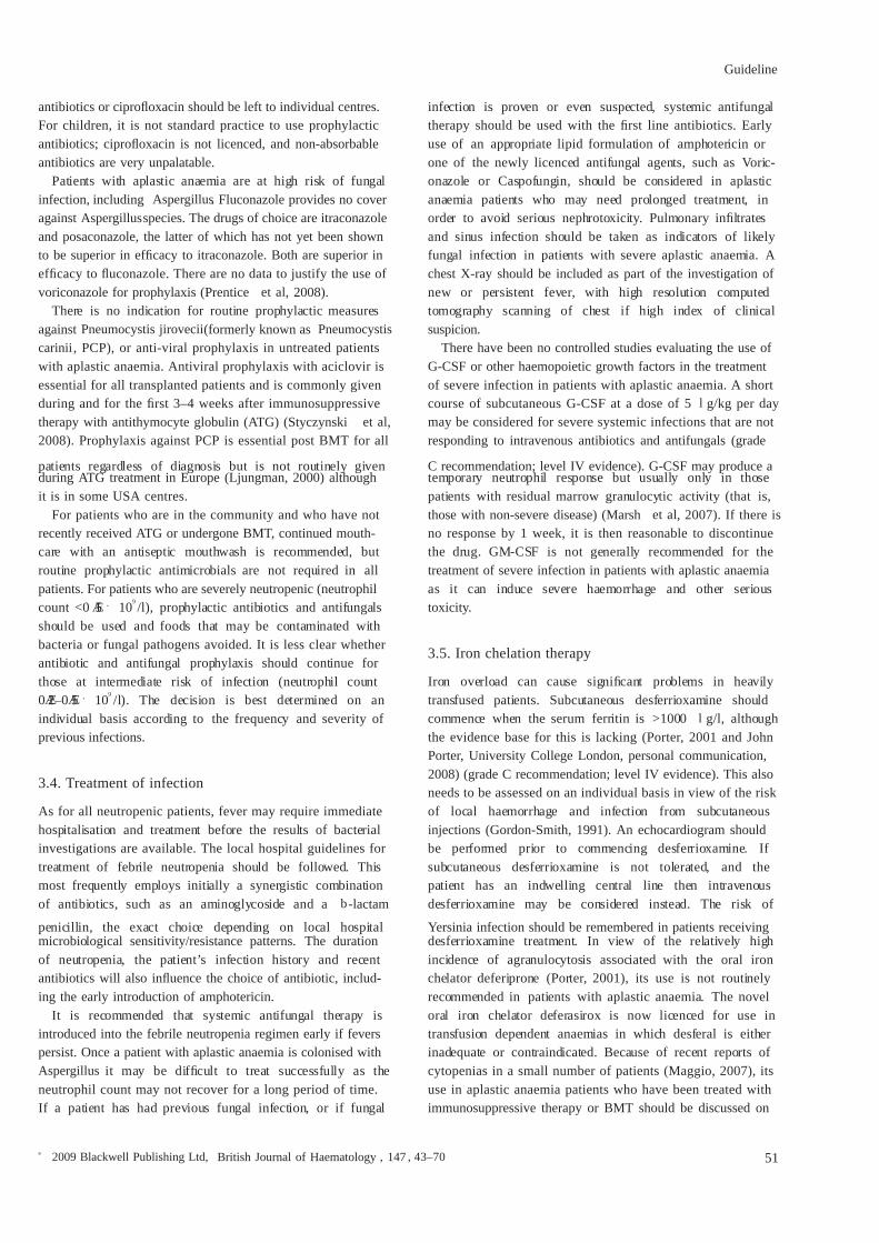
There have been no controlled studies evaluating the use of G-CSF or other haemopoietic growth factors in the treatmentof severe infection in patients with aplastic anaemia. A shortcourse of subcutaneous G-CSF at a dose of 5 l g/kg per day may be considered for severe systemic infections that are notresponding to intravenous antibiotics and antifungals (grade
C recommendation; level IV evidence). G-CSF may produce atemporary neutrophil response but usually only in thosepatients with residual marrow granulocytic activity (that is,those with non-severe disease) (Marsh et al , 2007). If there isno response by 1 week, it is then reasonable to discontinuethe drug. GM-CSF is not generally recommended for thetreatment of severe infection in patients with aplastic anaemiaas it can induce severe haemorrhage and other serioustoxicity.
3.5. Iron chelation therapy
Iron overload can cause signicant problems in heavily transfused patients. Subcutaneous desferrioxamine shouldcommence when the serum ferritin is >1000 l g/l, althoughthe evidence base for this is lacking (Porter, 2001 and JohnPorter, University College London, personal communication,2008) (grade C recommendation; level IV evidence). This alsoneeds to be assessed on an individual basis in view of the risk of local haemorrhage and infection from subcutaneousinjections (Gordon-Smith, 1991). An echocardiogram shouldbe performed prior to commencing desferrioxamine. If subcutaneous desferrioxamine is not tolerated, and thepatient has an indwelling central line then intravenousdesferrioxamine may be considered instead. The risk of
Yersinia infection should be remembered in patients receivingdesferrioxamine treatment. In view of the relatively highincidence of agranulocytosis associated with the oral ironchelator deferiprone (Porter, 2001), its use is not routinely recommended in patients with aplastic anaemia. The noveloral iron chelator deferasirox is now licenced for use intransfusion dependent anaemias in which desferal is eitherinadequate or contraindicated. Because of recent reports of cytopenias in a small number of patients (Maggio, 2007), itsuse in aplastic anaemia patients who have been treated withimmunosuppressive therapy or BMT should be discussed on
Guideline
ª 2009 Blackwell Publishing Ltd, British Journal of Haematology , 147 , 43–70 51
an individual patient basis. For iron over-loaded patientsfollowing response to ATG or successful BMT, venesection isthe standard way to remove iron.
3.6. Vaccinations
There have been anecdotal reports of vaccination producingbone marrow failure or triggering relapse of aplastic anaemia,so vaccinations, including inuenza vaccination, should only be given when absolutely necessary (Viallard et al , 2000;Hendry et al , 2002) (grade C recommendation; level IVevidence). All live vaccines should be avoided after BMT andATG treatment, indenitely. After BMT, aplastic anaemiapatients should be routinely vaccinated as recommended for allallogeneic BMT recipients.
3.7. Psychological and general support
Psychological support for the patient, family and close friends
is of great importance. Aplastic anaemia is a rare disease andrequires careful explanation of its nature, prognosis, as well asdiscussions on important issues such as pregnancy. Patientsshould be given the opportunity to be referred to a centre thatspecialises in the management of aplastic anaemia.
There is now an excellent patient support group in the UKfor patients with aplastic anaemia which can be contacted at:the Aplastic Anaemia Trust, AA and MDS Support Group, 16Sidney Rd, Borstal, Rochester, Kent ME13HF. Telephone:0870-487 0099, email: [email protected], website:http://www.theatt.org.uk.
The chronic nature and slow response to treatment shouldbe stressed early in the disease. The morale of the patient(family and close friends) and staff may sag when recovery hasnot occurred at 6 months or longer.
Recommendations(i) Prophylactic platelet transfusions should be given
when the platelet count is <10 · 109 /l (or <20 · 109 /lin the presence of fever).
(ii) Irradiated blood products should be given routinely toall patients having ATG treatment.
(iii) Transfusion of irradiated granulocyte transfusionsmay be considered in patients with life-threatening
neutropenic sepsis.(iv) The routine use of rHuEpo in aplastic anaemia is not
recommended.(v) A short course of G-CSF may be considered for severe
systemic infection that is not responding to intrave-nous antibiotics and anti-fungal drugs, but should bediscontinued after 1 week if there is no increase in theneutrophil count.
(vi) Prophylactic antibiotic and antifungal drugs should begiven to patients with neutrophil count <0 Æ2 · 109 /l.
(vii) Systemic antifungal therapy should be introduced intothe febrile neutropenia regimen early if fevers persist.
(viii) Iron chelation therapy should be considered when theserum ferritin is >1000 l g/l.
4. Specic treatment of aplastic anaemia:general commentsThe standard specic treatment for a newly diagnosed patientwith aplastic anaemia is either allogeneic stem cell transplan-tation from an HLA-identical sibling donor or immunosup-pressive therapy with a combination of ATG and ciclosporin.The results of transplantation for aplastic anaemia from amatched unrelated donor have recently been improved by usinga reduced intensity conditioning regimen, and this proceduremay be considered in young patients with severe disease who donot respond to treatment with ATG and ciclosporin.
It is essential that before specic treatment is given, thepatient is stabilised clinically in terms of controlling bleedingand treating infection. It is dangerous to give immunosup-pressive therapy in the presence of infection or uncontrolledbleeding (grade C recommendation; level IV evidence). Thepresence of infection is an adverse factor for outcome afterstem cell transplantation (grade B recommendation; level IIaevidence). However, it may sometimes be necessary to proceedwith BMT in the presence of active infection, particularly fungal infection, as the transplant offers the best chance of early neutrophil recovery, and delaying the transplant may risk progression of the fungal infection.
Because aplastic anaemia is a rare disease, the haematologistresponsible for the patient should contact a centre/specialist
with expertise in aplastic anaemia soon after presentation todiscuss a management plan for the patient. Care should beshared with the local hospital if possible.
Hospitals providing general haematology care at Level 2 (asdened by the Clinical Haematology Task Force for BCSH,2000) should be capable of the safe treatment of a patient withsevere aplastic anaemia with ATG, providing medical andnursing staff have experience of using ATG, including therecognition and management of its side effects. Level 4 care isrequired for related allogeneic BMT, providing the centre hasexperience in BMT for aplastic anaemia. British Society of Blood and Marrow Transplantation and European Group forBlood and Marrow Transplantation (EBMT) accreditation isrequired for centres to perform unrelated donor BMT.
How long should one wait after presentation before startingtreatment for the disease? Early spontaneous recovery occursinfrequently and, in practical terms, by the time the patient hasbeen stabilised clinically, the disease conrmed, its diseaseseverity assessed, potential sibling donor(s) HLA tissue typedand a management plan discussed in collaboration with anexpert specialist centre, specic treatment should not bedelayed much beyond this time.
Guideline
52 ª 2009 Blackwell Publishing Ltd, British Journal of Haematology , 147 , 43–70
Patients with aplastic anaemia should be followed up indef-initely to monitor for relapse and later clonal disorders, such asMDS, leukaemia, PNH and solid tumours. When childrenapproach adult age, arrangements should be made for theirsubsequent transfer to an adult unit for continued follow up.
Prednisolone should not be used to treat patients withaplastic anaemia (grade C recommendation; level IV evidence).Corticosteroids are ineffective; they encourage bacterial andfungal colonisation, and can precipitate serious gastrointestinalhaemorrhage in the presence of severe thrombocytopenia.Similarly, haemopoietic growth factors, such as G-CSF andrHuEpo, should not be used on their own in newly diagnosedpatients in the mistaken belief that they may cure the disease(grade C recommendation; level IV evidence). The use of haemopoietic growth factors in this way would lead to delay ingiving specic treatment, during which time the patient may become infected or allo-immunised (Marsh et al , 1994a;Marsh et al , 2007). The recommendations for specic treat-ment of aplastic anaemia are summarised in Figs 1 and 2.
Recommendations(i) Infection or uncontrolled bleeding should be treated
rst before giving immunosuppressive therapy. Thisalso applies to patients scheduled for BMT, although itmay sometimes be necessary to proceed straight toBMT in the presence of severe infection as a BMT may offer the best chance of early neutrophil recovery.
(ii) Haemopoietic growth factors, such as rHuEpo or G-CSF, should not be used on their own in newly diagnosed patients in an attempt to ‘treat’ the aplasticanaemia.
(iii) Prednisolone should not be used to treat patients withaplastic anaemia because it is ineffective and encour-ages bacterial and fungal infection.
5. HLA identical sibling donor transplantation
5.1. Results
Transplantation for severe aplastic anaemia from an HLAidentical sibling donor is now very successful with a 75–90%chance of long term cure (Passweg et al , 1997; Bacigalupo et al ,2000a; Locatelli et al , 2000; Adeset al , 2004; Gupta et al , 2004;Kahl et al , 2005; Champlin et al , 2007; Myers & Davies, 2009).Using high dose cyclophosphamide with ATG conditioning,graft failure is around 4–14%. GVHD remains a problem.Although acute GVHD grade III–IV 12–30%) appears to occurless commonly now, chronic GVHD still occurs in 30–40% of patients, (unless a Campath-1H based regimen is used whichreduces the risk to <5% (Gupta et al , 2004). Prior treatmentwith immunosuppressive therapy is associated with a worseoutcome and increased graft rejection (Ades et al , 2004;Kobayashi et al , 2006).
5.2. Indications for HLA-identical sibling BMT
Allogeneic BMT from an HLA-identical sibling donor is theinitial treatment of choice for newly diagnosed patients withaplastic anaemia if they (i) have severe or very severe aplasticanaemia ( see Section 2.4and Table IV for denitions of diseaseseverity), (ii) are younger than 40 years (although there iscontroversy concerning the upper age limit for BMT; see
Age of patient
≤ 40 years > 40 years
HLA identical sibling
Yes No
Response at 4 months
YesHLA id sib BMT
ATG + CSA
Supportivetherapy
Options
Maintain on CSA while FBCrising, then very slow taper,often over one/more years
Options:1. 3 rd ATG if previous
response to ATG2. Oxymetholone3. CRP using novel IST4. BMT using CRP with UCB or
haploidentical donor ?
ATG + CSA+G-CSF only as part
of clinical study
No
Consider MUDBMT if< 50 years (or 50–60* andgood performance status)
2nd ATG + CSAif no MUD
Response at 4 months
NoYes
Fig 1. Treatment of acquired severe aplastic anaemia. FBC, full blood count; CRP, clinical research protocol; IST, immunosuppressive therapy; UCB,umbilical cord blood; MUD, matched unrelated donor; ATG, antithymocyte globulin; CSA, ciclosporin; G-CSF, granulocyte colony-stimulatingfactor; BMT, bone marrow transplantation; HLA id sib, human leucocyte antigen-identical sibling. *For patients older than 60 years, there is currently insufcient data on the role of HSCT in severe AA although data for MDS suggests that this may be a future option (see text).
Guideline
ª 2009 Blackwell Publishing Ltd, British Journal of Haematology , 147 , 43–70 53
below) and (iii) have an HLA compatible sibling donor.(iv) For children who have non-severe aplastic anaemia and inwhom treatment is indicated, then HLA matched sibling donortransplant should be the rst choice, (Grade B recommenda-tion, level IIb evidence).
There is controversy concerning the upper age limit forBMT. Results of BMT using an HLA identical sibling donor areworse in patients >30 years of age compared with patients<30 years of age (Bacigalupo et al , 2000a), and particularly >40 years. The decision whether to treat patients aged30–40 years with ATG and ciclosporin or to transplant upfrontshould take into account the patient’s general medicalcondition. For patients >40 years who have failed immuno-suppressive therapy with ATG and ciclosporin, who have anHLA compatible donor and who are in good medicalcondition, BMT may be considered. A reduced intensity conditioning regimen may be preferable in such patients, asproposed by the EBMT Severe Aplastic Anaemia WorkingParty (Maury et al , 2007) in view of the high transplant-relatedmortality using high dose cyclophosphamide (Grade Brecommendation, level IIb evidence). A similar conditioningregimen may be indicated for patients between 30 and 40 years
of age, although there is currently no published data to supportthis approach.
5.3. Conditioning and GVHD prophylaxis regimen
The conditioning regimens and GVHD prophylaxisdescribed below refer specically to patients with acquiredaplastic anaemia. Patients with Fanconi anaemia and othertypes of inherited aplastic anaemia need special consider-ation and should not follow these pathways, as theconditioning regimen and GVHD prophylaxis are completely
different (Rosenberg et al , 2005) (grade B recommendation;level IIa evidence).
(a) Conditioning regimen for patients aged <30 years. Thepreparation used is a non-myeloablative and highly immunosuppressive regimen to help prevent graft rejectionand GVHD. The current standard regimen used is high dosecyclophosphamide 50 mg/kg · 4 (day ) 5 to ) 2) and ATG(Thymoglobuline, Genzyme 1 Æ5 vials/10 kg · 3 on days ) 5 to) 3), with methylprednisolone 2 mg/kg · 3 (day ) 5 to ) 3).(Methylprednisolone is not usually used for paediatric BMT).The recommended post-transplant immunosuppression is(i) ciclosporin 5 mg/kg per day, in two divided doses (thatis, 2Æ5 mg/kg BD), starting on day ) 1, and continuing for12 months with tapering beginning at 9 months to helpprevent late graft failure, and (ii) short course methotrexate15 mg/m 2 on day +1, then 10 mg/m 2 on days +3, +6, and +11(Schrezenmeier et al , 2000). The potential benet of usingATG with cyclophosphamide is unclear as a recently publishedprospective randomised study from the Centre for Blood andMarrow Transplant Research (CIBMTR) showed no signicantbenet in terms of graft rejection, GVHD and survival rates
using the combination of cyclophosphamide and ATGcompared with cyclophosphamide alone (Champlin et al ,2007). The study was underpowered to show differencesbetween the two groups, but the addition of ATG did notsignicantly improve outcome (Champlin et al , 2007).
(b) Conditioning regimens for patients aged >30 years. Forpatients between the ages of 30 and 50 years, who are potentialtransplant candidates, the best conditioning regimen is notknown. Patients who are>40 years of ageand who aremedically t enough for BMT (see above), may receive a reduced intensity
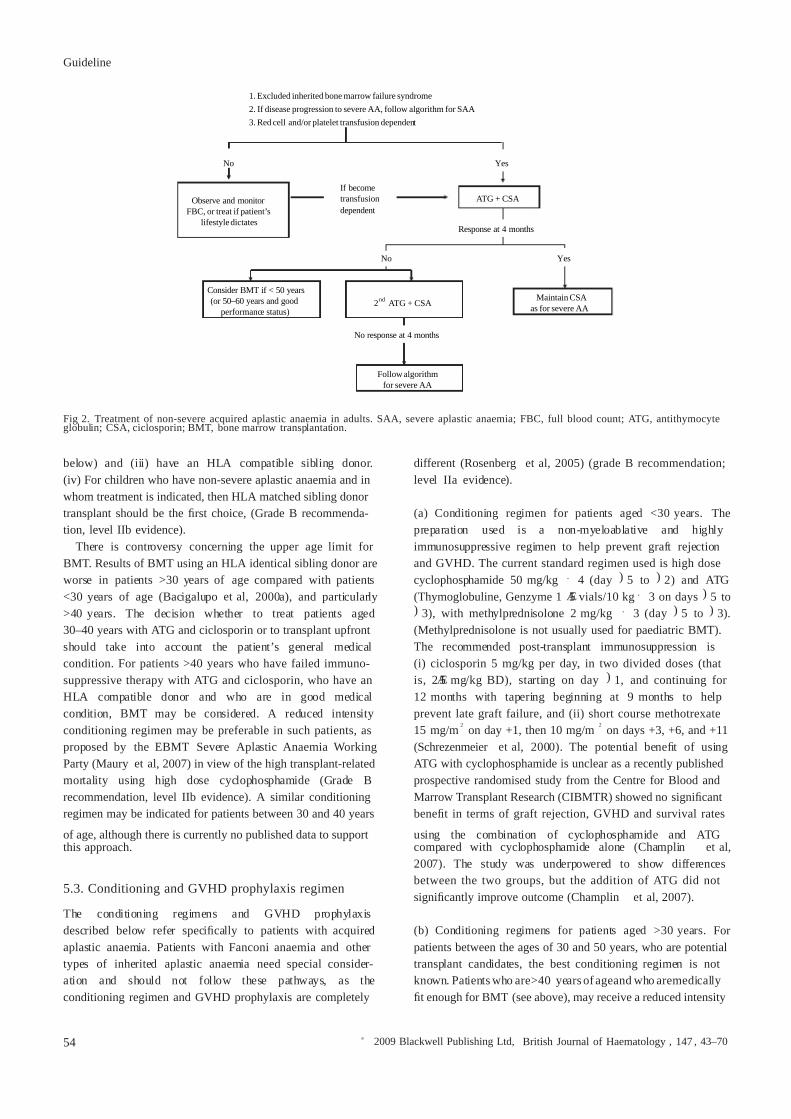
1. Excluded inherited bone marrow failure syndrome2. If disease progression to severe AA, follow algorithm for SAA3. Red cell and/or platelet transfusion dependent
No Yes
If becometransfusiondependent
Response at 4 months
No Yes
No response at 4 months
ATG + CSA
Consider BMT if < 50 years(or 50–60 years and good
performance status)
Follow algorithmfor severe AA
Maintain CSAas for severe AA
Observe and monitorFBC, or treat if patient’s
lifestyle dictates
2nd ATG + CSA
Fig 2. Treatment of non-severe acquired aplastic anaemia in adults. SAA, severe aplastic anaemia; FBC, full blood count; ATG, antithymocyteglobulin; CSA, ciclosporin; BMT, bone marrow transplantation.
Guideline
54 ª 2009 Blackwell Publishing Ltd, British Journal of Haematology , 147 , 43–70
conditioning regimen, using cyclophosphamide 1200 mg/m 2 ,udarabine 120 mg/m 2 and either ATG or Alemtuzumab(Gupta et al , 2004, Maury et al , 2007). A similar approachmay be considered for patients aged 30–40 years.
There is no indication for using irradiation-based regimensin HLA-identical sibling BMT for aplastic anaemia(Schrezenmeier et al , 2000) (Grade B recommendation, levelIIa evidence). Although irradiation reduces the risk of rejection,it confers no benet on survival and its use is associated with anincreased risk of later solid tumours and inevitable infertility, aswell as impaired growth and development in children.
5.4. Source and dose of stem cells
It is recommended that bone marrow stem cells, and notG-CSF mobilised peripheral blood stem cells (PBSC), shouldbe used (Schrezenmeier et al , 2007) (Grade B recommenda-tion, level IIb evidence). In a retrospective combined CIBMTR and EBMT study, earlier engraftment occurred with PBSC
although there was no difference in probability of neutrophilor platelet engraftment by day +30, and no difference in graftrejection compared with bone marrow transplants. Of majorconcern was signicantly worse survival for all patients, andmore chronic GVHD in younger patients, using PBSCcompared with bone marrow grafts (Schrezenmeier et al ,2007). There are other important reasons for using bonemarrow in children. Because most of the sibling donors willalso be children, it may be much easier to obtain bone marrow than PBSC. In addition, the collection of bone marrow stemcells avoids the exposure of G-CSF (Davies & Guinan, 2007).
It is important to give at least 3 · 108 nucleated marrow cells/kg because at lower doses the risk of graft rejectionincreases signicantly (Niederwieser et al , 1988). There are nodata on the minimum dose of CD34 + marrow cells to give inaplastic anaemia but it is recommend that at least 3 · 106 /kgshould be given (Russell et al , 1998).
The effect of sex-mis-match between donor and recipienthas recently been evaluated in a large retrospective study from the EBMT of patients undergoing HLA-identicalsibling or HLA-identical unrelated donor BMT for aplasticanaemia. Survival was signicantly better in patients withdonors from the same sex. Male patients with female donorshad an increased risk of severe GVHD compared torecipients of sex-matched grafts. In contrast, female patients
with male donors had an increased risk of graft rejection.These negative effects of donor/recipient sex-mismatchingwere abrogated by the use of ATG in the conditioningregimen (Stern et al , 2006).
Umbilical cord blood as an alternative source of stem cellsfor transplantation has been used in a small number of patientswith aplastic anaemia (Gluckman et al , 1997; Barker et al ,2001). Its use, however, is limited to small recipients because of the low number of haemopoietic cells that can be obtainedfrom a donation, despite their higher proliferative potentialcompared with bone marrow cells (Hows, 2001). Compared
with bone marrow transplants, umbilical cord blood trans-plants are associated with a lower risk of acute and chronicGVHD (Gluckman et al , 1997; Barker et al , 2001). Umbilicalcord blood transplantation may also be considered in childrenwho lack an HLA-identical sibling donor or a fully matchedunrelated adult donor. The role of double umbilical cord bloodtransplants in adults with aplastic anaemia is currently beingexplored (Mao et al , 2005; Myers & Davies, 2009), but themajor problem anticipated is failure of engraftment.
5.5. Post-transplant management
There is a signicant risk of late graft failure in aplasticanaemia following allogeneic BMT which is most commonly associated with discontinuing ciclosporin too early or low ciclosporin blood levels, and in the presence of progressivemixed chimaerism, as dened by >10% recipient cells or >15%increase over 3 months, using short tandem repeats by polymerase chain reaction (PCR) analysis of mononuclear
cells (McCann et al , 2007). Progressive mixed chimaerismpredicts a high risk of graft rejection. Stable mixed chimaerismis associated with excellent survival and a low risk of GVHD(Lawler et al , 2009). Therapeutic ciclosporin should be con-tinued for at least 9 months before gradually reducing the doseto zero over the following 3 months. For adults, ciclosporintrough blood levels should be maintained between 250 and350 l g/l. For children, lower ciclosporin levels are often used(150–200 l g/l), to avoid toxicity. Chimaerism should bemonitored particularly closely during the time of ciclosporinwithdrawal. If there is evidence of signicant mixed chimae-rism (see above) or a rising proportion of recipient cells, asassessed with sensitive techniques such as PCR of short tandemrepeats, there is a high risk of late rejection, and ciclosporinshould not be reduced or withdrawn at that time (McCannet al , 2000; Lawler et al , 2009).
Fertility is usually well preserved or near normal after BMTfor aplastic anaemia using high dose cyclophosphamide andwhere irradiation is not used (Sanders et al , 1996; Deeg et al ,1998). It is not necessary to arrange for sperm (or oocyte)cryopreservation pre-transplant, and it is very important thatall patients receive appropriate counselling regarding contra-ception following their transplant (Grade B recommendation,level IIb evidence). For older patients receiving a udarabine-based regimen (see above), because there is currently insuf-
cient data on fertility post transplant, cryopreservation of sperm or oocyte should be planned.
Patients can be advised that because irradiation is not given,the risk of second tumours is very low (Witherspoon et al ,1991; Socie et al , 1993; Ades et al , 2004).
Recommendations(i) Allogeneic BMT from an HLA-identical sibling donor is
the initial treatment of choice for newly diagnosedpatients if they have severe or very severe aplastic
Guideline
ª 2009 Blackwell Publishing Ltd, British Journal of Haematology , 147 , 43–70 55
anaemia, are < 40 years old and have an HLA-compat-ible sibling donor.
(ii) Patients with Fanconi anaemia and other types of inherited aplastic anaemia need special considerationand should not follow recommendations made in thisguideline.
(iii) There is no indication for using irradiation-basedconditioning regimens for patients undergoing HLAidentical sibling BMT for aplastic anaemia.
(iv) The recommended source of stem cells for transplan-tation in aplastic anaemia is bone marrow.
(v) Fertility is well preserved after high dose cyclophos-phamide conditioning in BMT for aplastic anaemia,and patients should be given appropriate contraceptiveadvice to prevent unwanted pregnancy. Until longerterm data is available in patients receiving udarabine-based regimens, cryopreservation of sperm and oocytesshould be planned.
6. Immunosuppressive therapy: antithymocyteglobulin (ATG) and ciclosporin
6.1. Results of treatment
Immunosuppressive therapy using the combination of ATGand ciclosporin is associated with response rates of between60% and 80% with current 5 year survival rates of around75–85% (Bacigalupo et al , 2000a; Bacigalupo et al , 2000b,Fuhrer et al , 2005; Locasciulli et al , 2007). A recent study hasshown that on multivariate analysis of response at 6 months,only younger age, absolute reticulocyte count (ARC) and
absolute lymphocyte count (ALC), correlate with response toATG. The lack of association with the absolute neutrophilresponse reected a high number of early deaths in patientswith very severe neutropenia. For patients with bothARC ‡ 25 · 09 /l and ALC ‡ 1Æ0 · 109 /l, the response was83% compared with 41% for those with lower counts (Schein-berg et al , 2009). For severe aplastic anaemia, the event-freesurvival and response rate to ATG alone is signicantly less thanwith the combination of ATG and ciclosporin (Bacigalupo et al ,2000a; Frickhofen et al , 2003), and for patients with non-severeaplastic anaemia the response to the combination of ATG andciclosporin is signicantly greater than with ciclosporin alone(Marsh et al , 1999). Response to ATG and ciclosporin isdelayed and response usually does not start much before3–4 months. This means that patients need to continue withregular red cell and platelet transfusional support and willremain neutropenic during this time period. Relapse may occurafter immunosuppressive therapy. This was previously reportedto be around 30% (Schrezenmeier et al , 1993) but with longeruse and slower tailing of ciclosporin the rate is closer to 10%(Bacigalupo et al , 2000b). Patients are at risk of later clonaldisease, 8% for MDS/AML, 10% for haemolytic PNH and 11%for solid tumours at 11 years (Frickhofen et al , 2003).
6.2. Indications
Immunosuppressive therapy is indicated for patients who arenot eligible for sibling donor BMT. This includes (i) patientswith non-severe aplastic anaemia who are dependent on redcell and/or platelet transfusions (ii) patients with non-severeaplastic anaemia who, although not transfusion-dependent,
may have signicant neutropenia and be at risk of infection(iii) patients with severe or very severe aplastic anaemia whoare >40 years of age and (iv) younger patients with severe orvery severe disease who lack an HLA-compatible sibling donor(Grade B recommendation; level IIb evidence). Children withnon-severe aplastic anaemia with an HLA-identical siblingdonor and who are transfusion-dependent, and particularly if the blood count is falling, may be considered for BMT.
For those patients with non-severe aplastic anaemia who arenot dependent on either red cell or platelet transfusions, andmaintain safe blood counts, it is reasonable to observe theblood count and monitor the patient regularly without initially instigating immunosuppressive therapy. The decision whetherand when to start treatment is usually determined by thepattern of the blood counts, the individual patient’s life-styleand choice, and older age ( see Section 6.4).
6.3. Administration
Antithymocyte globulin is a powerful immunosuppressivedrug and its use in severely neutropenic patients requires very careful monitoring, prophylaxis and treatment of fevers andinfections, as well as adequate (and sometimes intensive)platelet transfusional support (grade A recommendation; levelIb evidence).
In the UK, most of Europe and many other countries, thestandard preparation of ATG has until recently been horseATG (Lymphoglobuline; Genzyme). The rabbit preparation(Thymoglobuline; Genzyme) was usually reserved for secondor subsequent courses. From June 2007, supply of horse ATG(Lymphoglobuline) was withdrawn due to manufacturingdifculties maintaining quality control. Rabbit ATG (Thymo-globuline) is therefore now recommended as rst line treat-ment. Response rates to rabbit ATG are anticipated to besimilar to horse ATG, based on (i) response rates when rabbitATG is used for a second course (Di Bona et al , 1999;Scheinberg et al , 2006a) and (ii) both preparations have the
same immunogen (thymocytes), similar production methodand they bind to similar epitopes. To date, there have beenonly two studies using rabbit ATG as rst line treatment foraplastic anaemia. In a small single centre phase II study of 13patients with aplastic anaemia and 12 with low risk MDS,among the patients with aplastic anaemia, there were vecomplete responses and seven partial responses (Garg et al ,2009). Preliminary results from a Spanish retrospective multi-centre study of 72 patients, reported an overall response rate of 46% (Vallejo et al , 2009). For a second course of ATG, optionsinclude giving rabbit ATG again or using an alternative
Guideline
56 ª 2009 Blackwell Publishing Ltd, British Journal of Haematology , 147 , 43–70
preparation of horse ATG, such as ATGAM (Pharmacia andUpjohn Company, Kalamazoo, MI, USA).
Antithymocyte globulin is given for 5 days as a daily intravenous infusion over 12–18 h through a central venouscatheter. The daily dose of rabbit ATG is 1 Æ5 vials/10 kg body weight (one vial of rabbit ATG, Thymoglobuline, contains25 mg protein so the daily dose is 3 Æ75 mg/kg). A test dose of 1/10th of a vial (2Æ5 mg for rabbit ATG), diluted in 100 mlnormal saline and infused over 1 h, is often given beforehandand, if a severe systemic reaction or anaphylaxis occurs, furtherdoses of that preparation of ATG must not be given. Instead of giving a separate test dose, some centres give the rst 100 ml of the rst infusion very slowly over 1 h. A patient should not begiven ATG from the same animal source after anaphylaxis or asevere systemic reaction.
Immediate side effects are allergic and occur commonly,including fever, rigours, rash, hypertension or hypotension anduid retention. Each daily dose should be preceded by intravenous methylprednisolone and chlorphenamine. Platelet
transfusions should be given to maintain a safe platelet count(ideally >30 · 109 /l), but should not be given concurrently with ATG administration because of the anti-platelet activity of ATG. Prior to starting ATG, patients should be assessed toensure adequate platelet increments with random donorplatelets. Poor platelet increments should be investigatedbeforehand, as previously described ( see Transfusional support ).Patients are often nursed in isolation with reverse barriernursing. All fevers, even if suspected to be due to the ATG,should be treated with broad spectrum antibiotics. Intravenousmethylprednisolone (or oral prednisolone) and paracetamolare given at least 30 min before each daily dose of rabbit ATG at1–2 mg/kg per day (depending on individual study preference)and then orally, reducing the dose by half every 5 days, to helpprevent serum sickness, in line with current EBMT studies.Serum sickness typically occurs between day 7 and 14 from thestart of ATG treatment. If serum sickness occurs, intravenoushydrocortisone 100 mg six hourly should be commenced. Thecommon symptoms of serum sickness include arthralgia,myalgia, rash, fever, mild proteinuria and platelet consumptionoften necessitating increased platelet transfusion support.
Antithymocyte globulin must not be given as an out-patient.The patient should remain hospitalised from the start of ATGthrough the period when serum sickness occurs. If there isimmediate access to in-patient or day care facilities for
treatment of later complications, such as serum sickness,infection or bleeding, then admission for the 5 days of ATGtreatment alone can be considered (grade C recommendation;level IV evidence).
Oral ciclosporin at 5 mg/kg per day may be started either onthe rst day of ATG (in line with EBMT studies), or afterprednisolone has been discontinued, aiming to keep the troughciclosporin blood level between 150 and 250 l g/l for adultsand between 100 and 150 l g/l for children. The latterapproach helps reduce drug toxicity. A recent study in childrenshowed no evidence that maintaining higher ciclosporin blood
levels improves response rates further; higher blood levelsincrease the risk of ciclosporin toxicity (Saracco et al , 2008). Inaddition, there was a signicant risk of relapse with rapidtapering of ciclosporin and the authors recommend thatciclosporin should be continued for at least 12 months after amaximal response before starting to taper the drug (Saraccoet al , 2008). A very slow taper is recommended, for example,by 25 mg every 3 months. A similar approach in adults wouldseem prudent. Blood pressure, renal and liver function testsshould also be monitored regularly while on ciclosporin.
A second course of ATG is recommended if there is noresponse or relapse after the rst course. This should not begiven earlier than 3 months after the rst course because itusually takes around 3 months before a response occurs. Thereis a 30–60% chance of response to a second course (Tichelliet al , 1996; Scheinberg et al , 2006a); these gures reecttreatment with either two courses of horse ATG (Tichelli et al ,1996) or treatment with rabbit ATG after non-response to horseATG (Scheinberg et al , 2006a). When rabbit ATG is given for
the second course following an initial course of horse ATG, theresponse rate was only 30% for non-responders and 65% forrelapsing patients. A recent study from Japan has examinedprospectively the outcome of 52 children who have failed onecourse of IST, and who went on to either receive a secondcourse of ATG or an unrelated donor HSCT. The response to asecond course of ATG was only 11% with a 5-year failure-freesurvival of only 9Æ5% and three children had anaphylaxis toATG (Kosaka et al , 2008). There are currently no data onre-treatment with rabbit ATG. When horse ATG was stillavailable, it was possible to consider giving a third course of ATG. Those patients most likely to respond were those whohave shown response to previous ATG course(s). If patients hadshown no response to the rst or second courses, then thechance of responding to a third course was low (Gupta et al ,2005a). As when giving a rst or second course of ATG, a testdose must always be given beforehand, and if there is no severereaction one can then proceed with the full dose. Instead of giving a separate test dose, as discussed above, the rst 100 mlof the rst infusion can be given very slowly over 1 h.
6.4. ATG treatment in older patients
The decision whether to use ATG in older patients can bedifcult and requires careful assessment and discussion of the
risks with the patient. For older patients, the response rate andsurvival rate are lower compared with younger patients. Theresponse rate for patients aged >60, 50–59 and <50 years is37%, 49% and 57%, and 5-year survival is 50%, 57% and 72%,respectively (Tichelli et al , 1999). For patients aged >70 years,the 10-year survival is 33% compared with 60% for those agedbetween 50 and 70 years (A. Tichelli, unpublished observa-tions). Older patients (aged >60 years) also have a higher risk of serious cardiac events after ATG (Kao et al , 2008). Althoughthere is no upper age limit for ATG treatment, considerationfor treatment should be preceded by medical assessment to
Guideline
ª 2009 Blackwell Publishing Ltd, British Journal of Haematology , 147 , 43–70 57
exclude signicant co-morbidities and bone marrow exami-nation, including trephine and cytogenetics (and/or FISH) toexclude hypocellular MDS. Discussion with the patient shouldinclude the increased risk of mortality from bleeding, infectionand cardiac events associated with ATG treatment. For olderpatients who are not candidates for ATG, optimal supportivecare should be provided. Ciclosporin may be considered, butbecause of the increased risk of signicant renal toxicity andhypertension in older patients, a lower trough ciclosporinblood level or 100–150 l g/l is suggested. Oxymetholone may be useful in men but often causes unacceptable masculinisationin women. The risk of cardiac failure, liver toxicity, high serumcholesterol, impaired glucose tolerance and prostatism warrantfurther caution when used in older patients.
6.5. Denition of response
There has previously been no agreement regarding themeasurement of response to immunosuppressive therapy, with
the result that it has been difcult to compare response rates.New criteria for response have recently been accepted by anexpert committee on aplastic anaemia, and these are summar-ised in Table Va, b (Camitta, 2000). Responses should beconrmed by two or more blood counts at least 4 weeks apart,and should ideally be measured in patients who are notreceiving haemopoietic growth factors (Camitta, 2000).
6.6. Follow up of patients post ATG
Following treatment with ATG and ciclosporin, patientsshould be monitored carefully with regular FBC for evidenceof relapse, and also for later clonal disorders such as PNH,MDS and AML. At 3–4 months post-ATG, a screen for PNHshould be performed. Further bone marrow examinations withcytogenetics are indicated if there is evidence of relapse or
other change in the blood count or blood lm. A careful review of the blood lm is important to monitor for evidence of MDS.It is suggested that a PNH screen is performed annually in allpatients.
Recommendations
(i) Immunosuppressive therapy is recommended for (1)patients with non-severe aplastic anaemia who aretransfusion-dependent (2) patients with severe or very severe disease who are > 40 years old and (3) youngerpatients with severe or very severe disease who do nothave an HLA-identical sibling donor.
(ii) ATG is a powerful immunosuppressive drug and its usein severely neutropenic patients requires very carefulmonitoring, prophylaxis and treatment of fevers, andadequate (and sometimes intensive) platelet transfu-sional support.
(iii) ATG must only be given as an in-patient.
(iv) Ciclosporin should be continued for at least 12 monthsafter achieving maximal haematological response, fol-lowed by a very slow tapering, to reduce the risk of relapse.
7. Matched unrelated donor bone marrowtransplantation (MUD BMT)
7.1. Results
The role of MUD BMT in the treatment of severe aplasticanaemia is now clearer in view of recent improvements in
morbidity and mortality. Up until the late 1990s, long termsurvival was around only 30% with a high incidence of graftrejection, GVHD and severe infections (Passweg et al , 2006),although more encouraging results had been reported fromMilwaukee (Margolis et al , 1996) and from Japan (Koderaet al , 1999) in children and young adults. In many cases amyeloablative regimen incorporating, most frequently, irradi-ation had been employed. More recent data has shownimproved results using either a non-irradiation, udarabine-based regimen, as reported by the EBMT (Bacigalupo et al ,2005), or a low dose total body irradiation (TBI)-basedregimen (Deeg et al , 2006), with overall survival of between65% and 73% at 5 years. The EBMT protocol comprisesudarabine (30 mg/m 2 · 4), low dose cyclophosphamide(300 mg/m 2 · 4) and ATG for 4 days, with short course of both ciclosporin and methotrexate as GVHD prophylaxis. Theoverall 2-year survival is 73% but graft failure is 18% (and 35%in patients >14 years old). A similar protocol has beenemployed that uses Campath-1H instead of ATG (Gupta et al ,2005b). The current EBMT protocol has been modied forpatients >14 years, to include the addition of 2 Gy TBI, and areduction in ATG to 7 Æ5 mg in order to reduce the risk of EBVpost-transplant lymphoproliferative disorder (Bacigalupo et al ,
Table V. Criteria for response to immunosuppressive therapy inaplastic anaemia.
a. Response criteria for severe aplastic anaemiaNone Still severePartial Transfusion independent
No longer meeting criteria for severe diseaseComplete Haemoglobin normal for age
b. Response criteria for non-severe aplastic anaemiaNone Worse or not meeting criteria below Partial Transfusion independence (if previously dependent)
or doubling or normalisation of at least one cell lineor increase of baseline haemoglobin of >30 g/l
(if initially <6)or increase of baseline neutrophils of >0 Æ5 · 109 /l
(if initially <0Æ5)or increase of baseline platelets of >20 · 109 /l
(if initially <20)Complete Same criteria as for severe disease
Guideline
58 ª 2009 Blackwell Publishing Ltd, British Journal of Haematology , 147 , 43–70
2009). The EBMT Severe Aplastic Anaemia Working Party view is to avoid irradiation in children and young adults, evenat low doses, and to use udarabine instead. For older patients,the addition of low dose irradiation may be of benet inreducing graft rejection (Bacigalupo et al , 2005; Deeg et al ,2006) (grade B recommendation; level III evidence).
7.2. Indications
Matched unrelated donor bone marrow transplantation may be considered when patients full all the following criteria.They should:
(1) have a fully matched (at DNA level for both class I and IIantigens) donor
(2) be <50 years old (although patients aged 50–60 years may be considered if good performance status). There arecurrently insufcient data available on the use with HSCTin SAA patients >60 years of age (Gupta et al , 2008).However, for MDS patients transplanted using a reducedintensity conditioned HSCT, multivariate analysis of factors for overall survival, showed no signicant differ-ence for patients aged >60 compared with 50–60 years(Lim et al , 2009)
(3) have failed at least one course of ATG and ciclosporin (forboth adults and children), although in adults a secondcourse of ATG may be preferred if there are particularreasons not to proceed to MUD BMT after one failedcourse, based on individual patient circumstances
(4) have severe or very severe aplastic anaemia and(5) have no evidence of active infection and or acute bleeding
at time of BMT (Grade B recommendation, level IV
evidence). However, see comments under Psychologicaland general support
(6) be used as rst line treatment in patients with constitutionalaplastic anaemia and with no HLA matched sibling donor.
Even then, the decision to proceed with MUD BMT or asecond course of ATG is not always straightforward, especially when the patient may be clinically well. Full discussions aboutother treatment options and the natural history of aplasticanaemia should take place with the patient and family whomust be appropriately informed about the risks of MUD BMTin aplastic anaemia. Because results of MUD BMT for acquiredaplastic anaemia have improved signicantly over the last
5–10 years (Viollier et al , 2007), MUD BMT should no longerbe considered as a last resort after failing two courses of ATG,as previously recommended in these guidelines (Marsh et al ,2003). The disadvantages of continuing with ATG is that thepatient’s condition may continue to deteriorate with contin-uing sepsis and increasing iron overload, thus reducing thechance of a successful outcome at time of transplantation.Because aplastic anaemia is a rare disease and because of theparticular risks of MUD BMT in this condition, such aprocedure should only be done at centres with experience intransplantation for aplastic anaemia and accredited by the
European Blood and Marrow Transplant Registry for unrelateddonor BMT. Written guidelines from the United KingdomChildren’s Cancer Study group are currently in preparation forpaediatric BMT conditioning regimens.
7.3. Conditioning regimen
The current regimen recommended for younger patients by theEBMT is (i) Cyclophosphamide 300 mg/m 2 · 4 (ii) udara-bine 30 mg/m 2 · 4 (iii) ATG (Thymoglobuline, rabbit; Gen-zyme) 1Æ5 vials/10 kg · 4 (or Campath-1H 0 Æ2 mg/kg tomaximum dose of 10 mg/day · 5 pre-transplant) (iv) ciclo-sporin commencing on day ) 6 at 1 mg/kg per day to day ) 2,then 2 mg/kg per day from day ) 1 to +20, then 8 mg/kg perday orally and (v) methotrexate 10 mg/m 2 on day +1, then8 mg/m 2 on days +3 and +6, if using ATG instead of Campath-1H. For older patients, the addition of 200 cGy TBIwith reduced ATG (given for 2 days instead of four) may beconsidered (grade B recommendation; level III evidence).
It is acceptable to use either ATG or Campath-1H depend-ing on the patient’s previous exposure to ATG and individualcentre preference. Other approaches are valid within thesetting of prospective clinical trials. Patients should receivebone marrow and not PBSC as the source of stem cells and atleast 3 · 108 nucleated cells/kg should be infused. ( see Section4.1.2.1).
7.4. Timing of unrelated donor BMT search
An unrelated donor marrow search should be performed inpatients with severe aplastic anaemia who may be eligible for
unrelated donor transplantation and who do not have anHLA-identical sibling donor, at the time of the rst course of ATG, so that at the time of assessment of response to ATG(about 3 months), further information regarding the possibleavailability of a high resolution matched donor can be sought.For both children and adults, an unrelated donor transplantmay be considered if there is no response to a rst course of ATG, although in adults a second course of ATG may bepreferred if there are particular reasons not to proceed toMUD BMT after one failed course, based on individual patientcircumstances.
Recommendations(i) MUD BMT may be considered when a patient has a fully
matched donor, they are <50 years old (or 50–60 yearsold with good performance status), and have failed atleast one course of ATG and ciclosporin, have severeaplastic anaemia. There is currently insufcient data onoutcome for patients >60 years of age.
(ii) The optimal conditioning regimen for MUD BMT isuncertain, but currently a udarabine, non-irradiation-based regimen is favoured for younger patients.
Guideline
ª 2009 Blackwell Publishing Ltd, British Journal of Haematology , 147 , 43–70 59
8.1.1. High dose cyclophosphamide without stem cell support.The use of high dose cyclophosphamide (45 mg/kg · 4)without stem cell support has been proposed by one centre
as treatment for patients with newly diagnosed aplasticanaemia (Brodsky et al , 1996, 2001). However, a prospectiverandomised study comparing its use in combination withciclosporin against the gold standard of ATG and ciclosporinwas terminated prematurely because of an excess of early deaths and systemic fungal infections in the cyclophosphamidearm. The use of cyclophosphamide was associated withprofound and very prolonged pancytopenia resulting in asignicant increase in use of blood and platelet transfusions,days of intravenous antibiotics and amphotericin and in-patient days in hospital (Tisdale et al , 2000a, 2002). Forpatients refractory to ATG, high dose cyclophosphamideinduces a response in 70% of patients but does not eradicatePNH clones in all patients and later MDS has been reported(Brodsky et al , 2004).
Therefore, the use of high dose cyclophosphamide with-out stem cell support cannot be recommended in eithernewly diagnosed patients or patients who have failed ATGand ciclosporin in view of its serious toxicity and highmortality (Grade A recommendation, level Ib recommenda-tion).
8.1.2. Mycophenolate mofetil. Mycophenolate mofetil (MMF)inhibits the proliferation of B and T-lymphocytes and hasbeen used in the treatment and prevention of rejection in
organ transplantation as well as in the treatment of autoimmune disorders, such as ulcerative colitis,rheumatoid arthritis and multiple sclerosis. Its use in thetreatment of refractory aplastic anaemia is anecdotal andthere are no reported series of patients treated with the drug(Tisdale et al , 2000b). The EBMT SAA Working Party hasrecently performed a pilot study of 17 patients(Schrezenmeier et al , 2003) to assess its safety and efcacy in patients who are ineligible for BMT and refractory tostandard immunosuppressive therapy. However, no responseswere observed (Schrezenmeier et al , 2003). A retrospectivestudy from the US National Institutes of Health (NIH)
showed no improvement in response or reduction in relapseafter ATG and ciclosporin when MMF was added (Scheinberget al , 2006b). MMF appears to be ineffective in the treatmentof patients with refractory aplastic anaemia.
8.1.3. Alemtuzumab (Campath-1H). Campath-1H is currently under evaluation for the treatment of refractory aplasticanaemia in prospective trials at NIH in USA, andretrospectively by the EBMT, following reports of its efcacy in patients with autoimmune cytopenias, particularly autoimmune neutropenia (Willis et al , 2001).
8.2. Oxymetholone
Oxymetholone was used extensively in the treatment of aplastic anaemia for many decades before the availability of ATG and ciclosporin. In some patients, oxymetholone canstimulate erythropoiesis in particular but sometimes canproduce a trilineage response. Response to androgens, partic-
ularly if no PNH clone is present, raises the possibility of acongenital cause for the marrow failure. In combination withATG, it increases the response compared to ATG alone(Bacigalupo et al , 1993; Leleu et al , 2006). Oxymetholone ishepatotoxic and can cause liver dysfunction, clinical jaundice,hepatomas and peliosis hepatis. It must therefore be used withcaution, with regular monitoring of liver function tests andliver ultrasound. Because the drug causes virilization, it is oftenunacceptable to women. The drug is available on a namedpatient basis and is still useful as an option for those patientswho have failed several courses of ATG and ciclosporin, or incertain patients where standard immunosuppressive treatmentmay not be possible.
8.3. Should haemopoietic growth factors, such as G-CSF,be used with ATG and ciclosporin?
The rationale for using G-CSF after ATG and ciclosporin was toattempt to reduce the risk of infection during the 3 monthsbefore haematological, particularly neutrophil, response isexpected, and also to improve response (trilineage) to immu-nosuppressive therapy, as G-CSF may work in combinationwith other endogenous haemopoietic growth factors to stim-ulate haemopoietic stem cells. However, there are concernsabout the cost of using G-CSF long term and at high dose and
the potential increase in later clonal disorders, particularly fromstudies in Japan, which can only be fully appreciated with follow up of at least 10 years (Ohara et al , 1997; Kaito et al , 1998;Marsh, 2000; Socie et al , 2000). A pilot study of 100 patientstreated with ATG and ciclosporin and 3 months of G-CSF wasassociated with low mortality, a response rate of almost 80%and actuarial survival at 5 years of 87% (Bacigalupo et al ,2000a). However, a relatively small, prospective randomisedstudy comparing ATG, ciclosporin and G-CSF with ATG andciclosporin alone demonstrated no difference in response andsurvival between the two groups. Although there was noobvious increase in clonal disorders, the follow up of this study
was too short to evaluate this properly. (Locascuilli et al , 2001;Gluckman et al , 2002). A recent prospective randomised study of 101 adult patients from Japan comparing ATG andciclosporin with or without G-CSF showed a higher responserate at 6 months and a lower relapse rate in the G-CSF arm, butno difference in survival between the two arms. Although therewas no difference in the incidence of MDS and AML at 4 years,the follow up was too short to evaluate this adequately (Teramura et al , 2007). This question is also currently beingevaluated further in a larger prospective randomised multi-centre EBMT study, which has just closed and is awaiting
Guideline
60 ª 2009 Blackwell Publishing Ltd, British Journal of Haematology , 147 , 43–70
analysis. The EBMT Severe Aplastic Anaemia Working Party has recently reported results of a large retrospective study of 840patients treated with ATG and ciclosporin, of whom 43% alsoreceived G-CSF. The incidence of MDS/AML was 10Æ9% withG-CSF and 5Æ8% without G-CSF (Socie et al , 2007). Theroutine use of long termG-CSF afterATG and ciclosporin is notcurrently recommended outside prospective clinical trials(grade A recommendation; level Ib evidence).
A large prospective randomised study from China showedno benet in using both GM-CSF and erythropoietin withATG and ciclosporin, as well as conrming that the combi-nation of ATG and ciclosporin is superior to ATG alone interms of response and survival (Zheng et al , 2006).
Recommendations(i) The use of high dose cyclophosphamide without stem
cell support is not recommended in the treatment of aplastic anaemia.
(ii) MMF does not appear to be effective in the treatment of aplastic anaemia.(iii) The routine use of long term G-CSF, or other haemo-
poietic growth factors, after ATG and ciclosporin is notrecommended outside the setting of prospective clinicaltrials.
9. Management of aplastic anaemia in thepresence of an abnormal cytogenetic clone
As discussed previously (see section 2.8) an abnormal cyto-genetic clone can be detected in up to 12% of patients withaplastic anaemia at diagnosis (Appelbaum et al , 1989; Socieet al , 2000; Gupta et al , 2006). The most frequently observedabnormalities include trisomy 8, trisomy 6, 5q- and anomaliesof chromosomes 7 and 13. Often the abnormal clone is small,comprising only a small proportion of total metaphases, andnot infrequently, it may be transient and disappear spontane-ously or after haematological response to immunosuppressivetherapy (Mikhailova et al , 1986; Geary et al , 1999; Piaggioet al , 1999; Socie et al , 2000; Ishiyama et al , 2002; Gupta et al ,2006). From small reported series, the response rates toimmunosuppressive therapy appear to be similar to patientswith aplastic anaemia who lack an abnormal cytogenetic clone(Mikhailova et al , 1986; Geary et al , 1999; Ishiyama et al ,
2002), with good response seen particularly in those patientswith trisomies (Gupta et al , 2006). Maciejewski et al (2002)also reported a good response to immunosuppressive treat-ment in patients who acquired a trisomy 8 after treatmentcompared with a worse prognosis and high risk of leukaemictransformation for patients with monosomy 7.
In the absence of morphological evidence of MDS or AMLafter thorough review of blood and bone marrow slides ( seeSections 2.1, 2.2 and 2.11) a diagnosis of aplastic anaemia ratherthan hypocellular MDS/AML can usually be made condently.The presence of monosomy 7, however, is often more sinister
with a high risk of transformation to MDS or acute leukaemia.Monosomy 7 in children should be notied to the PaediatricMDS Registry and treated as MDS.
Patients with aplastic anaemia and an abnormal cytogeneticclone (except monosomy7) who arenot BMTcandidatesshouldbe managed in the same way as patients who lack an abnormalcytogenetic clone (grade C recommendation; level IV evidence).These patients should be treated with immunosuppressivetherapy (ATG and ciclosporin) and should not receive chemo-therapy as this will result in predictable worsening of pancyto-penia with the likelihood of irreversible marrow failure. If thepatient fulls the criteria for sibling BMT (i.e., they have severeaplastic anaemia, an HLA-identical sibling donor and are <30–40 years of age), they should be transplanted. For patients withmonosomy 7, for example, a myeloablative regimen may bepreferable. For other chromosome abnormalities, however,there is nodata to support this approachas thediseaseappearstofollow a course similar to that of aplastic anaemia (Geary et al ,1999;Piaggio et al , 1999; Ishiyama et al , 2002; Maciejewskiet al ,
2002). In this situation, the standard immunosuppressiveconditioning regimen for transplantation for aplastic anaemiashould be used (grade C recommendation; level IV evidence).The presence of an abnormal cytogenetic clone alone (exceptperhaps monosomy 7) is not an indication for BMT if thepatient does not have severe aplastic anaemia. The condition-ing regimen for children with an abnormal cytogenetic cloneshould be discussed with the Paediatric MDS Registry.
For patients with an abnormal cytogenetic clone, bonemarrow examination with cytogenetic analysis should berepeated every 6–12 months. If there is any evidence of dys-plasia or if blasts are seen, the patient can be considered forearly BMT. A rising proportion of abnormal metaphasesshould also alert one to the possibility of transformation.
Recommendations(i) The presence of an abnormal cytogenetic clone in the
presence of a otherwise typical aplastic anaemia, doesnot necessarily equate with a diagnosis of MDS or AML,as abnormal cytogenetic clones occur in up to 12% of patients with aplastic anaemia.
(ii) The presence of monosomy 7 is more often sinister witha high risk of transformation to MDS or AML.
10. Management of patients with aplasticanaemia who have a signicant PNH clone,resulting in clinical and/or laboratory evidenceof haemolysis
Patients with aplastic anaemia may later develop haemolyticPNH and conversely patients with haemolytic PNH can laterprogress to aplastic anaemia (Socie et al , 2000). A compre-hensive overview of the diagnosis and management of PNHhas recently been drawn up by the International PNH Interest
Guideline
ª 2009 Blackwell Publishing Ltd, British Journal of Haematology , 147 , 43–70 61
Group (Parker et al , 2005). Evolution to haemolytic PNH may be associated with worsening anaemia and reticulocytosis (orsometimes a rise in haemoglobin level) or recurrent pancyto-penia. Abdominal pain and jaundice should alert one to thepossible diagnosis of hepatic vein thrombosis (Budd Chiarisyndrome) and should be further investigated with Dopplerultrasound. The bone marrow in PNH is hypercellular witherythroid hyperplasia. Regular or intermittent blood transfu-sions may be required and the now standard pre-storageleucocyte-depleted red cells are safe to use. There is rarely theneed to use washed red cells to prevent acute haemolysis,except when this very occasionally occurs after transfusionwith leucocyte-depleted blood in PNH patients. All patientsshould receive regular folic acid supplementation 5 mg/day. If the patient becomes iron decient due to intravascularhaemolysis, oral iron supplementation should be given, butcautiously, as it can trigger acute haemolysis. It is recom-mended to start with a low dose, for example 200 mg onalternate days and then increase to 200 mg daily if no acute
haemolysis occurs (Packman, 1998).For patients with severe and/or frequent episodes of acute
intravascular haemolysis, prednisolone usually helps to reducethe degree of haemolysis, but continued use of high doses isusually limited by their side effects, necessitating an alternateday regimen at low dose of between 10 and 15 mg. Analternative option is to consider oral ciclosporin, maintainingtrough blood levels between 150 and 250 l g/l, which may improve the haemoglobin level as well as the neutrophil andplatelet counts (Van Kamp et al , 1995). Eculizumab (Alexion),a complement C5 blocking monoclonal antibody, has recently been shown to be effective at reducing haemolytic paroxysmsand blood transfusion requirements, and reducing the risk of thrombosis, in patients with haemolytic PNH (Hillmen et al ,2004, 2006, 2007; Hillet al , 2005).
It is still reasonable to consider ATG treatment for patientswith a signicant PNH clone (>50%). An increased risk of haemolysis during treatment and the period of serum sicknessis anticipated but this may be reduced by commencingprednisolone on the rst day of ATG and by using 2 mg/kginstead of 1 mg/kg prednisolone ( see Section 6.3).
Human leucocyte antigen-identical sibling BMT for haemo-lytic PNH is only indicated for those patients who laterdevelop severe aplastic anaemia or patients with multiple andlife-threatening venous thromboses (Saso et al , 1999; Parker
et al , 2005), although the effectiveness of Eculizumab inreducing venous thromboses may obviate the indication forBMT in patients with severe thromboses (Hillmen et al , 2007).
In patients with aplastic anaemia one can commonly detect a small PNH clone by ow cytometry in the absenceof haemolysis and in the presence of a hypocellular bonemarrow (Dunn et al , 1999; Socie et al , 2000). Most often themonocyte and neutrophil series are affected alone andusually the affected clone comprises only a small proportionof the cells. The PNH clone may vary, either increasingor decreasing in size or it may remain stable. It is
recommended that these patients are treated in exactly thesame way as for patients with aplastic anaemia who lack aPNH clone. Although one series reported a lower responserate to ATG (Schrezenmeier et al , 1995b), two subsequentstudies have shown similar response to ATG regardless of whether there is a small PNH clone present or not (Delordet al , 1998; Dunn et al , 1999).
The decision to start anticoagulation in aplastic anaemiapatients with a signicant PNH clone (>50% in granulocytes,Hall et al , 2003) is controversial: some centres routinely anticoagulate all patients, but others only start anticoagulation(i) after one episode of venous thrombosis or (ii) if there isreduced ow through the hepatic veins on Doppler ultrasoundscan or (iii) in the presence of recurrent abdominal pain.Routine anticoagulation is contraindicated in aplastic anaemiapatients with a platelet count <50 · 109 /l.
Recommendations
(i) Small PNH clones, in the absence of evidence of haemolysis, occur in up to 50% of patients with aplasticanaemia.
(ii) ATG is not recommended if there is a history of PNH-associated thrombosis (grade C recommendation; levelIV evidence).
(iii) All patients who are not transplanted should bescreened for PNH by ow cytometry every 12 months.
11. Management of aplastic anaemia inpregnancy
This is a difcult area. Aplastic anaemia can present inpregnancy although this may be due to chance and otherpossible causes should always be sought. The disease may remitspontaneously after termination, whether spontaneous ortherapeutic, and after delivery, but not in all cases and muchsupport may be needed. The disease often progresses duringpregnancy and there is a signicant risk of relapse in pregnancy in patients who have previously responded to immuno-suppressive therapy (Aitchison et al , 1989; Van Besien et al ,1991; Oosterkamp et al , 1998; Tichelli et al , 2002; Kwon et al ,2006). In contrast, after successful allogeneic BMT, pregnancy does not appear to trigger relapse of the disease (Sanders et al ,1996; Deeg et al , 1998; Kahl et al , 2005).
A recently reported EBMT SAA Working Party study evaluated the outcome of pregnancy and disease course among36 pregnancies in women previously treated with immuno-suppression (Tichelli et al , 2002). Almost half the pregnanciesinvolved a complication in the mother and/or baby. Therewere ve premature births and three abortions (one sponta-neous). All infants born alive had normal postnatal develop-ment. There were two cases of eclampsia and two maternaldeaths after delivery. Relapse of aplastic anaemia occurred in19% and a further 14% needed transfusion during delivery.
Guideline
62 ª 2009 Blackwell Publishing Ltd, British Journal of Haematology , 147 , 43–70
Most patients with relapsed aplastic anaemia or progressivethrombocytopenia during pregnancy were delivered by cae-sarian section. During pregnancy, blood counts changedsignicantly – haemoglobin levels and platelet countsdecreased while neutrophil counts increased. However, thecounts tended to return to pre-pregnancy values within1–6 months of delivery. Normal blood counts beforeconception did not guarantee freedom from relapse of aplasticanaemia during pregnancy.
It is possible for a patient with aplastic anaemia to gothrough pregnancy safely. The prognosis is better than it wasseveral decades ago, largely because of better supportive careparticularly in supply of blood products. From a recently reported single centre experience of 14 patients, treated withtransfusional support alone to maintain Hb >80 g/l andplatelet count >20 · 109 /l, there were no maternal deaths(Kwon et al , 2006). However, it is important to discuss withthe patient and family the potentially serious risks to both themother and baby. The nal decision whether to proceed with
the pregnancy or have a therapeutic abortion lies with thepatient after being fully informed of the risks.
Supportive care is the mainstay of treatment of aplasticanaemia in pregnancy and the platelet count should, if possible, be maintained above 20 · 109 /l with platelet trans-fusions. This recommendation is based on the BCSH guide-lines for treatment of ITP in pregnancy (Provan et al , 2003).There is an increased risk of alloimmunisation to red cell andplatelet transfusions during normal pregnancy and this risk isincreased further in aplastic anaemia ( see Section 4.2.1). ATG istoo hazardous to give during pregnancy, although there is onereported case of its use in late pregnancy in a patient with very severe aplastic anaemia who delivered a normal healthy baby (Aitchison et al , 1989). One can consider the use of ciclosporinin pregnancy. Data from renal transplant recipients shows thatciclosporin seems to be safe and does not increase the risk of malformations above the risk for the general population(Armenti et al , 1994; Stanley et al , 1999; Little et al , 2000;McKay & Josephson, 2006). If a patient needs transfusions or if the blood counts are falling towards levels that will soonrequire transfusional support, it is recommended to start oralciclosporin 5 mg/kg per day to maintain levels between 150and 250 l g/l (grade C recommendation; level IV evidence).Response to ciclosporin is delayed and may take between 6 and12 weeks.
Finally, it is essential that the patient and their blood countsare monitored frequently throughout pregnancy, initially monthly but later more frequently and according to diseaseseverity, and very close liaison with the obstetric team andgeneral practitioner is essential. The mode of delivery shouldbe determined on obstetric grounds.
Recommendations(i) There is a high risk (33%) of relapse of aplastic anaemia
in pregnancy.
(ii) Supportive care is the mainstay of treatment inpregnancy and the platelet count should be maintained> 20 · 109 /l, if possible.
(iii) It is safe to use ciclosporin in pregnancy.
Suggested topics for audit
1. The use of irradiated blood products in aplastic anaemiapatients.
2. The effectiveness and safety of iron chelation therapies inpatients with transfusion-dependent aplastic anaemia.
3. Comparison of infectious complications in aplastic anaemiapatients transplanted with ATG- or Alemtuzumab-condi-tioning regimens.
Disclaimer
While the advice and information in these guidelines is
believed to be true and accurate at the time of going to press,neither the authors, the British Society for Haematology northe publishers accept any legal responsibility for the contentsof these guidelines.
Acknowledgements and declarations of interest
JM is an advisor for, and has received honoraria from,Genzyme Therapeutics.
References
Ades, L., Mary, J.Y., Robin, M., Ferry, C., Porcher, R., Esperou, H.,Ribaud, P., Devergie, A., Traineau, R., Gluckman, E. & Socie, G.(2004) Long-term outcome after bone marrow transplantation forsevere aplastic anaemia. Blood , 103, 2490–2497.
Aitchison, R.G.M., Marsh, J.C.W., Hows, J.M., Russell, N.H. & Gor-don-Smith, E.C. (1989) Pregnancy associated aplastic anaemia: areport of ve cases and review of current management. British Journal of Haematology , 73, 541–545.
Alter, B.P. (2007) Diagnosis, genetics and management of inheritedbone marrow failure syndromes. American Society of Hematology.Education Program , 2007, 29–39.
Appelbaum, F.R., Barrall, J., Storb, R., Ramberg, R., Doney, K., Sale,G.E. & Thomas, E.D. (1989) Clonal cytogenetic abnormalities inpatients with otherwise typical aplastic anaemia. Experimental Hematology , 15, 1134–1139.
Bacigalupo, A., Hows, J.M., Gluckman, E., Nissen, C., Marsh, J., VanLint, M.T., Congiu, M., De Planque, M.M., Ernst, P. & McCann, S.(1988) Bone marrow transplantation (BMT) versus immuno-suppression for the treatment of severe aplastic anaemia (SAA): areport of the EBMT SAA Working Party. British Journal of Hae-matology , 70, 177–182.
Guideline
ª 2009 Blackwell Publishing Ltd, British Journal of Haematology , 147 , 43–70 63
Bacigalupo, A., Chaple, M., Hows, J., Van Lint, M.T., McCann, S.,Milligan, D., Chessells, J., Goldstone, A.H., Ottolander, J., VeerVan’t Korthof, E.T.E., Comotti, B., Coser, B., Broccia, G., Busi, A.,Locascuilli, A., Catalono, L., Battista, R., Arcese, W., Carotenuto, M.,Marmont, A.M. & Gordon-Smith, E.C. (1993) Treatment of aplasticanaemia (AA) with antilymphocyte globulin (ALG) and methyl-prednisolone (Mpred) with or without androgens: a randomised
trial from the EBMT SAA Working Party. British Journal of Haematology , 83, 145–151.Bacigalupo, A., Brand, R., Oneto, R., Bruno, B., Socie, G., Passweg, J.,
Locasciulli, A., Van Lint, M.T., Tichelli, A., McCann, S., Marsh, J.,Ljungman, P., Hows, J., Marin, P. & Schrezenmeier, H. (2000a)Treatment of acquired severe aplastic anaemia: bone marrow transplantation compared with immunosuppressive therapy – TheEuropean Group for Blood and Marrow Transplantation experience.Seminars in Hematology , 37, 69–80.
Bacigalupo, A., Bruno, B., Saracco, P., Di Bona, E., Locascuilli, A.,Gabbas,A., Dufour, C.,Arcese, W.,Testi,G., Broccia, G.,Marotenuto,M., Coser, P., Barbui, T., Leoni, P. & Ferster, A., for the EuropeanGroup for Blood and Bone Marrow Transplantation (EBMT)Working Party on Severe Aplastic Anemia and the Gruppo Italiano
Trapianti di Midollo Osseo (GITMO) (2000b) Antilymphocyteglobulin, cyclosporine, prednisolone and granulocyte colony stimu-lating factor for severe aplastic anemia: an update of the GITMO/EBMT study on 100 patients. Blood , 95, 1931–1934.
Bacigalupo, A., Locatelli, F., Lanino, E., Marsh, J., Socie, G., Maury, S.,Prete, A., Locasciulli, A. & Cesaro, S. (2005) Fludarabine, cyclo-phosphamide and ATG for alternative donor transplants in acquiredsevere aplastic anaemia – a report of the EBMT SAA Working Party.Bone Marrow Transplantation , 41, 45–50.
Bacigalupo, A., Locatelli, F., Lanino, E., Marsh, J., Socie, G. & Passweg,J. (2009) Fludarabine, cyclophosphamide with or without low doseTBI for alternative donor transplants in Acquired Aplastic Anemia(SAA): a report from the EBMT-SAA Working Party. Biology of Blood and Marrow Transplantation , 15, 5.
Barker, J.N., Davies, S.M., DeFor, T., Ramsay, N.K.C., Weisdorf, D.J. & Wagner, J.E. (2001) Survival after transplantation of unrelateddonor umbilical cord blood is comparable to that of humanleukocyte antigen-matched unrelated donor bone marrow: results of a matched-pair analysis. Blood , 97, 2957–2961.
Baumelou, E., Guiguet, M. & Mary, J.Y., the French CooperativeGroup for Epidemiological Study of Aplastic Anaemia (1993) Epi-demiology of aplastic anaemia in France: a case control study. 1.Medical history and medication use. Blood , 81, 1471–1478.
BCSH Clinical Haematology Task Force (2000) Guidelines on theprovision of facilities for the care of adult patients with haemato-
logical malignancies (including leukaemia and lymphoma and severebone marrow failure). In: Standard Haematology Practice/3 (ed. by K. Wood), pp. 241–252. Blackwell Publishing, Oxford.
Bean, M., Gordon, T., Appelbaum, F.R., Deeg, H.J., Schuening, F., Sale,G.E. & Storb, R. (1994) Gamma irradiation of pretransplant bloodtransfusions from unrelated donors prevents sensitisation to minorhistocompatibility antigens on dog leukocyte antigen-dened caninemarrow grafts. Transplantation , 57, 432–436.
Bennett, J.M. & Orazi, A. (2009) Diagnostic criteria to distinguishhypocellular acute myeloid leukemia from hypocellular myelodys-plastic syndromes and aplastic anemia: recommendations for astandardized approach. Haematologica, 94, 264–268.
Bodey, G.P., Bolivar, R. & Fainstein, V. (1982) Infectious complica-tions in leukaemic patients. Seminars in Hematology , 19, 193.
Brodsky, R.A., Sensenbrenner, L.L. & Jones, R.J. (1996) Completeremission in severe aplastic anemia after high dose cyclophospha-mide without bone marrow transplantation. Blood , 87, 491–494.
Durable treatment-free remission after high dose cyclophosphamidetherapy for previously untreated severe aplastic anemia. Annals of Internal Medicine, 135, 477–483.
Brodsky, R., Chen, A., Brodsky, I. & Jones, R. (2004) High-dosecyclophosphamide as salvage therapy for severe aplastic anaemia.Experimental Hematology , 32, 435–440.
Brown, K.E., Tisdale, J., Barrett, A.J., Dunbar, C.E. & Young, N.S.(1997) Hepatitis-associated aplastic anaemia. New England Journal of Medicine, 336, 1059–1064.
Camitta, B.M. (2000) What is the denition of cure for aplasticanemia? Acta Haematologica, 103, 16–18.
Camitta, B.M., Rappeport, J.M., Parkman, R. & Nathan, D.G. (1975)Selection of patients for bone marrow transplantation in severeaplastic anemia. Blood , 45, 355–363.
Casadevall, N., Nataf, J., Viron, B., Kolta, A., Kiladjian, J.-J., Martin-Dupont, P., Michaud, P., Papo, T., Ugo, V., Teyssandier, I., Varet, B.& Mayeux, P. (2002) Pure red cell aplasia and antierythropoietinantibodies in patients treated with recombinant erythropoietin. New England Journal of Medicine, 346, 469–475.
Champlin, R.E., Perez, W.S., Passweg, J., Klein, J.P., Camitta, B.M.,Gluckman, E., Bredeson, C. & Horowitz, M.M. (2007) Bone marrow transplantation for severe aplastic anemia: a randomised controlledstudy of conditioning regimens. Blood , 109, 4582–4585.
Chessells, J.M. (2001) Pitfalls in the diagnosis of childhood leukaemia.British Journal of Haematology , 114, 506–511.
Clark, A.D. & Butt, N. (1997) Ecstasy-induced very severe aplasticanaemia complicated by invasive pulmonary mucormycosis treatedwith allogeneic peripheral blood progenitor cell transplant.
Clinical and Laboratory Haematology , 19, 279–281.Dacie, J.V. & Lewis, S.M. (2001)Practical Haematology , 9th edn. (ed. by
S.M. Lewis, B.J. Bain & I. Bates), pp. 219–225. Churchill Livingstone,Elsevier.
Davies, J.K. & Guinan, E.C. (2007) An update on the management of severe idiopathic aplastic anaemia in children. British Journal of Haematology , 136, 549–564.
Deeg, H.J., Leisenring, W., Storb, R., Nims, J., Flowers, M.E., With-erspoon, R.P., Sanders, J. & Sullivan, K.M. (1998) Long term out-come after marrow transplantation for severe aplastic anaemia.Blood , 91, 3637–3645.
Deeg, H.J., O’Donnell, M., Tolar, J., Agarwal, R., Harris, R., Feig, S.A.,Territo, M.C., Collins, R.H., McSweeney, P.A., Copelan, E.A., Khan,
S.P., Woolfrey, A. & Storer, B. (2006) Optimisation of conditioningfor marrow transplantation from unrelated donors for patients withaplastic anemia after failure of immunosuppressive therapy. Blood ,108, 1485–1491.
Delord, C., Tooze, J.A., Saso, R., Marsh, J.C.W. & Gordon-Smith, E.C.(1998) Deciency of glycosylphosphatidylinositol-anchored proteinsin patients with aplastic anaemia does not affect response to immu-nosuppressive therapy. British Journal of Haematology , 101, 90–93.
Di Bona, E., Rodeghiero, F., Bruno, B., Gabbas, A., Foa, P., Locasciulli,A., Rosanelli, C., Camba, L., Saracco, P., Lippi, A., Jori, A.P., Porta, F.,De, R.V., Comotti, B., Iacopino, P., Dufour, C. & Bacigalupo, A.(1999) Rabbit antithymocyte globulin (r-ATG) plus cyclosporine and
Guideline
64 ª 2009 Blackwell Publishing Ltd, British Journal of Haematology , 147 , 43–70
granulocyte colony stimulating factor is an effective treatment foraplastic anaemia patients unresponsive to a rst course of intensiveimmunosuppressive therapy. British Journal of Haematology , 107,330–334.
Dokal, I. (2000) Dyskeratosis congenita in all its forms. British Journal of Haematology , 110, 768–779.
S.W., Kirby, M.R., Kumar, M.S., Rosenfeld, S. & Young, N.S. (1999)Paroxysmal nocturnal haemoglobinuria cells in patients with bonemarrow failure syndromes. Annals of Internal Medicine, 131, 401–408.
Fleming, L.E. & Timmeny, W. (1993) Aplastic anaemia and pesticides.An etiologic association? Journal of Occupational Medicine, 35, 1106–1116.
Frickhofen, N., Heimpel, H., Kaltwasser, J.P. & Schrezenmeier, H., forthe German Aplastic Anaemia Study Group (2003) Antithymocyteglobulin with or without cyclosporin A: 11-year follow-up of arandomised trial comparing treatments of aplastic anaemia. Blood ,101, 1236–1242.
Fuhrer, M., Baumann, I., Faldum, A., Niemeyer, C., Janka-Schaub, G.,Friedrich, W., Ebell, W., Borkhardt, A. & Bender-Goetz, C. (2005)
Immunosuppressive therapy for aplastic anaemia in children: amore severe disease predicts better survival. Blood , 106, 2102–2104.
Gafter-Gvili, A., Fraser, A., Paul, M. & Leibovici, L. (2005) Meta-analysis: antibiotic prophylaxis reduces mortality in neutropenicpatients. Annals of Internal Medicine, 142, 979–995.
Garg, R., Faderl, S., Garcia-Manero, G., Cortes, J., Koller, C., Huang,X., York, S., Pierce, S., Brandt, M., Beran, M., Borthakur, G., Kan-tarjian, H. & Ravandi, F. (2009) Phase II study of rabbitanti-thymocyte globulin, cyclosporine and granulocyte colony-stimulating factor in patients with aplastic anaemia and myelodys-plastic syndrome. Leukemia, doi:10.1038/leu.2009.28.
Geary, C.G., Harrison, C.J., Philpott, N.J., Hows, J.M., Gordon-Smith,E.C. & Marsh, J.C.W. (1999) Abnormal cytogenetic clones inpatients with aplastic anaemia: response to immunosuppressivetherapy. British Journal of Haematology , 104, 271–274.
Gluckman, E., Rocah, V., Boyer-Chammard, A., Locatelli, F., Arcese,W., Pasquini, R., Ortega, J., Souillet, G., Ferreira, E., Laporte, J.-P.,Fernandez, M. & Chastang, C., for the Eurocord Transplant Groupand the European Blood and Marrow Transplantation Group (1997)Outcome of cord-blood transplantation from related and unrelateddonors. New England Journal of Medicine, 337, 373–381.
Gluckman, E., Rokicka-Milewska, R., Hann, I., Nikiforakis, E.,Tavakoli, F., Cohen-Scali, S. & Bacigalupo, A. (2002) Results andfollow-up of a phase III randomised study of recombinant human-granulocyte stimulating factor as support for immunosuppressivetherapy in patients with severe aplastic anaemia. British Journal of Haematology , 119, 1075–1082.
Gordon-Smith, E.C. (1991) Acquired aplastic anaemia. In: Hematology,Basic Principles and Practice (ed. by R. Hoffman, E.J. Benz, S.J.Shattil, B. Furie & H.J. Cohen), pp. 160–171. Churchill Livingstone,New York.
Gordon-Smith, E.C., Marsh, J.C.W. & Geary, C.G. (1995) Is it time tostop using chloramphenicol on the eye? Prospective study of aplasticanaemia should give denitive answer. British Medical Journal , 311,450–451.
Gupta, V., Ball, S., Yi, Q., Sage, D., McCann, S., Lawler, M., Ortin, M.,Freires, M., Hale, G., Waldmann, H., Gordon-Smith, E. & Marsh, J.(2004) Favorable effect on acute and chronic graft-versus-hostdisease with cyclophosphamide and in vivo anti-CD52 monoclonal
antibodies for marrow transplantation from HLA-identical siblingdonors for acquired aplastic anemia. Biology of Blood and Marrow Transplantation , 7, 867–876.
Gupta, V., Gordon-Smith, E., Cook, G., Parker, A., Duguid, J., Wilson,K., Yi, Q. & Marsh, J. (2005a) A third course of anti-thymocyteglobulin in aplastic anaemia is only benecial in previousresponders. British Journal of Haematology , 128, 110–117.
Gupta, V., Ball, S., Sage, D., Ortin, M., Freires, M., Gordon-Smith, E. & Marsh, J. (2005b) Marrow transplants from matched unrelateddonors for aplastic anaemia using alemtuzumab, udarabine andcyclophosphamide based conditioning. Bone Marrow Transplanta-tion, 35, 467–471.
Gupta, V., Brooker, C., Tooze, J.A., Yi, Q.-L., Sage, D., Turner, D.,Kangasabapathy, P. & Marsh, J.C.W. (2006) Clinical relevance of cytogenetics abnormalities in adult patients with acquired aplasticanaemia. British Journal of Haematology , 134, 95–99.
Gupta, V., Carreras, J., Bajorunaite, R., Gale, R.P., Sabloff, M., Aljurf,M., Schrezenmeier, H., Socie , G., Passweg, J., Ringden, O., Pasquini,R., Marsh, J. & Eapen, M. (2008) Hematopoietic recovery andoverall survival after HLA-matched sibling transplants for olderpatients with Severe Aplastic Anemia (SAA). Blood (ASH Annual
Meeting Abstracts), 112, 2169.Hall, C., Richards, S. & Hillmen, P. (2003) Primary prophylaxis with
Heimpel, H. (1996) When should the clinician suspect a drug-inducedblood dyscrasia, and how should he proceed? European Journal of Haematology , 57(Suppl.) , 11–15.
Heimpel, H. (2000) Epidemiology and aetiology of aplastic anaemia.In: Aplastic Anaemia: Pathophysiology and Treatment (ed. by H.Schrezenmeier & A. Bacigalupo), pp. 97–116. Cambridge University Press, Cambridge, UK.
Hendry, C.L., Marsh, J.C.W., Gordon-Smith, E.C. & Sivakumaran, M.(2002) Relapse of severe aplastic anaemia after inuenza immuni-sation. British Journal of Haematology , 119, 283–284.
Hill, A., Hillmen, P., Richards, S., Elebute, D., Marsh, J., Chan, J.,Mojcik, C. & Rother, R. (2005) Sustained response and long termsafety of eculizumab in paroxysmal nocturnal haemoglobinuria.Blood , 106, 2559–2565.
Hillmen, P., Hall, C., Marsh, J.C.W., Elebute, M., Bombara, M.P.,Petro, B.E., Cullen, M.J., Richards, S.J., Rollins, S.A., Mojcik, C.F. & Rother, R.P. (2004) Effect of Eculizumab on hemolysis andtransfusion requirements in patients with paroxysmal nocturnalhemoglobinuria. New England Journal of Medicine, 350, 552–559.
Hillmen, P., Young, N.S., Schubert, J., Brodsky, R.A., Socie , G.,Muus, P., Ro th, A., Szer, J., Elebute, M.O., Nakamura, R., Browne,R., Risitano, A.M., Hill, A., Schrezenmeier, H., Fu, C.L., Macie-
jewski, J., Rollins, S.A., Mojcik, C.F., Rother, R.P. & Luzzatto, L.(2006) The complement inhibitor Eculizumab in paroxysmalnocturnal hemoglobinuria. New England Journal of Medicine, 355,1233–1243.
Hillmen, P., Muus, P., Du¨ hrsen, U., Risitano, A.M., Schubert, J.,Luzzatto, L., Schrezenmeier, H., Szer, J., Brodsky, R.A., Hill, A.,Socie, G., Bessler, M., Rollins, S.A., Bell, L., Rother, R.P. & Young,N.S. (2007) Effect of the complement inhibitor eculizumab onthromboembolism in patients with paroxysmal nocturnal hemo-globinuria. Blood , 110, 4123–4128.
Hows, J.M. (2001) Status of umbilical cord blood transplantation inthe year 2001. Journal of Clinical Pathology , 54, 428–434.
Guideline
ª 2009 Blackwell Publishing Ltd, British Journal of Haematology , 147 , 43–70 65
International Agranulocytosis and Aplastic Anaemia Study (1987)Incidence of aplastic anaemia: relevance of diagnosis criteria. Blood ,70, 1718–1721.
Ishiyama, K., Karasawa, M., Miyawaki, S., Ueda, Y., Noda, M., Wakita,A., Sawanobori, M., Nagai, H. & Nakao, S. (2002) Aplastic anaemiawith 13q-: a benign subset of bone marrow failure responsive toimmunosuppressive therapy. British Journal of Haematology , 117,
747–750.Issaragrisil, S., Kaufman, D., Anderson, T., Chansung, K., Leaverton,P., Shapiro, S. & Young, N., the Aplastic Anaemia Study Group(2006) The epidemiology of aplastic anaemia in Thailand. Blood 107,1299–1307.
Issaragrissil, S., Kauffmann, D.W., Anderson, T., Chansung, K.,Thamprasit, T., Sinjurachai, J., Piankijagum, A., Porakkham, Y.,Vannasaeng, S., Leaverton, P.E., Shapiro, S. & Young, N.S., theAplastic Anaemia Study Group (1997) Low drug attributability of aplastic anaemia in Thailand. Blood , 89, 4034–4039.
Jaffe, E.S., Harris, N.L., Stein, H. & Vardiman, J.W. (eds) (2001)Myelodysplastic syndromes. In: World Health Organisation Classi-cation of Tumours: Pathology and Genetics of Tumours of Haemato- poietic and Lymphoid Tissues. IARC Press, Lyon.
Kahl, C., Leisenring, W., Deeg, H.J., Chauncey, T., Flowers, M.,Martin, P., Sanders, J. & Storb, R. (2005) Cyclophosphamide andantithymocyte globulin as a conditioning regimen for allogeneicmarrow transplantation in patients with aplastic anaemia: a longterm follow up. British Journal of Haematology , 130, 747–751.
Kaito, K., Kobayashi, M. & Katayama, T. (1998) Long term adminis-tration of G-CSF for aplastic anaemia is closely related to the early evolution of monosomy 7 MDS in adults. British Journal of Haematology , 103, 297–303.
Kaminsky, E.R., Hows, J.M., Goldman, J.M. & Batchelor, J.R. (1990)Pretransfused patients with severe aplastic anaemia exhibit highnumbers of cytotoxic T lymphocyte precursors probable directed atnon-HLA antigens. British Journal of Haematology , 76, 401–405.
Kao, S.Y., Xu, W., Brandwein, J.M., Lipton, J.H., Messner, H.A.,Minden, M.D., Schimmer, A.D., Schuh, A.C., Yee, K. & Gupta, V.(2008) Outcomes of older patients ( ‡ 60 years) with acquiredaplastic anaemia treated with immunosuppressive therapy. British Journal of Haematology , 143, 738–743.
Kauffmann, D.W., Kelly, J.P., Jurgelon, J.M., Anderson, T., Issaragrisil,S., Wilhom, B.E., Young, N.S., Leaverton, P., Levy, M. & Shapiro, S.(1996) Drugs in the aetiology of agranulocytosis and aplasticanaemia. European Journal of Haematology , 57(Suppl.) , 23–30.
Keidan, A.J., Tsatalas, C., Cohen, J., Cousins, S. & Gordon-Smith, E.C.(1986) Infective complications of aplastic anaemia. British Journal of Haematology , 63, 503–508.
Kelsey, P., Murphy, M.F., Brown, M., Carrington, P., Hall, G., Jeffrey,R.R., Machin, S., Taylor, C. & Thomas, D. (2003) Guidelines for the
use of platelet transfusions. British Journal of Haematology , 122,10–23.
Killick, S.B., Win, N., Marsh, J.C., Kaye, T., Yandle, A., Humphries, C.,Knowles, S.M. & Gordon-Smith, E.C. (1997) Pilot study of HLAalloimmunisation after transfusion with pre-storage leucodepletedblood products in aplastic anaemia. British Journal of Haematology ,97, 677–684.
Kobayashi, R., Yabe, H., Hara, J., Morimoto, A., Tsuchida, M.,Mugishima, H., Ohara, A., Tsukimoto, I., Kato, K., Kigasawa, H.,Tabuchi, K., Nakahata, T., Ohga, S. & Kojima, S., for the JapanChildhood Aplastic Anemia Study Group (2006) Preceding
immunosuppressive therapy with antithymocyte globulin andciclosporin increases the incidence of graft rejection in children withaplastic anaemia who underwent allogeneic bone marrow transplantation from HLA-identical siblings. British Journal of Haematology , 135, 693–696.
Dohi, H., Okamoto, S., Hiraoka, A., Gondo, H., Tsuchida, M.,Harada, M., Asano, S., Juji, T., Sasazuki, T. & Takaku, F. (1999)Analysis of 500 bone marrow transplants from unrelated donors(UR-BMT) facilitated by the Japan Marrow Donor Program:conrmation of UR-BMT as a standard therapy for patients withleukaemia and aplastic anaemia. Bone Marrow Transplantation , 24,995–1003.
Kosaka, Y., Yagasaki, H., Sano, K., Kobayashi, R., Ayukawa, H.,Kaneko, T., Yabe, H., Tsuchida, Y., Ohga, S., Bessho, F., Nakahata,T., Tsikimoto, I. & Kojima, S., on behalf of the Japan ChildhoodAplastic Anemia Study Group (2008) Prospective multicenter trialcomparing repeated immunosuppressive therapy with stem-celltransplantation from an alternative donor as second-line treatmentfor children with severe and very severe aplastic anemia. Blood , 111,
1054–1059.Kurzrock, R., Paquette, R. & Gratwohl, A. (1997) Use of stem cell
factor (Stemgen, SCF) and lgrastim (G-CSF) in aplastic anaemiapatients who have failed ATG/ALG therapy. Blood , 90(Suppl. 1) ,173a.
Kwon, J.Y., Lee, Y., Shin, J.C., Lee, J.W., Rha, J.G. & Kim, S.P. (2006)Supportive management of pregnancy-associated aplastic anemia.International Journal of Gynaecology and Obstetrics, 95, 115–120.
Lancaster, T., Swart, A.M. & Jick, H. (1998) Risk of serioushaematological toxicity with use of chloramphenicol eye drops ina British general practice database. British Medical Journal , 316,667.
Lawler, M., McCann, S.R., Marsh, J.C., Ljungman, P., Hows, J., Van-denberghe, E., O’Riordan, J., Locasciulli, A., Socie, G., Kelly, A.,Schrezenmeier, H., Marin, P., Tichelli, A., Passweg, J.R., Dickenson,A., Ryan, J. & Bacigalupo, A., for the Severe Aplastic AnaemiaWorking Party of the European Blood and Marrow TransplantGroup (2009) Serial chimerism analyses indicate that mixed hae-mopoietic chimerism inuences the probability of graft rejectionand disease recurrence following allogeneic stem cell transplantation(SCT) for severe aplastic anaemia (SAA): indication for routineassessment of chimerism post SCT for SAA. British Journal of Haematology , 144, 933–945.
Leleu, X., Terriou, L., Duhamel, A., Moreau, A.-S., Andrieux, J.,Dupire, S., Coiteux, V., Berthou, C., Micol, J.-B., Guieze, R., Facon,T. & Bauters, F. (2006) Long-term outcome in acquired aplasticanemia treated with an intensied dose schedule of horse antilym-
phocyte globulin in combination with androgens. Annals of Hematology , 85, 711–716.
Lim, Z., Brand, R., Martino, R., Biezen, A., Finke, J., Bacigalupo, A.,Beelen, D., Devergie, A., Alessandrino, E., Willemze, R., Ruutu, T.,Boogaerts, M., Falda, M., Jouet, J.-P., Niederwieser, D.W., Kroger,N., Mufti, G. & de Witte, T.M., for the MDS subcommittee of theChronic Leukemia Working Party of the European group for Bloodand Marrow Transplantation (EBMT) (2009) Allogeneic hemato-poietic stem cell transplantation for patients 50 years or older withmyelodysplastic syndromes or secondary acute myeloid leukemia. Journal of Clinical Investigation, in press.
Guideline
66 ª 2009 Blackwell Publishing Ltd, British Journal of Haematology , 147 , 43–70
Little, M.A., Abraham, K.A., Kavanagh, J., Connolly, G., Byrne, P. & Walshe, J.J. (2000) Pregnancy in Irish renal transplant recipientsin the cyclosporine era. Irish Journal of Medical Science, 169, 19–21.
Ljungman, P. (2000) Supportive treatment of patients with severeaplastic anaemia. In: Aplastic Anaemia, Pathophysiology and Treat-ment (ed. by H. Schrezenmeier & A. Bacigalupo), pp. 137–153.
Cambridge University Press, Cambridge, UK.Locasciulli, A., Oneto, R., Bacigalupo, A., Socie, G., Korthof, E., Bek-assy, A., Schrezenemeir, H., Passweg, J. & Fuhrer, M. (2007) Out-come of patients with acquired aplastic anaemia given rst line bonemarrow transplantation or immunosuppression treatment in the lastdecade: a report from the European Group for Blood and Marrow Transplantation (EBMT). Haematologica, 91, 11–18.
Locascuilli, A., Arcese, W., Locatelli, F., Di Bona, E. & Bacigalupo, A.,Italian Aplastic Anaemia Study Group (2001). Treatment of aplasticanaemia with granulocyte-colony stimulating factor and risk of malignancy. Lancet , 357, 43–44.
Locatelli, F., Bruno, B., Zecca, M., Van Lint, M.T., McCann, S., Arcese,W., Dallorso, S., Di Bartolomeo, P., Fagioli, F., Locasciulli, A.,Lawler, M. & Bacigalupo, A. (2000) Cyclosporin A and short-term
methotrexate versus cyclosporin A as graft versus host disease pro-phylaxis in patients with severe aplastic anemia given allogeneicbone marrow transplantation from an HLA-identical sibling: resultsof a GITMO/EBMT randomized trial. Blood , 96, 1690–1697.
Maciejewski, J.P., Risitano, A., Sloand, E.M., Nunez, O. & Young, N.S.(2002) Distinct clinical outcomes for cytogenetic abnormalitiesevolving from aplastic anaemia. Blood , 99, 3129–3135.
Maggio, A. (2007) Light and shadows in the iron chelation treatmentof haematological diseases. British Journal of Haematology , 138, 407–421.
Mao, P., Zhu, Z., Wang, H., Wang, S., Mo, W., Ying, Y., Li, Q. & Xu,Y. (2005) Sustained and stable hematopoietic donor-recipient mixedchimerism after unrelated cord blood transplantation for adultpatients with severe aplastic anemia.
European Journal of Haema-tology , 75, 430–435.Margolis, D., Camitta, B.M., Pietoyga, D., Keever-Taylor, C., Baxter-
Lowe, L.A., Pierce, K., Kupst, M.J., French, J., Truitt, R., Lawton, C.,Murray, K., Garbrecht, F., Flomberg, N. & Casper, J. (1996) Unre-lated donor bone marrow transplantation to treat severe aplasticanaemia in children and young adults. British Journal of Haema-tology , 94, 65–72.
Marin, P. (2000) Clinical presentation, natural course and prognosticfactors. In: Aplastic Anaemia: Pathophysiology and Treatment (ed. by H. Schrezenmeier & A. Bacigalupo), pp. 117–136. CambridgeUniversity Press, Cambridge, UK.
Marsh, J.C.W. (2000) Hematopoietic growth factors in the patho-genesis and for the treatment of aplastic anemia. Seminars in
Hematology , 37, 81–90.Marsh, J.C.W., Socie, G., Schrezenmeier, H., Tichelli, A., Gluckman, E.,
Ljungman, P., McCann, S.R., Raghavachar, A., Marin, P., Hows,J.M. & Bacigalupo, A., for the European Bone Marrow TransplantWorking Party for Severe Aplastic Anaemia (1994a) Haemopoieticgrowth factors in aplastic anaemia: a cautionary note. Lancet , 344,172–173.
Marsh, J.C.W., Abboudi, Z.H., Gibson, F.M., Scopes, J., Daly, S., O’Shaunnessy, D.F., Baughan, A.S.J. & Gordon-Smith, E.C. (1994b)Aplastic anaemia following exposure to 3,4-methylenedioxymeth-amphetamine (Ecstasy). British Journal of Haematology , 88, 281–285.
Marsh, J., Schrezenmeier, H., Marin, P., Ilhan, O., Ljungman, P.,McCann, S., Socie, G., Tichelli, A., Passweg, J., Hows, J., Raghava-char, A., Locascuilli, A. & Bacigalupo, A. (1999) A prospectiverandomised multicentre study comparing cyclosporin alone versusthe combination of antithymocyte globulin and cyclosporin fortreatment of patients with non-severe aplastic anaemia: a reportfrom the European Blood and Marrow Transplant (EBMT) Severe
Aplastic Anaemia Working Party. Blood , 93, 2191–2195.Marsh, J.C.W., Ball, S.E., Darbyshire, P., Gordon-Smith, E.C., Keiden,A.J., Martin, A., McCann, S.R., Merceica, J., Oscier, D., Roques,A.W.W. & Yin, J.A.L., on behalf of the British Committee for Stan-dards in Haematology (BCSH) General Haematology Task Force(2003) Guidelines for the diagnosis and management of acquiredaplastic anaemia. British Journal of Haematology , 123, 782–801.
Marsh, J.C.W., Ganser, A. & Stadler, M. (2007) Hematopoietic growthfactors in the treatment of acquired bone marrow failure states.Seminars in Hematology , 44, 138–147.
Marsh, J.C.W., Socie, G., Tichelli, A., Schrezenmeier, H., Hochsmann,B., Risitano, A.M., Fuehrer, M., Bekassy, A., Korthof, E.T., Loca-sciulli, A., Ljungman, P., Bacigalupo, A., Camitta, B., Young, N.S. & Passweg, J. (2009) Should irradiated blood products be given rou-
tinely to all patients with aplastic anaemia undergoing immuno-suppressive therapy with antithymocyte globulin (ATG)? A survey from the EBMT Severe Aplastic Anaemia Working Party. 35thAnnual Meeting of the EBMT. Bone Marrow Transplantation ,43(S1), 315a.
Maury, S., Viollier, R., Oneto, R., Anderlini, P., Aljurf, M., Garban, F.,Cordonnier, C., Marsh, J., Bacigalupo, A. & Passweg, J., on behalf of the EBMT-SAA Working Party (2007) Overcoming the negativeimpact of age using udarabine based conditioning regimens forHLA-identical sibling HSCT in patients with severe aplasticanaemia. Bone Marrow Transplantation , 39(S1), Abstract 327.
McCann, S.R., Passweg, J., Storb, R. & Deeg, H.J. (2000) HLA identicalsibling bone marrow transplantation to treat severe aplastic anae-mia. In:
Aplastic Anaemia, Pathophysiology and Treatment (ed. by
H. Schrezenmeier & A. Bacigalupo), pp. 230–257. CambridgeUniversity Press, Cambridge, UK.
McCann, S., Passweg, J., Bacigalupo, A., Locasciulli, A., Locatelli, A.,Ryan, J., Schrezenmeier, H. & Lawler, M. (2007) The inuence of cyclosporin alone, or cyclosporin and methotrexate, on the inci-dence of mixed haematopoietic chimaerism following allogeneicsibling bone marrow transplantation for severe aplastic anaemia.Bone Marrow Transplantation , 39, 109–114.
McKay, D. & Josephson, M. (2006) Pregnancy in recipients of solidorgans – effects on mother and child. The New England Journal of Medicine, 354, 1281–1293.
Mikhailova, N., Sessarego, M., Fugazza, G., Cuima, A., De Filippi, S.,Van Lint, M.T., Bregante, S., Valeriamai, A., Mordini, N., Lam-
parelli, T., Gualandi, F., Occhini, D. & Bacigalupo, A. (1986)Cytogenetic abnormalities in patients with aplastic anaemia.Haematologica, 81, 418–422.
Montane , E., Ibanez, L., Vidal, X., Ballarin, E., Pig, R., Garcia, N. & Laporte, J.-R., the Catalan Group for the Study of Agranulocytosisand Aplastic Anemia (2008) Epidemiology of aplastic anemia: aprospective multicenter study. Haematologica 93, 518–523.
Muir, K.R., Chilvers, C.E.D., Harriss, C., Coulson, L., Grainge, M.,Darbyshire, P., Geary, C., Hows, J., Marsh, J., Rutherford, T., Taylor,M. & Gordon-Smith, E.C. (2003) The role of occupational andenvironmental exposures in the aetiology of acquired severe aplastic
Guideline
ª 2009 Blackwell Publishing Ltd, British Journal of Haematology , 147 , 43–70 67
anaemia: a case control investigation. British Journal of Haematology ,123, 906–914.
Myers, K.C. & Davies, S.M. (2009) Haemopoietic stem cell trans-plantation for bone marrow failure syndromes in children. Biology of Blood and Marrow Transplantation , 15, 279–292.
National Blood Service (NBS) (2007) Clinical Guidelines for the use of Granulocyte Transfusions. Information document INF/MED/MA/
006/02. http://hospital.blood.co.uk/library/pdf/INF_MED_MA_006_02_Granulocyte_Transfusions.pdf Niederwieser, N., Pepe, M., Storb, R., Loughran, Jr, T.P. & Longton,
G., for the Seattle Marrow Transplant Team (1988) Improvement inrejection, engraftment rate and survival without increase in graftversus host disease by high marrow cell dose in patients transplantedfor aplastic anaemia. British Journal of Haematology , 69, 23–28.
Ohara, A., Kojima, S. & Hamajima, N. (1997) Myelodysplasia andacute leukaemia as a late clonal complication in children withacquired aplastic anaemia. Blood , 90, 1009–1013.
Oosterkamp, H.M., Brand, A., Kluin-Nelemans, J.C. & Van-denbroucke, J.P. (1998) Pregnancy and severe aplastic anaemia:causal relation or coincidence? British Journal of Haematology , 103,315–316.
Packman, C.H. (1998) Pathogenesis and management of paroxysmalnocturnal haemoglobinuria. Blood Reviews, 12, 1–11.
Parker, C., Omine, M., Richards, S., Nishimura, J., Bessler, M., Ware,R., Hillmen, P., Luzzatto, L., Young, N., Kinoshita, T., Rosse, W. & Socie, G., for the International PNH Interest Group (2005) Diag-nosis and management of paroxysmal nocturnal haemoglobinuria.Blood , 106, 3699–3709.
Passweg, J.R., Socie, G., Hinterberger, W., Bacigalupo, A., Biggs, J.C.,Camitta, B.M., Champlin, R.E., Gale, R.P., Gluckman, E., Gordon-Smith, E.C., Hows, J.M., Klein, J.P., Nugent, M.L., Pasquini, R.,Rowlings, P.A., Speck, B., Tichelli, A., Zhang, M.J., Horowitz, M.M.& Bortin, M.M. (1997) Bone marrow transplantation for severeaplastic anaemia: has outcome improved? Blood , 90, 858–864.
Passweg, J., Perez, W., Eapen, M., Camitta, B., Gluckman, E., Hin-terberger, W., Hows, J., Marsh, J., Pasquini, R., Schrezenmeier, H.,Socie, G., Zhang, M.J. & Bredeson, C. (2006) Bone marrow trans-plants from mismatched related and unrelated donors for severeaplastic anaemia. Bone Marrow Transplantation , 37, 641–649.
Piaggio, G., Podesta, M., Pitto, A., Sessavego, M., Figari, O., Fugazza,G., Benvenuto, F., Bruno, B., Van Lint, M.T., Truini, M., Frassoni,F. & Bacigalupo, A. (1999) Coexistence of normal and clonalhaemopoiesis in aplastic anaemia patients treated with immuno-suppressive therapy (IST). British Journal of Haematology , 107,505–511.
Porter, J.B. (2001) Practical management of iron overload. British Journal of Haematology , 115, 239–252.
Prentice, A.G., Glasmacher, A., Hobson, R.P., Schey, S., Barnes, R.A.,Donnelly, J.P. & Jackson, G. (2008) BCSH Guidelines on themanagement of invasive fungal infection during therapy for hae-matological malignancy. http://www.bcshguidelines.com/pdf/IFI_therapy.pdf.
Provan, D., Newland, A.N., Norfolk, D., Bolton-Maggs, P., Lilleyman,J., Greer, I., May, A., Murphy, M., Ouwehand, W. & Watson, S., forthe British Committee for Standards in Haematology, GeneralHaematology Task Force (2003) Guidelines for the investigation and
management of idiopathic thrombocytopenic purpura in adults,children and pregnancy. British Journal of Haematology , 120, 574–596.
Roberts, H.J. (1997) Effect of pentachlorophenol exposure. Lancet , 19,279–281.
Rosenberg, P.S., Alter, B.P., Socie, G. & Gluckamn, E. (2005) Seculartrends in outcome for Fanconi anaemia patients who receive
transplants: implications for future studies. Biology of Blood and Marrow Transplantation , 11, 672–679.Russell, N.H., Gratwohl, A. & Schmitz, N. (1998) Developments in
allogeneic peripheral blood progenitor cell transplantation. British Journal of Haematology , 103, 594–600.
Sanders, J.E., Hawley, J., Levy, W., Gooley, T., Buckner, C.D., Deeg,H.J., Doney, K., Storb, R., Sullivan, K., Witherspoon, R. & Appelbaum, F.R. (1996) Pregnancies following high dose cyclo-phosphamide with or without high dose busulphan or totalbody irradiation and bone marrow transplantation. Blood , 87, 3045–3052.
Saracco, P., Quarello, P., Iori, A.P., Zecca, M., Longoni, D., Svahn, J.,Varotto, S., Vecchio, G.C., Dufour, C., Ramenghi, U., Bacigalupo, A.& Locasciulli, A. (2008) Cyclosporin A response and dependence in
children with acquired aplastic anaemia: a multicentre retrospectivestudy with long-term observation follow up. British Journal of Haematology , 140, 197–205.
Saso, R., Marsh, J., Cevreska, L., Szer, J., Gale, R.P., Rowlings, P.A.,Passweg, J.R., Nugent, M.L., Luzzatto, L., Horowitz, M.M. & Gor-don-Smith, E.C. (1999) Bone marrow transplants for paroxysmalnocturnal haemoglobinuria. British Journal of Haematology , 104,392–396.
Scheinberg, P., Nunez, O. & Young, N. (2006a) Re-treatment withrabbit antithymocyte globulin and ciclosporin for patients withrelapsed or refractory severe aplastic anaemia. British Journal of Haematology , 133, 622–627.
Scheinberg, P., Nunez, O., Wu, C. & Young, N. (2006b) Treatment of severe aplastic anaemia with combined immunosuppression:antithymocyte globulin, ciclosporin and mycophenolate mofetil.British Journal of Haematology , 133, 606–611.
Scheinberg, P., Fischer, S.H., Nunez, O., Wu, C.O., Sloand, E.M.,Cohen, J.I., Young, N.S. & Barrett, A.J. (2007) Distinct EBV andCMV reactivation patterns following antibody-based immuno-suppressive regimens in patients with severe aplastic anaemia. Blood ,109, 3219–3224.
Scheinberg, P., Wu, C.O., Nunez, O. & Young, N.S. (2009) Predictingresponse to immunosuppressive therapy and survival in severeaplastic anaemia. British Journal of Haematology , 144, 206–216.
Schrezenmeier, H., Marin, P., Raghavachar, A., McCann, S.R., Hows,J., Gluckman, E., Nissen, C., Van’t Korthof Veer, E.T., Ljungman, P.& Hinterberger, W. (1993) Relapse of aplastic anaemia after
immunosuppressive treatment: a report from the European BoneMarrow Transplantation Group SAA Working Party. British Journal of Haematology , 85, 371–377.
Schrezenmeier, H., Marsh, J.C.W., Stromyer, P., Heimpel, H., Gordon-Smith, E.C. & Raghavachar, A. (1995a) A phase I/II trial of recombinant human interleukin-6 in patients with aplastic anaemia.British Journal of Haematology , 90, 283–292.
Schrezenmeier, H., Hertenstein, B., Wagner, B., Raghavachar, A. & Heimpel, H. (1995b) A pathogenetic link between aplasticanemia and paroxysmal nocturnal hemoglobinuria is suggested by ahigh frequency of aplastic anemia patients with a deciency of
Guideline
68 ª 2009 Blackwell Publishing Ltd, British Journal of Haematology , 147 , 43–70
Schrezenmeier, H., Bacigalupo, A., Aglietta, M., Camitta, B., Frickho-fen, N., Fuhrer, M., Gluckamn, E., Gratwohl, A., Heimpel, H., Hows,J., Kojima, S., Locasciulli, A., Marmont, A., Marin, P., Marsh, J.,McCann, S., Passweg, J., Pasquini, R., Podesta, M., Socie, G.,Storb, R., Tichelli, A., Torok-Storb, B., Wodnat-Filipovicz, A. &
Young, N. (2000) Consensus document for treating aplastic anae-mia. Consensus document of a group of international experts. In: Aplastic Anaemia, Pathophysiology and Treatment (ed. by H.Schrezenmeier & A. Bacigalupo), pp. 308–318. Cambridge Univer-sity Press, Cambridge, UK.
Schrezenmeier, H., Bacigalupo, A., Dohner, H., Ramenghi, A., Loca-scuilli, A., Blundell, E., Platzebecker, U., Marsh, J., Raghavachar, A.,Reinold, H., Weh, E., Thiel, E. & Passweg, J. (2003) Mycophenolatemofetil as treatment of patients with acquired aplastic anaemiafailing to durably respond to standard immunosuppressive treat-ment. Bone Marrow Transplantation , 31(Suppl. 1) , 0363a.
Schrezenmeier, H., Passweg, J.R., Marsh, J.C.W., Bacigalupo, A.,Bredeson, C.N., Bullorsky, E., Camitta, B.M., Champlin, R.E., Gale,R.P., Fuhrer, M., Klein, J.P., Locasciulli, A., Oneto, R., Schattenberg,
A.V., Socie, G. & Eapen, M. (2007) Worse outcome and morechronic GVHD with peripheral blood progenitor cells than bonemarrow in HLA-matched sibling donor transplants for young pa-tients with severe acquired aplastic anemia. Blood , 110, 1397–1400.
Serious Hazards of Transfusion (SHOT) Annual Report (2006) http://www.shotuk.org/SHOT%20reports%20&%20summaries%202006.htm
Smith, M.T. (1996) Overview of benzene-induced aplastic anaemia.European Journal of Haematology , 57(Suppl.) , 107–111.
Socie, G., Henry-Amar, M., Bacigalupo, A., Hows, J., Tichelli, A.,Ljungman, P., McCann, S.R., Frickhofen, N., Veer-Korthof, E. & Gluckman, E. (1993) Malignant tumors occurring after treatment of aplastic anemia. The New England Journal of Medicine, 329, 1152–1157.
Socie, G., Rosenfeld, S., Frickhofen, N., Gluckman, E. & Tichelli, A.(2000) Late clonal diseases of treated aplastic anaemia. Seminars inHematology , 37, 91–101.
Socie, G., Mary, J.-Y., Schrezenmeier, H., Marsh, J., Bacigalupo, A.,Locasciulli, A., Fuhrer, M., Bekassy, A., Tichelli, A. & Passweg, J.(2007) Granulocyte colony stimulating factor for severe aplasticanaemia: a survey by the European Group for Blood and Marrow Transplantation. Blood , 109, 2794–2796.
Stanley, C.W., Gottlieb, R., Zager, R., Eisenberg, J., Richmond, R. & Moritz, M.J. (1999) Developmental well-being in offspring of women receiving cyclosporine post-renal transplant. TransplantationProceedings, 31, 241–242.
Stern, M., Passweg, J., Locasciulli, A., Socie, G., Schrezenemeier, H.,
Bekassy, A., Fuehrer, M., Korthof, E., McCann, S., Tichelli, A.,Zoumbos, N., Marsh, J.C., Bacigalupo, A. & Gratwohl, A. (2006)Inuence of donor/recipient sex matching on outcome of allogeneichematopoietic stem cell transplantation for aplastic anemia. Trans- plantation , 82, 218–226.
Styczynski, J., Einsele, M., de la Camara, R., Cordonnier, C., Ward,K.N., Kjungman, P. & Engelhard, D., for the European Conferenceon Infection in Leukemia (2008) Managemetn of HSV, VZV andEBV infections in patients with haematological malignancies andafter SCT: guidelines from the Second European Confernece onInfections in Leukemia. Bone Marrow Transplantation , 43, 757–770.
Sugimori, C., Chuhjo, T., Feng, X., Yamazaki, H., Takami, A., Ter-amura, M., Mizoguchi, H., Omine, M. & Nakao, S. (2005) Minorpopulations of CD55 ) CD59) blood cells predicts response toimmunosuppressive therapy and prognosis in patients with aplasticanaemia. Blood , 107, 1308–1314.
Teramura, M., Kimura, A., Iwase, S., Yonemura, Y., Nakao, S., Urabe,A., Omine, M. & Mizoguchi, H. (2007) Treatment of severe aplastic
anemia with antithymocyte globulin and cyclosporin A with orwithout G-CSF in adults: a multicenter randomized study in Japan.Blood , 110, 1756–1761.
Thomas, F.T., Griesedieck, G. & Thomas, J. (1984) Differential effectsof horse ATG and rabbit ATG on T cell and T cell subset levelsmeasured by monoclonal antibodies. Transplantation Proceedings ,16, 1561–1563.
Tichelli, A., Gratwohl, A., Nissen, C., Signer, E., Stebler Gysi, C. & Speck, B. (1992) Morphology in patients with severe aplastic anae-mia treated with antilymphocyte globulin. Blood , 80, 337–345.
Tichelli, A., Socie, G., Marsh, J., McCann, S., Hows, J., Schrezenmeier,H., Marin, P., Hinterberger, W., Ljungman, P., Ragavachar, A.,Vant-Veer Korthof, E., Gratwohl, A. & Bacigalupo, A. (1996)Cytogenetic abnormalities in aplastic anaemia. Bone Marrow
Transplantation , 7(Suppl. 1) , 268a.Tichelli, A., Passweg, J., Nissen, C., Bargetzi, M., Hoffman, T., Wod-
nar-Filipowicz, A., Signer, E., Speck, B. & Gratwohl, A. (1996)Repeated treatment with horse antilymphocyte globulin for severeaplastic anaemia. British Journal of Haematology , 100, 393–400.
Tichelli, A., Socie, G., Henry-Amar, M., Marsh, J., Passweg, J.,Schrezenmeier, H., McCann, S., Hows, J., Ljungman, P., Marin,P., Raghavachar, A., Locasciulli, A., Gratwohl, A. & Bacigalupo, A.(1999) Effectiveness of immunosuppressive therapy in olderpatients with aplastic anemia. Annals of Internal Medicine, 130,193–201.
Tichelli, A., Socie, G., Marsh, J., Barge, R., Frickhofen, N., McCann,S., Bacigalupo, A., Hows, J., Marin, P., Nachbaur, D., Symeonidis,A., Passweg, J. & Schrezenmeier, H., on behalf of the EuropeanGroup for Blood and Marriow Transplantation (EBMT) SevereAplastic Anemia Working Party (2002) Outcome of pregnancy and disease outcome among women with aplastic anemia treatedwith immunosuppression. Annals of Internal Medicine, 137, 164–172.
Tisdale, J.F., Dunn, D.E., Geller, N., Plante, M., Nunez, O., Dunbar,C.E., Barrett, A.J., Walsh, T.J., Rosenfeld, S.J. & Young, N.S. (2000a)High dose cyclophosphamide in severe aplastic anaemia: a rando-mised trial. Lancet , 356, 1554–1557.
Tisdale, J.F., Dunn, D.E. & Majewski, J. (2000b) Cyclophosphamideand other new agents for the treatment of severe aplastic anemia.Seminars in Hematology , 37, 102–109.
N.S. (2002) Late complications following treatment for severeaplastic anemia (SAA) with high-dose cyclophosphamide (Cy):follow-up of a randomized trial. Blood , 100, 4668–4670.
Tuzuner, N. & Bennett, J.M. (1994) Reference standards for bonemarrow cellularity. Leukemia Research, 18, 645–647.
Tuzuner, N., Cox, C., Rowe, J.M., Watrous, D. & Bennett, J.M. (1995)Hypocellular myelodysplastic syndromes (MDS): new proposal.British Journal of Haematology , 91, 612–617.
Vadhan-Raj, S. (2000) Clinical experience with recombinant humanthrombopoietin in chemotherapy-induced thrombocytopenia.Seminars in Hematology , 36, 28–34.
Guideline
ª 2009 Blackwell Publishing Ltd, British Journal of Haematology , 147 , 43–70 69
Vallejo, C., Montesinos, P., Rosell, A., Brunet, S., Perez, E., Petit, J.,Sevilla, J., Martin, A., Arranz, R. & Serna, J. (2009) Comparisonbetween lymphoglobuline and thymoglobuline-based immunosup-pressive therapy as rst-line treatment for patients with aplasticanaemia. Haematologica, 94 (Suppl. 2) , 451 abs. 1112.
Van Besien, K., Tricot, G., Golichowski, A., Padilla, L. & Hoffman, R.(1991) Pregnancy associated aplastic anaemia-report of 3 cases.
European Journal of Haematology , 47, 253–256.Van Kamp, H., van Imhoff, G.W., de Wolf, J.T., Smit, J.W., Halie,M.R. & Vellenga, E. (1995) The effect of cyclosporine on haema-tological parameters in patients with paroxysmal nocturnalhaemoglobinuria. British Journal of Haematology , 89, 79–82.
Viallard, J.F., Boiron, J.M., Pawens, M., Moreau, J.F., Randu, V.,Reiffers, J., Leng, B. & Pellegrin, J.L. (2000) Severe pancytopeniatriggered by recombinant hepatitis B vaccine. British Journal of Haematology , 110, 230–233.
Viollier, R., Socie, G., Tichelli, A., Bacigalupo, A., Korthof, E., Marsh,J., Cornish, J., Ljungman, P., Oneto, R., Bekassy, A., Fuehrer, M.,Maury, S., Schrezenmeier, H., van Lint, M.T., Wojcik, D., Locasci-ulli, A. & Passweg, J.R., for the working party aplastic anaemia(WPSAA) of the European Group for Blood and Marrow Trans-
plantation (EBMT) (2007) Recent improvement in outcome of unrelated donor transplantation for aplastic anaemia. Bone Marrow Transplantation , 41, 45–50.
Vulliamy, T. & Dokal, I. (2006) Dyskeratosis congenita. Seminars inHematology , 43, 157–166.
Vulliamy, T., Walne, A., Baskaradas, A., Mason, P., Marrone, A. & Dokal, I. (2005) Mutations in the reverse transcriptase componentof telomerase (TERT) in patients with bone marrow failure. Blood Cells, Molecules and Diseases., 34, 257–263.
Walne, A.J. & Dokal, I. (2009) Advances in the understanding of dyskeratosis congenita. British Journal of Haematology , 145, 164–172.
Wilholm, B.-E., Kelly, J.P., Kaufmann, D., Issaragrissil, S., Levy, M.,Anderson, T. & Shapiro, S. (1998) Relation of aplastic anaemia touse of chloramphenicol eye drops in two international case-controlstudies. British Medical Journal , 316, 666.
Williamson, L., Baglin, T., Copplestone, A., Darbyshire, P., Forna, K.,Gibson,B.,Knowles,S.M.,Morgan,G.,Norfolk,D.,Richards,A.,Todd,A., Warwick, R. & Webb, D. (1996) BCSH Guidelines for gammairradiation of blood components for the prevention of transfusion-associated graft-versus-hostdisease. TransfusionMedicine, 6, 261–271.
Willis, F., Marsh, J., Bevan, D., Killick, S., Lucas, G., Grifths, R.,Ouwehand, W., Hale, G., Waldmann, H. & Gordon-Smith, E. (2001)The effect of treatment with Campath-1H in patients with auto-immune cytopenias. British Journal of Haematology , 114, 891–898.
Witherspoon, R.B., Storb, R., Pepe, M., Longton, G. & Sullivan, K.M.(1991) Cumulative incidence of secondary solid malignant tumorsafter conditioning with chemotherapy alone. Blood , 79, 289–291.
Yin, S.N., Li, Y., Tian, F., Du, C. & Jin, C. (1987) Occupationalexposure to benzene in China. British Journal of Industrial Medicine,44, 192–195.
Yin, S.-N., Hayes, R.B., Linet, M.S., Li, G.-L., Dosemeci, M., Travis,L.B., Zhang, Z.-N., Li, D.-G., Chow, W.-H., Wacholder, S. & Blot,W.J., the Benzene Study Group (1996) An expanded cohort study of cancer among benzene-exposed workers in China. Environmental Health Perspectives, 104(Suppl. 6) , 1339–1341.
Ia Evidence obtained from meta-analysis of randomisedcontrolled trials
Ib Evidence obtained from at least one randomisedcontrolled trial
IIa Evidence obtained from at least one well-designedcontrolled study without randomisation
IIb Evidence obtained from at least one other type of well-designed quasi-experimental study*
III Evidence obtained from well-designed non-experimentaldescriptive studies, such as comparative studies,correlated studies and case studies
IV Evidence obtained from expert committee reportsor opinions and/or clinical experience of respectedauthorities
*Refers to a situation in which implementation is outwith the controlof the investigators, but an opportunity exists to evaluate its effect.
Classication of grades of recommendation
A Requires at least one randomisedcontrolled trial as part of a body of literature of overall good quality and consistency addressing specicrecommendation
Evidence levels Ia, Ib
B Requires the availability of well-conducted clinical studiesbut no randomised clinical trialson the topic of recommendation
Evidence levels IIa, IIb, III
C Requires evidence obtained fromexpert committee reports or
opinions and/or clinicalexperiences of respectedauthorities. Indicates an absenceof directly applicable clinicalstudies of good quality